Embed Size (px)
Citation preview
American Shoulder and Elbow Surgeons Elbow Curriculum
Section I: General Principles and Basic Science 1. Anatomy 2. Surgical Approaches 3. Elbow Arthroscopy General 4. Elbow Biomechanics 5. Outcome Evaluation Tools 6. Principles of Rehabilitation Section II: Trauma and Fractures 1. Distal Humeral Fractures: Evaluation 2. Distal Humeral Fractures: Internal Fixation 3. Distal Humeral Fractures: Role of Elbow Arthroplasty (Acute) 4. Nonunion of the Distal Humerus 5. Nonunion of the Distal Humerus: ORIF 6. Nonunion of the Distal Humerus: Elbow Arthroplasty 7. Simple Elbow Dislocation 8. Radial Head Fractures: Classification 9. Radial Head Fractures: Non-Operative Treatment 10. Radial Head Fractures: Radial Head Resection 11. Radial Head Fractures: Internal Fixation 12. Radial Head Fractures: Radial Head Arthroplasty 13. Olecranon Fractures 14. Coronoid Fractures 15. Monteggia Fractures 16. Complex Instability: Biomechanics 17. Complex Instability: Treatment 18. Chronic Dislocation Section III: Soft-Tissue 1. Biceps Tendon Rupture: Acute 2. Biceps Tendon Rupture: Chronic 3. Biceps Tendon Rupture: Complications 4. Triceps Tendon Rupture 5. Medial Epicondylitis 6. Lateral Epicondylitis Section IV: Instability and Athletic Injury 1. Basic Science of the Throwing Elbow 2. Medial Collateral Ligament: Anatomy and Evaluation 3. Medial Collateral Ligament: Biomechanics 4. Medial Collateral Ligament: Non-Operative Treatment 5. Medial Collateral Ligament: Operative Treatment 6. Lateral Collateral Ligament: Anatomy and Evaluation 7. Lateral Collateral Ligament: Anatomy and Evaluation 8. Lateral Collateral Ligament: Biomechanics 9. Lateral Collateral Ligament: Operative Treatment
10. Osteochondritis Dissecans 11. Valgus Impaction Overload Section V: Stiffness and Ectopic Bone 1. Basic Science of Ectopic Bone Formation- 2. Treatment of Ectopic Bone 3. Non-Operative Management of Elbow Stiffness 4. Open Treatment of Elbow Stiffness 5. Arthroscopic Treatment of Elbow Stiffness Section VI: Reconstruction 1. Elbow Arthrodesis 2. Interposition Arthroplasty 3. Linked Elbow Arthroplasty: Rationale 4. Unlinked Elbow Arthroplasty: Rationale 5. Osteoarthritis: Open Treatment 6. Osteoarthritis: Arthroscopic Management 7. Osteoarthritis: Elbow Arthroplasty 8. Inflammatory Arthritis: Open Synovectomy 9. Inflammatory Arthritis: Arthroscopic Management 10. Inflammatory Arthritis: Interposition Arthroplasty 11. Inflammatory Arthritis: Elbow Arthroplasty 12. Post-Traumatic Condition: Interposition Arthroplasty 13. Post-Traumatic Arthritis: Elbow Arthroplasty 14. Flail Elbow: Allograft 15. Flail Elbow: Elbow Replacement 16. Complications TEA: Implant Related: Infection 17. Complications TEA: Implant Related: Mechanical Failure 18. Complications TEA: Implant Related: Instability 19. Complications TEA: Implant Related: Periprosthetic Fractures 20. Complications TEA: Non-Implant Related: Extensor Mechanism 21. Complications TEA: Non-Implant Related: Neurovascular Injuries
Section VII: Other 1. Septic Arthritis 2. Hematoligic Arthritis 3. Neurotrophic Arthritis 4. Medial Nerve Entrapment 5. Ulnar Nerve Entrapment 6. Radial Nerve Entrapment 7. Olecranon Bursitis
ASES ELBOW CURRICULUM SECTION I: General Principles and Basic Science
ANATOMY
REVIEW ARTICLES King GJM, Morrey BF, and An KN: Stabilizers of the Elbow, J. Shoulder Elbow Surg. 1993; 2:165‐74. Summary: This review paper summarizes the important contributions of the osseous, capsular, and ligamentous anatomic structures to elbow stability and function. Learning Points:
a. The elbow joint is inherently stable due to the interlocking shape of its articular surfaces.
b. The joint capsule and collateral ligaments provide static constraint. c. The origin of the lateral collateral ligament complex on the lateral epicondyle is near
the axis of rotation, and therefore remains taut throughout the flexion extension arc of elbow motion.
d. The lateral ulnar collateral ligament (LUCL) originates on the lateral epicondyle and inserts on the cristae supinatoris of the ulna, and is an important stabilizer in both rotation and varus stability.
e. The anterior band of the medial collateral ligament (MCL) originates on the medial epicondyle and inserts on the sublime tubercle of the ulna and is the primary valgus stabilizer of the elbow.
Comments: An understanding of the functional anatomy of the elbow joint is critical to the elbow surgeon. Perhaps even more so than in other joints, the ligaments must be anatomically reconstructed to restore elbow stability, motion and function. This review paper succinctly describes the anatomy of the elbow, including the bony articulations as well as the origins and insertions of the key components of the medial and lateral collateral ligaments. Morrey BF and Sanchez‐Sotelo J: The Elbow and its Disorders, Fourth Edition, 11‐58. W.B. Saunders Co., Phil. PA, 2009. Summary: This chapter in the classic textbook on the elbow is a comprehensive description of the anatomy of the elbow joint. Learning Points:
a. The reader is referred to the text for the many Learning Points: in this
comprehensive review chapter. Comments: This chapter is critical to the elbow curriculum as a reference tool for a general understanding of elbow anatomy. CLASSIC REFERENCES Morrey BF, An KN: Articular and ligamentous contributions to the stability of the elbow joint, Am. J. Sports Med. 1983; 11: 315‐9. Summary: This paper compares the relative roles of articular, capsular and ligamentous (medial and lateral collateral ligaments) contributions to elbow stability. Learning Points:
a. The medial collateral ligament (MCL) provides 55% of valgus stability at 90 degrees of elbow flexion.
b. The lateral collateral ligament (LCL) provides only 9% varus stability at 90 degrees of elbow flexion, with the majority (75%) stability arising from the bony articulation.
c. In distraction, the MCL provides 78% stability compared to 10% for the LCL. Comments: Although this is a preliminary study using a small number of cadaveric specimens, it is the first to demonstrate the important role of the MCL in elbow stability. The authors propose that the MCL is the keystone, or primary stabilizer of the elbow to both valgus stress and distraction. Smith G, Hotchkiss RN: Radial head and neck fractures, anatomic guidelines for proper placement of internal fixation. J. Shoulder Elbow Surg., 1996; 5: 113‐7. Summary: This cadaveric study defines the safe zone for placement of internal fixation for radial head and neck fractures. Learning Points:
a. An arc of approximately 110 degrees of non‐articulating proximal radius (i.e., the "safe zone") exists at its articulation with the proximal ulna.
b. The extent of the safe zone may be determined by making reference marks from the lateral approach with the forearm in positions of neutral, full pronation, and full supination.
c. The safe zone extends approximately 65 degrees anterior and 45 degrees posterior to the reference mark made with the forearm in neutral rotation.
Comments:
Successful open reduction and internal fixation of radial head and neck fractures requires fixation of the proximal radius with a small or mini fragment plate and screws. In addition to recreating the normal anatomy of the proximal radius, this plate is placed in the nonarticular portion of the radial head to avoid impingement of the proximal radioulnar joint (PRUJ). This paper defines the safe zone for appropriate plate position and fixation. OTHER ARTICLES Yamaguchi K, Sweet F, Bindra R, Morrey BF, and Gelberman RH: The Extraosseous and Intraosseous Arterial Anatomy of the Adult Elbow, J. Bone Joint Surg. Am. 79: 1653‐62. Summary: This study describes the arterial circulation of the adult elbow. Learning Points:
a. The medial vascular arcade of the elbow is formed by the superior and inferior ulnar collateral arteries and the posterior ulnar recurrent artery.
b. The lateral vascular arcade of the elbow is formed by the radial and middle collateral arteries, the radial recurrent artery, and the interosseous recurrent arteries.
c. The posterior vascular arcade is formed by the medial and lateral arcades and the middle collateral artery.
d. The capitellum and the lateral trochlea are supplied by posterior perforating vessels of the radial recurrent, radial collateral, and interosseous recurrent arteries.
e. The medial trochlea is supplied by the ulnar collateral artery. f. A watershed area exists between the medial and lateral vascular supplies of the
trochlea, which corresponds to the trochlear groove. g. The olecranon is supplied medially by the posterior ulnar recurrent artery and
laterally by the interosseous recurrent artery. h. The radial head is supplied by the radial recurrent and interosseous recurrent
arteries. Comments: This paper is a comprehensive review of the blood supply to the adult elbow. An understanding of the circulation helps the shoulder and elbow surgeon to plan appropriate surgical approaches to elbow injuries. The watershed area of the trochlea explains the potential development of avascular necrosis in this region. An understanding of the posterior blood supply to the lateral elbow emphasizes the need to minimize posterior subperiosteal dissection to avoid injury to the lateral circulation. Timmerman LA, and Andrews JR: Histology and arthroscopic anatomy of the ulnar collateral ligament of the elbow, Am. Journal Sports Med. 1994; 22: 667‐71. Summary: This cadaveric study describes the anatomy of the ulnar collateral ligament (UCL) with its
anterior and posterior bands. Injuries to the UCL may be diagnosed with arthroscopy and the diagnostic criteria are described. Learning Points:
a. The capsule of the medial elbow joint consists of two layers of collagen fibers, with two distinct ligamentous bundles corresponding to the anterior and posterior portions of the ulnar collateral ligament.
b. The average width of the anterior bundle of the UCL is 6mm, and thickness ranges from 4 to 8mm.
c. The anterior band of the medial collateral ligament is the primary restraint to valgus instability of the elbow.
d. The anterior 20‐30% of the UCL may be seen through the arthroscopic anterior portal.
e. Injuries to the UCL may be diagnosed arthroscopically by increased opening of the ulnohumeral joint (3‐5mm) under valgus stress with the elbow at 70 degrees of flexion.
Comments: This article describes the macroscopic, microscopic, and arthroscopic appearance of the UCL and provides the average dimensions of this ligament. While clinical exam findings and MRI are helpful to diagnose UCL tears, the arthroscopic findings are the ultimate determinant of UCL tears that require surgical reconstruction. Keener JD, Chafik D, Kim HM, Galatz LM, Yamaguchi K. Insertional anatomy of the triceps brachii tendon. J Shoulder Elbow Surg. 2010 Apr; 19(3):399‐405. Epub 2010 Jan 13. PubMed PMID: 20056450. Summary: This cadaveric study describes the anatomy of the triceps tendon insertion at the ulna. Learning Points:
a. The lateral triceps expansion is a consistent finding with a width that is 70% of the central tendon width.
b. The central triceps tendon has a distinct, thickened medial edge that is confluent with the central tendon.
c. The mean tendon width is 20.9 mm. d. The mean tendon length at the olecranon footprint is 13.4 mm. e. The olecranon footprint is dome‐shaped, with the widest portion distal. f. One centimeter of olecranon bone can be safely removed from the olecranon
without disruption of the triceps insertion. Comments: This article provides information on the normal anatomy of the triceps tendon and its insertion into the olecranon. Since triceps repair is an important aspect of most surgical approaches for
total elbow arthroplasty, an understanding of the normal dimensions of the triceps tendon and its insertion are critical to a successful repair. Mazzocca AD, Cohen M, Berkson E, Nicholson G, Carofino BC, Arciero R, and Romeo AA: The anatomy of the bicipital tuberosity and distal biceps tendon, J. Shoulder Elbow Surg. 2007; 16: 122‐127. Summary: This cadaveric study describes the anatomy of the distal biceps tendon and its insertion at the bicipital tuberosity of the radius. Learning Points:
a. The bicipital tuberosity has a mean length of 22 mm and a mean width of 15mm. b. The insertion footprint of the distal biceps tendon is on the ulnar aspect of the
bicipital tuberosity ridge, which is of variable size and configuration. c. The biceps tendon passes over the ridge to insert on its ulnar aspect, but its
footprint does not include the ridge. Comments: Anatomic reconstruction of the distal biceps tendon is important after rupture to restore strength and motion in flexion and rotation. The single incision technique has been challenged for its inability to restore the distal biceps footprint. This study provides important information to allow anatomic recreation of distal biceps tendon function at the footprint. Weiss AP, and Hastings H: The anatomy of the proximal radioulnar joint. J. Shoulder Elbow Surg. 1992; 1: 93‐9. Summary: This cadaveric selective cutting study defines anatomic structures important to the stability of the proximal radioulnar joint. Learning Points:
a. The annular ligament is “cup‐shaped” with the wider area proximal. b. The radial fossa of the ulna subtended an arc of 66 degrees, or 18% of the radial
head circumference. c. The PRUJ is less stable in pronation than in supination regardless of the soft tissue
integrity. d. The annular ligament and the central band of the interosseous membrane are the
main stabilizers in pronation. e. The central band of the interosseous membrane is the main stabilizer in supination.
Comments: There are few studies which address the anatomy of the proximal radioulnar joint. This study describes the normal anatomy of the PRUJ and the ligaments important in its stability. The
PRUJ is more stable in supination than pronation, and this finding has important implications for immobilization after injury or surgery. Diliberti T, Botte MJ, Abrams RA. Anatomical considerations regarding the posterior interosseous nerve during posterolateral approaches to the proximal part of the radius. J Bone Joint Surg Am. 2000 Jun;82(6):809‐13. PubMed PMID: 10859100. Summary: This anatomic study defines the safe zone along the proximal radius to avoid injury to the posterior interosseous (PIN) branch of the radial nerve. Learning Points:
a. The average safe zone of the proximal radius is 52 mm with the forearm in pronation, with the minimum safe zone 38 mm.
b. In supination, the safe zone decreases on average to 33 mm with a minimum distance of 22 mm.
c. The PIN forms a more acute angle (47 degrees) with the radial shaft with the forearm in supination as compared to 28 degrees in pronation.
Comments: The lateral Kocher approach is the most commonly used approach to the radiocapitellar joint and to treat fractures of the capitellum and radial head. The PIN is at risk during this approach, particularly with the forearm in supination. To avoid PIN injury, the forearm should be held in pronation and dissection should not be performed more than 3 cms distal to the radiocapitellar joint.
SURGICAL APPROACHES REVIEW ARTICLES Cheung EV, Steinmann SP. Surgical approaches to the elbow. J Am Acad Orthop Surg. 2009 May;17(5):325‐33. Review. PubMed PMID: 19411644. Summary: A review of the most common surgical approaches to the elbow joint. Learning Points:
a. Posterior approaches are most useful for triceps repair, total elbow arthroplasty, and fixation of olecranon and distal humerus fractures. They generally require a large soft tissue flap, which can have subsequent wound healing problems.
b. Medial approaches are used for capsular release procedures, MCL repair, and fixation of coronoid fractures. The ulnar nerve must be protected during these approaches. The approach for coronoid fixation splits the two heads of FCU.
c. Lateral approaches allow for access to the radial head and capitellum, as well as treatment of extrinsic contractures.
Comments: This review is an excellent Summary: of the most common approaches to the elbow joint. The most common uses for each approach are described and help the surgeon to plan the approach based on surgical indications. Morrey BF: Limited and extensile triceps reflecting and exposures of the elbow in Master Techniques in Orthopaedic Surgery: The Elbow, 2nd Edition, Lippincott Williams Wilkins, Phila. PA, 2002: 3‐22. Summary: This chapter summarizes the exposures of the elbow joint with many useful illustrations and diagrams. Learning Points:
a. Lateral surgical exposures include the proximal lateral exposure, the Kocher approach, the mayo modified Kocher extensile posterior‐lateral, and the column procedure.
b. Posterior exposures include the extensile posterior medial (Bryan‐Morrey) approach. c. Medial Exposures include the Hotchkiss and medial column approach.
Comments: This is an important reference chapter for all elbow surgeons. The most commonly used approaches to the elbow are described, with useful illustrations defining each approach. The surgical indications for each approach are also described.
CLASSIC REFERENCES Kaplan, E: Surgical approach to the proximal end of the radius and its use in fractures of the head and neck of the radius., J. Bone and Joint Surg. Am. 1941; 23: 86‐92. Summary: This classic article describes the approach to the proximal radius utilizing the interval between the extensor digitorum communis (EDC) and extensor carpi radialis longus (ECRL). Learning Points:
a. A safe approach to the radial head and neck is between the between the extensor digitorum communis (EDC) and extensor carpi radialis longus (ECRL) with the forearm in pronation.
b. The incision should not extend more than two inches below the articular surface of the radius with the forearm in pronation.
c. The posterior branch of the radial nerve recedes approximately one inch from its position in pronation to its position in supination.
Comments: The classic Kaplan approach to the radial head and neck was described as a safe approach to address radial head and neck fractures. This article emphasizes placing the forearm in pronation to avoid injury to the posterior branch of the radial nerve (posterior interosseous nerve). The Kaplan approach is now a commonly used approach and is particularly useful in gaining access to the more anterior radial head and radiocapitellar joint. Bryan RS, Morrey BF. Extensive Posterior Exposure of the Elbow. Clin Orthop Relat Res. 1982 June; (166): 188‐192. Summary: A classic article by Bryan and Morrey describing a modified posterior approach to the elbow joint. Learning Points:
a. This approach provides excellent exposure to the elbow joint with preservation of the triceps mechanism.
b. From a straight posterior incision, the triceps mechanism is reflected medial to lateral, along with forearm fascia and posterior capsule, to expose the elbow joint.
c. The ulnar nerve must be identified at the medial border of the triceps and dissected free in order to protect it from the surgical field. Frequently the nerve is transposed anteriorly into the subcutaneous tissue.
d. This approach is useful for total elbow arthroplasty, distal humerus fracture fixation, and synovectomy in rheumatoid arthritis.
Comments: This triceps preserving posterior approach described by Bryan and Morrey may be used for total elbow arthroplasty, fracture fixation, and modified for treatment of complex injuries including ligament reconstruction. Originally described for use in total elbow arthroplasty and as a means to preserve triceps integrity and strength, it has become the utility approach to the elbow which may be used for multiple indications. VanGorder GW: Surgical approach in supracondylar “T” fractures of the humerus requiring open reduction. J. Bone and Joint Surg. 1940; 20: 278‐92. Summary: This classic posterior approach to the distal humerus utilizes a tongue‐shaped incision in the triceps mechanism and provides wide exposure to the distal humerus for fracture fixation. Learning Points:
a. With the patient in a prone position, a posterior midline skin incision is made. b. The superficial fascia covering the triceps muscle is incised in the shape of a long
tongue with its apex proximal and widened portion distal at the humeral condyles. This tongue should be composed of triceps fascia and the underlying triceps muscle should be left intact.
c. The underlying triceps muscle is incised in the midline and spread subperiosteally to expose the distal humerus.
d. At closure, the triceps muscle is reapproximated in the midline and the overlying tongue of fascia is repaired.
e. This operation is distinct from the “split triceps” approach which the author believes limits the exposure and requires further extension of the incision distally on the olecranon.
Comments: Although triceps sparing or preserving approaches are preferred for many indications to avoid triceps insufficiency, a more extensive exposure may be required in complex distal humeral fractures. This approach involves incision of the triceps mechanism to achieve this exposure, however the tongue‐shaped incision allows for a firm repair of the tendon and fascia at closure. The triceps tongue approach has recently gained in popularity for its use in total elbow arthroplasty. OTHER ARTICLES Ramsey ML. Surgical exposures for bicolumn distal humeral fractures. Instr Course Lect. 2009;58:509‐14. PubMed PMID: 19385560. Summary: This paper describes a posterior approach to the management of distal humeral fractures which can be extended if needed for more complex fractures.
Learning Points:
a. A posterior midline skin incision is used. b. The ulnar nerve is isolated and preserved and the medial triceps is exposed. c. A triceps preserving (triceps‐on) approach is used for most fractures. The anconeus
is elevated laterally to expose the capsule and the lateral collateral ligament complex is preserved.
d. If more exposure is needed, the dissection may be extended with an olecranon osteotomy to an anconeus flap trans‐olecranon approach, a triceps reflecting Bryan‐Morrey approach, or a triceps reflecting anconeus pedicle (TRAP) approach.
Comments: Distal humeral fracture fixation can be extremely difficult, and adequate exposure is critical to achieve success. However, preservation of the triceps mechanism is important for a successful result. This paper describes a stepwise approach to the management of distal humeral fractures that can be tailored to the severity of the fracture. The posterior utility approach may be extended to improve exposure in more difficult fractures. Mansat, P. and Morrey, BF: The Column Procedure: A Limited Lateral Approach for Extrinsic Contracture of the Elbow, J. Bone Joint Surg. Am. 80: 1603‐15. Summary: This paper describes a commonly used surgical approach centered on the lateral supracondylar osseous ridge for the treatment of elbow contractures. Learning Points:
a. The column procedure is a surgical approach where the muscles are elevated from the lateral supracondylar osseous ridge to gain access to the elbow joint capsule.
b. The procedure consists of arthrotomy, release of the capsule, and excision of osteophytes through a limited lateral incision.
c. The incision is a lateral Kocher incision centered over the lateral epicondyle. d. The origins of the extensor carpi radialis longus (ECRL) and brachioradialis are
elevated from the supracondylar ridge to expose the anterior capsule. e. The brachialis is swept off the anterior capsule and the anterior capsule excised. f. The triceps may be elevated from the posterior capsule and the posterior capsule
and osteophytes excised. g. Extrinsic elbow contractures typically involve the soft tissues only (capsule,
ligaments, muscles). h. The column procedure is a safe and effective approach for the treatment for
extrinsic elbow contractures. Comments: This classic paper describes a commonly used approach for the treatment of elbow contractures. Although arthroscopic release of elbow contracture has become popular, the
column approach is particularly useful for elbow surgeons less experienced with arthroscopy and in cases where arthroscopy is contraindicated (eg. prior surgery, fracture). The column procedure is a fundamental surgical approach with which all elbow surgeons should be familiar. Harty M and Joyce JJ: Surgical Approaches to the Elbow, J. Bone Joint Surg. Am. 46: 1598‐1606. Summary: This article is a review of the basic surgical anatomy of the elbow joint and its four main approaches. Learning Points:
a. The posterolateral approach requires reflection of the triceps from lateral to medial, while avoiding the ulnar nerve medially, radial nerve proximally, and posterior interosseus nerve distally.
b. The lateral approach enters the interval between the extensor carpi ulnaris (ECU) and anconeus and is useful for approaches to the radial head and capitellum.
c. The medial approach requires incising the deep fascia behind the medial epicondyle to expose the ulnar nerve and is useful for fixation of the medial epicondyle and ulnar nerve procedures.
d. The anterior approach enters the elbow joint between the medial border of the brachioradialis and the biceps tendon. Although often avoided due to its proximity to major neurovascular structures, this approach is useful for biceps tendon repair and excision of anterior compartment loose bodies.
Comments: This early instructional course lecture succinctly describes the standard surgical approaches and associated risks in exposing the elbow joint. Transverse sections of cadaveric specimens are used to demonstrate the relevant anatomy. Prokopis PM, Weiland AJ. The triceps‐preserving approach for semiconstrained total elbow arthroplasty. J Shoulder Elbow Surg. 2008 May‐Jun;17(3):454‐8. Epub 2008 Mar 24. Review. PubMed PMID: 18359644. Summary: This article describes an approach for placement of semi‐constrained elbow arthroplasty, using a posterior incision and medial dissection only. Learning Points:
a. Advantages to this approach include preservation of the triceps insertion on the olecranon and preservation of all lateral structures.
b. The ulnar nerve is isolated and transposed anteriorly. c. From the medial approach, the flexor carpi ulnaris (FCU) and medial collateral
ligament (MCL) are subperiosteally elevated and, along with triceps, reflected
laterally to expose the distal humerus, proximal ulna, and elbow joint. d. Forearm pronation promotes intra‐articular exposure, and is particularly helpful with
visualization of the ulnohumeral joint. Comments: This paper is one of several which describe a triceps preserving approach to total elbow arthroplasty. The approach is similar to the traditional Bryan‐Morrey approach, except that the dissection is stopped proximal to the triceps insertion on the olecranon and windows are used to visualize medially and laterally. This is an excellent approach to use for total elbow arthroplasty in the setting of fracture and in the uncomplicated total elbow where exposure of the olecranon is not required. Patterson SD, Bain GI, Mehta JA. Surgical approaches to the elbow. Clin Orthop Relat Res. 2000 Jan;(370):19‐33. Review. PubMed PMID: 10660699. Summary: This article is a review of the most useful surgical approaches to the elbow joint and applied surgical anatomy. Learning Points:
a. Surgical approaches to the elbow are divided into posterior, lateral, medial, anterior and global approaches.
b. Posterior approaches include Campbell, Kocher, Wadsworth, Bryan‐Morrey, Boyd, Taylor‐Scham, Mueller, and MacAusland.
c. Lateral approaches include Kocher, Kaplan, Herm‐Pfeiffer, and Gschwend. d. Medial approaches include Hotchkiss and Moleswoth‐Campbell. e. Anterior approaches include Henry and Urbaniak.
Comments: This comprehensive review of the surgical approaches to the elbow describes the applied surgical anatomy including the nerves, muscles and ligaments around the elbow. Each approach is detailed and the appropriate indications recommended. Tables are provided which summarize the approaches to the elbow and tissue planes used. Surgical diagrams help to define the specific tissue planes used with each approach. There is also an excellent Summary: table detailing surgical indications with both “recommended” and “alternate” surgical approaches.
ELBOW ARTHROSCOPY GENERAL Classic Articles Burman, M.S. Arthroscopy or the direct visualization of joints. An experimental cadaver study. J Bone Joint Surg Am, 1931; 13: 669‐695. Summary: Arthroscopy was performed in cadavers, including two or three elbows. The authors concludes "arthroscopy to be a key procedure in the study of joint physiology and pathology." Learning Points:
a. Although clinical application of elbow arthroscopy has existed for only about 25 years, orthopaedic surgeons have been experimenting with arthroscopy of the elbow since the 1930's. Many key concepts in arthroscopy, which are just as relevant today, are mentioned in this manuscript, including: "Position of the joint noticeably determines better or poorer visualization." "At times, one may use pressure to bring certain structures into better view..." "At times, it is also advisable to use motion...to orient us or bring out certain structures better."
Comments: This article has been reprinted in CORR 2001. Many of the early concepts regarding arthroscopy are still relevant, and this is the earliest report of elbow arthroscopy. Andrews, J.R., and Carson, W.G. Arthroscopy of the elbow. Arthroscopy, 1985; 1: 97‐107. Summary: This article introduces the modern technique for elbow arthroscopy. Arthroscopic treatment of the elbow, with various procedures for various etiologies, improved patient driven ratings of the involved elbow. The best results were obtained for removal of loose bodies. One transient median nerve injury was seen. Learning Points:
a. The most successful arthroscopic procedure of the elbow is loose body removal. b. Placement of the anterolateral, anteromedial, and posterolateral portals is
described. c. Elbow arthroscopy may be a safe and effective alternative to open surgery.
Comments: Although elbow arthroscopy techniques have evolved, procedures have expanded, and additional portals have been added to our arsenal, this study is the modern classic, and the first to report a clinical series of patients treated with elbow arthroscopy.
Review articles Abboud, J.A., Ricchetti, E.T., Tjoumakaris, F., and Ramsey, M.L. Elbow Arthroscopy: Basic Setup and Portal Placement. J Am Acad Orthop Surg, 2006; 14: 312‐318. Summary: This is a good review article, which includes all aspects of setup and portal placement for any arthroscopic elbow procedure. Learning Points:
a. Always establish the location of the ulnar nerve, and whether it subluxes, prior to attempting elbow arthroscopy.
b. Nerve injuries are the most commonly reported complication from elbow arthroscopy. Neurovascular injury has been reported with all portals, and involving all nerves and vessels about the elbow. Initial portal incisions should only go through skin, due to the proximity of cutaneous nerves.
c. Joint distention facilitates safer arthroscopic procedures, but care must still be taken when working close to the capsule.
d. Retractors improve visualization, and help to avoid nerve injury. Comments: A thorough knowledge of elbow anatomy, especially surface landmarks, is a minimum requirement for the safe performance of elbow arthroscopy. Although the authors do not contraindicate the perfomance of elbow arthroscopy under regional block, the surgeon should carefully consider the anesthesia options in light of their desire for a reliable neurological examination of the extremity in the immediate postoperative period. Dodson, C.C., Nho, S.J., Williams, R.J., III, and Altchek, D.W. Elbow Arthroscopy. J Am Acad Orthop Surg, 2008; 16: 574‐585. Summary: This is a good review article, with particular attention to preoperative planning, of elbow arthroscopy. Arthroscopic indications, including removal of loose bodies, treatment of valgus extension overload in pitchers, synovectomy, microfacture of osteochondral lesions, capsular release, osteophyte excision, and debridement of recalcitrant lateral epicondylitis, are discussed. Learning Points:
a. Elbow arthroscopy has been successfully used to treat a variety of pathologies, including osteoarthritis, rheumatoid arthritis, lateral epicondylitis, valgus extension overload, chondral defects, and capsular contracture.
b. Many elbow procedures can be performed arthroscopically using modern techniques. The most successful of these procedures is the removal of loose bodies.
c. Careful physical examination and thorough preoperative planning are essential parts of elbow arthroscopic treatment.
d. Most complications of elbow arthroscopy are transient neurovascular injuries. Comments: This review is a good reference for preoperative planning for elbow arthroscopy, and provides an overview of many of the (still expanding) techniques which can be performed arthroscopically. Additional Important Articles Kelly, E.W., Morrey, B.F., and O'Driscoll, S.W. Complications of Elbow Arthroscopy. J Bone Joint Surg Am, 2001; 83: 25‐34. Summary: The authors present a large retrospective series of 414 patients followed at least 6 weeks after elbow arthroscopy. Patients with osteoarthritis and inflammatory arthritis are included. Procedures in addition to diagnostic arthroscopy include synovectomy, joint surface debridement, lysis of adhesions, osteophyte removal, loose body removal, and capsular release. Four patients had joint infections (0.8%), and 50 total patients (11%) had minor complications, which included contracture less than 20 degrees, persistent portal drainage, and transient nerve dysfunction. No cases of permanent neurovascular injury are reported. Learning Points:
a. Elbow arthroscopy is a safe and effective tool for treatment of osteoarthritis and rheumatoid arthritis of the elbow.
b. Complications are common, but most are self‐limiting. c. Patients with capsular contracture or rheumatoid arthritis are at highest risk of
complication with elbow arthroscopy. d. Retractors help with exposure and decrease risk of nerve injury when performing
arthroscopic procedures about the elbow. Comments: A variety of elbow conditions are amenable to arthroscopic treatment. Knowledge of anatomy is critical to safe portal placement. Even in the hands of experienced elbow arthroscopists, complications such as transient neuropraxia, are common. Patients should be counseled regarding this risk preoperatively. Fortunately, major or permanent nerve injuries are uncommon. Savoie, F.H., III. Guidelines to Becoming an Expert Elbow Arthroscopist. Arthroscopy, 2007; 23(11): 1237‐1240. Summary: The author provides a step‐wise progression for surgeons learning arthroscopy of the elbow.
Learning Points:
a. The initial stage of elbow arthroscopy for any surgeon who is at the beginning of his/her learning curve for the procedure should be limited to diagnostic arthroscopy.
b. Synovectomy, and treatment of osteochondritis dissecans, arthrofibrosis, or minimally displaced intraarticular fractures should be reserved for the experienced elbow arthroscopist.
c. Learning portal safety and effective use of retractors are keys to advancing from beginner to experienced elbow arthroscopist.
Comments: Elbow arthroscopy has a higher complication rate than that reported for arthroscopy of the knee or shoulder, which may be more familiar to many surgeons. This article provides a useful and logical progression, with safety in mind, for the advancement of one's elbow arthroscopy skills. Poehling, G.G., Whipple, T.L., Sisco, L., and Goldman, B., III. Elbow Arthroscopy: A New Technique. Arthroscopy, 1989; 5(3): 222‐224. Summary: The authors modify the previously described supine technique for elbow arthroscopy to position the patient prone. Learning Points:
a. Prone positioning of the patient for elbow arthroscopy stabilizes the elbow and allows excellent visualization of the posterior joint.
b. Anesthesia may be more difficult when performing elbow arthroscopy prone due to the decreased ability to manage the airway in a prone patient.
Comments: Elbow arthroscopy may be done in the prone, supine, or lateral decubitus position. The surgeon should be familiar with the advantages and disadvantages of each position when deciding upon choice of position. O'Driscoll, S.W., and Morrey, B.F. Arthroscopy of the elbow: Diagnostic and therapeutic benefits and hazards. J Bone Joint Surg Am, 1992; 74(1): 84‐94. Summary: Seventy‐one elbows in 70 patients, followed for an average of 34 months, are included in this series. There were diagnostic benefits in 64% and therapeutic benefits in 70%. The procedure was successful in all patients who had isolated loose body removal, without other underlying pathology. There was a 10% complication rate, with no major complications. The authors also describe their use of lateral decubitus positioning for elbow arthroscopy.
Learning Points: a. Elbow arthroscopy is useful for both diagnostic and therapeutic indications. b. Lateral decubitus positioning facilitates elbow stability and posterior joint
visualization, while allowing the anesthesiologist access to the airway. c. Loose body removal at the time of elbow arthroscopy has a high success rate.
However, patients with additional concomitant pathologies have less favorable outcomes.
d. A posteromedial portal should not be used, in order to avoid injury to the ulnar nerve.
e. Elbow arthroscopy can be complicated by persistent portal drainage, mild contracture, and transient nerve palsy.
Comments: This article reports the expected therapeutic and diagnostic benefits of elbow arthroscopy. Although the majority of patients received a benefit, 17% did not. Nonetheless, it is a minimally invasive procedure. Complications are in the 10% range, but most of not severe. The surgeon should be aware of the pros and cons of each option for patient positioning. Flury, M.P., Goldhahn, J., Drerup, S., and Simmen, B.R. Arthroscopic and open options for surgical treatment of chondromatosis of the elbow. Summary: This is a retrospective study of 24 patients, comparing results of open versus arthroscopic treatment of elbow chondromatosis. Both yielded significant functional improvement, with a trend toward shorter recovery and greater patient satisfaction in the arthroscopic group. Learning Points:
a. Arthroscopy is an excellent option for treatment of chondromatosis of the elbow. b. Both open and arthroscopic treatments result in significant improvement in DASH
scores. c. The progression of degenerative arthritis, if present preoperatively, is not halted by
either open or arthroscopic treatment of chondromatosis. Comments: Synovial chondromatosis represents another indication for elbow arthroscopy, which has been shown to be successful. It is important to treat this disease in the early stages, as the progression of arthritis is not affected by synovectomy and removal of the chondral fragments from the joint. Schubert T., Dubuc, J.E., and Barbier O. A review of 24 cases of elbow arthroscopy using the DASH questionnaire. Acta Orthop Belg, 2007; 73(6): 700‐703.
Summary: Twenty‐four patients were evaluated using the Disabilities of Arm, Shoulder, and Hand (DASH) score at an average of 6 years after elbow arthroscopy. Average DASH score was 56, with an average pain rating of 2.6 out of 10. No permanent complications were seen. Learning Points:
a. Elbow arthroscopy appears to be safe and effective, with good pain relief and function, even at 6 years postoperatively.
Comments: This is the first study to report outcomes to 6 years after elbow arthroscopy, and to include use of the DASH score. It is difficult to generalize these results to all patients undergoing elbow arthroscopy, as the population is heterogeneous in terms of underlying pathology and arthroscopic procedures performed.
ELBOW BIOMECHANICS Beingessner DM, Stacpoole RA, Dunning CE, Johnson JA, King GJ. The effect of suture fixation of type I coronoid fractures on the kinematics and stability of the elbow with and without medial collateral ligament repair. J Shoulder Elbow Surg. 2007 Mar‐Apr;16(2):213‐7. Summary: Terrible triad injuries of the elbow are common and the management of type I coronoid tip fractures remains controversial. The authors examined the effect of type I coronoid fractures, with and without suture repair, on elbow kinematics in medial collateral ligament deficient and intact elbows. Eight specimens were tested on a custom elbow simulator. The authors found that type I coronoid fractures caused a small, but significant, change in elbow kinematics that was not corrected with suture repair. In medial collateral ligament deficient scenarios, type I coronoid fractures, with or without suture repair, did not significantly alter elbow kinematics. Learning Points:
a. Type I coronoid fractures only cause small changes in elbow kinematics that are not corrected with suture repair.
b. In terrible triad injuries with persistent instability after radial head repair/replacement and lateral collateral ligament repair, repair of the medial collateral ligament, rather than type I coronoid fixation, should be considered.
Comments: The management of type I coronoid fractures in complex elbow instability remains controversial. The clinically significant results of this biomechanical study favor avoidance of suture repair in small coronoid tip fractures. Bryce CD, Armstrong AD. Anatomy and biomechanics of the elbow. Orthop Clin North Am. 2008 Apr;39(2):141‐54, v. Review. Summary: This review article highlights the important kinematic and biomechanical properties of the elbow. Learning Points:
a. The primary elbow stabilizers are the ulnohumeral joint, anterior bundle of the medial collateral ligament and the lateral ulnar collateral ligament.
b. Secondary elbow stabilizers include the radial head, the common flexor and extensor origins on the epicondyles and the joint capsule.
c. The muscles that cross the elbow joint function as dynamic stabilizers. Comments: The authors provide a concise review of the biomechanical properties of the elbow.
Closkey RF, Goode JR, Kirschenbaum D, Cody RP. The role of the coronoid process in elbow stability. A biomechanical analysis of axial loading. J Bone Joint Surg Am. 2000 Dec;82‐A(12):1749‐53. Summary: The authors tested six fresh‐frozen cadaveric elbows with varying degrees of coronoid deficiency. An axial load was applied to the different elbow test states in varying degrees of flexion. The results demonstrated that greater than 50% coronoid deficiency lead to a significantly greater magnitude of posterior displacement. Learning Points:
a. In isolated coronoid fractures, type III fractures are more unstable than type II fractures.
b. Further biomechanical studies are required to replicate mechanisms of coronoid fracture in complex elbow instability (for example, including ligamentous injuries and rotational torques).
Comments: This was the first biomechanical study to examine the stabilizing role of the coronoid process. Although the experimental protocol may be considered rudimentary in relation to modern biomechanical testing, this landmark article stimulated other authors to further evaluate the coronoid process and its contributions to elbow stability. Dunning CE, Zarzour ZD, Patterson SD, Johnson JA, King GJ. Ligamentous stabilizers against posterolateral rotatory instability of the elbow. J Bone Joint Surg Am. 2001 Dec;83‐A(12):1823‐8. Summary: Twelve cadaveric specimens were examined to determine the stabilizing effect of the lateral ulnar collateral, radial collateral and annular ligaments. Sequential sectioning of the ligaments was performed followed by a testing protocol that included the pivot shift test, variation in forearm rotation and varus/valgus loading orientations. The results showed that release of either the lateral ulnar collateral or radial collateral ligaments in isolation had no significant effects on the parameters tested. A positive pivot shift test was only apparent after release of the entire lateral collateral ligament complex. Learning Points:
a. When the annular ligament is intact, either the lateral ulnar collateral or the radial collateral ligaments can be released without causing posterolateral rotatory instability.
b. Clinically significant posterolateral rotatory instability can only be induced after release of the entire lateral collateral ligament complex.
Comments:
This study confirmed, with an active elbow simulator, that the lateral ulnar collateral ligament is not the only constraint against posterolateral rotatory instability. The entire lateral collateral ligament complex is important with the radial collateral and annular ligaments having key roles in elbow stability. Morrey BF, Tanaka S, An KN. Valgus stability of the elbow. A definition of primary and secondary constraints. Clin Orthop Relat Res. 1991 Apr;(265):187‐95. Summary: This biomechanical study examined the role of the radial head and medial collateral ligament in resisting valgus forces. Six cadaveric specimens were tested in the gravity valgus position. Three specimens underwent release of the medial collateral ligament first and the other three had resection of the radial head first. The results indicated that isolated removal of the radial head does not significantly alter elbow kinematics. Isolated release of the medial collateral ligament, however, significantly increased abduction rotation. Release of both structures resulted in significant elbow joint laxity and subluxation. Learning Points:
a. The medial collateral ligament is the primary valgus stabilizer of the elbow. b. The radial head is a secondary valgus stabilizer. c. A radial head resection should not be conducted in an elbow with medial collateral
ligament insufficiency. Comments: Although several prior studies described the importance of the medial collateral ligament and radial head, this biomechanical study defined the primary and secondary valgus stabilizers. The clinically significant message is that a radial head resection is contraindicated in a medial collateral ligament insufficient elbow. Pollock JW, Brownhill J, Ferreira L, McDonald CP, Johnson J, King G. The effect of anteromedial facet fractures of the coronoid and lateral collateral ligament injury on elbow stability and kinematics. J Bone Joint Surg Am. 2009 Jun;91(6):1448‐58. Summary: Ten cadaveric elbows were tested on an elbow motion simulator to examine the effects of anteromedial coronoid fractures on elbow kinematics and stability. The three subtypes of anteromedial coronoid fractures were tested with and without lateral collateral ligament repair and with simulated small (2.5mm) and large (5mm) fracture fragments. Learning Points:
a. Larger anteromedial coronoid fractures alter elbow kinematics to a greater degree than smaller fragments.
b. Small anteromedial coronoid fractures may be managed with isolated repair of the lateral collateral ligament.
c. All subtype III fractures, even with lateral collateral ligament repair, were still unstable.
Comments: This is the first biomechanical study to examine anteromedial coronoid facet fractures in relation to varus posteromedial rotatory instability. This study highlights the importance of further clinical and biomechanical studies to further investigate this complex instability pattern. Schneeberger AG, Sadowski MM, Jacob HA. Coronoid process and radial head as posterolateral rotatory stabilizers of the elbow. J Bone Joint Surg Am. 2004 May;86‐A(5):975‐82. Summary: This cadaveric biomechanical study examined the effects of the radial head and coronoid in posterolateral rotator instability. Seven specimens were tested in various scenarios which consisted of, radial head intact, radial head resection, coronoid deficiencies (30%, 50% and 70% coronoid height), and radial head arthroplasty (bipolar and rigid). Learning Points:
a. Release of the lateral ulnar collateral ligament caused significant posterolateral rotatory instability (PLRI).
b. Repair of the lateral ulnar collateral ligament restored elbow stability to near normal levels.
c. Radial head excision significantly increased PRLI. d. A radial head prosthesis significantly enhanced stability, with a rigid prosthesis
performing better than a bipolar implant. e. An unrepaired 50% coronoid defect scenario could not be stabilized with a radial
head prosthesis alone. Comments: Radial head and coronoid fractures associated with an elbow dislocation, the so‐called terrible triad injury, were historically difficult to manage and had poor outcomes. Biomechanical and clinical research on this injury has improved our understanding of this complex injury and has lead to development of a treatment algorithm. This study was the first to specifically stress the elbow with a posterolateral rotatory torque and examine the common injury patterns that occur with a terrible triad injury. Søjbjerg JO, Ovesen J, Nielsen S. Experimental elbow instability after transection of the medial collateral ligament. Clin Orthop Relat Res. 1987 May;(218):186‐90. Summary: The authors studied the stabilizing effects of the anterior and posterior bundles of the medial collateral ligament in 12 cadaveric elbow specimens. The results indicated that the anterior bundle of the medial collateral ligament is the primary valgus stabilizer of the elbow.
Sectioning of the posterior bundle of the medial collateral ligament did not significantly affect elbow stability. Learning Points:
a. The anterior bundle of the medial collateral ligament is the primary valgus elbow stabilizer.
b. Repair or reconstruction of the anterior bundle of the medial collateral ligament effectively restores valgus elbow stability.
Comments: This study biomechanically examined the stabilizing effects of the anterior and posterior bundles of the medial collateral ligament. The observation that the anterior bundle is the primary valgus stabilizer has had a profound impact on the management of overhead throwing athletes with medial collateral ligament insufficiency. Van Glabbeek F, Van Riet RP, Baumfeld JA, Neale PG, O'Driscoll SW, Morrey BF, An KN. Detrimental effects of overstuffing or understuffing with a radial head replacement in the medial collateral‐ligament deficient elbow. J Bone Joint Surg Am. 2004 Dec;86‐A(12):2629‐35. Summary: Over‐lengthening of the radius with placement of a radial head prosthesis that is too thick, also termed over‐stuffing, has been associated with decreased range of motion, capitellar wear and arthritis. This biomechanical study assessed elbow kinematics and radiocapitellar joint pressures with varying degrees of radial head over‐lengthening and shortening. Learning Points:
a. An incorrectly sized radial head implant can significantly alter elbow kinematics and radiocapitellar joint pressures.
b. As little as 2.5mm of over‐lengthening significantly increase radiocapitellar joint pressures.
Comments: Placement of an incorrectly sized radial head implant is a reported cause of clinical failure after radial head arthroplasty. This study was the first to biomechanically identify the alterations in elbow joint kinematics and loading that occur with an incorrectly sized implant. This study highlights the importance of inserting a prosthesis that closely replicates the dimensions of the native radial head. Werner FW, An KN. Biomechanics of the elbow and forearm. Hand Clin. 1994 Aug;10(3):357‐73. Summary: This review article outlines the biomechanical properties of the elbow and forearm.

Learning Points: a. There is a complex interaction between the elbow, forearm and wrist. b. Forces transmitted to the elbow occur through a complex relationship between the
radius, ulna and interosseous membrane. Comments: This article reviews the important biomechanical principles at play in the elbow, forearm and wrist. Classic biomechanics literature is cited and discussed.
OUTCOME EVALUATION TOOLS Morrey BF, An KN, Chao EYS (1993) Functional evaluation of the elbow. In Morrey BF (ed.) The Elbow and Its Disorders, 2nd ed. Philadelphia: WB Saunders, 86–89. Summary: The authors describe the Mayo Elbow Performance Index (MEPI). The index is an aggregate score of four parts: Pain (45 points), ulnohumeral motion (20 points), stability (10 points), and function on five tasks (25 points). Higher scores indicate better function and scores have been further categorized as follows: excellent (90‐100), good (75‐89), fair (60‐74), and poor (<60). This scale is a modification of the scale described by Broberg and Morrey and replaces the strength measurement with the function on five tasks. Learning Points:
a. Assessment of elbow function following treatment can be undertaken with minimal effort using a simple rating instrument such as the MEPI.
Comments: The MEPI is a widely used scale that is inexpensive and easy to administer. The only equipment required is a goniometer to measure ulnohumeral range of motion. Critics have suggested that the scale inaccurately weights patients’ pain symptoms and as a result, the scale may be insensitive to change in disorders where pain is not a predominant symptom. The MEPI was not developed using a structured methodology and has not been subjected to rigorous validation. Widespread use of this scale has provided a growing body of literature that allows for comparison of treatment outcomes among various studies. Hudak PL, Amadio PC, Bombardier C. The Upper Extremity Collaborative Group (UECG). Development of an upper extremity outcome measure: the DASH (disabilities of the arm, shoulder and hand). Am J Ind Med. 1996 Jun;29(6):602‐8. Summary: The DASH is a standardized self‐report 30‐item questionnaire that assesses arm function. There is an optional component that assesses arm function with respect to employment and music/sports activities. Each item is scored on a five point scale with 1 = no difficulty and 5 = an inability to perform a task. The raw aggregate score is converted to a score between 0 and 100 with a higher score representing greater disability. Learning Points:
a. The DASH is a rating scale that was developed with the support of multiple stakeholders using a rigorous methodology including item generation, reduction, and testing to determine validity.
Comments:
The DASH is an inexpensive scale to administer that does not require specialized equipment or a trained examiner. The DASH is not joint or disease specific and is usually used in conjunction with a joint specific rating scale and a general measure of health. Score calculation is straightforward but requires an additional step that is further complicated when the optional component is included in the score. The length of the DASH is a shortcoming and has led to the development of the QuickDASH, an eleven‐item questionnaire that has similar measurement properties as the DASH. Beaton DE, Wright JG, Katz JN; Upper Extremity Collaborative Group. Development of the QuickDASH: comparison of three item‐reduction approaches. J Bone Joint Surg Am. 2005 May;87(5):1038‐46. Summary: Using three distinct conceptual frameworks, three 11‐item versions were prepared from items included in the original DASH. Repeat validation studies demonstrated that each of the shortened questionnaires had satisfactory measurement characteristics but the “concept/ retention” modeled questionnaire was most similar to the original DASH. Learning Points: The benefits of a simplified questionnaire include greater ease of administration, minimized testing burden, and reduced likelihood of absent or corrupt data. Shortening a questionnaire is problematic since the responsiveness may be altered if fewer items are included. The authors use systematic methodology to identify a shortened version of the original DASH that most closely approximates the measurement characteristics of the original DASH. Comments: In response to criticisms that the original DASH was too lengthy, the original developers of the DASH using 3 distinct methods to develop and validate a shortened version of the DASH. The version that most closely approximated the measurement characteristics of the original DASH is called the QuickDASH. Concise questionnaires are preferred over lengthy questionnaires and the authors discuss the dilemma associated with shortening an existing and validated questionnaire MacDermid JC. Outcome evaluation in patients with elbow pathology: issues in instrument development and evaluation. J Hand Ther. 2001 Apr‐Jun;14(2):105‐14. Summary: The development and reliability / validity testing of the Patient Rated Elbow Evaluation (PREE) is described in this publication. The PREE consists of 2 domains that investigate pain (five questions) and function (15 questions). Each question is rated on a 1 – 10 scale to yield a maximum of 50 points on the pain scale and 150 points in the function domain. The pain and function domains are equally weighted by dividing the function score by 3 to yield a maximum score of 50 points.
Learning Points: The author describes the process of questionnaire development and validation using test‐retest reliability and concurrent validity with the ASES‐e, DASH, and SF‐36. The author argues in favor of patient administered rating scales because they are patient centered and time efficient. Strengths of the PREE include its brevity, the ability for patients’ to self administer the evaluation, and the potential to measure change in function over time using a 10 point rating scale. The PREE does not directly measure patient satisfaction and the author encourages further validation studies. Comments: The author employs a previously described and rigorous methodology to develop the first validated patient rated outcome instrument for elbow pathology. The PREE is simple to complete, inexpensive and being increasingly utilized in clinical studies. The study also offers further validation of existing rating instruments. King GJ, Richards RR, Zuckerman JD, Blasier R, Dillman C, Friedman RJ, Gartsman GM, Iannotti JP, Murnahan JP, Mow VC, Woo SL. A standardized method for assessment of elbow function. Research Committee, American Shoulder and Elbow Surgeons. J Shoulder Elbow Surg. 1999 Jul‐Aug;8(4):351‐4. Summary: The American Shoulder and Elbow Surgeons‐Elbow (ASES‐E) is a two‐part standardized rating instrument developed by the Research Committee of the ASES. The first part is a patient form with three components: pain (1‐10 point scale) function (0‐3 point scale) and satisfaction (1‐10 point scale). The function component includes an assessment of both limbs. The second part is an examiner’s assessment of motion, stability, strength and the presence of 22 different physical findings of both limbs. Learning Points: The ASES‐E form was developed using item generation and reduction methodology. Reliability / validity testing is not described in the publication. The authors encourage use of the rating scale in conjunction with other instruments to gain a broadened perspective on patient health. Comments: This publication does not describe validity testing, but subsequent publications have provided validity support for this rating scale. Although the scale is lengthy and complex, it is inexpensive to administer, assesses the function of both limbs and provides a satisfaction score. A global rating scale is not described and using a 0‐3 point ordinal scale may be less responsive to disease change over time than a rating scale with a greater number of points. Dawson J, Doll H, Boller I, Fitzpatrick R, Little C, Rees J, Jenkinson C, Carr AJ. The development and validation of a patient‐reported questionnaire to assess outcomes of elbow surgery. J Bone Joint Surg Br. 2008 Apr;90(4):466‐73.

Summary: The authors describe the development and validation of a 12‐item patient rated assessment scale. Each item has anchored responses that are scored on a 0‐4 rating scale and items are clustered in three domains: pain, function and social/psychological well being. The final score for each domain is converted to a value expressed from 0‐100. Learning Points: This rating instrument attempts to simplify the outcome assessment using similar methodology that has been utilized for rating instrument development for other joints. An objective examiner assessment is not included in the score and the rating scale is intended to assess patients’ perception of outcome following elbow surgery and can be used in conjunction with other rating instruments to obtain examiner outcome information. Comments: This paper represents the most recent attempt to employ rigorous methodology for the development of an assessment instrument. The paper adds further validity to the use of the ASES‐E and MEPI rating scales. The scale is concise and easy to administer but does not include an examiner measurement of outcome, Broberg MA, Morrey BF. Results of delayed excision of the radial head after fracture. J Bone Joint Surg Am. 1986 Jun;68(5):669‐74. Summary: The authors describe a rating scale used for assessment of patients treated with radial head excision following fracture. The rating scale has four domains: range of motion, pain, strength and stability that provide a maximum aggregate score of 100 points. The total score is translated into rankings of excellent, good, fair and poor. Learning Points: Elbow rating scales can be easily applied to clinical practice. Elements of this rating scale can be found in all subsequent rating scales that were developed with more rigorous methodologies. Comments: This is one of the early attempts to report outcomes following elbow surgery with a rating instrument. The rating scale was not validated and has been subsequently revised to become the Mayo Elbow Performance Index. The revisions included replacing the strength measurement with a measure of elbow function on five tasks and altering the weighting of the components that contribute to the final score. Longo UG, Franceschi F, Loppini M, Maffulli N, Denaro V. Rating systems for evaluation of the elbow. Br Med Bull. 2008;87:131‐61. Summary:
This paper reviews 18 existing rating instruments used to assess elbow function. Although many instruments are available, none have all of the following desirable characteristics: validation information, concise and easy to use, patient rating, examiner rating and validation. Further work is necessary to develop an instrument that has all these features. Learning Points: The authors review each rating scale and provide a concise Summary: of each instrument Comments: The ideal rating instrument should include a number of characteristics described above. Whether a single instrument can satisfy the authors’ requirements without becoming unwealdy remains debatable. The role for disease‐specific instruments that have been developed for other joints is not discussed. Turchin DC, Beaton DE, Richards RR. Validity of observer‐based aggregate scoring systems as descriptors of elbow pain, function, and disability. J Bone Joint Surg Am. 1998 Feb;80(2):154‐62. Summary: Five elbow rating scales (Mayo Elbow Performance Index, the system of Broberg and Morrey, Ewald et al., The Hospital for Special Surgery, and Pritchard) were administered to a cohort of patients with elbow pathology. Each scale provides a raw score that is converted to a categorical ranking. There was good correlation of the raw scores among the rating scales, but the correlation of the categorical rankings was poor. The same cohort of patients also completed 2 self‐assessment scales (DASH and ASES‐E subjective component) a visual analogue scale describing pain and function and was assigned a categorical ranking by a physician examiner. Learning Points: Differences between patient rated outcome measures and examiner ratings are highlighted. The authors advise readers to familiarize themselves with the unique features of elbow rating scales and to exercise caution when comparing studies that use different rating scales when reporting outcomes for the treatment of elbow disorders. Comments: Differences among rating scales are described and the authors argue in favor of using multiple assessment methods when reporting the outcomes for the treatment of elbow disorders including patients’ self report of function, clinical examination and an assessment of pain. A number of new rating scales were developed and validated following publication of this paper. Sathyamoorthy P, Kemp GJ, Rawal A, Rayner V, Frostick SP. Development and validation of an elbow score. Rheumatology (Oxford). 2004 Nov;43(11):1434‐40. Summary:
The Liverpool Elbow Score includes a nine item patient answered questionnaire and a six item clinical examination. Patients and a panel of experts participated in item generation and reduction. The authors report test‐retest reliability, internal consistency, and correlation with other instruments including the DASH and the Nottingham Health Profile. The Liverpool Elbow Scale demonstrates good reliability, correlates with other measures of health and is sensitive to treatment interventions. Learning Points: This is the first elbow rating scale that used responsiveness to treatment as a component of the validation process. The authors highlight problematic areas in scale development and validation including the relative weighting of specific items and the need for patient reporting and clinical examination Comments: The Patient Answered Questionnaire was more responsive to change that the clinical examination and the authors debate the need for using clinical exam as a component of outcomes assessment.
PRINCIPLES OF REHABILITATION Armstrong AD, Dunning CE, Faber KJ, Duck TR, Johnson JA, King GJ. Rehabilitation of the medial collateral ligament‐deficient elbow: an in vitro biomechanical study. J Hand Surg Am. 2000 Nov;25(6):1051‐7. Summary: This in‐vitro biomechanical study examined the relative contribution of muscle activity and the effect of forearm position on the stability of the medial collateral ligament (MCL)‐deficient elbow. Learning Points: The authors use electromagnetic tracking to record ulnar rotation and varus / valgus position in elbows with an intact MCL and in the MCL deficient elbow. Passive range of motion resulted in greater instability than simulated active range of motion. The MCL deficient elbow had greater stability when the forearm was placed in a supinated position. Comments: Based on the study’s findings, the authors recommend a rehabilitation program for the MCL deficient elbow that includes early active range of motion to exploit the joint compressive forces generated by muscles crossing the elbow joint. In addition, forearm supination and maintaining the elbow in a vertical position effectively reduces instability. What remains unclear is the optimum rehabilitation required for MCL deficient elbows that have undergone reconstruction. Dunning CE, Zarzour ZD, Patterson SD, Johnson JA, King GJ. Muscle forces and pronation stabilize the lateral ligament deficient elbow. Clin Orthop Relat Res. 2001 Jul;(388):118‐24. Summary: This in‐vitro biomechanical study examined the relative contribution of muscle activity and the effect of forearm position on the stability of the lateral collateral ligament (LCL)‐deficient elbow. Learning Points: The authors use electromagnetic tracking to record ulnar rotation and varus / valgus position in elbows with an intact LCL and in the LCL deficient elbow. Passive range of motion resulted in greater instability than simulated active range of motion. The LCL deficient elbow had greater stability when the forearm was placed in a pronated position. Comments: The findings of this study can be contrasted to the report from Armstrong et al. The recommendation favoring active joint mobilization to improve stability is similar for the elbow with lateral ligament instability. In contrast, The LCL deficient elbow is most stable when the
forearm is pronated. The most appropriate rehabilitation for the elbow with combined medial and lateral instability remains undefined. Szekeres M, Chinchalkar SJ, King GJ. Optimizing elbow rehabilitation after instability. Hand Clin. 2008 Feb;24(1):27‐38. Summary: This review article describes rehabilitation employed following acute and chronic elbow instability. The general principles of rehabilitation including edema control, heat to increase tissue pliability and the role of splinting and mobilization exercises are described. Learning point:
a. A comprehensive review of elbow rehabilitation for instability is described. Comments: The review article provides recommendations for splints and range of motion exercises that are specific for the various elbow instability patterns. Detailed exercise protocols and illustrations that demonstrate the use of splints and motion exercises are included. Green DP, McCoy H. Turnbuckle orthotic correction of elbow‐flexion contractures after acute injuries. J Bone Joint Surg Am. 1979 Oct;61(7):1092‐5. Summary: The authors report their outcomes of turnbuckle splinting in fifteen patients with flexion contractures of the elbow. The contractures were the result of fractures in 6 patients, dislocations in 3 patients, fracture/dislocations in 2 patients, and post‐operative in 4 patients. The turnbuckle splints were fabricated and custom‐fitted by orthotists. At follow‐up, the average reduction in the flexion contracture was 37 degrees. Learning Point:
a. Turnbuckle splinting is safe and effective in treating elbow flexion contractures that have failed conventional therapy
Comments: The elbow joint is especially prone to stiffness after injury or surgery. Prevention of stiffness with early range of motion and therapy is paramount. In cases of stiffness nonresponsive to conventional physiotherapy, turnbuckle splinting is safe and effective. Morrey BF, Askew LJ, Chao EY. A biomechanical study of normal functional elbow motion. J Bone Joint Surg Am. 1981 Jul;63(6):872‐7. Summary: The authors studied 33 healthy volunteers to determine the degree of elbow motion required to conduct most activities of daily living. The results indicated that most activities could be
conducted with 100 degrees of elbow flexion/extension (30 to 130 degrees) and 100 degrees of prosupination (50 degrees pronation and 50 degrees supination). Learning Points:
a. A functional arc of elbow flexion/extension is 100 degrees (30 to 130 degrees) b. A functional arc of forearm rotation is 100 degrees (50 degrees supination and 50
degrees pronation)
Comments: This landmark article defined the functional arc of elbow motion required for most activities of daily living. The functional range defined by the authors has endured the test of time and remains a goal for many elbow rehabilitation protocols. Salter RB, Simmonds DF, Malcolm BW, Rumble EJ, MacMichael D, Clements ND. The biological effect of continuous passive motion on the healing of full‐thickness defects in articular cartilage. An experimental investigation in the rabbit. J Bone Joint Surg Am. 1980 Dec;62(8):1232‐51. Summary: The authors studied the effects of continuous passive motion (CPM), intermittent active motion and immobilization on articular cartilage defects created in the knees of rabbits. Full thickness articular cartilage defects were created in 120 adolescent rabbits and in 27 adult rabbits. The degree of healing was assessed by gross examination and by microscopic methods. The authors found that healing of the cartilage defects was greater in the rabbits managed with CPM. Learning Points:
a. Continuous passive motion lead to faster and more complete articular cartilage healing in rabbits than immobilization or intermittent active motion.
b. Continuous passive motion prevents intra‐articular adhesions. Comments: This landmark article coined the term Continuous Passive Motion (CPM). Initially, CPM was primary investigated as a method to stimulate articular cartilage healing and regeneration. Presently, CPM is used primary as a method to prevent joint contracture and stiffness after trauma or surgery. Salter RB, Field P. The effects of continuous compression on living articular cartilage. An experimental investigation. J Bone Joint Surg Am. 1960;42:31‐90. Summary: The authors conducted several experiments on animal models (monkey and rabbit) of continuous joint compression. After varying degrees and durations of joint compression, the animal joints were examined histologically and grossly. The authors found evidence of articular cartilage damage in all joints subjected to continuous compression of 6 days or greater.
Learning Points:
a. Immobilization of a joint in a forced position created areas of articular cartilage damage.
b. The degree of articular cartilage damage correlated with the duration of continuous compression.
Comments: This classic article defined the deleterious effects of joint immobilization and spawned the era of early joint motion and continuous passive motion protocols. Wilk KE, Reinold MM, Andrews JR. Rehabilitation of the thrower's elbow. Tech Hand Up Extrem Surg. 2003 Dec;7(4):197‐216. Summary: This article provides a comprehensive overview of all aspects of rehabilitation pertinent to the thrower’s elbow. Learning Points:
a. Rehabilitation following an elbow injury in the throwing athlete must be conducted in a systematic way to maximize function without compromising healing.
b. Injury and surgery specific rehabilitation protocols exist. Comments: This review article outlines the general principles of rehabilitation following injury to the thrower’s elbow. Disease, injury and surgery specific rehabilitation protocols are also discussed in detail.
SECTION II: Trauma and Fractures DISTAL HUMERUS FRACTURES: INTERNAL FIXATION Arnander, M. W., A. Reeves, et al. (2008). "A biomechanical comparison of plate configuration in distal humerus fractures." J Orthop Trauma 22(5): 332‐336. Summary, learning points and commentary: The authors performed a biomechanical comparison of plate configuration in distal humerus fracture, comparing 3.5 recon plates (Biomet) applied with standard nonlocking screws to the distal humerus with a gap model. The parallel plate configuration was found to be significantly stiffer and stronger than the perpendicular plate configuration when subjected to sagittal bending forces. This study confirms the results of prior studies showing the biomechanical superiority of parallel plate configuration compared to perpendicular plate configuration. Identical plates and screws were used for both groups and the only variation was the orientation of the lateral plate being placed either posteriorly or in the coronal plane or laterally in the sagittal plane. The study was well performed and confirmed the biomechanical advantages of parallel plate configuration. It would have been even better if they had performed varus stress at 90 degrees (essentially internal rotation stress from the distal humerus) as this would have been expected to show even greater differences. Doornberg, J. N., P. J. van Duijn, et al. (2007). "Surgical treatment of intra‐articular fractures of the distal part of the humerus. Functional outcome after twelve to thirty years." J Bone Joint Surg Am 89(7): 1524‐1532. Summary, learning points and commentary: 30 patients were reviewed an average of 19 years (range 12‐30 years) after ORIF of a distal humerus fracture. Approximately a third of the fractures were represented by each category: AO/ASIF C1, C2, and C3. Approximately a third were open. The ulnar nerves were not transposed. The results were considered excellent in 19, good in 7, fair in 1, and poor in 3. The average final arc of motion was 106 degrees, with an average satisfaction score of 8.8 out of 10. Subsequent procedures were required on 40 percent of the patients, distinguishing these fractures from many others. While the arc of motion averaged just over 100 degrees, no data are given on the individual limits of motion (extension and flexion respectively) for the patients. It is entirely possible that some patients did not obtain a functional arc of motion even though they may have had an arc of 100 degrees or more. For example, a patient with 10 to 110 degrees of flexion does not have a functional arc of motion. Such observations are common in patients with contractures. Arthritis was present in the vast majority (80%) of the patients, and this is a concern because we know that arthritis of the elbow may be progressive over years and yet may be coped with well while patients are young and/or are able to limit use of the elbow. The authors conclude that the short‐term results previously recorded are maintained over the long term and that satisfactory outcomes can be expected in 80 percent of patients. Based on the fact that no ulnar nerves had been transposed, and only one patient had an ulnar neuropathy at final follow‐up, the authors also questioned the routine transposition of the ulnar nerve.
This study represents an important contribution to our understanding of long‐term results of distal humerus fracture treatment. Whether or not the ulnar nerve needs to be transposed cannot be addressed from this study, but it would have been helpful to know in how many of the patients there were hardware placed in the vicinity of the nerve, especially underneath the nerve, as not all of the patients had a plate placed on the medial column. While distal humeral fractures can be quite complex, many of these fractures were likely of a more straight‐forward type. From the data, probably about a third were complex. This is a fairly small number (n=11) on which to base any predictive conclusions. With no data for the individual patients, nor a breakdown of the data by fracture type, it is possible that the more serious fractures had suboptimal outcomes. Henley, M. B., L. B. Bone, et al. (1987). "Operative management of intra‐articular fractures of the distal humerus." J Orthop Trauma 1: 24‐35. Summary, learning points and commentary: 33 distal humeral fractures over a 5‐year period were reviewed. Of the 10 AO/ASIF C2 or C3 fractures, 2 (20%) failed to unite and plate breakage occurred. These had both been fixed with one‐third tubular plates. They had a 50% complication rate with the use of these weaker plates and concluded that “poor fixation requiring prolonged postoperative immobilization will invariably produce unsatisfactory functional results.” They specifically recommended that strong plates are necessary and that one‐third tubular plates are inadequate compared with reconstruction plates. Jupiter, J. B., U. Neff, et al. (1985). "Intercondylar fractures of the humerus." J Bone Joint Surg 67‐A: 226‐239. Summary, learning points and commentary: Jupiter et al. reported that a consistent technique used by a single surgeon can yield satisfactory results in a high percentage of patients. However, the severity of the fracture is still critical. Of the 34 intercondylar fractures reviewed, satisfactory results were seen in all 15 patients with C1 and C2 fractures, in contrast to the 19 patients with C3 fractures, of whom 4 (21%) had poor results and 1 (5%) had a nonunion. Concerned about the stability of fracture fixation in these comminuted fractures, Jupiter later advocated triple plating. Kuhn, J. E., D. S. Louis, et al. (1995). "Divergent single‐column fractures of the distal part of the humerus." J Bone Joint Surg 77A: 538‐542. Summary, learning points and commentary: The authors report five cases of divergent single‐column fractures of the distal humerus in adolescent and young adult patients (age 13 to 20 years). They considered this fracture to be a unique fracture pattern, deserving special consideration. The reasons include the risk factor of a patent septal aperture between the coronoid and olecranon fossa, also referred to sometimes as a foramen. In addition, the authors treated these fractures differently from typical distal humerus fractures, as they
were able to be treated by closed reduction and percutaneous screw fixation with two screws; all healed. Only one required open reduction. They described the fracture to be intrinsically stable due to the integrity of the surrounding soft tissues. A septal aperture is reported to be present in 6 percent of the American population and is considered by the authors to be a risk factor for this injury. Since this anatomic variation is found bilateral in one‐third of the patients, it may be considered as a risk factor for the other elbow as well, as it occurred in one of their patients with bilateral fractures. I would agree that this is a unique fracture pattern and have also encountered this fracture in patients with central osteochondral defects in the trochlea. The stress‐riser effect is clearly a risk factor. Due to the long fracture surface, the low energy pattern of injury, and the integrity of the surrounding soft tissues, these can be treated with minimally‐invasive techniques. O'Driscoll, S. W. (2005). "Optimizing stability in distal humeral fracture fixation." J Shoulder Elbow Surg 14(1 Suppl S): 186S‐194S. Summary, learning points and commentary: A principle‐based approach to distal humeral fracture fixation is presented. The operative technique is divided into five steps, which are derived from a checklist of eight technical objectives. The technical objectives are derived from the need to satisfy two principles, (1) fixation in the distal fragment must be maximized, and (2) all fixation in the distal fragment must contribute to stability between the distal fragments and the shaft. This prevents failure of fracture fixation, which therefore maximizes the potential for union, and permits rehabilitation of the elbow with active and passive motion without concern for fixation failure. The concept of linking the plates together through the bone, thereby creating an architectural framework, for which intrinsic stability does not depend on the number of bone fragments nor the quality of the bone, offers substantial improvement in biomechanical stability for comminuted distal humeral fractures. This is accomplished by interdigitating the screws as they pass through the distal fragments from medial and lateral “parallel” plates placed in the sagittal plane. O'Driscoll, S. W., J. Sanchez‐Sotelo, et al. (2002). "Management of the smashed distal humerus." Orthop Clin North Am 33(1): 19‐33. Summary, learning points and commentary: Among the most dreaded of injuries that the orthopedic surgeon must treat is what can be referred to simply as the smashed distal humerus. In cases of supracondylar bone loss or severe comminution that preclude anatomic reduction and stable fixation, supracondylar shortening osteectomy is a viable option. This involves reshaping the distal end of the shaft to enhance contact and permit interfragmentary compression between the distal articular segment and the shaft and side to side on one or both sides. The distal segment must be translated anteriorly to create a pseudo‐fossa for the coronoid and radial head. It is acceptable to translate the distal segment medially or laterally, and also slightly anteriorly, provided that rotational and valgus alignment are maintained. The olecranon fossa is recreated with a burr. Stability is strong enough to permit early motion and rehabilitation. Sanchez‐Sotelo, J., M. E. Torchia, et al. (2007). "Complex distal humeral fractures: internal fixation
with a principle‐based parallel‐plate technique." J Bone Joint Surg Am 89(5): 961‐969. Summary, learning points and commentary: Thirty‐four consecutive complex distal humeral fractures (severe comminution, bone loss, and osteopenia (± open)) were fixed with two parallel plates applied (medially and laterally) in approximately the sagittal plane. Twenty‐six fractures were AO type C3, and fourteen were open. Neither hardware failure nor fracture displacement occurred in any patient. Union of thirty‐one of the thirty‐two fractures was achieved primarily. Five patients underwent additional surgery to treat elbow stiffness. At follow‐up, twenty‐eight elbows were either not painful or only mildly painful, and the mean flexion‐extension arc was 99°. The mean MEPS was 85 points. The result was graded as good or excellent for 27 elbows. This study showed that stable fixation and a high rate of union of complex distal humeral fractures can be achieved when a principle‐based surgical technique that maximizes fixation in the distal segments and stability at the supracondylar level is employed. The early stability achieved with this technique permits intensive rehabilitation to restore elbow motion. The difference between this report and others is that the author included only complex fractures, which normally represent a minority of the cases in most reports and usually yield unpredictable results. Sanchez‐Sotelo, J., M. E. Torchia, et al. (2008). "Complex distal humeral fractures: internal fixation with a principle‐based parallel‐plate technique. Surgical technique." J Bone Joint Surg Am 90 Suppl 2: 31‐46. Summary, learning points and commentary: Severe comminution, bone loss, and osteopenia at the site of a distal humeral fracture increase the risk of an unsatisfactory result, usually secondary to inadequate fixation. Thirty‐four consecutive complex distal humeral fractures were treated using a principle‐based parallel‐plate technique with two parallel plates applied (medially and laterally) in approximately the sagittal plane. The technique was specifically designed to satisfy two principles: (1) fixation in the distal fragments should be maximized and (2) screw fixation in the distal segment should contribute to stability at the supracondylar level. From the principles was derived a checklist of 8 technical objectives to achieved at the time of surgery. From this technical objective checklist, the operative procedure was developed into a standardized step‐wise method that included (1) Articular surface reduction, (2) Plate placement and provisional fixation, (3) Articular fixation, (4) Supracondylar compression and (5) Final fixation. The authors point out a common pitfall of placing a free screw into the distal fragments prior to the application of a plate. Such a screw does not contribute to supracondylar stability (principle 2) and is not as secure as it might have been if it had passed through a plate (principle 1). It also potentially interferes with the passage of the screws through the plate into the distal articular segment. Self, J., S. F. Viegas, et al. (1995). "A comparison of double‐plate fixation methods for complex distal humerus fractures." J Shoulder Elbow Surg 4: 10‐16. Summary, learning points and commentary: Self et al. documented that linking 2 plates together distally greatly increases stability of the construct. To begin with, they showed that parallel (sagittal plane) plating with 2 reconstruction plates resulted in greater stiffness than perpendicular plating with a reconstruction plate medially and another
posterolaterally. Most importantly, they found that failure of the 90°/90° plates occurred by the screws pulling out of the lateral column distally. They compared parallel plates (sagittal plane) with screws with parallel plates bolted together distally and proximally. They found that the construct was significantly stiffer and resulted in significantly higher loads to failure than those in which the plates were not linked together with bolts. This is the first study to propose the concept of linking the plates together and was supported by clinical experience with the same technique. It can also be achieved by interdigitation of screws within the distal humerus, thereby locking the screws together from opposing plates placed parallel to the sagittal plane. Stoffel, K., S. Cunneen, et al. (2008). "Comparative stability of perpendicular versus parallel double‐locking plating systems in osteoporotic comminuted distal humerus fractures." J Orthop Res 26(6): 778‐784. Summary, learning points and commentary: This paper is one of the more definitive biomechanical studies comparing the stability of perpendicular versus parallel plating systems for distal humeral fractures. The authors chose to use osteoporotic bone, mimicking fracture fixation in older patients. In addition, they chose a clinically realistic situation, comparing two different plate systems designed specifically for perpendicular versus parallel plate fixation. Both systems have been designed for locking screw fixation and therefore represent state of the art technology. A fracture with a supracondylar gap model was employed and fracture fixation impaired using 90/90 locked perpendicular 3.5 mm LCP distal humeral plating system (Synthes) and the parallel Mayo Clinic Congruent Elbow Plate System (Acumed). The parallel plate system provided significantly higher stability in compression and external rotation, and a greater ability to resist axial plastic deformation. Stability of the perpendicular plate system was more dependent on bone mineral density, indicating some potential loss of stability with the perpendicular plate system in osteoporotic bone. They concluded that the perpendicular plating system may not be as strong as the parallel plate system when plating a comminuted distal humerus fracture with an intraarticular component in osteoporotic bone. This well designed study compares two very realistic clinical scenarios depending on philosophies and techniques used to treat these fractures. Importantly, the authors recognized the mode of failure of distal humerus fractures, which is varus gravitational stress across the partially flexed elbow. This essential translates into an internal rotation torque on the distal humerus. A plate placed posteriorly on the lateral column puts the lateral column screws at their most vulnerable mode for failure, which is pull‐out from bone. This study confirms that what has been found in other studies regarding the biomechanical advantages of parallel sagittal plate fixation compared to 90/90 or perpendicular plate orientation with nonlocking plates is also true with locking plates. One weakness would be that it compared 2 different types of plates, leaving the possibility that plate design, and not just orientation, might have affected the outcomes. However, very few systems are designed to permit both options using the same types of plates.
DISTAL HUMERUS FRACTURES: ROLE OF ELBOW ARTHROPLASTY Cobb, T.K and Morrey, B.F: Total Elbow Arthroplasty as a primary treatment for distal humeral fractures in the elderly patients. J.Bone Joint Surg Am 1997;79:826‐32 Summary: Retrospective study of 21 distal humeral fractures in patients treated with Primary TEA. Mayo scores assessment rated 15 elbows excellent and 5 good. One patient was revised due to fall and ulna stem fracture. Learning Points:
a. TEA is indicated as the primary treatment of extensively comminuted intra‐articular fractures of the distal humerus in elderly patients, when ORIF is difficult to achieve, this is especially so if the patient has rheumatoid arthritis
b. Although the procedure is relatively simple compared to ORIF, some training in TEA is preferable
c. Patients have an improved range of motion and less rehabilitation than ORIF d. A triceps on exposure facilitates the rehabilitation e. The operative costs of ORIF and TEA are very similar f. The average delay to surgery was 7 days, which did not seem to affect the outcome.
A thorough medical evaluation of co‐morbidities and referral to the appropriate centre of surgical expertise is an optimal surgical plan.
Comments: This was the classic article that posed the question whether Semi‐constrained TEA rather than ORIF in elderly patients with a distal comminuted humeral fracture was the best solution for this difficult problem. A number of other studies have gone on to confirm these outcomes are reproducible in other centers. McKee, M et al: A multicenter prospective randomized controlled trial of open reduction internal fixation versus total elbow arthroplasty for displaced intra‐articular distal humeral fractures in elderly patients J Shoulder Elbow Surg 2009, 18; 3‐12 Summary: 42 patients were included in a multicenter prospective, randomized, controlled trial of open reduction‐internal fixation versus total elbow arthroplasty for displaced intra‐articular distal humeral fractures in patients over 65 years. Patients were assessed with Mayo score (MEP) and DASH at 6wks, 12 wks, 6mths, 12mths and 2 years. Learning Points:
a. Patients who underwent Semi‐constrained TEA had significantly better MEPs than ORIF: at 3mths(83 vs 65), 6mths (86 vs 68), 12mths(88 vs 72), 2yrs(86 vs 72). TEA DASH scores are better initially however pts accommodate by 2 yrs.
b. Approx 25% of distal comminuted humeral fractures are not amenable to ORIF and therefore the option of TEA needs to be available for this group of patients
c. Re‐operation rates were higher in ORIF but this did not reach significance. d. Ulna nerve symptoms were the most common complication in both groups (20%).
Both groups experienced stiffness and heterotopic ossification. e. Advanced age is not a contra‐indication to ORIF if good rigid fixation can be
achieved. Comments: This study is the only multicenter, prospective, randomized, controlled trial of open reduction‐internal fixation versus total elbow arthroplasty for displaced intra‐articular distal humeral fractures in patients over 65 years. It clearly shows TEA has better 2 year functional outcomes, than ORIF and TEA is the preferred treatment. McKee MD, Pugh DMW, Richards R.R, Pedersen E, Jones, C, Schemitsch, E.H: Effect of Humeral Condylar Resection on Strength and Functional Outcome After Semiconstrained Total Elbow Arthroplasty. J Bone Joint Surg Am. 2003;85:802‐807. Summary: Thirty‐two patients who underwent TEA for trauma were divide into two groups, those who had the condyles preserved and those who had them intact. Intact condyles group did not undergo ORIF for this to be so. The condylar resection group had the humeral implant inserted to the same depth as the intact group. Patients were then assessed with MEPS, isometric muscle testing, radiology at a min F/U of 14mths and a mean F/U of 57 mths. MEPS scores were essentially the same in both groups. With regard to strength compared to the normal elbow, there was no significant difference in strength of forearm rotation, wrist flex/ext or grip or pinch grip. There was a tendency for the intact condyle group to have more strength of elbow flex/ext. Learning Points: In the elderly patient condylar resection did not impact on functional and strength outcomes , except perhaps in resisted flex/ext of the elbow. There was no increased component failure in the short to medium term Summary: Condylar resection 1. reduces operating time, 2. avoids complex ORIF in the presence of a TEA, 3. Avoids painful non‐union of condyles, 4. Better surgical access to the humerus and especially the ulna, without the need to take down the triceps, thus facilitating the rehabilitation. Condylar retention has the theoretical advantage of augmenting implant stability, especially if an infection requires and excision arthroplasty is required for sepsis.
Mighell,M., Virani,M., Frankle,M and Pupello,D: Failed open reduction and internal fixation for elbow fractures converted to total elbow arthroplasty. In American Shoulder and Elbow Surgeons Closed Meeting. Chicago, 2006, p. e6 Summary: 28 cases of failed ORIF of Distal Humerus converted to TEA (avge age 66years). At 3 year follow‐up there was improved functional outcomes (ASES 36 to 65). These results were considered less than what the same unit had reported for Primary TEA for trauma. 6/28 (21%) required revision surgery. Learning Points:
a. Failed ORIF of distal humerus can be salvaged to TEA. b. The Outcomes however are inferior to a primary TEA for trauma and are associated
with an increased complication rate. Comments: Inappropriate ORIF of failed ORIF results in a poorer outcome compared to primary TEA for trauma in the elderly patient, especially if the patient is osteoporotic or has rheumatoid arthritis. Other Papers Kamineni, S and Morrey,B.F: Distal Humeral fractures treated with non‐custom total elbow replacement. J Bone Joint Surg Am. 86‐A:940, 2004. Comments: This study was a follow‐up to the original series of Cobb and Morrey from the Mayo clinic. It reported 43 patients with a seven year follow‐up. 19 patients had documented Rheumatoid arthritis. This study showed that the results of primary TEA for trauma were maintained over a longer period, although there was an incidence of revision (5% at 3years, 12% at 5 years). Chalidis,B, Papadopoulos, P, Sachinis,N, Dimitriou, C: One stage Shoulder and Elbow Arthroplasty after ipsilateral Fractures of the Proximal and Distal Humerus: Case Report. J. Orthop. Trauma Vol22, Number 4, April 2008, 282‐285 Comments: Ipsilateral TEA and shoulder arthroplasty has been described for arthritis, this article demonstrates the same approach with trauma and discusses the issues involved. Hughes,RE, Schneeberger, A.G, An, K. N., Morrey, B.F., O’Driscoll. S. W: Reduction of Triceps muscle force after shortening of the distal humerus: a computational model. J. Shoulder and Elbow Surg.6:444, 1997
Comments: If there is distal humeral bone loss, triceps strength is well maintained with humeral loss of up to 2cm. Chapter 52 Unlinked Arthroplasty: Distal Humeral Arthroplasty – Hughes, J.S pp720‐29 The Elbow and its Disorders: BF Morrey and J Sanchez‐Sotelo 4th ed 2009 Summary/Comments: There are very few reported series of hemiarthroplasty of the distal humerus for Distal Humeral Fractures. This chapter reviews these series and summarizes the authors experience with regard to this technique and represents the largest experience to date. Distal humeral hemiarthroplasty (DHH), although first described in 1947, has only recently emerged as a surgical option for a number of distal humeral disorders. The indications for DHH are currently evolving in terms of what is appropriate and achievable, and will continue to evolve as longitudinal studies provide more clinical data. Learning Points:
a. Acute trauma with involvement of specific columns of load bearing. b. Salvage of failed open reduction and internal fixation. c. Chronic malunion and non‐union. d. Avascular Necrosis of the trochlea with collapse of the subchondral plate. e. Tumour of the trochlea where adequate resection allows preservation of the bone
columns, epicondyles and collateral ligaments. The preferred surgical approach utilizes a chevron osteotomy. The ulna nerve is released but not necessarily transposed, depending on fracture involvement of the columns. This allows adequate exposure for either ORIF or DDH and preserves the collateral ligaments, which permit early mobilization. “Triceps on” approaches provide inadequate exposure and seem to produce an increased risk of instability. Sizing of the component is determined by the pre‐operative templating. There is a direct correlation between AP width of the condyles and the radii of the joint barrel. It is checked intra‐operatively by placing the different sized spools on the anterior sigmoid fossa and radial head and confirming that the centre of the radial head matches the centre of the capitellum whilst maintaining good trochlear congruency. Lastly, full elbow flexion should be demonstrable to avoid “overstuffing” of the joint (due to an oversized spool or a too distal placement of the component joint axis). Any tendency for rotatory instability or translocation of the joint is an indication that the prosthesis is misaligned relative to the ligament attachments. The results of the Latitude Distal Humeral Hemiarthroplasty in 29 patients was reviewed (23 acute and 6 chronic fractures), with a follow‐up of two to five years. There were 4 males and 25 females, with an age range of 29 to 89 years. The average range of movement was 26 to 129 degrees of extension/flexion and 164 degrees of Pronation/supination. Seventeen patients had
no pain (all acute fractures). The average pain score was 17.6 out of 25. The functional score was 27.5 out of 30 and the average ASES score 82.4 /100. All chevron osteotomies healed. Two patients required revision to a total elbow arthroplasty, one following a peri‐prosthetic fracture following a fall whilst the other patient persisitently exceeded load restrictions. Overall, the pain relief was better with acute compared to chronic fractures. Loss of articular cartilage in the sigmoid fossa and over the radial head may lead to incomplete pain relief, reduced function and uncertain longevity. This is reflected in the outcome assessments where the ASES score is better in acute cases compared to delayed/salvage procedures. Street, D.M and Stevens, P.S: A humeral replacement prosthesis for the elbow. Results in ten elbows. J. Bone Joint Surg. 56A:1147, 1974 Comments: Street and Stevens reported their experience of 10 distal humeral anatomical spools (no stem). Five patients had inflammatory arthritis and reported poor movement and implant instability. Five patients were post‐traumatic and 4 were painless and had good functional movement. They established the direct relationship between the AP diameter of the condyles and the radii of the capitellum and trochlea. Role of Capitellar Arthroplasty The role of capitellar hemiarthroplasty has not been reported other than as case reports. Its role is yet to be established. The experience of radiocapitellar arthroplasty has been described in relation to Radio‐capitellar arthritis following Traumatic Radio‐ulna Dissociation (Essex‐Lopresti) by Morrey. Heijink, A, Morrey, B.F, and Cooney, W.P: Radiocapitellar hemiarthroplasty for radiocapitellar arthritis: A report of three case. J. Shoulder Elbow Surg Vol 17, No2 pp e12‐15 A series of case reports, which record the experience of using Radiocapitellar Arthroplasty, for a diagnosis currently for which there are no good long term solution. Further follow‐up will be required but this option should remain on the “radar” as a potential option for future consideration.
DISTAL HUMERUS NONUNIONS: INTERNAL FIXATION Ackerman, G. and J. B. Jupiter (1988). "Non‐union of fractures of the distal end of the humerus." J Bone Joint Surg Am 70(1): 75‐83. Summary, learning points and commentary: The authors treated 15 of 20 patients with non‐union of the distal humerus by open reduction and internal fixation and bone grafting. Union occurred in all but one but results were suboptimal and long‐term disability was present in the majority of the patients. Patients with intra‐articular non‐unions did less well than those whose non‐unions were at the supracondylar level only. This is one of the earlier reports on treatment of non‐unions of the distal humerus, and while union was typically found to occur, the functional results were not particularly good and significant long‐term impairment occurred. Important advances in the management of these fractures were reported subsequent to this study, including release of contracture, mobilization, and external neurolysis of the ulnar nerve. Allende, C. and B. T. Allende (2009). "Post‐traumatic distal humerus non‐union: Open reduction and internal fixation: long‐term results." Int Orthop 33(5): 1289‐1294. Summary, learning points and commentary: The authors review 24 patients with distal humerus non‐unions treated surgically by internal fixation and bone grafting. The data in this paper are fairly limited, and therefore make it difficult to distinguish the results in different categories of patients or non‐union patterns. They reported satisfactory results but the average motion still did not achieve a functional arc, with flexion to only 110 degrees on average. Eight of the patients had mild‐to‐moderate pain. This paper has significant limitations including absence of discussion of bony union. Six patients required removal of the hardware because of discomfort at the elbow with motion. Gallay, S. H. and M. D. McKee (2000). "Operative treatment of nonunions about the elbow." Clin Orthop Relat Res(370): 87‐101. Summary, learning points and commentary: This is an excellent review of non‐unions of the elbow distal humeral non‐unions. While a little over a decade old, at this point, this review is comprehensive and objective in its coverage of the literature up until 2000 on this topic. The authors do an excellent job of discussing not only supracondylar non‐unions but also intracondylar non‐unions, and epicondylar non‐unions. This review article would serve as an excellent foundation from which to build knowledge and from which to approach distal humeral non‐unions. Helfet, D. L., P. Kloen, et al. (2003). "Open reduction and internal fixation of delayed unions and nonunions of fractures of the distal part of the humerus." J Bone Joint Surg Am 85‐A(1): 33‐40.
Summary, learning points and commentary: Fifty‐two patients with a delayed union (13) or non‐union (39) of the distal humerus were treated with ORIF, arthrolysis, and bone grafting. Fifty‐one of 52 healed. Fifteen (29 percent) needed additional surgery. Range of motion improved from 71 to 94 degrees. This paper offers encouragement for the management of distal humeral non‐unions. Lacking are details of the patients permitting one to discern between complex and otherwise more straightforward non‐unions. Fracture healing took up to 24 months, leading one to wonder why union would take that long and what additional insights might be obtained from the details of those types of cases. In addition, the range of motion averaging 94 degrees should not lead to the expectation of achieving a functional arc of motion (extension to 30 degrees as well as flexion to 130 degrees), as some of the patients who get 100 degrees or more still have a significant lack of either extension or flexion. Jupiter, J. B. and L. J. Goodman (1992). "The management of complex distal humerus nonunion in the elderly by elbow capsulectomy, triple plating, and ulnar nerve neurolysis." J Shoulder and Elbow Surg 1: 37‐55. Summary, learning points and commentary: The authors review 6 elderly patients with low supracondylar non‐unions associated with osteopenia and ulnar neuropathy as well as contracture. Using a combination of surgical technique including rigid internal fixation with bone grafting, adding a third plate to the conventional 2 plates normally used, and performing an ulnar nerve neurolysis and elbow capsulectomy, the authors achieved union in all 6 patients. 2 of the 6 patients achieved a functional arc of motion, and one patient developed avascular necrosis. At least 2 patients also healed with significant cubitus varus, indicating the challenges associated with establishing not only bony stability but also proper alignment of the elbow. This article established the importance of dealing with the ulnar nerve as a component of management of these non‐unions. The authors make a strong argument in favor of capsulectomy as well. Certainly, the rationale is clear and the biomechanical argument is found, but in the absence of evidence to prove that claim, I would caution against extensive soft tissue dissection because of the risk of avascular necrosis. The question of whether or not a 2‐stage operation, first to establish union, and second to establish motion, might be even preferable and may also come into consideration in the future. McKee, M., J. Jupiter, et al. (1994). "Reconstruction after malunion and nonunion of intra‐articular fractures of the distal humerus. Methods and results in 13 adults." J Bone Joint Surg Br 76(4): 614‐621. Summary, learning points and commentary: Thirteen patients with non‐unions and malunions of the distal humerus were reviewed after treatment including intra‐articular osteotomy as necessary and open reduction internal fixation with bone grafting if necessary. Eight of the thirteen patients had malunions, of which two also had non‐unions.
That left seven patients with non‐unions or a non‐union plus a malunion. 5 of the 13 patients achieved a functional arc of motion. Ulnar nerve function was improved in all patients. One patient developed avascular necrosis, which would be a concern with such devascularization of the distal humeral fragments. The improvement in function following an intra‐articular distal humeral non‐union is something that has not been entirely predictable in the past. Only 3 of the 13 patients had an intra‐articular non‐union. Those patients did not achieve a functional arc of motion after treatment of their non‐unions. I have had concern about treatment of intra‐articular non‐unions following AO type C3 fractures due to the erosion of the fragments that typically occurs and the limited ability to obtain stable fixation and union of those fragments. Perhaps with further advances in techniques, unions will become more predictable and functional outcomes more acceptable. Mitsunaga, M. M., R. S. Bryan, et al. (1982). "Condylar nonunions of the elbow." J Trauma 22: 787‐791. Summary, learning points and commentary: Thirty‐two patients with distal humeral non‐unions were evaluated. Twenty‐five were treated by ORIF and bone grafting while 7 were treated by total elbow arthroplasty. Of the 25 treated with ORIF and bone grafting, 22 healed but 6 needed further operations for repeat bone grafting and/or revision of the fixation. Six of the patients also required immobilization for longer than five months. While this represents one of the earlier reports on treatment of non‐unions of the distal humerus, much has changed since the time of that report relating to the surgical techniques. The cases illustrated principally involve the use of screws without plates or small plates that are not strong. With the advent of advanced techniques for distal humerus non‐union treatment, stable fixation should be able to be accomplished in a higher percentage of patients and reported in this series and prolonged immobilization should not be necessary except in most circumstances. Ring, D., L. Gulotta, et al. (2003). "Unstable nonunions of the distal part of the humerus." J Bone Joint Surg Am 85‐A(6): 1040‐1046. Summary, learning points and commentary: The authors reviewed 15 patients with unstable non‐unions of the distal humerus treated by internal fixation and bone grafting. Twelve of 15 healed, although 6 required additional surgery related to contracture, ulnar neuropathy, and/or hardware irritation. Excellent results were obtained in 2, good in 9, and fair in 1. However, of the 15 patients, only 2 achieved a functional arc of motion. This shows an important distinction, as the authors report the average arc of motion to be 95 degrees and interpreting it to compare “favorably with that in series of patients treated for fractures of the distal part of the humerus.” A functional arc of motion achieved in only 2 of 15 patients would be generally considered somewhat disappointing, indicating that management of these non‐unions is challenging. The authors distinguish these non‐unions as stable compared to non‐unions that are not unstable. However, there is no clear distinction between the two types of non‐unions to know whether or not

one is more difficult to manage or associated with a less favorable prognosis.
SIMPLE ELBOW DISLOCATION Classic References Linsheid RL, Wheeler DK. Elbow dislocation JAMA 1965;194:1171‐6 Protzman RR. Dislocation of the elbow joint. J Bone Joint Surg Am 1978;60:539‐541 References Josefsson PO, Johnell O, Gentz CF. Long‐term sequelae of simple dislocations of the elbow. J Bone and Joint Surg Am 1984;66:927‐30 Josefsson PO, Gentz CF, Johnell O. Surgical versus non‐surgical treatment of ligamentous injuries following dislocation of the elbow joint: a prospective randomized study. J Bone and Joint Surg Am 1987;69:605‐8 Melhoff TL, Noble PC, Bennett JB, Tulos HS. Simple dislocation of the elbow in the adult: Results after closed treatment. J Bone and Joint Surg Am 1988;70:244‐249 Rafai M, Largab A, Cohen D, Trafeh M. Pure posterior luxation of the elbow in adults: immobilization or early mobilization. A randomized prospective study of 50 cases. Chir Main 1999;18(4):272‐278 Maripuri SN, Debnath UK, Roa P, Mohanty K. Simple elbow dislocation among adults: A comparative study of two different methods of treatment. Injury 2007;38:1254‐1258 Review articles Mehta JA, Bain GI. Elbow dislocations in adults and children. Clin Sports Med 2004;23:609‐627 Kuhn MA, Ross G. Acute Elbow Dislocations. Orthop Clin N Am 2008;39:155‐161 deHaan J, Schep NW, Tuinebreijer WE, Patka P, den Hartog D. Simple elbow dislocations: a systematic review of the literature. Arch Orthop Trauma Surg 2010;130:241‐249 Linsheid RL, Wheeler DK. Elbow dislocation JAMA 1965;194:1171‐6 Summary: This article is a retrospective review of 110 elbow dislocations seen over a 15 year period at the Mayo Clinic. It reviews the demographics, associated injuries, and complications associated with acute treatment. The article also reviewed the long term results of treatment in 98 patients with sufficient follow‐up and noted generally good results for those with simple elbow dislocations.
Learning Points:
a. The majority of dislocations were posterior, and only 2 were anterior. Many were associated with avulsion fractures of the epicondyles or coronoid process.
b. Neurovascular complications were common, generally occurring in patients with more severe trauma.
c. Recurrent dislocation was uncommon, occurring in only 2 patients. Instability was not mentioned by patients in follow‐up unless they were questioned directly, and then was mentioned by only 4 patients.
d. Heterotopic bone formed in thirty‐two patients, but was limited in most cases. In only 4 cases the heterotopic bone was felt to interfere with return of function.
e. Closed reduction of the dislocation failed in only 4 cases. f. Results were best in patients with uncomplicated dislocations and generally poorer
in those with more serious initial injuries. Comments: The article represents the first large retrospective review of elbow dislocation. It lays the groundwork for subsequent studies of elbow dislocation and its associated complications. It notes that periarticular fractures adversely affect results and suggests that collateral ligament insufficiency may be a problem in a small subset of patients. The authors note the best results in patients with uncomplicated initial dislocations. Identification of these subsets of patients led to many subsequent studies regarding elbow stability. Protzman RR. Dislocation of the elbow joint. J Bone Joint Surg Am 1978;60:539‐541 Summary: This study looked retrospectively at the results of treatment of simple acute dislocations of the elbow at the United States Military Academy. Length of immobilization varied greatly depending on surgeon preference. Length of follow‐up averaged two years but could be as little as five months. The results suggested that immediate reduction with one to five days of immobilization led to the best results. Increasing the time of immobilization resulted in increased residual flexion contracture. Learning Points:
a. Approximately 10% of patients had an acute neurologic deficit, but all resolved. No patients had recurrent dislocation.
b. There was no correlation between heterotopic ossification and loss of extension, duration of immobilization, or time to return to full activity.
c. All patients had full pronation and supination. Average loss of extension was 7 degrees. Those patients immobilized less than five days had an average extension loss of 3 degrees. Those patients immobilized for more than 20 days averaged 21 degrees of extension loss. The authors recommended no more than five days of immobilization.
Comments: This study is a retrospective, observational study with no control group and treatment determined by various treating physicians. Although this study has many flaws, it was the first large series reviewing simple elbow dislocation in adults. It confirmed what was suggested in the series from Linsheid and Wheeler, simple elbow dislocations generally do well with conservative management when post reduction immobilization is brief. Josefsson PO, Johnell O, Gentz CF. Long‐term sequelae of simple dislocations of the elbow. J Bone and Joint Surg Am 1984;66:927‐30 Summary: This study analyzed the long term results of uncomplicated elbow dislocation with a minimum follow‐up of fifteen years and an average follow‐up of twenty four years. Only dislocations without significant fractures were included in the patient group, and 52 of 62 patients were available for long term evaluation. 28 patients were under 16 years old at the time of dislocation. No patients had a redislocation, symptoms of instability, or residual neurologic defect. The most common residual finding was a decrease in range of motion and slight degenerative changes in many elbows. Dislocation of the elbow without fracture generally has a good prognosis when treated non‐surgically. Learning Points:
a. No patients had a redislocation or complained of instability after dislocation without fracture.
b. There were no residual neurologic deficits. c. Eight patients had signs of instability on examination, but only one had to change his
work habits. d. The most common long term finding was decrease in range of motion, usually a loss
of extension. On average loss of extension was 12 degrees if the dislocation occurred as an adult.
e. Patients treated with plaster immobilization had no difference in outcome compared to simple bandage immobilization.
f. Heterotopic ossification occurred frequently and was associated with loss of extension, but not to a limiting degree.
Comments: The length of follow‐up in this study is remarkable. 52 of 62 patients who met the criteria for inclusion (dislocation without fracture) were available for long term follow‐up averaging 24 years. By limiting their study to patients without fractures, a very clear picture emerges regarding simple elbow dislocations. The results allow surgeons to reassure patients that non‐surgical treatment generally leads to good result with little long term sequellae. However, more than half of the patients were children at the time of injury, and stiffness was more common in the adult group.
Josefsson PO, Gentz CF, Johnell O. Surgical versus non‐surgical treatment of ligamentous injuries following dislocation of the elbow joint: a prospective randomized study. J Bone and Joint Surg Am 1987;69:605‐8 Summary: Thirty consecutive adult patients with elbow dislocations without fractures were prospectively randomized into surgical and non‐surgical treatment groups. The surgical treatment group had medial and lateral explorations with ligament repair followed by a two week immobilization, while the non‐surgical group only had a two week immobilization. Twenty eight patients were available for follow‐up. There was no statistical difference in range of motion in the two groups, but loss of extension was common to both groups. No patients complained of instability, subluxation or redislocation after treatment. The results of this study do not support surgical treatment for uncomplicated elbow dislocations. Learning Points:
a. All elbow were examined under anesthesia at an average of four days post injury and all elbows had medial ligamentous instability, but 16 also had lateral ligamentous instability.
b. In all 15 elbows surgically explored both the medial and lateral collateral ligaments were found to be totally ruptured, although only eight elbows showed lateral instability.
c. The period of immobilization in plaster was between two and three weeks in both groups, but the authors note that the time of immobilization could be less in more stable elbows.
d. A loss of extension was the most common complaint in both groups with slightly better range of motion in the non‐surgically treated group. There was no loss of pronation or supination in either group.
e. In both groups, none of the patients complained of instability, subluxation, or redislocation but most thought the injured elbow was not as good as the unaffected elbow.
f. Non‐surgical management of an uncomplicated elbow dislocation is effective and surgical repair is not required.
Comments: A well designed prospective randomized study can resolve controversy as to the best choice of treatment. This study clearly does not support surgical repair of the ligaments in an unstable elbow after acute dislocation without a concomitant fracture. The design of the study made it clear that both medial and lateral collateral ligaments are usually disrupted in an acute elbow dislocation, but recurrent dislocation and instability are uncommon. Of note, a majority of patients noted the affected elbow was “not as good” as the unaffected side, suggesting that some residual problems may have been present. Melhoff TL, Noble PC, Bennett JB, Tulos HS. Simple dislocation of the elbow in the adult: Results after closed treatment. J Bone and Joint Surg Am 1988;70:244‐249
Summary: The results of treatment for simple dislocation in the adult were evaluated in this retrospective study. Fifty‐two adult patients were evaluated an average of 34 months after injury with a minimum of 12 months. All were simple dislocations without fracture treated with closed reduction and immobilization. The final outcome was dramatically affected by the duration of immobilization, with lengthier immobilization leading to poorer outcomes Learning Points:
a. Three comparative groups were created based on length of immobilization: Group I 0‐13 days, Group II 14‐24 days, Group III >24 days.
b. Loss of extension was 5.1 degrees in Group I increasing to 30.1 degrees in Group III. Loss of flexion was 2.7 degree in Group I and 18.6 degrees in Group III.
c. No substantial degenerative changes were noted. Heterotopic ossification was noted in 55%, most frequently in the collateral ligaments, but it did not correlate to impairment of motion.
d. Overall 60% of patients reported residual symptoms as a consequence of dislocation and 45% reported pain at last follow‐up.
e. Immobilization for more than two weeks prevented any chance for an excellent result and immobilization for more than four weeks always yielded a fair or poor result.
Comments: The importance of this article is noting the strong correlation between immobilization and poor results. It makes clear that the best results follow early mobilization after injury. The author’s recommendations for early active range of motion as soon as pain will allow has become the generally recommended approach to simple elbow dislocation. Because the study is limited to adults, it is not skewed by the resiliency of the child’s recovery, and reveals that residual pain and stiffness are often problematic in the adult. There was selection bias depending on the treating physician and only 52 of 90 patients (56%) were available for follow‐up. Rafai M, Largab A, Cohen D, Trafeh M. Pure posterior luxation of the elbow in adults: immobilization or early mobilization. A randomized prospective study of 50 cases. Chir Main 1999;18(4):272‐278 Summary: This is a randomized controlled trial comparing early mobilization to immobilization for three weeks. All elbows included were stable after reduction. 26 patients were immobilized and 24 patients were mobilized after three days. Loss of motion was more frequent in the immobilized group. There was no difference in pain or heterotopic ossification. There were no redislocations or instability episodes. Learning Points:
a. Mechanism of randomization is not revealed.
b. Patients immobilized for three weeks had greater loss of motion than those treated with 3 days of immobilization and early motion.
c. There were no recurrent dislocations, there was no difference in pain, no recurrent dislocation or subluxations in both groups.
Comments: This is the second randomized controlled trial regarding treatment of simple elbow dislocation in the literature. It is available in French. Although this is a randomized controlled trial, there are no details regarding randomization. No p values are reported to assess significance of extension or flexion losses. The conclusions here reiterate those of several previous authors. Maripuri SN, Debnath UK, Roa P, Mohanty K. Simple elbow dislocation among adults: A comparative study of two different methods of treatment. Injury 2007;38:1254‐1258 Summary: This study retrospectively compared 20 adults treated with plaster immobilization and 22 adults treated with a sling for simple elbow dislocation. The treatment method was dependent upon the treating physician, all injuries were low energy impact falls. Plaster immobilization averaged 14 days with a range from 12 to 20 days. Patients using a sling were allowed early movement to pain tolerance. Minimum two year follow‐up was required. MEPI (Mayo Elbow Performance Index) scores and Quick DASH (Disabilities of the Arm, Shoulder and Hand) scores were compared. MEPI, Quick DASH scores were significantly better in the sling group and time off work was significantly less in the sling group. Learning Points:
a. There were no poor results in the group treated with a sling and early motion. b. There was no redislocation in the early mobilization group and one dislocation in the
plaster immobilized group, suggesting that early mobilization does not lead to instability.
c. Functional outcome was better and return to work was faster in patient treated with a sling and early mobilization.
d. This study is retrospective, with treatment selection based solely on physician preference.
Comments: Despite its flaws as a retrospective study with significant selection bias, this study confirms that early mobilization is superior to immobilization in treating simple elbow dislocations. It is one the few studies to use functional outcome scores for assessment. Mehta JA, Bain GI. Elbow dislocations in adults and children. Clin in Sports Med 2004;23:609‐627 Summary:
This general review of elbow dislocations begins with applied anatomy and pathomechanics. It reviews classification, epidemiology and clinical presentation. Radiologic investigations are discussed, as well as treatment of simple and complex dislocations in the adult and children and the outcome of treatment. Learning Points:
a. An elbow dislocation occurs in three stages. The lateral ligamentous complex is usually torn first, followed by the anterior and posterior capsule. The medial collateral ligament is generally the last to be injured. Posterolateral rotatory instability can occur after stage 1.
b. On the lateral side the common finding is complete avulsion of the lateral ligamentous sheet from the humerus. Avulsion on the medial side usually occurs from medial epicondyle along with the anterior capsule in the absence of fracture.
c. Elbow dislocations are classified as simple if they are without associated fractures. Further sub‐classification is based on the direction of displacement in relation to the distal humerus. Posterior dislocation is most common, and there is little difference between posterior, posteromedial or posterolateral in terms of management. Anterior dislocation may involve triceps disruption or olecranon fracture and often require surgical correction.
d. Following simple posterior dislocation, it is imperative to evaluate the joint for stability. If the joint dislocates spontaneously or there is joint incongruency, surgery may be required. If the elbow dislocates in extention, a hinged brace with an extension block and forearm pronation may be needed to maintain reduction. If more than 45 degrees of extension is needed to maintain reduction, surgical repair should be considered.
e. In children, entrapment of the medial epicondyle or osteochondral fracture may prevent reduction.
f. Early mobilization as comfort allows is recommended with repeat evaluation and radiographs at 5 to 7 days post reduction.
Comments: A well written, detailed review of elbow dislocation. The authors include intraoperative photos and drawings that are helpful in understanding the pathology associated with dislocation. A separate discussion of dislocation in the child emphasizes the potential problems in the immature patient. Kuhn MA, Ross G. Acute Elbow Dislocations. Orthop Clin N Am 2008;39:155‐161 Summary: A concise review of treatment of acute elbow dislocation, this article emphasizes the clinical management of simple elbow dislocation with helpful treatment suggestions. It reviews the author’s post dislocation treatment protocol with a detailed physical therapy outline. Learning Points:
a. The mechanism of dislocation is an axial directed force with forearm external rotation. Extension and varus moment disrupts the lateral collateral ligament first, followed by the anterior capsule and finally the medial ulnar collateral ligament.
b. Most dislocations are simple, without associated fractures. A complete neurologic assessment should be documented before treatment.
c. The prone position for relocation is suggested. Forearm supination with pressure on the olecranon is usually successful.
d. Most reductions are stable. Post reduction instability requiring 50 to 60 degrees of flexions to remain stable may require surgical intervention.
e. A physical therapy program based on early functional activities is outlined in detail for the first week after dislocation.
Comments: This review is helpful for any orthopaedist who will be treating acute elbow dislocations. Its focus is on clinical management and suggestions for evaluation, reduction and post‐reduction treatment of a simple elbow dislocation. The detailed post dislocation therapy protocol may be particularly helpful for those orthopaedists and therapists who only occasionally treat elbow dislocations. deHaan J, Schep NW, Tuinebreijer WE, Patka P, den Hartog D. Simple elbow dislocations: a systematic review of the literature. Arch Orthop Trauma Surg 2010;130:241‐249 Summary: This systematic review of the literature analyzes the data from studies comparing different techniques for the treatment of simple elbow dislocations. Only two randomized controlled studies comparing treatment of simple elbow dislocation were found in the literature. Six observational comparative studies were identified. Important data was missing from three observational studies, and these were excluded from the authors conclusions. The authors concluded that no difference was found between surgical treatment and plaster immobilization of the elbow joint. Better range of motion, less pain, better functional scores, shorter disability and shorter treatment were seen after functional treatment versus immobilization. The conclusions apply only to stable elbow joints after dislocation. Learning Points:
a. There are only two randomized controlled studies regarding treatment of simple elbow dislocation available in the literature. Josefsson et al compares operative treatment versus conservative treatment with plaster immobilization. Rafai et al compares fuctional treatment and plaster immobilization.
b. In the eight studies reviewed, only one recurrent dislocation was mentioned in 342 patients (0.3%).
c. Severity of dislocation is a confounding variable in observational studies since those with more severe dislocations may have had longer periods of immobilization.
d. Nearly all cited studies included only stable joints after reduction. Only one study, Josefsson et al, included all post‐reduction elbows. Several studies did not report on stability after reduction, and those patients with instability may have been excluded.
Comments: An extensive database search revealed only two randomized controlled studies of the treatment of simple elbow dislocation, and only 6 observational comparison series. After review the authors conclude that the data supports two findings: 1.) There is no difference between surgical treatment and immobilization. 2.) Better ROM, less pain, better function, shorter disability, and shorter treatment time is needed after functional treatment versus plaster immobilization. This represents the evidenced based treatment conclusions available in the literature today.
RADIAL HEAD FRACTURES: CLASSIFICATION Mason ML. Some observations on fractures of the head of the radius with a review of 100 cases. Br. J. Surgery 42: 123‐132, 1959. Summary: Mason reviewed 100 radial head fractures a classified them into 3 types: 1) Fissure or marginal fracture without displacement (62 fractures); 2) Marginal sector fracture with displacement (20 fractures); 3) Comminuted fracture involving the whole head of the radius (18 fractures). At a time when the only operative treatment for a fracture of the radial head was excision, Mason struggled with Type 2 fractures. Fifteen Mason 2 fractures were treated nonoperatively and 5 with resection of the radial head. He considered 8 of the 15 nonoperatively treated fractures as “borderline” for operative treatment, with an average loss of 60 degrees of forearm rotation in this group, with most of the loss in four patients. Mason lamented the difficulty predicting which patients with displaced partial head fractures would lose motion. Learning Points:
a. Mason’s classification was very simple: nondisplaced, displaced partial head, displaced whole head.
b. He offered no objective criteria for defining or measuring displacement. c. In fact, he struggled with what to do with partial head fractures.
Comments: Mason’s paper is notable for the lack any discussion or associated ligament injury or fracture, which are now recognized to be important determinants of management and outcome. He mentions distal radioulnar problems as that was a particular concern in the first half of the twentieth century. One should note than any use of percentages of head involvement or millimeters of displacement are not attributable to Mason. Broberg MA, Morrey BF. Results of treatment of fracture‐dislocations of the elbow. Clin Orthop Relat Res. 1987 Mar;(216):109‐19. Summary: In a review of 24 patients with ulnohumeral dislocation associated with radial head fracture Broberg and Morrey introduced three modifications of Mason’s classification: 1) They included fracture of the radial neck; 2) They introduced criteria of a minimum of 30% of the articular surface and 2 millimeters displacement to define “displacement”; and 3) They included a Type 4 fracture involving concomitant dislocation of the elbow. Using a rating scale they develop (and which is still in use) the results were excellent in three (12%), good in 15 (62%), and fair in six patients(25%), and they felt these results were better than “thought previously”. They recommended nonoperative treatment for partial head fractures and excision for whole head fractures, as well as no more than one month of cast immobilization. Learning Points:
a. Repeat dislocation is uncommon in the absence of an associated coronoid fracture, even when the radial head is excised.
b. Although they introduced Type 4, they didn’t really use it, instead finding value in knowing if the fracture involved part or all of the head.
c. This paper introduced the 30%/2 millimeter criterion for Type 2 fractures, but this not based on scientific analysis.
Comments: This paper and one the year prior addressing delayed excision of the radial head introduced concepts with regard to radial head excision and the rating of elbow function and arthrosis that are still in use today. Hotchkiss RN. Displaced fractures of the radial head internal fixation or excision? J Am Acad Orthop Surg 1997; 5:1‐10. Summary: In this review article, Hotchkiss recommended a modification of Mason’s classification in which Type 2 fractures were considered amenable to open reduction and internal fixation and Type 3 fractures, unfixable. Partial and whole head was not specified and the classification is based on this intraoperative judgment. Learning Point:
a. The Hotchkiss classification emphasized that not all fracture of the radial head are repairable.
Comments: At first glance this seems like a very practical and useful classification; however, the radiological and intraoperative criteria by which one concludes that a fracture is “unrepairable” are not specified and remains a matter of judgment. Guitton TG, van der Werf HJ, Ring D. Quantitative three‐dimensional computed tomography measurement of radial head fractures. J Shoulder Elbow Surg. 2010 Jun 19. [Epub ahead of print] Summary: Using quantitative analysis of radial head fracture fragments using 3D CT scans from 46 adult patients with 3 Mason type 1, 26 type 2 fractures (7 stable [27%] and 19 unstable [73%]), and 17 Type 3 fractures (all unstable), it was found that partial head fractures (Mason type 2) are usually multi‐fragmented (19 of 26 [73%]) and often have small fragments by volume (32 fragments) and surface area (46 fragments) criteria, particularly when the fracture is displaced and unstable; and only 4 of the 17 patients (25%) with whole‐head fractures (Mason type 3) had greater than 3 fragments, but 9 of 17 fractures (69%) with 3 or fewer fragments had small fragments. The authors concluded that partial‐head fractures are often complex and difficult to
repair (small fragments), and most whole‐head fractures have 3 or fewer fragments, but many of those fragments are small and may be difficult to repair. Learning Points:
a. Partial head fractures can have multiple small fragments, some of which are difficult or impossible to repair.
b. Most whole head fractures are repairable according to a criterion of 3 or fewer fragments, but even those fractures may create fragments that are small and difficult to repair.
Comments: Quantitative analysis of radial head fractures has the potential to give us more precise information regarding fracture characteristics that might affect management. Prosthetic replacement of even partial radial head fractures that cannot be repaired may be necessary to restore stability to an injured elbow/forearm and this analysis demonstrates that this is due to fragmentation and small, irreparable fragments. Rineer CA, Guitton TG, Ring D. Radial head fractures: loss of cortical contact is associated with concomitant fracture or dislocation. J Shoulder Elbow Surg. 2010 Jan;19(1):21‐5. Summary: Among 121 consecutive Mason Type 2 (partial head) displaced greater than 2 mm 30 (25%) were classified as having contact between the fragment and the intact radial neck (cortical contact), and 91 (75%) were classified as have a gap between the fragment(s) and the radial neck. Ten (33%) with cortical contact were part of a complex elbow injury, and 83 of 91 fractures (91%) without cortical contact were part of a complex elbow injury (P < .01). Among the Mason type 2 fractures, loss of cortical contact was a significant predictor of a complex elbow injury in both bivariate and multivariable analyses, with an odds ratio of 21 (95% confidence interval, 7‐59). Learning Point:
a. One factor that may help distinguish partial radial head fractures that merit operative repair as part of a complex traumatic elbow instability from stable isolated fractures that may not benefit from surgery is loss of contact between the fragment(s) and the intact radial neck.
Comments: These data emphasize that the stability of a partial radial head fracture is important in classfication. Not only are unstable fractures more likely part of a complex injury pattern, they also have less soft tissue attachments, and may create multiple fracture fragments, some of which are too small to repair. Sheps DM, Kiefer KR, Boorman RS, Donaghy J, Lalani A, Walker R, Hildebrand KA. Can J Surg. 2009 Aug;52(4):277‐282. The interobserver reliability of classification systems for radial head
fractures: the Hotchkiss modification of the Mason classification and the AO classification systems. Summary: Five observers classified 43 radial head fractures according to Hotchkiss’ modification of Mason’s classification and the AO classification. The mean percent agreement was 72% for the Hotchkiss classification (89% when Types 2 and 3 were consolidates) and 38% for the AO classification (67% when the consolidated at the Group level). The agreement (kappa statistic) was moderate (0.59; 0.76 consolidated) for the Hotchkiss classification and fair (0.26; 0.46 consolidated) for the AO classification. Learning Points:
a. There is moderate or greater observer variation in the classification of radial head fractures.
b. Observer variation decreases with greater simplification of the classification system. Comments: Classification systems that are simple and emphasize distinct, important fracture characteristics are likely to be more reliable. van Riet RP, Morrey BF. Documentation of associated injuries occurring with radial head fracture. Clin Orthop Relat Res. 2008 Jan;466(1):130‐4. Epub 2008 Jan 3. Summary: A modification of Mason’s classification with a lettering system that indicates associated injuries as follows: coronoid fracture (c); olecranon fracture (o); injury to the lateral collateral ligament (l); injury to the medial collateral ligament (m); and injury to the forearm and distal radioulnar joint (d). The authors found the classification 98% reproducible. Learning Points:
a. Associated injuries are integral to the treatment of fractures of the radial head and can be included in the classification of radial head fractures.
Comments: The concept of accounting for associated injuries in a classification system is a good one. Fracture of the olecranon and coronoid can be diagnosed radiographically, but the ligament injuries may only be diagnosable with examination under anesthesia. Doornberg J, Elsner A, Kloen P, Marti RK, van Dijk CN, Ring D. Apparently isolated partial articular fractures of the radial head: prevalence and reliability of radiographically diagnosed displacement. J Shoulder Elbow Surg. 2007 Sep‐Oct;16(5):603‐8. Epub 2007 Apr 19. Summary:
Using Broberg and Morrey’s modification of Mason’s classification, 119 radiographically visible partial fractures of the radial head not associated with other wrist, forearm, or elbow injury, 101 were classified as Mason type 1 (85%), 11 as borderline between Mason type 1 and Mason type 2 fractures (9%), and 7 as Mason type 2 fractures (6%). The intraobserver reliability of the classification of Mason type 1 and type 2 fractures was excellent (mean kappa, 0.85), but the interobserver reliability was only moderate (multirater kappa, 0.45). Learning Points:
a. Isolated radial head fractures (no associated fractures or ligament injuries) are uncommonly displaced more than 2 millimeters.
b. Observers have only moderate agreement regarding which fractures are displaced by radiographic criteria.
Comments: Low disease prevalence magnifies the shortcomings of diagnostics tests, particularly false positive diagnosis. Given that displaced isolated radial head fractures are uncommon, the limited reliability of the diagnosis of fracture displacement might lead to overdiagnosis of displacement. van Riet RP, Morrey BF, O'Driscoll SW, Van Glabbeek F. Associated injuries complicating radial head fractures: a demographic study. Clin Orthop Relat Res. 2005 Dec;441:351‐5. Summary: A review of 333 adult radial head fractures identified 223 Mason Type 1 (67%), 46 Mason Type 2 (14%), and 64 Mason Type 3 (19%) fractures. Associated injuries were diagnosed in 118 patients (39%) including 53 coronoid fractures (16%) and 45 elbow dislocations (14%). Initial nonoperative treatment was elected in 235 (71%) patients, 6 of whom (3%) eventually elected operative treatment. Open reduction and internal fixation failed in 6 of 66 patients (9%). Resection was used in 51 patients with prosthetic replacement in 31 (9%), one of which was revised. Ligament repair was performed in 44 patients. Concurrent coronoid fracture was the most common reason for referral to the tertiary medical center. Learning Points:
a. Among 110 displaced fractures and 7 nondisplaced fractures, ORIF was used in 66 (56%), resection without replacement in 20 (17%), and prosthetic replacement in 31 (27%).
b. Rates of conversion from initial treatment choice were 2.5% for nonoperative treatment, 9% for ORIF, 0% for resection without replacement, and 3.2% for prosthetic replacement.
Comments: This large data set provides useful information about the relative prevalence of the various Mason fracture types, treatment choices, and change of treatment. They should be interpreted in light of the fact that these data were collected at a referral center that is likely to see patients
with more complex fractures and more patients that are dissatisfied with the initial treatment choice. Itamura J, Roidis N, Mirzayan R, Vaishnav S, Learch T, Shean C. Radial head fractures: MRI evaluation of associated injuries. J Shoulder Elbow Surg. 2005 Jul‐Aug;14(4):421‐4. Summary: Magnetic resonance imaging (MRI) was used to diagnose associated injuries in 24 patients with an acute displaced radial head fracture (Mason type II and III) without documented dislocation or tenderness at the distal radioulnar joint. The medial collateral ligament was injured in 13 (54%), the lateral collateral ligament in 18 (80%), and both collateral ligaments in 12 (50%). There were capitellar osteochondral defects in 7 (29%), capitellar bone bruises in 23 (96%), and loose bodies in 22 (92%). Learning Points:
a. Displaced fractures are likely to have associated ligament and capitellum injury. Comments: It’s difficult to be certain that the signal changes on MRI correlate with complete ligament rupture, but clearly an injury that causes displacement of a radial head fracture requires some elbow subluxation that at least stretches the collateral ligaments and bruises the capitellum. Morgan SJ, Groshen SL, Itamura JM, Shankwiler J, Brien WW, Kuschner SH. Reliability evaluation of classifying radial head fractures by the system of Mason. Bull Hosp Jt Dis. 1997;56(2):95‐8. Summary: Twenty practicing orthopaedic surgeons classified 25 sets of radiographs of patients with isolated radial head fractures (no associated fracture or ligament injury) according to Mason’s classification twice. Complete agreement was seen in only 16% of the cases. The inter‐observer had a median Kappa of 0.54 at the first observation and 0.63 at the second observation. Kappa was not used to assess intra‐observer variability, but it was estimated that a median of 78% of the time surgeons did not agree with themselves at the second rating. Learning Points:
a. There is notable variability between and among observers using the Mason classification.
Comments: This study used the original rather than the Broberg and Morrey modified Mason classification.
RADIAL HEAD FRACTURES: NON‐OPERATIVE TREATMENT Akesson T, Herbertsson P, Josefsson PO, Hasserius R, Besjakov J, Karlsson MK. Primary nonoperative treatment of moderately displaced two‐part fractures of the radial head. J Bone Joint Surg Am. 2006 Sep;88(9):1909‐14. Summary: Forty‐nine adults (15 men and 34 women) with a mean age of 49 years with a Mason Type 2 fracture of at least 30% of the articular surface of the radial head that was displaced 2 to 5 mm were treated nonoperatively: 27 with early mobilization and 22 with brief immobilization (mean, two weeks; range, one to four weeks). A mean of nineteen years later, 6 patients had had a delayed radial head excision, and 40 of 49 had no subjective complaints, eight were slightly impaired as the result of occasional elbow pain, and one had daily pain. The average flexion, extension, and supination of the injured elbow were slightly less than the uninjured elbow (137 degrees vs. 139 degrees and ‐3 degrees versus; 86 degrees vs. 88 degrees respectively). Learning Points:
a. Displaced partial articular (Mason 2) fracture create limited impairment and disability.
Comments: This paper did not separate radial neck fractures from articular fractures and did not clearly explain why 6 patients had operative treatment. At least 6 of 49 patients (12%) did not do well, but it’s unclear how to identify this subset of patients and offer them operative treatment. Nonetheless these data are fairly convincing that most displaced partial articular fractures do not merit operative treatment. Herbertsson P, Josefsson PO, Hasserius R, Karlsson C, Besjakov J, Karlsson M. Uncomplicated Mason type‐II and III fractures of the radial head and neck in adults. A long‐term follow‐up study. J Bone Joint Surg Am. 2004 Mar;86‐A(3):569‐74. Summary: One hundred patients (70 women and 30 men) an average of forty‐seven years of age had a displaced fracture of the radial head or neck (Mason types 2 or 3) without other fracture or dislocation. An average of 19 years after nonoperative treatment in 78 (with delayed radial head resection in 9; 12%), radial head resection in 19, ORIF in 2, and lateral collateral ligament repair in one patient 77 patients had no symptoms in the injured elbow, 21 had occasional pain, and two had daily pain. The injured elbows had a slight flexion and extension deficits compared with the uninjured elbows. The prevalence of degenerative changes was higher in the injured elbows than in the uninjured ones (76% compared with 16%, p < 0.001). Learning Points:
a. Isolated radial head fractures generally do well, expect for greater arthrosis in the long term and elective radial head resection in about 10% after nonoperative treatment.
Comments: The mixture of treatments and the failure to explain the delayed radial head resections make this data somewhat difficult to interpret. Presumably the arthrosis is ulnohumeral rather than radiocapitellar—likely there were unrecognized ligament injuries and even spontanteously reduced dislocations among these patients. Herbertsson P, Josefsson PO, Hasserius R, Karlsson C, Besjakov J, Karlsson MK. Displaced Mason type I fractures of the radial head and neck in adults: a fifteen‐ to thirty‐three‐year follow‐up study. J Shoulder Elbow Surg. 2005 Jan‐Feb;14(1):73‐7. Summary: Thirty‐two patients (20 women and 12 men) with a mean age of 46 years (range, 22‐69 years) were reexamined an average of mean of 21 years (range, 15‐33 years) after nonoperative treatment of a nondisplaced (Mason Type 1) fracture of the radial head. Three patients had occasional elbow pain and there was not objective impairment. There was more radiographic degeneration in the fractured elbow than in the uninjured elbow (85% vs. 4%, P < .001). Learning Points:
a. Minimally displaced fractures of the radial head do well with nonoperative treatment, even in the long term.
Comments: The prevalence of mild degenerative changes on radiographs demonstrates that even a minor injury changes the elbow permanently, but there is no role for operative treatment. Akesson T, Herbertsson P, Josefsson PO, Hasserius R, Besjakov J, Karlsson MK. Displaced fractures of the neck of the radius in adults. An excellent long‐term outcome. J Bone Joint Surg Br. 2006 May;88(5):642‐4. Summary: Twenty‐three patients (20 women and 3 men) aged 22 to 73 years were evaluated 14 to 25 years after a displaced fracture of the radial neck. Nineteen had nonoperative treatment (13 with early mobilization and 6 with cast immobilization one to four weeks) and four had operative treatment (3 excision, 1 ORIF). Two patients had slight impairment and occasional elbow pain. Learning Points:
a. Displaced radial neck fractures generally do well. b. Although the authors recommend operative treatment for greater than 4mm of
displacement, the operative indications are still uncertain.
Comments: It’s difficult to learn anything about the indications for operative treatment based on this series, but in general radial neck fractures do well with nonoperative treatment even when displaced. Khalfayan EE, Culp RW, Alexander AH. Mason type II radial head fractures: operative versus nonoperative treatment. J Orthop Trauma. 1992;6(3):283‐9. Summary: Ten patients with displaced partial radial head fractures (Mason Type 2) were treated by open reduction and internal fixation and 16 treated nonoperatively were evaluated and average of 18 months after injury. The operatively treated group had a mean elbow score of 92 compared to 77 in the nonoperative group (p < 0.01). Nonoperatively treated patients had a higher incidence of articular depression, displacement, and joint narrowing. Learning Points:
a. In this retrospective series, operative treatment was superior to nonoperative treatment for displaced partial radial head fractures.
Comments: There is almost certainly a selection bias and other biases in this retrospective study. While it points out that operative treatment might have advantages and prospective randomized trial is needed to be certain. Ring D, Psychoyios VN, Chin KR, Jupiter JB. Nonunion of nonoperatively treated fractures of the radial head. Clin Orthop Relat Res. 2002 May;(398):235‐8. Summary: Five fractures of the radial head and neck treated nonoperatively were diagnosed with radiographic nonunion. Only one was treated operatively. Learning Points/Comments: The radial neck may not heal even when minimally displaced, but is rarely symptomatic and is usually an incidental finding that does not need treatment. Miller GK, Drennan DB, Maylahn DJ. Treatment of displaced segmental radial‐head fractures. Long‐term follow‐up. J Bone Joint Surg Am. 1981 Jun;63(5):712‐7. Summary: Good results were documented at an average follow‐up of more than 10 years in 20 of 34 radial head displaced fractures of the radial head, six excised and the remainder treated nonoperaively. The prognosis was not affected by the extent of the depression, the size of the fragment, or the degree of comminution.

Learning Points/Comments: Similar to the other long term studies, these data support nonoperative treatment, but do not help us determine the indications for surgery, which seems to be helpful in a subset.
RADIAL HEAD FRACTURES: EXCISION Antuña SA, Sanchez‐Márquez JM, Barco R. Long‐term results of radial head resection following isolated radial head fractures in patients younger than forty years old. J Bone Joint Surg Am. 2010 Mar;92(3):558‐66. Summary: Twenty‐six patients that had radial head excision for treatment of a fracture (6 Mason Type 2 and 20 Mason Type 3 fractures) when they were less than 41 years of age at Hospital La Paz in Madrid were evaluated an average of 25 years after surgery. Twenty‐one patients (81%) had no elbow pain. The mean elbow flexion was from 9 to 139 degrees and the mean forearm rotation from 84 degrees of pronation to 85 degrees of supination. The mean Mayo Elbow Performance Score was 95 points (92% good or excellent results) and the mean Disabilities of the Arm, Shoulder and Hand score was 6 points. Three patients had wrist pain (two mild, one moderate). The mean carrying angle was 21 degrees in the involved elbow compared with 10 degrees in the uninvolved elbow. All of the patients had radiographic signs of arthrosis (17 mild, 9 moderate) and the degree of arthrosis did not correlate with function. Learning Points:
a. Patients that have radial head excision as treatment for a displaced fracture of the radial head develop arthrosis and slightly increased valgus alignment, but have limited pain and excellent function on average over the long term.
b. Wrist pain is uncommon after radial head excision. Comments: When treating an individual patient with problems after radial head excision (e.g. elbow or wrist pain), it is tempting to ascribe those problems to elbow or forearm instability or elbow arthrosis. Based on these data we need to be careful about such associations as most patients do very well after radial head excision for fracture, even in the long‐term and even with elbow arthrosis and valgus alignment. Health providers are prone to overinterpret such objective findings and anatomical differences because we see largely symptomatic patients. It’s worthwhile to call back the patients that are going well many years after their injury, because it gives us a more accurate representation of how the average person recovers from injury or illness. This is quite relevant to the debate regarding the indications for radial head prosthetic arthroplasty in an elbow with competent elbow and forearm ligament support. It’s possible that the long term problems associated with a prosthesis may be worse that those associated with the lack of radiocapitellar support. Sanchez‐Sotelo J, Romanillos O, Garay EG. Results of acute excision of the radial head in elbow radial head fracture‐dislocations. J Orthop Trauma. 2000 Jun‐Jul;14(5):354‐8. Summary: Ten patients with elbow dislocation and fracture of the radial head were evaluated an average 4.6 years after radial head excision at Hospital La Paz in Madrid. Nine of 10 patients had good
or excellent results. Four patients had pain (3 mild, 1 moderate). The mean flexion was 7.5 to 140 degrees, and mean pronation and supination were 85.5 and 83.5 degrees, respectively. Eight patients had arthrosis (4 Grade 1 and 4 Grade 2). Two patients had mild wrist pain. Learning Points:
a. Patients with radial head excision and elbow dislocation have results similar to those with radial head fracture alone with perhaps slightly worse arthrosis over time.
Comments: It’s difficult to make conclusions based on a small series and the data are not as strong as the more recent series from this hospital. The discussion section of this paper speculated that the arthrosis might have been less if radiocapitellar contact had been maintained with radial head fixation or prosthetic replacement, but there are no data to support this theory as of yet. Ikeda M, Sugiyama K, Kang C, Takagaki T, Oka Y. Comminuted fractures of the radial head: comparison of resection and internal fixation. Surgical technique. J Bone Joint Surg Am. 2006 Mar;88 Suppl 1 Pt 1:11‐23. Summary: Fifteen patients with a Mason Type 3 fracture treated with radial head excision followed and for an average of 10 years were compared with 13 patients treated with open reduction and internal fixation and followed for an average of 3 years. The average flexion contracture was 15 degrees after excision and 7 degrees after open reduction and internal fixation. The carrying angle and ulnar positive variance were greater in patients treated with excision (8.2 degrees vs. 1.9 mm and 1.5 vs. 0.5 mm respectively. The average Broberg and Morrey was significantly lower in patients treated with excision (81 vs. 91 points; p = 0.003). Learning Points:
a. While patients treated with radial head fixation did better than patients treated with excision, the cohorts were not entirely comparable, most notably the follow‐up.
Comments: The most notable objective differences were in the carrying angle (which is difficult and likely unreliable to measure) and the ulnar variance, which is known to change a bit with radial head excision, but creates few problems. My opinion is that these data do support the theory that the elbow is better off with radiocapitellar contact, a repaired radial head in particular. But the benefits of a repaired radial head should be balanced against the risks of surgery, reoperation, nonunion, etc. Herbertsson P, Josefsson PO, Hasserius R, Besjakov J, Nyqvist F, Karlsson MK. Fractures of the radial head and neck treated with radial head excision. J Bone Joint Surg Am. 2004 Sep;86‐A(9):1925‐30.
Summary:Sixty‐one radial head fractures (39 Mason type 2, 10 Mason type 3, and 12 Mason type 4) were evaluated a mean of 18 years (range, 11 to 33 years) after treatment (43 treated with immediate and 18 treated with delayed radial head excision). Only 10% had daily pain and the average arc of flexion extension was 94% of the opposite uninjured side. Seventy‐three percent of the injured elbows had radiographic signs of ulnohumeral arthrosis compared to 7% of the opposite uninjured elbows. There were no differences between early and late head resection. Learning Points:
a. An average of nearly decades after radial head fracture treated with excision (likely a displaced, unstable fracture that is part of a more complex injury pattern), most elbows have arthrosis, but few have more than slight pain and the average motion is near normal.
Comments: Consistent with other studies, anatomical abnormalities (i.e., loss of radiocapitellar arthrosis) and impairment (e.g. arthrosis and loss of motion) do not correlate directly with symptoms and disability. These results temper enthusiasm for implanting a radial head prosthesis based on the rationale of delaying arthrosis and impairment. Ring D, Jupiter JB, Zilberfarb J. Posterior dislocation of the elbow with fractures of the radial head and coronoid. J Bone Joint Surg Am. 2002 Apr;84‐A(4):547‐51. Summary: Seven of 11 elbows with terrible pattern elbow injuries redislocated in a splint after manipulative reduction. Five, including all four treated with resection of the radial head, redislocated after operative treatment. Learning Points:
a. Radiocapitellar contact is an important element of elbow stability and this is particularly notable among very unstable injuries such as the terrible triad.
Comments: Displaced, unstable fractures of the radial head are associated with other fractures or ligament injuries. And excision of the radial head can be risky. Lindenhovius AL, Felsch Q, Doornberg JN, Ring D, Kloen P. Open reduction and internal fixation compared with excision for unstable displaced fractures of the radial head. J Hand Surg Am. 2007 May‐Jun;32(5):630‐6. Summary: Unstable, displaced fractures of the radial head treated with either excision (15 patients) or ORIF (13 patients) were compared. Three patients had instability after radial head excision. Two failures in the ORIF cohort had radial head excision: 1 had early hardware loosening and 1
developed a nonunion. An average of 17 years after injury motion did not differ between cohorts, but average Disabilities of the Arm, Shoulder, and Hand (DASH) score (5 points in the ORIF cohort vs. 15 points in the excision cohort) and arthrosis (5 mild, 2 moderate, 1 severe in the excision cohort vs. 1 mild, 1 moderate in the ORIF cohort) favored the ORIF cohort. Learning Points:
a. Maintaining radiocapitellar contact has definite advantages for maintaining stability of the elbow and likely advantages in terms of reducing long‐term arthrosis.
Comments: This study must be interpreted based on the fact that the patients were treated decades ago. For instance, ORIF vs. excision is a strange comparison given that unstable fractures of the radial head are expected to be associated with other ligament injuries or fractures and excision is unwise when stability is an issue. Nowadays it would be ORIF vs. prosthetic replacement. Keeping in mind that the techniques and screws for ORIF used 20 years ago were very different from what we use today, the results of ORIF might be expected to be even better today. Coleman DA, Blair WF, Shurr D. Resection of the radial head for fracture of the radial head. Long‐term follow‐up of seventeen cases. J Bone Joint Surg Am. 1987 Mar;69(3):385‐92. Summary: Among seventeen patients evaluated eight to forty‐six years after resection of the radial head, few had pain, and elbow and forearm motion, and strength were near normal. There was an average 2 millimeters positive ulnar variance and 9 degrees of valgus. Learning Points:
a. Radial head resection in the absence of other ligament injuries or fractures (something unusual and rarely considered today) has good long‐term results.
Comments: The short and long‐term outcomes of radial head fractures may be more directly related to the overall pattern of injury, with simple injuries doing relatively well. For 100 years patients have surgeons have wondered whether resection of the radial head leads to problems at the DRUJ in the absence of ligament injury. There is substantial data like these that suggest that the small changes in anatomy are relatively inconsequential and should not be over‐interpreted or overvalued as a potential target for operative intervention. Postacchini F, Morace GB. Radial head fracture treated by resection. Long‐term results. Ital J Orthop Traumatol. 1992;18(3):323‐30. Summary: Seventeen years after resection of the radial head, the results were satisfactory in 72% of patients with isolated fractures, none of the patients treated with partial resection of the radial head, and 60% of the patients with associated ligament injuries or fractures. Half the patients
had reformation of a radial head, 55% had an increase in elbow valgus, and 80% had changed in radioulnar relative length—none of which was related to symptoms. Learning Points:
a. Partial resection is unwise and complete resection has better results when there are no other ligament injuries or fractures.
Comments: Again, these data reflect older indications and treatments. Goldberg I, Peylan J, Yosipovitch Z. Late results of excision of the radial head for an isolated closed fracture. J Bone Joint Surg Am. 1986 Jun;68(5):675‐9. Summary: An average of 16.4 years after resection of the radial head, 86% of patients were satisfied and 94% returned to their occupation. Loss of 30 degrees or more of flexion of the elbow or of pronation or supination of the forearm was observed in only 3 patients. Proximal radial migration of more than one millimeter occurred in eight patients (22 per cent) and arthrosis was present in all of the patients, but neither were associated with symptoms. Learning Points:
a. Another study documenting relatively good results after radial head resection for relatively simple injuries.
Comments: The authors mentioned that given the good results of nonoperative treatment of isolated Mason 2 fractures of the radius, they no longer recommend resection, which I agree with. That can be considered a historical treatment for that subset of fractures.
RADIAL HEAD FRACTURES: OPEN REDUCTION AND INTERNAL FIXATION Businger A, Ruedi TP, Sommer C. On‐table reconstruction of comminuted fractures of the radial head. Injury. 2010 Jun;41(6):583‐8. Epub 2009 Nov 22. Summary: Six patients with fractures of the entire radial head that were repaired by removing the fragments, repairing them, and replacing them in the elbow were reviewed an average of 112 months after surgery. The patients had healed fractures, near full motion, and minimal disability. Learning Points: Repair of completely devitalized fragments of the radial head often leads to union without osteonecrosis. Comments: This is a select series and therefore unlikely to represent the average result. While it shows what is possible, neither patient, nor surgeon should expect these results given that others have not been able to achieve them predictably. Lindenhovius AL, Felsch Q, Ring D, Kloen P. J Trauma. 2009 Jul;67(1):143‐6. The isolated partial articular fractures of the radial head. Summary: An average of 22 years after open reduction and internal fixation (5 with plate and screws, 11 with screws alone) of a stable, displaced partial articular (Mason Type 2) fracture and implant removal in 14 of 16 patients, the average arc of elbow flexion and extension was 129 degrees and the average arc of forearm rotation was 166 degrees, with 3 unsatisfactory results according to the Mayo Elbow Performance Index and 13 unsatisfactory results according to the Steinberg classification. Screws were too long in 2 patients, two had infection, and one had a transient posterior interosseous nerve palsy. Learning Points:
a. The best results for open reduction and internal fixation are good, but on average, over the long‐term (at least using these older techniques) they do not outperform nonoperative treatment for stable, displaced partial articular factures (no gap between fragments—only stepoff).
Comments: Current techniques may outperform nonoperative treatment, but we need randomized trials to be sure.
Koslowsky TC, Mader K, Gausepohl T, Pennig D. Reconstruction of Mason type‐III and type‐IV radial head fractures with a new fixation device: 23 patients followed 1‐4 years. Acta Orthop. 2007 Feb;78(1):151‐6. Summary: Twenty‐three complex radial head fractures treated with ORIF followed an average of 2 years. There were no nonunions and the Mayo Elbow Performance Index was good or excellent in 20 patients and fair in 3. Learning Points:
a. Some authors still report good results with open reduction and internal fixation. Comments: This paper should be interpreted in light of the fact that it is a report of the results of a new fixation device. Ikeda M, Sugiyama K, Kang C, Takagaki T, Oka Y. Comminuted fractures of the radial head: comparison of resection and internal fixation. Surgical technique. J Bone Joint Surg Am. 2006 Mar;88 Suppl 1 Pt 1:11‐23. Summary: Fifteen patients with Mason 3 fracture that were excised were evaluated an average 10 years after surgery and compared to 13 patients treated with ORIF and evaluated an average of 3 years after surgery. Patients treated with excision had slightly less motion, slightly greater carrying angle, slightly greater ulnar positive variance, and slightly lower Broberg and Morrey and American Shoulder and Elbow Surgeons scores on average. Learning Points: Some evidence suggests that maintaining the radial head improves the alignment and function of the elbow compared to excision. Comments:
a. These data should be interpreted in light of the longer follow‐up in the excision cohort, which might explain the differences in outcome.
Ikeda M, Yamashina Y, Kamimoto M, Oka Y. Open reduction and internal fixation of comminuted fractures of the radial head using low‐profile mini‐plates. J Bone Joint Surg Br. 2003 Sep;85(7):1040‐4. An average of 28.5 months after ORIF of 10 displaced whole head fractures of the radial head (and implant removal in 9) all were healed, the elbow motion was near full, and the results were excellent in three patients, good in six and fair in one. Learning Points:
a. Another paper with relatively good results shows what is possible. Comments: The routine removal of implants may have improved the results. Ring D, Quintero J, Jupiter JB. Open reduction and internal fixation of fractures of the radial head. J Bone Joint Surg Am. 2002 Oct;84‐A(10):1811‐5. Summary: After open reduction and internal fixation the result was unsatisfactory in none of the 15 patients with a simple partial articular fracture, 4 of 15 patients with a comminuted Mason Type‐2, 13 of the fourteen patients with a Mason Type‐3 fracture with greater than 3 fragments (3 early failures, 6 nonunions), and one of 12 with a Type‐3 fracture that split the radial head into two or three simple fragments. Learning Points: Comminution, particularly for fractures of the entire head, compromises the results of internal fixation. Most of the whole head fractures with more than 3 fragments had either early failure or nonunion. Comments: Some people regard this as a landmark article for documenting that a least a subset of complex fractures of the radial head do not do well with open reduction and internal fixation. Esser RD, Davis S, Taavao T. Fractures of the radial head treated by internal fixation: late results in 26 cases. J Orthop Trauma. 1995;9(4):318‐23. Summary: Among 26 patients evaluated an average of 7 years after open reduction and internal fixation of the radial head good or excellent results were achieved in all Mason type 2 and 3 fractures, and 4 of 6 fractures associated with an elbow dislocation. Excision of the radial head improved function in both patients with unsatisfactory results. Learning Points/Comments: One of the earlier papers documented difficulty with some of the fractures that had associated elbow dislocation, but otherwise good results. Khalfayan EE, Culp RW, Alexander AH. Mason type II radial head fractures: operative versus nonoperative treatment. J Orthop Trauma. 1992;6(3):283‐9. At a mean follow‐up of 18 months, 10 fractures treated by open reduction and internal fixation were compared to 16 cases treated nonoperatively. The operative group had a mean elbow score of 92 and 90% good/excellent results and the nonoperative group had a mean elbow score of 77 and 44% good/excellent results.
Learning Points/Comments: This is one of the few comparative studies of operative vs. nonoperative treatment of stable, displaced, partial radial head fractures, but it is retrospective and likely there is some selection bias, among other biases. King GJ, Evans DC, Kellam JF. Open reduction and internal fixation of radial head fractures. J Orthop Trauma. 1991;5(1):21‐8. Summary: Among 14 elbows followed an average of 32 months after open reduction and internal fixation of a displaced fracture of the radial head the average elbow score for Mason fractures was 97 points (100% good or excellent results) compared to 73 for Mason Type 3 fractures (33% good/excellent) Learning Points/Comments: One of the first papers to point out that comminuted whole head fractures don’t do as well with open reduction and internal fixation. Geel CW, Palmer AK, Ruedi T, Leutenegger AF. Internal fixation of proximal radial head fractures. J Orthop Trauma. 1990;4(3):270‐4. Summary: Among 19 patients treated with ORIF, 5 had a slightly restricted range of motion of less than 10 degrees extension and flexion as well as less than 8 degrees pronation and supination, without signs of arthritis. Learning Points/Comments: This is one of the early series describing the use of small plates and screws for ORIF radial head.
RADIAL HEAD ARTHROPLASTY van Riet RP, Sanchez‐Sotelo J, Morrey BF. Failure of metal radial head replacement. J Bone Joint Surg Br. 2010 May;92(5):661‐7. Summary: Forty‐seven radial head implants were removed over a 10‐year period, most (31) for pain and loosening. Stiffness (18), instability (9), and infection (2) were also issues. Eleven were too large. Learning Points: Prosthetic radial heads have drawbacks and sometimes need to be removed, particularly when they are too large. Comments: Since many radial head implants are inserted loosely intentionally, and radiographic signs of loosening of these intentionally loose prostheses do not correlate with pain, the association of radiographic findings with pain should be considered complex and somewhat subjective. Frank SG, Grewal R, Johnson J, Faber KJ, King GJ, Athwal GS. Determination of correct implant size in radial head arthroplasty to avoid overlengthening. J Bone Joint Surg Am. 2009 Jul;91(7):1738‐46. Summary: In a cadaver‐based study, medial ulnohumeral joint incongruity was apparent radiographically after overlengthening of the radius by 6 mm or greater, but intraoperative lateral ulnohumeral joint gap was a reliable indicator of overlengthening. Learning Points/Comments: Overlengthening is one of the most common pitfalls of a radial head prosthesis. Radiographics are not adequate to diagnosis this problem. Shore BJ, Mozzon JB, MacDermid JC, Faber KJ, King GJ. Chronic posttraumatic elbow disorders treated with metallic radial head arthroplasty. J Bone Joint Surg Am. 2008 Feb;90(2):271‐80. Summary: Thirty‐two radial head arthroplasties inserted a mean of 2.4 years after injury for nonunion or malunion of the radial head, elbow instability after radial head excision, or failure of a silicone prosthesis were evaluated an average of 8 years after reconstructive surgery. Sixty‐six percent had good or excellent results and none were revised. Learning Points/Comments: Radial head arthroplasty can be a useful and durable reconstructive procedure.
Ruan HJ, Fan CY, Liu JJ, Zeng BF. A comparative study of internal fixation and prosthesis replacement for radial head fractures of Mason type III. Int Orthop. 2009 Feb;33(1):249‐53. Epub 2007 Oct 16. Summary: Fourteen patients treated with cemented bipolar radial head prostheses were compared to 8 patients treated with ORIF. Thirteen of 14 arthroplasties had good or excellent results compared to 1 of 8 treated with ORIF. Learning Points/Comments: In this small retrospective comparative study, arthroplasty was better than ORIF in the short term. Doornberg JN, Parisien R, van Duijn PJ, Ring D. Radial head arthroplasty with a modular metal spacer to treat acute traumatic elbow instability. J Bone Joint Surg Am. 2007 May;89(5):1075‐80. Summary: A radial head prosthesis with an intentionally loose modular radial head replacement was used in 27 patients with traumatic elbow instability. In two patients the prosthesis was removed during elbow contracture release or to treat infection. An average of forty months postoperatively the arc of elbow flexion and extension averaged 111 degrees, and the arc of forearm motion averaged 160 degrees. All elbows were stable, and 22 of 27 had a good or excellent result according to the Mayo Elbow Performance Index. Radiographic signs of lucency (present in 17 patients) did not correlate with pain. Learning Points/Comments:
a. Some radial head prostheses are intentionally loose and radiographic signs of loosening are not meaningful.
b. When used for the most complex injuries, radial head prostheses can help restore stability and achieve good elbow function in most patients.
Grewal R, MacDermid JC, Faber KJ, Drosdowech DS, King GJ. Comminuted radial head fractures treated with a modular metallic radial head arthroplasty. Study of outcomes. J Bone Joint Surg Am. 2006 Oct;88(10):2192‐200. Summary: Twenty‐six patients with a displaced radial head fracture associated with other injuries (mostly elbow dislocations—22 patients) had metal prosthetic radial head arthroplasty with a loose, smooth, straight neck modular prosthesis. None of the implants were revised and slight to moderate impairment of motion was noted. Learning Points:
a. A loose, smooth neck, spacer arthroplasty helps restore stability to the elbow.
b. It is intentionally loose and not intended to, be solidly fixed to the neck. Comments: There is more extensive published data about loose, smooth stem radial head arthroplasty than the other types, a bit about cemented stems, and very little about in‐growth stems. Chapman CB, Su BW, Sinicropi SM, Bruno R, Strauch RJ, Rosenwasser MP. Vitallium radial head prosthesis for acute and chronic elbow fractures and fracture‐dislocations involving the radial head. J Shoulder Elbow Surg. 2006 Jul‐Aug;15(4):463‐73. Summary: Sixteen patients followed an average of 33 months after Vitallium radial head prosthetic arthroplasty had 5 excellent, 10 good, and 1 poor result using the Mayo Elbow Performance Index, with stable elbows in all. Learning Points/Comments: Another series confirming that radial head arthroplasty can help restore elbow stability with limited problems. Dotzis A, Cochu G, Mabit C, Charissoux JL, Arnaud JP. Comminuted fractures of the radial head treated by the Judet floating radial head prosthesis. J Bone Joint Surg Br. 2006 Jun;88(6):760‐4. Summary: Among 12 patients evaluated an average of 5 years after a cemented stem bipolar radial head arthroplasty had 6 excellent, four good, one fair and one poor result using the Mayo Elbow Performance Index. Learning Points/Comments: The early results of bipolar prosthesis demonstrate that they can be effective for helping stabilize the elbow. Doornberg JN, Linzel DS, Zurakowski D, Ring D. Reference points for radial head prosthesis size. J Hand Surg Am. 2006 Jan;31(1):53‐7. Summary: Quantitative 3‐dimensional computed tomography analysis found that the average distance between the planes defined by the radial head articular surface and the lateral edge of the coronoid articular surface was ‐0.9 mm. Learning Points:
a. A useful guideline to avoid overstuffing is to place the plane of the articular surface of the radial head even with or just slightly more proximal than the lateral edge of the coronoid articular surface.
Comments: Overstuffing the radiocapitellar joint by placing a prosthesis that is too long (axial length) is the primary pitfall of prosthetic radial head arthroplasty and this and similar guidelines can be useful. Birkedal JP, Deal DN, Ruch DS. Loss of flexion after radial head replacement. J Shoulder Elbow Surg. 2004 Mar‐Apr;13(2):208‐13. Summary: Four patients with limited flexion and overstuffing of the radiocapitellar joint by an oversized modular radial head prosthesis had improved flexion and notable capitellum articular damage after removal of the prosthesis. Learning Points/Comments: One of the first series to point out the pitfalls of placing a prosthesis that is too large. Moro JK, Werier J, MacDermid JC, Patterson SD, King GJ. Arthroplasty with a metal radial head for unreconstructible fractures of the radial head. J Bone Joint Surg Am. 2001 Aug;83‐A(8):1201‐11. Summary: Twenty‐four consecutive radial head replacements evaluated an average of 39 months after surgery, demonstrated mild to moderate arm‐specific disability (Disabilities of the Arm, Shoulder and Hand score = 17 +/‐ 19) with 17 good or excellent results according to the Mayo Elbow Performance Index. Seventeen of 25 patients had asymptomatic bone lucencies surrounded the stem of the implant. Learning Points/Comments:
a. Radial head replacement can help stabilize the elbow and forearm with few complications.
b. An intentionally loose, smooth necked prosthesis has lucencies in the neck, but these seem inconsequential.
Popovic N, Lemaire R, Georis P, Gillet P. Midterm results with a bipolar radial head prosthesis: radiographic evidence of loosening at the bone‐cement interface. J Bone Joint Surg Am. 2007 Nov;89(11):2469‐76. Summary: Fifty‐one comminuted fractures of the radial head replaced with a cemented bipolar radial head prosthesis were evaluated a mean of 8.4 years after surgery. There were 39 good or excellent results according to the Mayo Elbow Performance Index, but there were radiographic changes reflecting or suggesting progressive osteolysis in thirty‐seven patients.
Learning Points: a. The biopolar prosthetic interface creates particles that lead to osteolysis
Comments: Downsides of bipolar prostheses include osteolysis, dissociation of the head from the neck, and less stability (in biomechanical testing). Harrington IJ, Sekyi‐Otu A, Barrington TW, Evans DC, Tuli V. The functional outcome with metallic radial head implants in the treatment of unstable elbow fractures: a long‐term review. J Trauma. 2001 Jan;50(1):46‐52. Summary: Twenty patients that had a loose, smooth, radial head arthroplasty were reviewed an average of 12 years after surgery. There were 16 good or excellent results according to the Mayo Elbow Performance Index. There were no apparent problems associated with long term metal prosthetic‐cartilage contact. Learning Points/Comments: These long‐term data suggest that, provided the prosthesis is sized correctly, the metal radial head prostheses are well tolerated.
OLECRANON FRACTURES AND NONUNIONS Villanueva P. Osorio F. Commessatti M. Sanchez‐Sotelo J. Tension‐band wiring for olecranon fractures: Analysis of risk factors for failure. J Shoulder Elbow Surg 2006; 15: 351‐356. Summary: The authors reviewed the results of 37 consecutive patients with olecranon fractures treated with a tension band technique. Although the overall functional results were satisfactory, the presence of associated instability and fractures of the radial head and coronoid were associated with poor outcomes. Learning Points:
a. Patients with simple fracture patterns treated with tension band technique have good functional results at an average follow up of 4 years.
b. Osteoarthritic changes are common, and they are related to the severity of the fracture and the length of the follow up.
c. Patients with associated radial head fractures, coronoid fractures or instability do poorly with this technique.
Comments: This paper has the value of presenting the results of a single technique for the whole spectrum of olecranon fractures. It clearly shows that complex fractures, and those associated with instability, should not be underestimated. Tension band fixation is not appropriate under these circumstances. The results support the indication of plate fixation for the complex olecranon fracture. Michele AA. Olecranon Fractures: A method of wiring. J Bone and Joint Surg Am 1948; 30‐A: 234‐235 Summary: This classical manuscript presents one of the original techniques of olecranon fracture fixation with wires Learning Points:
a. The technique described in this paper is one the very first depicting the usefulness of steel wire fixation of olecranon fractures.
b. The author emphasizes the possibility of initiating early range of motion after stable fixation with wires.
c. Bony union was achieved in all 12 cases presented with good functional results. d. The author recognizes that the few cases with limited motion were due to narrowing
of the greater sigmoid notch. Comments:
The value of this classical paper is that it probably represents the precursor of later tension band techniques popularized by the AO group. It anticipates some technical tricks which we know today are important in achieving a good result as avoiding narrowing of the sigmoid notch and the necessity of achieving stable fixation of the fracture through proper wire tensioning. Newman SDS, Mauffrey C, Krikler S. Olecranon Fractures. Injury 2009; 40: 575‐581 Summary: This review article offers a comprehensive overview of olecranon fracture treatment options from immobilization to plate fixation. The authors give guidelines on preferred fixation methods, their outcomes and complications. Learning Points:
a. The incidence of kirschner wires migration after a tension band technique is probably related more to the improper impaction of the proximal end of the wire into the ulna than to the cortical penetration of the distal wire anteriorly.
b. The use of an isolated intramedullary screw for fixing stable fractures may be an option, but it has shown a greater loss of fixation than tension band wiring. Newer locking nails may be an alternative.
c. When a plate is used for fixation, it should be placed dorsally, over the tension side of the olecranon. The addition of a screw inserted along the medullary canal adds extra stability.
d. Excision of the fractured fragment is a reasonable option in some elderly patients. The resection should not exceed 50% of the trochlear notch, as this would significantly increase the risk of instability.
Comments: This article nicely presents an up to date review of treatment option for olecranon fractures. The indications given by the authors are scientifically based and cover the whole spectrum of fracture severity. It is a very good start point for anyone interested in this topic. Gartsman GM, Sculco TP, Otis JC. Operative treatment of olecranon fractures. Excision or open reduction with internal fixation. J Bone Joint Surg 1981; 63:718‐721 Summary: The authors reviewed the results of 107 patients with olecranon fractures who underwent surgical treatment. Fifty‐three patients were treated by primary excision of the fractured fragments and 54 with open reduction and internal fixation. There were no significant differences between both groups with regard to elbow function or biomechanical strength testing. Patient undergoing open reduction and internal fixation had a higher rate of complications, including hardware intolerance. Learning Points:
a. This study compares the results of fixation and fragment excision in a group of patients with olecranon fractures. The average age of this group was 57 years and included both simple and more complex fractures. Fixation was mainly performed with lag screws. No significant clinical differences were found at a minimum follow up of two years. Complications, related mainly to hardware intolerance, were more common after fixation. Only one patient who underwent excision of a fragment compromising 70% of the greater sigmoid notch had instability which was resolved with a short period of inmobilization.
b. Isokinetic and isometric muscle testing was performed in a subgroup of 29 patients who returned for a detailed examination. No differences between these two groups were found.
Comments: This paper shows that olecranon fragment excision may be an option for olecranon fractures. Despite the technique of fixation in this group of patients would not probably be considered optimal today, the fact that all fractures healed adds more value to these results. Our understanding of elbow biomechanics and the availability of newer implants has made the treatment of olecranon fractures more reliable, but this article highlights the role of olecranon excision and triceps reattachment in selected cases. Buijze G, Kloen P. Clinical evaluation of locking compression plate fixation for comminuted olecranon fractures. J Bone and Joint Surg Am 2009; 91‐A: 2416‐2420 Summary: This article reports on nineteen patients with comminuted fractures of the olecranon treated with a contoured locking compression plate with unicortical screws and an additional intramedullary screw. All fractures healed uneventfully with satisfactory clinical results. Learning Points:
a. Comminuted fractures of the olecranon should be fixed with a plate and not with traditional tension‐band techniques.
b. Locking plates applied in a bridging configuration are an alternative to conventional compression plates. The addition of an intramedullary screw adds rigidity to the construct.
c. Unicortical locked screws may be a good alternative to bicortical screws in order to avoid a potential mechanical conflict with the intramedullary screw.
Comments: This article represents the first published report on locking compression plates in the treatment of comminuted olecranon fractures. The novelty added by the authors is the possibility of using unicortical screws which do not interfere with the intramedullary lag screw. This technique has proved to lead to a high rate of union and good clinical results, representing a valid alternative to conventional compression plating or even to locking plates with bicortical screws.
Macko D, Szabo RM. Complications of tension‐band wiring of olecranon fractures. J Bone and Joint Surg Am 1985 Dec; 67‐A: 1396‐1401 Summary: This article reported the complication rate on twenty patients who underwent tension‐wiring of olecranon fractures. Seventeen patients had a complication, with symptomatic prominence of the kirschner wires being the most common, present in fifteen cases. Learning Points:
a. The rate of bony union after tension‐band wiring of olecranon fractures is very high, even in open fractures
b. The most common complication of this type of fixation was prominence of the kirschner wires, occurring in the vast majority of cases
c. The most common finding in those cases with wire migration was inappropriate bending of the proximal part of the kirschner wire, which should be ideally bent 180º and inserted flush with the cortex of the proximal fragment.
Comments: This article represents one of the first published reports on the most common, well known, complication of tension‐band wiring of olecranon fractures. Migration of the kirschner wires is still a common problem today, and the recommendation made by the authors regarding the importance of proper bending of the proximal end of the wire is quite pertinent in order to decrease its incidence. Morrey BF. Current concepts in the treatment of fractures of the radial head, the olecranon and the coronoid. J Bone and Joint Surg Am 1995; 77‐A: 316‐327 Summary: This review article presents a systematic classification of olecranon fractures which is therapeutically oriented. Learning Points:
a. Olecranon fractures can be classified according to their displacement and stability in three types with two subtypes in each group depending on the degree of comminution.
b. Undisplaced fractures can be treated conservatively. Displaced fractures should be fixed.
c. Unstable fractures with a high degree of comminution should be fixed with a neutralizing plate.
d. The article highlights the value of fragment excision and triceps advancement in very comminuted fractures of the proximal ulna in elderly patients.
Comments:
This review article is extremely useful in acquiring basic concepts on the treatment of olecranon fractures. It gives a simple and systematic classification of fractures which can be used as a guide for the optimal treatment. The author gives technical tips and tricks which the reader would find very interesting when dealing with complex fractures. Papagelopoulos PJ, Morrey BF. Treatment of nonunion of olecranon fractures. J Bone and Joint Surg Br 1994; 76‐B: 627‐635 Summary: This article reports the results in 24 patients treated for nonunion of olecranon fractures. Operations included excision of the fragment, osteosynthesis and joint replacement. Fifteen of the 16 patients treated with osteosynthesis achieved bone union. Learning Points:
a. Nonunion after an olecranon fracture is uncommon. It occurs more frequently after complex fractures.
b. Nonunion of the olecranon is often associated either with elbow stiffness or instability which should be addressed at the time of surgery.
c. Nonunion in the young patient should be reconstructed with rigid fixation. The addition of a corticocancellous bone plate fixed with screws to both sides of the nonunion increases the chance of healing.
d. Older patients with evidence of degenerative change are better treated with joint replacement.
Comments: This paper presents a mixed series of nonunions of the olecranon. The greater value of this article is the description of the bone plate technique to fix proximal ulna nonunions. The information is detailed so the reader can get a good sense of what was done in every case and which was the clinical and radiographic outcome. Hutchinson DT, Horwitz DS, Ha G, Thomas CV, Bachus KN. Cyclic loading of olecranon fracture fixation constructs. J Bone Joint Surg 2003; 85: 831‐837 Summary: This in vitro biomechanical study evaluates multiple olecranon fixation techniques under physiologic cyclic loads. The use of a 7.3‐mm screw in conjunction with a tension band provided better fixation of simulated displaced transverse fractures than did the use of Kirschner wires in conjunction with a tension band or the use of a screw only. Learning Points:
a. The use of a 7.3‐mm screw in conjunction with a tension band provided better fixation of simulated displaced transverse fractures than did the use of Kirschner wires in conjunction with a tension band or the use of a screw only.
b. In none of the constructs did the AO tension band result in compression across the osteotomy gap.
c. The authors question the validity of the tension band concept in olecranon fracture fixation and recommend passive rather than active range of motion in the immediate postoperative period to limit fracture distraction.
Comments: This study shows that the tension band technique with and intramedullary screw is biomechanically the most stable construct. Anterior cortex penetration on the proximal ulna by the Kirschner wires does not add any value to the tension band technique regarding fixation stability. If we translate this finding to the clinical practice, it would probably be wise to avoid active motion to decrease the risk of nonunion and hardware migration. Kozin SH, Berglund LJ, Cooney WP, Morrey BF, An KN. Biomechanical analysis of tension band fixation for olecranon fracture treatment. J Shoulder Elbow Surg 1996; 442‐448. Summary: This article assessed the strength of various tension band fixation methods with wire and cable applied to simulated olecranon fractures to compare stability and potential failure. Learning Points:
a. Tension band fixation is more resistant to tensile forces exerted by the triceps than compressive forces.
b. Distal placement of the wire transversely through the ulna, either anterior or dorsal to the kirschner wires, does not make any difference with regard to the stability of the construct.
c. The use of a multifilament, stronger cable, instead of a 1.2 mm steel wire, does not increase the stability of the fixation.
Comments: This paper confirms that the standard configuration of the tension band technique with a 1.2 mm steel wire place transversely through the ulna dorsally to the intramedullary wires yields to the same stability than a stronger cable or a wire placed anteriorly.
CORONOID FRACTURES Steinmann SP. Coronoid Process Fracture. J Am Acad Orthop Surg 2008; 16: 519‐529 Summary: This paper reviews the current status of treatment for coronoid fractures. It gives information on the classification of these injuries as it guides the therapeutic options available. Tips and tricks on surgical approach and on several fixation techniques are presented. Learning Points:
a. Coronoid fractures may be caused by two mechanisms: a valgus and supination moment to the forearm (valgus posterolateral rotatory instability) producing a transverse fracture of various sizes, or a varus and posteromedial rotation force on the forearm (varus posteromedial rotatory instability) leading to an anteromedial coronoid fracture.
b. Adequate preoperative imaging studies are essential to establish the pattern of the fracture. Patients suspected of having a coronoid fracture on plain radiographs should undergo a CT scan to better categorize the fracture and to assist in the surgical strategy.
c. Coronoid fractures in the setting of a terrible triad should be treated according to the size of the fragment and to associated injuries present. Fractures affecting more than 50% of the coronoid height, especially when the repair of the soft tissues has been suboptimal, should be fixed. Fixation can be accomplished with screws from the posterior cortex of the ulna or with suture fixation if the fragment is small or comminuted.
d. Anteromedial coronoid fractures should be identified early and fixed with screws or a buttress plate from a medial approach if the fracture is comminuted. Failure to treat these fractures acutely leads to bad clinical results due to rapid degenerative disease secondary to joint incongruency.
Comments: This is a very comprehensive article which gives very useful information on the diagnosis and management of coronoid fractures. It reviews the most important literature on this issue and can be recommended to any surgeon dealing with the traumatic elbow. Doornberg JN, de Jong IM, Lindenhovius ALC, Ring D. The anteromedial facet of the coronoid process of the ulna. J shoulder Elbow Surg 2007; 16: 667‐670 Summary: This paper presents the results of a quantitative analysis of tridimensional computed tomography scans to evaluate the degree to which the anteromedial facet of the coronoid protrudes as a distinct process separate from the proximal ulnar metaphysis. Learning Points:
a. An average of 58% of the anteromedial facet of the coronoid is unsupported by the proximal ulnar metaphysic and diaphysis.
b. The anteromedial facet of the coronoid is vulnerable to varus injury forces. When there is a varus stress injury to the elbow, the lateral collateral ligament may be ruptured, and if the force is severe enough, it may cause a fracture of the most anteromedial aspect of the coronoid leading to a varus posteromedial rotatory instability pattern.
c. Treatment of this type of injury should include repair of the torn ligament and open reduction and fixation of the coronoid through a medial approach.
Comments: Fractures of the anteromedial coronoid have been well recognized recently as a distinct pattern of fracture produced by a varus mechanism. This study gives an anatomic explanation to the vulnerability of the medial coronoid which is unsupported by the proximal ulnar metaphysis. Fixation of the fracture should restore elbow kinematics, and therefore avoid early degenerative changes of the elbow joint. Moon JG, Zobitz ME, An KN, O´Driscoll SW. Optimal screw orientation for fixation of coronoid fractures. J Orthop Trauma 2009; 23: 277‐280. Summary: This experimental biomechanical study compares the fixation stability of transverse coronoid fractures achieved by a screw inserted in the anteroposterior direction with that achieved by a screw inserted in the posteroanterior direction. Placement of the coronoid screw form posterior to anterior was biomechanically superior. Learning Points:
a. Although the purchase of lag screws is expected to be better with a small near fragment than with a small far fragment, the results of this study showed the opposite. This counterintuitive finding may be explained by the fact that screws placed from anterior come down on the oblique surface of the coronoid, in which interfragment compression is compromised.
b. In addition to this biomechanical study, placing the screw from the posterior cortex is technically easier and allows simple screw removal should it be necessary.
Comments: This biomechanical study is simple and well designed. This information is very practical for elbow surgeons, confirming the adequacy of what the majority of surgeons have been performing routinely. Adams JE, Hoskin TL, Morrey BF, Steinmann SP. Management and outcome of 103 acute fractures of the coronoid process of the ulna. J Bone Joint Surg 2009; 91‐B: 632‐635. Summary:
This paper presents the results of 103 acute coronoid fractures treated over a period of 5 years at the same institution. At a mean follow up of 3.4 years, the results were strongly related with the severity of the fracture according to the classification of Regan and Morrey. Patients with an associated radial head fracture or plate fixation of the coronoid fracture had the worst functional prognosis. Learning Points:
a. Operative fixation of coronoid fractures improves the outcome of previous reports where the majority of fractures were treated conservatively.
b. The best clinical outcomes were found in less severe fractures, affecting less than 50% of the coronoid height, when these patients were treated conservatively.
c. Fixation of the coronoid fracture with plates was associated with a higher rate of complications and secondary surgical procedures.
d. Over‐treatment of less severe injuries, as type I fractures, should be avoided. Comments: Fixation of coronoid fractures seems to be critical to restore elbow stability in acute trauma, especially when the coronoid fracture has associated osseous or ligamentous injuries. However, the surgeon should be aware that small fractures do not need fixation routinely. When a severe fracture of the coronoid needs fixation, it is desirable to elect a fixation technique with enough but minimal hardware to avoid stiffness or fixation failure. In this regard, patients should be advice of the possibility of requiring a second operation to address these problems. Pollock JW, Brownhill J, Ferreira LM, McDonald CP, Johnson JA, King GJ. The effect of anteromedial facet fractures of the coronoid and lateral collateral ligament injury on elbow stability and kinematics. J Bone Joint Surg 2009; 91: 1448‐1458 Summary: This in vitro biomechanical study evaluates the influence of the size of anteromedial coronoid fractures and concomitant lateral collateral ligament injury on elbow stability. The results show that small (less than 2.5 mms) anteromedial coronoid fractures may be managed with isolated repair of the lateral collateral ligament while larger fragments should be treated with internal fixation in addition to ligament repair. Learning Points:
a. When the lateral collateral ligament is repaired, in the varus position, small fractures(< 2.5mm) of the anteromedial rim of the coronoid do not alter significantly the kinematics of the elbow.
b. Larger fractures of the anteromedial facet resulted in varus an internal rotation instability in spite of lateral ligament repair.
Comments: Although clinical studies are necessary to validate the results of this study, it seems that larger fractures of the anteromedial coronoid, especially those affecting the rim and the tip or the rim
and the sublime tubercle should be fixed in order to restore normal elbow kinematics. Lateral collateral ligament repair would not be sufficient to guaranty elbow stability under these circumstances. Hausman MR, Klug RA, Qureshi S, Goldstein R, Parsons BO. Arthroscopically assisted coronoid fracture fixation. A preliminary report. Clin Orthop Relat Res 2008; 466: 3147‐3152 Summary: This paper presents the indications and technique of arthroscopic fixation of coronoid fractures. The technique was used in four patients with good results and no significant complications. Learning Points:
a. Arthroscopic reduction and fixation of coronoid fractures would be indicated in type I and type II fractures.
b. Fixation can be performed with screws from the posterior cortex of the ulna. If the fragment is too small or comminuted, capsule fixation may be the only possible feasible procedure.
c. The results of this procedure in a very short series of patients seem to be good, with a low rate of complications.
Comments: This brief report on arthroscopic treatment of coronoid fractures opens a new approach to the treatment of these fractures. However, the majority of coronoid fractures are seen in the context of a complex injury to the elbow with associated osseous and ligamentous injuries which need to be addressed. Very few patients, in our experience, would benefit of this appealing approach. Surgical expertise in elbow arthroscopy is required before this procedure is undertaken. O´Driscoll SW, Jupiter JB, Cohen MS, Ring D, McKee MD. Difficult elbow fractures: Pearls and pitfalls. Instr Course Lect 2003; 52:113‐134 Summary: This paper reviews some of the more difficult fractures of the elbow. It includes a novel classification of coronoid fractures which is based on the mechanism of injury and the anatomic location. It also describes a previously unrecognized mechanism of injury producing coronoid fractures, the posteromedial varus rotatory instability. Learning Points:
a. A seven part coronoid fracture classification system based on the anatomic location of the fracture is described. The classification comprises three main types: type I, transverse fracture of the tip of the coronoid; type II, fracture of the anteromedial facet of the process; and type III, fracture of the process at its base.
b. Type I fractures are usually caused by a valgus and supination moment to the forearm, and are the most common pattern seen in terrible triads.
c. Type II fractures (anteromedial), are seen with a different pattern of instability: the posteromedial varus rotatory instability.
d. Anteromedial coronoid fractures are almost always associated with an avulsion of the lateral collateral ligament. Both injuries should be addressed at the time of surgery.
Comments: This is the first article that made us aware of the existence of a different type of coronoid fractures associated with a varus posteromedial mechanism. It clearly describes the new classification of coronoid fractures which is based on its anatomical location and mechanism of injury. Although the reliability of this classification system needs to be tested, it obviously enhances our understanding of coronoid process fracture patterns. Reagan W, Morrey B. Fractures of the coronoid process of the ulna. J Bone and Joint Surg Am 1989 Oct; 71‐A: 1348‐1354 Summary: This article reports the results of coronoid fractures according to the size of the fragment. The authors found a clear correlation of the clinical outcome with the type of fracture: 92 percent of the patients who had a type I fracture had satisfactory results whilst only 20 per cent of patients with type III fractures achieved a good outcome. Learning Points:
a. The coronoid fracture is commonly associated with unstable elbow dislocations. b. Coronoid fractures can be categorized according to the size of the fragment with
regard to the height of the coronoid as avulsion (type I), less than 50% (type II), and more than 50% (type III).
c. The clinical results after coronoid fractures are clearly related to the size of the fragment. Type III fractures had a poor clinical outcome in 80% of cases. For this reason the authors recommend surgical fixation of type III fractures.
d. Prolonged immobilization was the main reason for developing stiffness postoperatively, and should therefore be avoided.
Comments: This paper represents probably one of the pioneer research articles in modern elbow surgery. Several crucial points were brought up by the authors. The classification of coronoid fractures is still used today as a therapeutic guide. The insistence in avoiding prolonged immobilization is a key factor in any elbow procedure, and the recommendation of fixing type III fractures has been very useful for many elbows throughout the world. Schneeberger AG, Sadowski MM, Jacob HAC. Coronoid Process and Radial Head as Posterolateral Rotatory Stabilizers of the Elbow. J Bone and Joint Surg Am 2004; 86‐A: 975‐982
Summary: This experimental study evaluates the role of the radial head and the coronoid process as posterolateral rotatory stabilizers of the elbow in a cadaver biomechanical model. Radial head resection and removal of more than 50% of the coronoid caused a severe rotatory instability laxity which was not completely stabilized by radial head replacement alone. Learning Points:
a. Isolated radial head resection with intact both coronoid and lateral ligaments yields to a mild degree of increased posterolateral laxity.
b. When the radial head was resected and 30% of the coronoid height removed, the elbow was clearly unstable posterolateraly. Adding a radial head implant in this setting restored almost normal stability.
c. Radial head resection associated with removal of more than 50% of the coronoid caused severe instability with overt dislocation of the elbow which could not be completely corrected with isolated implantation of radial head prosthesis.
Comments: This experimental work gives very valuable information on the biomechanics of terrible triad injuries. The results reported by the authors show that type III coronoid fractures associated with radial head excision cause severe instability. Moreover, this posterolateral rotator instability cannot be solved only with implantation of a radial head arthroplasty and repair of the lateral ligament complex. Clinical data correlate well with these findings, as we know that type III coronoid fractures should be fixed in order to restore stability. In fact, many of the persistent instabilities that we see after terrible triad injuries are caused by deficient coronoids which were not fixed at the time of surgery. Doornberg JN, Ring D. Fracture of the anteromedial facet of the coronoid process of the ulna. J Bone Joint Surg 2006; 88: 2216‐2224 Summary: This paper presents the results of eighteen patients with a fracture of the anteromedial facet of the coronoid process. At an average of 26 months after the injury, six elbows, all with no specific treatment of the coronoid fracture or with loss of fixation, showed elbow subluxation and early arthrosis. All twelve patients who had adequate fixation of the fracture had a satisfactory outcome. Learning Points:
a. Several patterns of injury can be observed in association with fracture of the anteromedial coronoid. Those associated with varus subluxation usually have an avulsion of the lateral collateral ligament, or a concomitant fracture of the olecranon. When the fracture of the anteromedial coronoid is associated with a complete elbow dislocation it is usually a small fragment, is associated with a radial head fracture or has a second fracture line at the base of the coronoid.
b. Patients in whom the anteromedial coronoid fracture is not fixed or with a failed fixation have almost uniformly an unsatisfactory clinical result. Patients who undergo early fixation of the fracture had a good outcome.
c. Fixation of medial coronoid fractures should be performed through a medial approach. Buttressing the fracture with a plate is recommended, especially if there is comminution at the fracture site.
Comments: This paper presents the largest series available of acute anteromedial fractures of the coronoid, and gave us very valuable information on how we should deal with this injury. Although anteromedial coronoid fractures are more commonly seen in the setting of a varus stress injury, and have an associated lateral ligament avulsion, it is important to recognize that it is possible to have a medial coronoid fracture in association with elbow dislocation. If we fail to detect this injury, the elbow will rapidly go into arthrosis with no good reconstructive solution.
MONTEGGIA FRACTURES Nakamura K, Hirachi K, Uchiyama S, Takahara M, Minami A, Imaeda T, Kato H. Long‐term clinical and radiographic outcomes after open reduction for missed Monteggia fracture‐dislocations in children. J Bone and Joint Surg Am 2009; 91: 1394‐1404 Summary: This article reports the long‐term clinical and radiographic outcomes after open reduction for 22 patients with a missed Monteggia fracture. Good results were obtained in the majority of cases, especially if the patients were younger than 12 years or were treated within 3 years after the injury. Learning Points:
a. Missed Monteggia Lesions can be satisfactorily treated by bending and elongation osteotomy of the ulnar and reconstruction of the annular ligament.
b. At an average follow up of 7 years, 17 of the 22 patients had a completely reduced radial head, and 21 patients had an excellent or good clinical result.
c. The results were better for those patients younger than 12 years and when the surgical procedure was performed within 3 years after the injury.
Comments: This article shows that missed Monteggia lesions may be corrected with bending osteotomy of the ulna and reconstruction of the anular ligament. Good results can be expected if the patients are treated soon after the initial injury and if they are younger than 12 years old. The technique should be carefully planned, achieving an average bending of 11‐15 º with 5 to 7 millimeters of elongation. Ring D, Jupiter JB, Simpson NS. Monteggia Fractures in Adults. J Bone and Joint Surg Am 1998; 80: 1733‐1744. Summary: This article presents the results of 48 adult patients with a Monteggia fracture treated by open reduction at an average follow up of 6.5 years. The most prevalent type of fracture was a Bado type II (posterior dislocation of the radial head). Forty patients had a satisfactory result according to the Mayo Elbow Performance Score. Unsatisfactory outcomes were more common in patients with Bado type II injuries with concomitant fracture of the radial head. Learning Points:
a. The results presented in this study are better than those reported in earlier series suggesting that stable anatomical fixation of the ulnar fracture (including associated fracture fragments of the coronoid process) with a plate and screws inserted with use of current techniques of fixation leads to a satisfactory result in most adults who have a Monteggia fracture.
b. The posterior (Bado type‐II) fracture is the most common type of Monteggia fracture in adults.
c. Problems with the elbow related to fractures of the coronoid process and the radial head, which are common with Bado type‐II Monteggia fractures, remain the most challenging elements in the treatment of these injuries.
Comments: The treatment of Monteggia fractures in adults with compression plating of the ulna significantly improves the outcome of these injuries. While Bado type I fractures (anterior dislocation of the radial head) are more common in children, posterior fractures (Bado type II) are more prevalent in adults. Careful assessment and treatment of associated coronoid or radial head fractures is crucial to achieve a good result. Konrad GG, Kundel K, Kreuz PC, Oberst M, Sudkamp NP. Monteggia Fractures in Adults. Long‐term results and prognostic factors. J Bone and Joint Surg 2007; 89‐B: 354‐360 Summary: This article presents the results of 47 adult patients with a Monteggia fracture treated by open reduction at an average follow up of 8.4 years. The results were correlated with the type of fracture. The following factors were found to be correlated with a poor clinical outcome: Bado type II fracture, Jupiter type IIa fracture, fracture of the radial head, coronoid fracture, and complications requiring further surgery. Learning Points:
a. Bado type II fractures are the most commonly encountered in adult patients. Coronoid fractures are associated only with type II injuries.
b. The results correlate with the type of fractures according to both the classification system of Bado and Jupiter: poorer results can be expected in Bado type II fractures and Jupiter type II‐A and II D (affecting the coronoid).
c. No correlation was found between the age or gender of the patient and the final outcome.
d. Patients with associated radial head fractures had a poorer outcome regardless of the form of treatment.
e. Bado type I fractures, although uncommon in adults, have an excellent results in the majority of cases.
Comments: This paper confirms that good outcomes should be expected if proper internal fixation of the ulna is performed. The prognosis may be worse when there is an associated fracture of the coronoid or radial head. Every attempt should be made to fix the coronoid fragment in an anatomical position. Associated radial head fractures should be internally fixed if there is a simple fracture pattern, or replaced if the is severe comminution and the coronoid fixation is precarious.
Ring D, Jupiter JB, Waters PM. Monteggia fractures in children and adults. J Am Acad Orthop Surg 1998; 6: 215‐224 Summary: This paper reviews the diagnostic pitfalls, indications and results of treatment of Monteggia fractures both in children and adult population. It highlights the improvement in clinical results with the use of conventional plating of the ulna, and the importance of addressing concomitant injuries. Learning Points:
a. Monteggia fractures in children are more commonly Bado type I with anterior displacement of the radial head. The ulnar fracture usually presents as a plastic deformation or a greenstick incomplete fracture which can be treated with closed reduction and immobilization. Unstable fractures should be internally fixed.
b. Chronic Monteggia fractures in children are difficult to treat. Reconstructive procedures with ulnar osteotomy or anular ligament reconstruction have unpredictable results. It is critical to detect this injury early in order to avoid permanent radial head dislocation.
c. Monteggia fractures in adults should be treated by open reduction and stable internal fixation of the ulna. Type II injuries tend to have a poorer prognosis, especially if they are associated with coronoid and radial head fractures.
d. It is important to differentiate Monteggia fractures, in which there is a dislocation of the proximal radioulnar joint, with transolecranon fracture‐dislocations of the elbow in which the radial head is not dislocated from the ulna, but there is an anterior dislocation of both bones through the fractured olecranon.
Comments: This review article gives an excellent overview of Monteggia injuries. It clarifies the mechanism of this injury and establishes the differences with what has been called the “Monteggia variants”. One can find very useful and clear guidelines for treatment ulnar fractures in children, emphasizing the importance of fixing unstable fractures. It gives very valuable information for anyone interested in studying this injury more deeply. Rodgers WB, Waters PM, Hall JE. Chronic Monteggia Lesions in Children. Complications and results of reconstruction. J Bone and Joint Surg 1996; 78‐A: 1322‐1329 Summary: This article presents the results of delayed reconstruction of missed Monteggia fractures in 7 children. There was not a standard surgical protocol, and the procedures varied from isolated anular ligament reconstruction to ulnar osteotomy according to surgeon´s preferences. The authors found a very high rate of complications, including permanent nerve injuries. The majority of cases showed loss of motion. Learning Points:
a. This case series report presents the results of an experienced group of pediatric surgeons treating missed Monteggia fractures. Overall, the results are very unpredictable with a strikingly high rate of complications.
b. One weakness of this study is the lack of homogeneity in the approach to these patients, and the great variety of surgical techniques employed.
c. Some of the complications related to ulnar osteotomy can be attributed to poor fixation. The authors recommend rigid synthesis of the ulna with a plate. Additionally, due to the high rate of nerve related complications, they also recommend surgical dissection and identification of both the radial and the ulnar nerve.
d. The authors conclude that proper treatment of an acute Monteggia fracture‐dislocation is much simpler and more successful than any reconstructive procedure.
Comments: This paper highlights the difficulty in treating chronic Monteggia fractures in children. Treatment of these injuries varies from doing nothing to complex surgical procedures including anular ligament reconstruction and ulnar osteotomy. Although more recent reports have shown much better results with bending and elongation osteotomies of the ulna, one should be aware of the complexity of this technique and should alert the patient and the family of the possible risks and complications. Speed JS, Boyd HB. Treatment of fractures of ulna with dislocation of head of radius (Monteggia Fracture). JAMA 1940;115:1699‐1705. Summary: This classical study presents the early experience of the Campbell Clinic with sixty‐two Monteggia fractures. The series includes fractures in children and adults, both acute and chronic. The results of operative treatment are improved by rigid fixation of the ulna. The Boyd Approach is recommended for treating the ulnar fracture and the radial head dislocation through the same incision. Learning Points:
a. Closed reduction and immobilization in adult yields to unsatisfactory results due to residual angular displacement of the ulna. Instead, open reduction and internal fixation with a vitallium plate and screws is recommended.
b. The radial head should be secured in place with a strip of fascia. c. Both procedures can be safely performed through a single dorsal incision, elevating
the anconeus and the supinator muscles from the ulna. The radial nerve is protected in the substance of the supinator muscle.
d. In children, the ulna can often be satisfactorily aligned and the radial head replaced by closed manipulation.
e. Chronic malunion of the ulna with dislocation of the radial head in children should be treated by ulnar osteotomy and bone grafting.
Comments: This is a pioneer paper on the treatment and outcomes of Monteggia lesions. Many of the recommendations given by the authors are still valid today, and some remarks on clinical outcomes could be extrapolated to contemporary practice. Although not commonly used today due to the increased risk of heterotopic ossification, the authors very nicely describe the Boyd approach for treating this injury. This paper is, in my opinion, very illustrative and worth reading to recognize the magnificent work of our predecessors. Bado JL. The Monteggia lesion. Clin Orthop 1967;50:71‐76 Summary: This classical paper proposes a classification of Monteggia fracture‐dislocations according to the direction of displacement of the radial head. The author includes some Monteggia “equivalents” which could be related with the mechanism of injury of the original description, including the pulled elbow or elbow dislocation with associated ulnar fracture. Learning Points:
a. Type I: Fracture of the ulnar diaphysis at any level with anterior angulation at the fracture site and an associated anterior dislocation of the radial head.
b. Type II: Fracture of the ulnar diaphysis with posterior angulation at the fracture site and a posterolateral dislocation of the radial head.
c. Type III: Fracture of the ulnar metaphysis with a lateral or anterolateral dislocation of the radial head.
d. Type IV: Fracture of the proximal third of the radius and ulna at the same level with an anterior dislocation of the radial head.
e. Type I fractures are more commonly seen in children and young adults while type II fractures are more common in adults.
Comments: This classification system has gained popularity and is widely used today for describing Monteggia fractures. The Monteggia “equivalents” proposed by the author have not proved to be caused by the same mechanism of injury and should probably not be considered a variation of the original description because they have distinctive pathoanatomic features with different management considerations. Jupiter JB, Leibovic SJ, Ribbans W, Wilk RM. The Posterior Monteggia Lesion. J Orthop Trauma , 1991; 5: 395‐402 Summary: The authors report the results of 13 surgically treated posterior Monteggia fracture‐dislocations of the elbow. The majority of cases had an associated fracture of the radial head. The common features of these fractures are presented and a Sub‐classification of Bado type II injuries is proposed according to the involvement of the coronoid process.
Learning Points: a. Proximal ulnar fractures in posterior Monteggia lesions present characteristically
with an anterior triangular fragment close to the coronoid process. b. This injury is very commonly associated with a radial head fracture. c. Depending on the configuration of the fracture in relation to the coronoid process,
Bado type II injuries can be sub‐classified: Type II‐a and type II‐d affect the coronoid process, while Type II‐b and Type II‐c are distal to the coronoid. Type II‐a and type II‐d have a worse prognosis.
d. Residual radioulnar subluxation leads to poor functional outcome.
Comments: This paper brought into our attention the relevance of the fracture configuration for the final clinical outcome of Bado type II Monteggia lesions. When the coronoid is involved, the prognosis is less predictable and every attempt should be made to achieve stable and anatomic reduction of the coronoid process. Additionally, this study confirmed that posterior Monteggia lesions are very commonly associated with a fracture of the radial head in association to the posterior dislocation. Egol KA, Tejwani NC, Bazzi J, Susarla A, Koval KJ. Does a Monteggia Variant Lesion Result in a Poor Functional Outcome? A retrospective study. Clin Orthop 2005; 438: 233‐238 Summary: The authors retrospectively reviewed the results of 20 patients after operative fixation of ipsilateral fractures of the proximal ulna, radial head or neck and radial head dislocation. The average Broberg and Morrey score was 79, with almost half of the patients requiring revision surgery. The functional outcome of patients suffering a Monteggia variant with associated radial head fracture are clearly worse than those previously reported for pure Monteggia lesions with isolated radial head dislocation. Learning Points:
a. The association of a radial head fracture in adult Monteggia lesions is common, especially in Bado type II injuries.
b. When the radial head is fractured, the functional prognosis is worse, regardless of the specific treatment of the radial head fracture.
c. Although the number of cases does not allow a valid statistical analysis, the authors did find a tendency to better clinical results when the radial head fracture was not fixed or replaced. This probably reflects that more severe injuries were more aggressively treated.
d. Globally, almost half of the patients had a poor outcome according to the Bromberg and Morrey Score. Physicians should counsel patients that functional impairment is common after these complex high‐energy injuries.
Comments:
This article shows that a Monteggia variant lesion has a worse functional prognosis than Monteggia injuries with pure radial head dislocation. In this study, patients with more complex fractures were treated with radial head fixation, replacement or excision. We cannot extract from the results which form of treatment of the radial head is better. However, it seems logical to think that simple radial head fractures should be internally fixed; while more comminuted ones should be either replaced or resected depending on the intraoperative stability achieved. If there is an associated coronoid fracture with poor fixation, and the surgeon is concerned about elbow stability, the radial head should be probably replaced. Bell Tawse AJS. The treatment of malunited anterior Monteggia fractures in children. J Bone Joint Surg 1965; 47‐B: 718‐723 Summary: In this classical paper, the author presents the results of an original technique for surgical correction of six chronic anterior dislocations of the radial head with an annular ligament reconstruction. Learning Points:
a. The technique involves reconstruction of the annular ligament with a pedicled flap of the triceps tendon which is turned down, wrapped around the radial head and fixed to the ulna.
b. Five out of the six cases, including one patient with congenital dislocation of the radial head achieved a satisfactory clinical result with maintenance of the radial head reduction.
c. If the triceps tendon is weak, the author recommends a similar procedure with a free graft harvested from the fascia lata.
Comments: This classical paper, to which almost everyone interested in this topic refers to, presents a clever technique for reconstructing the annular ligament. Although the results presented by the author are very good, and it really merits the insight of searching an elegant solution for a difficult problem, we know that it would not be possible in the great majority of cases, to maintain the radial head in position unless the procedure is accompanied with an ulnar osteotomy.
COMPLEX INSTABILITY: BIOMECHANICS Articular and ligamentous contributions to the stability of the elbow joint Morrey B and An K Amer J Sports Med 1983; 11(5):315‐319 Summary: This investigation was performed on 4 cadaveric specimens as a means to establish a reproducible design for future studies involving greater numbers of specimens and additional data points. The authors measured decreases in resistance as the medial and lateral ligaments, along with the anterior capsule, were sectioned in specific order. The authors found that in full extension, the MCL, anterior capsule, and bony architecture contributed equally to valgus stress resistance, whereas at 90 degrees the MCL provides 55% of stability. With varus stress in extension, the anterior capsule(32%) and bony articulation (55%) provided the majority of stability with a small(14%) contribution from the LCL. At 90 degrees 75% of stability comes from the bony articulation with lesser contributions from the LCL and anterior capsule. Learning Points:
a. The anterior band of the MCL is a critical structure for valgus stability. b. A biomechanical study that is reproducible allows for further investigation. c. The bony articulation provides a substantial degree of stability with varus or valgus
stress. Comments: This article is one of the first biomechanical studies to evaluate the contributions of articular geometry and ligaments to the stability of the elbow. This work led to numerous other investigations and biomechanical studies which have since greatly improved our understanding of elbow stability, particularly in the setting of trauma. Beingessner DM. Dunning CE, Gordan KD, Johnson JA, King GJW. The Effect of Radial Head Excision and Arthroplasty of Elbow Kinematics and Stability. J Bone and Joint Surg Am 2004; 86: 1730‐1739. Summary: Radial head excision causes altered elbow kinematics and increased laxity. The findings of this biomechanical study reinforce the principle that arthroplasty alone does not render stability to elbows with complex ligamentous injuries. Learning Points:
a. Utilizing an in vitro elbow simulator, testing was performed that demonstrated that when the collateral ligaments were intact, valgus angulation increased after radial head excision.
b. In this same model radial head replacement resulted in valgus angulation that was similar to that seen with the native radial head intact with the forearm maintained in pronation or supination.
c. In the more complex ligamentous injuries radial head excision resulted in significant varus‐valgus instability.
d. Radial head arthroplasty alone failed to restore stability in these complex injuries. e. Only with repair of the ligaments and radial head replacement was the elbow
rendered stable. Comments: This biomechanical study can be considered to represent a worst case scenario, as is often seen in elbows with higher‐energy injuries. There are two salient points to the above referenced biomechanical study (1) Caution must be exercised when the decision is made to excise the radial head that this will not lead to greater instability (2) In the setting of complex instability with comminuted radial head fractures both the radial head must be replaced in conjunction with ligamentous repair. Dunning CE, Zarzour DSZ, Patterson SD, Johnson JA, King GJW. Ligamentous Stabilizers Against Posterolateral Rotatory Instability of the Elbow. J Bone and Joint Surg Am. 83: 1823‐1828, 2001. Summary: This biomechanical study was performed to determine if either an intact radial collateral ligament alone or an intact lateral ulnar collateral ligament alone is sufficient to prevent posterolateral rotatory instability when the annualr ligament is intact. Learning Points:
a. The lateral ligamentous complex is comprised of both the radial collateral ligament and the lateral ulnar collateral ligament.
b. This study focused on elbow stability after sequential sectioning of the two main components of the ligamentous complex.
c. Analysis of varus‐valgus laxity demonstrated that there was no difference in stability between the intact specimens and the specimens with either the radial collateral ligament (p> 0.05) or the lateral ulnar collateral ligament intact (p> 0.05).
d. However with both components sectioned there was an increase in varus‐valgus laxity (p< 0.0001).
e. Regardless of the cutting sequence, forearm pronation and supination had no effect. f. None of the twelve specimens had a positive pivot shift test until the entire lateral
ligament was sectioned. Comments: This study is in agreement with the work of O’Driscoll et al. that posterolateral rotatory instability involves 15 degrees of external rotatory subluxation. None of the pivot shift tests of the present study were deemed to be clinically positive nor did the magnitude of external
rotation exceed 15 degrees until the entire lateral ligamentous comples is sectioned. In cases of complex elbow trauma it is imperative that the entire lateral complex be repaired in the acute setting to prevent recurrent post operative instability. Hull JR, Owens JR, Fern SE, Wayne JS, Boardman ND. Role of the coronoid process in varus osteoarticular stability of the elbow. J Shoulder Elbow Surg 2005; 14: 441‐446 Summary: This biomechanical study demonstrated that the coronoid represented a critical varus stabilizer of the elbow. In addition the data suggests that the coronoid may contribute more to stability in extension than in flexion. Learning Points:
a. The elbow is an inherently stable joint due to the bony architecture of the ulnohumeral joint.
b. Fractures of the coronoid destabilize the elbow’s ulnohumeral articulation by disrupting its congruent architecture.
c. The study purpose was to evaluate incremental removal of the coronoid process on varus osteoarticular stability of the elbow joint.
d. Resistance to varus displacement tended to decrease more as a percent of control at lower flexion angles than at higher flexion angles after removal of more than 50% of the coronoid.
e. With removal of 33‐50% of the coronoid dislocation did not occur. The residual osteoarticular stability was provided by an intact radial head.
f. Resistance to varus displacement is provided by a force couple through tension of the lateral collateral ligamentous complex and a joint reaction force through the medial facet of the coronoid.
Comments: This publication confirms the primacy of the coronoid in the osteoarticular architecture of the ulnohumeral articulation. In complex fractures it is critical that coronoid fractures are repaired to prevent early and late complications including acute redislocation, chronic instability and posttraumatic arthrosis. Pomianowski S, Morrey BF, Neale PG, Park MJ, O’Driscoll SW, An KN, Contribution of Monoblock and Bipolar Radial head Prostheses to Valgus Stability of the Elbow. J Bone and Joint Surg Am, 2001; 83: 1829‐1834. Summary: This biomechanical study was performed to evaluate the stabilizing effect of radial head replacement in a cadaver elbows with a deficient medial collateral ligament. In conclusion, all implants provided some stability to the medial collateral ligament‐deficient elbows. None of the prostheses restored stability to the same degree as provided by the native radial head.
Learning Points: a. In this cadaveric biomechanical study the greatest mean valgus laxity was observed
after medial collateral ligament release and radial head resection (11.1 +/‐ 5.6 degrees).
b. All implants provided some stability, but none restored stability in the medial collateral ligament‐deficient elbows to the same degree as was provided by the native radial head.
Comments: The inability of any prosthesis to function as well as the radial head suggests that open reduction and internal fixation to restore radial head anatomy is preferable to replacement. However, in cases of complex instability the radial head assumes a greater role in the osteoarticular stability of the joint and tenuous fixation should never be accepted.
COMPLEX INSTABILITY: TREATMENT Complex Elbow Instability Tashjian R and Katarincic J J Am Acad Orthop Surg 2006; 14:278‐286 Summary: This article is an excellent Summary: of the anatomy, classifications, and descriptions of various types of complex instability as well as treatment and outcomes. This article provides surgeons a background of etiology, mechanisms, and anatomy to better understand how to treat these injuries. Learning Points:
a. There are no good long term outcome studies for this problem. b. Understanding the anatomy of the elbow is critical to a reasonable functional
outcome. c. Both fractures and soft tissues should be addressed, but adequate repair can usually
be achieved through a lateral approach only, although a posterior or combined medial/lateral approach can be used.
d. Coronoid fractures in the setting of instability should usually be fixed, regardless of size.
e. Excision of a radial head is rarely indicated; ORIF or replacement is usually recommended, especially with medial ligamentous injury.
f. With posterior monteggia lesions, fixation of the coronoid is critical for an optimal outcome.
g. Dynamic external fixation can be applied if residual instability is present following treatment of bone and soft tissue injuries.
Comments: As this Summary: article highlights, there are no good level 1 or 2 studies regarding treatment and outcomes of these complex, heterogeneous injury patterns. This review article provides a rational and relatively current approach to these injury patterns. As with all complex elbow injuries, patients should be counseled preoperatively about reasonable expectations. Management of acute complex instability of the elbow with hinged external fixation Yu J, Throckmorton T, Bauer R, Watson J, Weikert D J Shoulder and Elbow Surg 2007; 16:60‐67 Summary: This paper reports the largest series of complex elbow instability treated acutely (placed at a mean of 26 days from injury) with a hinged external fixator. In addition to placement of the fixator, soft tissues were repaired and fractures fixed in selected cases. At a mean of 2.1 years, the average flexion/extension arc was 93 degrees and the average pronation/supination arc was 96 degrees. Posttraumatic arthrosis was moderate or severe in 55% of patients at follow‐
up, although arthrosis did not correlate with functional outcome. Mean Mayo Elbow Performance Index was 75. Learning Points:
a. Residual instability requiring external fixation or additional operations leads to reasonable, but not uniformly good outcomes.
b. Some arthrosis, pain, and loss of function is to be expected following surgical management.
c. Complication rates with external fixation are high, but may be related more to injury severity than external fixation.
d. A congruent reduction with early motion is critical for a reasonable result.
Comments: Most of these fixators were placed after the index procedure (typically performed elsewhere), when it was determined the elbow was unstable similar to a series published by McKee et al. (JBJS Br 1998). Fixators are useful when acute repair of primary or secondary stabilizers does not restore adequate stability, which can frequently be assessed intraoperatively to avoid additional procedures. If there is any concern of residual instability, repair of the medial ligaments can be performed and if instability is still present, a fixator can be applied. Patients outcome expectations should be tempered. Management of Unstable Elbows Following Complex Fracture‐Dislocations—the “Terrible Triad” Injury Zeiders G and Minoo P J Bone Joint Surg Am. 2008;90:75‐84 Summary: This article is relatively large and recent series of “terrible triad” elbow fracture dislocations. The authors outline a very specific protocol to deal with these injuries. The authors report on 32 patients followed for a mean of 3 years treated with repair of the corocoid/capsule complex, repair/replacement of the radial head when necessary, and repair of ligaments. All patients had a functional arc of motion, and lost a mean of 12 degrees of extention and 14 degrees of flexion. The average DASH score was 23. Hinged elbow fixators were used liberally if there was any resulting instability. Learning Points:
a. Results are better when instability is recognized at the time of surgery, and addressed with soft tissue and bony repair. Hinged elbow fixators should be used if any residual instability remains.
b. Three‐dimensional computerized tomographic reconstructions are very useful to facilitate preoperative planning and to stage the treatment.
c. Standardized protocols can be used with success to treat these injuries. d. As always, early motion is critical.
Comments: This article provides a unique perspective given the available literature which clearly demonstrates inferior results and early arthrosis when persistent subluxation and instability of the joint exist after initial treatment, even if the instability is recognized and treated secondarily. This would suggest that surgeons should be willing to perform any of the necessary steps in the algorithms listed to achieve a stable, congruent reduction throughout the arc of motion. Surgeons should be familiar with all techniques listed in this and other articles to achieve this goal and optimize results. Results of Treatment of Fracture‐Dislocations of the Elbow Broberg M and Morrey B Clin Orthop Rel Res 1987; 218:109‐119 Summary: This classic article on fracture‐dislocations of the elbow is one of the first large series of this type of injury published. The authors report on 24 patients with an ulnohumeral dislocation associated with a radial head fracture 2‐35 years after injury. Three excellent, 15 good, and 6 fair results were obtained and they noted no late instability in this group. The best results were found in Mason type 2 injuries treated with closed reduction or Mason type 3 injuries treated with early excision. Prolonged immobilization was associated with poor results. Learning Points:
a. Patients with worse results in this series were generally due to pain from radial head fractures. Treatment of the radial head with fixation or replacement is an important component of treating these injuries.
b. 92% of patients had mild, moderate, or severe arthritis at latest follow‐up, but the arthritis did not seem to correlate with functional results. Longer follow‐up tended to demonstrate increased arthrosis.
c. Immobilization should be limited, as the best results are achieved with early motion. Comments: This is one of the first series to recognize not only the importance of treating the radial head, but also early mobilization. While current treatment guidelines differ somewhat from those in this article, the common theme of treating these injuries (even in current literature) is that most patients will have some residual dysfunction and arthrosis following treatment. Standard Surgical Protocol to Treat Elbow Dislocations with Radial Head and Coronoid Fractures Pugh D, Wild L, Schemitsch E, King G, McKee M J Bone Joint Surg 2004; 86‐A(6):1122‐1130 Summary: This article outlines a standard and widely used treatment protocol for “terrible triad” elbow fracture‐dislocations. The authors report on not only their treatment protocol, but also the
results of this protocol used in 36 elbows with this condition. Mean follow‐up was 34 months, with an average flexion/extension arc of 112 +/‐ 11 degrees and an average pronation/supination arc of 136 +/‐ 16 degrees. The mean Mayo Elbow Performance Score was 88, and concentric stability was restored to all but two elbows. Early motion was emphasized in the postop rehab. Learning Points:
a. The subject of speculation in the literature, a coronoid fracture may be pathonogmonic for an episode of posterior elbow instability.
b. The majority of these injuries may be treated through a lateral (extended Kocher) approach, with fixation of the coronoid first through the radial head defect.
c. The lateral ligaments may be addressed through this incision, and unless gross instability is present following this approach, no additional treatment is necessary on the medial side.
d. Early motion, when possible, is beneficial to reduce the incidence of debilitating elbow stiffness.
Comments: This protocol represents a rational and systematic approach to these complex injuries. Restoration of the anterior capsule/coronoid, radial head, and lateral ligaments generally allows enough stability for early motion, but medial repair and/or a hinged fixator can be used when necessary as the goal of surgical treatment for elbow trauma is adequate stability for motion. Fixation of the coronoid in this setting becomes much more difficult if the radial head is partly or completely intact.
CHRONIC DISLOCATION Classic Reference Naidoo, K. S.: Unreduced posterior dislocations of the elbow. J Bone Joint Surg Br, 64(5): 603‐6, 1982. Summary: This article discusses the results of 23 patients treated for chronic (one month – 2 years) elbow dislocations with open reduction, transolecranon Kirschner wire fixation, and V‐Y triceps lengthening. 10 of 23 patients gained greater then a 90˚ flexion arc and no cases of recurrent dislocation, with short‐term follow‐up (4 weeks to 2 years) were reported. Learning Points:
a. The typical spectrum of pathophysiology associated with chronic elbow dislocation is discussed and delineated, including: o Shortening of the triceps. o Adhesions and contracture of the anterior capsule and collateral ligaments. o Dense fibrous tissue within the greater sigmoid notch, olecranon and coronoid
fossae. o Presence of heterotopic ossification. o Malunion of periarticular fractures, when present. o Preservation of the articular surface, with covering granulation tissue
membrane. b. Successful reduction and restoration of a functional arc is possible following
concentric reduction and stabilization, followed by rehabilitation to regain motion. c. Successful outcome can be achieved late, even in patients dislocated for up to 2
years, and in adults, not just children. Comments: Prior to this classic article, most series describing treatment of chronically dislocated elbows reported poor outcomes in adults and in patients treated after 3 months, or in adult patients compared to pediatric patient series. Naidoo demonstrated that successful outcomes may still be possible in patients where concentric reduction and maintenance of stability can be obtained surgically, even when treated much later than 3 months after index dislocation. Review Papers Morrey, B. F.: Acute and Chronic Instability of the Elbow. J Am Acad Orthop Surg, 4(3): 117‐128, 1996. Summary:
This review article discusses the pathophysiology, diagnosis, and management of acute, chronic and recurrent simple and complex elbow dislocations. The review section on chronic elbow dislocation reviews the pertinent pathologic changes and treatment options. Learning Points:
a. Pathologic changes associated with chronic dislocation are reviewed, including shortened triceps, scarring of the capsule, attenuation of one collateral ligament with concomitant contracture of the other (usually the medial collateral ligament), with preservation of the articular cartilage.
b. Stepwise surgical management including capsular release, reduction, collateral ligament repair/reconstruction, and use of a distraction external fixator is discussed.
c. These complex conditions/procedures carry a high complication rate, with the literature reporting a 10% incidence of nerve injury, infection, and heterotopic bone. 70% of patients undergoing late reduction can obtain satisfactory results, defined as 60 to 115˚ of elbow flexion‐extension, and 90˚ forearm rotation.
Comments: This succinct review article covers much of the pertinent pathophysiology, management pearls and results typical in the management of acute, recurrent and chronic elbow instability. Lyons, R. P., and Armstrong, A.: Chronically unreduced elbow dislocations. Hand Clin, 24(1): 91‐103, 2008. Summary: This review article is a detailed overview of the ligamentous and osseous pathoanatomy, biomechanical considerations, operative management, and results of management of both simple and complex chronic instability. Learning Points:
a. Fibrous tissue can fill the articular joint space fairly early, even within a few days, following unreduced dislocation, blocking attempts at closed reduction.
b. Available literature reviewed demonstrates that closed reduction of chronically unreduced simple dislocation is unlikely to be successful after 21 days.
c. Triceps contracture, nearly always present to some degree, may be handled in two fashions depending upon degree of flexion contracture: if flexion is greater than 110˚ no triceps reconstruction is necessary, as the muscle will often stretch. Flexion less than 100˚ may require tricepsplasty.
Comments: This detailed review of this complex problem offers detailed discussion of the pathoanatomy and a stepwise approach to its management. It clearly delineates the available literature regarding results, and discusses salvage situations requiring external fixation and arthroplasty. Additional Original Articles
Jupiter, J. B., and Ring, D.: Treatment of unreduced elbow dislocations with hinged external fixation. J Bone Joint Surg Am, 84‐A(9): 1630‐5, 2002. Summary: 5 patients with chronically unreduced simple elbow dislocation were managed with open release and hinged external fixation at an average of 11 weeks (6 – 30 weeks) post injury. Open reduction included release of the lateral soft tissue envelope, including the lateral ulnar collateral ligament complex. All five patients maintained a stable concentric reduction at final follow‐up (minimum 1 year, average 38 months), with average range of motion 123˚ flexion‐extension and full forearm rotation. Learning Points:
a. The lateral elbow soft tissues, including the remnant of the lateral ligament complex, are scarred and require release to gain entrance to the joint and allow for reduction. Anatomic repair of the released lateral tissue sleeve is recommended, but formal reconstruction with ligament grafting may not be required.
b. Scarred anterior and posterior capsular tissues required full and complete release. c. Fibrous and granulation tissue was present within the ulno‐trochlear articulation in
all patients, requiring removal to allow reduction. d. Maintenance of a concentric reduction with a hinged external fixator allows for early
range of motion exercises and resulted in a stable elbow in all patients.
Comments: This small series of patients demonstrates that successful outcomes can be obtained with the use of hinged external fixation following release of contracted capsular and ligamentous tissues and anatomic concentric reduction. No attempt was made by the authors to perform ligament reconstruction, advocated by many previous series. Mahaisavariya, B.; Laupattarakasem, W.; Supachutikul, A.; Taesiri, H.; and Sujaritbudhungkoon, S.: Late reduction of dislocated elbow. Need triceps be lengthened? J Bone Joint Surg Br, 75(3): 426‐8, 1993. Summary: This article reported the results of two groups of patients treated for chronic (greater then 1 month, but less then 3 months) dislocated elbows without fractures. 36 patients underwent lengthening of the triceps during open reduction (Group A), compared to 34 patients whose triceps were not lengthened (Group B). Group B results demonstrated great flexion at final follow‐up and improved early stability of the elbow. Learning Points:
a. Simple dislocation can often be reduced within 3 months from injury without lengthening of the triceps.
b. Triceps lengthening, when performed, may impact early stability of the elbow, in this series necessitating trans‐ulnohumeral pin fixation to maintain stability.
c. Triceps lengthening may impact final flexion‐extension range of motion, with a 30˚ flexion contracture observed in patients undergoing triceps lengthening compared to those that did not.
Comments: This series demonstrated that open reduction can be performed as late as 3 months post‐injury without concomitant triceps lengthening. Further, tricepsplasty can result in decreased range of motion and may impact early stability of the elbow, and should be avoided if possible when open reduction is performed. Papandrea, R. F.; Morrey, B. F.; and O'Driscoll, S. W.: Reconstruction for persistent instability of the elbow after coronoid fracture‐dislocation. J Shoulder Elbow Surg, 16(1): 68‐77, 2007. Summary: This article reviews the outcome of management of 21 patients with persistent complex instability of the elbow with associated coronoid fracture or deficiency. All patients were managed by three key principles: restoration of the integrity of the ulnohumeral joint (coronoid bone loss or fracture), reduction and stabilization of the ulnohumeral joint, allowance of early, active range of motion. Associated injuries included 18 radial head fractures, 7 olecranon fractures. Average delay of treatment was 11 weeks from injury (3 days – 34 weeks). 62% (13/21) of patients had a satisfactory objective outcome (Mayo Elbow Performance Score, MEPS) and 81% had a satisfactory subjective outcome. Learning Points:
a. Delay of definitive treatment greater than 7 weeks had a negative impact on outcome, with only 1 of 7 patients treated after 7 weeks obtaining a satisfactory objective outcome by MEPS. 12/14 treated within 7 weeks had satisfactory MEPS outcomes.
b. Only 56% of coronoid reconstruction or augmentation yielded satisfactory MEPS outcomes, with the majority of failures secondary to coronoid graft resorption.
c. At final assessment only 13 elbows (62%) were concentrically reduced, and a residually subluxed joint was more likely to have developed grade II – III arthrosis.
d. 71% of patients developed at least 1 perioperative complication, including recurrent instability, infection, etc.
Comments: This article demonstrates the substantial challenges facing the surgeon when treating recurrent complex instability, and highlights the critical role of the coronoid in preserving elbow stability, as well as the challenges in trying to salvage coronoid deficiency. Further, it clearly delineates the negative impact delay of treatment and arthrosis have on successful management of these complex conditions.
Ramsey, M. L.; Adams, R. A.; and Morrey, B. F.: Instability of the elbow treated with semiconstrained total elbow arthroplasty. J Bone Joint Surg Am, 81(1): 38‐47, 1999. Summary: This article reviews the results of short‐term follow‐up of 19 patients with flail or grossly unstable elbows managed with semiconstrained total elbow arthroplasty. At average follow‐up of 72 months (25‐128 months), 16 of 19 elbows rated good‐excellent scores by Mayo Elbow Performance Scores, with an average score of 84, and all elbows maintained stability. Average arc of motion was 26 – 113˚, or an overall arc of 87˚. Three elbows were revised secondary to loosening (humeral component, 1) or prosthetic fracture (ulnar component, 2). Learning Points:
a. Management of flail or grossly unstable elbows with elbow arthroplasty may yield successful outcome in short to mid‐term follow‐up.
b. A semiconstrained implant is advocated to enable restoration of a stable elbow. c. Long‐standing flail elbows often present with contracted soft tissues, including the
triceps mechanism. This may require shortening of the humeral bone to enable stable reduction and relieve stresses on the bone‐prosthetic interface. Up to 2 cm of humeral shortening above the roof of the olecranon fossa (total shortening of 4 ‐ 5 cm) can be performed without significantly compromising the triceps function or flexion‐extension arc.
d. Total elbow arthroplasty for flail elbow results in an “extreme clinical situation in which high mechanical demands are placed on the implant”, and therefore appropriate soft tissue balancing and release of all contracted tissues is paramount for successful function and longevity of the implants.
Summary: This article presents the short‐term results of arthroplasty management of a very challenging patient population, the flail elbow with chronic instability. From this review, we appreciate the critical role of humeral shortening and soft tissue balancing in these situations, enabling a stable articulation with decreased implant stresses. Mighell, M. A.; Dunham, R. C.; Rommel, E. A.; and Frankle, M. A.: Primary semi‐ constrained arthroplasty for chronic fracture‐dislocations of the elbow. J Bone Joint Surg Br, 87(2): 191‐5, 2005. Summary: This article reviews the outcome of 6 elderly patients with chronic complex instability of the elbow and arthritis managed with semiconstrained total elbow arthroplasty. At short to mid‐term follow‐up (58 months, 24‐123) range of motion improved from 33˚ to 121˚. Mean ASES scores improved from 17.8 to 70.8. 2 of 6 patients required revision for periprosthetic fracture (1), and polyethylene wear (2). Learning Points:
a. Triceps lengthening was not indicated or required in any patient. b. A linked implant is required to maintain stability due to insufficiency of the collateral
ligaments. c. Although successful short to mid‐term results are demonstrated, 50% of implants
demonstrated polyethylene wear at latest follow‐up, 2 of 6 requiring revision.
Comments: Similar to the review by Ramsey et al., this series demonstrates that the management of chronic complex elbow instability with total elbow arthroplasty is extremely challenging but can yield successful restoration of function and relief of pain. Critical to success is the appropriate soft tissue balancing and release of contracture to prevent external stresses on the polyethylene articulation of the implants.
SECTION III: Soft‐Tissue BICEPS TENDON RUPTURE: ACUTE Classic references Boyd HB, Anderson LD. A method for reinsertion of the distal biceps brachii tendon. J Bone Joint Surg Am. 1961; 43: 1041‐1043. Morrey BF, Askew LJ, An KN, Dobyns JH. Rupture of the distal tendon of the biceps brachii. A biomechanical study. J Bone Joint Surg Am. 1985 Mar; 67(3): 418‐21. Outstanding review article Sutton KM, Dodds SD, Ahmad CS, Sethi PM. Surgical treatment of distal biceps rupture. J Am Acad Orthop Surg. 2010 Mar; 18(3):139‐48. Important concept articles Epidemiology
Safran MR, Graham SM. Distal biceps tendon ruptures: incidence, demographics, and the effect of smoking. Clin Orthop Relat Res. 2002 Nov;(404): 275‐83.
Anatomy Mazzocca AD, Cohen M, Berkson E, Nicholson G, Carofino BC, Arciero R, Romeo AA. The anatomy of the bicipital tuberosity and distal biceps tendon. J Shoulder Elbow Surg. 2007 Jan‐Feb; 16(1): 122‐7. Seiler JG 3rd, Parker LM, Chamberland PD, Sherbourne GM, Carpenter WA. The distal biceps tendon. Two potential mechanisms involved in its rupture: arterial supply and mechanical impingement. J Shoulder Elbow Surg. 1995 May‐Jun;4(3):149‐56. Partial Tears Dellaero DT, Mallon WJ. Surgical treatment of partial biceps tendon ruptures at the elbow. J Shoulder Elbow Surg. 2006 Mar‐Apr; 15(2): 215‐7. Physical Exam O'Driscoll SW, Goncalves LB, Dietz P. The hook test for distal biceps tendon avulsion. Am J Sports Med. 2007 Nov;35(11):1865‐9.
Nesterenko S, Domire ZJ, Morrey BF, Sanchez‐Sotelo J. Elbow strength and endurance in patients with a ruptured distal biceps tendon. J Shoulder Elbow Surg. 2010 Mar; 19(2): 184‐9. Biomechanics Mazzocca AD, Burton KJ, Romeo AA, Santangelo S, Adams DA, Arciero RA. Biomechanical evaluation of 4 techniques of distal biceps brachii tendon repair. Am J Sports Med. 2007 Feb; 35(2): 252‐8. Surgical Techniques Bone tunnel: Karunakar MA, Cha P, Stern PJ. Distal biceps ruptures. A followup of Boyd and Anderson repair. Clin Orthop Relat Res. 1999 Jun;(363): 100‐7. Endobutton: Bain GI, Prem H, Heptinstall RJ, Verhellen R, Paix D. Repair of distal biceps tendon rupture: a new technique using the Endobutton. J Shoulder Elbow Surg. 2000 Mar‐Apr;9(2):120‐6. Suture Anchor: John CK, Field LD, Weiss KS, Savoie FH 3rd. Single‐incision repair of acute distal biceps ruptures by use of suture anchors. J Shoulder Elbow Surg. 2007 Jan‐Feb; 16(1): 78‐83. Interference Screw: Heinzelmann AD, Savoie FH 3rd, Ramsey JR, Field LD, Mazzocca AD. A combined technique for distal biceps repair using a soft tissue button and biotenodesis interference screw. Am J Sports Med. 2009 May; 37(5): 989‐94. Boyd HB, Anderson LD. A method for reinsertion of the distal biceps brachii tendon. J Bone Joint Surg Am. 1961; 43: 1041‐1043. Summary: This article was the first to describe a two‐incision approach for anatomic reinsertion of the distal biceps tendon into the radial tuberosity. Previously, a single anterior incision was used, but case reports showed a high rate of radial nerve injury with this approach. The authors advocated addition of a posterior incision that allowed for visualization of the radial tuberosity and avoidance of the neurovascular structures located within the antecubital fossa. Learning Points:
a. The ruptured tendon is identified through a curvilinear incision over the anterior aspect of the elbow.
b. An incision over the posterolateral aspect of the elbow is made with the elbow flexed.
c. After detachment of the muscles on the lateral olecranon, the ruptured tendon is passed posteriorly between the radius and ulna and retrieved via the posterior incision. Pronation allows exposure to the radial tuberosity and reattachment.
d. Boyd‐Anderson two‐incision technique avoided the dangers of deep dissection in the antecubital fossa. However, it requires dissection of the muscles off the lateral aspect of the olecranon.
Comments: Before this article was published, there was a surgical trend toward non‐anatomic restoration of the distal biceps tendon to the brachialis secondary to case reports showing radial nerve injury with a single anterior approach. The authors of this paper noted that suturing the distal biceps to the brachialis tendon caused loss of the powerful supination strength of the biceps. Their technique allowed for anatomic restoration of the distal biceps tendon and avoidance of the potential complications of a single anterior approach. Morrey BF, Askew LJ, An KN, Dobyns JH. Rupture of the distal tendon of the biceps brachii. A biomechanical study. J Bone Joint Surg Am. 1985 Mar; 67(3): 418‐21. Summary: This article reported on ten patients diagnosed with distal biceps tendon ruptures. Seven patients were treated operatively, while three were managed non‐operatively. Surgery included acute anatomic reinsertion to the radial tuberosity in five patients, delayed anatomic reinsertion in one patient and delayed non‐anatomic insertion to the brachialis tendon in one patient. Near normal strength in flexion and supination at 15‐20 months was observed in those patients treated acutely with anatomic reinsertion. Delayed anatomic insertion into the radial tuberosity resulted in improved flexion and supination strength, but some residual weakness at 36 months. Insertion at the brachialis muscle showed near normal elbow flexion strength, but markedly decreased supination. Conservative treatment showed significant decrease in strength with both flexion and supination. Learning Points:
a. Normal to near‐normal elbow flexion and forearm supination can be achieved with anatomic surgical reinsertion of the distal biceps tendon to the radial tuberosity.
b. Conservative treatment does not lead to an incapacitating loss of strength in flexion of the elbow or supination of the forearm. However, the loss is significant enough and treatment should be tailored to the activity level of the individual patient.
c. An average of 30% loss of flexion strength and a 40% loss in supination strength are seen in the patients managed non‐operatively.
d. Heterotopic ossification was seen in one patient treated acutely at an outside institution and required excision by the authors.
Comments: Prior to this classic article, many case studies showed excellent elbow function regardless of surgical treatment method. This article demonstrated the loss of supination with non‐anatomic
insertion to the brachialis, as well as posing the possibility that time to surgery may affect post‐operative strength. The authors also introduced a modification of the Boyd and Anderson technique by employing a high‐speed burr rather than a bone flap for tendinous reinsertion into the radial tuberosity. This article also showed that heterotopic ossification of the proximal radioulnar joint is a potential pitfall of the Boyd and Anderson technique. Sutton KM, Dodds SD, Ahmad CS, Sethi PM. Surgical treatment of distal biceps rupture. J Am Acad Orthop Surg. 2010 Mar; 18(3): 139‐48. Summary: This article reviews the treatment options for distal biceps tendon ruptures. It is well known that distal biceps ruptures are relatively uncommon compared to proximal ruptures, but more recent data shows that the incidence may be as high as 10%. The mechanism of injury is well understood and characterized by an eccentric force applied to an elbow flexed at 90o. Cadaveric analysis has shown that two distinct portions of the tendon exist; each better suited for a certain motion. The short head is located on the distal portion of the tuberosity, which allows it to be a powerful flexor of the elbow. The long head is located more proximally, which increases its leverage for supination. Patients typically present with sudden onset of sharp pain with or without a painful “pop.” On inspection, proximal retraction of the muscle belly may be seen and ecchymosis may be appreciated in the antecubital fossa. Examination is typically significant for weakness in flexion and supination, with supination primarily affected. Palpation for an intact tendon is best performed using the Hook test, which has been shown to have a 100% sensitivity and specificity of diagnosing a rupture. Healthy, active patients are best treated surgically with early surgical intervention. The anterior single‐incision approach historically was used for repair, but was associated with a high rate of neurovascular injury. Subsequently a two‐incision approach was devised that decreased the incidence of neurovasculary injury, but added heterotopic ossification was seen. Subsequent modification with a muscle‐splitting approach for exposure of the radial tuberosity appears to have decreased the likelihood of heterotopic ossification, though not completely. Most recently, newer fixation devices have allowed for a less invasive single anterior incision, which requires less extensive dissection. As a result, the single‐incision anterior approach has seen a subsequent increase in popularity once again. Learning Points: Demographics
a. Overall incidence is 1.2 per 100,000 patients. b. The dominant extremity is implicated in approximately 86% of cases and there is a
significant male predominance of 93%. c. Injury typically occurs in the fifth decade of life. d. There is a 7.5 times greater risk of distal biceps tendon rupture in patients that
smoke. Anatomy
a. Cadaveric analysis of the radial tuberosity has shown it possesses a mean length of 22‐24 mm and mean width of 15‐19 mm.
b. Two distinct heads of the distal biceps tendon exist and each is anatomically situated to provide a certain action.
Fixation a. The anterior single‐incision approach has regained popularity with the introduction
of newer fixation methods. b. Biomechanical studies show that the cortical button technique exhibits the highest
peak load to failure in biomechanical studies, while the suture anchor and interference screw techniques yield the least displacement.
c. All techniques have shown good functional results with a limited number of complications.
d. Currently, postoperative management consists of early rehabilitation and return to activities of daily living.
Comments: This article wonderfully demonstrated how the treatment of distal biceps tendon ruptures has evolved over time. It seems that much of the advancements in the treatment of this injury were devised in order to avoid or reduce the risk of newly recognized complications. Boyd and Anderson created a two‐incision approach to avoid the neurovascular complications of the single‐incision approach. Morrey subsequently modified this two‐incision technique in order to decrease the risk of heterotopic ossification. Most recently, advancements in the field of biomechanics have created newer devices with stronger constructs that allow for early rehabilitation and return to normal activities. These devices allow for a less extensive dissection in the antecubital fossa and caused a resurgence in the single‐incision anterior approach. All of this has helped benefit and improve patient outcome. Safran MR, Graham SM. Distal biceps tendon ruptures: incidence, demographics, and the effect of smoking. Clin Orthop Relat Res. 2002 Nov;(404): 275‐83. Summary: This article retrospectively reviewed complete distal biceps tendon ruptures in a defined population within a geographical area in California. Fourteen patients were included over a five‐year span in the study group. Thirteen of them were managed surgically via anatomic reinsertion on the radial tuberosity. The authors found the mechanism of injury in all cases to be an unintentional eccentric load to a flexed, shortened and contracted biceps muscle. This injury occurred most often in the fourth decade of life and typically involved the dominant extremity. Data also showed a 7.5 times higher risk of complete distal biceps tendon rupture in smokers compared to nonsmokers. Learning Points:
a. Distal biceps tendon ruptures typically result from an excessive eccentric tension forcing the arm from a relatively flexed position to an extended one.
b. Smoking may increase the risk of developing a distal biceps tendon rupture. c. In a general population sample, this injury pattern is seen most often in the
dominant elbow of men who are in their fourth decade of life.
d. Incidence of this injury was seen in 1.2 per 100,000 patients per year and may be greater than originally thought.
Comments: This article, although retrospective, did analyze and include all patients seen at a multispeciality medical group. The authors point out that data from previous articles on distal biceps ruptures was based only on a select group of individuals without knowledge of the overall population. This article may provide a more accurate incidence of the overall rate of distal biceps tendon rupture in the general population. Mazzocca AD, Cohen M, Berkson E, Nicholson G, Carofino BC, Arciero R, Romeo AA. The anatomy of the bicipital tuberosity and distal biceps tendon. J Shoulder Elbow Surg. 2007 Jan‐Feb; 16(1): 122‐7. Summary: This article analyzed cadaveric radii to determine the internal and external osteology of the radius and bicipital tuberosity, as well as the anatomy of the biceps tendon insertion footplate. 178 dried cadaveric radii were examined via nine measurements to determine the exact dimensions of the bicipital tuberosity. 48 dried radii underwent further analysis with a CT scanner to determine bicortical, unicortical and styloid angle. Finally, eighteen fresh cadaveric radii were dissected under loupe magnification to establish the insertion of the biceps tendon on the tuberosity. They found that the bicortical thickness averaged 15±1 mm and a unicortical thickness averaged 13±1 mm. Also, it was discovered that the distal biceps tendon inserts on the ulnar side of the tuberosity with the insertion footprint occupying approximately 63% of the length and 13% of the width of the tuberosity. No correlation was seen between any of these anatomic measurement and age, race or gender of the subject. Learning Points:
a. The bicipital tuberosity has an average length of 22±3 mm and an average width of 15±2 mm.
b. There are morphologic variants of the bicipital tuberosity with a majority of individuals having a single, small or medium type ridge.
c. The distal biceps tendon footprint has a unique ribbon‐like insertion along the ulnar side of the radial tuberosity. This functions as a pulley to increase the mechanical advantage of the musculotendinous unit.
Comments: This article was the first cadaveric study to clearly define the anatomy of the biceps tendon insertion on the radial tuberosity. It showed the different morphological variants of the tuberosity and well as the insertion location of the tendon on the tuberosity. It specifically outlined the location for anatomic surgical repair of an avulsed distal biceps tendon.
Seiler JG 3rd, Parker LM, Chamberland PD, Sherbourne GM, Carpenter WA. The distal biceps tendon. Two potential mechanisms involved in its rupture: arterial supply and mechanical impingement. J Shoulder Elbow Surg. 1995 May‐Jun; 4(3): 149‐56. Summary: This article evaluated the potential causes of distal biceps tendon ruptures by dissecting and examining forty fresh‐frozen cadaveric upper extremities in order to determine arterial supply and sites of impingement. Injection of India ink into the brachial artery showed that branches directly off that artery provided blood supply to the proximal one‐third of the tendon, while branches of the posterior interosseous recurrent artery supplied the distal insertion of the tendon. Between these two sources a hypovascular area averaging 2.14 centimeters was seen. Light microscopy verified a vascular rich paratenon covering most of the distal biceps tendon. However, this thick covering was reduced to a single cell layer in the hypovascular area observed on injection studies. Evaluation for impingement was performed with the use of computed axial tomography, and showed that the width of the radioulnar tunnel in pronation was approximately 48% less than in supination. Learning Points:
a. The distal biceps tendon is covered by a paratenon that is typically rich in blood vessels.
b. A hypovascular zone exists in the distal biceps tendon just proximal to its insertion in the radial tuberosity that may be a potential cause for rupture.
c. The radioulnar space available for the biceps tendon is significantly less in pronation than supination.
d. Any bony irregularity bordering the osseous radioulnar space could further decrease space for the tendon, which could cause impingement.
Comments: This article was one of the first to focus more on the pathogenesis of this injury pattern, as many articles at this time were focusing specifically on treatment. It allowed a more detailed microscopic and anatomic understanding of this injury. The location of the hypovascular zone helps to explain why tears typically occur in such close proximity to the tuberosity. Dellaero DT, Mallon WJ. Surgical treatment of partial biceps tendon ruptures at the elbow. J Shoulder Elbow Surg. 2006 Mar‐Apr; 15(2): 215‐7. Summary: This article reported on seven patients with the diagnosis of a partial tear of the distal biceps tendon found on MRI. These patients were referred to the authors’ institution after failing conservative treatment and subsequently underwent operative management. Surgical repair included the use of suture anchors through a single‐incision modified Henry approach. All seven patients returned to their previous activity level with normal range of motion in flexion, pronation and supination equal to the contralateral upper extremity. Two transient
neuropraxias of the lateral antebrachial cutaneous nerve were encountered post‐operatively, but resolved by one‐year follow‐up. Learning Points:
a. Partial biceps tendon ruptures do not usually have the typical signs of complete rupture such as ecchymosis, swelling and palpable defect.
b. Reliable findings on physical exam include pain with resisted supination and tenderness to palpation over the biceps tendon. However, these findings can be nonspecific necessitating the need for magnetic resonance imaging to confirm the diagnosis.
c. Partial tears can initially be treated conservatively, but if symptoms persist surgical intervention results in excellent outcomes with the suture anchor technique.
Comments: This article was the first to prospectively look at seven consecutive cases of partial biceps tendon ruptures. As the authors discuss, this study cannot state that all patients with partial tears require surgery, as these seven patients were referred to the authors after many months of pain. As a result, conservative management should always be the first step in management of both atraumatic and acute cases of partial biceps tendon rupture. Nesterenko S, Domire ZJ, Morrey BF, Sanchez‐Sotelo J. Elbow strength and endurance in patients with a ruptured distal biceps tendon. J Shoulder Elbow Surg. 2010 Mar; 19(2): 184‐9. Summary: This article reported on ten patients with a diagnosis of complete distal biceps tendon rupture that underwent strength and endurance testing of the involved and uninvolved extremity. Muscle strength was tested isometrically in all 10 patients with shoulder flexed at 45o, elbow flexed at 90o and the forearm in neutral position. Muscle fatigability was tested isokinetically in 9 patients and was always performed after strength testing. The peak torque was statistically lower in the involved limb for both flexion (30% loss) and supination (50% loss), however fatigue indexes were similar on the involved and uninvolved sides for both flexion and supination. Learning Points:
a. A statistically significant decrease in flexion and supination strength is seen following rupture of the distal biceps tendon.
b. Although patients may complain of subjective fatigue, fatigue indexes were similar in both involved and uninvolved limbs.
c. When non‐operative treatment is warranted, rehabilitation should concentrate on improving strength and not endurance.
Comments: This article helps instruct patients on the expected lose in function if an attempt at non‐operative management is made. However, the long‐term loss is uncertain, as there was a trend
seen between time elapsed since rupture and peak torques, with increased strength the further out from injury. However, given the low power in the study this was not found to be statistically significant. One final point is that despite no detectable change in endurance, patients still complain subjectively of fatigue. This is thought to be due to a lower initial peak torque at the start of the fatigue cycle. O'Driscoll SW, Goncalves LB, Dietz P. The hook test for distal biceps tendon avulsion. Am J Sports Med. 2007 Nov; 35(11):1865‐9. Summary: This article reported on a cohort of 45 patients that were examined preoperatively with a clinical examination test prior to surgical exploration for partial or complete avulsion of the distal biceps tendon. “The Hook Test” is performed with the elbow flexed to 90o with the forearm in full supination. The examiner’s index finger is brought in from the lateral side of the antecubital fossa beneath the lateral edge of the biceps tendon in an attempt to hook the tendon. If the index finger is able to “hook” under the tendon this indicates a normal intact tendon. The Hook test was abnormal in all 33 patients with complete biceps avulsion and intact in all twelve patients with partial detachment. However, although intact in all patients with partial avulsion, nine patients had pain when the test was performed. A smaller subset of the 45 patients underwent MRI pre‐operatively, which diagnosed partial tears in eleven of twelve patients and complete tears in eleven of thirteen patients. Learning Points:
a. The Hook test is a highly sensitive and specific examination tool to test the integrity of the biceps tendon in both acute and chronic presentations.
b. Modification of the test can be performed in patients suspected of partial avulsion by pulling vigorously on the tendon after it has been hooked with a finger. If pain is elicited this may be indicative of a partial avulsion.
c. The Hook test had a higher sensitivity and specificity than pre‐operative magnetic resonance imaging (MRI).
Comments: This article presents a very simple, but reliable test to determine the integrity of the distal biceps tendon. The authors point out that despite obvious clinical signs, injuries to the distal biceps tendon can be missed. This is especially true if the brachialis tendon is mistaken for an intact biceps tendon, which can occur in large muscular men. In the antecubital fossa, the brachialis tendon is intramuscular and no potential space between the tendon and the muscle in which it sits. Therefore, it cannot be “hooked” like the biceps tendon. Mazzocca AD, Burton KJ, Romeo AA, Santangelo S, Adams DA, Arciero RA. Biomechanical evaluation of 4 techniques of distal biceps brachii tendon repair. Am J Sports Med. 2007 Feb; 35(2): 252‐8. Summary:
This article reported the biomechanical analysis of four treatment methods for distal biceps tendon repair. Sixty‐three fresh‐frozen cadaveric elbows were dissected via an anterior approach with removal of the biceps tendon from the radial tuberosity. The tendons were then either repaired with a bone tunnel, cortical button, interference screw, or suture anchor technique. Subsequently, repaired tendons were cyclically loaded with 50 N of force from zero to ninety degrees. Nine failures occurred during cyclic testing, although none occurred in the cortical button or bone‐tunnel groups. Failures were seen most commonly in the suture anchor group, and were typically related to suture breakage. Two failures also occurred in the interference screw group and were related to fatigue fracture during screw insertion. Although a small amount of displacement occurred in all groups, no statistically significant difference was found between them. Load to failure was found to be greater with the cortical button than with any of the other techniques. Learning Points:
a. All four techniques are likely able to tolerate early passive and active range of motion, but concern lies with the suture anchor technique.
b. The cortical button technique appears to be the strongest repair technique with no failures during cyclic or physiologic loading.
c. Displacements seen with the bone‐tunnel, cortical button and interference screw are less likely to compromise tendon‐bone apposition and healing because the remnant tendon is buried in the radial tuberosity by means of a bone trough/socket.
d. Suture breakage observed with the suture anchor technique may compromise tendon‐bone apposition and necessitate a more conservative post‐operative regimen.
Comments: At the time of publication, this article biomechanically compared the three most common treatment methods for acute distal biceps tendon ruptures and also added the interference screw technique. Although used successfully in ligamentous surgeries of the knee, it had not been applied to this upper extremity injury. Although two failures occurred secondary to fatigue fracture with screw insertion, the data showed the interference screw to be biomechanically comparable to the other popular techniques for biceps tendon repair. Also, the two failures seen with this technique could be dismissed by the fact that cadaveric elbows typically come from elderly females with osteopenic/osteoporotic bone. Since this injury typically affects males aged thirty‐five to fifty, these fatigue fractures should not be expected in bones with significant bone mineral density. Karunakar MA, Cha P, Stern PJ. Distal biceps ruptures. A followup of Boyd and Anderson repair. Clin Orthop Relat Res. 1999 Jun;(363): 100‐7. Summary: This article presented twenty‐one distal biceps ruptures that occurred in a cohort of twenty patients managed surgically using the bone tunnel technique previously described by Boyd and Anderson. Seven complications were seen, with heterotopic ossification being the most
common. No reruptures or wound complications were seen during final postoperative evaluation at 44 months. Flexion arcs were diminished in only one upper extremity, while pronation and supination arcs were diminished in four. However, isokinetic testing revealed a 48% weakness in elbow supination and 14% in flexion. DASH scores showed excellent or good outcomes in all patients. Learning Points:
a. The bone tunneling technique was associated with increased rate of complications as well as diminished strength and endurance postoperatively, but was linked with a moderate to high level of patient satisfaction.
b. Despite being a well‐tested method for distal biceps repair, heterotopic ossification is still a major complication seen with a two‐incision approach.
Comments: The main critique in this study is that the authors failed to describe whether a muscle‐splitting approach or periosteal elevation approach was used to expose the radial tuberosity. The complications seen in this study are not uncommon with this technique, but the high incidence is. It would have been nice to know exactly which approach was used because this may explain some of the reasons certain complications were seen. Also, it was somewhat confusing that a large number of patients experienced weakness, but patient satisfaction and DASH scores showed favorable results. It is difficult to determine if this was caused by the definition of weakness used by the authors, or if patient satisfaction with this procedure is not correlated with strength. No rationale was explained for the definition of weakness used. Bain GI, Prem H, Heptinstall RJ, Verhellen R, Paix D. Repair of distal biceps tendon rupture: a new technique using the Endobutton. J Shoulder Elbow Surg. 2000 Mar‐Apr; 9(2): 120‐6. Summary: This article reported on 12 male patients treated with a new technique to repair distal biceps tendon avulsions. This surgery involved the use of a cortical button that delivers and locks the tendon into a hole created in the radial tuberosity. Postoperatively, good range of motion and full muscle strength were achieved in all patients. The only complication seen was an abscess that required incision and drainage. 5 cadaveric upper extremities were also dissected to determine the proximity of important neurovascular structures to the biceps tendon. With this technique, a hole must be drilled through the posterior cortex of the radius to allow for passage of the button. Steinman pins were advanced through the cadaveric proximal radii in a direct anterior to posterior trajectory (0o) and posterolateral trajectory (45o) in order to determine proximity to the posterior interosseous nerve. They found that the nerve was much closer to the pin when drilled at a 45o angle, and in one situation the Steinman pin actually pierced the nerve. Learning Points:
a. The cortical button utilizes a single, small anterior approach and avoids the excessive dissection and deep fixation seen with other surgical techniques.
b. Fluoroscopy should be used to confirm the position of the button prior to wound closure.
c. The Endobutton holes in which to place the suture are much larger than suture anchors and allow for increased suture strand diameter, which theoretically increases strength of repair.
d. When drilling through the posterior cortex of the radius, it is imperative to drill in a direct anterior to posterior trajectory with the arm in 90o of supination.
e. Handheld retractors offer adequate exposure of the radial tuberosity while minimizing the risk of injury to neurovascular structures.
Comments: This article provided an alternative technique for distal biceps tendon rupture that utilizes a small anterior approach, which theoretically lessens the risk of neurovascular complications. However, more importantly, this article illustrates the important benefits of cadaveric research. Using their anatomical results, the authors were able to deduce a safe trajectory for drilling of the posterior radial cortex for button passage. This is a prime example of research providing safer outcomes for patients. It maps out a so‐called “safe zone” from 0o‐30o where drilling the posterior cortex of the radius and needle passage of the button will not lead to potential posterior interosseous nerve injury. John CK, Field LD, Weiss KS, Savoie FH 3rd. Single‐incision repair of acute distal biceps ruptures by use of suture anchors. J Shoulder Elbow Surg. 2007 Jan‐Feb; 16(1): 78‐83. Summary: This article reported on 53 patients who underwent operative intervention for an acute (<6 week), complete rupture of the distal biceps tendon. Surgery included the use of two suture anchors centered in the radial tuberosity approximately one centimeter apart. A minimum of two‐year follow‐up showed 46 excellent outcomes and seven good outcomes according to the Andrews‐Carson scoring system. No fair or poor outcomes were noted. However, two patients developed heterotopic ossification at the elbow with subsequent loss of motion, and one patient experienced a transient radial nerve palsy that resolved within eight weeks of surgery. Learning Points:
a. Anchor fixation to the radial tuberosity using two suture anchors provides secure fixation that allows for early controlled motion.
b. Although a limited single‐incision approach is utilized, this technique is not without complications.
c. Avoid the use of self‐retaining retractors for this limited surgical exposure. d. Periodic release of surgical retractors may prevent nerve injury. e. The Andrews‐Carson scoring system takes into account subjective findings such as
pain, swelling and activity limitation, as well as objective findings of range of motion (flexion, sagittal arc, and pronation/supination).
Comments:
This was not the first article to look at the efficacy of using suture anchors to repair a distal biceps tendon avulsion. However, it had a large sample size that was followed long‐term for two years. Patients had good to excellent results defined by the Andrews‐Carson score, but it should be noted that no objective strength testing was performed. This makes it difficult to fully assess the overall restoration of patient’s function postoperatively with this technique. Heinzelmann AD, Savoie FH 3rd, Ramsey JR, Field LD, Mazzocca AD. A combined technique for distal biceps repair using a soft tissue button and biotenodesis interference screw. Am J Sports Med. 2009 May; 37(5): 989‐94. Summary: This article evaluated outcomes in 31 patients who underwent acute distal biceps tendon repair with a soft tissue button and interference screw. Home physical therapy was begun at one week post‐operatively with patients performing active motion and activities of daily living by two to three weeks. Patients were followed for at least three months or until they had resumed normal activities. Post‐operative Andrews‐Carson elbow scores were significantly increased compared to initial presentation. Full supination strength was seen in all but four elbows and full strength with flexion initiation was seen in all but two elbows. No reruptures occurred despite the early rehabilitation protocol, but one case of heterotopic ossification and two transient neuropraxias involving the superficial radial nerve occurred. Telephone interviews performed at approximately 24 months showed that all patients were back to their previous activity level or employment with no deterioration in function. Learning Points:
a. A combined technique utilizing a soft tissue button and interference screw provides enough repair strength to permit early aggressive rehabilitation.
b. Early return to activities of daily living allows the patient to be increasing independent soon after surgery.
c. Interference screw fixation provides firm contact between the tendon and the ulnar aspect of the tuberosity, which has been shown in anatomic studies to be the exact location of the normal tendon.
Comments: Strong repairs are integrally important because they allow for aggressive early rehabilitation and faster return to normal function. Over the past few years new fixation methods have developed that have several advantages to a simple bone tunnel repair. First they allow for a limited surgical exposure, and are a main reason for the return of the single‐incision approach. Also, these new fixation methods have been studied biomechanically and have been shown to withstand large forces, which allows early rehabilitation.
BICEPS TENDON RUPTURE: CHRONIC Classic reference
Hang DW, Bach BR Jr, Bojchuk J. Repair of chronic distal biceps brachii tendon rupture using free autogenous semitendinosus tendon. Clin Orthop Relat Res. 1996 Feb;(323): 188‐91.
Outstanding review article
Hamer MJ, Caputo AE. Operative treatment of chronic distal biceps tendon ruptures. Sports Med Arthrosc. 2008 Sep;16(3):143‐7.
Important concept articles
Darlis NA, Sotereanos DG. Distal biceps tendon reconstruction in chronic ruptures. J Shoulder Elbow Surg. 2006 Sep‐Oct; 15(5): 614‐9.
Hallam P, Bain GI. Repair of chronic distal biceps tendon ruptures using autologous hamstring graft and the Endobutton. J Shoulder Elbow Surg. 2004 Nov‐Dec; 13(6): 648‐51. Kaplan FT, Rokito AS, Birdzell MG, Zuckerman JD. Reconstruction of chronic distal biceps tendon rupture with use of fascia lata combined with a ligament augmentation device: a report of 3 cases. J Shoulder Elbow Surg. 2002 Nov‐Dec;11(6):633‐6. Levy HJ, Mashoof AA, Morgan D. Repair of chronic ruptures of the distal biceps tendon using flexor carpi radialis tendon graft. Am J Sports Med. 2000 Jul‐Aug; 28(4):538‐40.
Sanchez‐Sotelo J, Morrey BF, Adams RA, O'Driscoll SW. Reconstruction of chronic ruptures of the distal biceps tendon with use of an achilles tendon allograft. J Bone Joint Surg Am. 2002 Jun; 84‐A(6): 999‐1005.
Wiley WB, Noble JS, Dulaney TD, Bell RH, Noble DD. Late reconstruction of chronic distal biceps tendon ruptures with a semitendinosus autograft technique. J Shoulder Elbow Surg. 2006 Jul‐Aug; 15(4):440‐4.
Hang DW, Bach BR Jr, Bojchuk J. Repair of chronic distal biceps brachii tendon rupture using free autogenous semitendinosus tendon. Clin Orthop Relat Res. 1996 Feb;(323): 188‐91. Summary: This article discussed the use of a tendon graft for a chronic distal biceps tendon rupture. During surgery, the tendon was found to be severely retracted and could not be advanced to the radial tuberosity for primary repair. The authors decided to use a semitendinosus autograft, which had previously gained acceptance for anterior cruciate ligament (ACL) repair. At six months following surgery, the patient had full range of motion with full strength to
resisted elbow flexion and forearm supination. At one year, isokinetic testing was performed which showed a strength deficit of 13% with flexion and 14% with supination. However, no pre‐operative testing was performed for comparison. Learning Points:
a. Chronic distal biceps tendon ruptures represent a surgical challenge due to proximal retraction and shortening of the tendon.
b. The use of tendon grafts help to restore length and bridge the deficit between the radial tuberosity and the shortened biceps tendon.
c. The semitendinosus tendon is an alternative graft choice for distal biceps tendon reconstruction.
d. Previous data regarding ACL repair has shown minimal donor site morbidity and no significant hamstring weakness following harvest of the semitendinosus tendon.
Comments: This was one of the first articles to discuss the use of tendon grafts to reconstruct a distal biceps tendon. Prior to this, chronic cases of rupture typically underwent non‐anatomic repair to the brachialis muscle or were managed conservatively. The use of tendon grafts for biceps reconstruction was still not widely performed at this time as only one prior article from Sweden had described biceps reconstruction with a fascia lata graft. Hamer MJ, Caputo AE. Operative treatment of chronic distal biceps tendon ruptures. Sports Med Arthrosc. 2008 Sep; 16(3):143‐7. Summary: This article reviews the treatment options for chronic distal biceps tendon ruptures and reviews key concepts reported in previous studies on this topic. Initially, these injuries were managed conservatively, but research has shown poor functional results with non‐operative treatment. Studies have shown a 25‐40% decrease in supination strength and 25‐30% decrease in flexion strength with conservative management. As a result, there has been a recent trend toward operative fixation. Possible operative techniques for chronic biceps tendon ruptures include tenodesis to the brachialis muscle, primary repair, or graft reconstruction. In many instances the integrity of the tendon does not allow for strong fixation with primary repair and reconstruction must be attempted. At this time, reconstruction is advised preoperatively in patients that require full supination and extension, and intraoperatively if the quality of the remaining tendon is poor or approximation to the radial tuberosity cannot be made with the elbow in less than 70o of flexion. Many grafts exist, including both autografts and allografts, with the most common being Achilles tendon allograft and autologous semitendinosus tendon. Overall, reconstruction has shown good functional outcomes with a wide variety of graft sources, but no large randomized control study has been performed to assess clinical outcomes. Learning Points:
a. Chronic pain, weakness and decreased endurance at the elbow are seen in patients who are treated conservatively for chronic distal biceps ruptures.
b. Chronic repairs have been associated with a higher rate of complication versus acute repairs due in large part to loss of the bicipital tunnel and need for extensive anterior dissection.
c. In defining this injury, evidence of tendon pathology and lacertus fibrosis integrity are more important than overall time elapsed since injury.
d. When intact, the lacertus fibrosis prevents biceps retraction after rupture. e. Although no specific guidelines exist, indications for reconstruction include poor
quality of the biceps remnant, inability to approximate to the tuberosity in a certain degree of flexion or high patient demands requiring full supination and extension
f. Achilles tendon allograft is commonly used for graft reconstruction as it offers an abundant amount of tissue and avoids donor site morbidity.
Comments: This article showed the change in management of chronic distal biceps tendon ruptures over time. Initially, non‐operative treatment was the norm, but a gradual trend toward operative repair occurred as studies showed deficits in strength and motion with conservative therapy. Now graft reconstruction occurs most commonly for chronic injuries that meet certain criteria. However, almost all of the data regarding proper treatment of these injuries comes from small cohort groups or case studies. Although the review authors advocate for a large, randomized control trial this may be difficult given the relatively infrequency of this injury. Darlis NA, Sotereanos DG. Distal biceps tendon reconstruction in chronic ruptures. J Shoulder Elbow Surg. 2006 Sep‐Oct; 15(5): 614‐9. Summary: This article reported on seven patients with chronic distal biceps tendon ruptures that were treated with reconstruction using Achilles tendon allograft. Surgery consisted of a single anterior incision using allograft attached to the radial tuberosity with suture anchors and then secured to the remaining biceps tendon via a Pulvertaft weave. All patients had full muscle strength at final follow‐up with a mean elbow flexion of 145o and a pronation arc of 170o. No complications were seen post‐operatively and all patients returned to their previous occupations with only one requiring duty restrictions. Learning Points:
a. Many patients with chronic distal biceps tendon ruptures experience paresthesias in the lateral antebrachial cutaneous nerve distribution.
b. Inability to approximate the biceps remnant to the radial tuberosity with the elbow in less than 70o of flexion is an indication that reconstruction should be performed over primary repair.
c. Reconstruction is a viable option in individuals who present with a distal biceps tendon rupture more than four weeks after initial injury.
d. Suture anchors for tuberosity fixation allow for a single incision and less extensive dissection than seen with the tendon‐bone graft reconstruction technique.
e. Allograft avoids the issues of graft harvesting and donor site comorbidity that is seen with autologous graft techniques.
Comments: This article showed a different method for reconstruction than Sanchez‐Sotelo et al. 2002. Both utilized an Achilles tendon allograft, but this study used suture anchors to attach the graft to the radial tuberosity. This technique was carried out with minimal dissection through a single anterior incision without adversely effecting patient outcome. Also, this was the first article to advocate reconstruction when the remnant tendon cannot be approximated to the tuberosity with the elbow in 70o of flexion. This concept is still used today, but some authors state that up to 90o of flexion is acceptable before reconstruction becomes necessary. Hallam P, Bain GI. Repair of chronic distal biceps tendon ruptures using autologous hamstring graft and the Endobutton. J Shoulder Elbow Surg. 2004 Nov‐Dec; 13(6): 648‐51. Summary: This article described nine patients with distal biceps tendon ruptures who underwent operative intervention on average of 4.1 months post‐injury. Surgery consisted of tendon reconstruction utilizing semitendinosus autograft and a cortical button. Mobilization was allowed one‐week postoperatively. The mean visual analog score for pain significantly decreased postoperatively, with all patients able to return to their previous occupation. Excellent functional results were reported via the Mayo Clinic Elbow score, and measurements of both forearm and elbow motion showed excellent range of motion. Learning Points:
a. The Endobutton technique allows for prefabrication, which creates a strong link and increased strength between the remnant tendon and graft.
b. Biomechanical studies on the Endobutton have shown superior pullout strength allowing for early active mobilization postoperatively.
c. Reconstruction appears to substantially improve supination with only mild improvement in flexion.
d. As in a primary repair, the Endobutton technique allows for repair through a single anterior incision and avoidance of extensive surgical dissection.
Comments: This article took a technique previously performed solely for primary acute tendon repair and expanded its use to include reconstruction for chronic rupture. The same advantages observed with primary repair were again seen in this study, with the added benefit of prefabrication. This avoids the tying of suture knots deep in the forearm and allows for each knot to be directly visualized for integrity. This integrity, along with previous biomechanical studies on Endobutton strength, allows the surgeon to allow early, active postoperative motion.
Mobilization was begun one week after surgery, while previous studies waited at least three weeks before allowing motion. Kaplan FT, Rokito AS, Birdzell MG, Zuckerman JD. Reconstruction of chronic distal biceps tendon rupture with use of fascia lata combined with a ligament augmentation device: a report of 3 cases. J Shoulder Elbow Surg. 2002 Nov‐Dec; 11(6): 633‐6. Summary: This article reported three cases of chronic distal biceps tendon ruptures treated with reconstruction using fascia lata autograft and a synthetic augmentation device. Surgery consisted of a two‐incision muscle‐splitting approach for reconstruction, and a separate incision on the ipsilateral thigh for harvesting of the fascia lata graft. The graft was subsequently tubularized around a ligament augmentation device and sutured to the radial tuberosity. At final follow‐up, all patients were pain‐free with no functional limitations, and had full strength on manual muscle testing of flexion and supination. However, isokinetic testing showed a wide variety of flexion and supination strength for the three repairs. Learning Points:
a. Reconstruction can safely be carried out using a bone‐tunnel technique and fascia lata autograft.
b. The patient identifies alleviation of pain and increased motion as successful outcomes with reconstruction despite persistent deficits on isokinetic strength testing.
c. Augmentation of the fascia lata graft with a synthetic device was successful in restoring functional outcome, but has largely been abandoned in favor of Achilles tendon allograft.
Comments: The use of ligament augmentation devices was initially for reconstructive repair of the knee. The device was thought to combat the early degeneration and loss of strength that occurs prior to bone‐ligament apposition. However, no significant advantages were shown in clinical knee trials, and complications such as reactive synovitis and effusions were observed. As a result, authors who had applied this technique in distal biceps reconstructions have abandoned it in favor of other grafts. Interestingly, the authors in this study have also stopped using this technique secondary to limited availability. Levy HJ, Mashoof AA, Morgan D. Repair of chronic ruptures of the distal biceps tendon using flexor carpi radialis tendon graft. Am J Sports Med. 2000 Jul‐Aug; 28(4): 538‐40. Summary: Over a seven‐year period at the authors’ institution, sixteen repairs were performed on distal biceps tendon ruptures. This article reported on five of those sixteen patients who were treated with reconstruction for a chronic rupture. Surgery consisted of a single anterior incision across the elbow crease for exposure of the biceps tendon and tuberosity, and multiple small incisions
of the distal forearm to harvest one‐half of the ipsilateral flexor carpi radialis tendon. In all five cases the double‐looped graft was secured proximally to the remnant tendon and attached to the tuberosity by the use of two suture anchors. All patients returned to full activities of daily living with full range of motion in elbow and forearm rotation. Biomechanical testing of dominant extremity repairs showed greater than normal supination strength and endurance compared with the contralateral side, and near normal flexion strength and endurance. Learning Points:
a. A single incision reconstruction, utilizing suture anchors and a portion of the flexor carpi radialis tendon, shows good functional results postoperatively.
b. Graft harvesting of the flexor carpi radialis tendon can be performed successfully on the ipsilateral extremity through small distal incisions without any functional deficits in wrist motion.
Comments: This article illustrated another viable graft option for reconstruction of a chronic distal biceps tendon rupture. As in previous articles using different graft options, they found good functional results. This adds further validity that equal outcomes can be achieved regardless of graft selection, and choice should be based on the comfort level of the operating surgeon. Sanchez‐Sotelo J, Morrey BF, Adams RA, O'Driscoll SW. Reconstruction of chronic ruptures of the distal biceps tendon with use of an achilles tendon allograft. J Bone Joint Surg Am. 2002 Jun; 84‐A(6): 999‐1005. Summary: This article reported on four patients who underwent reconstruction of a chronic distal biceps tendon rupture using Achilles tendon‐calcaneus composite allograft. This allograft allowed for the option of bone‐to‐bone fixation between the graft and radial tuberosity. A two‐incision approach was performed to gain access to both the retracted tendon and the radial tuberosity. All four patients had full range of elbow and forearm motion with a Mayo elbow score of 100 points. No complications were seen at final follow‐up. Learning Points:
a. Substantial gap between the ruptured tendon and the radial tuberosity, as well as poor tendon quality, are potential indications for reconstruction over primary repair.
b. Tendon‐bone allograft allows the option of bone‐to‐bone fixation with the radial tuberosity, which may increase strength of reconstruction.
c. In patients with chronic distal biceps ruptures, tendon reconstruction with allograft can provide excellent clinical results.
Comments: This article was the first to propose the use of a bone‐tendon graft to reconstruct a distal biceps tendon. The authors found excellent functional results with this technique and did not have
any instance of complications. This article provided further evidence for the role of surgery in chronic biceps ruptures that fail initial conservative treatment. Wiley WB, Noble JS, Dulaney TD, Bell RH, Noble DD. Late reconstruction of chronic distal biceps tendon ruptures with a semitendinosus autograft technique. J Shoulder Elbow Surg. 2006 Jul‐Aug; 15(4): 440‐4. Summary: This article reported on seven patients treated with tendon reconstruction and compared them to an equal group of non‐operative patients to determine differences in clinical outcomes. Surgery in the operative group utilized an autologous semitendinosus tendon graft via the two‐incision modified Boyd‐Anderson technique. Flexion and supination strength were restored and no loss of motion was seen when compared to the contralateral extremity. Learning Points:
a. Surgical reconstruction of a chronic distal biceps injury not only increases strength and range of motion, but also relieves discomfort compared to non‐operative treatment.
b. Autograft semitendinosus reconstruction significantly improves flexion strength when compared to non‐operative management.
c. Patients report symptomatic and functional improvement with reconstruction compared to preoperative assessment and are typically satisfied with the final result.
Comments: This study differed from most other articles on this topic in that it had a matched cohort of patients to compare and was not simply a case report series. They showed that patient satisfaction was much improved after reconstructive surgery despite a lack of significant objective results. Most importantly, it allowed patients adequate function to return to their previous profession and normal daily activities.
BICEPS TENDON RUPTURE: COMPLICATIONS Classic references
Failla JM, Amadio PC, Morrey BF, Beckenbaugh RD. Proximal radioulnar synostosis after repair of distal biceps brachii rupture by the two‐incision technique. Report of four cases. Clin Orthop Relat Res. 1990 Apr;(253):133‐6.
Kelly EW, Morrey BF, O'Driscoll SW. Complications of repair of the distal biceps tendon with the modified two‐incision technique. J Bone Joint Surg Am. 2000 Nov;82‐A(11):1575‐81.
Outstanding review article
Cohen MS. Complications of distal biceps tendon repairs. Sports Med Arthrosc. 2008 Sep; 16(3): 148‐53.
Important concept articles Bisson L, Moyer M, Lanighan K, Marzo J. Complications associated with repair of a distal biceps rupture using the modified two‐incision technique. J Shoulder Elbow Surg. 2008 Jan‐Feb;17(1 Suppl):67S‐71S.
Katolik LI, Fernandez J, Cohen MS. Acute failure of distal biceps reconstruction: a case report. J Shoulder Elbow Surg. 2007 Sep‐Oct;16(5):e10‐2.
Katzman BM, Caligiuri DA, Klein DM, Gorup JM. Delayed onset of posterior interosseous nerve palsy after distal biceps tendon repair. J Shoulder Elbow Surg. 1997 Jul‐Aug; 6(4): 393‐5. Links AC, Graunke KS, Wahl C, Green JR 3rd, Matsen FA 3rd. Pronation can increase the pressure on the posterior interosseous nerve under the arcade of Frohse: a possible mechanism of palsy after two‐incision repair for distal biceps rupture‐‐clinical experience and a cadaveric investigation. J Shoulder Elbow Surg. 2009 Jan‐Feb; 18(1): 64‐8.
Sotereanos DG, Sarris I, Chou KH. Radioulnar synostosis after the two‐incision biceps repair: a standardized treatment protocol. J Shoulder Elbow Surg. 2004 Jul‐Aug; 13(4): 448‐53. Wysocki RW, Cohen MS. Radioulnar heterotopic ossification after distal biceps tendon repair: results following surgical resection. J Hand Surg Am. 2007 Oct; 32(8): 1230‐6. Failla JM, Amadio PC, Morrey BF, Beckenbaugh RD. Proximal radioulnar synostosis after repair of distal biceps brachii rupture by the two‐incision technique. Report of four cases. Clin Orthop Relat Res. 1990 Apr;(253): 133‐6. Summary:
This article reported on four cases of proximal radioulnar synostosis seen with the Boyd‐Anderson two‐incision approach to distal biceps tendon repair. All initial repairs involved periosteal elevation of the ulna to gain exposure to the radial tuberosity. Subsequently, all four patients underwent excision of the synostosis, with two developing recurrences. Learning Points:
a. Radioulnar synostosis is characterized by a gradual progressive loss of forearm rotation over a period of 3‐6 months postoperatively.
b. Subperiosteal reaction of the ulna, bone dust during creation of the bone tunnel, and developing hematoma between the radius and ulna have all been implicated as potential causes of radioulnar synostosis.
c. A muscle‐splitting approach through the extensor muscle mass avoids exposure of the ulna and may decrease the chance of synostosis formation.
Comments: This article showed the potential risk of periosteal elevation of the ulna in order to gain exposure to the radial tuberosity. Although, the Boyd‐Anderson two‐incision approach reduced the risk of sensory neuropathy it added a new complication not previously seen with the single incision approach. Kelly EW, Morrey BF, O'Driscoll SW. Complications of repair of the distal biceps tendon with the modified two‐incision technique. J Bone Joint Surg Am. 2000 Nov; 82‐A(11):1575‐81. Summary: This article reported on twenty‐three complications seen in seventy‐four operative distal biceps repairs using the muscle‐splitting two‐incision technique. The most common complications included persistent anterior elbow pain (six) and sensory nerve paresthesias (five). Only one rerupture occurred, and although four patients developed heterotopic ossification none went on to develop a proximal radioulnar synostosis. The overall complication rate with repairs performed less than ten days since injury was lower than those for subacute and delayed repairs (24% compared to 38% and 41% respectively). A complication rate of 38% was seen in cases performed by the two most experienced surgeons, while a 19% complication rate was seen in surgeons with limited experience. Learning Points:
a. Nerve injury and persistent anterior elbow pain are two of the most common complications with the modified two‐incision approach. Elbow pain was seen more frequently in patients that underwent a delayed repair.
b. There is a trend towards fewer complications with early operative treatment of distal biceps tendon avulsions.
c. The two‐incision muscle‐splitting approach lowers the risk of radioulnar synostosis by avoiding exposure of the ulna.
d. Surgical experience is not associated with any increased risk of complications.
e. This approach has a similar complication rate to a single anterior approach with suture anchors.
Comments: The article does a good job of addressing why there is a trend towards higher complications with delayed repair. They found that as the interval between injury and surgery increased, the tunnel traveled by the tendon to the radial tuberosity fills with granulation tissue. This makes dissection to the tuberosity much more extensive and increases the risk of neurovascular injury. Also, the findings in this study showed that this technique could be performed with excellent results regardless of surgeon experience with the technique. Cohen MS. Complications of distal biceps tendon repairs. Sports Med Arthrosc. 2008 Sep; 16(3): 148‐53. Summary: This article subdivides complications into three main categories with specific detail directed toward the devastating complications of posterior interosseous nerve (PIN) injury and heterotopic ossification (HO). Overall, the risk of complications has been shown to increase when surgical repair is delayed. This is in part to the extensive surgical dissection needed to remove scar tissue and identify the shortened and retracted biceps tendon seen with chronic ruptures. Also, different complications are seen more frequently depending on the surgical approach utilized. A single anterior incision increases the risk of posterior interosseous nerve injury but lessens the risks of HO formation, while a two‐incision approach has been shown to increase HO risk while diminishing the risk of radial nerve injury. Learning Points: Nerve injury
a. The two most commonly injured nerves are the lateral antebrachial cutaneous nerve and the posterior interosseous nerve.
b. Typically injury is temporary, but some instances of permanent damage have been described.
c. Extensive surgical dissection and overzealous retraction have been implicated as potential causes of this complication.
Rerupture a. Inadequate initial attachment of tendon to tuberosity or poorly compliant patient
are two main causes of rerupture b. Rerupture is a rare complication as other muscles surrounding the biceps share
common muscle actions and protect against excessive strain on the repair. c. Graft options for reconstruction following rerupture include fascia lata, hamstring
and flexor carpi ulnaris autograft, or Achilles allograft. Heterotopic Ossification (HO)
a. Occurs in up to 15% of cases. b. Presents as soft tissue swelling with progressive forearm motion loss and warmth at
the surgical incision.
c. Typically occurs between two and twelve weeks after surgery. d. Avoidance of ulna exposure by a muscle‐splitting approach may limit the risk of HO
formation seen with the two‐incision approach. e. Plain radiographs are adequate to assess maturity of HO. f. Surgical resection is best performed early once maturity of HO occurs – which
typically occurs at 3‐5 months. g. Prophylactic agents include NSAIDs and radiation, and should be initiated soon after
surgery. h. Surgical resection can lead to functional recovery of elbow and forearm motion.
Comments: This article does an excellent job in comprehensively reviewing the different complications seen with distal biceps tendon repair. The overall theme appears to be that some complications can be devastating, but typically occur only infrequently. In most instances, patients achieve near‐normal strength, motion, and endurance allowing them to return to a functional level close to their pre‐injury status. Bisson L, Moyer M, Lanighan K, Marzo J. Complications associated with repair of a distal biceps rupture using the modified two‐incision technique. J Shoulder Elbow Surg. 2008 Jan‐Feb; 17(1 Suppl): 67S‐71S. Summary: This article retrospectively reviewed forty‐five consecutive patients who were treated with a modified two‐incision approach for distal biceps avulsion. Post‐operative follow‐up averaged six months, and in that time fourteen complications were identified. Overall complication rate was twenty‐seven percent. Nerve injury, specifically to the lateral antebrachial cutaneous nerve, was the most common complication seen. Loss of forearm rotation was seen in five patients, with three showing evidence of heterotopic ossification. The incidence of complications in patients having surgery within the first fourteen days after injury was twenty percent, while those operated longer since time of injury was forty percent. However, this difference was not significant. Learning Points:
a. Nerve injury and loss of forearm rotation were the most common complications seen with the muscle‐splitting two‐incision approach to distal biceps tendon repair. The lateral antebrachial cutaneous nerve was the most common nerve injured.
b. As noted previously by Kelly et al. 2000, there is a trend toward fewer complications with early repair of distal biceps tendon avulsion.
c. Although a true radioulnar synostosis was not seen, heterotopic ossification in and around the substance of the supinator muscle can lead to loss of forearm rotation with this approach.
Comments:
This article reiterated the findings of Kelly et al. 2000, specifically in regards to most common complications, surgeon experience, and trend toward fewer complications with early repair. The main issue with this paper was that follow‐up time was very brief and some instances of heterotopic ossification may have been missed. Overall, this article provided more proof that early surgical repair may decrease post‐operative complications. The problem is that because distal biceps tendon avulsions are so rare, it is difficult to perform a study that is significantly powered to see a difference with early versus late treatment of these injuries. Katolik LI, Fernandez J, Cohen MS. Acute failure of distal biceps reconstruction: a case report. J Shoulder Elbow Surg. 2007 Sep‐Oct; 16(5): e10‐2. Summary: This article described a case of an acute rerupture of a distal biceps tendon repair. The patient initially underwent a delayed repair approximately four weeks after initial injury. After repair, the tendon was found to be somewhat tense with passive elbow extension to approximately 70o. On postoperative day 3, the patient experienced acute anterior elbow pain after lifting a small suitcase and was diagnosed with a rerupture. Tendon reconstruction using autologous hamstring graft was performed and postoperative management included 3 weeks of immobilization in a long‐arm splint with gradual physical therapy. At six‐month follow‐up, patient had 10o of decreased forearm pronation compared to his unaffected side with approximately equal grip strength. Learning Points:
a. Rerupture of a distal biceps tendon repair is exceedingly rare, especially in the acute repair setting.
b. Chronic tears are best managed with graft reconstruction because tendon length typically shortens over time.
c. Direct repair of a chronic avulsion may cause excessive tensioning and increase the risk of rupture of the tendon.
Comments: The complication in this article was most likely related to poor patient compliance, but the chronicity of the injury likely played a significant role as well. It again brings up the debate of the proper treatment of chronic injuries to the distal biceps tendon. If concern arises during surgical exploration about the length and tension of the biceps tendon, it may be wise to err on the side of caution and perform a graft reconstruction rather than a direct repair. Katzman BM, Caligiuri DA, Klein DM, Gorup JM. Delayed onset of posterior interosseous nerve palsy after distal biceps tendon repair. J Shoulder Elbow Surg. 1997 Jul‐Aug; 6(4): 393‐5. Summary: This article described a case of delayed onset posterior interosseous nerve palsy following distal biceps tendon repair utilizing a Boyd‐Anderson approach. Patient was neurovascularly intact until approximately 4 months postoperatively when he began to experience finger and thumb
extensor weakness. Electrophysiological studies showed evidence of severe PIN entrapment and patient subsequently underwent surgical decompression and neurolysis. Intraoperatively, the nerve was entrapped in a dense collection of scar tissue that extended into the supinator adjacent to the repaired biceps tendon. After resection, postoperative course demonstrated a full recovery of thumb and finger extension at one year. Learning Points:
a. Delayed onset of posterior interosseous nerve palsy can be seen following distal biceps tendon repair and is likely due to excessive postoperative scarring.
b. Posterior interosseous nerve injury typically occurs immediately postoperatively and is related to either direct injury or excessive retraction/hyperpronation during surgery.
Comments: This article was the first to describe a case of posterior interosseous nerve injury that was delayed in onset. Although, it is a simple case report study it does provide another potential cause of PIN palsy after distal biceps repair. Links AC, Graunke KS, Wahl C, Green JR 3rd, Matsen FA 3rd. Pronation can increase the pressure on the posterior interosseous nerve under the arcade of Frohse: a possible mechanism of palsy after two‐incision repair for distal biceps rupture‐‐clinical experience and a cadaveric investigation. J Shoulder Elbow Surg. 2009 Jan‐Feb; 18(1): 64‐8. Summary: This article reported on compression of the posterior interosseous nerve (PIN) beneath the arcade of Frohse and supinator in six cadaveric upper extremities. It also described four cases of PIN palsy following two‐incision distal biceps repair witnessed by the authors. A pressure system was created using a balloon catheter apparatus, and pressure measurements were made in 30o increments from full supination to maximum pronation with the elbow in extension and 90o of flexion. The authors defined maximal pronation as the force used in the operating room to adequately expose the biceps insertion on the radius through the posterior incision. They found that pressures were effectively equal from full supination to 30o of pronation. However, pressures were significantly higher as the forearm was brought from 30o to maximal manual pronation. Learning Points:
a. Maximum pronation during surgical exposure of the radial tuberosity can cause compression of the posterior interosseous nerve beneath the arcade of Frohse and supinator.
b. Avoidance of long periods of uninterrupted, excessive pronation intraoperatively may reduce the incidence of posterior interosseous nerve palsy.
c. PIN palsy after distal biceps repair has a variable course and may be permanent or resolve over time.
Comments: This was the first article to hypothesize that excessive pronation during radial tuberosity exposure may cause nerve compression in the area under the arcade of Frohse. Prior to this study, many possible causes of PIN palsy had been considered. These included direct injury, surgical retraction, tourniquet ischemia, or postoperative scarring. Sotereanos DG, Sarris I, Chou KH. Radioulnar synostosis after the two‐incision biceps repair: a standardized treatment protocol. J Shoulder Elbow Surg. 2004 Jul‐Aug; 13(4): 448‐53. Summary: This article described the postoperative course of eight patients who underwent early resection of a synostosis after a distal biceps tendon repair. Surgery consisted of two dissection planes created through a single dorsal incision: an intramuscular plane to protect the posterior interosseous nerve, and a separate plane for resection of the synostosis. One patient required an additional anterior incision to fully remove the synostosis. Post‐operative management consisted of two doses of radiotherapy with physical therapy started on postoperative day one. All patients had zero degrees of forearm rotation that improved to an average of 155o post‐operatively. Supination strength was found to be eighty percent of the contralateral limb at final follow‐up, and only one of the eight patients subsequently experienced pain with activities of daily living. No evidence of recurrence was seen in any of the eight patients. Learning Points:
a. Most radioulnar synostosis can be safely removed through a single posterolateral incision. At times, an anterior incision will be needed to fully remove the synostosis.
b. Functional recovery of forearm motion can be obtained with synostosis excision. c. The posterior interosseous nerve is at risk for injury with this approach and must be
protected through a separate intramuscular interval. d. Adequate hemostasis with bone wax after removal of the synostosis and post‐
operative radiotherapy may help prevent synostosis recurrence. e. Maturity of the synostosis can be determined from simple radiographs and does not
require alkaline phosphatase levels or bone scans. Comments: This article stressed the importance of protecting the posterior interosseous nerve, and did so without the need of an additional posterior incision. The authors bring up an excellent hypothesis for the cause of radioulnar synostosis even when the muscle splitting approach is used initially for tendon repair. During anterior dissection of the tunnel, blunt instrumentation can potentially contact both the radius and ulna creating a potential path for synostosis development. Although a postoperative course of NSAIDs was not used in this study, there was no evidence of synostosis recurrence following excision. Wysocki RW, Cohen MS. Radioulnar heterotopic ossification after distal biceps tendon repair: results following surgical resection. J Hand Surg Am. 2007 Oct; 32(8): 1230‐6.
Summary: This article reported on eight consecutive patients treated with heterotopic ossification (HO) excision after an initial distal biceps tendon repair. Surgery consisted of a single lateral approach in which the posterior interosseous nerve was not identified. All patients received both single dose radiotherapy and three weeks of indomethacin postoperatively. Excision led to improved measurements of elbow and forearm motion. However, patients had a statistically significant average loss of 9o forearm pronation compared to their normal, contralateral upper extremity. These patients were then compared to a matched cohort of eight patients who underwent uncomplicated repair of a distal biceps avulsion. The two groups had no statistically significant difference in arc of forearm motion, isokinetic torque, endurance strength or DASH score at final follow‐up. Learning Points:
a. Surgical resection of motion‐limiting heterotopic ossification can lead to a functional recovery of elbow and forearm motion.
b. The use of indomethacin may be an additional prophylactic agent to prevent recurrence of heterotopic ossification.
c. Although not significant, there is a trend toward worse function DASH scores in patients undergoing HO excision compared to those with uncomplicated distal biceps tendon repairs.
Comments: This article showed another approach to excise a radioulnar synostosis. Post‐operative management was similar to Sotereanos et al. 2004 with the addition of three weeks of Indomethacin. As in that study, no recurrences were seen at final follow‐up. Although controversy still remains regarding post‐operative prevention of HO recurrence, both indomethacin and radiotherapy can be used in conjunction with aggressive physical therapy with great success.
TRICEPS TENDON RUPTURE Anzel SH, Covey KW, Weiner AD, Lipscomb PR. Disruption of Muscles and Tendons: An Analysis of 1,014 Cases. Surgery 1959;45:406‐414. Summary: This paper reviewed all of the muscle or tendon disruptions seen at one institution between 1945 and 1954 to quantify the relative incidence of different disruptions. 1,014 disruptions in 781 patients were appreciated during the allotted time period giving an incidence rate of 70 disruptions per 100,000 patients per year. There were 8 disruptions of the triceps muscle or tendon, meaning that triceps disruption accounted for less than 1% of all disruptions. The paper also analyzed the causes of disruptions, categorizing the etiologies as lacerations, direct injuries, stress ruptures or miscellaneous. Learning Points:
a. Triceps tendon ruptures are very rare, accounting for less than 1% of muscle or tendon disruptions in this large series.
Comments: This paper quantified on a large scale what had previously been postulated via smaller case series and case reports, namely the relative incidence of different muscle or tendon ruptures. It must be noted, however, that this paper predates the advent of magnetic resonance imaging which can aid in the diagnosis of muscle or tendon tears, especially partial tears that may not show overt clinical deficits. Were a similar study performed today the incidence of triceps disruption would presumably not increase, and if anything it would probably decrease, since a greater number of partial tears in more commonly torn tendons, such as the rotator cuff, would likely be appreciated. Yeh PC, Dodds SD, Smart LR, Mazzocca AD, Sethi PM. Distal Triceps Rupture. J Am Acad Orthop Surg 2010 Jan; 18(1): 31‐40. Summary: This is a review article on distal triceps tendon rupture. It covers triceps anatomy, presentation, physical exam findings, diagnostic imaging and management, including the authors’ preferred technique for surgical management. Learning Points:
a. Triceps tendon rupture is among the least common of reported tendon ruptures, with men twice as likely to be affected as women.
b. Triceps tendon rupture has been associated with weight lifting and anabolic steroid use. Other reported risk factors include local steroid injection, olecranon bursitis and hyperparathyroidism.
c. The mechanism of injury is usually a sudden eccentric loading of a contracting triceps, such as a fall onto an outstretched hand or during weight lifting. Lacerations and open injuries can also result in tendon rupture.
d. The rupture is usually at the insertion of the tendon onto the olecranon, although there have been reported cases of rupture at the myotendinous junction and within the muscle belly.
e. Pain, swelling and body habitus can make the initial physical examination difficult. Physical exam findings suggestive of a rupture are tenderness on palpation, swelling and ecchymosis on the posterior elbow. Additional signs include a palpable defect proximal to the olecranon, the inability to actively extend against resistance, and a positive modified Thompson squeeze test.
f. Lateral radiographs may show the “flake sign,” which is characterized by flecks of avulsed osseous material from the olecranon. MRI can show the tendon rupture.
g. Partial tears of <50% or partial tears of >50% in sedentary patients are treated non‐operatively with splint immobilization for 4 weeks. Partial tears of >50% in active patients or complete tears are treated with surgical repair. The authors present their preferred repair technique using the anatomic footprint of the triceps tendon.
h. Surgically repaired chronic ruptures (>6 weeks) have shown improvement although the outcomes are inferior to those for an acute repair. Thus, surgical repair should ideally be performed within the first 2 weeks following the rupture.
Comments: This article provides a nice review on the topic of distal triceps tendon rupture. Different surgical techniques for repair are presented with special attention given to the preferred technique of the authors. Due to the rarity of this problem, only level IV evidence exists on the risk factors and management of a rupture. As such, evidence regarding the optimal treatment algorithm and surgical technique is limited. van Riet RP, Morrey BF, Ho E, O’Driscoll SW. Surgical Treatment of Distal Triceps Ruptures. J Bone and Joint Surg Am 2003; 85(10): 1961‐1967. Summary: This article is a retrospective case series of 23 surgeries in 22 patients with either an acute or chronic triceps tendon rupture. Eight elbows had complete tendon ruptures and 15 were partial ruptures. Fourteen ruptures underwent a primary repair while nine required allograft augmentation. Primary repair and reconstruction both resulted in good objective outcomes and high patient satisfaction, however delayed treatment with reconstruction lead to a prolonged time to recovery, with regards to range of motion and strength, when compared to primary repair of acute ruptures. Learning Points:
a. The diagnosis of triceps tendon rupture was missed on initial presentation in 10 of the 22 patients. Triceps tendon rupture is difficult to diagnose in the acute setting due to pain and swelling. However, certain physical exam findings are seen in most
patients with a rupture, notably ecchymosis, triceps weakness, a palpable defect in the tendon, and a decreased range of motion.
b. Repair of acute ruptures within 3 weeks of the injury resulted in a quicker recovery of range of motion and strength compared to delayed reconstruction of chronic ruptures.
c. Reconstruction with an allograft is a more challenging and complex procedure than primary suture repair.
d. Six of the nine ruptures that underwent a delayed reconstruction were partial ruptures. Thus not all partial ruptures heal with conservative management leading the authors to advocate for early surgical intervention in these cases.
Comments: This retrospective case series presents the largest reported series of patients with distal triceps tendon rupture in the literature. The high rate of missed diagnoses (10 of 22) highlights the necessity for a high index of suspicion and a good physical examination when seeing a patient that presents with pain and swelling about the elbow. The proper diagnosis is important since surgical repair of an acute rupture within 3 weeks appears to lead to the best recovery of triceps function. Unlike the authors of a number of other articles, these authors advocate for the primary repair of an acute partial rupture of the tendon given the high rate (6 of 9) of chronic partial ruptures that they ended up reconstructing. Of note, the so‐called “flake sign,” that has aided in the diagnosis of triceps tendon rupture in numerous reports, was only seen in four of the patients in this series. Mair SD, Isbell WM, Gill TJ, Schlegel TF, Hawkins RJ. Triceps Tendon Ruptures in Professional Football Players. Am J Sports Med 2004;32(2):431‐434. Summary: This retrospective case series reviewed 21 distal triceps tendon ruptures in 19 NFL football players over a 6‐year period. Fifteen of the 19 players were linemen. The most common mechanism of injury was eccentric loading of a contracting triceps. Five players had received local corticosteroid injections for presumed olecranon bursitis prior to rupture. Eleven of the ruptures were complete and treated with early surgical repair. At final follow‐up (average 3 years) all eleven of these elbows had full range of motion without pain or weakness. Ten of the ruptures were partial and initially treated conservatively. Six of the ten partial ruptures healed without any residual pain or weakness. One partial rupture went on to complete rupture with return to football. Three players finished the season and then had a surgical repair for residual pain and weakness. All but one of the 19 athletes subsequently played at least one more season of professional football (one retired after the injury). Learning Points:
a. Return to sport following triceps tendon rupture is a realistic goal. b. Complete ruptures treated with early surgical repair provide excellent results. c. Some partial ruptures do well with conservative management but others will
eventually require surgical intervention for desired results.
Comments: This article presents one of the largest case series on triceps tendon rupture. It addresses the important issue of return to sport since a majority of the patients who sustain this injury are athletes or weight lifters who are subjected to eccentric loading of their contracting triceps. Based on these results it appears that a return to athletics is certainly a realistic goal following this injury, however, there was no analysis of whether these football players regained their pre‐injury level of play. One could assume that since there was no discernible pain or weakness after healing had occurred that level of play was not adversely affected. Of note though, objective strength testing was not possible given the retrospective nature of the study. Yeh PC, Stephens KT, Solovyova O, Obopilwe E, Smart LR, Mazzocca AD, Sethi PM. The Distal Triceps Tendon Footprint and a Biomechanical Analysis of 3 Repair Techniques. Am J Sports Med 2010;38(5):1025‐1033. Summary: This cadaveric study investigated the footprint of the distal triceps tendon and biomechanically compared three repair techniques for distal triceps tendon rupture. The footprint was measured in 27 cadaveric elbows and was found to average 466mm2. Additionally it was found that the lateral expansion of the triceps tendon was roughly half the average width of the tendon itself at its insertion. Each elbow had its triceps tendon dissected off and was then randomized to receive one of three repair techniques: the classic transosseous cruciate repair, a suture anchor repair, or what the authors termed an “anatomic” repair to allow maximal surface area coverage of the repaired tendon to the footprint. The anatomic repair, on average, covered 86% of the footprint while the cruciate and suture anchor repairs covered 31% and 48% respectively. Cyclic loading of the tendons following repair found that the anatomic repair resulted in the least amount of displacement when compared to the other two repair techniques (p<0.05). Load at yield and peak load were statistically similar for all three repair techniques. Learning Points:
a. The distal triceps tendon footprint covers a large area of the olecranon. b. Attempting to maximize coverage of the large footprint using an “anatomic” repair
led to less displacement of the repaired tendon during biomechanical testing when compared to two other “non‐anatomic” repair techniques.
c. The lateral expansion of the distal triceps tendon is substantial in size and may play an important role in stability and strength in the repaired tendon.
Comments: This paper, using the anatomic repair concept coopted from arthroscopic double row suture bridge rotator cuff repair, biomechanically assesses a new repair technique for the distal triceps tendon that aims to better recreate the pre‐rupture anatomy. When compared to two other repair options, this anatomic repair was found to have less displacement following cyclic loading. The clinical correlation of such a finding is unknown since no clinical studies have
compared the techniques. However, one could postulate that there is a clinical advantage to less gap formation during the healing process. In addition to better healing of the repair, less restriction would be necessary in post‐operative rehabilitation. Were it easier to get the numbers for a clinical study on this rare injury, it would be interesting to compare these techniques in a clinical study and determine the clinical significance of these findings. Sanchez‐Sotelo J, Morrey BF. Surgical Techniques for Reconstruction of Chronic Insufficiency of the Triceps. J Bone Joint Surg Br 2002;84:1116‐1120. Summary: This paper describes two surgical technique options for treating a chronically insufficient triceps and the results of these techniques in seven patients. Five patients had triceps insufficiency resulting from elbow replacement. The other two patients had traumatic triceps tendon ruptures. One technique was an anconeus rotation flap and the other used an Achilles tendon allograft. The anconeus flap was used when the anconeus was found to be of good quality and to still have partial continuity with the lateral aspect of the triceps. If this was not the case an Achilles allograft was used. Four patients received the anconeus flap while three received the Achilles allograft. At an average follow‐up of 33 months, six of the seven patients had good results with respect to pain, strength and range of motion, while the anconeus flap repair failed in the one patient. Learning Points:
a. Chronic triceps ruptures are more difficult to repair than acute ruptures due to retraction of the muscle‐tendon unit and variable quality of the ruptured tendon.
b. Two surgical options for repair of chronic triceps ruptures are an anconeus rotation flap and use of an Achilles tendon allograft.
c. An anconeus flap is a viable option if the anconeus muscle is of good quality and still maintains continuity with the lateral triceps.
d. The Achilles allograft is a viable option when the anconeus is of poor quality or there are large defects in the triceps tendon and the anconeus cannot provide enough coverage.
Comments: Chronic ruptures of the distal triceps tendon provide a more challenging surgical problem than acute ruptures because retraction and poorer tendon quality do not allow direct repair. Therefore other techniques must be used to achieve successful results. Since triceps tendon ruptures remain a rare clinical entity there is little evidence to support any method of repair over another. However this paper details two surgical techniques for chronic triceps insufficiency that were successful in six out of seven patients, thus providing the surgeon facing this difficult clinical problem a couple of tested (albeit on a very small scale) options. Celli A, Arash A, Adams RA, BF Morrey. Triceps Insufficiency Following Total Elbow Arthroplasty. J Bone Joint Surg Am 2005 Sept;87(9):1957‐1964.
Summary: This paper retrospectively reviews triceps insufficiency following total elbow arthroplasty over a twenty‐year span at one institution. Of 887 elbow replacements, 16 elbows in 14 patients were afflicted with triceps insufficiency. The triceps tendon was completely ruptured in four, partially ruptured in five, completely avulsed from the olecranon in six and partially avulsed from the olecranon in one. Three repair techniques were used depending on the quality of the triceps tendon and the status of the olecranon. These techniques were direct suture repair, anconeus rotation flap and Achilles tendon allograft. Twenty‐three procedures were needed to reconstruct the triceps in the 16 elbows because two elbows required a total of nine procedures before good triceps function was restored. At final follow‐up (average 67 months) the ability to extend against gravity was restored in 15 of 16 elbows. Using the Mayo Elbow Performance Score, 14 of 16 elbows were considered to have satisfactory outcomes, with 11 of those considered excellent. Learning Points:
a. Triceps insufficiency is a possible complication following total elbow arthroplasty even with use of the triceps sparing approach, as was the case in 15 of the 16 elbows in this series. In this series 1.8% of elbow athroplasties over a twenty‐year period had this complication.
b. While traumatic rupture can be the etiology for triceps insufficiency following elbow arthroplasty (2 of the 16), more commonly there is no apparent injury (14 of the 16). It is important to be cognizant of this when a patient reports pain or weakness in elbow extension following their elbow replacement.
c. The type of surgical repair selected depends on the quality of the triceps tendon, tendon retraction, and the status of the olecranon.
Comments: As the indications for elbow arthroplasty increase and the number of procedures subsequently increase, the complications of the procedure will also be encountered at an increased rate. One of these complications is triceps insufficiency and was found in 1.8% of elbows in this large series. Therefore knowledge on acceptable treatment options for this problem is important. This paper serves to examine the problem and three possible treatment options and reports on the results of each option. Of particular interest is the treatment algorithm presented on the last page of the paper that provides a straightforward way to decide whether direct suture repair, anconeus rotational flap or Achilles allograft should be attempted. Levy M, Goldberg I, Meir I. Fracture of the Head of the Radius with a Tear or Avulsion of the Triceps Tendon: A New Syndrome? J Bone Joint Surg Br 1982;64(1):70‐72. Summary: This case series reports on 16 patients who experienced a combined injury pattern of the elbow that included a ruptured triceps tendon following a fall onto an outstretched hand. Fifteen of the patients had a concomitant radial head fracture, while one had a fracture of the capitellum. The symptoms of the fracture, pain and tenderness at the radial head, dominated the clinical
picture and acted to obscure the clinical signs of the triceps tendon rupture. Three of the triceps ruptures were considered partial in nature and treated non‐operatively while the other 13 required surgical repair. Learning Points:
a. Since radial head fractures and distal triceps tendon ruptures have a similar mechanism of injury, a fall onto an outstretched hand, one must consider a combined injury when a patient presents with a fracture.
b. The signs and symptoms of a fracture will likely overshadow the signs and symptoms of a triceps tendon rupture.
c. Lateral radiographs showing the “flake sign” and an MRI can assist in making the diagnosis when other signs and symptoms may be overshadowed by the fracture.
Comments: This paper shed light on the rare combination of radial head fracture and distal triceps tendon rupture injuries following elbow trauma. Diagnosing an isolated distal triceps tendon rupture can be difficult due to pain and swelling, and this diagnosis is made even more difficult when there is a concomitant fracture of the radial head or capitellum obscuring the classic signs and symptoms. Therefore the clinician must be aware of this combined injury pattern and consider it when faced with a patient that suffered a radial head fracture, especially since early repair of the tendon rupture leads to better results. Madsen M, Marx RG, Millett PJ, Rodeo SA, Sperling JW, Warren RF. Surgical Anatomy of the Triceps Brachii Tendon: Anatomical Study and Clinical Correlation. Am J Sports Med 2006;34(11):1839‐1843. Summary: This paper presents the findings of a cadaveric study prompted by an unusual clinical case. The case involved a patient with an isolated rupture of the tendon of the medial head of the triceps. The patient presented with three months of progressive weakness of elbow extension when the elbow was flexed >90°. While performing surgery to repair the triceps, it was found that there was a ruptured deep tendon that was separate from the more superficial triceps tendon. Subsequently, eight cadaveric elbows were dissected and all found on gross inspection to have a separate tendon for the medial head of the triceps that was deep to the rest of the triceps tendon. While it grossly appeared to have a separate insertion, histologic evaluation on three other cadaveric elbows revealed that the tendon of the medial head and the tendon of the other two heads of the triceps were actually confluent as they inserted into the olecranon. The patient’s deep tendon was repaired and with two years of follow‐up the results were excellent with no weakness and a full return to previous activities. Learning Points:
a. The medial head of the triceps has a separate tendon, deep to the tendon for the other two heads, until the tendon insertion onto the olecranon where there is one confluent tendon.
b. The medial head is muscular almost to its insertion with a small amount of tendon blended with the muscle distally.
c. Because of this anatomy, isolated rupture of the deep tendon is possible. In this patient the clinical manifestation of this was weakness of elbow extension with the elbow flexed >90°.
d. MRI can help diagnose an isolated deep tendon rupture. e. Isolated rupture of the deep tendon of the triceps will not be initially evident with
surgical exposure of the superficial triceps tendon. However the superficial tendon can be longitudinally divided to expose the ruptured deep tendon.
Comments: This is an interesting paper identifying that the anatomy of the distal triceps is slightly different than it had classically been presented. A separate deep triceps tendon that only becomes confluent with the superficial triceps tendon at the insertion means that isolated ruptures of either of these two tendons can occur. The presentation here of an isolated deep tendon rupture represents a rare variant of an already rare pathology. However, when a patient presents with pain and weakness on extension but some of the classic signs of triceps tendon rupture, such as a palpable defect and the “flake sign” on radiographs, are not apparent, the clinician must have a high index of suspicion to diagnose an isolated deep tendon rupture. MRI can be very helpful in making this diagnosis. Belentani C, Pastore D, Wangwinyuvirat M, Dirim B, Trudell DJ, Haghighi P, Resnick D. Triceps Brachii Tendon: Anatomic‐MR Imaging Study In Cadavers with Histologic Correlation. Skeletal Radiol 2009;38:171‐175. Summary: This paper aimed to correlate MRI findings with anatomical findings to elucidate the true nature of the distal triceps tendon. Twelve cadaveric elbows underwent an MRI and were then dissected to reveal the anatomy. Histology was performed to further examine the tendon insertion. Learning Points:
a. Sagittal MRI images provide the best visualization of the triceps tendon. b. On MRI the triceps tendon has a bipartite appearance and appears to insert on the
olecranon separate from the common tendon. However histologic examination revealed a common insertion for the deep and superficial parts of the tendon. They become confluent just proximal to their insertion.
c. It is possible to rupture solely the deep or superficial triceps tendon. d. Isolated deep tendon ruptures will not be initially evident during surgical exposure
of the common distal triceps tendon. e. Partial triceps tendon ruptures are characterized by a small fluid‐filled defect within
the tendon with edema in the surrounding subcutaneous tissue.
f. Complete triceps tendon ruptures are characterized by a large fluid‐filled gap between the tendon and the olecranon with a large amount of edema in the surrounding subcutaneous tissue.
Comments: MRI is an important tool in the diagnosis and treatment planning for patients with a distal triceps tendon rupture, especially with an isolated rupture of the deep tendon where the clinical manifestations may be ambiguous. Knowledge of the correlation between the MRI images and the actual anatomy is essential. This study aims to clarify this relationship. It also confirms that the distal triceps tendon consists of deep and superficial components that join as they insert onto the olecranon.
MEDIAL EPICONDYLITIS Ciccoti MC, Schwartz MA, Ciccotti MG. Diagnosis and treatment of medial epicondylitis of the elbow. Clin Sports Med; 23(2004): 693‐705. Summary: This Summary: of medial epicondylitis describes the anatomy, pathophysiology and appropriate treatments for medial epicondylitis. Pathophysiology is described as an incomplete reparative process rather than inflammation. Treatment is usually nonoperative although operative treatment is described. Learning Points:
a. Medial epicondylitis is not an inflammatory process but rather a tendinous microtearing which results in an incomplete reparative response.
b. Predominant muscles affected are the pronator teres and flexor carpi radialis; however, other muscles involved include the palmaris longus, flexor digitorum superficialis and flexor carpi ulnaris.
c. The pathophysiology is further described as a disruption of the normal collagen architecture, followed by an immature vascular response and then an incomplete reparative response.
d. Physical exam shows that the pain is worse with pronation and wrist flexion. The tenderness is also noted to be 5‐10 mm distal and anterior to the medical epicondyle.
e. Treatment is predominantly nonoperative, although both nonoperative and surgical interventions are described.
f. Surgery is indicated for patients with refractory symptoms that undergo nonoperative treatment for at least 6 months.
Comments: This article is a good Summary: of medial epicondylitis, which is less common than lateral epicondylitis. The article describes the relevant anatomy and also describes the proximity of the ulnar nerve to the medial epicondyle. After describing the medial collateral ligament (MCL), the authors describe the pathophysiology with emphasis on the important point that this condition results from tendinosis, not inflammation. Diagnosis is often done clinically with the use of MRI for patients with confounding medial elbow symptoms. Nonoperative treatment is the cornerstone of care for medial epicondylitis. Surgical treatment is outlined and consists of (1) excision of the pathologic portion of the tendon, (2) enhancement of local vascularity to stimulate a healing response, (3) firm reattachment of any elevated tendon origin back to the epicondyle, (4) repair of the resultant defect, and (5) management of any concurrent ulnar nerve or ulnar collateral ligament pathology. Rinner CA, Ruch DS. Elbow Tendinopathy and Tendon Ruptures: Epicondylitis, Biceps and Triceps Ruptures. J Hand Surg; 34A(2009): 566‐576.
Summary: This article is a Summary: of how medial epicondylitis is similar to the more common lateral epicondylitis. This article describes various innovative nonoperative treatment modalities such as botulinum toxin and autologous blood injections. Also described are various surgical treatment modalities. Learning Points:
a. Lateral epicondylitis is 4‐7 times more common than medial epicondylitis; however, the pathophysiology is the same and treatment algorithms are similar.
b. Nonoperative treatment is the primary mode of treatment, beginning with NSAIDS, ice, rest, and appropriate physical therapy.
c. Other treatments currently in use include botulism toxin injection, autologous blood injection and extracorporeal shock wave therapy.
d. Treatments that are experimental in that they have not been widely in clinical practice include iontophoresis, nitric oxide, acupuncture, and pulsed electromagnetic field (PEMF).
e. Surgical options include open release, arthroscopic release, and percutaneous release.
f. The authors also noted the use of radiofrequency microtenotomy. Comments: Standard nonoperative treatment for medial epicondylitis consists of NSAIDs, ice, rest, and appropriate physical therapy for stretching and strengthening. These authors highlight the recently published information and treatments available for epicondylitis. Injections of botulinum toxin and autologous blood have been used by various investigators in the treatment of epicondylitis. Another form of treatment is extracorporeal shock wave therapy; however, studies show little to no benefit of this therapy. Other modalities that have been recently studied include iontophoresis, pulsed electromagnetic field therapy, topical nitric oxide, and acupuncture. These various therapies may give physicians other options for nonoperative care, which is the mainstay of treatment. Zonno A, Manuel J, Merrell G, Ramos P, Akelman E, DaSilva M. Arthroscopic Technique for Medial Epicondylitis: Technique and Safety Analysis. Arthroscopy; 26(5) (2010): 610‐616. Summary: This article describes a study investigating whether debridement of the medial epicondyle can be done arthroscopically. The authors describe the anatomy of medial epicondylitis specific to reference points seen on arthroscopy. The study involved four matched pairs of arms that were scoped and had a wide debridement. Measurements were taken of distance between the decortication and ulnar nerve and the decortication and MCL attachment. Learning Points:
a. Four matched pairs of elbows were used in this study; all eight arms underwent a medial epicondyle release via an arthroscopic approach.
b. The flexor‐pronator origin was identified on the anterosuperior medial epicondyle, and it was noted that the pronator teres and flexor carpi radialis were just proximal to the MCL.
c. After appropriate wide debridement was done, an anterior arthrotomy was done to visualize the debridement and important structure.
d. The mean distance between the decortication and ulnar nerve was 20.8 mm (range 14.4‐25.1). The mean distance between the decortication and MCL was 8.3 mm (range 5.9‐10.4).
e. The authors advised that patients with ulnar nerve subluxation underwent open medial epicondyle debridement.
f. The authors concluded that an arthroscopic approach is safe. Comments: This is the first study demonstrating measurements that are important when performing arthroscopic release for medial epicondylitis. The two main structures on the medial aspect of the elbow include the ulnar nerve and origin of the MCL. Two medial portals were made, and the flexor‐pronator origin was identified to orient surgeon. The pathologic region is the area where the deep flexor‐pronator fibers insert onto the anteromedial epicondyle and is just proximal to the MCL. After debridement was completed, measurements showed that the mean distance between the decortication and ulnar nerve was 20.8 mm (range 14.4‐25.1) and between the decortication and MCL was 8.3 mm (range 5.9‐10.4). These distances that arthroscopic debridement appears safe due to the wide debridement done in the laboratory and adequate distances recorded. The authors noted that a wide decortication was done so that it was easy to identify the area macroscopically when an anterior arthrotomy was done. The authors state the procedure is doable and reproducible for surgeons experienced with elbow arthroscopy. Hume PA, Reid D, Edwards T. Epicondylar Injury in Sport: Epidemiology, type, mechanism, assessment, management and prevention. Sports Med; 36(2) (2006): 151‐170. Summary: This article is a Summary: of epicondylitis as seen in athletes. Although the generic term for medial epicondylitis is “golfer’s elbow,” this process is often seen in those participating in tennis and baseball and in javelin throwers. The authors found that abnormal mechanics (equipment and technique) often lead to epicondylar injury. The article describes an injury prevention protocol to adequately train athletes to prevent this injury. Learning Points:
a. The authors point out that athletes have this injury more often than those with occupational or home hazards.
b. Athletes that have a greater chance for developing medial epicondylitis: pitchers, novice tennis players, rock climbers, javelin throwers, weightlifters, and golfers.
c. The authors state that the elbow joint has a physiological valgus, which leads to lateral compression and medial traction/tension.
d. Due to the chronic, degenerative nature of medial epicondylitis, it is thought that prevention is more effective than treatment.
e. The authors describe use of an injury prevention protocol that includes muscle balancing, monitoring of training volume, and change of biomechanics (technique and/or equipment).
Comments: According to this literature review, prevalence of chronic epicondylosis seemed to be greater in novice athletes as compared to expert athletes. The diagnosis of epicondylitis is mainly a clinical diagnosis once the history and physical exam are complete. Management is initially rest, ice, NSAIDS, and cortisone injection if needed. A physical therapy program should then be started to aid in healing and to increase range of motion and strength. Operative treatment can be used for refractory cases. Injury prevention protocols are being developed that focus on appropriate training volume, intensity, and duration. Emphasis is also placed on appropriate biomechanics, equipment, and technique because studies demonstrate that often novice players have this problem but trained athletes do not. Descatha A, Leclerc A, Chastang JF, Roquelaure Y. Medial epicondylitis in occupational settings: prevalence, incidence and associated risk factors. J Occup Environ Med 45(2003): 993‐1001. Summary: This article states that although medial epicondylitis is more common in athletes, the disease process can occur in an occupational setting. Approximately six hundred subjects with a known repetitive work environment for their occupation were examined by medical professionals to determine prevalence and incidence of medial elbow pain. These subjects were then re‐examined 3 years later to determine the extent of their medial elbow symptoms. Prevalence initially was described at 3.8% and at 3 years was 5.2%, with incidence calculated at 4.5%. Although there was no statistical association with repetitive work, there was an association with years on the job, smoking, forceful work (as deemed by the workers when answering a questionnaire), and work satisfaction. Learning Points:
a. Initially, 1757 subjects were examined by physicians and completed a questionnaire for the study. 598 patients were ultimately selected because they fulfilled criteria of a diagnosis of medial epicondylitis and a treating physician who was available after 3 years when these subjects were re‐examined.
b. Initially a prevalence of medial epicondylitis of 3.8% was calculated, and 3 years later it was found to be 5.2%.
c. An incidence of 4.5% was calculated, which includes those patients that improved and patients with a new diagnosis.
d. No association was found between medial epicondylitis and repetitive work. e. Associations were found between medial epicondylitis and the number of years on
job, smoking history, forceful work, and work satisfaction.
f. 81% of people were found to have recovered from the first diagnosis of medial epicondylitis.
Comments: Medial epicondylitis occurs in athletes more often than in an occupational setting; however, this study evaluates the relationship between occupation and symptoms. Initially 1757 patients were diagnosed for medial epicondylitis by a treating physician. After 3 years and return of their treating physician, 598 were ultimately determined to be the analyzable cohort. Although there was no direct correlation with repetitive work as reported by the worker in a questionnaire, there was a relationship between medial epicondylitis and forceful work. The authors also found that workers with medial epicondylitis had a significantly higher prevalence of other work related musculoskeletal disorders. 81% of patients recovered fully at 3 years from their initial diagnosis of medial epicondylitis. This study demonstrates that medial epicondylitis does develop in occupational settings and not just athletic activity. Walz DM, Newman JS, Konin GP, Ross G. Epicondylitis: Pathogenesis, Imaging and Treatment. Radiographics 2010. Summary: The authors describe normal and abnormal pathology of epicondylitis as seen via MRI. Normal imaging shows the common flexor tendon origin and appears as a low‐signal‐intensity band. Abnormal imaging has increased signal in this band indicating signs of medial epicondylitis. This band is separate from the MCL and visualized in the MRI images. Edema can be seen during chronic cases of medial epicondylitis and is illustrated with increased signal surrounding the tendon in the muscle. The article also describes the use of ultrasound as well to define epicondylitis. Learning Points:
a. The article explains the normal anatomy, but also states that often the flexor‐pronator musculature is hypertrophied in professional throwing athletes.
b. This article points out the anatomy of the MCL, which is comprised of three ligamentous bands with the most important being the anterior band of the anterior bundle.
c. Diagnostic imaging can be useful for diagnosis of a complicated clinical elbow problem.
d. MRI findings include a range from intratendinous thickening to a fluid‐signal‐intensity gap indicative of disease.
e. A grading system has not been developed using MRI findings. f. Ultrasound is used to depict the flexor origin and to determine whether there is
disease with heterogenous echogenicity. Comments: MRI is becoming more useful for diagnosis of epicondylitis, especially with more knowledge of this entity and better imaging capability. This article fully describes normal anatomy of the
elbow with respect to the tendons, bone, and nerve architecture. MRI findings specific to epicondylitis include a fluid signal gap within the tendon and surrounding edema within the muscle. MRI is also useful to depict the extent of the disease and useful for treatment algorithms. Plancher KD, Halbrecht J, Lourie GM. Medial and lateral epicondylitis in the athlete. Clin Sports Med; 15(1996): 283‐305. Summary: This article describes both medial and lateral epicondylitis with respect to epidemiology, anatomy, physical examination, and radiological findings. There also is a brief Summary: of treatment for these entities. This article also states that often medial epicondylitis occurs with lateral epicondylitis or ulnar nerve dysfunction and therefore should not be viewed as an isolated entity. Learning Points:
a. Medial epicondylitis can occur with lateral epicondylitis, and each needs to be treated separately.
b. Athletes often develop medial epicondylitis; however, people who have jobs that have repetitive motions of using a hammer or a screwdriver develop this condition.
c. Patients with medial epicondylitis develop a weak grasp and pain in the anterior portion of the medial epicondyle.
d. Physical exam can show pain, ulnar collateral ligament (UCL) laxity, and ulnar nerve symptoms as well as a decrease in grip strength.
e. Calcifications are seen in 20%‐30% of patients. f. Surgical treatments have been documented with an 83%‐88% good‐excellent rating.
Comments: Grip is a useful physical exam finding that is specifically identified in patients with medial epicondylitis. Grip strengths can be used for diagnosis and to monitor the progress of treatment. This is one of the first review articles on this condition. This article notes particular sports that are prone to develop medial epicondylitis: tennis, golf, bowing, baseball, javelin throwing, and racquetball. The authors voice that nonoperative treatment is the mainstay of treatment, but that surgical options are good. Usually the athlete needs 4‐6 months to return to previous level of activity, although some patients need a year. Ciccotti MG. Epicondylitis in the Athlete. Inst Course Lec; 48(1999): 375‐381. Summary: This review of epicondylitis in the athlete notes the condition most often results from repetitive stress and overuse; however, epicondylitis also occurs due to a single event. The article also states that radiology studies such as MRI are often useful for precise identification of pathology for treatment.
Learning Points: a. Medial epicondylitis occurs due to repetitive stress and overuse, but cases can occur
with a single event. b. Improper techniques often lead to the development of medial epicondylitis. c. The pathology is disruption of normal parallel orientation of collagen fibers by
fibroblasts and infiltration of vascular granulation‐like tissue. d. MRI is useful in throwing athletes for precise identification of pathology. e. NSAIDs are useful in treating accompanying synovitis.
Comments: Epicondylitis is identified in those athletes that have repetitive stress and overuse in their sporting activity, although it can occur with single event. This article is useful to describe the importance of proper technique and equipment to prevent epicondylitis. Physical exam findings are tenderness approximately 5 mm distal and anterior to the medial epicondyle and not directly over this portion of the humerus. This article suggests that arthroscopy will play a role in the surgical treatment of epicondylitis. Ciccotti MG, Ramani MN. Medial Epicondylitis. Tech Hand Up Extrem Surg; 7(2003): 190‐196. Summary: This article describes medial epicondylitis and the surgical treatments that exist for this process. Goals of surgical intervention are excision of pathology, repair of the defect, and reattachment of the tendon. The article also describes that phases of nonoperative treatment to outline for physicians what is important for complete treatment. Learning Points:
a. The surgical technique is excision of the pathology, repair of the remaining defect, and reattachment of the tendon.
b. Pitchers have the most stress on their medial elbow during the late cocking and accelerating phase of throwing.
c. There are three phases of nonoperative treatment: medications and rest, formal physical therapy, and substitution of proper technique and equipment.
d. It is also necessary to address the UCL or ulnar nerve pathology for complete treatment of medial elbow problems.
Comments: This article briefly describes the epidemiology and diagnosis of medial epicondylitis and then focuses on the 3 phases of nonsurgical treatment. Phase 1 has the primary goal of relief of pain. Cessation of the offending activity is important as well as ice, NSAIDs, and a possible cortisone shot. A physical therapy regimen should be started mainly for modalities. Phase 2 consists of a guided rehabilitation program that emphasizes range of motion stretching, and strengthening. Gradually activity‐specific exercises are added. Phase 3 occurs when the athlete returns to activity. Proper equipment and technique is emphasized in this phase. If all three phases are completed, the athlete has a greater than 85% chance to resolve the problem.
Vangsness CT, Jobe FW. Surgical treatment of medial epicondylitis. J Bone Joint Surg Br; 73(1991): 409‐411. Summary: This article describes a study of 35 patients available for follow up who had surgery over a defined 10‐year period to examine success of surgical intervention. Of these patients, 35 completed a questionnaire and 16 of these patients were able to return after 10 years for a physical examination and radiographs. There was a 34/35 (98%) good or excellent outcome for those who required surgical intervention. Learning Points:
a. 35 of the initial 38 consecutive patients failed conservative management and were also available for follow up for this study.
b. Average age of these patients was 43 years, and average follow up was 85 months. c. Physicians attempted 6 months of nonoperative treatment prior to surgical
treatment of medial epicondylitis. d. 35 patients completed the questionnaire about symptoms, approximately five years
after the set time period, and 16 of those were available for a physical exam with radiographs.
e. 20 patients had medial epicondylitis that was caused by athletic activity, and the condition was occupational in origin in the remaining 15 as stated by the patients.
f. 24 of 35 patients had an excellent result, and 10 of 35 patients had a good result. g. The authors concluded that surgical treatment of medial epicondylitis can be
beneficial and gives predictably good results. Comments: This article was one of the first studies on the surgical treatment of medial epicondylitis. Of 38 patients who underwent surgical treatment in the ten year time period, 35 were available to complete the questionnaire. Of the 35 patients, 33 had their dominant arm affected by medial epicondylitis and 20 attributed their epicondylitis to sporting activities. Results were excellent in 24 patients and good in 10 patients. This study demonstrated that surgical treatment can give predictably good results for those patients with refractory medial epicondylitis. This was the first article to demonstrate that surgical treatment for those that failed nonoperative treatment can be successful and should be done.
LATERAL EPICONDYLITIS The Classics Cyriax, J.H. The Pathology and Treatment of Tennis Elbow. J Bone and Joint Surg Am 1936; 18: 921‐941. Summary: The author presents an overview of the pathology and treatment of Tennis elbow, citing all of the available series of work to that date, including H P Major’s letter in the British Medical Journal in response to a statement on Lawn tennis elbow, commonly cited as the first reference to lateral epicondylitis. He also presents his own series of 20 cases. Remarkably, although written in 1936 the hypothesis of etiology and treatment are in many ways identical to our thinking today.
Learning Points:
a. Using only the power of observation, physical examination and logical thinking the author reviews the literature and comes to the conclusion that the most likely pathology involves the ECRB tendon origin.
b. Additional pathology may occur in the synovial fringe (plica) or in the orbicular (annular) ligament.
c. Most non‐surgical and surgical treatments are successful.
Comments: This is an amazing Summary: of lateral epicondylits, with citations of the giants of orthopaedic surgery (Codman, Hohman, Bankart, Elmslie, etc. and includes an excellent bibliography of original work on tennis elbow.
Bosworth, D.M. The Role of the Orbicular Ligament in Tennis Elbow. J Bone and Joint Surg 37A 527‐533 Summary: In this 1955 article David Bosworth describes the lateral ligament complex, including the 3 main components described today. He also detailed 4 separate operations, dealing with the major components of potential pathology of lateral epicondylits, including splitting of the common extensors to allow resection of the fibrous tissue that demonstrated hyaline degeneration (now know as the Nirschl lesion). Learning Points:
a. The pathology of lateral epicondylitis has been recognized for a number of years b. A critical review of one’s own results will result in advancing the understanding of
the pathology and an improvement in results. Comments:
Although Cyriax had an excellent Summary: in 1936, this report in 1955 really opened our understanding of the pathology of tennis elbow and was amazingly advanced. Bosworth describes the lateral ligament complex, including the connections to the annular (orbicular) ligament, the attachment to the ulna and radius from the lateral epicondyle and it’s sometimes role in lateral epicondylitis, the disorganized matrix noted at the origin of the ECRB and describes an improved, muscle splitting approach to access the pathology.
Boyd, H.B. and Mcleod,Jr A.C. Tennis Elbow. J Bone Joint Surg Am 1973 55: 1183‐1187. Summary: In this report of 15 years of tennis elbow patients from the famed Campbell Clinic in Memphis, Tn. Dr Boyd reports the success of non‐operative treatment in 834 patients and the results of sugery in 28 patients using a modified Bosworth type 3 operation where the proximal part of the annular ligament, the synovial fringe (plica) are resected and the lateral epicondyle decorticated and the tendon repaired. Learning Points:
a. Non operative treatment works in a majority of patients. b. Observation and correction of Pathology usually results in successful results.
Comments: This is an excellent clinical study of lateral epicondyltis. It also reflects the thinking of one of the leading educators of his time. This report also credits Runge’s 1873 article as the first citation of lateral epicondylitis.
Nirschl, R.P. and Pettrone, F.A. Tennis Elbow. The surgical treatment of lateral epicondylitis. J Bone Joint Surgery Am 1979 61: 832‐839. Summary: The authors describe the clinical results of 1213 cases of lateral epicondylitis, including the results of surgical intervention in 88 elbows in 82 patients. They describe the disorganized matrix that occurs as the results of tearing and incomplete healing of the ECRB attachment, the primary pathologic lesion of lateral epicondylitis currently known as the “Nirschl lesion”. They also describe the surgical procedure to resect the lesion and improve blood supply to the area, currently known as the Nirschl operation. Learning Points:
a. The primary pathologic lesion involved in lateral epicondylitis is the disorganized attachment of the ECRB tendon.
b. Close observation is important to remove all the pathologic tissue. c. It is necessary to improve local blood supply in order to achieve a successful result.
Summary:
Nirschl and Pettrone specifically defined lateral epicondylitis as an injury to the origin of the ECRB and demonstrated success with surgical excision. They describe the microscopic histology of the lesion.
Baker, C. L. Jr. ; Murphy, K.P.; Gottlob, C.A.; and Curd, D.T.: Arthroscopic classification and treatment of lateral epicondylitis. J Shoul Elbow Surg 2000 9: 475‐482.
Summary: The authors report the initial clinical series of 40 patients with 42 lateral releases performed under arthroscopic guidance. The technique was based on cadaveric studies previously published in J Arth Rel Res 15:259‐264 in 1999 and represents a paradigm shift in the understanding and management of lateral epicondylitis. They present a classification system and documented satisfactory results in 37 of 39 patients.
Learning Points:
a. The authors describe the technique of arthroscopic release of the ECRB tendon. b. The return to work rate and percentage of satisfactory results was superb.
Comments: Dr. Baker’s trio of articles on the arthroscopic technique for lateral epicondylitis is superb. In addition to the 2 cited above he published a long term follow up on 30 of these patients in Am J sports Med 36: 254‐260, 2008 showing continued satisfactory results.
Szabo, S.J.; Savoie, F.H. 3rd; Field, L.D.; Ramsey, J.R. Hosemann, C.D. Tendinosis of the extensor carpi radialis brevis: an evaluation of three methods of operative treatment. J Shoulder Elbow Surg 2006 15 721‐727.
Summary: The authors present an excellent comparison study of 3 surgical techniques for the treatment of lateral epicondylitis. Although there was no statistical difference between percutaneous, arthroscopic or open release there was a much higher success rate in the open and arthroscopic groups despite having symptoms for a much longer period.
Learning Points:
a. Conservative management is usually successful. b. Operative treatment was effective. c. 41% of the patients managed arthroscopically had additional pathology that was
required treatment.
Comments: There is some controversy as to the “best” method of operative management of lateral epicondylitis. This article was an attempt to quantify and compare the expected results. However, although it was still a level 4 study with non random patient selection, it was
apparent that all 3 forms of surgical intervention could be successful if used for the correct indications.
Kalainov, D.M. and Cohen, M.S. Posterolateral rotatory instability of the elbow in association with lateral epicondylitis. A report of three cases.
Summary: The authors presented a series of patients with concomitant instability and lateral epicondylitis in the absence of previous surgery, indicating that instability may pre‐date and even be one of the causes of lateral epicondylitis.
Learning Points:
a. PLRI and lateral epicondylitis may both be present in an elbow with lateral sided pain and dysfunction.
b. Failure to diagnose and correct both problems will result in an unsatisfactory result.
Comments: Although O’Driscoll postulated that PLRI often results from iatrogenic injury during tennis elbow surgery it was Cohen who found that they may actually co‐exist. This excellent case study demonstrates the need for careful physical examination to determine the exact pathology. The excellent study by Bosworth demonstrated the proximity of the ligament and tendon structures and is an excellent article to review prior to reading this one.
McCallum, S.D.; Paoloni, J.A.; Murrell, G.A. Five year prospective comparison study of topical glyceryl trinitrate treatment of chronic lateral epicondylosis at the elbow.
Summary: The authors had previously published on the short term effectiveness of glyceryl trinitrate in the management of tennis elbow with excellent results. In this follow up study they re‐look at the same group, finding the results were not sustained long term.
Learning Points:
a. Short term effectiveness is not the same as a cure. b. Topical preps in the absence of continued rehabilitation will most likely not be
effective. c. It is most likely that other topical treatments‐diclofenac, iontophoresis patches, etc
that show the same early effectiveness would have the same long term effect.
Comments: George Murrell is one of the leading researchers of our time and has again proved his effectiveness by re‐studying a group of patients to see if the initial success was preserved. This article illustrates 2 key points: the short term value of topical preps and the value of long term rehabilitation.
Smith, A.N. ; Castle, J.A. ; Ruch, D.S. Arthroscopic resection of the common extensor origin: anatomic considerations. J Shoulder Elbow Surg. 2003 12 375‐379.
Summary: The authors describe the anatomy of both the ECRB and EDC origins in relation to the view obtained during arthroscopic resection and give guidelines on how to avoid accidently injuring the radial ulno‐humeral ligament complex.
Learning Points:
a. Arthroscopic resection can completely debride the ECRB origin and can also remove the EDC origin if necessary.
b. Remaining anterior to a line bisecting the radial head prevents injury to the radial ulno‐humeral ligament complex.
Comments: The author’s have added to our understanding of the anatomy of the lateral side as viewed arthroscopically. They provide excellent guidelines for the adequate resection of the appropriate tissue while protecting the lateral ligaments.
Peerbooms, J.C.; Sluimer, J. Bruijn, D.J.; Gosens, T. Positive effect of an autologous platelet concentrate in lateral epicondylitis in a double‐blind randomized controlled trial: platelet‐rich plasma versus corticosteroid injection with a 1‐year follow‐up.
Summary: The authors conducted a randomized control trial of Platelet rich plasma vs. corticosteroid injection of 100 patients with lateral epicondylitis. The results were tabulated using the DASH score, with success determined by a 25% reduction in VAS or DASH after 1 year. Seventy three per cent of the PRP group had satisfactory results while only 51% of the corticosteroid group were successful. They concluded that PRP was more efficacious at 1 year than corticosteroid injection but recommended further study into long term success and cost effectiveness before fully recommending it for all chronic lateral epicondylitis patients.
Learning Points:
a. Both PRP and corticosteroid injections helped at least half of the patients treated. b. The PRP group had an increased success of an additional 22%.
Comments: We are all under pressure by both patients and companies to utilize PRP preps to manage a variety of tendon injuries. It is extremely important that we use scientific principles to protect our patients and determine adequacy. This is a quality study, indicating some positive effects of the specific type of PRP used in this study. Further and critical study of the various types of PRP is necessary to determine whether or not each version is effective. A longer follow‐up period (see the Murrell study) would also help to know if true healing of the ECRB occurred.
SECTION IV: Instability and Athletic Injury BASIC SCIENCE OF THE THROWING ELBOW Historical Articles Morrey BF, An K‐N. Articular and ligamentous contributions to the stability of the elbow joint. Am J Sports Med 1983; 11: 315‐19. Summary: Four cadaveric elbows were used to study the relationship of articular geometry and ligamentous structures in providing elbow stability at full extension and at 90˚ of elbow flexion. In full extension, the medial collateral ligament, anterior capsule and bony articulation contributed equally to valgus stability. In 90˚ of flexion, however, the medial collateral ligament provides 55% of the valgus stability, with no change in the contribution of the joint articulation. Varus stability in full extension was afforded primarily by the joint articulation (55%), the anterior capsule (32%) and a small contribution from the radial collateral ligament (14%). At 90˚ of flexion the joint articulation provides 75% of the varus stability with the anterior capsule offering 13% and the radial collateral ligament 9%. Learning Points:
a. The anterior capsule provides significant varus and valgus stability in full extension but does not provide this stability in 90˚ of flexion.
b. The medial collateral ligament is the primary valgus stabilizer of the elbow assuming greater amounts of stability as the elbow is ranged from extension to 90˚ of flexion.
Comments: With this biomechanical study, Morrey and An were the first to identify the medial collateral ligament as the primary valgus stabilizer of the elbow. Pappas AM, Zawacki RM, Sullivan TJ. Biomechanics of baseball pitching. A preliminary report. Am J Sports Med 1985; 13(4): 216‐22. Summary: Three distinct phases of throwing, which include cocking, acceleration and follow through, were studied using high speed cinematography on fifteen major league pitchers. An electromagnetic digitizer and minicomputer were used to analyze one hundred forty‐seven pitches. Learning Points:
a. The cocking phase is the time period between the initiation of windup and the moment of maximum shoulder external rotation. The phase accounts for close to 80% of the entire pitching sequence and lasts approximately 1500 ms.
b. The acceleration phase starts at the moment of maximum shoulder external rotation and ends with ball release accounting for 2% of the pitch sequence and lasting an average 50 ms.
c. Peak angular velocities averaging 6,180 deg/sec for shoulder internal rotation and 4,595 deg/sec of elbow extension were measured during the acceleration phase.
d. Peak accelerations of approximately 500,000 deg/sec/sec were found in the elbow during the acceleration phase.
e. The follow‐through phase begins with ball release and ends with a cessation of motion, lasting approximately 350 ms.
Comments: Enormous stresses are placed on the elbow during the acceleration phase and the initial stages of the follow‐through phase. During the acceleration phase of throwing angular velocities extending the elbow are estimated to reach an average 4,595 deg/sec which must then be decelerated in the follow‐through phase. Failure of the dynamic muscle forces to control deceleration requires forearm motion to be stopped by the ligamentous and bony components of the elbow, causing the olecranon to traumatically abut the posterior compartment towards full extension. Articles with Important Concepts Morrey BF, Tanaka S, An K‐N. Valgus stability of the elbow. A definition of primary and secondary constraints. Clin Orthop Relat Res 1991 Apr; 265: 187‐95. Summary: The valgus stabilizing structures of the elbow were examined in six cadaveric elbows with a tracking device in a model simulating active motion and muscle activity. Release of the medial collateral ligament complex and radial head removal were performed in varying orders to determine each structures contribution to valgus stability against the effect of gravity. The medial collateral ligament was defined as the primary constraint of the elbow joint to valgus stress and the radial head as a secondary constraint. Learning Points:
a. The anterior bundle of the medial collateral ligament is the primary valgus stabilizer of the elbow.
b. The radial head is a secondary valgus stabilizer of the elbow and is significant only if the anterior bundle of the medial collateral ligament is absent.
c. In an otherwise intact joint, the radial head does not appreciably contribute to normal elbow valgus or axial rotational stability.
d. Removal of both the medial collateral ligament complex and the radial head results in a grossly unstable elbow, leading to subluxation or dislocation with applied stress.
Comments:
This classic article was the first study to define the medial collateral ligament as the primary valgus stabilizer of the elbow and the radial head as a secondary stabilizer. Furthermore, it showed that removal of the radial head did not lead to valgus instability if the anterior bundle of the medial collateral ligament remained intact. These observations are clinically relevant with regards to the treatment of radial head fractures. The findings show that radial head resection with concomitant medial collateral ligament injury will lead to gross instability. Internal fixation, instead of excision, of the radial head should be performed for radial head fracture with a medial collateral ligament tear. Radial head replacement should be reserved for situations when stability of the elbow cannot be achieved with internal fixation of the radial head. Fleisig GS, Andrews JR, Dillman CJ, Escamilla RF. Kinetics of baseball pitching with implications about injury mechanisms. Am J Sports Med 1995; 23(2): 233‐9. Summary: High‐speed motion analysis was used to calculate elbow and shoulder kinetics in 26 high‐level adult pitchers. All pitchers competed at the college or professional level and the fastest of three pitches thrown into the strike zone during testing averaged 84 mph. Learning Points:
a. Large forces and torques exerted at the shoulder and elbow joint during pitching are thought to lead to overuse injuries.
b. Maximum valgus torque in the elbow reach 64 Nm during late cocking and early acceleration phases of throwing.
c. The estimated requirement of the ulnar collateral ligament to resist valgus torque is 34.6 Nm.
d. The load on the ulnar collateral ligament during pitching appears to approach its maximum load capacity.
Comments: Overuse injuries are thought to result from the large forces and torques exerted at the elbow during pitching. During the late cocking and early acceleration phases of throwing the valgus torques are estimated to reach 64 Nm and resisted primarily by the anterior bundle of the medial collateral ligament. The authors estimate that the ulnar collateral ligament is required to resist a valgus torque of 34.6 Nm which approaches the maximum load of the ligament reported in previous cadaveric studies. Callaway GH, Field LD, Deng X‐H, Torzilli PA, O’Brien SJ, Altchek DW, Warren RF. Biomechanical evaluation of the medial collateral ligament of the elbow. J Bone and Joint Surg Am 1997 Aug; 79(8): 1223‐31. Summary: The role of the medial collateral ligament under valgus loading was studied by the anatomical dissection and biomechanical testing on twenty‐eight cadaveric elbows. Sequential cutting of
the components of the medial collateral ligament was performed while the rotation caused by valgus torque was measured in 30˚, 60˚, 90˚ and 120˚ of elbow flexion. Learning Points:
a. Anatomic dissection revealed that the anterior bundle of the medial collateral ligament is composed of two bands that tighten in reciprocal fashion during elbow flexion and extension.
b. The anterior band is the primary restraint to valgus stress at 30˚, 60˚ and 90˚ of flexion and the anterior and posterior bunds are co‐primary restraints at 120˚ of flexion. The posterior band is a secondary restraint at 30˚ and 90˚ of flexion.
c. The posterior bundle of the medial collateral ligament is a secondary stabilizer to valgus rotation only at 30˚ of flexion.
d. Clinical examination for complete tears of the anterior bundle of the medial collateral ligament should be performed at 90˚ of elbow flexion.
e. The anterior bundle should be the primary focus of medial collateral ligament reconstruction since it is the major stabilizer to valgus stress.
Comments: This study suggests that the anterior and posterior bands of the anterior bundle of the medial collateral ligament have different biomechanical roles. These structures may be injured separately depending on the degree of elbow flexion at the time of injury. The anterior band is more susceptible between full extension and 90˚ of flexion when it is the primary valgus stabilizer whereas combined anterior and posterior band injuries may occur at greater degrees of flexion when the bands are co‐primary restraints to valgus stress. The authors suggest that the focus of medial collateral ligament reconstruction be the anterior bundle and that a doubled tendon graft may allow a more anatomic reconstruction of the anterior and posterior bands. Ochi N, Ogura T, Hashizume H, Shigeyama Y, Senda M, Inoue H. Anatomic relation between the medial collateral ligament of the elbow and the humero‐ulnar joint axis. J Shoulder Elbow Surg 1999 Jan/Feb; 8: 6‐10. Summary: The anatomic relationship between the proximal attachment of the medial collateral ligament and the humero‐ulnar axis was examined in ten cadaveric elbows. Microscopic dissection of the medial collateral ligament was performed to isolate specific fiber bundles. The length of the individual fiber bundles was measured with a charge‐coupled device video camera. Learning Points:
a. The deep middle bundle of the anterior oblique ligament was found to be isometric during elbow flexion.
b. The origin of the deep middle bundle of the anterior oblique ligament was found near the center of the trochlea and considered to almost be on the humero‐ulnar joint axis.
c. The humeral origin of the deep middle bundle of the anterior oblique ligament is the best point through which to pass a tendon graft when reconstructing the anterior oblique ligament.
Comments: This study found that the deep middle bundle of the anterior oblique ligament was isometric during elbow range of motion and that the humeral origin of the deep middle bundle was almost located on the humero‐ulnar joint axis. These findings are clinically applicable because recreating the anatomy of the deep middle bundle during medial collateral ligament reconstruction results in an isometric ligament repair. Werner SL, Murray TA, Hawkins RJ, Gill TJ. Relationship between throwing mechanics and elbow valgus in professional baseball pitchers. J Shoulder Elbow Surg 2002 March/April; 11(2): 151‐55. Summary: This study investigates the relationship between valgus elbow stress in professional baseball pitchers and the kinematic parameters of pitching mechanics. High‐speed video data was collected in game situations from 40 professional pitchers. Statistical analysis was used to relate elbow valgus stress to kinematic parameters of pitching. Four parameters were found responsible for 97% of the variance of valgus elbow stress. Learning Points:
a. The four parameters identified to be responsible for 97% of the variance in valgus elbow stress include shoulder abduction angle at stride foot contact, peak shoulder horizontal adduction angular velocity, elbow angle at the instant of peak valgus stress and peak shoulder external rotation torque.
Comments: This study provides insights into the effects of pitching mechanics effect on elbow valgus stress. This information can be used as a foundation to create methods that reduce valgus stress at the elbow during throwing, potentially decreasing the prevalence of valgus extension injury in baseball pitchers. Park MC, Ahmad CS. Dynamic contributions of the flexor‐pronator mass to elbow valgus stability. J Bone and Joint Surg Am 2004 Oct; 86‐A(10): 2268‐74. Summary: Muscle forces were simulated on six cadaveric elbows and kinematic data was obtained prior to and following simulated medial ulnar collateral ligament tear. The valgus angle was calculated at both 30˚ and 90˚ of elbow flexion under several conditions. The biceps, brachialis and triceps were simulated concomitantly during flexor carpi ulnaris, flexor digitorum superficialis, flexor digitorum superficialis and flexor carpi ulnaris and pronator teres loading conditions.
Learning Points: a. The flexor carpi ulnaris and flexor digitorum superficialis provide significant dynamic
stability to the elbow. b. The flexor carpi ulnaris provides the greatest stability in the medial ulnar collateral
ligament deficient elbows at 30˚ and 90˚ of flexion suggesting that it is the primary dynamic stabilizer of the elbow.
c. The flexor digitorum superficialis is a secondary dynamic stabilizer of the elbow. d. The pronator teres provides the least dynamic stability of the flexor‐pronator
muscles. Comments: The findings in this study support the importance of dynamic stabilizers to the elbow. Muscle injury and dysfunction may lead to the onset of symptoms in pitchers with underlying elbow laxity or insufficiency given the interrelationship between the dynamic stabilizing forces of the muscles and the static stabilizing forces of the ligaments. Therefore, the function of the flexor carpi ulnaris and flexor digitorum superficialis muscles should be optimized in overhead athletes with elbow laxity or insufficiency and morbidity to these muscles should be minimized during surgical approaches to the elbow. Davis JT, Limpisvasti O, Fluhme D, Mohr KJ, Yocum LA, ElAttrache NS, Jobe FW. The effect of pitching biomechanics on the upper extremity in youth and adolescent baseball pitchers. Am J Sports Med 2009; 37(8): 1484‐91. Summary: One hundred sixty nine youth and high school pitchers, between the ages of 9 and 18, were analyzed while throwing fastballs using a quantitative motion analysis system and high‐speed video. Each participant’s age, humeral internal rotation torque, elbow valgus load and calculated pitching efficiency was compared to the correct performance of the 5 common pitching parameters of leading with the hips, hand‐on‐top position, arm in throwing position, closed‐shoulder position and stride foot toward home plate. Youth pitchers (aged 9‐13) who performed 3 or more parameters correctly were found to have lower humeral internal rotation torque, lower elbow valgus load and higher pitching efficiency. The authors concluded that proper pitching mechanics may help to prevent shoulder and elbow injuries in youth pitchers. Learning Points:
a. The performance of more pitching parameters correctly is associated with lower humeral internal rotation torque and elbow valgus load and higher pitching efficiency in youth pitchers.
b. Proper pitching mechanics may reduce shoulder and elbow injuries in youth pitchers.
Comments: This is the first study to quantify the effect that common pitching errors have on elbow stress and pitching efficiency. The 5 parameters used in the study are simple errors, typically
considered poor mechanics, which can be easily detected by parents and coaches. The study provides simple, scientifically based evidence which can be used for the development of pitching instructions that can prevent shoulder and elbow injuries in youth pitchers. The study did not find a correlation between the correct performance of the pitching parameters and elbow joint stresses and pitching efficiency in adolescent pitchers (aged 14‐18). The authors speculate that overuse, rather than poor mechanics, may be the primary cause of elbow and shoulder injuries in adolescent throwers. Aguinaldo AL, Chambers H. Correlation of throwing mechanics with elbow valgus load in adult baseball pitchers. Am J Sports Med 2009; 37(10): 2043‐48. Summary: This laboratory study measured the effects of sequential body motion on elbow valgus torque during baseball pitching. Three‐dimensional motion analysis was used to measure whole body kinematics and kinetics during pitching in 69 collegiate, minor and major league pitchers. Late trunk rotation, reduced shoulder external rotation and increased elbow flexion were found to be associated with reduced elbow valgus torque. Learning Points:
a. Elbow valgus torque is most influenced by maximum shoulder external rotation, elbow flexion at peak valgus load, elbow flexion at ball release, timing of maximum elbow flexion, onset of trunk rotation before ball release and elbow valgus loading rate.
b. Reduced elbow valgus torque seen in pitchers with late trunk rotation, reduced shoulder external rotation and increased elbow flexion.
c. Significantly higher valgus torques were seen in the elbows of pitchers with a sidearm delivery when compared to the more common overhand delivery.
Comments: Six biomechanical variables of sequential body motion were identified that had significant correlations with valgus elbow torque. The combination of late trunk rotation, reduced shoulder external rotation and increased elbow flexion were the most closely related to decreased amounts of valgus torque produced during throwing. Sidearm pitching was found to have a significantly higher amount of valgus torque generated in the elbow during throwing. The variables identified in this study can be used to improve pitching mechanics and potentially decrease elbow injuries in baseball pitchers.
MEDIAL COLLATERAL LIGAMENT: ANATOMY AND EVALUATION Morrey BF, An K: Articular and ligamentous contributions to the stability of the elbow, Am J Sports Med, Vol 11, No 5, 1983, 315 – 319. Summary: A study of 4 elbow specimens investigating the relationship of the articular geometry and ligamentous structures in providing stability to the elbow at full extension and 90 degrees of elbow flexion. Valgus stability was equally provided by the MCL, anterior capsule, and bony articulation in extension whereas at 90 degrees of flexion the MCL becomes the primary stabilizer. This paper also looked at the contributions to varus stability. Learning Points:
a. Valgus stability was equally provided (approximately 30%) by the MCL, anterior capsule, and bony architecture with the elbow in extension.
b. With the elbow in 90 degrees of flexion the MCL became the primary valgus stabilizer (55%).
Comments: This was the first study to support that the MCL complex is the primary stabilizer the valgus load of the elbow. Sojbjerg JO, Ovesen J, Nielsen S: Experimental elbow instability after transaction of the medial collateral ligament, CORR 218, May, 1987, 186‐190. Summary: Twelve cadaveric specimens underwent sequential sectioning of the MCL and then a valgus load was applied through a passive arc of motion. The anterior bundle of the MCL was identified as the primary valgus stabilizer throughout the full elbow range of motion. Learning Points:
a. The anterior bundle of the medial collateral ligament is the primary valgus stabilizer to the elbow throughout full range of motion.
Comments: This article expanded upon the original article my Morrey et al, 1983 to show that the anterior bundle of the medial collateral ligament is the primary valgus stabilizer of the elbow through an arc of motion and not just at 90 of elbow flexion. Morrey BF, Tanaka S, An K: Valgus stability of the elbow: A definition of primary and secondary constraints, CORR, 265 Apr, 1991, 187 – 195. Summary:
This paper looks at the valgus stabilizing structures of the elbow using a tracking device in a biomechanical model simulating active motion and muscle activity. The MCL was sectioned in varying order to removing the radial head. Each structures contribution to stability was determined. The primary constraint to valgus load of the elbow was the medial collateral ligament complex and the secondary constraint was the radial head. Learning Points:
a. The medial collateral ligament complex is the primary constraint to valgus load. With the absence of the radial head and otherwise intact elbow joint, no significant alteration of 3D joint motion was detected.
b. The radial head is the secondary constraint to valgus load. With the radial head intact and isolated MCL release, valgus instability increased 6 – 8 degrees.
c. Loss of both the MCL and the radial head creates gross instability and subluxation. Comments: This is a classic article and was the first to indentify the MCL as the primary valgus constraint to the elbow with the radial head as an important secondary constraint. This information was very important for the treatment of the radial head fracture. It was better understood that if the medial collateral was torn in the event of a comminuted radial head fracture that excision of the head would lead to gross valgus instability of the elbow, however if the MCL was intact, excision would be better tolerated. This paper shows us that it is particularly important to reconstruct a comminuted radial head fracture if the MCL is torn and if adequate stability cannot be achieved with fixing the native head then consideration should be given to radial head replacement. Callaway GH, Field LD, Deng XH,Torzilli PA, O’Brien SJ, Altchek DW, Warren RF: Biomechanical evaluation of the medial collateral ligament of the elbow: JBJS (Am), 79 (A), No. 8, 1997, 1223 – 1231. Summary: This is a biomechanical study investigating 28 cadaveric elbows in order to determine the role of the medial collateral ligament (MCL) under valgus loading. The MCL was identified as having a anterior, posterior, and transverse bundle. It was determined that the anterior bundle is the major constraint (primary valgus stabilizer) to valgus load. The anterior bundle was further divided into an anterior band and posterior band. It was determined that the anterior and posterior bands of the anterior bundle tighten reciprocally with the anterior band being tighter in extension and the posterior band tighter in flexion. The anterior band remained the co‐primary restraint throughout the arc of flexion and it was suggested that some of its fibers may be isometric. Learning Points:
a. The anterior bundle of the medial collateral ligament is the primary stabilizer to valgus load.
b. The anterior and posterior bundles tighten reciprocally with the anterior bundle tight in extension and the posterior bundle tight in flexion.
c. The anterior bundle was further divided into an anterior and posterior band and they also tighten in reciprocal fashion with anterior band tight in extension and posterior band tight in extension, suggesting possible isometric fibers in the anterior band.
Comments: This is a classic paper and is important for a few reasons. It was one of the first papers to support that the anterior bundle of the MCL is the primary valgus stabilizer. Secondly, it identified that the anterior portion of this bundle likely played more of an important role for stability of the joint. In addition, although this paper is focused on the joint stability, it also helped us to understand that releasing the posterior bundle of the MCL is important when dealing with elbow contractures. Contracture and scarring of the posterior bundle will limit elbow flexion and it can safely be released during elbow contracture surgery without creating significant valgus instability. Floris S, Olsen BS, Dalstra M, Sojbjerg JO, Sneppen O: The medial collateral ligament of the elbow joint: Anatomy and Kinematics, JSES, 1998: 345 – 351. Summary: This is a biomechanical study that looks at the kinematics of selective MCL dissections. The anterior bundle of the MCL was identified as the primary constraint to valgus and internal rotator forces, the posterior bundle was a tertiary constraint. Learning Points:
a. The anterior bundle of the medial collateral ligament is the primary constraint to valgus load.
Comments: This paper further supports that the anterior bundle of the medial collateral ligament is the main valgus stabilizer of the elbow. Armstrong AD, Ferreira LM, Dunning CE, Johnson JA, King GJ: The medial collateral ligament of the elbow is not isometric: an in vitro biomechanical study. Am J Sports Med, Jan‐Feb, 32(1), 2004, 85‐90. Summary: This study examined twelve cadaveric specimens and recorded the change in length of the MCL throughout the arc of passive motion using an electromagnetic tracking device. True isometry of the MCL was not found but an area of “nearly” isometric was found. Learning Points:
a. Isometric fibers do not exist within the anterior bundle of the MCL on the medial epicondyle but an area on the lateral aspect of the attachment site of the anterior bundle of the MCL near the anatomic axis of rotation was close to being isometric.
Comments: Prior to this study, previous authors (Fuss et al. 1991. Regan et al. 1991, Ochi et al. 1999) had suggested that there was an isometric band of the anterior bundle of the MCL. However, no study prior to this one quantified that the middle or central portion of the anterior bundle was isometric. This was the first study to quantify isometry and also provided important insight into the where the single drill hole for a single strand ligament reconstruction should be located on the medial epicondyle in order to closely restore this “close” to isometric band. Eygendaal D, Olsen BS, Jensen SL, Seki A, Sojbjerg JO: Kinematics of partial and total ruptures of the medial collateral ligament of the elbow. JSES, 8(6), 1999. 612‐616. Summary: Kinematic study of partial and total ruptures of the MCL with selective transaction. Valgus and internal rotator stress were applied to the elbows. Partial ruptures did not lead to medial joint opening. Valgus and internal rotator laxity were significant after 100% transaction of the anterior bundle of the MCL. Learning Points:
a. Valgus and internal rotator laxity were significant after 100% transaction of the anterior bundle of the MCL and was maximum between 70 and 90 degrees of elbow flexion.
Comments: This paper would support testing valgus laxity of the elbow at 70 to 90 degrees of elbow flexion rather than 30 degrees of elbow flexion which had been previously reported. O'Driscoll SW, Lawton RL, Smith AM. The "moving valgus stress test" for medial collateral ligament tears of the elbow. Am J Sports Med. Feb;33(2), 2005:231‐9. Summary: The authors describe a new clinical test for valgus elbow instability called the “moving valgus stress test”. Learning Points:
a. For the moving valgus stress test the elbow is flexed and extended with the constant valgus torque and the test is considered positive if the medial elbow pain is reproduced and maximum between 70 and 120 degrees, mean angle 90 degrees.
b. The moving valgus stress test was highly sensitive (100%) and specific (75%) in detecting medial collateral ligament insufficiency by surgical exploration or arthroscopic valgus stress testing.
Comments: This clinical test provides us with a valuable clinical test for MCL. Chen FS, Rokito AS, Jobe FW: Medial elbow problems in the overhead throwing athlete, JAAOS, 9(2), Mar‐Apr, 2001, 99‐113 Comments: This is a classic review article describing the anatomy, throwing biomechanics, evaluation, and treatment of MCL injuries. Safran M, Ahmad CS, Elattrache NS: Current Concepts: Ulnar collateral ligament of the elbow, J. Arthros Rel Res, 21(11), 2005, 1381‐1395 Comments: This is a good review article of the general anatomy, biomechanics, pathophysiology, clinical examination, imaging studies, and treatment.
MEDIAL COLLATERAL LIGAMENT: BIOMECHANICS Armstrong AD, Dunning CE, Faber KJ, Duck TR, Johnson JA, King JW: Rehabilitation of the medial collateral ligament deficient elbow: An invitro Biomechanical Study: J Hand Surg, 25 A(6), 2000, 1051 – 1057. Summary: This is a biomechanical study that investigated the relative contributions of muscle activity and the effect of forearm position on the stability of the medial collateral ligament deficient elbow. Overall, following MCL transaction the elbow was more stable with the forearm supinated than pronated with passive forearm rotation. Simulated active motion of the elbow reduced the instability of the elbow seen during passive flexion of the elbow. Learning Points:
a. Forearm supination is able to render the MCL deficient elbow stable with passive range of motion of the elbow.
b. Passive range of motion of the elbow with the forearm in pronation created significant instability of the elbow.
c. Simulated active range of motion of the elbow rendered the elbow stable regardless of forearm position despite all medial stabilizers cut.
Comments: Up until publication of this paper the optimal method to rehabilitate the MCL deficient elbow has not received much attention. Therapy protocols did not have much scientific evidence. This paper clearly showed us that rehabilitation of the MCL deficient elbow would be optimal with the forearm in supination for passive range of motion and splinting of the elbow. Active mobilization provided compressive forces to the joint to allow for stability of the joint even in the face of all medial stabilizers cut regardless of forearm position. Subsequent articles support these findings (Pomianowski et al, 2001 and Seiber et al, 2009). Armstrong AD, Dunning CE, Faber KJ, Johnson JA, King GJ: Single strand ligament reconstruction of the medial collateral ligament restores valgus elbow stability, JSES, 11 (1), Jan‐Feb, 2002, 65‐71 Summary: This study looked at the contribution of the central third of the anterior bundle of the MCL to stability of the elbow. The central band of the anterior bundle of the MCL was found to be an important valgus stabilizer of the elbow and a single strand reconstruction of the MCL was able to provide stability to the elbow. Learning Points:
a. The central third (3mm) of the anterior bundle of the MCL provided valgus stability to the elbow with all other stabilizers cut. Valgus stability was maintained to the
same integrity, through an arc of motion, as the intact elbow until this central band was cut.
b. A one strand reconstruction was able to restore elbow varus/valgus stability to the MCL deficient elbow.
Comments: This article was the first to explore the feasibility of a one strand reconstruction of the MCL deficient elbow in a biomechanical model, up until this paper was published only 2 strand reconstructions had been reported clinically. The potential advantage of having only one drill hole at the ulna, thus putting the ulnar nerve at less risk, was also highlighted. Ahmad CS, Lee TQ, ElAttrache NS: Biomechanical evaluation of a new ulnar collateral ligament reconstruction technique with interference screw fixation, Am J Sports Med, 31(3), 2003, 332‐337. Summary: This is a cadaveric biomechanical study examining fixation strength and kinematics of an intact elbow compared to a reconstructed ligament reconstruction utilizing screw fixation at single drill holes at the medial epicondyle and the ulna. Learning Points:
a. The specimens were tested with a single load to failure and no significant difference was found from the intact and the reconstructed elbows for valgus stability.
Comments: This paper biomechanically showed that a single strand reconstruction of the MCL with interference screws was able to provide stability similar to that of an intact situation but it also only tested with a single load to failure and there was no comparison to the more traditional MCL reconstructions ie. docking, Jobe Armstrong AD, Dunning CE, Ferreira LM, Faber KJ, Johnson JA, King GJW: A biomechanical comparison of four reconstruction techniques for the medial collateral ligament deficient elbow, JSES, 14(2), 2005, 207‐215. Summary: This is a biomechanical study that looked at 4 different reconstruction techniques for the MCL, comparing to the intact ligament. The intact ligament and reconstructions were loaded in a cyclic fashion. Learning Points:
a. The peak load to failure of all 4 MCL reconstructions was inferior compared to the native ligament.
b. There was no difference in strength between the docking and the single strand reconstruction using an endobutton for ulnar fixation.
c. Both the docking and endobutton fixation techniques were stronger than the figure eight or interference screw reconstruction techniques. The interference reconstruction showed failure at the tendon screw interface with tearing of the tendon.
Comments: This biomechanical study compares single strand ligament reconstruction techniques to the classic Jobe and docking reconstruction methods with a cyclic loading protocol. A new single strand fixation with an endobutton for fixation at the ulna was described and found to have equivalent strength to the docking technique that is used clinically. The failure of the interference construct was hypothesized to be related to the metal anchors, possible microtearing of the tendon at the time of screw insertion and improvements in interference screw design were recommended. Mc Adams et al,(2007) and Hurbanek et al.(2009) have since published biomechanical studies with improved interference screw techniques with improved strength of repair. Park M, Ahmad CS: Dynamic Contributions of the flexor‐pronator mass to elbow valgus stability, JBJS (Am), 86A (10), 2004, 2268‐2274. Summary: This is a biomechanical study that looked at the varying dynamic contributions of the flexor pronator mass to valgus stability in a cadaveric model. The flexor carpi ulnaris was the primary stabilizer and the flexor digitorum was a secondary stabilizer. Pronator teres provided the least dynamic stability. Learning Points:
a. Simulated contraction of the flexor carpi ulnaris at 30 and 90 degrees of elbow flexion provided the greatest stability in MCL deficient cadaveric elbow compared to other flexor pronator mass muscles. This findings suggests that the flexor carpi ulnaris is a primary dynamic stabilizer of the medial part of the elbow.
b. Co‐contraction of the flexor digitorum superficialis suggested it is a secondary stabilizer.
Comment: These findings support the flexor carpi ulnaris as an important primary dynamic stabilizer of the elbow. Two previous classic EMG studies (Glousman et al, 1992 and Hamiliton et al, 1996) of the flexor pronator mass in pitchers had shown that the EMG activity of the flexor pronator muscle activity was decreased in MCL deficient elbows, which was the direct opposite of what they expected to find. It was concluded in these studies that the flexor pronator mass did not provide stability during throwing in the MCL deficient elbow. However it was unclear if the flexor pronator mass in these earlier studies had been injured prior to the EMG study and that the EMG results may suggest that valgus instability may be more symptomatic when dynamic muscle forces are not optimally functioning. When considering injury prevention, surgical techniques, and rehabilitation for athletes, particular attention should be paid to optimizing the
function of the flexor carpi ulnaris and the flexor digitorum superficialis muscles. Limited dissection of the flexor pronator muscle mass (Smith et al, 1996) is important to reduce morbidity to the important dynamic stabilizers. Kamineni S, Hirahara H, Pomianowki S, Neal PG, O’Driscoll SW, Elattrache N, An KN, Morrey BF: Partial posteromedial olecranon resection: A kinematic study, JBJS(Am), 85A(6), 2003, 1005‐1011. Summary: This is a biomechanical study that looked at the effect of sequential resection of 3mm steps of the posteromedial olecranon on elbow kinematics under valgus loading. Valgus angulation of the elbow increased with all resections with a marked increase at 9mm resection. This paper concluded that any amount of normal posteromedial olecranon bone removed in a throwing athlete would lead to increased instability. For a high level throwing athlete only removal of osteophyte bone should occur, leaving all normal bone to limit valgus instability. Learning Points:
a. Resection of the posteromedial part of the olecranon in 3mm increments increased valgus angulation. Valgus angulation increased further with 6mm and 9mm resections.
b. Posteromedial olecranon resection should not extend beyond the abnormal osteophytes to include normal olecranon bone.
Comments: The biomechanical study shows us the importance of resecting only osteophytes (abnormal bone) in athletes with posteromedial impingement. Resection into normal bone increases the risk of increasing valgus instability.
MEDIAL COLLATERAL LIGAMENT: NON‐OPERATIVE TREATMENT Rettig AC, Sherrill C, Snead DS, Mendler JC, Mieling P: Nonoperative treatment of the ulnar collateral ligament injuries in throwing athletes, Am J Sports Med, 29(1), 2001, 15‐17. Summary: This paper reports the results of nonperative treatment for 31 throwers with ulnar collateral ligament injury from 1994‐1997. Nonoperative treatment with minimum 3 months rest and rehabilitation exercises allowed for a 42% of athletes to return to their previous level of competition. Learning Points:
a. Nonoperative treatment with a minimum of 3 months of rest showed a return to sport rate of 42%.
b. The average return was 24.5 weeks after diagnosis. c. There were no predictive findings in the patients’ history or physical exam that could
help to predict the success of nonoperative treatment. Comment: This is the first paper to report the results of nonoperative treatment in throwers with MCL injuries. This paper demonstrated the importance of a good rehabilitation program and that some athletes may return to sport without surgical treatment. Josefsson PO, Johnell O, Gentz CF: Long term sequelae of simple elbow dislocation of the elbow. JBJS(Am), 66(6), 1984, 927‐930. Summary: A review of 52 patients, 24 years following a simple elbow dislocation. Symptomatic valgus instability was uncommon but mild valgus instability with manual testing was present in 15% of the patients which required one person to change their line of work. The most significant finding was a loss of terminal extension in a third of the patients thought to be related to slight degenerative changes in the elbow similar to that found with valgus extension overload syndrome. The decreased extension rarely caused functional problems or symptoms. Learning Points:
a. Following simple elbow dislocations associated with MCL tears, mild degenerative changes may be develop over the longterm with mild valgus instability.
b. Most patients are asymptomatic unless they have an occupation or sport that loads the elbow into valgus.
Comments: As a general rule, patients with traumatic MCL injuries following simple elbow dislocation will functionally do well, unless they participate in activities that require valgus overloading of the elbow. Worsening degrees of residual valgus elbow laxity may increase a patients risk for
future degenerative changes in the elbow and thus chronic pain. Eygendaal et al (2001) showed that medial instability following posterolateral elbow dislocation correlated with elbow arthrosis. Their patient group showed more functional limitations. Dines JS, Frank JB, Akerman M, Yocum LA: Glenohumeral internal rotation deficits in baseball players with ulnar collateral ligament insufficiency, Am J Sports Med, 37(3), 2009, 566‐570. Summary: This paper compared 29 baseball players with MCL injuries to match 29 baseball players with no history elbow, shoulder, or cervical spine injury. They found a higher incidence of glenohumeral internal rotation deficit in athletes with MCL injury. Learning Points:
a. Compared to matched controls, baseball players with MCL injury had significantly decreased internal rotation of the throwing shoulder and a total decrease in the total arc of motion. There was no difference in the external rotation range of motion.
b. All patients presenting with symptomatic valgus instability of the elbow should have a thorough examination of the ipsilateral shoulder, in particular a range of motion assessment looking for internal rotation deficit.
Comments: This paper highlights the importance of a full examination of the throwing athlete. This paper shows that there is an association of valgus elbow instability and glenohumeral rotation deficit. All parts of the kinetic chain of throwing should be assessed to maximize nonoperative treatment of the athlete or to maximize postoperative rehabilitation. Fleisig GS, Weber A, Hassell N, Andrews JR: Prevention of elbow injuries in youth baseball pitchers, Curr Sports Med Rep, 8(5), 2009, 250‐4. Comments: This is a good review article highlighting the youth baseball pitcher and the risk for elbow injury and early preventative measures. Wilk KE, Simpson CD, Cain EL, Dugas JR, Andrews JR: Shoulder Injuries in the overhead athlete, JOSPT, 39(2), 2009, 38‐54. Comments: This is a good review article discussing the mechanics of throwing and the different approaches to treating the “throwing shoulder” non‐operatively.
MEDIAL COLLATERAL LIGAMENT: OPERATIVE TREATMENT Jobe FW, Stark H, Lombardo SJ: Reconstruction of the ulnar collateral ligament in athletes, JBJS (Am), 68, 1986, 1158 – 63. Summary: This is the classic paper describing the figure 8 technique (Tommy John) procedure for MCL reconstruction. A novel MCL reconstruction technique is described in 16 athletes with MCL deficient elbow. 10 out of 16 patients were able to return to previous level of sport. Learning Points:
a. This is the initial classic description of MCL reconstruction using a tendon graft weaved through drill holes in the medial epicondyle (3 drill holes) and ulna (2 drill holes) in a figure 8 fashion.
b. Surgical exposure through flexor pronator resection and take down. c. All patients underwent ulnar nerve transposition with 2 out of 16 patients having
ulnar neuropathy postoperatively, both required a second procedure and recovered completely. Three other patients described transient ulnar nerve symptoms that eventually resolved.
Comments: This was the first paper to describe the “Tommy John” procedure for MCL deficient elbow. The reconstruction was initially described with take down of the flexor pronator mass and routine ulnar nerve transposition. These days, a muscle splitting approach is favoured and ulnar nerve transposition in not routine but may be considered if the patient is symptomatic preoperatively. Conway JE, Jobe FW, Glousman RE, Pink M: Medial instability of the elbow in throwing athletes. Treatment by repair or reconstruction of the ulnar collateral ligament. JBJS (Am), 74, 1992, 67‐83. Summary: This paper reports on the result of 71 patients operated on for MCL insufficiency. 56 patients underwent the classic Jobe reconstruction for the MCL with transaction of the flexor pronator mass and ulnar nerve transposition. Learning Points:
a. 68% return to sport rate. b. 21% incidence of ulnar neuropathy.
Comments: This is a report of a larger series of the classic Jobe reconstruction of the MCL deficient elbow with a high rate of ulnar neuropathy.
Smith GR, Altchek DW, Pagnani MJ, Keeley JR: A muscle splitting approach to the ulnar collateral ligament of the elbow. Neuroanatomy and operative technique. Am J Sports Med, 24, 1996, 575‐80. Summary: This article describes the muscle splitting approach for MCL reconstruction. Anatomy and surgical technique are described. Learning Points:
a. A “safe zone” for the muscle splitting approach of the flexor pronator mass. It extends from the medial epicondyle to approximately 1 cm distal to the distal insertion of the MCL on the tubercle of the ulna. The median nerve is at risk anteriorly and the ulnar nerve posteriorly.
b. 22 patients underwent MCL reconstruction through this interval with no muscle denervation.
Comments: This muscle splitting approach is standard approach used today for MCL reconstruction. It less invasive then the original approach described by Jobe et al. Careful consideration should be given to the neurovascular risks. Azar FM, Andrew JR, Wilk KE, Groh D: Operative treatment of ulnar collateral ligament injuries of the elbow in athletes. Am J Sports Med, 28, 2000, 16‐23 Summary: This paper describes the modified Jobe technique in 91 throwers with surgical approach through a muscle splitting approach (Smith et al, 1996) and subcutaneous ulnar nerve transposition. Learning Points:
a. Jobe technique through muscle splitting approach shows 81% return to sport rate b. One patient with ulnar nerve symptoms postoperative that resolved at 10 months.
Comments: This was the first report of the Jobe MCL reconstruction technique through a muscle splitting approach with a higher return to sport rate. The ulnar nerve was still transposed but a much lower incidence of ulnar neuropathy was reported. Thompson WH, Jobe FW, Yocum LA, Pink MM: Ulnar collateral ligament reconstruction in athletes: muscle splitting approach without transposition of the ulnar nerve. JSES, 10, 2001, 152‐7. Summary:
This is a report of 83 patients that underwent the Jobe reconstruction with muscle splitting approach without ulnar nerve transposition. Learning Points:
a. 82% return to sport rate. b. 5 % incidence of ulnar neuropathy.
Comments: Another clinical series showing higher return to sport rate compared to original description and lower rate of ulnar neuropathy. This paper provided further support to the muscle splitting approach. Rohrbough JT, Altchek DW, Hyman J, Williams RJ, Botts JD: Medial collateral ligament reconstruction of the elbow using the docking technique, Am J Sports Med, 30(4), 2002, 541‐548. Summary: This paper reports its results using the Docking technique for MCL reconstruction. The study group consisted of 36 athletes with MCL insufficiency. A new docking technique for MCL reconstruction is described with a 92% return to sport rate. Learning Points:
a. The key elements of the docking procedure include a muscle splitting approach without routine ulnar nerve transposition, routine arthroscopic assessment and treatment of associated lesions with docking the two ends of the tendon graft into a single humeral tunnel.
b. 92% return to sport rate. Comment: This was the first clinical report of MCL reconstruction using the docking technique which showed a higher return to sport rate than previous reports. Paletta et al, 2006, Koh et al, 2006, and Dodson et al, 2006 later show a 92%, 95%, and 90% return to sport rate using this technique. Bowers et al, 2010 have more recently a modification of the docking technique using a 3 strand graft and arthroscopy was no longer routinely performed. Dines JS, ElAttrache NS, Conway JE, Smith W, Ahmad CS. Clinical outcomes of the DANE TJ technique to treat ulnar collateral ligament insufficiency of the elbow, Am J Sports Med, 35, 2007, 2039‐44. Summary: The authors report their results and technique for the DANE TJ MCL reconstruction in 22 athletes. An 85% return to sport rate is reported. Learning Points:
a. The DANE TJ reconstruction technique is a single strand ligament reconstruction technique (hybrid) with interference screw fixation on the ulnar side and docking technique fixation on the humeral side.
b. 85% return to sport rate. c. Authors propose this technique as a solution for revision cases or sublime tubercle
fracture. Comments: This is the first clinical report of the use of a single strand ligament reconstruction of the MCL of the elbow, up until this paper only reports of this technique have been biomechanical cadaveric investigations. Wilson FD, Andrews JR, Blackburn TA, McCluskey G: Valgus extension overload in the pitching elbow. Am J Sports Med, 11, 1983, 272‐83. Summary: This is a report of 5 cases of valgus extension overload syndrome of the elbow. Surgical excision of the posteromedial olecranon osteophyte allowed early return to sport. Learning Points:
a. In a retrospective review of 100 surgically treated chronic elbow problems, 5 patients were identified to have an isolated valgus extension overload mechanism. Pain occurred with forced extension and valgus strain.
b. Open resection of the posteromedial osteophyte through a posterolateral approach allowed for return to sport at previous level.
c. Valgus extension overload is present when the posteromedial olecranon impinges into the medial olecranon fossa due to excessive valgus load causing formation of osteophytes.
Comments: This was the first paper to report about valgus extension overload syndrome. This paper provides a good understanding of this entity. Although an open posterolateral approach was utilized in this paper for resection, a number of approaches have been described more recently including arthroscopic approaches. It is important to appreciate that pain with extension or when releasing the ball when throwing, may be related to posteromedial impingement. Savoie FH, Trenhaile SW, Roberts J, Field LD, Ramsay JR: Primary Repair of ulnar collateral ligament injuries of the elbow in young athletes: A case series of injuires to the proximal and distal ends of the ligament, Am J Sports Med, 36(6), 2008, 1066‐1072. Summary: The authors report their results of acute repair of the MCL in 60 patients who were considered non‐professional athletes. They report good to excellent results in 93% of patients and suggest that in the younger non‐professional athlete that graft reconstructions may not be necessary.
Learning Points:
a. The MCL ligament was repaired using anchors on the medial epicondyle or the sublime tubercle depending on whether it was a proximal or distal avulsion. In some cases the ligament was repaired with suture and drill holes.
b. 56/60 (93%) of patients returned to sport by 6 months at the same or higher level of competition.
c. The authors suggest that in younger patients without chronic attritional stress of years of high level competition that the ligament is likely of better quality making it feasible to consider direct repair rather than reconstruction allowing for a more rapid return to sport.
Comments: This paper provides readers with a new approach to MCL injury in younger athletes that may not necessarily require reconstruction which has been considered the standard treatment. In the younger non‐professional athlete it may be reasonable to consider direct repair with faster recovery periods. Richard el al, 2008 later reported similar findings with direct repair of the MCL. Vitale MA, Ahmad CS: The outcome of elbow ulnar collateral ligament reconstruction in overhead athletes: A systematic review, Am J Sports Med, 36(6), 2008, 1193‐1205. Comment: This is a good systemic review article of the surgical treatment options for the MCL deficient elbow. The paper highlights that currently in the literature there are no prospective cohort or randomized control trials for MCL reconstruction techniques. The overall rate of return to sport is 83% (range 68‐ 95%), time to return to sport 9.8‐26.4 months. The overall complication rate is 10% with the most common complication ulnar neuropathy (6%). Recent advances to improve results have been the muscle splitting approach and the docking reconstruction technique.
LATERAL COLLATERAL LIGAMENT INJURY Anatomy O’Driscoll SW, Horii E, Morrey BF, Carmichael SW. Anatomy of the ulnar part of the lateral collateral ligament of the elbow.ClinAnat1992;5:296‐303. Anatomical study that describes the lateral ulnar collateral ligament (LUCL). The LUCL originates on the lateral epicondyle, blends with the fibers of the annular as it arches superficial to it, then curves to insert on the tubercle of the supinator crest of the ulna. It is distinct at its insertion, but not at its origin where its fibers blend with those of the common extensor origin. Imatani J, Ogura T, Morito Y, Hashizume H, Inoue H. Anatomic and histologic studies of lateral collateral ligament complex of the elbow joint. J Shoulder Elbow Surg 1999;8:625‐627. Cadaveric study that shows that the lateral ulnar collateral ligament adheres closely to the supinator, the extensor muscles, its intermuscular fascia, and the anconeus muscle and lies posterior to the radial collateral ligament. The lateral ulnar collateral ligament itself was identified with microscopy as a slender, poor structure consisting of the thick area of the posterolateralcapsuloligamentous layer. Hannouche D, Begué T. Functional anatomy of the lateral collateral ligament complex of the elbow. SurgRadiolAnat 1999;21:187‐191. The anterior and medial bundles of the lateral collateral ligament have close relationship with the annular ligament and a common proximal course. Isolated section of the medial bundle of the lateral collateral ligament induces only minor laxity of the elbow joint. Combined divisions of the medial and the anterior bundles at their humeral insertion, or the medial bundle and the annular ligament at their ulnar insertion results in a reproducible subluxation of the ulno‐humeral joint. Thus posterolateralrotatory stability of the elbow joint is largely maintained by the lateral collateral ligament complex composed of three elements: the anterior and medial bundles of the lateral collateral ligament, and the annular ligament. Seki A, Olsen BS, Jensen SL, Eygendaal D, Sojbjerg JO. Functional anatomy of the lateral collateral ligament complex of the elbow: configuration of Y and its role. J Shoulder Elbow Surg 2002;11:53‐9. Anatomic and biomechanics studies using selective ligament cutting that show that the lateral collateral ligament functions as a complex with a Y structure and not as an isolated linear ligament. The lateral collateral ligament complex includes a superior, an anterior and posterior band, and the annular ligament. Biomechanics Morrey BF, An KN. Articular and ligamentous contributions to the stability of the elbow joint. Am J Sports Med 1983;11:315‐9.
In this cadaveric serial sectioning study the authors noted that the lateral collateral ligament (LCL) complex contributes 14% and 9% of the total resistance to varus stress at 0° and 90° elbow flexion, respectively. Morrey BF, An KN. Functional anatomy of the ligaments of the elbow.ClinOrthop 1985;201:84‐90. This cadaveric study reveals an important insertion of the posterior portion of the lateral collateral ligament to the ulna at the cristasupinatoris. The flexion axis is shown to pass through the origin of the lateral collateral ligament, so the length of this structure was not changed during elbow flexion and can be considered as isometric. O’Driscoll SW, Morrey BF, Korineck S et al. Elbow subluxation and dislocation: a spectrum of instability. ClinOrthop 1992;280:186‐97. The authors have characterized a spectrum of elbow instability: stage 1, the LUCL only is injured; stage 2, the remainder of the LCL complex, the anterior capsule, and the posterior capsule fail; stage 3A, the injury progresses through the posterior bundle of the MUCL; stage 3B, disruption of the anterior bundle of the MUCL occurs. Olsen BS, Vaesel MT, Sojbjerg JO, helmig P, Sneppen O. Lateral collateral ligament of the elbow joint. Anatomy and kinematics. J Shoulder Elbow Surg 1996;5(2pt1):103‐112. This study suggests the lateral collateral ligament to be an important stabilizer of the humero‐ulnar joint and the radial head in forced varus and external rotation. The humero‐ulnar stability was independent of the forearm rotation. Olsen BS, Sojbjerg JO, Dalstra M, Sneppen O. Kinematics of the lateral ligamentous constraints of the elbow joint. J Shoulder Elbow Surg 1996;5:333‐341. This study revealed that the annular ligament and the lateral ulnar collateral ligament are of minor importance as constraints when cut separately, whereas the lateral collateral ligament is a significant preventer of elbow joint laxity. The lateral collateral ligament complex (LCLC) was observed to be a complex structure of ligamentous fibers rather than discreet bands. The LCLC forms a ligamentous constraint between the lateral humeral epicondyle and the ulna, stabilizing the elbow joint and forming a base for radial head stability and rotation. Cohen MS, Hastings H II. Rotatory instability of the elbow. The anatomy and role of the lateral stabilizers. J Bone Joint Surg Am 1997;79:225‐233. Dissection of the lateral aspect of the elbow revealed a broad conjoined insertion of the lateral collateral and annular ligaments onto the proximal aspect of the ulna. This insertion was bilobed (type I) or braod (type II). Serial sectioning studies revealed primary and secondary stabilizers of the lateral aspect of the elbow. In addition to the lateral collateral ligament and the annular ligament, the extensor muscle origins provide stability through fascial bands and intermuscular septa. The authors concluded that posterolateralrotatory instability of the elbow was the result of attenuation or disruption of both the ligamentous and the muscular origins from the lateral epicondyle of the humerus. Injury to these structures should be avoided during operative exposures for procedures such as surgical treatment of lateral epicondylitis, radial
head surgery, or capsular relase for stiff elbows. If injured and repaired postoperative immobilization must be in pronation to protect healing of the lateral complex. Dunning CE, Zarsour ZD, Patterson SD, Johnson JA, King GJ. Ligamentous stabilizers against posterolateralrotatory instability of the elbow. J Bone Joint Surg Am 2001;83:1823‐1828. The goal of this anatomic study was to determine whether either an intact radial collateral ligament alone or an intact lateral ulnar collateral ligament alone is sufficient to prevent posterolateralrotatory instability when the annular ligament is intact. The results suggest that when the annular ligament is intact, either the radial collateral ligament or the lateral ulnar collateral ligament can be transected without inducing posterolateralrotatory instability of the elbow. The clinical implication is that surgical approaches to the lateral side of the elbow that violate only the anterior or posterior half of the lateral collateral ligament should not result in posterolateralrotatory instability of the elbow. Dunning CE, Zarzour ZDS, Patterson SD, Johnson JA, King GJW. Muscle forces and pronation stabilize the lateral ligament deficient elbow. ClinOrthop 2001;388:118‐124. Anatomic study that showed that during passive elbow flexion, stability of the lateral collateral ligament deficient elbow was similar to the intact elbow with the forearm held in pronation, but not similar to the intact elbow when maintained in supination. This instability with the forearm supinated was reduced significantly when simulated active flexion was done. The stabilizing effect of muscle activity suggests physical therapy of the lateral collateral ligament deficient elbow should focus on active rather than passive mobilization, while avoiding shoulder abduction to minimize varus elbow stress. Passive mobilization should be done with the forearm maintained in pronation. King GJW, Dunning CE, Zarsour ZDS, Patterson SD, Johnson JA. Single‐strand reconstruction of the lateral ulnar collateral ligament restores varus and posterolateralrotatory stability of the elbow. J shoulder Elbow Surg 2002;11:60‐4. Three different techniques of lateral ulnar collateral ligament (LUCL) reconstruction were evaluated in 8 cadaveric elbows. Reconstruction of the LUCL was performed in a randomized sequence, consisting of proximal single‐strand, distal single‐strand, and double‐strand tendon grafts. These reconstructions restored elbow stability to that of the intact state. There was no significant difference in stability between the single‐ and double‐strand repair technique. Deutch SR, Olsen BS, Jensen SL, Tyrdal S, Sneppen O. Ligamentous and capsular restraints to experimental posterior elbow joint dislocation. Scand J Med Sci Sports 2003;13:311‐316. Cadaveric study that showed that the primary stabilizers against pathological external forearm rotation (PEFR) in the extended elbow were the anterior capsule and the lateral collateral ligament complex (LCLC), whereas in the flexed elbow the anterior capsule did not have a stabilizing effect. In flexed joint positions, the LCLC seems to be the only immediate stabilizer against PEFR, and thereby against posterolateral instability and possibly against posterior dislocation. The medial collateral ligament did not have any immediate stabilizing effect, but it prevented the final step the posterior dislocation.
Beuerlein MJ, Reid JT, Schemitsch EH, McKee MD.Effect of distal humeral varus deformity on strain in the lateral ulnar collateral ligament and ulnohumeral joint stability. J Bone Joint Surg Am 2004;86:2235‐2242. Biomechanical study on 11 cadaveric elbows that showed that cubitusvarus deformity increases strain in the lateral ulnar collateral ligament, with a corresponding increase in ulnohumeral joint‐opening consistent with the posterolateralrotatory instability of the elbow seen clinically. Takigawa N, Ryu J, Kish VL, Kinoshita M, Abe M. Functional anatomy of the lateral collateral ligament complex of the elbow: morphology and strain. J Hand Surg Br 2005;30:143‐147. Cadaveric investigation of the anatomy of the lateral ulnar collateral ligament (LUCL) that show 3 types of LUCL: type I, bilobed, with longitudinal fibers inserting onto the annular ligament and a second bundle inserting distally along the ulna; type II, conjoined, with the lateral ligaments inserting as a broad single expansion with a smooth transition between the proximal the proximal and distal fibers; type III, broad single expansion along with a thin membranous fibers. The maximum strain in the proximal fibers were observed between 50° and 60° flexion. There were significantly larger than those in the distal fibers. There were not influenced by forearm position. McAdams TR, Masters GW, Srivastava S. The effect of arthroscopic sectioning of the lateral ligament complex of the elbow on postero‐lateral rotatory stability. J Shoulder Elbow Surg 2005;14:298‐301. This cadaveric study evaluates the relative roles of the radial collateral ligament, the lateral ulnar collateral ligament, and the overlying musculature in posterolateralrotatory instability of the elbow. After sequential sectioning of the lateral collateral ligament complex, arthroscopic and fluoroscopic evaluation of the lateral pivot shift test was done. Minimal instability was noted after the isolated section of the radial collateral ligament or the lateral ulnar collateral ligament with no difference on stability between the section of the two ligaments. Complete instability occurred only after sectioning the overlying musculature. This study suggests that injury to both the radial collateral ligament and lateral ulnar collateral ligaments is necessary to cause significant posterolateralrotatory instability of the elbow. Furthermore, the overlying musculature plays an important role in overall stability. Moritomo H, Murase T, Arimitsu S, Oka K, Yoshikawa H, Sugamoto K.The in Vivo isometric point of the lateral ligament of the elbow. J Bone Joint Surg Am 2007;89:2111‐17. The present study suggests that the isometric point for the lateral ulnar collateral ligament graft origin is approximately 2 mm proximal to the center of the capitellum. The radial collateral ligament is essentially isometric, but the lateral ulnar collateral ligament is not. The lateral ulnar collateral ligament is loose in elbow extension and becomes tight with elbow flexion. Fraser GS, Pichora JE, Ferreira LM, Brownhill JR, Johnson JA, King GJW. Lateral collateral ligament repair restores the initial varus stability of the elbow: An in vitro biomechanical study. J Orthop Trauma 2008;22:615‐23. This biomechanical study has tested the effectiveness of transosseous sutures of the LCL and the influence of ligament tensioning on the initial kinematics and stability of the elbow. The
suture tested passed through a humeral bone tunnel entering at the center of curvature of the capitellum near the lateral epicondyle and exited in the lateral supracondylar ridge. Results showed that this transosseous repair of the LCL restored the initial kinematics of the elbow at 20 N of tension. Greater magnitudes of initial repair tension in vitro overcorrected the varus instability causing the elbows to track in excessive valgus an internal rotation. Charalambous CP, Stanley JK, Siddique I, Aster A, Gagey O. Posterolateralrotatory laxity following surgery to the head of the radius. Biomechanical comparison of two surgical approaches. J Bone Joint Surg Br 2009;91:82‐7. This biomechanical study demonstrates that the surgical approach to the radial head can influence posterolateral laxity. The Wrightington approach consisting of an osteotomy of the ulnar insertion of the lateral ligament complex produces less posterolateralrotatory laxity compared to the Kocher approach that consists of incising through the lateral ulnar collateral ligament. Goren D, Budoff JE, Hipp JA. Isometric placement of lateral ulnar collateral ligament reconstructions: A biomechanical study. Am J Sports Med 2010;38:153‐159. In this biomechanical study the authors could not locate truly isometric points for tunnel placement for LUCL reconstruction. The position of most isometric tunnel placement was on the supinator crest 16 to 20 mm distal to the proximal margin of the radial head for the proximal wall of the ulnar tunnel, and between the 3:00 and 4:30 o’clock positions on the lateral epicondyle for the posterior/distal wall of the humeral tunnel. Evaluation O’Driscoll SW, Bell DF, Morrey BF. Posterolateralrotatory instability of the elbow. J Bone Joint Surg Am 1991;73A:440‐446. Important paper that describes the clinical presentation of posterolateralrotatory instability of the elbow. A diagnosis test has been described “the pivot shift test” or “posterolateralrotatory instability test”: this test involves supination of the forearm and application of a valgus moment and an axial compression to the elbow while it is flexed from full extension; the elbow is reduced in full extension and must be subluxated as it is flexed in order to obtain a positive test result; flexion produces a sudden palpable and visible reduction of the radiohumeral joint. Principle of surgical treatment has been defined: restoration the functional integrity of the ulnar part of the lateral collateral ligament. Potter HG, Weiland AJ, Schatz JA, Paletta GA, Hotchkiss RN. Posterolateralrotatory instability of the elbow: usefulness of MR imaging in diagnosis. Radiology 1997;204:185‐9. The first radiological study that shows that with use of appropriate pulse sequences, MR imaging can be effective to diagnose lateral collateral ligament tears. Carrino JA, Morrison WB, Zou KH et al. Lateral ulnar collateral ligament of the elbow: optimization of evaluation with two‐dimensional MR imaging. Radiology 2001;218:118‐25.
Cadaveric study that show the interest of intermediate‐weighted imaging with high spatial resolution and MR arthrography to enable the diagnosis of LUCL tears. Terada N, Yamada H, Toyama Y.The appearance of the lateral ulnar collateral ligament on magnetic resonance imaging. J Shoulder Elbow Surg 2004;13:214‐16. This study assesses the appearance of the lateral ulnar collateral ligament of 20 asymptomatic elbows on magnetic resonance imaging. The lateral ulnar collateral ligament was identified as a hypointense structure originating from the lateral epicondyle and inserting on the proximal ulna in 50% of the cases and was ambiguous in the other 50%. The identified lateral ulnar collateral ligament images included areas of high signal intensity, which was confusing because it suggested ligamentous disruption within the ligament. The authors concluded that magnetic resonance imaging is not reliable for diagnosing lateral ulnar collateral ligament injuries at the present time. Coonrad RW, Roush TF, Major NM, Basamania CJ. The drop sign, a radiographic warning sign of elbow instability. J Shoulder Elbow Surg 2005;14:312‐317. The drop sign corresponding to an ulno‐humeral distance of 4 mm or more on the lateral radiographs may be present on immediate post‐reduction lateral radiographs, and usually disappears after muscle loading with mobilization. Concern is warranted only when the sign is still present on follow‐up radiographs with patient’s arm in a cast or after mobilization is initiated. Arvind CH, Hargreaves DG. Tabletop relocation test: a new clinical test for posterolateralrotatory instability of the elbow. J Shoulder Elbow Surg 2006;15:707‐8. Description of a test for the assessment of posterolateralrotatory instability. The patient is asked to stand in front of a table. The hand of the symptomatic arm is placed over the lateral edge of the table. The patient is initially asked to perform a press‐up maneuver with the elbow pointing laterally. This maintains the forearm in supination. Pressure is pushed down through the hand onto the table, as the elbow is allowed to flex. In the presence of posterolateralrotatoryinstability positive apprehension and a reproduction of pain occur as the elbow reaches approximately 40° of flexion. Regan W, Lapner PC. Prospective evaluation of two diagnostic apprehension signs for postero‐lateral instability of the elbow. J Shoulder Elbow Surg 2006;15:344‐6. Two tests to diagnose posterolateral instability were evaluated prospectively: the chair sign and the pushup sign. Both tests were effective in aiding the diagnosis of PLRI. They seem to be more sensitive than the pivot‐shift sign in the awake patient. Non‐Operative Management Josefsson PO, Gentz CF, Johnell O, Wendeberg B. Surgical versus nonsurgical treatment of ligamentous injuries following dislocation of the elbow joint. A prospective randomized study. J Bone Joint Surg Am 1987;69:605‐608.
Prospective randomized study of 30 consecutive patients who had dislocation of the elbow who were treated non‐surgically or surgically after reduction of the elbow. All of the surgically treated elbows showed complete rupture or avulsion of both the medial and lateral collateral ligaments, and in about half of these patients the muscle origins were found to be torn from the humeral epicondyles. At follow‐up there were no difference statistically significant between the 2 groups. Eygendaal D, Verdegaal SH, Obermann WR, Van Vugt AB, Pöll RG, Rozing PM.Posterolateral dislocation of the elbow joint: Relationship to medial instability. J Bone Joint Surg Am 2000;82:555‐560. The authors evaluated the association between the long‐term results of conservative treatment of simple posterolateral dislocation of the elbow and the presence of persistent medial or valgus elbow instability. They showed that conservative treatment of postero‐dislocation of the elbow joint could lead to persistent valgus instability that is associated with a worse overall clinical and radiographic result. The authors advocated the use of a hinged brace for an additional 6 weeks after the initial period of immobilization to prevent development of long‐term valgus instability and secondary degenerative changes. Operative Treatment Osborne G, Cotterill P. Recurrent dislocation of the elbow. J Bone Joint Surg Br 1966;48:340‐346. Initial description of recurrent dislocation of the elbow that pointed out the usual damage of the lateral ligament complex. A surgical repair has been proposed: tightening of the posterolateral capsule and the lateral ligament complex. O’Driscoll SW, Bell DF, Morrey BF. Posterolateralrotatory instability of the elbow. J Bone Joint Surg Am 1991;73A:440‐446. Important paper that describes the clinical presentation of posterolateralrotatory instability of the elbow. Principle of surgical treatment has been defined: restoration the functional integrity of the ulnar part of the lateral collateral ligament. Nestor BJ, O’Driscoll SW, Morrey BF. Ligamentous reconstruction for posterolateralrotatory instability of the elbow. J Bone Joint Surg Am 1992;74:1235‐1241. After having described the physiopathology and clinical presentation of posterolateral rotator instability the authors presented in this study the surgical treatment and the results of surgery. Eleven consecutive patients were operated using the Palmaris longus tendon as a graft in 7, the triceps fascia in one, and only by advancing and imbricating the radial collateral ligament complex in 3. All of the patients had symptoms that suggested instability but only one patient had a demonstrable lateral pivot‐shift sign. Under anesthesia, the test was positive for 7 patients. Diagnosis was confirmed with radiographic examination, examination under fluoroscopy, and arthrogram. The surgical technique is described: Kocher incision, posterior reflection of the anconeus, tendon graft passed through an osseous tunnel just posterior to the crest of the supinator, humeral tunnel at the point of isometry expanded posteriorly and
superiorly to create 2 other holes, tendon graft drawn through the humeral tunnels and tied to itself after it recrosses the joint the elbow 30° flexed and fully pronated. The extremity is immobilized in a cast for 4 weeks with elbow flexed 90° and the forearm fully pronated, then a hinged splint limiting extension beyond 30° is worn for 6 weeks. In this series stability was obtained in 10 out of 11 patients, and 7 had an excellent functional result. Smith JP 3rd, Savoie FH 3rd, Field LD. Posterolateralrotatory instability of the elbow.Clin Sports Med 2001;20:47‐58. Description of an arthroscopic technique to repair the lateral structures in patients with PLRI. The repair used arthroscopic suturing techniques to tighten the posterolateral capsule and the radial ulnohumeral ligament. Gurley DJ, Savoie FH. Arthroscopic and open radial ulnohumeral ligament (RUHL) reconstruction for PLRI of the elbow (SS‐89). Arthroscopy 2003; suppl 1; vol 19 (5):153. Retrospective study comparing the authors’ cumulative experience with an arthroscopic technique and with open repair. This study showed that arthroscopic plication of the RUHL can be as successful as open repair. Olsen BS, Sojbjerg JO. The treatment of recurrent posterolateral instability of the elbow. J Bone Joint Surg Br 2003;85:342‐346. The authors reported the results of reconstruction and reinforcement of the lateral collateral ligament complex using triceps tendon graft in 18 consecutive patients with posterolateral instability. The graft was harvested from the mid‐portion of the triceps tendon, and its insertion into the humerus was reinforced using a bone anchor. Bone anchors were also used to fix the graft in a trough created in the proximal part of the supinator crest. At 44 months average follow‐up (14‐88) the elbow was stable in 14 patients; 3 had some minor limitation of movement; 13 had no or only occasional slight pain, 15 returned to their normal level of activity and 17 were satisfied with the outcome. There was only one failure. McKee MD, Schemitsch EH, Sala MJ, O’Driscoll SW.The pathoanatomy of lateral ligamentous disruption in complex elbow instability. J Shoulder Elbow Surg 2003;12:391‐6. Observational studies to determine the lateral soft‐tissue injury pattern in 10 patients with elbow dislocation and 52 with fracture‐dislocation that required open operative repair. Disruption of the lateral collateral ligament ligament complex was seen in all 62 elbows. Concomitant rupture of the common extensor origin was found in 66% of the cases. The authors underlined the necessity to repair these lateral soft‐tissue structures as an integral part of the surgical strategy for elbow dislocation and fracture‐dislocation that require operative treatment. Sanchez‐Sotelo J, Morrey BF, O’Driscoll SW.Ligamentous repair and reconstruction for posterolateralrotatory instability of the elbow. J Bone Joint Surg Br 2005;87:54‐61. Most important study evaluating the results of ligamentous repair or reconstruction for posterolateralrotatory instability of the elbow. Repairs (12 cases) were performed by reinsertion at the isometric point through bone tunnels on the lateral condyle. Reconstructions
(32 cases) were performed when the tissue of the lateral complex was of poor quality with a tendon autograft or allograft. The tendon of Palmaris longus was the autograft of choice. When absents, autografts or allografts from the tendons of semitendinosis or the plantaris were used. At a mean of 6 years follow‐up, stability was obtained in all patients but five. Two elbows became stable after a second procedure. Better results were obtained in patients with a post‐traumatic aetiology, those with subjective symptoms of instability at presentation, and those who had an augmented reconstruction using a tendon graft compared to those that had ligament repair. The results do not seem to deteriorate with time. The outcome of this procedure is however less predictable in patients with no subjective instability. Spahn G, Kirschbaum S, Klinger HM, Wittig R. Arthroscopic electrothermal shrinkage of chronic psoterolateral elbow instability: good or moderate outcome in 21 patients followed for an average of 2.5 years. ActaOrthop 2006;77:285‐9. This study suggests that arthroscopic bipolar ligament shrinkage is sufficient for the treatment of chronic posterolateralrotatory elbow instability with an average of 30 months follow‐up. The result was moderate in 10 patients and good in 11 patients. Gong HS, Kim JK, Oh JH, Lee YH, Chung MS, Baek GH. A new technique for lateral ulnar collateral ligament reconstruction using the triceps tendon.Techn Hand Surg 2009;13:34‐36. The authors describe a single‐strand LUCL reconstruction using the triceps tendon. Through a posterolateral incision, a triceps tendon graft of 10 x 1 cm is harvested from the midportion of the triceps, with its distal attachment on the olecranon preserved and augmented with sutures. This graft is passed from the medial to the lateral cortex of the proximal ulna to emerge at the insertion point of the proximal strand of the conventional double‐strand technique. The graft is tightened, passed through a drill hole made on the lateral epicondyle, and sutured to itself. Dzugan SS, Savoie FH, Field LD, Gurley D. Arthroscopic radial ulnohumeral ligament reconstruction (SS23). Arthroscopy 2010;suppl 1;vol 26 (6):e12. The authors showed again the value of arthroscopic repair and/or plication of RUHL to treat PLRI of the elbow. Surgical findings included avulsion of the entire complex from the humerus in 7 patients, mid‐substance tearing and stretching in 10 patients, and a combination of both in 3. Four patients had acute or subacute repairs and 10 had the addition of an anchor to supplement the arthroscopic suture plication. Acute repairs produced the best results functionally with the majority of patients returning to normal activities. O’Driscoll SW, Spinner RJ, McKee MD et al. Tardy posterolateralrotatory instability of the elbow due to cubitusvarus. J Bone Joint Surg Am 2001;83:1358‐69. Interesting study that underlines the causal relationship between cubitusvarus and posterolateral instability of the elbow. The physiopathology of this relationship is well described: with cubitusvarus, the mechanical axis, the olecranon, and the triceps line of pull are all displaced medially. The repetitive external rotation torque on the ulna permitted by these deformities can stretch the lateral collateral ligament complex and lead to posterolateralrotatory instability. Thus, cubitusvarus deformity secondary to supracondylarmalunion or congenital deformity of the distal part of the humerus may not

always be a benign condition and may have important long‐term clinical implications. Treatment consisted of reconstruction of the lateral collateral ligament and osteotomy. Ligament reconstruction alone may provide an excellent result in some patients with <15° varusangulation; however ligament reconstruction without osteotomy places greater stress on the repair and the authors advocated that patients with varus deformity of >15° underwent corrective osteotomy in association.
OSTEOCHONDRITIS DISSECANS Baker CL, 3rd, Romeo AA, Baker CL, Jr. Osteochondritis Dissecans of the Capitellum. Am J Sports Med. 2010. Summary: This recent article provides a comprehensive review of osteochondritis dissecans of the capitellum, including pathogenesis, presentation, diagnostic imaging, differential diagnosis, nonoperative and operative intervention, and outcomes. The authors offer a treatment algorithm based on their preferred approach to management. Learning Points:
a. OCD, which is described as a a focal disorder with limited capacity for healing, affecting young adolescent overhead or weight‐bearing athletes, should be distinguished from conditions such as Panner disease.
b. Pathogenesis of OCD is most likely due to a combination of microtrauma and repetitive overuse injury to a structure with tenuous epiphyseal blood supply.
c. Capitellar OCD typically affects overhead or weight‐bearing athletes between the ages of twelve and seventeen, and presents with insidious onset of diffuse activity‐related pain relieved with rest, mechanical symptoms, flexion contracture, and tenderness over the radiocapitellar joint.
d. Supplementing traditional AP and lateral views of the elbow with an AP in forty‐five degrees flexion increases the sensitivity of plain radiographs in detecting OCD lesions and loose bodies. MRI has become the standard modality for detecting early lesions and assessing lesion stability, which is critical in formulating a treatment plan.
e. Nonoperative management consisting of prolonged of rest with progressive return to activity dictated by symptoms, is most effective in early, stable lesions defined by an open physis, normal motion, and subchondral flattening or radiolucency on imaging.
f. Indications for operative intervention include failed nonoperative management, loose bodies, or unstable lesions defined by a closed physis, restriction of motion greater than twenty degrees, or radiographic fragmentation. Options include open or arthroscopic fragment removal, curettage or drilling of the lesion, reconstruction with osteochondral graft, chondrocyte implantation, or closing‐wedge osteotomy.
g. Fragment excision has been associated with improved pain and motion, with variable return to preinjury activity level, and is most effective for lesions accounting for less than fifty percent of capitellar width.
h. Ostechondral autograft is most appropriate for advanced lesions with unstable, displaced, and detached fragments, or those lesions that extend to the lateral capitellar border that might engage the radial head due to an absence of a circumferential border of articular cartilage.
Comments:
This review article effectively summarizes the current evidence regarding diagnosis and treatment of capitellar OCD. As the majority of published literature on this topic consists of retrospective reviews of small series, consensus does not yet exists regarding treatment of this entity. However, the authors offer an evidence based approach beginning with nonoperative management of stable lesions, and moving to arthroscopic drilling, fragment removal with microfracture, or fixation for unstable or persistently symptomatic lesions. Osteochondral autograft is reserved for displaced fragments, lateral defects, or failed previous arthroscopic intervention. Bauer M, Jonsson K, Josefsson PO, Linden B. Osteochondritis dissecans of the elbow. A long‐term follow‐up study. Clin Orthop Relat Res. 1992(284):156‐160. Summary: This article reported on thirty‐one patients with capitellar OCD treated nonoperatively or with fragment removal, with mean follow‐up of twenty‐three years. Thirteen patiens remained symptomatic on final evaluation, and degenerative joint disease was observed in nineteen patients, with associated diminished motion. Learning Points:
a. The authors believe the the predominance of right elbows and male patients in this series confirms the role of trauma in the etiology of OCD.
b. Normal radiographs on final evaluation in twelve patients does indicate that complete healing is possible in certain cases.
c. The most common complaints in symptomatic individuals on final evaluation were reduced motion and pain on effort. Loss of extension occurred more commonly than loss of flexion or rotation.
d. This loss of motion occurred more commonly in patients older than sixteen years, and was associated with the presence of degenerative joint disease.
e. In this series, a high rate of persistent symptoms as well as degenerative joint disease was observed on long‐term follow‐up.
f. The diameter of the radial head increased in eighteen patients by an average of four millimeters.
Comments: This study with long‐term follow‐up of patients treated for osteochondritis dissecans both nonoperatively and operatively offers valuable insight regarding the long‐term sequelae of this condition. Patients in this series were older (14‐57 years), with longer duration of symptoms (1 week – 12 years) than in other series, which may have affected the results. Nevertheless, the study underscores the high rate of persistent symptoms, including pain, restricted motion, and degenerative disease, particularly in older patients. However, it also demonstrates that complete healing is possible, occuring in twelve patients in the series. Nonoperative and operative intervention were not compared.
Baumgarten TE, Andrews JR, Satterwhite YE. The arthroscopic classification and treatment of osteochondritis dissecans of the capitellum. Am J Sports Med. 1998;26(4):520‐523. Summary: This article reviews outcomes of seventeen elbows in sixteen patients with OCD treated with arthroscopic fragment removal and abrasion chondroplasty. Thirteen elbows demonstrated pain relief. Thirteen patients returned to their preinjury activity level. Flexion and extension contractures improved by fourteen and six degrees, respectively. An arthroscopic classification system, with correlation to treatment strategies, is offered. Learning Points:
a. This series included patients from ten to seventeen years of age. The majority of cases affected the dominant extremity of boys who participated in throwing athletics. Those cases affecting the nondominant extremity involved weight‐bearing activities such as gymnastics or weight lifting.
b. A classification system based on arthroscopic findings guides treatment: ‐ Grade I: smooth but soft, ballotable articular cartilage
nonoperative treatment, drilling ‐ Grade II: fibrillation or fissuring of articular cartilage
nonoperative treatment, debridement to stable rim, abrasion ‐ Grade III: exposed bone, fixed osteochondral fragment
fragment removal, abrasion ‐ Grade IV: loose, nondisplaced fragment
fragment removal, abrasion ‐ Grade V: displaced fragment
removal of loose body, abrasion c. The dual direct lateral portal approach is described for arthrosopic visualization of
capitellar OCD lesions. d. Based on this algorithm, favorable outcomes with respect to pain, motion, and
return to preinjury activity level, are possible with arthroscopic treatment of symptomatic OCD in this patient population.
Comments: This investigation provides evidence supporting arthrosopic management of symptomatic OCD in young overhead and weight‐bearing athletes, with improved pain, motion, and return to athletics in the short term. It should be noted, however, that twenty‐four percent of elbows had persistent pain, albeit mild, and nineteen percent of patiens were unable to return to their preinjury activity level. Most importantly, the article describes an arthroscopic classification system that guides treatment, with drilling alone reserved for lesions with intact articular cartilage, fragment removal with abrasion chondrplasty recommended for lesions with nondiplaced fragments but disrupted cartilage, and removal of loose bodies with abrasion chondroplasty reserved for lesions with diplaced fragments.
Byrd JW, Jones KS. Arthroscopic surgery for isolated capitellar osteochondritis dissecans in adolescent baseball players: minimum three‐year follow‐up. Am J Sports Med. 2002;30(4):474‐478. Summary: This article reported on ten adolescent baseball players with capitellar OCD managed arthrosopically. All ten patients demonstrated excellent results in the short term based on an objective elbow rating system, while only four patients returned to their preinjury level of competitive athletics. Learning Points:
a. The characteristics of this cohort confirm that this condition affects the dominant extremity of adolescent patients between the ages of eleven and sixteen years, who participate in throwing athletics.
b. These patients commonly present with pain and mechanical symptoms, and demonstrate flexion contractures more commonly than extension contractures.
c. History (including duration of symptoms), physical examination, and diagnostic imaging correlate poorly with grade of lesion as defined by the classification system described by Baumgarten et al, with a tendency to underdiagnose or underestimate the extent of the lesion.
d. MR arthrography may improve the ability to detect and accurately characterize capitellar OCD.
e. Excellent outcomes based on subjective symptoms as well as motion can be achieved with arthroscopic management of capitellar OCD in young overhead athletes; however, return to preinjury level of athletic competition is unpredictable. The extent of the lesion on arthroscopy does not correlate with the ability to return to sport.
f. Extension of the lesion to the lateral capitellar border may portend a poor prognosis due to loss of circumferential subchondral support.
Comments: This article provides valuable insight regarding outcomes of arthrosopic management of OCD in a cohort of adolescent competitive baseball players. Further, the authors utilize an arthroscopic classification system of OCD, and attempt to correlate intraoperative findings with clinical outcomes. While they found excellent outcomes in all patients, they concede the outcome instrument was intended for use in elbow osteoarthritis, and may therefore not be as relevant in this population. They also discovered that only a small number of patients returned to preinjury level of competition, which conflicts with other reports, while all returned to sport at some level. Finally, the authors demonstrate the poor correlation of the classification system with clinical outcomes and return to sport. Davis JT, Idjadi JA, Siskosky MJ, ElAttrache NS. Dual direct lateral portals for treatment of osteochondritis dissecans of the capitellum: an anatomic study. Arthroscopy. 2007;23(7):723‐728.
Summary: This controlled laboratory study examines the anatomic implications of the use of dual direct lateral portals for the arthroscopic management of capitellar OCD. None of the lateral ligamentous structures were violated as a result of portal creation. Seventy‐eight percent of the sagittal surface area of the capitellum was accessible through these portals. Learning Points:
a. Dual direct portals are created one centimeter apart, in reference to a spinal needle placed at the midpoint of a line extending from the lateral epicondylar ridge to the olecranon.
b. The minimum distance from the ulnar and radial direct lateral ports to the lateral ligamentous structures was two and zero millimeters, respectively, while no structure was violated as a result of portal creation.
c. No case of posterolateral rotatory instability was observed as a result of portal creation.
d. With the exception of small portal holes in line with muscle fibers, the anconeus was found to be intact after dual direct lateral portal creation. Further, as the radial nerve innervates this muscle proximal to the portal sites, no disruption of innervation was observed.
e. Seventy‐eight percent of the sagittal surface area, and one hundred percent of the coronal surface area, was accessible via the dual direct lateral portal approach by flexing the elbow to 143 degrees.
Comments: Previous literature has established the utility of dual direct lateral portals in the arthrosopic management of capitellar OCD. This is the first study to establish the safety of these portals with respect to the critical lateral ligamentous structures. Further, the authors demonstrate that the majority of the capitellum is accessible via this approach. They stipulate that significant flexion is required for this exposure, and that perhaps the supine position facilitates this flexion. They also suggest that for smaller patients, a 2.7 millimeter arthroscope may be required to work in a more restricted space. Kijowski R, De Smet AA. MRI findings of osteochondritis dissecans of the capitellum with surgical correlation. AJR Am J Roentgenol. 2005;185(6):1453‐1459. Summary: This article described MRI findings of ten adolescent patients with capitellar OCD, and attempted to correlate this findings with arthroscopic examination of the lesions with respect to stability. Four patients were found to have unstable lesions based on intraoperative findings. MRI findings in three of these patients consisted of a peripheral ring of high signal intensity on T2‐weighted images, while MRI of the last patient demonstrated an cyst underlying the lesion. Learning Points:
a. All lesions in this study occurred in the anterolateral aspect of the capitellum. b. The authors define an unstable lesion as one in which the overlying articular
cartilage was soft and compressible or disrupted. c. The thin articular cartilage of the capitellum cannot be easily visualized based on
traditional MRI techniques using a 1.5Tesla device. d. Central signal intensity of the lesion, both on T1‐ and T2‐weighted images, was
variable in appearance. e. All lesions demonstrated a surrounding rim of low signal intensity on T1‐weighted
images. All stable lesions demonstrated low signal intensity peripherally on T2‐weighted images as well. Unstable lesions demonstrated either a peripheral ring of high signal intensity or the presence of an underlying cyst on T2‐weighted images.
f. Counterintuitively, loose bodies were discovered in all patients with stable lesions, and in none of the patients with unstable lesions. MRI accurately detected the presence of loose bodies in every case.
g. A pseudodefect of the capitellum, a normal variant consisting of the abrupt transition between articular cartilage of the posteroinferior capitellum and the adjacent lateral epicondyle, should not be mistaken for an OCD lesion.
Comments: This study provides useful information regarding the correlation between preoperative MRI findings of capitellar OCD and stability as determined based on arthroscopic examination of the lesion. The authors stipulate that this correlation is similar to that of the femoral condyles or talar dome. This information has practical application to the formulation of an effective treatment strategy in determining those patients that may benefit from nonoperative management. The study also aids in the avoidance of pitfalls in managing this condition, including the detection of loose bodies even in stable lesions and the recognition of normal variants such as pseudodefects. Kusumi T, Ishibashi Y, Tsuda E, et al. Osteochondritis dissecans of the elbow: histopathological assessment of the articular cartilage and subchondral bone with emphasis on their damage and repair. Pathol Int. 2006;56(10):604‐612. Summary: This article reports the histopathological findings of osteochondral plugs and loose bodies harvested from twenty five adolescent athletes with capitellar OCD. The harvested tissue demonstrated findings consistent with degenerative change similar to osteoarthritis, including the presence of markers of apoptosis as well as matrix metalloproteinases. Further, separation of the lesion from the underlying capitellum occurred either at the deep articular cartilage or subchondral bone, with the former more common in early lesions. Learning Points:
a. The ICRS classification system for OCD was utilized in this study: Grade I: stable lesions with soft but continuous articular cartilage Grade II: partial discontinuity
Grade III: complete discontinuity but nondisplaced Grade IV: dislocated or loose fragment
b. Structural findings including discontinuity of the cartilage surface, chondrocyte cloning, and fibrosis of the cartilage matrix were discovered in the tissue samples. These findings are suggestive of a degenerative process, consistent with injury followed by attempted repair.
c. The staining pattern within the articular cartilage of assays for apoptosis and matrix metalloproteinases were similar in the tissue samples to those in osteoarthritis.
d. Separation occurred either at the level of the deep articular cartilage or subchondral bone. Separation at the deep articular cartilage was prominent in Grade II lesions, while separation at the level of subchondral bone occurred most often in higher grade lesions.
e. Necrosis was observed in the subchondral bone of the tissue samples, but was explained as a secondary phenomenon resulting from fracture.
Comments: This histopathological investigation provides invaluable insight into the underlying etiology of capitellar OCD. The authors postulate that capitellar OCD occurs as a result of injury due to repetitive trauma based on histopathology consistent with a degenenerative process. They refute avascular necrosis as a contributing factor, as they feel the pattern of necrosis observed in this study was more suggestive of a secondary phenomenon resulting from fracture of the OCD lesion. Takahara M, Mura N, Sasaki J, Harada M, Ogino T. Classification, treatment, and outcome of osteochondritis dissecans of the humeral capitellum. J Bone Joint Surg Am. 2007;89(6):1205‐1214. Summary: This article reports on 106 patients with capitellar OCD treated either with nonoperative measures or operative procedures consisting of fragment removal, fragment fixation with bone graft, or reconstruction with osteochondral autograft. Based on the clinical outcomes, the authors distinguish stable lesions from unstable lesions, and develop a treatment algorithm based on this distinction. Learning Points:
a. The authors offer the following radiographic classification of capitellar OCD utilized in this study: Grade I: localized flattening or radiolucency Grade II: nondisplaced fragment Grade III: displaced or detached fragment
b. Time from onset of symptoms to presentation was significantly longer in the group with closed physis. In turn, radiographic grade and range of motion upon presentation in this group were significantly worse, suggesting a relationship between duration of symptoms, radiographic grade, and loss of motion.
c. Range of motion on initial presentation was associated with final outcome with regard to pain, ability to return to sport, and radiographic findings.
d. Nonoperative management of patients with an open physis consisting of rest led to significantly better healing, improved pain, return to sport, and radiographic findings compared to patients with closed physis.
e. Operative management of patients with a closed physis lead to significantly better outcomes with regard to pain in comparison to patients with an open physis.
f. Patients who underwent fixation or osteochondral allograft reconstruction demonstrated improved outcomes with regard to pain in comparison to patients who underwent fragment removal alone.
g. For small defects accounting for less than fifty percent of capitellar width, fragment removal alone demonstrated similar outcomes compared to fixation or reconstruction.
h. Stable lesions, defined by an open physis, radiographic flattening or radiolucency, and near‐normal motion, can be managed nonoperatively primarily with rest. Unstable lesions, characterized by a closed physis, radiographic fragmentation, and motion restriction greater than twenty degrees, should be managed with fixation or reconstruction unless the defect is less than fifty percent of capitellar width.
Comments: This investigation provides critical prognostic insight into the aspects of capitellar OCD lesions that may affect ultimate outcome based on a comprehensive review of a large number of patients with medium‐ to long‐term follow‐up. Based on their data, the authors provide a straight‐forward evidence‐based algorithm to identify those patients that may benefit from nonoperative or operative intervention. Takahara M, Ogino T, Sasaki I, Kato H, Minami A, Kaneda K. Long term outcome of osteochondritis dissecans of the humeral capitellum. Clin Orthop Relat Res. 1999(363):108‐115. Summary: This article reports long‐term follow‐up of fifty‐three patients with capitellar OCD managed nonoperatively or with open fragment removal. Fifty percent of patiens treated nonoperatively, and forty‐six percent of patients treated operatively, demonstrated poor outcomes as defined by persistent elbow symptoms with activities of daily living. Learning Points:
a. In the conservatively managed group, seventy‐eight percent of patients noted unchanged or worse motion, none returned to preinjury level of athletis, and fifty percent noted poor assessment of residual elbow symptoms.
b. In the operatively treated group, seventy‐two percent of patients noted unchanged or worse motion, forty‐nine percent of patient returned to preinjury level of athletics, and forty‐six percent noted poor assessment of residual elbow symptoms.
c. A poor outcome was observd in fifty percent of patients with advanced lesions, in sixty‐four percent of patients with evidence of osteoarthritis on initial radiographs, and one hundred percent of patients with a large residual osteochondral defect.
Comments: Despite the methodological weaknesses of this investigation, it provides valuable information regarding long‐term follow‐up of a relatively large number of patients with OCD of the capitellum. The article underscores the large percentage of patients with this condition that may experience residual symptoms. Further, the authors illustrate the importance of radiographic appearance of the lesion, presence of concomitant ostearthritis on presentation, and the size of the residual defect after intervention on ultimate outcome. Yamamoto Y, Ishibashi Y, Tsuda E, Sato H, Toh S. Osteochondral autograft transplantation for osteochondritis dissecans of the elbow in juvenile baseball players: minimum 2‐year follow‐up. Am J Sports Med. 2006;34(5):714‐720. Summary: This article reported the results of eighteen adolescent baseball players with capitellar OCD treated with osteochondral allograft transplantation for fixation of nondisplaced fragments or reconstruction of displaced fragments. Eighty‐nine percent of patients demonstrated good to excellent results, and ninety‐four percent of patients returned to throwing sports. Learning Points:
a. A direct posterior approach with elevation of the anconeus and significant elbow flexion greatly facilitates osteochondral transplantation of the capitellum.
b. All patients with nondisplaced fragments demonstrated resolution of a radiolucent zone and of abnormal signal in the subchondral bone at three months postoperatively. In all patients with displaced fragments, a cartilage cap was visible at the site of surgery on serial postoperative MRI.
c. Patients with nondisplaced fragments treated with osteochondral autograft fixation or reconstruction demonstrated significant improvement in subjective outcome. Sixty‐six percent of these patients returned to their preinjury level of athletics. The objective outcome did not improve in this group, and this was attributed to the good range of motion in these patients preoperatively.
d. Patients with displaced fragments treated with osteochondral autograft reconstruction demonstrated significant improvement in subjective and objective outcomes. Eighty‐nine percent of these patients returned to preinjury level of athletics.
e. Patients with displaced fragments and a wide osteochondral defect greater than fifteen millimeters demonstrated persistently limited motion postoperatively, as well as persistent irregularity of the capitellar articular surface.
Comments:
This investigation evaluated a series of eighteen adolescent baseball players with capitellar OCD treated with osteochondral autograft transplantation using subjective and objective outcome scores, as well as serial MRI every three months for one year. While significant literature exists on this procedure in other anatomic areas, this study offers valuable insight into the results of this procedure when applied to the capitellum. The results seem to indicate that patients with displaced fragments may benefit the most from this procedure with respect to improved pain, motion, and return to sport, while patients with nondisplaced fragments may also realize some degree of improved pain with osteochondral autograft fixation or reconstruction.
VALGUS IMPACTION OVERLOAD Historical Articles Bennett GE. Elbow and shoulder lesions of baseball players. Am J Surg 1959; 98: 484‐92. Summary: This article written by GE Bennett in 1959 first explains the pathologic process in the posterior compartment of the elbow in baseball players. He explained that the tip of the olecranon and adjacent humeral condyles are constantly traumatized by throwing a baseball. He believed that this repetitive trauma results in “osteochondritis with exfoliation of cartilage which may produce loose bodies, synovial thickening or semiattached cartilaginous masses which obstruct and limit extension of the elbow.” He further stated that this was the most common lesion seen in baseball players. Learning Points:
a. Repetitive trauma to the olecranon tip and adjacent humeral condyles from throwing a baseball causes a specific pathologic process in the posterior compartment of the elbow.
Comments: This article was the first published attempt to describe the pathologic process that has now become known as valgus extension overload. The article laid the foundation for this concept that multiple authors have since expanded on. Slocum, DB. Classification of elbow injuries from baseball pitching. Tex Med 1968; 64(3): 48‐53. Summary: In this classic article written in 1968, Dr. Slocum separated conditions seen in the thrower’s elbow into three groups: medial tension injuries, lateral compression injuries and extension overload injuries. Dr. Slocum also identified the pronator teres and wrist and hand flexors, the medial ligaments, especially the anterior portion, and the radiohumeral joint as the major stabilizers of the elbow. Furthermore, Slocum stated that frequent impingement of the olecranon in the olecranon fossa results in osteophyte formation and removal of these osteophytes usually relieves symptoms of pain and discomfort. Learning Points:
a. The three types of injuries in the throwing arm are classified as medial tension and lateral compression injuries sustained during delivery and valgus extension injuries occurring during the follow‐through.
Comments:
Dr. Slocum separated elbow injuries in pitchers into three groups that included medial tension injuries, lateral compression injuries and extension overload injuries. During the follow through phase of throwing, Dr. Slocum speculated that extension injuries were caused by the “interplay of torsional, traction and extension forces.” He is one of the first authors to identify chronic overuse as a major contributor to elbow injury in pitchers and recommend the advent of preventative rules to protect immature players from these types of injuries. King JW, Brelsford HJ, Tullos HS. Analysis of the pitching arm of the professional baseball pitcher. Clin Orthop Relat Res 1969; 67: 116‐23. Summary: Fifty major and minor league baseball pitchers were evaluated with a questionnaire and underwent physical examination in search of underlying factors which produce deformities or clinical entities that effect the development or performance of the professional baseball player. Additionally, high‐speed motion photography was used to analyze each pitchers throwing technique to identify elements that are responsible for pathologic changes. The authors found that during the pitching cycle the elbow is placed in a position of extreme valgus strain and concluded that this strain is the fundamental element responsible for producing injuries in the elbow of the pitching arm. Learning Points:
a. Flexion contractures of the elbow in the pitching arm were found in 50% of the pitchers evaluated and seen more commonly in older individuals.
b. The repetitive extreme valgus strain placed on the elbow during the pitching cycle results in a number of injuries in the pitching elbow.
c. The most common clinical entity was “medial stress syndrome” and consisted of the triad of medial soft tissue insufficiency, posteromedial compartment impingement and lateral compartment chondrosis.
Comments: King described the condition caused by chronic valgus stress in the pitchers elbow as “medial elbow‐stress syndrome”, which consists of the triad of medial soft tissue insufficiency, posteromedial compartment impingement and lateral compartment chondrosis. He further explained that the combination of medial elbow stress and cubitus valgus cause the olecranon process to jam into the olecranon fossa causing osteochondral loose bodies at the tip of the olecranon. Wilson FD, Andrews JR, Blackburn TA, McCluskey G. Valgus extension overload in the pitching elbow. Am J Sports Med 1983; 11(2): 83‐8. Summary: Five elite baseball pitchers presented with pain between the acceleration and follow‐through phases of throwing. This pain resulted in a gradual loss of control leading to ineffective pitching. An osteophyte was found on the posteromedial aspect of the olecranon in all five
pitchers. Each of the pitchers failed conservative management and eventually underwent surgical excision of the posteromedial osteophyte through an open posterolateral approach. Postoperatively, all of the players returned to pitching with maximum effectiveness for at least one full season. Learning Points:
a. On physical examination all of the pitchers had pain with valgus stress and extension of the elbow.
b. Mechanism of injury in valgus extension overload syndrome is the “wedging effect of the olecranon into the olecranon fossa” that is caused by the excessive valgus stress applied to the elbow during the early phase of acceleration.
c. Posteromedial olecranon osteophyte formation due to impingement of the olecranon in the olecranon fossa during the early acceleration phase of pitching can be a major source of pain in a pitcher.
d. An axial radiograph with the arm lying on the cassette, the elbow flexed to 110 degrees and the beam angled at 45 degrees with respect to the ulna puts the medial aspect of the olecranon on profile.
e. An active physical therapy program and thorough analysis of pitching mechanics is the first step in management.
f. Surgical management of valgus extension overload syndrome with osteophyte excision allowed early return of function.
Comments: This classic article by Wilson and Andrews published the first series of pitchers with valgus extension overload syndrome. They noticed a posteromedial osteophyte on the olecranon process in all of the pitchers and postulated that the osteophyte was the result of a “wedging effect of the olecranon on the olecranon fossa” that occurred during the acceleration phase of pitching. They were the first to show that surgical excision of the osteophyte was a successful treatment option. Review Articles Ahmad CS, ElAttrache NS. Valgus extension overload syndrome and stress injury of the olecranon. Clin Sports Med 2004; 23: 665‐76. Summary: The pathomechanics of valgus extension overload syndrome and olecranon stress fracture in the throwing elbow are similar. Both result from the combination of repetitive abutment of the olecranon into the olecranon fossa and valgus torques, which result in impaction and shear along to posteromedial aspect of the olecranon. Physical examination often reveals loss of motion, crepitus and posteromedial olecranon tenderness in both conditions. Arthroscopic debridement is indicated for patients with valgus extension overload that fail conservative management. Internal fixation is necessary for olecranon stress fractures that do not heal with non‐operative management.
Learning Points:
a. Posteromedial olecranon osteophytosis that results in painful chondromalacia and eventual osteophyte formation is the result of repetitive shearing of the olecranon in the olecranon fossa.
b. The olecranon is subject to injury from both valgus and extension forces during the acceleration and deceleration phases of pitching.
c. Thorough examination of the medial collateral ligament is mandatory when valgus extension overload is identified.
d. Conservative treatment of valgus extension overload consists of a period of rest, nonsteroidal anti‐inflammatory drugs, evaluation of pitching mechanics with correction of mechanical flaws and supervised rehabilitation.
e. Arthroscopic debridement or limited incision arthrotomy with posterior compartment decompression is indicated in patients with valgus extension overload that fail non‐operative management.
f. During surgical management of valgus extension overload it is important to remove only the osteophyte and not the normal olecranon.
Comments: This review article provides a concise review on valgus extension overload and olecranon stress fracture in the throwing elbow. The topics of anatomy and biomechanics, clinical evaluation, and treatment are covered for both valgus extension overload and olecranon stress injury. Furthermore, a brief discussion of the reported clinical results is included for each of the two conditions. Articles with Important Concepts Andrews JR, Timmerman LA. Outcome of elbow surgery in professional baseball players. Am J Sports Med 1995; 23(4): 407‐13. Summary: This is a retrospective study on seventy two consecutive professional baseball players who underwent arthroscopic or open elbow surgery by the senior author over a 5‐year period. The series reports a 73% rate of return to baseball at the same or higher level of competition following surgery. One third of the players required two or more additional surgeries. Patients with a posteromedial olecranon osteophyte were found to have the highest rate of reoperation. Learning Points:
a. Posteromedial olecranon osteophytes were the most common finding in these patients, followed by injuries of the ulnar collateral ligament and ulnar nerve neuritis.
b. Twenty five percent of the patients requiring additional surgery underwent ulnar collateral ligament reconstruction after the removal of a posteromedial olecranon osteophyte as the initial procedure.
c. Repetitive microtrauma of throwing is a source of continuous stress on the medial structures of the elbow.
d. An interaction exists between the olecranon and the medial collateral ligament in maintaining valgus stability with overhead throwing.
e. Stability on the medial side of the elbow is critical to a successful surgical outcome. Comments: Andrews and Timmerman showed in this retrospective review of 72 consecutive professional baseball players that elbow surgery in the professional athlete can result in successful return to play. They identified posteromedial olecranon osteophytes as the most common finding and were also the subgroup of patients that were found to have the highest reoperation rates. Twenty five percent of the patients requiring multiple operations initially underwent posteromedial olecranon osteophyte excision and later had ulnar collateral ligament reconstruction. This finding caused the authors to speculate that they had initially underestimated the incidence of ulnar collateral ligament injury in these patients. Reddy AS, Kvitne RS, Yocum LA, ElAttrache NS, Glousman RE, Jobe FW. Arthroscopy of the elbow: A long‐term clinical review. Arthroscopy 2000 Sep; 16(6): 588‐94. Summary: Reddy and colleagues retrospectively reviewed the clinical results of 187 elbow arthroscopies performed at the Kerlan‐Jobe Clinic. The procedures were performed by seven different surgeons using all three standard operating positions including supine, lateral decubitus and prone. Posterior impingement was the most common diagnosis (51%), followed by loose bodies (31%) and degenerative joint disease (22%). At an average follow‐up of 42 months, 88 of 104 patients (85%) rated their results as good or excellent. There were three surgical complications (1.6%) identified. Two of these patients had persistent drainage from an arthroscopic portal that resolved within 7 days on oral antibiotics and one patient sustained an ulnar nerve transection and was taken back to the operating room emergently for microsurgical nerve repair and submuscular ulnar nerve transposition. The authors concluded that arthroscopy of the elbow is a safe and efficacious procedure. Learning Points:
a. The most common diagnosis was posterior impingement in 51% of patients, loose bodies in 31% of patients and degenerative joint disease in 22% of patients.
b. Patients with posterior impingement, loose bodies and osteochondritis dissecans had better outcomes postoperatively than patients with degenerative joint disease.
c. The overall complication rate with elbow arthroscopy is low. d. Elbow arthroscopy is a safe, efficacious procedure, regardless of patient positioning
or arthroscopic portals used, as long as proper technique is utilized.
Comments: At the time of publication, this article was the largest group of patients followed‐up after elbow arthroscopy reported in the literature. The patient population included professional and collegiate athletes, recreational athletes and non‐athletic individuals. The large, diverse patient population, number of surgeons involved, and variety of procedures performed provides useful information on the risks and benefits of elbow arthroscopy. The results show that elbow arthroscopy is a safe and effective surgical option for a number of pathologic conditions about the elbow. Kamineni S, Hirahara H, Pomianowski S, Neale PG, O’Driscoll SW, ElAttrache N, An KN, Morrey BF. Partial posteromedial olecranon resection: a kinematic study. J Bone and Joint Surg Am 2003 June; 85‐A(6): 1005‐11. Summary: The kinematic effects of increasing valgus and varus torques and posteromedial olecranon resections were studied in twelve cadaveric elbows using an electromagnetic tracking device. Three sequential 3 mm resections of the olecranon from 0 mm to 9 mm, angled at 45˚ to the sagittal, coronal and transverse planes were performed. Two valgus and two varus torques were applied prior to and following each of the resections. Sequential partial resection of the olecranon led to a stepwise increase in valgus angulation of the elbow when a valgus torque was applied. Learning Points:
a. Excessive resection of normal olecranon increases the demands on the medial collateral ligament and increases valgus instability of the elbow.
b. The removal of normal olecranon may potentially lead to medial collateral ligament compromise due to the increased valgus laxity.
c. Posteromedial olecranon resection for the treatment of valgus extension overload syndrome should be limited only to osteophytes.
Comments: The authors found that increased posteromedial olecranon excision resulted in a stepwise increase in the valgus angulation of the elbow when a valgus torque was applied. This suggests that excessive removal of normal olecranon in a throwing athlete may increase the strain on the medial collateral ligament potentially leading to medial collateral ligament insufficiency. The authors conclude that resection of the posteromedial osteophyte in throwing athletes should be limited to the osteophyte and not include normal olecranon bone. Ahmad CS, Park MC, ElAttrache NS. Elbow medial ulnar collateral ligament insufficiency alters posteromedial olecranon contact. Am J Sports Med 2004; 32(7): 1607‐12. Summary: Contact area and pressure between the posteromedial trochlea and olecranon with the application of valgus torque following simulated partial and full medial ulnar collateral ligament
tears were measured in seven cadaveric elbows. Measurements were obtained with the elbow in 30˚ and 90˚ of flexion. The contact area decreased and the pressure increased with increasing amounts of medial ulnar collateral ligament insufficiency for all ligament and loading conditions for each elbow flexion angle. Learning Points:
a. Medial ulnar collateral ligament insufficiency alters the contact area and pressure between the posteromedial trochlea and olecranon.
b. Symptomatic posterior impingement may result from existing asymptomatic medial elbow laxity from medial ulnar collateral ligament insufficiency.
c. Osteotomy of the posteromedial olecranon for symptomatic posterior impingement may convert an asymptomatic medial ulnar collateral ligament insufficiency into an unstable and painful elbow.
d. Thorough evaluation of the medial ulnar collateral ligament is recommended in every patient that presents with posteromedial elbow pain.
Comments: This study demonstrates that existing medial ulnar collateral ligament insufficiency created in cadavers causes alterations in contact area and pressure between the posteromedial trochlea and olecranon. These alterations may lead to olecranon osteophytosis that manifests as extension overload syndrome. The formation of posteromedial osteophytes may create a clinically stable elbow despite the medial ulnar collateral ligament injury. This has clinical implications if the osteophyte is surgically excised as the surgery may convert a stable asymptomatic medial ulnar collateral ligament injury into an unstable and painful one. All patients that present with posteromedial elbow pain should undergo a thorough medial ulnar collateral ligament evaluation. Kamineni S, ElAttrache NS, O’Driscoll SW, Ahmad CS, Hirohara H, Neale PG, An K‐N, Morrey BF. Medial collateral ligament strain with partial posteromedial olecranon resection. A biomechanical study. J Bone and Joint Surg Am 2004 Nov; 86‐A(11): 2424‐30. Summary: Strain in the anterior bundle of the medial collateral ligament as a function of increasing applied torque and posteromedial olecranon resections was investigated using an electromagnetic tracking device in seven cadaveric elbows. Three sequential resections were performed in 3‐mm increments, from 0 to 9 mm. Resection of greater than or equal to 6 mm, an increase in flexion angle and an increase in valgus torque led to an increase in the strain in the anterior bundle of the medial collateral ligament. Learning Points:
a. Resection of the posteromedial bone of the olecranon beyond the margin of the osteophyte increases the strain in the anterior bundle of the medial collateral ligament, potentially placing the ligament in jeopardy of rupture.
b. Resection of the posteromedial olecranon for the treatment of valgus extension overload syndrome should be limited to the osteophytes only.
Comments: This study showed that strain in the anterior bundle of the medial collateral ligament increased with increasing flexion angle, valgus torque and olecranon resection beyond 3 mm. A non‐uniform change in strain related to 3 mm of resection was seen. This suggests that posteromedial olecranon resection greater than 3 mm places the anterior bundle of the medial collateral ligament in danger of rupture. The authors recommend resection of the posteromedial osteophytes only in the surgical management of valgus extension overload syndrome.
SECTION V: Stiffness and Ectopic Bone BASIC SCIENCE Lournev V., Ramachandran R., Woscyna M., et al. Identification of Progenitor Cells That Contribute to Heterotopic Skeletogenesis. J Bone Joint Surg Am. 2009 91‐A (3):652‐63. Summary: Individual who have fibrodysplacia ossificans progressiva develop an ectopic skeleton because of a genetic dysregulation of BMP signaling in the presence of inflammatory triggers. Two mouse models were used to induce heterotopic bone formation. The contribution of labeled cells to fibroproliferative lesions, cartilage, and bone was evaluated histologically by light and fluorescence microscopy. Tie2‐expressing progenitor cells, which are endothelial precursors, respond to an inflammatory trigger, differentiate through an endochondral pathway, contribute to every stage of thee heterotopic endochondral anlagen, and form heterotopic bone in response to overactive BMP signaling. Thus, the ectopic skeleton appears to be constructed in part by cells of vascular origin. Data strongly suggest that dysregulation of the BMP signaling pathway and an inflammatory microenvironment are both required for the formation of fibrodysplasia progressive like lesions. Learning Points:
a. Despite extensive efforts, the lineages of progenitor cells that respond to BMP signaling and directly contribute to formation of ectopic bone have remained elusive.
b. Tie2‐expressing progenitor cells, which are endothelial precursors, respond to an inflammatory trigger, differentiate through an endochondral pathway, contribute to every stage of thee heterotopic endochondral anlagen, and form heterotopic bone in response to overactive BMP signaling.
c. The ectopic skeleton appears to be constructed in part by cells of vascular origin. d. Data strongly suggest that dysregulation of the BMP signaling pathway and an
inflammatory microenvironment are both required for the formation of fibrodysplasia progressive like lesions.
Comments: These cell lineage tracing studies provide new insight into the cellular pathophysiology of heterotopic ossification. Therapeutic regulation of specific cell lineages involved in BMP induced heterotopic ossification holds promise for the treatment of fibrodysplasia ossificans progressive and possibly other more common disorders of heterotopic ossification. Banovac K, Sherman AL, Estores IM, Banovac F. Prevention and treatment of heterotopic ossification after spinal cord injury. J Spinal Cord Med. 2004; 27(4): 376‐82. Summary:
Heterotopic ossification is a frequent complication after spinal cord injury. This article explains the etiology of HO; present advances in prevention, diagnosis, and management of this complication. Provides a suggested algorithm for clinical management. Learning Points:
a. In studies in which HO was induced experimentally 2 factors were found to be prerequisites for ectopic ossification: one factor was tissue expression of bone morphogenetic proteins (BMPs) and the other was ischemic degeneration of the involved muscle.
b. BMP‐2, BMP‐4, BMP‐7 have been recognized as potent bone inducers. The sources of BMP in muscle are mesenchymal and endothelial cells. The target cells in the muscle for BMPs are mesenchymal stem cells and microvascular smooth muscle cells.
c. During tissue repair, mesenchymal stem cells under the influence of BMPs may switch their differentiation pathway from myogenic and fibrogenic pathway to osteogenic pathway that leads to the formation of ectopic bone.
d. Microvascular smooth cells, also called pericytes play an important role in angiogenesis of regenerating tissue after trauma, but if stimulated by BMP, may differentiate into bone forming cells.
e. Tissue trauma most likely is the initiating factor in the pathogenesis of HO. f. Preventive use of non‐steroidal anti‐inflammatory agents reduces the incidence of
HO by a magnitude of 2 to 3. Comments: This article explains what is thought to be the current etiology of heterotopic bone formation in the spinal cord injured patient. The specific details of how HO is formed are still unknown. The principles explained of bone formation, prevention and management apply to HO around the elbow. M.H Segenschmiedt, A.R. Goldmann, R.Wolfel, D.Hohmann, H.Beckand R. Sauer. Prevention of Heterotopic ossification (HO) after total hip replacement: randomized high versus low dose radiation. Radiotherapy and Oncoly. 1994 Feb; 30 (2): 184‐6. Summary: In a prospective randomized study 60 hips at high risk of HO were randomized into low dose radiation 5 x 2 Gy (32 patients), high dose radiation of either 10 x 2Gy (8 patients) or 5 x 3.5 Gy (20 patients). 7% of patients developed treatment failure. A delay of radiotherapy over POD 4 was significantly correlated with failure. The results suggest no difference between high dose and low dose radiotherapy. Learning points:
a. Radiotherapy is an effective method of preventing HO. b. Low dose radiation is as effective for Ho prevention as high dose radiation. c. Radiotherapy should be administered not later than POD 4.
Comments: There are few randomized studies that compare high vs low dose radiation in the prevention of HO. This study showed that a low dose of radiation could be used without affecting its efficacy. It also showed that radiation should be administered early (up to POD 4) for it to be effective, Vavken P., Castellani L., Sculco T. Prophylaxis of Heterotopic Ossification of the Hip. Clinical Orthopedics and Related Research (2009) 467:3283‐3289. Summary: A review of the literature was done to perform a meta‐analysis of randomized, controlled trials to assess effectiveness and complications of NSAIDS and radiation in the prevention of HO. Nine studies reporting on effectiveness and complications including 1295 patients were identified. No evidence for a statistical or clinical significant difference was found between NSAIDS or radiation in the prevention of HO. Learning Points:
a. HO is a well known complication of hip surgery, but it can be avoided by prophylactic treatment with NSAIDS or radiotherapy.
b. Pooling evidence from the literature has found no difference in the effectiveness of radiation or NSAIDs in the prevention of HO.
c. An analysis of complications associated with NSAIDs or radiation showed no differences.
d. The cost of radiotherapy is 45 times higher than that of NSAID use. Comments: What has been learned about hip HO can be applied to the elbow. In this meta‐ analysis there was no difference in effectiveness and complications with the use of NSAIDs compared to radiotherapy. NSAIDs use is more cost effective. Naragi F., De Coster TA, Moneim MS; Miller RA; Rivero D. Heterotopic ossification, Orthopedics, 1996 (2): 145‐51. Summary: Heterotopic ossification is a well recognized condition frequently encountered by the orthopedic surgeon. HO can be a complication of extreme severity. This article is a review of the literature and attempts to clarify the definition, and delineates the etiology, incidence, risk factors, and current modes of prophylaxis and treatment of various types of heterotopic ossification. Learning points:
a. Heterotopic ossification is formation of mature, lamellar bone in soft tissues. This is not to be confused with myositis ossificans, which is mature bone formation solely as a result of inflammation in striated muscle. Both are contrasted to ectopic
calcification, which is mineralization of soft tissue structures usually due to trauma. Ectopic calcification has the radiographic appearance of amorphous, well‐circumscribed structure without trabecular pattern.
b. There are three basic forms of heterotopic ossification: traumatic, neurogenic, and myositis ossificans progressive.
c. Three conditions have been proposed for heterotopic ossification: osteogenic precursor cell, inducing agents, and a permissive environment.
d. The only well established risk factors include previous formation of HO, male sex, age over 60.
e. Irradiation and NSAIDs are of no value in cases of established ectopic bone. f. Both radiation and NSAIDs have been shown to delay or inhibit the process of bony
ingrowth. g. Serial bone scans that demonstrate progressive decreasing uptake indicate relative
maturity and a favorable prognosis after excision. h. In neurogenic heterotopic ossification, poor neurologic recovery and persistent
spasticity are associated with high recurrence rates after surgical excision. Comments: This is an excellent review on HO. The definition, etiology, classification, treatment and prevention are reviewed. Mc Carthy E.F., Sundaram M. Heterotopic Ossification: a review. Skeletal Radiology 2005 (34): 609‐619. Summary: The genetic, neurogenic, post traumatic, post‐surgical and “reactive” causes of heterotopic ossification are reviewed. Causes of heterotopic ossification and current concepts of its pathogenesis are discussed. Learning points:
a. Four factors are necessary in the pathogenesis of heterotopic bone. First, there must be an inciting event. The second factor is that there is a signal from the site of injury. Third, there must be a supply of mesenchymal cells whose genetic machinery is not fully committed. The last factor is an appropriate environment which is conducive to the continued production of heterotopic bone.
b. The genetic disease fibrodysplasia ossificans progessiva and progressive osseous heteroplasia are the most severe manifestations of heterotopic bone formation.
c. Myositis ossificans circumscripta is a reactive process characterized by the intramuscular proliferation of fibroblasts, new bone and occasionally, cartilage. In this self limited disorder and osseous mass develops next to bones and joints.
d. Heterotopic bone develops around joints in patients immobilized after traumatic neurologic lesions, Heterotopic bone forms in 20‐25% of patients paralyzed from spinal cord injury. The cause of HO formation in these patients is unknown.
e. Following total hip surgery. 2‐7% of patients develop extensive heterotopic bone in periarticular soft tissues. Post operative radiation or NSAIDs can reduce the incidence of HO formation.
Comments: Several causes of HO formation are discussed and current concepts on pathogenesis are covered. Kaplan F. Glaser D., Hebela N. Shore E,. Heterotopic Ossification, J Am Acad Orthop Surg 2004; 12:116‐125. Summary: Little is known about the molecular pathways that lead to heterotopic bone formation. Research into two rare heritable and developmental forms, fibrodysplasia ossificans progressive and progressive osseous heteroplasia, has provided clinic, pathologic and genetic insights. Learning Points
a. In fibrodysplasia ossificans progressiva, over expression of bone morphogenetic protein 4 and under expression of multiple antagonists of this protein highlight the potential role of a potent morphogenic gradient.
b. The genetic cause of progressive osseous hyperplasia has been identified as inactivating mutations in the alpha subunit of the gene coding for the stimulatory G protein of adenyl cyclase.
c. Gene therapy with BMP antagonists seems to be a promising potential treatment for FOP.
d. Better understanding of the complex developmental and molecular pathology of these disorders may lead to more effective strategies to prevent and treat other, more common forms of heterotopic bone.
Comments: The study of diseases that form heterotopic bone, provide an understanding of the pathways and pathogenesis of HO. Prevention and treatment may be improved by understanding these pathways. The authors make an excellent presentation on what is known about these two rare genetic conditions and how their study might lead to new treatments for HO formation. Balboni T., Gobezie R, Mamon H. Heterotopic Ossification: Pathophysiology, Clinical Features, and the role of Radiotherapy for Prophylaxis. Int. J. Radiation Oncology Biol. Phys., Vol, 65, No 5, pp 1289‐1299, 2006. Summary: The etiology of HO is incompletely understood. HO results from the presence of osteoprogenitor cells pathologically induced by an imbalance in local systemic factors. The two most commonly employed modalities for prophylaxis are nonsteroidal anti‐inflammatory drugs
and radiation therapy. This review discusses HO pathophysiology, clinical features, and the role of radiotherapy for prophylaxis. Learning Points:
a. Soft tissue calcifications can be divided into two categories‐ dystrophic and metastatic.
b. Dystrophic calcification is calcium deposition that occurs in the setting of soft tissue insult. HO is one etiology of dystrophic calcification distinguished histologically by the presence of trabecular pattern characteristic of bone.
c. Metastatic calcification is characterized by the development of diffuse pathologic calcification resulting from an elevated calcium phosphate product as seen in renal failure and hyperparathyroidism.
d. There are three recognized etiologies of HO: traumatic, neurogenic and genetic. e. Radiation therapy has emerged as an effective modality for preventing HO for
patients at high risk after surgeries of the hip. The available data support single fraction treatment given <4 h preoperatively or < 72 h postoperatively.
Comments: This review discusses the pathophysiology, local and systemic factors in HO, Incidence and risk factors for HO, the management of HO, the rationale for the use of radiation in HO prophylaxis, indomethacin in the prevention of HO and the side effects of radiation. Cipriano C.A., Pill S., Keenan M.A. Heterotopic Ossification Following Traumatic Brain Injury and Spinal Cord Injury. J Am Acad Orthop Surg 2009 Nov; 17 (11): 689‐97. Summary: Heterotopic ossification associated with neurologic injury, or neurogenic heterotopic ossification, tends to form at major synovial joints surrounded by spastic muscles. The associated decline in range of motion may greatly limit activities of daily living. Management of HO is aimed at limiting its progression and maximizing function of the affected joint. Surgical excision of HO should be considered in cases of joint ankylosis or significantly decreased range of motion before complications arise. Patient selection, timing of excision and postoperative prophylaxis are important components of proper management. Learning Points:
a. The reported incidence of clinically significant HO is 11% in patients with traumatic brain injury (TBI) and 20% in patients with spinal cord injury (SCI).
b. Trauma, either from forcible passive movement or as a result of direct injury, greatly increases the incidence of HO in neurologically impaired patients.
c. HO usually develops in neurologically impaired limbs and occurs more often in spastic than flaccid limbs. It tends to form between the muscle planes surrounding joints rather than within muscles or joints themselves.
d. In the elbow it forms anteriorly from the humeral shaft to the ulna without involvement of the radial head. Posteriorly from the humeral shaft to the olecranon, ulnar nerve entrapment is common.
e. An elevated level of alkaline phosphatase appears to be a reliable predictor of HO formation.
f. Diphosphonates, NSAIDs and radiotherapy are the mainstay of early treatment and prevention. Physical therapy serves as an adjunctive role in the prevention of HO formation.
g. The generally recommended time for HO excision is approximately 6 months when secondary to trauma, 1 year when secondary to SCI, and 1.5 years when secondary to TBI.
h. High level neurologic function is the best single predictor of a good surgical result and is associated with the lowest rate of HO recurrence.
Comments: This is an excellent review on the etiology, pathogenesis and management of HO formation specifically after neurologic injury. Ellerin B., et al. Current Therapy in the Management of Heterotopic Ossification of the Elbow. Am J of Physical Med & Rehab Vol 78 (3), May 1999, 259‐71. Summary: Unlike HO formation about the hip, HO of the elbow has not been extensively investigated, leaving its optimal management ill‐defined. The authors review risk factors, clinical anatomy, physical findings, proposed mechanisms, and current practice for treatment and prevention of HO. Four cases of elbow injury are presented and conclusions are drawn from their treatment. The authors summarize their Institutional practices. Learning Points:
a. The mechanism of HO remains to be fully elucidated, a number of risk factors have been identified and are described in the paper.
b. At the microscopic level, the requirements for HO appear to be an osteogenic precursor cell (mesenchymal pluripotent cell), and inducing agent, and an environment conducive to osteogenesis. BMP must be present to induce differentiation into osteocytes.
c. The fundamental prerequisite for surgical intervention is not the mere presence of HO but peripheral nerve compromise, pain, or an impaired range of motion.
d. Bone scan are not needed for HO around the elbow. Before surgery is undertaken, no radiographic progression should be documented on serial radiographs taken at least one month apart.
e. In the case of brain injured patients it is recommended that resection not be done earlier than 6 mo after the first radiographic appearance of HO and deferred until 1 yr has passed.
f. Radiation should be started as soon as possible post operatively. Timing should be within 4 hours before or 48 hours after.
g. Combined radiation and NSAIDs is probably better than single alone. h. Physical therapy involves assisted active range of motion exercises, gentle terminal
stretch, and terminal resistance training. The joint should be moved within its pain free range of motion but not beyond.
i. If range of motion at 6 weeks is significantly below its intra‐operative range, manipulation under anesthesia should be considered.
j. Prognosis is related to the severity of preoperative HO and to the patient/s overall neurological condition. The different outcomes appear to be related to the ability to engage in active postoperative physical therapy.
Comments: The authors review the pathogenesis of bone formation, treatment methods such as radiotherapy, Biphosphonates and NSAIDs. Give surgical and post surgical treatment recommendations. The role of physical therapy is explained. Case examples are given.
TREATMENT OF HETEROTOPIC OSSIFICATION Neal B., Rodgers A., Clark T., et al. A systematic survey of 13 randomized trials of non‐steroidal anti‐inflammatory drugs for the prevention of heterotopic bone formation after major hip surgery. Acta Orthop Scand 2000; 71 (2): 122‐ 128. Summary: A systematic survey of randomized trials to determine the effects of peri‐operative NSAIDs on the occurrence of heterotopic bone formation, gastrointestinal side effects and long term clinical outcomes after major hip surgery. 13 trials with 4,129 individuals were identified. The authors concluded that routine prophylaxis against heterotopic bone formation with NSAIDs may be a useful adjuvant therapy for patients undergoing major hip surgery, but the overall balance of risks and benefits requires assessment in a large scale randomized trial Learning Points:
a. This survey shows that peri‐operative regimens involving medium to high doses of NSAIDs significantly reduce the risk of developing heterotopic bone formation after hip surgery.
b. Aspirin may provide less effective prophylaxis than other NSAIDs. c. Peri‐operative NSAIDs appear to produce between one third to two thirds reduction
in the risk of heterotopic bone formation after mayor hip surgery. d. There remains some uncertainty about short term side effects and much uncertainty
about effects on long term clinical outcome. Comments: There in vast literature about the effectiveness of NSAIDs in preventing the development of HO after major hip surgery. The side effects physiologic and long term clinical effects on hip replacements (effect on bony ingrowth) are not know. Until these effects are studied further it will not be clear if NSAIDs are better than and as safe as radiotherapy for the prevention of HO. Ayers D., Pellegrini V., Mc Collister E. Prevention of Heterotopic Ossification in High Risk Patients by Radiation Therapy. Clin Orthop and Related Research, Num 263, Feb,1991,: 87‐92. Summary: Heterotopic Ossification is a frequent occurrence of total hip arthroplasty. Low dose radiation therapy effectively prevents HO in high risks patients when treatment is begun early in the post operative period. Limited radiation portals are used in patients receiving cementless prostheses. A rectangular radiation portal obliquely oriented to the prosthesis enables radiation treatment of the periarticular soft tissues while avoiding exposure of the bone prosthesis interface. Radiation therapy is the only treatment used to prevent HO that is delivered locally and not systemically. Low‐dose radiation using a limited radiation portal is the treatment of choice to prevent HO in high risk patients after cementless THA. Learning Points:
a. Radiation therapy in the immediate post operative period effectively prevents HO. b. Radiation therapy should be reserved for the select group of patients known to be
high risk for the development of HO. c. There are concerns about the effects of perioperative NSAIDs on maximum failure
loads and the interface of porous coated prostheses. Radiation therapy is the only modality that can be used to prevent periarticular ossification while avoiding exposure of the bone prosthesis interface.
d. No sarcomas have been reported secondary to radiation therapy at doses less that 3000 cGy delivered within a three week period.
Comments: This study focuses on the effectiveness of low dose radiation for the prevention of HO in high risk patients. Lessons learned from the hip can be applied for prevention of HO after elbow surgery. Cipriano C.A., Pill S., Keenan M.A. Heterotopic Ossification Following Traumatic Brain Injury and Spinal Cord Injury. J Am Acad Orthop Surg 2009 Nov; 17 (11): 689‐97. Summary: Heterotopic ossification associated with neurologic injury, or neurogenic heterotopic ossification, tends to form at major synovial joints surrounded by spastic muscles. The associated decline in range of motion may greatly limit activities of daily living. Management of HO is aimed at limiting its progression and maximizing function of the affected joint. Surgical excision of HO should be considered in cases of joint ankylosis or significantly decreased range of motion before complications arise. Patient selection, timing of excision and postoperative prophylaxis are important components of proper management. Learning Points:
a. The reported incidence of clinically significant HO is 11% in patients with traumatic brain injury (TBI) and 20% in patients with spinal cord injury (SCI).
b. Trauma, either from forcible passive movement or as a result of direct injury, greatly increases the incidence of HO in neurologically impaired patients.
c. HO usually develops in neurologically impaired limbs and occurs more often in spastic than flaccid limbs. It tends to form between the muscle planes surrounding joints rather than within muscles or joints themselves.
d. In the elbow it forms anteriorly from the humeral shaft to the ulna without involvement of the radial head. Posteriorly from the humeral shaft to the olecranon, ulnar nerve entrapment is common.
e. Elevated levels of alkaline phosphatase appears to be a reliable predictor of HO formation.
f. Diphosphonates, NSAIDs and radiotherapy are the mainstay of early treatment and prevention. Physical therapy serves as an adjunctive role in the prevention of HO formation.
g. The generally recommended time for HO excision is approximately 6 months when secondary to trauma, 1 year when secondary to SCI, and 1.5 years when secondary to TBI.
h. High level neurologic function is the best single predictor of a good surgical result and is associated with the lowest rate of HO recurrence.
Comments: This is an excellent review on the etiology, pathogenesis and management of HO formation specifically after neurologic injury. Ellerin B., et al. Current Therapy in the Management of Heterotopic Ossification of the Elbow. Am J of Physical Med & Rehab Vol 78 (3), May 1999, 259‐71. Summary: Unlike HO formation about the hip, HO of the elbow has not been extensively investigated, leaving its optimal management ill‐defined. The authors review risk factors, clinical anatomy, physical findings, proposed mechanisms, and current practice for treatment and prevention of HO. Four cases of elbow injury are presented and conclusions are drawn from their treatment. The authors summarize their Institutional practices. Learning Points:
a. The mechanism of HO remains to be fully elucidated, a number of risk factors have been identified and are described in the paper.
b. At the microscopic level, the requirements for HO appear to be an osteogenic precursor cell (mesenchymal pluripotent cell), and inducing agent, and an environment conducive to osteogenesis. BMP must be present to induce differentiation into osteocytes.
c. The fundamental prerequisite for surgical intervention is not the mere presence of HO but peripheral nerve compromise, pain, or an impaired range of motion.
d. Bone scan are not needed for HO around the elbow. Before surgery is undertaken, no radiographic progression should be documented on serial radiographs taken at least one month apart.
e. In the case of brain injured patients it is recommended that resection not be done earlier than 6 mo after the first radiographic appearance of HO and deferred until 1 yr has passed.
f. Radiation should be started as soon as possible post operatively. Timing should be within 4 hours before or 48 hours after.
g. Combined radiation and NSAIDs is probably better than single alone. h. Physical therapy involves assisted active range of motion exercises, gentle terminal
stretch, and terminal resistance training. The joint should be moved within its pain free range of motion but not beyond.
i. If range of motion at 6 weeks is significantly below its intraoperative range, manipulation under anesthesia should be considered.
j. Prognosis is related to the severity of preoperative HO and to the patient/s overall neurological condition. The different outcomes appear to be related to the ability to engage in active postoperative physical therapy.
Comments: The authors review the pathogenesis of bone formation, treatment methods such as radiotherapy, Biphosphonates and NSAIDs. Give surgical and post surgical treatment recommendations. The role of physical therapy is explained. Case examples are given. Hastings H., Graham T. The classification and treatment of heterotopic ossification about the elbow and forearm. Hand Clin, 1994 Aug; 10(3): 417‐37. Summary: Successful treatment of HO about the forearm and elbow relies on a working understanding of the risk factors, the pathophysiology and pathoanatomy, and the potential role of reconstructive procedures. These are discussed by the authors. Learning Points:
a. In HO the number of osteoblasts is typically higher than normal, and the concentration of osteoclasts is roughly double that of non pathologic bone. The bone formed in ectopic locations is not covered by periosteum with a functioning cambium layer. It is highly organized with a structure similar to that found in native locations.
b. In the elbow HO is often diffuse and does not follow anatomic structures or planes. c. Surgical excision of Ho is reviewed in detail, approaches are recommended
depending on the location of the HO around the elbow and forearm. d. HO classification at the forearm and elbow are reviewed
Comments: This is one of the earlier articles with specific emphasis in HO around the elbow and forearm. Pathogenesis, prophylaxis, classification, surgical and non‐surgical treatment are reviewed in detail. Lindenhovius A., Jupiter J. The post Traumatic Stiff Elbow, J of hand Surg Am. 2007 Dec: 32(10):1605‐23. Summary: Restoration of joint motion in the post traumatic stiff elbow can be a difficult, time – consuming, and costly challenge. In this review of the literature, the biologic response to trauma, the possible etiologic events that may lead to fibrosis of the capsules and heterotopic ossification, nonsurgical and surgical management of stiffness, and expected outcomes of treatment will be discussed. Learning points:
a. Prevention of HO is based on 3 principles: (1) disrupting the signaling pathways, (2) altering the relevant progenitor cells in the target tissue, and (3) modifying the environment conducive to heterotopic osteogenesis. Anti‐inflammatory drugs, preoperative irradiation and post‐operative etidronate may all have an inhibitive effect.
b. The use of NSAIDS has been related to reduce formation of HO. c. Diphosphonates that interfere with the calcification of osteoid are considered a poor
choice, as rebound calcification is to be expected after discontinuation. d. Gene therapy with BMP antagonists might offer some potential for future
treatment, as a preventive effect on the formation of HO has been shown in an experimental animal model.
e. Radiation prevents stem cells from differentiating into osteoblasts. An inhibiting effect has been described in doses ranging from 600 to 1000 cGy.
f. Technetium‐99m bone scans are no longer used to evaluate metabolic bone activity, because they do not provide useful prognostic information. The progression of Ho should be evaluated radiographically.
g. Most authors agree that surgical excision safely proceed as soon as maturity is seen radiographically.
h. In skeletally immature individuals, non‐ bridging HO may resorb over time, therefore it may be wise to wait longer in children.
i. Good functional results after an open release for posttraumatic stiffness have been described in several case series.
j. Young age and arthrosis have been associated with less favorable outcomes after contracture release, whereas patients with HO due to burns and head injury seem to have better functional outcomes.
k. The most common complications of open contracture release include neuropathies, infection, and recurrence of elbow stiffness and /or heterotopic bone.
l. Arthroscopic release of contractures should be performed by experienced elbow arthroscopists sue to the inherent risk of nerve injury.
Comments: The authors do an extensive review of the current literature regarding elbow contractures and their treatment including the management of HO around the elbow. Banovac K, Sherman AL, Estores IM, Banovac F. Prevention and treatment of heterotopic ossification after spinal cord injury. J Spinal Cord Med. 2004; 27(4): 376‐82. Summary: Heterotopic ossification is a frequent complication after spinal cord injury. This article explains the etiology of HO; present advances in prevention, diagnosis, and management of this complication. Provides a suggested algorithm for clinical management. Learning Points:
a. In studies in which HO was induced experimentally 2 factors were found to be prerequisites for ectopic ossification: one factor was tissue expression of bone morphogenetic proteins (BMPs) and the other was ischemic degeneration of the involved muscle.
b. BMP‐2, BMP‐4, BMP‐7 have been recognized as potent bone inducers. The sources of BMP in muscle are mesenchymal and endothelial cells. The target cells in the muscle for BMPs are mesenchymal stem cells and microvascular smooth muscle cells.
c. During tissue repair, mesenchymal stem cells under the influence of BMPs may switch their differentiation pathway from myogenic and fibrogenic pathway to osteogenic pathway that leads to the formation of ectopic bone.
d. Microvascular smooth cells, also called pericytes play an important role in angiogenesis of regenerating tissue after trauma, but if stimulated by BMP, may differentiate into bone forming cells.
e. Tissue trauma most likely is the initiating factor in the pathogenesis of HO. f. Preventive use of nonsteroidal anti‐inflammatory agents reduces the incidence of
HO by a magnitude of 2 to 3. Comments: This article explains what is thought to be the current etiology of heterotopic bone formation in the spinal cord injured patient. The specific details of how the HO is formed are still unknown. The principles explained of bone formation, prevention and management apply to HO around the elbow. Viola R., Hastings H., Treatment of Ectopic Ossification about the Elbow. Clin Ortho and Rel Reseach. 2000. Number 370, pp 65‐86. Summary: The current study outlines the author’s treatment of patients with ectopic bone about the elbow. Clinical and radiologic evaluation is described. Anatomic and functional classification is reviewed. Non surgical and surgical treatment is described in detail. Learning Points:
a. Direct elbow trauma is the most common cause of ectopic ossification. b. Monitoring of serum alkaline phosphatase levels to assess maturity of HO is not
indicated. c. Most cases of HO appear by 6 weeks after injury. d. Several recent studies show that active HO can be excised without an increased
recurrence rate. e. Functional and anatomic classifications are described. f. The risk factors for HO are: elbow trauma, neural axis trauma, burns, diffuse
idiopathic skeletal hyperostosis, and hypertrophic osteoarthritis in men, ankylosis spondylitis, Paget’s disease, and a history of ectopic ossification.
g. The authors criteria for elbow contracture release includes: (1) functionally limiting elbow stiffness resulting from a flexion and extension arc less than 100 degrees or a pronation and supination arc less than 100 degrees; (2) radiographic union of fractures; (3) radiographic evidence of intact ulnohumeral joint articular surfaces; (4) ectopic ossification at any stage of maturity in plain radiographs; (5) stabilization of traumatic brain injury (if any) and demonstration of competence and motivation to complete the critical therapy program after surgery: and elbow soft tissue stability.
h. Recent reports suggest that 80 to 90% of patients will regain functional elbow flexion and extension and pronation and supination.
i. Timing for surgical intervention to excise ectopic bone requires that that all fractures have united, initial wounds have healed, and inflammation has subsided. Safe surgical intervention can be performed at 3 to 6 months after injury.
Comments: With this publication Dr. Hastings updates his previously published article on this subject (1994). The surgical management of ectopic bone around the elbow and forearm are discussed in detail. Approaches for excision are described depending on the anatomic location of the ectopic bone, Naragi F., De Coster TA, Moneim MS; Miller RA; Rivero D. Heterotopic ossification, Orthopedics, 1996 (2): 145‐51. Summary: Heterotopic ossification is a well recognized condition frequently encountered by the orthopedic surgeon. HO can be a complication of extreme severity. This article is a review of the literature and attempts to clarify the definition, and delineates the etiology, incidence, risk factors, and current modes of prophylaxis and treatment of various types of heterotopic ossification. Learning points:
a. Heterotopic ossification is formation of mature, lamellar bone in soft tissues. This is not to be confused with myositis ossificans, which is mature bone formation solely as a result of inflammation in striated muscle. Both are contrasted to ectopic calcification, which is mineralization of soft tissue structures usually due to trauma. Ectopic calcification has the radiographic appearance of amorphous, well‐circumscribed structure without trabecular pattern.
b. There are three basic forms of heterotopic ossification: traumatic, neurogenic, and myositis ossificans progressive.
c. Three conditions have been proposed for heterotopic ossification: osteogenic precursor cell, inducing agents, and a permissive environment.
d. The only well established risk factors include previous formation of HO, male sex, age over 60.
e. Irradiation and NSAIDs are of no value in cases of established ectopic bone.
f. Both radiation and NSAIDs have been shown to delay or inhibit the process of bony ingrowth.
g. Serial bone scans that demonstrate progressive decreasing uptake indicate relative maturity and a favorable prognosis after excision.
h. In neurogenic heterotopic ossification, poor neurologic recovery and persistent spasticity are associated with high recurrence rates after surgical excision.
Comments: This is an excellent review on HO. The definition, etiology, classification, treatment and prevention are reviewed. Banovac K, Sherman AL, Estores IM, Banovac F. Prevention and treatment of heterotopic ossification after spinal cord injury. J Spinal Cord Med. 2004; 27(4): 376‐82. Summary: Heterotopic ossification is a frequent complication after spinal cord injury. This article explains the etiology of HO; present advances in prevention, diagnosis, and management of this complication. Provides a suggested algorithm for clinical management. Learning Points:
a. In studies in which HO was induced experimentally 2 factors were found to be prerequisites for ectopic ossification: one factor was tissue expression of bone morphogenetic proteins (BMPs) and the other was ischemic degeneration of the involved muscle.
b. BMP‐2, BMP‐4, BMP‐7 have been recognized as potent bone inducers. The sources of BMP in muscle are mesenchymal and endothelial cells. The target cells in the muscle for BMPs are mesenchymal stem cells and microvascular smooth muscle cells.
c. During tissue repair, mesenchymal stem cells under de influence of BMPs may switch their differentiation pathway from myogenic and fibrogenic pathway to osteogenic pathway that leads to the formation of ectopic bone.
d. Microvascular smooth cells, also called pericytes play an important role in angiogenesis of regenerating tissue after trauma, but if stimulated by BMP, may differentiate into bone forming cells.
e. Tissue trauma most likely is the initiating factor in the pathogenesis of HO. f. Preventive use of nonsteroidal anti‐inflammatory agents reduces the incidence of
HO by a magnitude of 2 to 3. CommentS: This article explains what is thought to be the current etiology of heterotopic bone formation in the spinal cord injured patient. The specific details of how the HO is formed are still unknown. The principles explained of bone formation, prevention and management apply to HO around the elbow.
NON‐OPERATIVE MANAGEMENT OF ELBOW STIFFNESS Morrey BF, Askew LJ and Chao, EY. A biomechanical study of normal functional elbow motion. J Bone Joint Surg Am 1981 July;63(6):872‐877. Summary: Classic study measuring elbow and forearm range of motion with various activities of daily living using a triaxial electrogoniometer in 33 normal patients. The majority of functional tasks could be completed with a 100 degree arc of elbow (30‐130) and forearm (50 pronation – 50 supination) motion. Learning Points:
a. The elbow is fairly tolerant to mild/moderate losses in elbow and forearm range of motion.
b. The functional arc of elbow motion was determined to be 30 to 130 degrees of flexion. Personal hygiene activities, dressing or eating that require hand to head or neck motion require flexion of 100 to 140 degrees.
c. The functional arc of forearm motion found in this study was 50 degrees of pronation and 55 degrees of supination. The majority of positions needs for personal hygiene are performed in supination of 0‐55 degrees. Most activities of daily living require both pronation and supination within the 100 degree functional arc.
d. Gender does not affect functional elbow range of motion when performing similar tasks.
Comments: This study is the classic report of functional elbow and forearm range of motion needed for most activities of daily living. This study often serves as a basis for surgical indications, along with other factors, in patients with elbow contractures. The relevance of this study holds true as most measured activities are still clinically applicable. Its noteworthy that moderate elbow flexion contracture would probably be well tolerated based on this data. However, this study did not examine higher the necessary motion with recreational or sporting activities and does not account for individual occupational demands. Green DP and McCoy H. Turnbuckle orthotic correction of elbow flexion contractures after acute injuries. J Bone Joint Surg Am 1979 July;61(7):1092‐1095. Summary: This article is a retrospective review of 15 patients with post‐traumatic or postsurgical flexion contractures of the elbow treated with a turnbuckle type extension assist brace. The mean flexion contracture improved from 57 degrees before treatment to 23 degrees after treatment. There were 3 cases with minimal improvement and no treatment complications. Learning Points:
a. The use of a custom turnbuckle extension assist brace is useful in the treatment of post‐traumatic or postsurgical flexion contractures of the elbow but will not fully recover loss of elbow motion.
b. Although the number of patients is small, the turnbuckle splint is less effective for the treatment of elbow contractures with intra‐articular damage.
c. The use of a turnbuckle extension assist elbow splint can regain elbow extension motion without the loss of elbow flexion motion.
Comments: This early study is one of the few reports demonstrating the potential benefits of a turnbuckle type extension assist elbow brace. Like other treatment modalities, the use of this type of therapy is best reserved for motivated patients with minimal articular damage of the joint. The study population was selected to include only patients with flexion contractures of the elbow. The recommended frequency and duration of use of this splint was not well defined in this study and the results should be tempered by the 40% loss to follow‐up rate of the study population. Mehlhoff TL, Noble PC, Bennett JB and Tullos HS. Simple dislocation of the elbow in the adult. Results after closed treatment. J Bone Joint Surg 1988 Feb; 70(2);244‐249. Summary: This classic study reports the results of 52 adults with simple elbow dislocations treated conservatively. At a mean follow‐up of 34 months, 60 percent of patients had residual symptoms. Fifteen percent of patients had a flexion contracture of 30 degrees or more, 45 percent had some residual pain and with valgus stress pain was seen in 35 percent of cases. Prolonged immobilization correlated with worse flexion contracture. Learning Points:
a. Simple elbow dislocations are associated with moderate rates of residual mild disability with conservative treatment.
b. The mean loss of elbow flexion was 12 degrees and loss of elbow flexion was 6 degrees following conservative treatment of simple elbow dislocations.
c. Prolonged immobilization (>3‐4 weeks) is associated with worse clinical outcomes – increased pain and worse flexion contractures. The mean elbow flexion contracture was 5 degrees for patient immobilized for less than 2 weeks and 30 degrees for those immobilized for more than 25 days.
d. Heterotopic ossification was seen in 55% of simple elbow dislocations but did not affect the clinical outcome.
e. Recurrent gross instability of the elbow was not seen in any patient however pain with valgus stress activities, indicating possible residual laxity, was seen in 18 elbows (35 percent).
Comments:
This classic article shows the result of conservative management of simple elbow dislocations in adults is influenced by the duration of immobilization. In this series, immobilization of longer than 3 to 4 weeks was associated with greater loss of motion, especially flexion and greater residual elbow pain. The authors concluded that immobilization of longer than 2 weeks prevented any chance for an excellent result while immobilization of 4 weeks or more always resulted in a fair or poor result. This results of this study are somewhat limited by the high attrition rates and the lack of description and control of the postoperative care, duration of splinting and physical therapy, etc. However, this study highlights the importance of a brief period of immobilization followed by early motion in simple elbow dislocations with a low chance of recurrent instability. Scherwing DJ, Beaudet M and Carvell JE. Reverse dynamic slings: Results of use in the treatment of post‐traumatic flexion contractures of the elbow. Injury 1991 May;22(5):400‐402. Summary: This study reports the results of use of a reversed dynamic sling for the treatment of post‐traumatic flexion contractures of the elbow joint. Twenty‐one patients were treated with a standardized protocol noting a mean pretreatment flexion contracture of 55 degrees which improved to 34 degrees following treatment. No complications were noted. Learning Points:
a. The reversed dynamic sling is a helpful adjunct treatment for post‐traumatic flexion contractures of the elbow joint with a mean correction of 21 degrees of established contractures.
b. At 6 months follow‐up most patients treated with this modality maintain their respective gains in motion.
Comments: The reverse dynamic sling is another method of conservative treatment of post‐traumatic flexion contractures that may be of some clinical benefit. However, the complexity of the setup combined with the time‐intensive treatment make this method of treatment impractical compared to conventional treatment modalities. In addition, the gains in elbow range of motion with this treatment are less than those reported with other types of conservative treatment/bracing. Bonutti PM, Windau JE, Ables BA and Miller BG. Static progressive stretch to reestablish elbow range of motion. Clin Orthop 1994 June;303:128‐134. Summary: This study reports the results of static progressive splinting for the treatment of post‐traumatic contractures of the elbow in 20 patients. The patients underwent self‐directed use of the splint 30 minutes per day typically for 1 to 3 months. The mean improvement in elbow flexion was 14 degrees and flexion was 17 degrees for a total arc of improvement of 31 degrees.
Learning Points:
a. The use of a commercially available static progressive stretch elbow splint can improve elbow range of motion in patients with post‐traumatic elbow contractures that have failed other treatment methods.
b. Gains in both elbow flexion and extension are possible with no measured loss of motion in the opposite direction while focusing on motion in one direction.
c. The use of this splint is patient directed and requires less time commitment than other types of bracing and splinting (30 minutes per day in each direction).
d. The typical duration of treatment is 1 to 3 months but will vary for each patient.
Comments: This small series demonstrates the potential usefulness of static progressive stretching for recalcitrant elbow contractures. The potential advantages of this modality primarily relate to patient compliance as the degree of stretch is self directed and the time commitment needed for treatment is minimal. The gains in range of motion in this series were modest which may reflect the difficult patient population that had failed other treatment modalities. There is no clear advantage of this modality in terms of improvement in elbow motion compared to other types of conservative treatment of elbow contractures. Gelinas JJ, Faber KJ, Patterson SD and King GJW. The effectiveness of turnbuckle splinting for elbow contractures. J Bone Joint Surg Br 2000 Jan;82(1):74‐78. Summary: This study reports the results of 22 patients with elbow contracture treated with a custom turnbuckle type splint. All patients had plateaued with supervised physical therapy. Patients were followed until the point that splinting was discontinued and 18 patients were further evaluated at a minimum of 6 months following treatment. Significant improvements were seen in the total arc of motion (mean 76 degrees to 100 degrees), extension (32 degrees to 26 degrees) and flexion (108 degrees to 127 degrees). Three patients subsequently underwent operative release of their elbow contracture. Learning Points:
a. Custom turnbuckle can be used effectively to improve elbow motion in patients with a variety of causes of elbow contracture who have plateaued in physical therapy.
b. Improvements in both elbow flexion (19 degrees) and extension (6 degrees) were seen.
c. The use of the turnbuckle splint, in this series, called for 20 hours per day treatment. d. Most patients maintained their gains in elbow motion at a minimum of 6 months
following treatment. e. No complications were seen with the use of a custom turnbuckle splint. f. Three patients were not satisfied with their elbow motion following splinting and
underwent operative release. .
Comments: This article provides some evidence that the use of a turnbuckle type static progressive stretch splint is helpful to improve elbow motion in patients with recalcitrant elbow contractures. The improvements in elbow motion seen in this series were modest compared to other reports of static progressive stretch splinting which may have reflected the less severe contractures seen in this series. One of the primary limitations of the applicability of this study is the recommended period of splint use, which was 20 hours/day. The authors commented that compliance with the splinting regimen was an issue and difficult to monitor. Chinchalkar SJ and Szekeres M. Rehabilitation of elbow trauma. Hand Clin 2004 Nov; 20:363‐374. Summary: This review article discusses the rationale and goals behind the rehabilitation programs used for treating elbow trauma. A review of the types of therapeutic exercise, the rationale for the use of various modalities and the types of elbow splinting and bracing are provided. Learning Points:
a. The elbow joint is vulnerable to joint contracture and limitation of function following trauma.
b. The various stages of healing following trauma should be managed with appropriate application of rehabilitation principles and therapeutic exercise.
c. The appropriate use of modalities and bracing/splinting are effective adjuncts to exercise to facilitate recovery of function following elbow trauma.
Comments: This review article provides a good discussion regarding rehabilitation principles of managing patients with elbow trauma. All aspect of therapy ranging from the types of exercise, the rationale and use of modalities and the adjunctive use of bracing and splinting are provided. Appropriate literature is reviewed for the outcomes of these treatments, revealing the need for further research for most of these treatment strategies. Doornberg JN, Ring D and Jupiter JB. Static progressive splinting for post‐traumatic elbow stiffness. J Orthop Trauma 2006 July;20(6):400‐404. Summary: This study reports the clinical results of the use of a commercially available static progressive stretch splint for the treatment of post‐traumatic elbow stiffness in 29 patients. Significant improvements were seen in both elbow flexion (23 degrees), extension (15 degrees) and total arc of flexion (38 degrees) at a mean of 11 months follow‐up. The patient treated with this modality of conservative or primary surgical management of their injury showed greater improvements in range of motion and final motion compared to those treated with splinting after surgical contracture release.
Learning Points: a. The use of a commercially available static progressive stretch elbow splint can
significantly improve elbow range of motion in patients with post‐traumatic elbow stiffness.
b. Improvements in elbow flexion (23 degrees), extension (15 degrees) and total arc of motion (38 degrees) were seen at a mean of 11 months follow‐up.
c. Patients treated with static progressive stretch after conservative or primary surgical treatment of the original injury showed greater improvements in total flexion (mean 51 degrees) compared to those elbow treated with capsulectomy (mean 12 degrees).
d. The treatment regimen of patients in this series varied considerably but the amount of splint use was 30 minutes/3 times per day.
e. Complications of the static progressive stretch splint were rare; however 3 patients required further surgery (2 cases of heterotopic ossification and 1 unresolved ulnar neuropathy).
Comments: This study is a supports the use of static progressive stretch splinting in the treatment of post‐traumatic elbow contractures. Given the retrospective nature of the study the splinting regimen and duration were not controlled. However, this series shows significant improvements in elbow range of motion, both flexion and extension, with the use of the splint. Subgroup analysis showed this modality to be less effective in patients after capsular release. Despite the encouraging early results of this treatment modality further studies are needed to identify the ideal patient population and treatment regimen for static progressive stretch therapy and comparisons are needed against other treatments such as dynamic and serial splinting. Evans PJ, Nandi S, Maschke S, Hoyen HA and Lawton JN. Prevention and treatment of elbow stiffness. J Hand Surg 2009 April;34:769‐778. Summary: The elbow joint is prone to joint contracture following injury. Limitations in elbow and forearm motion can be very disabling. This article reviews the appropriate steps in preventing elbow stiffness after injury and the evaluation, workup and treatment of patients with elbow joint contractures. Learning Points:
a. The elbow is prone to contracture following injury or other intrinsic disease processes.
b. Prevention of contracture is paramount following injury. c. Causes of joint contracture include intrinsic, extrinsic and mixed etiologies. d. Proper workup of patients with elbow contracture includes a thorough history,
radiographs and physical examination.
e. This review article highlights pertinent literature regarding nonoperative management of elbow contractures.
Comments: This review article is well‐written and thorough. The article focuses on proper steps to prevent elbow stiffness following trauma. The appropriate steps in the diagnosis, classification and management of elbow stiffness are covered. The types of splinting and bracing methods currently available are reviewed as well as pertinent literature. Additionally, strategies for the surgical management of recalcitrant elbow contractures are outlined for the reader. Ulrich SD, Bonutti PM, Seyler TM, Marker DR, Morrey BF and Mont MA. Restoring range of motion via stress relaxation and static progressive stretch in post‐traumatic elbow contractures. J Shld Elbow Surg 2010;19:196‐201. Summary: This study reported the results of 37 patients with post‐traumatic elbow contractures treated with a commercially available static progressive stretch splint. All patients had plateaued in physical therapy before the initiation of the splinting protocol. The mean duration of the self‐directed splinting program was 10 weeks. The mean improvement in total arc of motion was 26 degrees, flexion was 15 degrees and extension was 11 degrees. Thirty‐five of 37 patients had a satisfaction score of 8 or greater on a 0 to 10 scale. Learning Points:
a. Static progressive stretch splinting is effective in treating post‐traumatic elbow contractures.
b. Static progressive splinting can help restore elbow range of motion in patients that plateau in physical therapy.
c. With static progressive splinting, the mean improvement in total arc of motion was 26 degrees, extension improved from a mean of 30 to 19 degrees and flexion improved from 110 to 125 degrees.
d. The use of static progressive splinting has high patient satisfaction rates, low chance of complications and is less time intensive than other types of splinting/bracing.
Comments: This article represents the largest series to date focused on the treatment of post‐traumatic elbow stiffness with the use of a commercially available static progressive elbow stretch splint. This article provides further evidence that static progressive splinting is effective in the treatment of post‐traumatic elbow contractures. Significant improvements were seen in both flexion and extension range of motion. Similar to other studies improvements in motion were seen in patients that had plateaued in physical therapy. The splinting regimen in this study was better controlled that other reports dedicated to the use of this modality. Further research is needed to better define the indications and timeline needed for cost‐effective treatment.
OPEN TREATMENT OF ELBOW STIFFNESS Morrey BF, Askew LJ and Chao, EY. A biomechanical study of normal functional elbow motion. J Bone Joint Surg Am 1981 July;63(6):872‐877. Summary: Classic study measuring elbow and forearm range of motion with various activities of daily living using a triaxial electrogoniometer in 33 normal patients. The majority of functional tasks could be completed with a 100 degree arc of elbow (30‐130) and forearm (50 pronation – 50 supination) motion. Learning Points:
a. The elbow is fairly tolerant to mild/moderate losses in elbow and forearm range of motion.
b. The functional arc of elbow motion was determined to be 30 to 130 degrees of flexion. Personal hygiene activities, dressing or eating that require hand to head or neck motion require flexion of 100 to 140 degrees.
c. The functional arc of forearm motion found in this study was 50 degrees of pronation and 55 degrees of supination. The majority of positions needs for personal hygiene are performed in supination of 0‐55 degrees. Most activities of daily living require both pronation and supination within the 100 degree functional arc.
d. Gender does not affect functional elbow range of motion when performing similar tasks.
Comments: This study is the classic report of functional elbow and forearm range of motion needed for most activities of daily living. This study often serves as a basis for surgical indications, along with other factors, in patients with elbow contractures. The relevance of this study holds true as most measured activities are still clinically applicable. It is noteworthy that moderate elbow flexion contracture are probably be well tolerated based on this data. However, this study did not examine the necessary motion with recreational or sporting activities and does not account for individual variations in occupational demands. Husband JB and Hastings H. The lateral approach for operative release of post‐traumatic contracture of the elbow. J Bone Joint Surg Am 1990 Oct;72(9):1353‐1358. Summary: This study reported the results of 7 patients with posttraumatic elbow contracture treated with a lateral based approach and release of the anterior and posterior elbow capsule. At a mean follow‐up of 38 months, elbow extension improved from a mean of 45 to 12 degrees and flexion improved from 116 to 129 degrees. Learning points:
a. Post‐traumatic elbow contractures can be successfully managed with an open release from the lateral aspect of the joint allowing access to both the anterior and posterior compartments of the joint.
b. A combination of operative release followed by early and aggressive range of motion and splinting was utilized to regain elbow motion.
c. One complication was reported – transient ulnar neuropathy. Comments: This classic article was the first to describe the now commonly utilized lateral approach to the elbow used to treat contractures of the elbow joint. A good description of the operative technique is given which includes and extensile Kocher approach, release of the common extensor muscles form the lateral supracondylar ridge and exposure and release of the anterior and posterior elbow capsule. More commonly, the exposure now is performed sparing the lateral collateral ligament origins. Although this series is small, the results show this technique to be safe and effective in treating post‐traumatic elbow contractures. Mansat P and Morrey BF. The column procedure: A limited lateral approach for extrinsic contracture of the elbow. J Bone Joint Surg Am 1998 Nov;80(11):1603‐1615. Summary: This article reported the clinical results of 38 elbows treated with open release performed through a specific lateral (column procedure) approach. At a mean of 43 months the mean arc of elbow flexion/extension improved from 49 to 94 degrees. At latest follow‐up, 89% of elbows showed improvement in elbow range of motion and 82% of the elbows had a good or excellent functional result using the Mayo elbow performance score. A poor result was seen in 6 elbows. Learning Points:
a. Extrinsic causes of elbow contracture can be successfully treated with a limited lateral approach to the elbow joint.
b. Improvements in range of motion can be expected in both elbow flexion and extension following the column procedure.
c. The greatest gains in motion are seen in elbows with severe preoperative contractures (<60 degrees total range of motion) and in those elbows with loss of both extension and flexion preoperatively.
d. A mean loss of 28% of the gain in range of motion was noted from the time of surgery to final follow‐up.
e. 4 elbows (11%) suffered complications including 2 postoperative hematomas and 2 transient ulnar neuropathies.
Comments: This classic article was the first to report the surgical technique and clinical results of the column procedure which is a limited lateral elbow approach sparing the lateral collateral ligaments. The article describes the surgical technique including methods of access to the contracted soft tissues and boney blocks to range of motion very well. Analysis of the data
shows that patients with more severe contractures and loss of both flexion and extension motion will benefit most from this procedure. In this series, contractures from childhood and younger age were risk factors for poorer results. Severe loss of elbow flexion was uncommon in this series, therefore, an approach to the medial aspect of the elbow was performed only in cases of preoperative ulnar nerve symptoms. Morrey BF. Post‐traumatic contracture of the elbow. Operative treatment, including distraction arthroplasty. J Bone Joint Surg Am 1990 April;72(4):601‐618. Summary: This reports details the clinical results of 26 consecutive patients with post‐traumatic elbow contracture treated with open release, release with distraction arthroplasty or interposition arthroplasty with distraction. Twenty patients had a combined intra‐articular and extra‐articular joint contracture and 6 patients had an isolated extra‐articular joint contracture. The mean arc of motion preoperatively improved from 30 to 96 degrees with most patients having two years clinical follow‐up. Learning Points:
a. Distraction arthroplasty combined with interposition arthroplasty can improve elbow range of motion in patients with post‐traumatic elbow joint contracture with intra‐articular derangement. The surgery is best indicated for joint stiffness rather than pain as the chief complaint.
b. Interposition arthroplasty is indicated when greater than 50% of the articular cartilage of the joint is damaged, when the articular cartilage is avulsed from manipulation or when malunion of the joint surface necessitating joint surface contouring is justified.
c. Distraction arthroplasty is indicated when interposition arthroplasty is performed or when extensive anterior and posterior releases have been performed raising the concern for instability.
d. Six patients underwent isolated release of the extrinsic contracture without distraction arthroplasty. The mean arc of elbow flexion extension improved from 32 to 74 degrees. Of the 14 patients that underwent release with distraction arthroplasty, the mean arc of elbow motion improved from 32 to 99 degrees. Of the 6 elbows treated with interposition arthroplasty and distraction arthroplasty, the mean arc of elbow motion improved from 27 to 107 degrees.
e. Mild to moderate elbow instability developed in 3 patients with distraction arthroplasty, one with associated interposition arthroplasty.
f. Complications were seen in 27% of patients (8 elbows), 4 complications required further surgery.
Comments: This article is one of the few studies dedicated to the treatment of elbow contractures that includes patients with significant intrinsic causes of the joint contracture. The article provides a comprehensive discussion regarding appropriate indications for surgery and surgical technique.
The surgical technique for interposition arthroplasty is complicated as is the postoperative management of these patients. The results of this study indicate encouraging short‐term results in this difficult patient population but, due to the complexity of the problem, may not be applicable to the surgeon without extensive experience with this treatment strategy. Complications with this technique are common but usually manageable. In the absence of developing deep infection, no bridges are burned for future management of these patients such as total elbow arthroplasty. Morrey BF. Surgical treatment of extraarticular elbow contracture. Clin Orthop Related Res 2000;370:57‐64. Summary: This review article highlights the common etiologies of extrinsic elbow contracture and provides a thorough discussion of appropriate examination, diagnostic tests, operative strategies and the aftercare of managing these patients. Learning Points:
a. Extrinsic elbow contractures can complicate simple or more complex elbow trauma. b. Appropriate workup includes a thorough history, physical examination and
radiographs of the elbow. c. Operative strategy is dictated by the presence of heterotopic bone, the severity of
the contracture and the need to address ulnar nerve pathology. d. Appropriate surgical management and aftercare will improve elbow range of motion
in approximately 90% of patients.
Comments: This review article provides a comprehensive review of the extrinsic causes of elbow contracture and outlines the appropriate steps in the proper evaluation and the surgical management of this disorder. Surgical planning and the operative technique are well covered. The author advocates a lateral column approach for most extrinsic elbow contractures followed by the use of CPM for three days, a patient directed exercise program and use of static splinting for 3 to 6 months. Pertinent outcome studies are highlighted. Jupiter JB, O’Driscoll SW and Cohen MS. The assessment and management of the stiff elbow. Instr Course Lect 2003;52:93‐111. Summary: This comprehensive review article reviews the appropriate steps in evaluating and managing patients with elbow contractures. Specific emphasis is given to both open and arthroscopic elbow releases and the evaluation and management of heterotopic bone and distal humerus nonunions. Learning Points:
a. Elbow contractures can be classified as simple or complex disorders which affect the complexity of surgery and potential complications.
b. Elbow contractures can be successfully managed with both open and arthroscopic techniques depending on the complexity of the case and the experience of the surgeon.
c. Improvements in elbow range of motion are seen in 90‐95% of open releases with approximately 80% of patients achieving a functional arc of elbow motion (30 to 130 degrees.
d. Heterotopic ossification of the elbow is common after trauma, burns or CNS injuries and requires careful evaluation and altered surgical strategies to achieve a successful surgical result.
e. Distal humerus fracture nonunion can often be successfully managed with operative reconstruction combined with open release techniques as long as there is minimal articular surface damage.
Comments: This excellent review article highlights the important steps in the diagnosis and surgical management of elbow contractures. The pathophysiology and classification of stiffness are defined. Operative treatment strategies and surgical techniques are well described including open releases from the medial and lateral aspect of the joint, managing the ulnar nerve and arthroscopic release. The outcomes of operative releases are given through meta‐analysis rather than technique specific outcomes or detailed review of the literature. Little attention is given to the controversial topic of postoperative aftercare. Special attention is directed towards the pathology and treatment of heterotopic ossification of the elbow the management of distal humerus fracture nonunion. Aldridge JA, Atkins TA, Gunneson EE and Urbaniak JR. Anterior release of the elbow for extension loss. J Bone Joint Surg Am 2004 Sep;86(9): 1955‐60. Summary: This paper retrospectively studied the clinical results of anterior release of the elbow for flexion contractures. The study group was comprised of 77 patients treated with open release and either continuous passive motion or extension splinting. The preoperative mean extension improved from 52 to 20 degrees while the mean flexion motion increased from 111 to 117 degrees postoperatively. Learning Points:
a. Anterior elbow release combined with selected posterior release and debridement allows significant improvement in elbow extension range of motion (mean 52 degrees preoperatively to 20 degrees postoperatively).
b. This surgical approach can be used for a variety of post‐traumatic etiologies resulting in loss of elbow extension.
c. The mean increase in total range of motion was greater in patients treated with continuous passive motion (45 +/‐ 3 degrees) compared to those treated with extension splinting (26 +/‐ 5 degrees) postoperatively.
d. Complications of this procedure primarily consisted of postoperatively nerve palsies (5 ulnar nerve, 2 radial nerve, 1 lateral antebrachial cutaneous nerve, 1 posterior interosseus nerve) 4 of which required further surgery.
Comments: The majority of studies dedicated to the results of open elbow release describe either a lateral or medial or combined approach. This study represents the largest series of patients treated with an anterior release of the elbow. This article provides only a summary of the surgical technique and refers to a previous publication (Urbaniak JR et al. JBJS Am 1985;67:1160‐4) for the details of the operative procedure. Overall, this technique can be used for successfully for patients with primary loss of elbow flexion. This study is also unique in that the use of CPM was shown improve final elbow motion compared to extension splinting. Ruch DS, Shen J, Chloros GD, Krings E, Papadonikolakis A. Release of the medial collateral ligament to improve flexion in post‐traumatic elbow stiffness. J Bone Joint Surg Br 2008 May;90(5):614‐618. Summary: This study reports the clinical results of 14 patients with post‐traumatic elbow stiffness treated with sequential release of the medial collateral ligament to improve flexion motion. Routine ulnar nerve decompression was performed as well as transposition of the nerve in 7 elbows. The mean elbow flexion improved from 96 to 130 degrees and the mean extension improved from 43 to 22 degrees at a mean of 25 months. The mean Broberg and Morrey elbow score improve from 54 to 87 points. Learning Points:
a. Release of the medial collateral ligament (focused on preserving the anterior bundle of the MCL) can effectively improve elbow flexion range of motion in cases of post‐traumatic elbow stiffness.
b. This surgical approach combined with supervised physical therapy and use of static splinting improved both flexion and extension range of motion in these patients.
c. To gain flexion motion, the authors recommend release of the ulnar nerve, release of the posterior bundle of the MCL followed by the release of the transverse and posterior aspect of the anterior bundle of the MCL, as necessary to regain motion.
d. The ulnar nerve should be assessed for stability intraoperatively following release to determine if the nerve should be transposed.
e. Significant improvement in function was realized although pain relief with this procedure was not consistent.
f. No cases of iatrogenic valgus instability of the elbow were encountered. Comments:
The results of this study confirm long‐held beliefs that loss of flexion of the elbow associated with trauma require release of the ulnar nerve and posterior band of the medial collateral ligament to safely regain flexion motion. This study also showed that this technique does not result in valgus instability of the elbow as long as a portion of the anterior bundle of the MCL is preserved (it is worth mentioning that the authors excluded all cases of elbow dislocation from this series). Interestingly, the elbows in this series also demonstrated improvement in extension motion even though the anterior capsule was not released, which may have resulted in the early use of physical therapy and aggressive motion with these patients. This study highlights the importance of addressing the medial side of the elbow in patients with loss of elbow flexion motion and has shown safe and effective improvements in range of motion and function when the medial elbow is properly addressed. Wada T, Ishii S and Miyano US. The medial approach for operative release of post‐traumatic contracture of the elbow. J Bone Joint Surg Br 2000 Jan;82(1):68‐73. Summary: This study reports the results of operative release of post‐traumatic elbow contractures through a medial approach with release of the posterior band of the medial collateral ligament in 14 consecutive patients. Combined release of the anterior and posterior capsule, removal of heterotopic bone and release of the ulnar nerve was also performed in all patients. At a mean of 57 months, the mean elbow flexion improved from 89 to 127 degrees, mean extension improved from 43 to 17 degrees and the mean total arc of motion improved from 46 to 110 degrees. One wound healing complication was noted. Learning Points:
a. A medial approach with removal of heterotopic bone, release of the anterior and posterior capsule and release of the posterior band of the MCL was effective in improving severe post‐traumatic elbow contractures in patients with recalcitrant stiffness.
b. An anterior capsulectomy was necessary in 8 patients and an accessory lateral release with lateral heterotopic bone excision was needed to maximize elbow motion.
c. A self‐directed exercise program combined with 6 to 8 weeks of physical therapy was utilized to prevent recurrence of contracture. Continuous passive motion was used in the first 7 cases but was not felt to be beneficial.
d. Contractures related to fractures did not recover as much elbow motion as those related to injury without fracture (44 degree improvement compared to 80 degrees, respectively).
e. No cases of postoperative ulnar nerve palsy or valgus instability of the elbow were noted.
Comments: This article is one of the few reports dedicated to open release of the elbow joint from the medial aspect. This article highlights the importance of recognizing and treating contracture of
the posterior band of the MCL to regain flexion motion and prevent iatrogenic ulnar nerve complications. This series had a high rate of heterotopic ossification (13 of 14 elbows) necessitating open release, sometimes combined with a lateral approach. The longer‐term follow‐up of these patients is the strength of the study. No analysis is given to the effect of CPM on postoperative motion although the authors felt that there was no benefit to CPM in these patients. Postoperative radiographs were not obtained; therefore, the possible recurrence of heterotopic ossification is not addressed. Lindenhovius ALC, van de Luijtgaarden K, Ring D and Jupiter J. Open elbow contracture release: Postoperative management with and without continuous passive motion. J Hand Surg Am 2009 Apr/May; 34A:858‐865. Summary: This study reported the clinical results of open elbow release followed by use of a continuous passive motion (CPM) machine compared to a matched series of patients that underwent release but did not use CPM postoperatively. At both early and later follow‐up periods, there were no differences between groups in final range of motion, improvement in range of motion, final Mayo elbow performance or ASES scores or complications. Learning Points:
a. The use of continuous passive motion after open release of severe elbow contractures does not improve early or late improvements in elbow range of motion, functional outcome scores or the risk of need for further surgery.
b. Improvements in elbow range of motion were seen in the patients in this series irrespective of the use of CPM. In the CPM cohort, the mean total arc of elbow flexion/extension motion improved from 38 to 96 degrees at mean of 19 months follow‐up. In the no CPM cohort, the mean total arc of motion improved from 42 to 101 degrees.
c. Routine release and/or transposition of the ulnar nerve does not completely prevent ulnar nerve dysfunction in patients with severe elbow contractures.
d. The use of continuous passive motion after elbow release does not reduce the need for other motion‐gaining therapies such as dynamic or static progressive stretch splinting in the early postoperative period.
Comments: This article is a good attempt by the authors to answer the question: Is the use of CPM beneficial following open elbow release? The authors were able to create a well‐matched control group without the use of CPM in terms of diagnosis, age and severity of contracture. In this series, CPM was of no benefit, either early or late, to the final range of motion or functional outcomes. Noteworthy for this study was complexity of the surgical cases and severity of the contractures. Nearly one quarter of both groups experienced post‐operative ulnar nerve dysfunction despite the routine release of the nerve at the index procedure. The findings of this study should be interpreted with caution given the postoperative therapy and CPM protocols were not standardized or monitored for patient compliance.
ARTHROSCOPIC TREATMENT OF ELBOW STIFFNESS Krishnan SG, Harkins DC, Pennington SD, Harrison DK, Burkhead WZ. Arthroscopic ulnohumeral arthroplasty for degenerative arthritis of the elbow in patients under fifty years of age. J Shoulder Elbow 2007;16:443‐48. Summary: This article reported on eleven patients aged less than 50 years managed arthroscopically with ulnohumeral arthroplasty. Surgery included olecranon spur and loose body removal. A complete posterior capsular release was done. A 1.5 to 2 cm fenestration was made in the olecranon fossa. Coronoid spur removal and anterior capsular release with capsulectomy was accomplished. Radial head resection and open ulnar nerve exposure was done in one patient each. Total arc of motion improved by 73º. Average Mayo Elbow Performance Scores improved by 31 points with 10 excellent results and one good result. Learning Points:
a. Arthroscopic ulnohumeral arthroplasty provides satisfactory results regarding ROM and pain relief in patients <50 years old, at least in the short term.
b. Debridement is recommended to begin in the posterior compartment which allows visualization through the fenestration to decompress and manage the tighter arthritic anterior compartment.
c. Both anterior and posterior capsular releases are recommended in addition to the arthroplasty to regain nearly full ROM.
d. Ulnar nerve transposition is recommended if preoperative limitation of elbow extension is greater than 60º or flexion is less than 90º.
e. Inpatient continuous passive motion (CPM) in the initial 24‐hour post operative period with regional axillary block is considered crucial in maintaining ROM gains.
Comments: Significant improvements in range of motion and pain relief have been documented with open ulnohumeral arthroplasty as originally described by Outerbridge and Kashiwagi. There is no statistical evidence in the literature to support arthroscopic humeral fenestration of the stiff elbow; however, many surgeons are including this procedure in the arthroscopic treatment of the stiff elbow. Radial head excision was done in only one patient in this study. This provides validity to humeral fenestration as an isolated bony debridement procedure for the stiff elbow. As the authors state, complete anterior and posterior capsular releases should be included as well. Savoie FH, Nunley PD, Field LD. Arthroscopic management of the arthritic elbow: indications, technique, and results. J Shoulder Elbow Surg 1999;8:214‐19. Summary: This article reported on 24 arthritic elbows treated with the arthroscopic modification of the Outerbridge‐Kashiwagi procedure. The procedure included debridement, partial resection of
coronoid and olecranon spurs, and fenestration of the olecranon fossa. Most patients (75%) also had a radial head resection. All patients had a significant decrease in pain and the mean arc of motion improved by 81º. There were 20 excellent, 2 good, and 2 fair results. Learning Points:
a. The procedure begins anteriorly utilizing the proximal medial and proximal lateral portals. Coronoid spur removal is accomplished. Radial head excision is done if necessary. Anterior capsular release was not routinely performed.
b. Posterior compartment surgery was done viewing from the standard posterolateral portal. Olecranon spur removal and olecranon fossa debridement was accomplished through the posterior central portal. The fenestration was done and widened until full flexion (≥135º) and extension (≤‐5º) was achieved or until the medial and lateral columns were encountered. Posterior capsular release was not routinely done.
c. Patients were treated with continuous passive motion (CPM) beginning in the recover room and then at home for one week postoperatively.
d. Complications were minimal and comparable to the open procedure. e. Overall improvements in range of motion in this group of patients were greater than
that achieved in previous reports with the open operation.
Comments: Although previous descriptions of arthroscopic humeral fenestration were in the literature, this article reports on the arthroscopic management of the entire arthritic elbow—not just the treatment of the posterior impingement lesion. The advantages of the arthroscopic approach are emphasized such as the avoidance of muscle tendon unit detachment which is required by the open operation. This allows for unrestricted immediate range of motion and strengthening. When a symptomatic and arthritic radiocapitellar articulation is present, radial head excision is recommended as an important adjunct to the overall treatment of the arthritic elbow. Interestingly, anterior and posterior capsular releases were not emphasized in this publication. Geib TM, Savoie FH. Elbow arthroscopy for posttraumatic arthrosis. Inst Course Lect 2009;58:473‐80. Summary: Elbow trauma can lead to capsular thickening, brachialis contracture, and ultimately loss of motion. This review article highlights the intrinsic and extrinsic factors involved in the posttraumatic stiff elbow. The arthroscopic technique for the management of posttraumatic arthrosis is detailed. The primary focus for the surgeon is avoiding nerve injury, which is an increased risk in the posttraumatic elbow. Learning Points:
a. The PIN should be exposed (via the transbrachioradialis approach of Lister) if an anteriorly displaced radial head fracture is present or if there is anterior heterotopic bone present. The ulnar nerve should be exposed if there are large osteophytes or extra‐articular fragments over the ulnar nerve.
b. Surgery begins anteriorly utilizing proximal medial and lateral portals which are farther from the median and radial nerves and are therefore safer. The anterior capsule is excised from medial to lateral septums exposing the underlying brachialis. Bony debridement is then addressed to include restoration of the radial fossa, radial head excision (if necessary), and coronoid spur removal.
c. The posterior compartment is addressed utilizing posterior central and posterior lateral portals. The adherent triceps must be released from the humerus to allow for full flexion. The olecranon fossa is cleared of scar and adhesions and the olecranon spur is removed.
d. The medial and lateral gutters are then debrided of contracted soft tissue taking care to avoid the ulnar nerve adjacent to the medial gutter capsule. An open exposure of the ulnar nerve is recommended if sever scarring of the medial gutter is present. If necessary, fossa fenestration is accomplished as a final step.
e. The previously excised olecranon is evaluated for adequate medial and lateral resection. It is usually the medial and lateral impingement on the columns that limits extension, not the center of the olecranon.
Comments: Complication rates are higher when treating the posttraumatic stiff elbow, especially with regard to nerve injury. This article emphasizes that reality and gives the surgeon specific surgical pearls to avoid such a catastrophic complication. Specifically, the importance of initial exposure of the ulnar nerve and PIN nerve prior to the arthroscopic procedure is addressed. Haapaniemi T, Berggren M, Adolfsson L. Complete transection of the median and radial nerves during arthroscopic release of the post‐traumatic elbow contracture. Arthroscopy 1999;15:784‐7. Summary: This case report presents a devastating complication of complete transection of the median and radial nerves during arthroscopic release in the posttraumatic stiff elbow. This particular case was a fracture dislocation with a 1 cm radial head subluxation and several fracture fragments adjacent to the coronoid. Despite the joint incongruity, the stiffness was attributed to soft tissue contracture and arthroscopy for release was performed at 4 months after the initial injury. This report emphasizes the inherent risks of nerve injury during elbow arthroscopy, especially in the posttraumatic elbow. Learning Points:
a. In elbow joints with posttraumatic contracture, scarring can displace the skeletal anatomy making correct placement of portals difficult.
b. A non‐congruent elbow joint also complicates correct portal placement. Opencapsular release should be considered such that the underlying cause of joint incongruency can be addressed as well as the soft tissue contracture.
c. In the posttraumatic elbow, the joint capsule has a reduced compliance and capacity which prevents capsular distention. Consequently, the working distance between neurovascular structures and joint surface is markedly reduced.
d. Capsular release should be done at the proximal humeral attachment of the capsule. More proximally, the nerves and vessels are protected by the brachialis muscle as opposed to the midline of the articulation where the neurovascular structures are much closer to the capsule.
Comments: Elbow arthroscopy is a relatively difficult procedure even in joints with normal anatomy. In the posttraumatic elbow with capsular contracture, it is often difficult to pre‐insufflate the joint with even as much as 10ml of fluid. This situation can be compared to attempting to operate with the arthroscope inside a 10ml syringe. The vital neurovascular structures are much closer to the articular joint surface where work is being done. This article highlights the potential devastating complication that can be the result. In the setting of posttraumatic arthrosis, elbow arthroscopy should be done by highly experienced elbow arthroscopists. Otherwise, open surgery should be done. Steinmann SP, King GJW, Savoie FH. Arthroscopic treatment of the arthritic elbow. JBJS Am 2005;87:2114‐21. Summary: This review article summarizes the important concepts regarding the arthroscopic treatment of the arthritic elbow. The arthroscopic technique is detailed to include positioning, portal placement, and the surgical steps to treat the arthritic elbow. Specific indications for arthroscopic treatment are addressed and include primary degenerative arthritis, posttraumatic contracture, and rheumatoid arthritis. Potential complications are discussed as well. Learning Points:
a. The joint is distended with 20‐30 ml of normal saline making capsular penetration much easier with a blunt trocar.
b. In order to achieve greater flexion in the osteoarthritic elbow, the posterior capsule needs to be released and surgery should begin in the posterior compartment. If the greatest lack of motion is in extension, then the procedure should begin in the anterior compartment to perform an anterior capsular release.
c. It is important to check the medial and lateral sides of the olecranon for osteophytes, as they are often overlooked and only the tip of the olecranon is addressed. Osteophytes in the fossa must be removed as well.
d. Anteriorly, the radial and coronoid fossae, must be cleared of osteophytes in addition to coronoid spur excision. All bone removal is usually completed prior to anterior capsular release. Extreme caution should taken when removing capsule directly anterior to the radial head as the radial nerve lies just anterior to the capsule in this location.
e. The elbow is splinted in full extension overnight. Motion begins post operative day one. Continuous passive motion (CPM) is recommended in those with significant pre‐operative motion deficits and begins with the maximal settings which were achieved in the operating room.
f. Static splinting is recommended as an alternative to CPM in those patients with significant motion deficits.
g. If a significant improvement in flexion was achieved at surgery and preoperative flexion was ≤ 90º, ulnar nerve transposition is recommended to prevent a postoperative ulnar neuropathy.
h. In the rheumatoid elbow where arthroscopic synovectomy is considered, the best result are seen in elbows without substantial osseous deformity and with some preserved articular cartilage (Mayo grade 1 or 2).
Comments: This review article provides an overview of the arthroscopic treatment of the arthritic elbow. Osteoarthritis, inflammatory arthritis, and posttraumatic arthritis have all been treated arthroscopically. Special considerations that should be given to each of these arthritic conditions are thoroughly detailed. Adams JE, Wolff LH, Merten SM, Steinmann SP. Osteoarthritis of the elbow: Results of arthroscopic osteophyte resection and capsulectomy. J Shoulder Elbow Surg 2008;17:126‐31. Summary: This article presents the results of arthroscopic treatment of 42 elbows with primary osteoarthritis. Patients underwent arthroscopic debridement of osteophytes and capsulectomy. Neither fenestration of the olecranon fossa nor radial head excision was done in any patient. The mean flexion‐extension arc improved by 26º with significant improvements seen in the Mayo Elbow Performance Index and subjective pain scores at 2 years minimum follow up. Good to excellent results were seen in 81%. Learning Points:
a. One patient developed heterotopic ossification (HO) requiring open surgical excision. Prophylaxis was recommended with oral indomethacin.
b. All patients were treated with continuous passive motion (CPM) or a dynamic bracing program. Neither modality proved superior at postoperative follow up.
c. The arthroscopic technique provides excellent visualization and access to all areas of the joint. Obtaining a similar view using open techniques would require large exposures with presumably increased morbidity.
d. A complete anterior capsular resection was done in each case. The authors opined that the complete capsular resection provided for better range of motion postoperatively and lessened the possibility of recurrent scar formation and contracture.
e. Particular care should be taken when removing capsule adjacent to the radial head as the posterior interosseous nerve lies just anterior to the capsule in that area. The
shaver should never be placed to suction. Instead, the outflow of the shaver should be placed to gravity.
Comments: Predictable improvements in range of motion and pain relief are again demonstrated with the arthroscopic debridement of the elbow limited by primary osteoarthritis. The authors place particular importance on the release and resection of the anterior capsule in order to achieve optimal improvements in range of motion. No patient underwent radial head excision or fenestration of the olecranon fossa. Interestingly, the increase in flexion‐extension arc of 26º was much less than that reported by Savoie, et al., where most patients underwent radial head excision and all had fenestration of the olecranon fossa. Jones GS, Savoie FH. Arthroscopic capsular release of flexion contractures (arthrofibrosis) of the elbow. Arthroscopy 1993;3:277‐83. Summary: This article reports on 12 patients with elbow flexion contractures treated arthroscopically. The procedure included an anterior proximal capsular release and posterior debridement of scar tissue and osteophytes. Short term follow up showed improvements in flexion contracture (35º), supination (39º), and pronation (8º). One patient sustained a permanent posterior interosseous nerve palsy. Learning Points:
a. The elbow is splinted in extension overnight and aggressive active and passive range of motion is recommended on post op day one.
b. Anterior capsular release should begin proximally at the humeral attachment of the capsule, not at the level of the joint. The neurovascular structures are further from the capsule in this more proximal location, and the brachialis muscle serves as a protective layer.
c. The posterior interosseous nerve palsy occurred in a patient with an anteriorly displaced radial head malunion that healed attached to the anterior capsule. This resulted in the capsule being released near the radial head instead of more proximally at the humeral attachment of the capsule. Therefore, fracture of the radial head and neck can distort normal anatomy making neurologic injury more likely.
d. If there has been prior surgery around the lateral side of the elbow or a displaced radial head fracture is present, posterior interosseous nerve exposure should be done prior to arthroscopy so that the nerve can be protected at all times. Otherwise, an open procedure should be done.
Comments: This article represents one of the earliest reports of arthroscopic capsular release for arthrofibrosis of the elbow. Improvements in range of motion and pain can be predicted. However, this report more importantly highlights the potential devastating complication of
complete nerve transection, even in the hands of the most experienced elbow arthroscopists. In the setting of posttraumatic arthrofibrosis, significant distortion of anatomy can be present which certainly increases the risk of devastating nerve injury. The surgeon should proceed with extreme caution in these situations, opting for a safer open approach if arthroscopic experience is limited. Kelly EW, Bryce R, Coghlan J, Bell S. Arthroscopic debridement without radial head excision of the arthritic elbow. Arthroscopy 2007;23:151‐6. Summary: This article reports on the results of 25 arthritic elbows treated arthroscopically. All patients had grade 3 or 4 chondromalacia of the radiocapitellar articulation. The technique included debridement of anterior and posterior compartment osteophytes. The capitellum and radial head were debrided and marginal osteophytes were removed, but formal radial head excision was not done. Anterior capsular release was not emphasized. The posterior olecranon fossa was recontoured, but fenestration was not done. The average flexion‐extension arc improved by 21º. Andrews‐Carson scores improved by an average of 51 points. The authors concluded that resection of the radial head was not necessary to obtain satisfactory improvements in pain relief and motion with moderate to severe chondral loss in the radiocapitellar joint. Learning Points:
a. It has been shown that progressive ulnohumeral arthritic changes can occur after radial head excision for fracture. Therefore, preserving the secondary restraining effect of the radial head in the arthroscopic treatment of the arthritic elbow has theoretical merit.
b. Even with grade 3 and 4 changes of the radiocapitellar joint, most patients obtained improvement in pain and motion with the radial head left intact.
Comments: This article does show that satisfactory, short term results can be obtained with arthroscopic debridement of osteophytes and re‐contouring of the radial head, leaving the secondary stabilizer intact. The authors claim comparable results to those of Savoie where a similar number of patients were treated with radial head excision when radiocapitellar arthritis was present. A control group where a direct comparison of those treated with and without radial head excision certainly would add strength to the suggestions of this study. Nonetheless, this study is important regarding it’s questioning of the necessity and possible long term effects of radial head excision in the arthroscopic treatment of elbow arthritis.
Cohen AP, Redden JF, Stanley D. Treatment of osteoarthritis of the elbow: a comparison of open and arthroscopic debridement. Arthroscopy 2000;16:701‐6. Summary: This study compares the open Outerbride‐Kashiwagi (O‐K) procedure with arthroscopic debridement and fenestration of the olecranon fossa for the arthritic elbow. Those patients
treated arthroscopically achieved significantly better pain relief than those treated open; however, significantly greater improvements in flexion were seen with those treated with the open procedure. There were no significant differences between the two groups. The authors concluded that the arthroscopic procedure may be better suited for those patients where painful symptoms predominate. In the presence of a significant flexion loss, the open procedure was recommended. Learning Points:
a. The arthroscopic technique simply fenestrated the fossa while viewing from the anterior compartment. No posterior debridement or release was done. Only 8 of 26 patients treated arthroscopically achieved improvement in flexion with the average improvement being only 4º. This highlights the necessity of a significant posterior soft tissue release (which is part of the open O‐K procedure) when lack of flexion is present. Simply fenestrating the fossa does not improve flexion.
b. Only modest improvements in extension (3º for the arthroscopic group, 6º for the open group) were seen. Neither the arthroscopic nor the open procedure included release of the anterior capsule. This reflects the belief that extension loss is largely a result of contracture of the anterior soft tissues.
c. Anterior capsular release should be included in the surgical treatment when extension loss is a significant problem.
Comments: This publication represents the only comparative evaluation of arthroscopic and open treatment of the arthritic elbow. This study does not necessarily prove one technique better than the other. More importantly, it highlights important principals in the treatment of the stiff elbow, whether being treated by open technique or arthroscopically. Specifically, Anterior and posterior soft tissue releases should be included when extension and flexion deficits, respectively, are present. McLaughlin RE, Savoie FH, Field LD, Ramsey JR. Arthroscopic treatment of the arthritic elbow due to primary radiocapitellar arthritis. Arthroscopy 2006;22:63‐9. Summary: This article reports on a consecutive series of 36 patients with the primary diagnosis of radiocapitellar arthritis who underwent either early arthroscopic radial head excision (8 elbows) or late arthroscopic radial head excision combined with ulnohumeral arthroplasty (28 elbows). Patients were selected based on the initiating area of arthritis being in the radiocapitellar joint. Patients undergoing only radial head excision had a 20‐point higher average overall postoperative Andrews‐Carson rating than those undergoing both procedures. Greater improvements in total arc of motion was seen in the radial head excision group (62º compared to the combined procedure group (46º). Learning Points:
a. Patients treated early in the disease process (isolated radiocapitellar arthritis) had greater overall success than those who received surgery once diffuse arthritic changes had occurred.
b. The suggestion is made that the earlier intervention (prior to the progression of arthritis to include the remainder of the elbow joint) resulted in the better results in the group with isolated radiocapitellar arthritis.
c. The radiographic findings consistent with the isolated radiocapitellar arthritis group included 1) a mushroom shaped, expansible radial head 2) a slight posterior subluxation of the radial head relative to the capitellum and 3) early onset of arthritic changes to the posterior aspect of the radiocapitellar joint. These radiographic findings defined a subset of patients who would be better treated with early radial head excision.
Comments: This work shows that arthroscopic radial head excision certainly provides an effective tool in the treatment of radiocapitellar arthritis. Better short term results were seen in those patients with the isolated procedure compared to those whose arthritis had progressed to the remainder of the elbow joint and required more extensive surgery. In the select group of patients with the above mentioned deformities of the radiocapitellar joint, early excision is suggested to be the more conservative option as compared to delayed excision. An interesting follow up to this study would be an assessment of the early excision group for the development of ulnohumeral arthritis which could arguably result from the loss of the secondary stabilizing effect of the radial head.
SECTION VI: Reconstruction ELBOW ARTHRODESIS Bilic R, Kolundzic R, Bicanic G, Korzinek K: Elbow arthrodesis after war injuries. Mil Med 2005; 170(2):164‐166. Summary: The authors report on nine cases treated surgically for war injuries. The authors used a combination of external and internal fixation. Surgery was performed an average of 8.5 months after the injury and follow‐up was from eight to eleven years. The external fixator was left tin place for 12.5 months. Eight of nine patients had good results for day to day activities. Learning Points:
a. The authors used a tourniquet, posterior approach and cancellous bone graft from the iliac crest.
b. Two Schanz screws were placed in the humerus and two in the ulna. c. A compression screw and washer were placed from the proximal ulna to the distal
humerus. d. Arthrodesis in 90 degrees flexion and neutral rotation was suggested.
Comments: This technique should be considered an alternative to internal fixation. The length of immobilization in an external fixator was significant. The author reserves the use of external fixation for infected cases. McAuliffe JA, Burkhalter WE, Ouellette EA, Carneiro RS: Compression plate arthrodesis of the elbow. J Bone Joint Surg Br. 1992;74(2):300‐304. Summary: Fifteen patients with an arthrodesis of the elbow using a plate were reviewed. Infection and bone loss were common due to high speed trauma. Arthrodesis was achieved in all but one case. One patient required an amputation due to deep infection. Learning Points:
a. The metal was partly exposed in eight wounds. b. After removal of the metal all wounds healed secondarily and none had clinical or
radiographic signs of sepsis at follow‐up. c. Union was achieved with relative certainty compared to other techniques without
an increased risk of refracture. Comments:
The author prefers this technique when performing elbow arthrodesis. It is usually possible to achieve stable internal fixation. Bone grafting is not usually required unless there is significant underlying bone loss. O’Neill OR, Morrey BF, Tanaka S, An KN: Compensatory motion in the upper extremity after elbow arthrodesis. Clin Orthop Relat Res 1992;281:89‐96. Summary: Ten subjects were asked to complete a series of tasks with and without immobilization of the elbow. A brace was used to simulate elbow arthrodesis at 50, 70, 90 and 110 degrees of elbow flexion. Motion was assessed objectively and compensatory motion of the wrist and shoulder assessed. Learning Points:
a. Elbow arthrodesis at any angle results in a significant impairment. b. The adjacent shoulder and wrist joints cannot compensate to allow for completion
of activities following arthrodesis of the elbow. Comments: This article provides the scientific basis which confirms the clinical observation that there is no “good” position for elbow arthrodesis. Arthrodesis is reserved as salvage procedure when arthroplasty is not possible. Nagy SM 3rd, Szabo RM, Sharkey NA: Unilateral elbow arthrodesis: the preferred position. J South Orthop Assoc 1999;8(2):80‐85. Summary: Twenty five volunteers had their elbows immobilized for 24 hours in 45 and 90 degrees of flexion. Twenty‐two of the twenty‐five volunteers preferred 90 degrees of flexion. Learning Points:
a. Standard functional testing revealed significant limitation in each position of immobilization.
b. The study suggested that, for most patients, the position of 90 degrees immobilization was preferred to 45 degrees.
c. Only two positions of immobilization were studied. Comments: Since only two positions were assessed the authors correctly concluded that factors such as age, sex, occupation and dominance of the extremity should be considered when choosing a position for elbow arthrodesis. This author’s preference is to position the elbow at 60 degrees when performing elbow arthrodesis.
Ring D, Jupiter JB, Toh S: Transarticular body defects after trauma and sepsis: arthrodesis using vascularized fibular transfer. Plast Reconstr Surg 1999;104(2):426‐434. Summary: Ten patients with bony defects between 3 and 21 cm. (mean 9 cm.) underwent arthrodesis using a vascularized fibular transfer. One patient had an elbow arthrodesis. Nine patients achieved arthrodesis after the index procedure. Learning Points:
a. Preservation of limb length is of greater importance in the lower extremity. b. The vascularized bone grafting technique is a viable option for limb salvage in the
face of an infected transarticular bony defect. Comments: The author reserves this technique for rare cases of massive bone loss. Most patients requiring elbow arthrodesis and be treated by approximation of the humerus and ulna and conventional, non‐vascularized bone grafting. Tang C, Roidis N, Itamura J, Vaisshnau S., Shean C, Stevanovic M: The effect of simulated elbow arthrodesis on the ability to perform activities of daily living. J Hand Surg (Am) 2001;26(6):1146‐50. Summary: The elbow of twenty‐four volunteers was immobilized in 20 degree increments from 30 to 130 degrees. Functional scores were maximal at 110 degrees. Learning Points:
a. The results of the study supported a clear advantage in subjects’ upper extremity functional abilities with the elbow immobilized at 110 degrees when dealing with normal upper extremity joints.
b. Clinicians need to assess the impact of arthrodesis on the ability to perform specific tasks, in addition to being aware of the impact of arthrodesis on Summary: scores.
Comments: The author prefers to reserve positioning of the elbow at 110 degrees (or greater) for the rare patient requiring bilateral arthrodesis.
INTERPOSITION ARTHROPLASTY Classic References Kita M. Arthroplasty of the elbow using J‐K membrane. An analysis of 31 cases. Acta Orthop Scand 1977;48(5):450‐5. Summary: This is a classic article looking at the results of 31 patients treated with interposition arthroplasty. In this series patients had various etiologies for their elbow arthritis including septic arthritis, fracture or fracture dislocation as well as inflammatory arthritis. Learning Points:
a. The authors used a portion of the patient’s tensor fascia lata and prepared it in a specific manner to create what was coined as the J‐K membrane.
b. A posterior approach was preferred, the olecranon was cut and reflected, the humero‐ulnar joint was re‐contoured to its anatomical curvature and the radial head was usually resected.
c. The end of the humerus was covered with the J‐K membrane. d. The authors concluded from the study that patients with infectious origins of their
arthritis fared poorly and considered infection as a relative contraindication for future considerations of this procedure. The best indication for the arthroplasty was functional disability due to rheumatoid arthritis, because in these cases the anatomical shape of the condyle was often preserved.
Comments: This is a fairly classic article looking at the utilization of interposition arthroplasty using a unique membrane fashioned from the patient’s fascia lata into the J‐K membrane. Morrey BF. Post‐traumatic contracture of the elbow. Operative treatment, including distraction arthroplasty. J Bone Joint Surg Am 1990;72:601‐18. Summary: This article looks at 26 consecutive patients who had posttraumatic contracture of the elbow and were treated by operative release alone or by release and distraction arthroplasty with or without fascial interposition. In this study, they found that the role of distraction arthroplasty and the treatment of this problem can be gratifying, but the technique is demanding and the rate of complications is high. Learning Points:
a. The operative technique described by Dr. Morrey in this article is detailed and exacting, which allows for a careful understanding of the technical aspects of this procedure.
b. This article introduced the concept of allowing motion of the joint while a hinged external fixator, that permits rotation about a pin through the axis of rotation of the joint, distracts the joint surfaces.
c. Classification of causes of posttraumatic contractures of the elbow as intrinsic or extrinsic helps in determining the operative plan. Distraction arthroplasty accompanied with fascial interposition in patients who have extensive intraarticular changes appear to be effective in restoring a functional arc of motion to the elbow.
Comments: Prior to this publication, we had no other publication that covers this operative technique in such detail with the use of multiple figures and an extensive surgical technique section to allow the surgeon to better understand how to utilize this technique in their practice. Review Articles Wright PE, Froimson AI, Morrey BF. Interposition Arthroplasty of the Elbow. In: Lampert R, editor. The Elbow and its Disorders. Philadelphia: Saunders; 2000. Summary: This chapter is an overview chapter written by Dr. Morrey and co‐authors going through the historical aspects of interposition arthroplasty, indications, contraindications, preferred techniques, results and complications. Learning Points:
a. The basic concept behind interposition arthroplasty is to help young individuals who have lost use of their elbows by utilizing skin and other substances to cover the humerus to create a new articular surface.
b. The basic indications for interposition arthroplasty are either incapacitating pain or loss of motion in an individual less than 30 years of age with rheumatoid arthritis and less than 60 years of age with traumatic arthritis.
c. The best indication for this procedure is posttraumatic painful loss of motion not complicated by sepsis.
d. Fascia lata interposition arthroplasty, cutis arthroplasty as well as Achilles tendon allograft are discussed and figures are provided to assist in describing the techniques utilized.
e. Failure of interposition arthroplasty typically will present with recurrence of pain, re‐ankylosis or instability. Prosthetic replacement is the salvage procedure of choice that can be readily performed if there is no underlying infection.
Comments: This chapter concisely summarizes the topic of interposition arthroplasty and provides a thorough and concise review of the literature on interposition arthroplasty.
Froimson AI, Morrey BF. Interposition arthroplasty of the elbow. In: Morrey BF, editor. Master techniques in orthopaedic surgery: the elbow. Philadelphia: Lippincott Williams and Wilkins; 2002, p. 391‐408. Summary: This is an excellent chapter in Dr. Morrey’s textbook on the elbow that covers the technical aspects of interposition arthroplasty surgery with multiple illustrative pictures and cases. Learning Points:
a. Although there have been significant advances in elbow arthroplasty, the ongoing need to address severe arthritis in a patient in the third, fourth or fifth decade of life continues to warrant consideration of this alternative procedure to restore motion and relieve discomfort.
b. The surgical approach for performing this procedure is a posterior approach. The posterior incision is preferred so as to always expose the ulnar nerve.
c. Determination is made as to whether the ligament tissue is adequate or necessitates reconstruction. If it is inadequate, strips of Achilles graft can be taken medially and laterally to allow for reconstruction. This typically does require the construct to be protected with an external fixator.
Comments: Typically in my practice, this chapter tends to be the one referred to in the education of our residents and fellows in preparation for assisting in this surgical procedure. Gramstad GD, Galatz LM. Management of elbow osteoarthritis. J Bone Joint Surg Am 2006;88:421‐30. Summary: This current concepts review article discusses the etiology, prevalence and presentation, evaluation, non‐operative management and surgical management of elbow osteoarthritis. Learning Points:
a. Primary osteoarthritis of the elbow appears to begin in the lateral aspect of the elbow specifically the radiocapitellar joint.
b. The secondary cause of elbow osteoarthritis includes trauma, osteochondritis dissecans, synovial chondromatosis, and valgus extension overload.
c. While the etiology of osteoarthritis remains somewhat elusive, the common assertion that the elbow is not a weight bearing joint should not suggest that the elbow does not bear a load. The resultant forces generated at the ulnohumeral joint have been shown to reach one‐half times body weight during normal daily activities. Forces up to 3 times body weight occur during strenuous lifting across both the ulnohumeral and the radiocapitellar joint. Dynamic loading during throwing produces forces more than six times body weight.
Comments: This is a fairly comprehensive and current review of the literature on the management of elbow osteoarthritis. It should be part of the core reading curriculum for residency programs and provides a concise overview for the busy surgeon out in practice. Additional References Ljung P, Jonsson K, Larsson K et al. Interposition arthroplasty of the elbow with rheumatoid arthritis. J Shoulder Elbow Surg 1996;5(2 Pt 1):81‐5. Summary: This article reviewed the outcomes of 35 elbow interposition arthroplasties in 33 patients with rheumatoid arthritis. Learning Points:
a. The operative procedure was predominantly a triceps splitting posterior approach. b. In the posterior approach, both collateral ligaments were divided and the ulnar
nerve was exposed and mobilized. c. The long‐term clinical results of elbow interposition arthroplasty for rheumatoid
arthritis were found to be good in terms of pain relief, but only fair in terms of joint mobility and stability and as a whole were considered inferior to those of total elbow replacement.
d. Authors concluded that total elbow replacement was the first choice in the surgical treatment of the painful elbow with rheumatoid arthritis and cartilage destruction, Larsen stage III to V.
Comments: This article describes the utilization of a bovine collagen membrane for interposition arthroplasty. Clinical results of the study are important to understand as the results were considered good in terms of pain relief, but only fair in terms of joint mobility and stability and the radiographic follow‐up of the elbow showed progressive destruction to higher Larsen stage in half of the elbows. Radiographic measurements utilized in the study revealed humeral bone loss in two‐thirds of the elbows, and ulnar bone loss in one‐third of patients treated with this technique. Cheng SL, Morrey BF. Treatment of the mobile, painful arthritic elbow by distraction interposition arthroplasty. J Bone Joint Surg Br 2000;82:233‐8. Summary: This article reported on 13 patients treated with distraction interposition arthroplasty using fascia lata. Although less reliable than prosthetic replacement, distraction interposition arthroplasty is a useful option in the treatment of young high demand patients with arthritis of the elbow.
Learning Points: a. Dr. Morrey in 1990 described the modification of interposition arthroplasty with the
use of an external fixator device applied across the elbow, distracting the articular surfaces. This allows for immediate passive movement of the elbow while protecting the interposed graft material and repaired soft tissues.
b. The results of this study show 9 of 13 patients had satisfactory relief from their pain. c. From this study, it appears that an elbow with underlying stability has the greatest
correlation with success. The four elbows that were unstable before surgery continued to have instability after surgery and three were unsatisfied with the procedure because of continued pain.
Comments: This is a classic article reviewing the outcomes of interposition arthroplasty in 13 patients utilizing a standardized technique popularized by Dr. Morrey of distraction interposition arthroplasty (DIA). Larson AN, Morrey BF. Interposition arthroplasty with an Achilles tendon allograft as a salvage procedure for the elbow. J Bone Joint Surg Am 2008;90:2714‐23. Summary: This article reports on 69 elbows treated with interposition arthroplasty with an Achilles tendon allograft. The study again emphasizes that interposition arthroplasty is a salvage procedure that may be indicated in young active patients with severe inflammatory or posttraumatic arthritis. However, the authors do not recommended this procedure with patients who present with preoperative instability on physical examination. Learning Points:
a. This is the first published series looking at the use of Achilles tendon allograft for interposition arthroplasty of the elbow.
b. This study reconfirmed the point that there was significant association between poor results from interposition arthroplasty and any preoperative elbow instability as these patients consistently had worse Mayo Elbow Performance Score (MEPS) and DASH scores as compared with the cohort of patients with no preoperative elbow instability.
c. There was a 16% rate of revision at a mean of six years postoperatively to a total elbow arthroplasty as compared to a 31% rate of revision to elbow arthroplasty when the senior author published his results utilizing the fascia lata graft.
d. The robust Achilles tendon allograft may provide more longevity for the interposition arthroplasty as compared with a fascial interspersion graft perhaps leading to fewer revisions to total elbow arthroplasty in the early postoperative years.
e. There was no evidence that the Achilles tendon interposition arthroplasty resulted in substantial humeral, radial or ulnar bone loss.
Comments: Interposition arthroplasty, through studies such as this one, has been shown to be a safe intermediate term procedure that preserves bone stock for future revisions. Interposition arthroplasty may be a reasonable interim treatment for severe elbow arthritis giving improved motion with fairly good functional outcomes, subjective satisfaction surpassing 80% and successful conversions to total elbow arthroplasty if needed. Morrey BF, Schneeberger AG. Anconeus arthroplasty: a new technique for reconstruction of the radiocapitellar and/or proximal radioulnar joint. J Bone Joint Surg Am 2002;84‐A(11):1960‐9. Summary: This article is an anatomic cadaver study looking at 25 specimens. In addition, 14 patients underwent variations of the anconeus interposition options and 12 had satisfactory overall subjective results. Learning Points:
a. The average length and width of the in situ anconeus muscles were 9.1 +/‐ 1.4 cm with a range of 6 cm to 12 cm and 3.3 +/‐ 0.7 cm with a range of 1.5 to 5 cm, respectively.
b. The length of the anconeus was approximately one‐third of length of the ulna. c. There are 3 variations of the surgical technique for this described. d. In general all 3 techniques involve the mobilization of the anconeus from distal to
proximal with preservation of the fascial attachment at the lateral margin of the triceps, which protects its blood supply through the recurrent posterior interosseous artery.
e. Type I interposition, the muscle is interposed between the capitellum and the resected radial head folded or rolled on itself to serve as an interposition between the radius and capitellum.
f. Type II, the anconeus is rotated first into the joint over the capitellum and between it and the radius or the resected proximal part of the radius and then it is inserted between the radial head and the ulna at the lesser sigmoid notch.
g. Type III, the muscle is “wrapped” around the radial neck to address radioulnar impingement.
h. The specific pathoanatomy determined the type of anconeus interposition that was performed.
i. Type I interposition was done in patients in whom the majority of the symptoms are related to radiocapitellar impingement, type II interposition was performed when both the radiocapitellar and the proximal radioulnar joint was arthritic and type III procedure was done in patients in whom radioulnar synostosis or ankylosis extended proximally to include the proximal radioulnar articulation.
j. Residual radiocapitellar and/or proximal radioulnar joint pain and dysfunction following fracture dislocation or another traumatic injury of the elbow is relatively uncommon, but extremely difficult to manage. Since there is no consistently
reliable, readily available solution for these problems, proximally based anconeus rotation arthroplasty with several terminal variations was developed.
k. The anatomic studies undertaken in this article demonstrate that there is sufficient length to achieve the technical goals of all three methods of interposition regardless of the size of the muscle.
Comments: This article goes over in detail the anatomy of the anconeus as well as the various alternative utilizations of the anconeus in treating the radiocapitellar and radioulnar joint. This article provides a solution to surgeons attempting to manage the difficult problems that can be associated with radiocapitellar and proximal radioulnar pathology.
LINKED ELBOW ARTHROPLASTY: RATIONALE Complications: Mechanical Complications of Linked Replacement Throckmorton TW, Zarkadas PC, Sanchez‐Sotelo J, Morrey BF: Failure mechanisms of a linked semiconstrained total elbow arthroplasty for post‐traumatic arthritis. A report with 2‐20 year follow‐up. J Bone Joint Surg 92:1432‐1441, 2010. Summary/Learning Points: It has long been assumed and recognized that linked implants’ major mode of failure is a mechanical one, typically loosening, whereas the failure of an unlinked implant is instability. There is emerging data to suggest that this is not the case. This particular review is one of 84 patients followed for an average of 9 years. All patients had post‐traumatic arthritis. Of these 84, 19% of the implants failed for one cause or another. The most common cause of failure after 5 years was infection, whereas intermediate failure was bushing wear. Late failure after more than 10 years was relatively uncommon since most of the failures occurred early.
a. Bushing wear was a cause of failure in approximately 9% of the 84 patients. b. Component fracture occurred in approximately 3% of instances. The overall
infection rate was 3% which is less than seen in the rheumatoid population. c. Importantly, 75% of the failures were in patients who were less than 60 years old at
the time of failure (P<.03). d. At final follow‐up 75% of these patients continued to be pleased with their implant.
Comments: Although mechanical failure of total elbow arthroplasty is well recognized, this study demonstrated that loosening of the implant was not a major problem as has been anticipated. Bushing wear did occur in 7 of the 84, and is a relatively problem to manage with bushing revision. The distribution of problems is somewhat broader than what might have been anticipated. In general the authors also observe that initially the satisfactory results that are recorded in three to five years so gradually deteriorate with time. General Reference: Linked Implants Morrey BF: Linked Arthroplasty. Chapter 21 In: Shoulder and Elbow Arthroplasty. Williams GR, Yamaguchi K, Ramsey ML, Galatz LM (eds), Lippincott Williams & Wilkins, Philadelphia, 2005, 315‐332. Summary/Learning Points: The advantages of the linked device is that the joint is immediately stable with little likelihood for spontaneous disassembly. This broadens the indications for joint replacement arthroplasty allowing management of a spectrum of disease states. The disadvantages is that a captured articulation potentially places increased stress at the articulation as well as at the bone‐cement interface. Hence, when managing deformity, unless properly balanced, linked implants may
show increased wear or loosening. Unfortunately, unlinked implants are not suitable under these circumstances.
a. Linked implants have been demonstrated generically to have more favorable long‐term survival than unlinked implants.
b. Linked implants are typically felt to be more forgiving from a technical standpoint and easier to insert.
c. Linked implants allow the management of the full spectrum of pathology from acute fractures to established arthrosis to revision.
d. Depending upon the degree of flexibility of the system, linked implants provide an opportunity for managing most pathology without a custom device.
Comments: The unlinked more anatomic devices continue to be attractive on a theoretical basis. However, from a practical standpoint the literature supports the use of linked devices for virtually all conditions at least to date. The literature awaits the documentation of some of the more current and contemporary designs, particularly the convertible implant whereby a device can be converted from an unlinked to a linked device. The long‐term survival for the linked implant is dependent upon the underlying diagnosis. Aseptic loosening with the linked implant in the rheumatoid group is only approximately 10% with follow‐up approaching 20 years. The initial outcome of managing a spectrum of traumatic conditions is favorable but has also been shown to deteriorate with time principally due to mechanical and longer term septic complications. Linked Vs. Unlinked Implants Levy JC, Loeb M, Chuinard C, Adams RA, Morrey BF: Effectiveness of revision following linked versus unlinked total elbow arthroplasty. J Shoulder Elbow Surg 18:457‐462, 2009. Summary/Learning Points: This is the first assessment from the same institution in which an aggregate of unlinked designs are compared to an aggregate of linked implant designs. The importance of this study is that it demonstrates that with several hundred in each sample, the unlinked implant is statistically superior to the unlinked designs. This is particularly impressive since over half the patients with the linked implant are for posttraumatic conditions which are known to be more difficult to manage. Over 90% of the unlinked devices were used in the rheumatoid population.
a. The selection factor for the linked and unlinked at the Mayo Clinic over this period were different with the majority of unlinked implants being used in the rheumatoid population.
b. The survival curves for the two were dramatically different and statistically superior for the linked implant.
c. The linked implants that were used over this period of time were of four separate designs. The linked implants also were of multiple designs.
d. Of importance, however, is that the ability to revise the unlinked implant is more reliable than the revision of a linked device.
e. When revising a failed unlinked implant the success rate is statistically better when revision is with a linked implant as opposed to revision using an unlinked device.
Comments: This is the first study to compare an experience from the same institution with the same group of surgeons, the outcome of two different design philosophies. The importance is that the linked implant was markedly superior to the unlinked implant, at least based on the designs available and the understanding of the technique up to this point of time. The unlinked implants, however, were superior to the linked devices in terms of the reliability of the revision if the revision converted the unlinked implant to a linked device. Overall the authors have discontinued using unlinked implants based on this experience. Below are the total procedures conducted at Mayo with these designs in the time period studied: Linked Elbows:Coonrad Morrey - 242Coonrad 2 - 3Coonrad 1 - 60 Pritchard Walker - 38 Schlein - 5 GSB - 4 Unlinked Elbows:Mayo - 63Prichard ERS - 41 Capitellar Condylar - 45London - 2
UNLINKED ELBOW ARTHROPLASTY: RATIONALE Classic Articles Ewald FC, Simmons ED, Sullivan JA, Thomas WH, Scott RD, Poss R, Thornhill TS, Sledge CB. Capitellocondylar total elbow replacement in rheumatoid arthritis. J Bone and Joint Surg Am 1993 Apr; 75 (4): 498‐507. Summary: The article presents medium to long‐term (average follow‐up 69 months) clinical data on 202 patients undergoing unlinked elbow replacement. The patients had good clinical results in terms of pain relief and functional status with low rates of complication. The preoperative score increased from 26 to 91 points with improvement of range of motion. Learning Points:
a. Only 3 elbows required revision due to aseptic loosening with another 3 requiring revision for dislocation.
b. Wound problems developed in 15 patients with ulnar nerve issues in 6 patients. c. Initially the authors thought that the radial head may be leading to further
constraint and subsequent loosening. Therefore the radial head replacement was abandoned. This has been questioned based on long term results.
d. For stability, surgical technique should ensure that the components do not distract more that 2‐3 mm after implant. Anterior capsulectomy should not be performed as this can lead to instability.
Comments: This involves a retrospective study with 3 different subgroup based on the components used or the surgical approach utilized which weakens the study. The authors did not separate the results within these groups which makes the data difficult to interpret. Instability did not appear to be a significant issue even if ligament deficiency present as long as the components were appropriately tensioned. The authors feel surgical technique is a large factor toward success. Similar results have not been reported using this implant. This study involved patients with rheumatoid arthritis only. Ikavalko M, Tiihonen R, Skytta ET, Belt EA. Long‐term survival of the Souter‐Strathclyde total elbow replacement in patients with rheumatoid arthritis. J Bone and Joint Surg Br 2010 May; 5: 656‐660. Summary: This article presents long‐term clinical data on a large number (n=522) of unlinked Souter‐Strathclyde elbow replacements. The article reports overall good results with survival rates at 5‐, 10‐, 15‐, and 19‐ years of 96%, 89%, 83%, and 77%, respectively. They also describe changes in the design to improve outcome in terms of prosthesis longevity, stability, and function throughout the duration of the study.
Learning Points:
a. The authors recommend longer stem humeral components (3.6‐5.6‐fold increase in loosening with short stems).
b. Metal backed ulnar components as opposed to all poly components led to less loosening (8.4‐28.2‐fold increase in loosening with all poly ulnar components).
c. Retentive ulnar components were not associated with loosening. d. 18% revision rate at 10 years due to loosening (70%), dislocation (20%), infection
(10%). Comments: The results are similar to those reported by other authors (Ruzing, Talwaker) and shows loosening rates comparable to linked designs. This is disappointing as decreased aseptic loosening has been felt to be one of the possible advantages of the unlinked design. Although the dislocation rate is low, this does occur and is a disadvantage specific to the unlinked prosthesis which raises the question as to whether the theoretical advantages of the unlinked prosthesis outweigh this disadvantage. This was a retrospective study with typical limitations such as no standardized pre‐op data and a change in the design during the study making it difficult to interpret data completely. These were only performed in patients with rheumatoid arthritis. Review Articles Ring D. Instability after total elbow arthroplasty. Hand Clin 2008; 24: 105‐112. Summary: This review article summarizes the issues of instability after total elbow replacement. This is an issue exclusive to the unlinked design which continues to have significant popularity internationally. The unlinked design is felt to possibly preserve bone stock and provide more normal kinematics which has not been proven. The unlinked design is only felt appropriate for patients with minimal bone loss and deformity and good ligament restraint. Those that do dislocate generally end up requiring further surgery whereas those that end up subluxed generally do not have functional problems. The rate of dislocation is felt to decrease with improved surgical technique. Learning Points:
a. In order to avoid instability, the authors recommend maintaining the medial and lateral collateral ligaments as well as the anterior capsule with secure repair of the triceps.
b. There is question as to whether the radial head should be preserved or a radial head prosthesis implanted to assist with instability.
c. Attempts at soft tissue reconstruction have generally been ineffective (6 out of 9 failed in author’s study), but the authors still recommend one attempt at soft tissue
reconstruction as revision to a linked prosthesis is a very challenging procedure fraught with complications such as perforation of the humerus.
Comments: Instability following elbow replacement is a very challenging problem which is best avoided. Based on current literature, the surgeon should have significant experience with this implant to avoid instability. Indications for performing this procedure should be limited. With such a low success rate with soft tissue reconstruction for an unstable prosthesis, I personally question the author’s recommendation for one attempt at soft tissue stabilization and feel revision to a linked prosthesis seems a more appropriate initial step. Newer designs have been developed to convert unlinked to linked prostheses which appears to be an advantage in this scenario as will be discussed in the Gramstad article. Hargreaves D, Emery R. Total elbow replacement in the treatment of rheumatoid disease. Clin Orthop 1999 Sep; 366: 61‐71. Summary: This is a review article for elbow replacements in patients with rheumatoid arthritis that has a section on unlinked prostheses. No review article exists specifically on unlinked elbow prostheses. Table 1 provides a nice overview of the various prostheses as a Summary. The unlinked prosthesis requires preservation of bone stock and ligament stability and is hoped to provide a long‐lasting implant with minimal long‐term loosening. Although elbow replacement is less commonly performed than hip and knee replacements, the incidence is increasing. Elbow replacements in patients with rheumatoid arthritis have historically done better than patients with post‐traumatic arthritis. Learning Points:
a. Early attempts at stemless designs failed due to humeral component subsidence. In addition, some early stem designs (Kudo 3) were found to have stem breakages leading to modification of design.
b. Designs vary between less constrained (Kudo) with concerns with instability and more constrained (Souter‐Strathclyde, Ewald) with concern for potential loosening. Although the article states the Ewald is more constrained this was not found to be the case in the below biomechanics study by Kamineni.
c. Norway prosthesis (1997) shown to have good results with minimal change in design over the years.
d. Kudo and Dent have had success with revision replacements of unlinked designs with minimal problems with instability.
e. Survivorship studies with both linked and unlinked designs show similar rates of longevity.
Comments: Although this is an older review article, more recent review articles on elbow replacement do not discuss the unlinked prosthesis in this detail. This is due mostly to the fact that the linked
prosthesis has become the more commonly performed procedure. Therefore most recent elbow replacement articles discuss the linked design. Clearly, if instability is noted at time of implant, a linked prosthesis should be utilized. However, unlinked designs appear to have a role in patients with rheumatoid arthritis in stable, non‐deformed joint. Biomechanics Kamineni S, O’Driscoll SW, Urban M, Garg A, Berglund LJ, Morrey BF, An KN. J Bone Joint Surg Am 2005 Sep; 87A: 2019‐2027. Summary: This is a cadaveric study assessing the stability of various unlinked elbow prostheses comparing them to the biomechanics of the native elbow joint. There is significant variability between prostheses. The Souter‐Strathclyde and Kudo prosthesis most closely approximate the behavior of the human elbow joint. Learning Points:
a. The peak torque and constraint ratio significantly increased with increasing compressive load for the implants as well as for the normal elbow.
b. In valgus displacement, the Souter‐Strathclyde implant had the highest and the Sorbie‐Questor had the smallest peak torque. The Souter‐Strathclyde also had the highest constraint ratio while the Ewald had the smallest. Therefore, the Souter‐Strathclyde is more of a constrained prosthesis than the others.
c. In varus displacement, the Kudo had the highest and the Ewald had the smallest peak torque constraint ratio.
Comments: These implants are pictured in this article which is interesting as those that most closely resemble the anatomy of the elbow do not resemble the native elbow’s biomechanics. There is a large difference in the geometry of these implants which is unique as most joint replacements such as knee, hip, or even linked elbow replacements are fairly similar. Further evaluation both biomechanically and clinically is necessary to determine the prosthesis design that offers the best results. For instance, the Souter‐Strathclyde had the highest constraint and yet in the article by Ikavalko, prosthesis longevity appears comparable to less constrained unlinked designs (Ewald) or linked designs. Important/New Concepts Gramstad G, King GJ, O’Driscoll SW, Yamaguchi K. Elbow arthroplasty using a convertible implant. Tech Hand Upper Extremity Surg 2005; 9(3): 153‐163. Summary: This reviews the literature and reasoning for developing modular elbow prostheses with convertibility between linked and unlinked prostheses (e.g. the Tornier Latitude and Depuy
Acclaim). It discusses a novel technique for dealing with the issue of desire for an unlinked implant while avoiding instability issues. This includes placing a linked prosthesis if instability noted at the time of implant with later conversion to an unlinked prosthesis through a minimally invasive approach. Learning Points:
a. The authors point out that as unlinked prostheses have become more contained this has paradoxically led to increased prosthetic loosening as seen with linked implants.
b. The authors feel there is compelling evidence that the radial head is important for stability in the unlinked prosthesis.
Comments: It would appear a prosthesis with convertibility provides advantages that should be considered in the future design of implants. However, other than a small article discussing the early results of the Acclaim prosthesis there is little data on the clinical results with this type of implant. For instance, do the convertibility designs have some flaws that will not be recognized until long‐term studies provided? This would certainly be the preferred option for those unlinked prostheses that become unstable as noted in the Ring article. Wright TW, Wong AM, Jaffe R. Functional outcome comparison of semiconstrained and unconstrained total elbow arthroplasties. J Shoulder Elbow Surg 2000 Nov/Dec; 9(6): 524‐531. Summary: This study compares the functional outcome in terms of activities of daily living in patients undergoing either linked (n=14) or unlinked (n=12) replacement in a 2 year follow‐up study. Learning Points:
a. No significant functional difference was found between the 2 groups. b. The rate of revision and complication was similar within both groups as well
although the unconstrained group did require 2 early surgeries one for instability. c. There was no difference in the amount of progressive radiolucent lines in the two
subgroups, but the minimal follow‐up was only 2 years. Comments: Although this study attempts to answer a good question, the numbers appear too small and there are some limitations in the study (such as different patient populations and short‐term follow‐up) to make sweeping statements. Nonetheless, the improved kinematics that are hoped to occur with unlinked implants are not found in this study. Because instability did occur in the unlinked group, this again calls into question the real advantage of the unlinked design. A large, long‐term prospective study comparing linked and unlinked designs both for function and longevity would be difficult but very helpful in providing direction for the orthopedic surgeon.
Mori T, Kudo H, Iwano K, Juji T. Kudo type‐5 total elbow arthroplasty in mutilating rheumatoid arthritis‐ a 5‐11 year follow‐up. J Bone and Joint Surg Br 2006 Jul; 7: 920‐924. Summary: Historically, the unlinked prosthesis is used only in patients with minimal deformity or bone loss whereas the linked prosthesis is reserved for patients with significant deformity. This article reports 5‐11 yr f/u results on patients (n=14) with significant deformity undergoing unlinked replacement with good results. Massive bone defects were treated with autologous bone grafts. The authors feel that unstable rheumatoid elbows can be successfully replaced with unlinked components. Learning Points:
a. There were no major complications such as infection, subluxation, or loosening in this study.
b. Mayo Elbow scores were excellent in 8, good in 5, and fair in 1. c. No evidence of significant bone resorption was seen. d. The authors feel it is important to leave the MCL intact while the LCL is
repaired/protected in these patients. e. Better stability was noted in those with a 30 degree flexion contracture.
Comments: This stretches the indications for the utilization of this unlinked design with apparent good results by authors very experienced with the technique. As most of the above studies, describe performing this procedure in patients with good bone stock and minimal deformity. However, even the authors state that “a high level of surgical expertise is necessary and therefore this does not justify the general use of the unlinked design for a mutilans elbow”. Further study by authors with less experience verifying these results with an unlinked implant is necessary to determine the role of the unlinked designed in this patient population. Kalogrianitis S, Sinopidis C, El Meligy A, Frostick SP. Unlinked elbow arthroplasty as primary treatment for fractures of the distal humerus. J Shoulder Elbow Surg 2008 Mar/Apr; 17(2): 287‐292. Summary: This article is the first to report results using unlinked elbow prostheses for acute distal humerus fractures (n=9). The patients were followed for a mean of 3.5 years. 2 patients had underlying rheumatoid arthritis. The overall results were good suggesting this may be a viable option for these difficult fractures that has previously been reserved for linked prostheses. Learning Points:
a. All patients were stable and able to perform ADLs at last follow‐up. b. All patients had satisfactory pain relief with median Mayo Elbow score of 95.
Comments:
This is the first report of unlinked elbow replacement for distal humerus fractures. The results are positive, but with small numbers and medium‐term follow‐up, further study is necessary to ensure that the unlinked prosthesis is as effective as has been reported in the studies using linked designs. It would make sense that this would be effective for distal humeral fractures as often times these patients have adequate bone stock and no ligamentous instability. This is as opposed to patients with rheumatoid arthritis where bone deformity and instability are common. However, if instability is appreciated during implant, a linked design should be utilized. It would seem that only authors with experience with the unlinked design would consider this implant based on the current literature. Levy JC, Loeb M, Chuinard C, Adams R, Morrey BF. Effectiveness of revision following linked versus unlinked total elbow arthroplasty. J Shoulder Elbow Surg 2009; 18: 457‐462. Summary: This study evaluates patients undergoing revision elbow replacement from either linked (n=55) or unlinked (n=67) prostheses. Survivorship, evaluated over an 18 year period, for the initial and revision elbow replacement is calculated and comparisons are made between revisions done after a failed primary linked or unlinked design. In this study, primary linked designs displayed significantly better long‐term survivorship. Learning Points:
a. Survival was 56% at 367 months for unlinked device; survival was 84% at 371 months for the linked device.
b. When the unlinked prosthesis was revised to a linked device, this was more reliable than when the unlinked was revised to another unlinked device. 1 year survival 84% vs. 47%.
c. A second revision was required in 12 of the 35 elbows (30%) in the linked cohort as compared to 14 of 50 elbows (28%) in the unlinked group.
Comments: This study is limited in its retrospective nature. In fact, 5 different unlinked and 6 different linked prostheses were used during the study by 14 different surgeons with patients with different baseline diagnoses and age making it difficult to draw true conclusions from the data. Nonetheless, given the large number of patients and the long follow‐up, the data is reasonable to consider. This is the first study to analyze and directly compare survival rates of linked and unlinked implants at the same institution. The expectation that the survival rate would be better in the unlinked design given less strain on the prosthesis was not born out in this study.
OSTEOARTHRITIS: OPEN TREATMENT Classic References Morrey BF. Ulnohumeral Arthroplasty. In: Morrey BF, editor. Master Techniques in Orthopaedic Surgery: The Elbow. Philadelphia: Lippincott Williams Wilkins; 2002, p. 369‐80. Summary: This classic chapter written by Dr. Morrey discusses in detail the indications and contraindications for ulnohumeral arthroplasty. Learning Points:
a. More so than not the ulnar nerve needs to be decompressed with this procedure. b. The approach may be a simple triceps splinting technique versus elevation of one‐
third to one‐half of the triceps attachment fibers from the tip of olecranon. c. Proper placement for the foraminectomy is important. d. Often times, the more challenging portion of this case is removing the coronoid
osteophyte and a portion of the coronoid process. This is usually done by fully flexing the elbow, which allows the coronoid process to appear in the orifice created by the trephine.
Comments: While the popularity of ulnohumeral arthroplasty has decreased in recent years, this is a technique that is worth understanding and having in your armamentarium of treatment options for challenging cases of ulnohumeral arthritis that are not necessarily amenable to arthroscopic treatment or implant arthroplasty. O'Driscoll SW. Elbow Arthritis: Treatment Options. J Am Acad Orthop Surg 1993;1(2):106‐16. Summary: This review article nicely summarizes the various etiologies of elbow arthritis as well as the nonsurgical and surgical treatment options. Learning Points:
a. This article systematically goes through the various surgical treatment options for dealing with arthritis advancing from arthroscopic treatment options to synovectomy to the use of a core osteotomy and progressing into interpositional arthroplasty, arthrodesis, and total elbow arthroplasty.
Comments: While this article is now 17 years old, the principles of treatment that are advocated in this article continue to apply and are routinely still utilized and advocated. I find this article a must read for any surgeon who treats the adult elbow.
Review Articles Morrey BF. Primary degenerative arthritis of the elbow: ulnohumeral arthroplasty. In: Morrey BF, editor. The elbow and its disorders. Philadelphia: Saunders; 2000, p. 799‐808. Summary: This chapter discusses primary degenerative arthritis of the elbow and the role of ulnohumeral arthroplasty. It reviews the incidence and etiology of degenerative arthritis of the elbow, the radiographic features, the nonoperative and operative treatment, and discusses the technique of ulnohumeral arthroplasty in detail. Learning Points:
a. Degenerative arthritis of the elbow afflicts males more so than females at a ratio of approximately 4:1.
b. The most common reason that patients seek medical treatment is the loss of extension of the elbow.
c. The characteristic pain is at terminal extension in almost all patients and at terminal flexion in about 50% of patients.
Comments: This chapter describes the technique for debridement, which was originally described by Outerbridge and popularized and performed by Kashiwagi in Japan. In performing this procedure, patient selection is extremely important and terminal extension pain is the best indication for the procedure. In addition, when performing this procedure the placement of the trephine should be emphasized. If the trephine is placed too distally, the competence of the trochlea may be impaired; if it is placed too laterally, the capitellum may be violated and if it is placed too medially, the medial column may be weakened. Cheung EV, Adams R, Morrey BF. Primary osteoarthritis of the elbow: current treatment options. J Am Acad Orthop Surg 2008;16(2):77‐87. Summary: This article provides an overview that is comprehensive and current about the diagnosis and treatment of primary osteoarthritis of the elbow. The authors review the technique and outcomes of various procedures. Learning Points:
a. While there are various techniques for dealing with primary osteoarthritis of the elbow, the procedure of choice for each patient demands appropriate understanding of the indications for each procedure, the surgical technique, and the personal technical limitations of each treating surgeon.
Comments:
This article provides the reader with an in‐depth overview of treatment options one must consider when seeing patients in the office with osteoarthritis of the elbow. Additional Articles Tsuge K, Mizuseki T. Debridement arthroplasty for advanced primary osteoarthritis of the elbow. Results of a new technique used for 29 elbows. J Bone Joint Surg Br 1994;76(4):641‐6. Summary: This classic article reports on a method of debridement arthroplasty for advanced primary osteoarthritis of the elbow. Learning Points:
a. The approach described by the authors here involves the posterolateral skin incision: the ulnar nerve is exposed, any entrapment of the cubital tunnel is treated by neurolysis and medial epicondylectomy.
b. The triceps and anconeus are reflected off the olecranon maintaining continuity with the ulnar periosteum.
c. The humeral origins of the brachioradialis and extensor carpi radialis longus are also freed subperiosteally and retracted anterodistally to expose the radial collateral ligament, which is divided by a Z‐incision to facilitate later repair.
d. This technique essentially dislocates the elbow allowing for extensive visualization. Comments: The surgical exposure and technique utilized in this article has a limited role, as it does potentially violate the triceps attachment and the collateral ligaments. However, while the utilization of this technique may be limited, surgeons should be familiar with this approach as a potential alternative technique that can be utilized in particularly challenging surgical cases. Mansat P, Morrey BF. The column procedure: a limited lateral approach for extrinsic contracture of the elbow. J Bone Joint Surg Am 1998;80(11):1603‐15. Summary: This article studies the outcomes of 38 elbows with extrinsic contracture that were treated operatively with a limited lateral approach to the anterior and posterior aspects of the capsule. The author has noted the column procedure was associated with low rate of complication and was safe and effective for the treatment of a limitation in flexion or extension resulting from an extrinsic contracture of the elbow. Learning Points:
a. The skin incision utilized in the column procedure is the modified Kocher skin incision.
b. With the column procedure, a release of the origin of the extensor carpi radialis longus and distal fibers of the brachioradialis from the humerus provides direct access to the superolateral aspect of the elbow capsule.
c. A useful instrument for this procedure is a modified knee retractor with an angle of 130 degrees, which allows one to protect the brachialis, radial nerve and the brachial artery anteriorly.
Comments: Utilization of the column procedure is safe and reliable; however, the etiology of the patient’s contracture must be carefully considered and discussed before the proper operation is chosen. Forster MC, Clark DI, Lunn PG. Elbow osteoarthritis: prognostic indicators in ulnohumeral debridement‐‐the Outerbridge‐Kashiwagi procedure. J Shoulder Elbow Surg 2001;10(6):557‐60. Summary: This study consisted of 43 consecutive patients who underwent ulnohumeral debridement for elbow osteoarthritis. Learning Points:
a. This study tried to identify preoperative and operative findings that would potentially predict outcome.
b. The authors found that patients who benefit the most from the ulnohumeral debridement were those who had the duration of symptoms of less than two years with considerable pain and one or more posterior loose bodies at the index operation.
c. The nature of the osteoarthritis, the range of elbow joint movement, and the radiographic scores were not predictive of outcome.
d. Patients with associated cubital tunnel syndrome had an increased chance of a good outcome. This was felt to be in part due to the nerve decompression that was performed.
e. This study was interesting in that they found that the presence of anterior loose bodies was associated with a decreased incidence of good outcomes with the Outerbridge‐Kashiwagi procedure.
f. It is theorized that the formation of anterior loose bodies may be a result of erosion of the radiohumeral joint and of course, the radiohumeral joint is not treated by the Outerbridge‐Kashiwagi procedure. This may explain why the number of patients with good postoperative results is reduced when anterior loose bodies are present.
Comments: Understanding the potential prognostic indicators for good results for ulnohumeral arthroplasty is helpful in guiding a surgeon’s decision process with their patients.
Antuna SA, Morrey BF, Adams RA et al. Ulnohumeral arthroplasty for primary degenerative arthritis of the elbow: long‐term outcome and complications. J Bone Joint Surg Am 2002;84‐A(12):2168‐73. Summary: This article reviews the results of 46 elbows with primary osteoarthritis treated with ulnohumeral arthroplasty. Learning Points:
a. The operative approach utilized in this paper included exposure by elevating the medial 25% of the triceps attachment to the olecranon if the ulnar nerve was to be decompressed or moved.
b. In this procedure, the authors recommended the concomitant capsular release be done through a column procedure in addition to the ulnohumeral arthroplasty as the posterior skin incision allows for this to occur.
c. The authors recommend that patients with preoperative flexion limited to 90 degrees to 100 degrees, in which there was expected to be an increase in motion of approximately 30 degrees to 40 degrees postoperatively undergo prophylactic decompression or transposition of the ulnar nerve.
d. The authors have also recommended that all patients with preoperative ulnar nerve symptoms even if they are mild are treated with mobilization of the nerve.
e. In addition, the authors do not recommend manipulation of the elbow in the early postoperative period if the nerve has not been decompressed or translocated.
Comments: The authors here make a very significant contribution to the treatment of elbow stiffness by discussing the method of treatment that they have found to be important in avoiding complications with the ulnar nerve. I routinely perform and recommend ulnar nerve exploration and decompression in patients undergoing treatment for elbow arthritis and stiffness, particularly in the face of preoperative flexion of less than 100 degrees. Wada T, Isogai S, Ishii S et al. Debridement arthroplasty for primary osteoarthritis of the elbow. J Bone Joint Surg Am 2004;86‐A(2):233‐41. Summary: This article looks at 33 elbows with primary osteoarthritis treated with debridement arthroplasty. The surgical approach utilized here was a posteromedial approach. In this study, the author has found a stable and reliable long‐term result with regard to relief of pain and gains in range of motion. Learning Points:
a. The medial approach to the elbow is effective for the treatment of advanced primary osteoarthritis of the elbow, especially in patients with ulnar nerve symptoms as well as medial osteophytes.
b. The major advantage of the posteromedial approach is that it keeps the anterior oblique bundle and the ulnar nerve under direct vision.
c. The authors here noted a slight loss of extension over time after the procedure. d. The lower rate of recurrence of osteophytes over the medial edge of the ulna is a
possible explanation for the improved long‐term results compared to other authors who have looked at long term results of debridement arthroplasty.
Comments: While this is not my preferred approach, being comfortable with the various approaches to the elbow such as the posteromedial approach is of critical importance in being able to treat the variety of pathology one can encounter around the elbow. Tashjian RZ, Wolf JM, Ritter M et al. Functional outcomes and general health status after ulnohumeral arthroplasty for primary degenerative arthritis of the elbow. J Shoulder Elbow Surg 2006;15(3):357‐66. Summary: In this study, 17 patients with primary degenerative arthritis of the elbow were evaluated to assess the functional outcomes of ulnohumeral arthroplasty. Learning Points:
a. This study suggests that currently used categoric elbow outcome scoring systems (Mayo Elbow Performance Score (MEPS) and the HSS elbow score) that rely on pain, motion, and stability may not accurately reflect the patient’s true residual functional status.
b. The MEPS and the HSS scoring systems include objective and subjective measures. c. The DASH score is entirely dependent on the patient’s subjective self‐assessment,
and eliminates or ignores surgeon or observer bias. d. It was found that functional outcomes appeared to be closer to normal than the
objective parameters of the MEPS and HSS scoring system would suggest. e. The results of this study suggested discrepancy between patient derived functional
outcome measures (DASH) and physician derived scoring systems (MEPS & HSS) in patients after ulnohumeral arthroplasty.
Comments: The functional outcomes and general health status of patients who have undergone ulnohumeral arthroplasty may be inaccurately reflected by categoric outcome tools used in previously reported series on this topic. This suggests that the objective measures may not be critical components of the functional outcome of patients with degenerative arthritis of the elbow. Therefore the subjective outcomes of ulnohumeral arthroplasty may be better then what has been reported in other studies using categoric outcomes such as the MEPS.
OSTEOARTHRITIS: ARTHROSCOPIC MANAGEMENT Classic Ogilvie‐Harris DJ, Gordon R, MacKay M: Arthroscopic treatment for posterior impingement in degenerative arthritis of the elbow. Arthroscopy 1995 Aug;11(4):437‐43. Summary: This is a retrospective review of 25 patients who underwent elbow arthroscopy for the treatment of elbow arthritis. While the title suggests that only posterior pathology was addressed. Anterior loose bodies were removed if found, before proceeding to debride the posterior compartment more extensively. Patients had significant improvement in all measures of the Mayo Elbow Performance Score at an average of 35 months Learning Points:
a. Addressing the pathology associated with arthritis consists of loose body removal, recreation of the olecranon fossa and removal of olecranon osteophyte. This treatment is effective at improving end range impingement pain of elbow osteoarthritis.
Comments: While the technique of positioning and portal placement have evolved from this early report, the concepts of stepwise approach for the treatment of arthritis is well described. The results in this study compared reasonably well to previous report of results from open surgery. They showed that the boney pathology associated with elbow arthritis can effectively be remove through a scope Redden JF, Stanley: Arthroscopic fenestration of the olecranon fossa in the treatment of osteoarthritis of the elbow. Arthroscopy. 1993;9(1):14‐6. Summary: The arthroscopic Outerbridge‐Kashiwigi is described with short‐term follow‐up on 11 patients. Length of follow‐up averages 16 months. The patients improved symptoms related to loose bodies (Catching, etc.) and pain. Learning Points:
a. The procedure described is the first attempt to address both anterior and posterior compartments during elbow arthroscopy.
Comments: This procedure has fallen out of favor because it indirectly approaches the anterior joint pathology and because the olecranon fossa is oval and not round. However, at the time of this publication, only open surgery had been described to treat anterior and posterior compartments of elbow arthritis.
Reviews Ramsey ML. Elbow arthroscopy: basic setup and treatment of arthritis. Instr Course Lect. 2002;51:69‐72. Summary: This instructional course takes the reader on a step by step journey on patient selection, set‐up, equipment, and technique of arthroscopic elbow debridement and release. Comments: This is a complete “How to” guide to performing arthroscopy to treat elbow arthritis. Cheung EV, Adams R, Morrey BF: Primary osteoarthritis of the elbow: current treatment options. J Am Acad Orthop Surg. 2008 Feb;16(2):77‐87. Summary: This review article includes evaluation, treatment, and results of arthroscopic and open treatment. Comments: This review describes well the gamut of treatment option available to treat patients with elbow arthritis. In addition, there is an excellent section on the ulna nerve and its role in elbow stiffness. This it is not exclusively an arthroscopic treatment review, but this article provides a thorough understanding of arthroscopy’s roll in the treatment armamentarium. Other Notable Articles Savoie FH III, Nunley PD, Field L: Arthroscopic management of the arthritic elbow: Indications, technique, and results. J Shoulder Elbow Surg 1999; 8: 214‐219. Summary: This retrospective review of a series of 24 patients with several types of arthritis that underwent an arthoscopic debridement of both the anterior and posterior compartments. The overall Andrews/Carson scores significantly improved for the group overall. With 22 of the 24 patients having excellent (20 patients) or good (2 patients) outcomes. Comments: The results of this article were encouraging that elbow arthritis and stiffness could be effectively treated arthroscopically. The routine excision of the radial head during debridement has since fallen out of favor (See Kelly below). In addition the diagnosis for the procedure was most commonly post‐traumatic and not primary arthritis in this series.
Kelly EW, Bryce R, Coghlan J, Bell S: Arthroscopic debridement without radial head excision of the osteoarthritic elbow. Arthroscopy 2007; 23: 151‐156. Summary: Twenty‐four patients with 25 arthritic elbows underwent an arthoscopic debridement. One elbow was subjectively unchanged while the remaining were considered “better” or “much better” by the patients. Twenty‐one of the elbows were excellent (14 patients) or good (7 patients) by Andrews/Carson. These results were despite the presence of radiocapitallar joint arthritis and no radial head excision was performed. Comments: Due to concerns of the long –term implication of radial head excision in a high demand patient population and previous reports routinely excises the radial head (See Savoie above). This report found good results even when the radial capitellar joint was arthritic grade III‐IV changes found at arthoscopy. Also the technique describes capsule release if soft tissue contracture is present. Cohen AP, Redden JF, Stanley D: Treatment of osteoarthritis of the elbow: A comparison of open and arthroscopic debridement. Arthroscopy 2000; 16: 701‐706. Summary: Eighteen patients who underwent open Outerbridge‐Kashiwagi procedure were compared to 26 patients who had undergone arthrosopic O‐K procedure. Both groups had an overall improvement in the range of motion and pain but there was no significant difference noted between the treatment groups. Comments: This early report had a trend toward better results in the arthroscopic group but the small groups size did not provide enough power to detect a significant difference. Adams JE, Wolff LH 3rd, Merten SM, Steinmann SP: Osteoarthritis of the elbow: results of arthroscopic osteophyte resection and capsulectomy. J Shoulder Elbow Surg. 2008 Jan‐Feb;17(1):126‐31. Summary: The results of 42 arthroscopic procedures performed for primary elbow arthritis. The patients were followed up at an average of 177 weeks. The average range of motion improved in all directions as did the pain and Mayo Elbow Performance Scores (MEPS). Thirty‐two percent of elbows were rated excellent and 49% were rated as good by MEPS. Comments: The results of this study, again found that most elbows improved after arthroscopy with the final result most likely to be “good” twice as often as “excellent”. Also, the ulna nerve being either decompressed or transposed during the procedure in 10 of the 44 procedures.

Interestingly, the authors attribute many of the poorer results as being related to ulna nerve irritation.
OSTEOARTHRITIS: ELBOW ARTHROPLASTY Debridement arthroplasty for osteoarthritis of the elbow (Outerbridge‐Kashiwagi procedure) Vingerhoeds B, Degreef I, De Smet L Summary: This article describes the results following the Outerbridge‐Kashiwagi (or O‐K) procedure in 15 patients (16 elbows) followed for a mean of 20 months. This procedure was designed to treat symptoms of mild to moderate degenerative arthritis of the elbow. This procedure involved a posterior triceps splitting approach with removal of olecranon osteophytes and posterior loose bodies, following by fenestration of the olecranon fossa with coronoid osteophyte and anterior loose body removal. The authors added radial head excision at their discretion. They found that the mean arc of motion improved from 94 degrees to 114 degrees, and the Mayo elbow Performance Index improved from 63 to 88. 13 patients were much better or better and two were no better. Learning Points:
a. The O‐K procedure is for mild to moderate arthritis only in the younger patient. b. With failed nonoperative management, surgical options to date include arthroscopic
debridement and open debridement of the elbow with or without radial head excision. None of these procedures reliably improves pain and motion back to normal, but can provide the patient with at least short term improvement in their symptoms.
Comments: Elbow arthritis in the middle aged or younger patient can be difficult to treat. The authors of this article present the results of open debridement as a means of improving the pain and functional loss associated with arthritis. Although not perfect, they demonstrate that open debridement is a viable alternative to or complimentary procedure with arthroscopic debridement, and should likely be performed prior to considering arthroplasty in the younger patient. Debridement Arthroplasty for Primary Osteoarthritis of the Elbow Wada, T., Isogai, S., Ishii, S., Yamashita, T. 2004 Journal of Bone and Joint Surgery ‐ Series A 86 (2), pp. 233‐241. Summary: 33 elbows in 32 patients with OA of the elbow were treated with aggressive debridement. 23/32 had a posteromedial approach and 9 patients required an additional lateral incision. Patients were followed for 10 years. 3 of the 32 patients required reoperation. 27/32 elbows had ulnar nerve compression at the elbow. The authors performed preoperative CT scans on these patients and they identified osteophytes on the medial ridge of the coranoid and olecranon. The indications for surgery were (1) long‐standing pain that was not responsive to conservative treatment, including anti‐inflammatory medication and modification of activity for
three months, combined with (2) loss of extension of >30° and/or <110° of flexion. Learning Points:
a. This is a classic article both in the description of the pathology that develops as a result of osteoarthritis as well as the subsequent operative treatment. There are several surgical approaches to this group of patients including the Outerbridge‐Kashiwagi technique, debridement using a lateral approach, and arthroscopic osteocapsular arthroplasty among others. However, none of these have a ten‐year follow up as the authors in this paper have produced. It seems that this technique does provide improvements in most patients but these do decline with follow up. Additionally, the authors associate OA with ulnar neuropathy. This provides evidence for routine medially‐based exposure, while 1/3 will require an additional lateral side approach.
Comments: This article makes a strong argument to perform this technique as opposed to the Outerbridge‐Kashiwagi technique (ulnohumeral arthroplasty) as primary procedure for advanced OA of the elbow with loss of motion. Additionally, when performing these open procedures the surgeon should consider concurrently performing a subcutaneous ulnar nerve transposition. By avoiding fenestration of the elbow, it is possible that the patients bleed less postoperatively and therefore have less postoperative swelling. This article is a necessity for anyone treating patients with osteoarthritis of the elbow. Review: Total elbow arthroplasty: A systematic review of the literature in the English language until the end of 2003 Little CP, Graham AJ, Carr AJ. JBJS (Br) 87‐B(4). 437 Summary: This was a systematic review of the English language literature regarding TEA including papers written until 2003. The authors excluded case reports, abstracts and endoprosthetic reconstruction following tumor excisions in their analysis. They focused on published series based on outcomes of TEA to get an overview of the results and complications of the procedure reported over time. The authors included a total of 86 articles in their review that reported on 3618 TEAs performed; the patient population had a mean age of 58 years and a mean follow up of 60 months. RA, post traumatic arthritis and osteoarthritis patients were included in the review, although RA patients made up the majority of patients studied. In terms of outcomes, the authors report an overall proportion of patients with good or excellent results as 78%, although it should be noted that the scoring system varied between papers evaluated. In general, there were a higher proportion of excellent and good results following TEA with sloppy hinge devices than for unlinked or fixed hinge implants (82%, 78% and 73% respectively). Furthermore, the results showed similar outcomes for RA patients and post traumatic arthritis patients (83% and 81% respectively). ROM improvements averaged 26 degrees with linked implants restoring a slightly better ROM than unlinked components; post traumatic patients had the greatest improvement overall in ROM. Survival analysis showed an overall revision rate
of 13% over the average 5 year follow up. Complications were also evaluated, with rates reported between 14 and 80%. The overall rate of deep infections averaged 5%, however, when broken down by year of publication, papers published prior to 1979 showed a 6.9% rate of infection, from 1979‐1989 it was 6.1% and after 1989 it was 4%. The authors attributed this decrease to a relative increase in the use of antibiotic impregnated cement after 1989. Overall post op disruption of the extensor mechanism was seen in 3%; the rates varied depending on the approach technique: 0.56% with a triceps turndown, 2.8% when the triceps was taken down in continuity with the ulnar periosteum, and 11% when the triceps insertion was released from the ulna. Loosening was seen in 9% and radiolucencies in 14%. Implant failure was noted overall at 4%. The literature noted an overall rate of ulnar nerve lesions at 5% (120 of 2416). Of these patients, preoperative symptoms were recorded in only 13 of the 86 papers evaluated; the rates from these papers were between 0 and 19%. 49 papers documented transient ulnar nerve lesions between 0 and 37%. Learning Points:
a. Good to excellent results can be seen in TEA in the vast majority of patients (78%). b. RA and PTA patients may have similar rates of good and excellent outcomes. c. ROM improvements are seen in all disease processes, with PTA patients improving
the most. d. Complication rates can be high. e. Antibiotic impregnated cement may reduce infection rates. f. Extensor mechanism complications can vary depending on the approach. g. Ulnar nerve lesions are common, but preop and post op analysis of ulnar nerve
function may be underreported. Comments: Nerve injuries associated with tea are probable higher in practice then reported as witnessed by the large variation in incidence (0‐37%)This may be due to underlying neurological problems in this group or related to the nerve being stretched during surgery or compressed from edema postoperatively. Even though there are nerve changes associated with tea most patients are satisfied with the end result this Primary OA of the elbow: Current treatment options Cheung E, Adams R, Morrey B JAAOS 2008:16(2) Summary: This is a review article about current treatment options for primary OA of the elbow. The authors describe operative treatment and results for arthroscopic osteocapsular debridement, open ulnohumeral arthroplasty, distraction interposition arthroplasty and total elbow arthroplasty. The article introduces OA of the elbow as a relatively uncommon disease affecting <2% of the population. They proceed to describe the pathologic changes as characterized by relative preservation of articular cartilage and maintenance of joint space with hypertrophic osteophyte formation and capsular contracture. Patients complain of pain, stiffness,
mechanical symptoms and weakness. The condition is most commonly seen in men with a history of heavy use of the arm (manual laborers, weight lifters, throwers). Arthroscopic debridement: This procedure is performed in patients usually <60 years old who complain of impingement pain at the extremes of motion. The procedure involves decompression of impinging osteophytes, capsular release, and joint debridement with removal of loose bodies. Risks of the surgery include injury to surrounding neurovascular structures – the median, ulnar and radial nerve have all been reported as sites of injury following elbow arthroscopy. Results of this procedure have generally been favorable: various authors have shown improvements in arc of motion of 40‐80 degrees; the majority of patients evaluated had significant gains in the Mayo Elbow Performance Scores and the DASH scores and a high patient satisfaction rate. Open ulnohumeral arthroplasty: The indications are similar as for arthroscopic osteocapsular arthroplasty. The procedure involves excising osteophytes on the olecranon tip and the coronoid as well as loose body removal. The open procedure allows for fenestration of the distal humerus (OK procedure) as well as a lateral column procedure if necessary. The lateral column procedure is designed to decrease flexion contracture and involves elevation of the brachioradialis and ECRL to allow for excision of the anterior and posterior capsules. Clinical outcomes have shown significant improvements in arc of motion (Antuna showed improvement from 79 deg preop to 101 postop) and improvements in MEPS. Long term results (mean of 6.3 years) have been promising, with good to excellent results maintained in 85% of patients studied in Phillips series; however, other authors (Minami et al) reported slightly lower rates. Distraction Interposition Arthroplasty: This procedure is rarely indicated and reserved primarily for the young active patient (<65) with end stage OA who does not want to live with the lifetime restrictions needed after TEA. The procedure involves open joint debridement and resurfacing of the distal humerus with fascia lata, dermis, or Achilles tendon allograft followed by placement in a distraction ex‐fix for 4 weeks. A series in posttraumatic arthritis showed 62% of patients having a satisfactory result based on the MEPS at 5.3 years. Furthermore, the complication rate in this group was significant. TEA: TEA is indicated for patients older than age 65 when other treatments for OA have failed; aseptic loosening rates are higher in TEA for younger patients. Pain relief and improvement in ROM are generally good, but complication rate can be up to 43%. Learning Points:
a. Primary OA of the elbow is rare and seen in younger, more active patients who place high demands on the elbow.
b. Nonoperative treatment with NSAIDS and intraarticular cortisone injections should be the first line of treatment.
c. Several surgical options exist to improve ROM and decrease pain. d. Arthroscopic and open techniques of debridement have been similarly successful. e. Distraction interposition arthroplasty is an option, but studies for primary OA are
limited. f. TEA should be reserved for older patients with fewer functional demands who are
willing to accept the lifetime restrictions of TEA (no lifting >10 lbs, no repetitive lifting >2 lbs) as aseptic loosening rates are high for younger, more active patients.
Comments: This is the most current and up to date review on the treatment of osteoarthritis of the elbow. I have found that interposition or distraction arthroplasty are almost never indicated for patients with true oa but may be useful in post traumatic oa same is true of tea as a treatment for oa. Since these patients are more active it seems that the outcomes for this disease as compared to an inflammatory arthritis are not as worthwhile. Finally, this article is helpful in understanding the patterns of oa about the elbow so establishing a treatment algorithm is possible OA of the elbow: Results of arthroscopic osteopyte resection and capsulectomy Adams J, Wolff L, Steinmann S JSES 2008 Summary: This is a retrospective review of 41 (42 elbows) patients with a mean age of 52 (range 20‐73) who underwent arthroscopic osteophyte resection, removal of loose bodies and capsulectomy. The group was followed for a minimum of two years. Outcomes were measured with the Mayo Elbow Performance Index (MEPI), range of motion in flex/ext as well as pro/sup. All patients were treated with postoperative heterotopic ossification prophylaxis including indomethocin (25mg tid or 75 mg qd) as well as either static ROM splinting or CPM. The article does a thorough job explaining the arthroscopic procedure performed. They describe stripping the anteromedial capsule from the humerus, loose body removal and osteophyte removal from the coronoid and radial head. The anterior capsule is fully resected, and then osteophytes were removed from the olecranon tip and medial and lateral sides, as well as the olecranon fossa. The authors noted significant improvements in flexion (117 deg to 131 deg), extension (21 deg to 8 deg), and supination (70 to 78 deg). MEPI scores improved from 67.5 to 84.4. Furthermore, 81% of patients reported their results as good or excellent. There were 2 complications in this series, both of which required an additional surgical procedure. One patient had HO despite prophylaxis, another had ulnar nerve dysethesias. The authors conclude that arthroscopic debridement, osteophyte removal and capsulectomy are an effective treatment for primary OA of the elbow. Learning Points:
a. Primary OA of the elbow is rare and can be seen in a younger population including laborers, athletes, and patients who require UE assistance for ambulation (i.e.: wheelchair users, crutch users).
b. Arthroscopic debridement, loose body removal, and capsulectomy can significantly improve ROM in flexion/extension and supination and improve the MEPI.
c. HO prophylaxis may or may not be necessary after the procedure. d. Complication rates are low, and satisfaction is generally high after this procedure.
Comments: The use of less invasive operative treatment for oa of the elbow provides and invaluable tool in the arsenal of the shoulder and elbow surgeons. Initially, it was felt open surgeries were necessary to obtain adequate restoration of elbow motion but with contributions from the mayo group alternative arthroscopic management is almost comparable in the osteoarthritic. The Journal of Arthroplasty Vol. 13 No. 7 1998 Case Report Total Elbow Arthroplasty in Primary Osteoarthritis of the Elbow Tomasz K. W. Kozak, FRACS, Robert A. Adams, RPA,‐ and Bernard F. Morrey, MD Summary: This is a review of patients with osteoarthritis who were treated with total elbow arthroplasty. 5 patients out of 500 total elbow replacements were indicated with OA. Of those patients, 4/5 had major complications and 2/5 required revision procedures. The patients were followed for an average of 4 years postoperatively. Learning Points:
a. OA of the elbow can be treated with various non‐implant arthroplasty procedures, and total elbow is rarely the ideal procedure. Patients must be aware of the strict limitations a total elbow arthroplasty will impose, and if they have high activity expectations alternative surgical procedures should be considered.
Comments: OA of the elbow is uncommon; complaints of pain are often from impingement from osteophytes or mechanical symptoms from multiple loose bodies. Surgical treatment such as loose body removal, capsular release, and osteophytes removal can be effective in reducing mechanical symptoms. Patients who have pain at rest and who are older, sedentary, and/or physiologically challenged can achieve improvements in function and pain relief. Patients who undergo TEA for OA need close follow up, since their implant may be challenged and component wear is likely accelerated.
INFLAMMATORY ARTHRITIS: OPEN SYNOVECTOMY Brumfield RH Jr, Resnick CT. Synovectomy of the elbow in rheumatoid arthritis. J Bone Joint Surg 67A:16, 1985. Summary: The authors report the results of a retrospective study of combined open synovectomy and radial head resection to treat grade III rheumatoid involvement in 42 elbows. The indication for surgery was pain that had not responded to non‐operative treatment. An extended Kocher lateral approach was used in most of the cases to perform synovectomy and radial head resection. A medial approach was used in cases with preoperative ulnar neuritis in order to perform an anterior transposition of the ulnar nerve. At mean follow‐up of seven years the pain was decreased in 36 of the 42 cases. There were no significant changes in elbow motion. There was recurrence of synovitis in 13 of 42 cases and there was no significant change in the grade of radiographic involvement. Learning Points:
a. Synovectomy and radial head resection is effective in the presence of advanced joint involvement.
b. The major benefit of synovectomy and radial head resection is pain relief. Comments: Although the authors reported significant pain relief the retrospective nature of this study and the fact that the patients assessed their preoperative pain level at the time of follow‐up suggests a bias that may call into question the authors’ conclusions that synovectomy and radial head excision is an effective treatment for advanced disease. They note contrasting findings compared to other authors. Many of their cases were performed prior to the advent of reliable total elbow arthroplasty at a time that synovectomy was the only alternative for these patients. Gendi NST, Axon JMC, Carr AJ, Pile KD, Burge PD, and Mowat AG. Synovectomy of the elbow and radial head excision in rheumatoid arthritis. Predictive factors and long‐term outcome. J Bone Joint Surg 79B:918; 1997. Summary: The authors performed a survivorship analysis of rheumatoid arthritis patients who had elbow synovectomy and radial head excision. They defined failure as need for revision surgery with elbow synovectomy or total elbow replacement, or presence of severe pain. The failure rate after the first year was 19%. The cumulative survival decreased by 2.6% each year thereafter. At an average follow‐up of 6.5 years the overall failure rate was 46%. They identified several factors that predicted outcome. Limitation of supination‐pronation motion of greater 50% predicted a success rate of greater than 70%, while limitation of elbow flexion arc to less than 60° was associated with a failure rate of 60%. Longer duration of symptoms also appeared to predict a poor outcome.
Learning Points:
a. The relationship between improvement in supination‐pronation and outcome supports the recommendation that radial head excision should be combined with elbow synovectomy.
b. The fact that failure of elbow synovectomy occurs overtime justifies the procedure as an alternative to total elbow replacement.
Comments: The authors seem to be making the argument that the results of elbow synovectomy are not durable. In contrast to this thought is the fact that a substantial percentage of patients do not have early failure of synovectomy. Thus, elbow synovectomy appears to be an effective management option for treating rheumatoid patients with elbow pain and limited motion.
Inglis AE, Ranawat CS, Straub LR. Synovectomy and debridement of the elbow in rheumatoid arthritis. J Bone Joint Surg 53A:652‐662;1971. Summary: Pain and limitation of motion were the primary indication for surgery in this series. All of the patients had Class III or IV disease. The mean age was 51 (range 37 to 70 years). The authors used a direct posterior incision and an olecranon osteotomy and the anconeus and extensor carpi ulnaris were dissected off of the ulna to expose the radial head. After mean follow‐up of three and a half years (range 1 to 6 years), all of the patients had relief of pain at rest and reduced elbow pain associated with systemic arthritic flare‐ups. 17 of the 28 elbows had improvement in elbow flexion arc; mean improvement was 33 degrees. Overall, the mean improvement in elbow flexion arc was 12.5 degrees. Most of the elbows did not have change in forearm rotation. Based upon the authors’ rating system 14 patients had a good result, 6 had a satisfactory result, and 4 were considered to be a failure. The procedures in three of the four failures were complicated by spontaneous ankylosis. Learning Points:
a. The status of distal radio‐ulnar joint should be considered when evaluating the elbow in a rheumatoid patient and if indicated the wrist should be treated first.
b. Elbow synovectomy better preserves stability than fascial arthroplasty. Comments: The authors provided important perspective relative to the use of elbow synovectomy in rheumatoid patients. They recommended considering the status of the distal radio‐ulnar joint when treating rheumatoid arthritis of the elbow and suggest treating the wrist first if surgery would otherwise be required. They also Comments that synovectomy preserves elbow joint stability which is a substantial advantage of this procedure over fascial arthroplasty in the rheumatoid population. Similar to other reports some elbows had significant, although not dramatic, improvement in elbow flexion arc. Interestingly, most of the elbows in this series did not have a change in forearm rotation.
Kauffman JI, Chen AL, Stuchin S, Di Cesare PE. Surgical Management of the Rheumatoid Elbow. J Am Acad Orthop Surg 2003;11:100‐108. Summary: This article is a very good overview of the subject of treatment of the rheumatoid elbow including all aspects of surgical management. Mäenpää HM, Kuusela PP, Kaarela K, Kautaininen HJ, Lehtinen JT, Belt EA. Reoperation rate after elbow synovectomy in rheumatoid arthritis. J Shoulder Elbow Surg 12:480; 2003. Summary: In this study the authors specifically studied both the outcome of and the need for subsequent elbow surgery in a relatively large cohort of 88 rheumatoid patients (103 elbows) that they treated with open synovectomy. The primary indication for synovectomy was elbow pain, without significant instability or bony defects, which had not responded to treatment with medications and corticosteroid injection. More than half of the elbows were Larsen grade 2 or less and the elbow joint involvement observed at surgery and was mild in 64 (62%), moderate in 32 (31%), and severe in 7 (7%). The procedure was performed through a posterolateral exposure with radial head resection in 46 cases and anterior capsular release in 14. The mean follow‐up was 5.2 years (range 1‐8 years). There were no changes in elbow flexion arc or forearm rotation. There was reduction in elbow pain. Subjective assessment of 86 of the elbows (83%) was excellent or good in 51 (59%). Survivorship to reoperation, resynovectomy or total elbow replacement, was 77% at 5 years post‐operative. Resynovectomy was performed in elbows that had the original synovectomy for early stage disease. The survivorship from total elbow replacement was statistically better for elbows that had synovectomy for Larsen grade 0‐2 radiographic elbow involvement. Learning Points:
a. Synovectomy in less severely involved rheumatoid elbows has similar results compared to treatment of more advanced cases.
b. Patients with later stage disease are more likely to progress over mid‐term follow‐up to need total elbow replacement.
Comments: In contrast to many of the reports of elbow synovectomy for treatment of rheumatoid arthritis the authors reported on the outcomes of less severely involved elbows. The authors suggest that in earlier stage disease synovectomy may prevent further deterioration. This is a more recent series than most other reports and many of the patients were treated with disease modifying antirheumatic drugs. This may have a substantial impact on the outcomes after synovectomy, especially in early stage disease. The need for additional surgery might be biased by surgeons’ and patient expectations’ and is a factor that is difficult to control from one study to another.
Porter BB, Park N, Richardson C, Vainio K. Rheumatoid arthritis of the elbow: The results of synovectomy J Bone Joint Surg 56B:427;1974. Summary: This report includes the largest number of cases of elbow synovectomy performed for rheumatoid arthritis. The authors evaluated 154 of a total of 282 elbows that were treated at one center. In most cases pain was the indication for surgery while in a small number of cases stiffness and limitation of motion was the primary indication. Early in the series a lateral surgical approach was used. However, later a combined medial and lateral approach was used. 71 percent of the patients were satisfied with the clinical results. Learning Points:
a. The results were not affected by the radiological grade of disease, the duration of follow‐up, and age of the patient.
b. Poorer results were associated with female gender, duration of disease less than 5 years, patients treated primarily for limited motion rather than elbow pain, single lateral surgical approach, and recurrent synovitis.
c. There were no complaints attributable to instability. Comments: The authors appropriately discussed the inherent difficulties of assessing the outcome treatment of the elbow in a group of patients who suffer from a system disorder that frequently and substantially affects other parts of the upper extremity. They were unable to determine whether the severity of the disease at the time of surgery affected the results because there were essentially no elbows with early stage disease. The most important finding of this study was that synovectomy and radial head excision are not an effective treatment for rheumatoid arthritis of the elbow in patients with limited motion without significant pain. Rymaszewski LA, Mackay I, Amis AA, Miller JH. Long‐term effects of excision of the radial head in rheumatoid arthritis. J Bone Joint Surg 66B:109; 1984. Summary: In this report the authors focused on determining the effect of radial head excision on the results of elbow synovectomy in rheumatoid arthritis. They reviewed 38 of 52 elbows that they treated after a mean follow‐up of 6 years (range 2 to 15 years). A lateral Kocher approach was used in all except 2 cases and silastic sheeting was interposed in 15 cases. Although the sheeting tended to fragment and displaced they did not think that this affect stability. In 18 cases the patients were very pleased with the results. In the other cases the results were either initially good but subsequently deteriorated beginning after 2.5 years or had either no relief or worsened after the procedure. Learning Points:
a. Radial head excision results in varus/valgus instability. b. Radial head malalignment is common.
Comments: The purpose of radial head excision performed in conjunction with elbow synovectomy is two‐fold. First, removal of the radial head facilitates exposure to the elbow joint in order to perform a more thorough synovectomy. In addition, the radial head excision is thought to eliminate pain from the radiocapitellar joint and to improve forearm rotation. Most authors combine the procedures and typically report significant pain relief and large degrees of improvement of forearm rotation. The authors of this study note the effect of radial head excision performed for traumatic injury including proximal radial migration affecting the wrist and valgus overload leading to ulnohumeral arthritis. Treatment of rheumatoid patients is clearly different from the trauma situation. Although their observations are probably true they did not study the results of elbow synovectomy with and without radial head excision. Thus, their conclusion that the poor results were due to the radial head excision may not be valid. In fact one might alternatively consider that they good results were the result of the radial head excision. Schemitsch EH, Ewald FC, Thornhill TS. Results of total elbow arthroplasty after excision of the radial head and synovectomy in patients who had rheumatoid arthritis. J Bone Joint Surg 78A:1541;1996. Summary: The authors report the results of a retrospective study of patients with rheumatoid arthritis who were treated with an unconstrained capitocondylar total elbow replacement. They compared a cohort of patients who had total elbow replacement after failure of previous synovectomy and radial head excision to a non‐consecutive matched group of patients who had primary total elbow replacement. The mean follow‐up for both cohorts was 4 years (range 2‐14 years). The synovectomy and radial head excision was performed at a mean of 6 years (range 1‐18 years) prior to the total elbow replacement. Learning Points:
a. The patients without prior synovectomy and radial head excision had statistically better functional outcome scores, improvement in outcome, and reduction in pain.
b. There was no statistically significant difference in the range of elbow flexion arc. c. Primary total elbow replacement improves supination to the same degree as
synovectomy and radial head resection. d. Elbows with previous synovectomy had a significantly greater rate of post‐operative
complications which were primarily instability and ulnar neuropathy. e. Semi‐constrained total elbow should be used when treating patient with previous
synovectomy and radial head excision. Comments: The findings relative to post‐operative complications, specifically elbow instability and ulnar neuropathy, are specific to the unconstrained total elbow replacement that was used in this study. The authors emphasize technical points and aspects of post‐operative management that
might reduce the risk of post‐operative instability. Instead, use of a semi‐constrained implant would probably obviate this problem. This study does not analyze the outcomes of synovectomy and radial head excision and one cannot conclude that synovectomy and radial head excision should be abandoned. Unconstrained total elbow is not optimal treatment for elbows with instability and elbows with previous synovectomy are more likely to be unstable. Arthroscopic synovectomy may be less likely to have the same problems. Based upon their long‐term experience the authors recommended using elbow synovectomy and radial head excision as a primary procedure for most rheumatoid patients while considering primary total elbow replacement in older low demand patients. Tulp NJA, Winia WPCS. Synovectomy of the elbow in rheumatoid arthritis: Long‐term results. J Bone Joint Surg 71B:664‐666;1989. Summary: The authors retrospectively evaluated 50 rheumatoid patients (61 elbows) who were treated with elbow synovectomy. The mean follow‐up was 6.5 years (range 4‐10 years). 70% of the patients had a satisfactory result. Longer‐term follow‐up at 8.4 years (range 6‐10 years) was obtained for 22 patients with 27 elbows. The success rates at shorter and longer term were equivalent. They did not find a difference between the results of early and late synovectomies. Learning Points:
a. A substantial percentage of patients with rheumatoid arthritis have successful mid‐term and longer term outcomes.
b. Elbow synovectomy has a relatively high early success rate that justifies use of this procedure to treat symptomatic patients.
Comments: The findings of this study are consistent with those of other studies. The effect of timing of synovectomy appears controversial especially in light of more recent reports of the longer term outcomes of the use of total elbow arthroplasty in rheumatoid patients.
Whaley A, Morrey BF, Adams R. Total elbow arthroplasty after previous resection of the radial head and synovectomy. J Bone Joint Surg 87B:47‐53;2005. Summary: The authors performed a case‐control study to determine the effect of previous elbow synovectomy and radial head excision on the outcome of semi‐constrained total elbow arthroplasty. There 15 elbows in both the study group and control group. The elbow synovectomy and radial head excision were performed at a mean of 8.9 years (range 0.6‐30.9 years) prior to the total elbow arthroplasty. Although there were more complications in the study group they did not appear to be attributable to the previous surgery. There was a greater proportion of excellent results in the study group as well as a greater mean Mayo Elbow Performance Score.
Learning Points: a. Previous elbow synovectomy and radial head replacement do not have an obvious
detrimental effect on the outcome of semi‐constrained total elbow arthroplasty for rheumatoid arthritis.
Comments: The study group had a greater proportion of patients with pre‐operative severe radiographic findings. This might account for the increased but statistically insignificant incidence of ulna fracture. The study group had synovectomy at a younger age which prolonged the need for a total elbow arthroplasty. These findings support the concept of non‐arthroplasty surgery with elbow synovectomy as a first‐line surgical treatment of a painful and dysfunctional rheumatoid elbow. Woods DA, Williams JR, Gendi NST, Mowat AG, Burge PR, Carr AJ. Surgery for rheumatoid arthritis of the elbow: A comparison of radial head excision and synovectomy with total elbow replacement. J Shoulder Elbow Surg 8:291;1999. Summary: In this study the authors compared the outcomes of unconstrained total elbow replacement (TER) performed in 38 patients (45 elbows) to the outcomes of elbow synovectomy with radial head excision (RHES). The authors randomly selected an age matched control patient who had undergone synovectomy from a larger series of 175 patients. Pre‐operative pain, duration of pre‐operative symptoms and radiographic grade of disease were worse, although not statistically significant, in the total elbow replacement group. Radial head excision and synovectomy was performed through either a lateral or posterolateral approach. 27 of the 45 patients who had radial head excision and synovectomy had improvement in pain level while 13 of the 45 were considered a failure due to moderate or severe pain. 11 of these cases were revised to total elbow replacement. 43 of the 45 cases of total elbow replacement had improvement in pain; the remaining 2 had moderate pain at follow‐up. Although both groups had significant improvement in pain the improvement in the TER group was greater. The improvement in elbow flexion arc was modest and similar between the two groups. There was greater improvement in forearm rotation in the RHES group. Functional outcome was similar. Although the complication rate was greater in the TER group many of the complications occurred in patients who had previously undergone RHES. Survivorship at 10 years was 85% for the TER group and 69% for the RHES group.
Learning Points:
a. Although radial head excision and synovectomy results improvement for a majority of patients, a substantial percentage will eventually require total elbow replacement.
b. RHES can prolong the time before total elbow replacement. Comments:
The authors concluded that TER and RHES are equally effective at improving range of motion and function and that TER was significantly better at reducing pain. The chronology of treatment preferences is not considered. In earlier years synovectomy might have been preferred due to inexperience with and early evolution of total elbow replacement. Instability of the total elbows is a complication of unconstrained implant design which was probably not the ideal choice for the patients who had prior RHES. Authors recommend considering RHES as an alternative to TER in patients with predominantly lateral elbow pain and limited forearm rotation. Instability of the ulnohumeral joint was considered a contraindication to RHES. They point out the high rate of complications of total elbow replacement.
INFLAMMATORY ARTHRITIS: ARTHROSCOPIC MANAGEMENT Classic Article Lee, B.P.H., and Morrey, B.F. Arthroscopic Synovectomy of the Elbow for Rheumatoid Arthritis: A Prospective Study. J Bone Joint Surg [Br] 1997;79‐B:770‐2. Summary: The authors report 93% good to excellent short‐term clinical results after arthroscopic synovectomy in 11 patients (14 elbows) with rheumatoid arthritis. Results deteriorated over the duration of follow‐up (average 42 months), and 4 patients required total elbow arthroplasty. Learning Points:
a. Arthroscopic synovectomy for rheumatoid arthritis of the elbow provides a minimally invasive solution with good to excellent short‐term clinical results.
b. Patients should be counseled regarding the deterioration of results over time, and the possible need for further procedures in the future.
c. Patients should be counseled regarding the risk of neuropraxia with elbow arthroscopy.
d. Preoperative nerve function should be carefully assessed and documented. Comments: This was an early study reporting clinical outcomes for arthroscopic elbow synovectomy in rheumatoid patients. This follow‐up, along with the fact that the results deteriorated over time, provides useful information in counseling patients who are considering arthroscopic synovectomy. Patient expectations should take into account the likely need for further procedures, although the performance of arthroscopic synovectomy may provide temporary relief. It is not yet known whether total elbow arthroplasty, if needed as a subsequent procedure, will be more difficult if a patient has undergone arthroscopic synovectomy. Review Articles Kauffman, J.I., Chen, A.L., Stuchin, S., and DiCesare, P.E. Surgical Management of the Rheumatoid Elbow. J Am Acad Orthop Surg, 2003; 11: 100‐108. Summary: This is an excellent review article on the pathologic basis of rheumatoid disease of the elbow, and the surgical treatment options thereof. Learning Points:
a. Synovectomy is an excellent treatment option for patients with low grade arthritis involvement.
b. Radial head excision may improve forearm rotation, but is contraindicated in cases of preoperative instability.
c. Rheumatoid patients undergoing synovectomy with or without radial head resection may require further procedures, including total elbow arthroplasty.
d. Arthroscopic treatment of the rheumatoid elbow is technically demanding. Surgeons should decide arthroscopic versus open treatment on the basis of their comfort and experience with each approach.
Comments: The authors point out the paper by Schemitsch et al, which reports less favorable outcomes in those patients undergoing secondary total elbow arthroplasty after synovectomy and radial head excision, as compared to those undergoing primary total elbow arthroplasty. This comparison has not yet been studied with arthroscopic synovectomy, rather than open, as well as with and without radial head excision. Steinmann, S.P., King, G.J.W., and Savoie, F.H., III. Arthroscopic Treatment of the Arthritic Elbow. J Bone Joint Surg Am, 2005; 87: 2113‐2121. Summary: This is a comprehensive review on arthroscopy of the elbow in all varieties of arthritis, including a description, with clinical photographs, of positioning and portal placement. Indications and complications are also discussed. A section specifically addressing rheumatoid arthritis is included. Learning Points:
a. A thorough understanding of surface landmarks, as well as deep anatomy, is crucial to success for the arthroscopic elbow surgeon.
b. The most commonly used portals for elbow arthroscopy, along with the nerves at risk with each portal, are reviewed.
c. Synovectomy, osteophyte removal, radial head excision, and capsulectomy can all be performed arthroscopically.
d. Arthroscopic synovectomy for rheumatoid arthritis of the elbow is most successful in patients with mild arthritis. Results deteriorate over time, especially in patients with more advanced arthritis.
e. Radial head excision may be considered if the patient has a deficit of forearm rotation due to radiocapitellar deformity in the presence of an intact medial collateral ligament.
f. Patients with rheumatoid arthritis and capsular contracture are at highest risk for nerve injury with arthroscopic treatment of the elbow.
Comments: Excellent review of arthroscopy of the elbow, with specific Summary: of its application in rheumatoid patients. As the authors correctly point out, the arthroscopic elbow procedure for rheumatoid arthritis with the most supporting literature is for arthroscopic synovectomy.
Overall, the literature is lacking with regard to arthroscopic treatment options for the rheumatoid elbow, with little to no long‐term follow‐up available. Additional Important Articles Horiuchi, K., Momohara, S., Tomatsu, T., Inoue, K., and Toyama, Y. Arthroscopic Synovectomy of the Elbow in Rheumatoid Arthritis. J Bone Joint Surg Am. 2002; 84: 342‐347. Summary: The clinical results of arthroscopic synovectomy in rheumatoid arthritis patients, followed for an average of 67 months, are reported. Results are compared based on the degree of preoperative arthritis, according to the Larsen grading system. Results were significantly improved from preoperative Mayo score to 2 years postoperative, but declined somewhat over the course of continued follow‐up. Only patients with early arthritis (Larsen 1 or 2) had sustained improvement, and poor results were seen in those patients with Larsen Grade 4 involvement. Five patients had recurrent synovitis, and two required total elbow arthroplasty. Learning Points:
a. Arthroscopic synovectomy has good to excellent short‐term functional outcomes and pain relief.
b. Patients with lesser degrees of arthritis have better and more sustained clinical improvement following arthroscopic elbow synovectomy.
c. Synovitis may recur following arthroscopic synovectomy. d. Initial postoperative results of arthroscopic synovectomy may deteriorate with time,
and additional procedures, including total elbow arthroplasty, may be indicated. Comments: This article helps to delineate those patients most likely to receive long‐term benefit from arthroscopic synovectomy (low grades of arthritis) of the elbow, from those less likely to have sustained benefit. Expected outcomes and duration of improvement are useful in preoperative counseling of rheumatoid patients contemplating arthroscopic synovectomy. Menth‐Chiari, W.A., Ruch, D.S., and Poehling, G.G. Arthroscopic Excision of the Radial Head: Clinical Outcome in 12 Patients with Post‐traumatic Arthritis After Fracture of the Radial Head or Rheumatoid Arthritis. Arthroscopy, 2001; 17(9): 918‐923. Summary: This is a retrospective review reporting clinical outcomes in 12 patients who underwent arthroscopic radial head excision, at an average follow‐up of 39 months. Only 2 of these patients had rheumatoid arthritis, and the results are not separated according to indication for the procedure. The majority of the patients had undergone previous arthroscopic synovectomy. All but one of the 12 patients had good pain relief and relief of mechanical symptoms, with no objective evidence of instability. Average flexion improved from 111
degrees preoperatively to 136 degrees postoperatively. The underlying diagnosis of the one dissatisfied patient is not given. Learning Points:
a. Radial head excision can safely be performed arthroscopically, with reasonably good pain relief and improved flexion.
b. However, the application of this in rheumatoid patients is reported in only 2 patients, and long‐term outcomes are not reported.
Comments: Caution should be exercised when considering radial head excision in rheumatoid patients, as the disease process is often accompanied by bony deformity and attenuation of the ligaments. Resection of the radial head in this setting could lead to instability, though it is not reported here. If indicated, resection of the radial head can be successfully performed utilizing an arthroscopic approach. Kelly, E.W., Morrey, B.F., and O'Driscoll, S.W. Complications of Elbow Arthroscopy. J Bone Joint Surg Am. 2001; 83: 25‐34. Summary: This is a large retrospective review of 414 elbow arthroscopy cases, for multiple etiologies, followed for a minimum of 6 weeks. Complication rates are reported. Overall, there were 4 infected joints (0.8%), and 50 (11%) of patients had prolonged wound drainage, contracture less than 20 degrees, or transient nerve palsy. The authors conclude that complications are more common than previously reported for elbow arthroscopy, but note that most were minor and resolved without intervention. Contracture and rheumatoid arthritis were the most significant risk factors for complications. Learning Points:
a. Arthroscopic treatment of rheumatoid elbows has a higher complication rate than elbow arthroscopy for other etiologies. Patients should be counseled regarding this risk preoperatively. In particular, transient nerve palsies are more common. All patients with a nerve palsy in this series had resolution of the palsy without the need for further intervention.
b. The risk of serious complications with elbow arthroscopy, even in rheumatoid patients, is low.
Comments: This article is the largest and probably the most quoted article reporting the incidence of complications such as nerve palsy following elbow arthroscopy. However, many additional case reports with additional complications, including some permanent nerve injuries, do exist. The elbow surgeon should be aware of the potential complications of arthroscopic treatment, and weigh them against the advantages of arthroscopy (better visualization of the entire joint,
faster recovery, better cosmesis) as well as their own experience, in deciding the best approach to treatment of inflammatory arthritis of the elbow. Tanaka, N., Sakahashi, H., Hirose, K., Ishima. T., and Ishii, S. Arthroscopic and Open Synovectomy of the Elbow in Rheumatoid Arthritis. J Bone Joint Surg Am. 2006;88:521‐525. Summary: This article reports clinical and radiographic outcomes at an average of 13 years after arthroscopic or open synovectomy in 58 elbows (53 patients) with rheumatoid arthritis. There was no significant difference in clinical outcomes between those with arthroscopic treatment and those with open treatment. Patients with a very limited preoperative flexion arc (less than 90 degrees) had better results with arthroscopic than open treatment. Synovitis recurrence was 21% in the arthroscopic group, versus 10% in the open group. The radiographic staging of arthritis increased during follow‐up in all patients. Learning Points:
a. Arthroscopic and open synovectomy for rheumatoid arthritis of the elbow are both effective treatment options with good medium‐term clinical results.
b. Arthroscopic synovectomy may be preferable to open synovectomy in patients with a flexion arc less than 90 degrees preoperatively.
c. Synovectomy, whether open or arthroscopic, does not halt the radiographic progression of rheumatoid arthritis of the elbow.
d. Synovitis recurs in 10‐21% of patients following synovectomy. Comments: Prior to publication of this article, the results of open and arthroscopic synovectomy of the elbow in rheumatoid patients had not been directly compared. In order to justify the newer arthroscopic technique, it was necessary that it be shown to be at least as good as the tradition gold‐standard open technique. Both methods appear to be effective, and the decision should be driven by surgeon experience and comfort with arthroscopic techniques. This is the longest available follow‐up of arthroscopic elbow synovectomy in rheumatoid patients. Additionally, the effect of the limitation of flexion‐extension motion on the results of synovectomy was not previously studied. Although results were better arthroscopically in this study, severe restriction of flexion‐extension motion may make arthroscopy technically more difficult to perform. Patients should be counseled that synovectomy may provide symptomatic relief and functional improvement, but synovitis may recur, and advancement of the arthritis radiographically will still be expected postoperatively. Nemoto, K., Arino, H., Yashihara, Y., and Fujikawa, K. Arthroscopic synovectomy for the rheumatoid elbow: A short‐term outcome. J Shoulder Elbow Surg, 2004; 13: 652‐655. Summary: This article reports clinical and radiographic results of 11 elbows (10 rheumatoid patients) after arthroscopic synovectomy. The elbows ranged in severity from Larsen grade 1 to 4, and all
grades of involvement had significant improvement in visual analog pain rating and function. Flexion arc was significantly improved in stages 3 or less, but not in stage 4. Average follow‐up was 37 months. Learning Points:
a. Arthroscopic synovectomy for rheumatoid arthritis has better results in lower grades of arthritic involvement. However, even patients with advanced arthritis (grade 4) can benefit.
b. Radiographic progression of arthritis continues to occur postoperatively following arthroscopic elbow synovectomy.
Comments: This is a small series with short‐term follow‐up, and the only functional outcome measure reported in this study is the Japanese Orthopaedic Association scoring system, which is unfamiliar to most Americans. Regardless, visual analog pain scale scores and range of motion, in degrees, did improve in the short term. Sustainability of these results in the long term, especially in more advanced arthritis, is not known.
INFLAMMATORY ARTHRITIS: INTERPOSITION ARTHROPLASTY
Smith, MA; Savidge, GF; Fountain, EJ. Interposition arthroplasty in the management of advanced hemophilic arthropathy of the elbow. J Bone Joint Surg 65B:436‐40, 1983.
Summary: This article reported on six patients with severe hemophilic arthropathy of the elbow treated with limited excision arthroplasty with interposition using a sheet of silicone rubber. There were no complications. All patients experienced pain relief and improved range of motion. Additionally, the incidence of spontaneous hemorrhage into the elbow was reduced with considerable cost benefit.
Learning Points:
a. A number of different membrane materials have been used in performing interposition arthroplasty.
b. Interposition arthroplasty in patients with hemophilic arthropathy can lower the incidence of joint hemorrhage.
Comments: This article highlighted the potential improvements in pain and range of motion in patients with hemophilic arthropathy. Beyond these improvements, a critical added benefit was the reduction in spontaneous hemorrhage with its significant cost savings. Uuspaa, V. Anatomical interposition arthroplasty with dermal graft. A study of 51 elbow arthroplasties on 48 rheumatoid patients. Z Rheumatol 46: 132‐5, 1987.
Summary: Interposition arthroplasties of the elbow without ulnar nerve transposition was performed in 67 patients (70 elbows) with rheumatoid arthritis. At an average follow‐up of 3 years, 51 arthroplasty were reexamined. All patients had either Larson grade IV (45 elbows) or grade V (6 elbows). The mean flexion contracture improved from 38 degrees preoperatively to 25 degrees postoperatively. Mean flexion improved from 90 degrees preoperatively to 109 degrees postoperatively. Ulnar nerve symptoms were present in 13 patients before surgery. Eight patients had ulnar nerve symptoms postoperatively, five of which had had previous operations of the joint.
Learning Points:
a. Range of motion can be improved in patients with rheumatoid arthritis following interposition arthroplasty.
b. Interposition arthroplasty can be performed in patients with advance rheumatoid arthritis with expected improvements.
c. Previous surgery of the elbow places the ulnar nerve at risk. Comments:
This article reports on a large group of patients undergoing interposition arthroplasty with a dermal (cutis) graft for advanced rheumatoid arthritis. Range of motion and pain improved.
Ljung, P; Jonsson, K; Larsson, K; Rydholm, U. Interposition arthroplasty of the elbow with rheumatoid arthritis. J Shoulder Elbow Surg 5:81‐5, 1996. Summary: This article reports the results of interposition arthroplasty with a bovine collagen membrane in 35 patients with rheumatoid arthritis. Pain relief was good in these patients. However, the results for range of motion and stability were only fair. Radiographic assessment demonstrated progression to a higher Larsen stage in half of the elbows. Seven early postoperative complications, two major (dislocation and infection) and five minor, occurred. Three elbows subsequently required total elbow replacement. The authors concluded that interposition does not halt disease progression and subsequent total elbow arthroplasty was required in 9% of patients. The long‐term results of interposition arthroplasty were found to be inferior to total elbow arthroplasty. The authors recommend total elbow replacement as the first choice in the surgical treatment of the painful elbow with rheumatoid arthritis and cartilage destruction.
Learning Points:
a. Interposition arthroplasty for rheumatoid arthritis improves pain. b. Range of motion and stability are not predictably improved. It should be pointed
out that the collateral ligaments were not preserved in this operation. c. Interposition arthroplasty does not halt progression of rheumatoid destruction of
the joint. Comments: When reviewing this article, the impact that the surgical technique has on the results needs to be considered. Pain was improved while range of motion was not. Over half of the patients had instability compromising the results of surgery. The collateral ligaments were released at surgery and not reconstructed. Failure of surgery and progression of the rheumatoid process contributed to the need to convert to total elbow arthroplasty in one out of ten patients.
Cheng, SL; Morrey, BF. Treatment of the mobile, painful arthritic elbow by distraction interposition arthroplasty. J Bone Joint Surg 82B:233‐8, 2000.
Summary: Thirteen patients with mobile painful arthritic elbows were treated by distraction interposition arthroplasty using fascia lata. Only three of thirteen were treated for inflammatory arthritis. An elbow distractor/fixator was applied for three to four weeks to separate the articular surfaces and to protect the fascial graft. In the three patients who had inflammatory arthritis, two (66%) had satisfactory pain relief and outcome measured by the Mayo Elbow Performance score. These results parallel the results obtained for the overall group. One patient with inflammatory arthritis (4 in the overall group) required revision to total elbow arthroplasty. Preoperative and postoperative instability was associated with an unsatisfactory result.
Although less reliable than total elbow replacement, distraction interposition arthroplasty was recommended in the treatment of young, high‐demand patients with arthritis of the elbow.
Learning Points:
a. This study was performed in patients with arthritis but a mobile, painful elbow as a result of their disease.
b. Autogenous fascia lata was used as an interposition membrane. c. A hinged distractor was applied to protect the fascial graft. d. The results of distraction, interposition arthroplasty are similar in patients with
inflammatory and posttraumatic arthritis. Comments: This article discusses a select group of patients with painful destruction of their joint as the primary indication for surgery. Historically, interposition arthroplasty was performed for stiffness or ankylosis. Therefore, the results in this group of patients address the role of interposition arthroplasty in treating pain from destruction of the joint surface. Instability preoperatively and postoperatively was factors contributing to unsatisfactory results. While the results were satisfactory in about two‐thirds of the patients, about one‐third had unsatisfactory results, many requiring conversion to total elbow arthroplasty. When performing this operation, the surgeon must be comfortable with the possibility of performing a total elbow arthroplasty if the operation yields an unsatisfactory result.
INFLAMMATORY ARTHRITIS ELBOW ARTHROPLASTY Ikävalko M, Tiihonen R, Skyttä ET, Belt EA. Long‐term survival of the Souter‐Strathclyde total elbow replacement in patients with rheumatoid arthritis. J Bone Joint Surg Br. 2010 May;92(5):656‐60. Summary: This article reports the outcome of 522 elbows in 403 patients with rheumatoid arthritis who underwent a Souter‐Strathclyde total elbow replacement. By the end of 2007, there had been 66 revisions for aseptic loosening in 60 patients. The mean length of follow‐up was 11 years. The survival rates at five, ten, 15 and 19 years were 96%, 89%, 83% and 77% respectively. The short‐stemmed humeral components had a higher risk of revision for aseptic loosening than the long‐stemmed components. The all‐polyethylene ulnar components had higher risk of revision for aseptic loosening than the metal‐backed ulnar components. The use of retentive ulnar components was not associated with an increased risk of aseptic loosening compared to non‐retentive implants. Learning Points:
a. Unlinked total elbow arthroplasty is a successful treatment for rheumatoid arthritis. b. Implant design has a significant influence on the outcome of elbow arthroplasty. c. Stemmed metal backed components improve the longevity of elbow arthroplasty.
Comments: Unlinked total elbow arthroplasty is a durable procedure in patients with rheumatoid arthritis. The importance of stemmed metal backed components as a design feature in elbow arthroplasty is confirmed. The similar survival of both retentive and non‐retentive components suggest that at least for this relatively constrained unlinked arthroplasty, the additional constraint of a snap fit ulnar component was not detrimental. Linkage of a convertible elbow arthroplasty should be considered if there are any concerns regarding instability of an unlinked implant noted intraoperatively. Skyttä ET, Eskelinen A, Paavolainen P, Ikävalko M, Remes V. Total elbow arthroplasty in rheumatoid arthritis: a population‐based study from the Finnish Arthroplasty Register. Acta Orthop. 2009 Aug;80(4):472‐7. Summary: Total elbows performed between 1982 to 2006 were followed in the Finnish arthroplasty register. The survival rates of the Souter‐Strathclyde (n = 912), i.B.P./Kudo (n = 218), Coonrad‐Morrey (n = 164), and NESimplavit/Norway (n = 63) were compared. The most frequent reason for revision was aseptic loosening (47%). The 10‐year survivorship for the whole cohort was 83%.There were no differences in survival rates between the different TEA designs. The authors reported a 1.5‐fold elevated risk of revision in hospitals which did not specialize in the treatment of patients with rheumatoid arthritis.
Learning Points: a. The outcome of total elbow arthroplasties was similar in patients with rheumatoid
arthritis regardless of implant design. b. The outcome to total elbow arthroplasty was better in centers with higher volumes.
Comments: The survival of elbow arthroplasty for rheumatoid arthritis would appear to depend more on the experience of surgeon than the implant design selected. Little CP, Graham AJ, Karatzas G, Woods DA, Carr AJ. Outcomes of total elbow arthroplasty for rheumatoid arthritis: comparative study of three implants. J Bone Joint Surg Am. 2005 Nov;87(11):2439‐48. Summary: Three groups of thirty‐three patients who had undergone elbow arthroplasty with the Souter‐Strathclyde, Kudo, or Coonrad‐Morrey implant for the treatment of rheumatoid arthritis were compared. Patients were followed for about five years. The groups were comparable in terms of age, sex, and duration of follow‐up. All three implant procedures relieved pain and improved flexion. The five‐year survival rates were 85% for the Souter‐Strathclyde, 93% for the Kudo, and 90% for the Coonrad‐Morrey implants. Learning Points:
a. The early outcomes of three different types of total elbow arthroplasties were similar in patients with rheumatoid arthritis.
b. While instability and loosening were more common with the unlinked Kudo and Souter implants, the linked Coonrad‐Morrey arthroplasty had signs of osteolysis around the precoat ulnar components which were concerning with respect to the potential for future loosening.
Comments: While not a randomized trial, this study was the first to compare the outcomes of different designs of elbow arthroplasties performed by a single surgeon. The prosthesis design had little effect on the early outcome of elbow arthroplasty in patients with rheumatoid arthritis. Design changes have subsequently been made to each of these implants which may improve their survivorship. Long‐term studies are needed to evaluate whether the higher incidence of initial instability with unlinked implants is offset by the potential for increased loosening with more constrained linked devices. Tanaka N, Kudo H, Iwano K, Sakahashi H, Sato E, Ishii S. Kudo total elbow arthroplasty in patients with rheumatoid arthritis: a long‐term follow‐up study. J Bone Joint Surg Am. 2001 Oct;83‐A(10):1506‐13. Summary:
Type‐3 Kudo total elbow arthroplasties were performed in forty‐seven patients (fifty elbows) with rheumatoid arthritis. The survival rate of the prosthesis was 90% at sixteen years. The mean Mayo elbow performance scores were 43 points initially and 77 points at final followup. The incidence of radiolucency about the humeral and ulnar components was 100% and 8.9% respectively at long‐term follow‐up. Learning Points:
a. The survival rate of the type‐3 Kudo unlinked elbow arthroplasty was 90% at 16 years.
b. Radiolucent lines around the smooth stainless steel humeral components were concerning.
c. Sectioning the ulnar collateral ligament was associated with an increased range of motion and improved implant survival.
d. The incidence of ulnar neuropathy with in‐situ release without transposition of the ulnar nerve was low.
e. Flexion contracture was not improved with this unlinked TEA. Comments: The survivorship of the Kudo total elbow arthroplasty was excellent. Design modifications to the humeral component (cobalt –chrome stem and titanium plasma spray) since this series was reported may reduce the incidence of humeral loosening. In spite of sectioning and no repair of the collateral ligaments, symptomatic instability of this highly unconstrained unlinked device was uncommon. Gill DR, Morrey BF. The Coonrad‐Morrey total elbow arthroplasty in patients who have rheumatoid arthritis. A ten to fifteen‐year follow‐up study. J Bone Joint Surg Am. 1998 Sep;80(9):1327‐35. Summary: Sixty‐nine patients (seventy‐eight elbows) who had rheumatoid arthritis were managed with a Coonrad‐Morrey total elbow arthroplasty over a five year period. The Mayo elbow score improved from 42 preoperatively to 87 postoperatively. Five bushings were completely worn, and six were partially worn. Complications occurred in eleven elbows and were serious, necessitating reoperation, in ten. Delayed complications included three avulsions of the triceps, two deep infections, two ulnar fractures, and one fracture of an ulnar component. Two elbows were revised for aseptic loosening. No patient had persistent ulnar neuritis or serious skin complications. The rate of survival of the prosthesis was 92 percent at ten years, with 86 per cent good or excellent and 14 per cent fair or poor results. Learning Points:
a. The survival rate of the Coonrad‐Morrey prosthesis was 92 percent at ten years. b. The range of motion improved from preoperatively. c. Complications, when they occur, commonly required revision surgery.
Comments: The functional outcome and survival of the Coonrad‐Morrey loose‐hinge linked prosthesis was excellent for the management of rheumatoid arthritis. While bearing wear was common in this series, clinical instability was not seen and none of the patients had had a revision for bearing wear at the time of review. While the complication rates at this specialist centre were lower than other studies, they remain problematic. Ewald FC, Simmons ED Jr, Sullivan JA, Thomas WH, Scott RD, Poss R, Thornhill TS, Sledge CB. Capitellocondylar total elbow replacement in rheumatoid arthritis. Long‐term results. J Bone Joint Surg Am. 1993 Apr;75(4):498‐507. Summary: 202 unlinked capitellocondylar total elbow replacements in patients with rheumatoid arthritis were reviewed at an average of six years. Improvement occurred in pain, function, and range of motion except extension. Revision of the prosthesis was necessary in three elbows because of loosening without infection, and in three additional elbows because of dislocation of the prosthesis. Complications included deep infection in three elbows; problems related to the wound in fifteen; permanent, partial ulnar‐nerve palsy in six; and dislocation in seven. Learning Points:
a. Excellent outcomes with an unlinked prosthesis are possible in patients with rheumatoid arthritis.
b. Early improvements in function were maintained over time. c. Elbow extension did not improve significantly. d. Instability was less common in this series than other reports. e. The incidence of complications decreased as experience with the implant increased. f. The role of the radial head prosthesis was unclear. Implants that incorporated a
radial head prosthesis did not dislocate and their survivorship were similar to those without a radial head.
Comments: This large series of unlinked total elbow arthroplasties demonstrates the effectiveness and durability of this procedure using an implant which is no longer commercially available. Problems with the soft tissue healing, triceps insufficiency and instability decreased over time. The importance of release of the ulnar nerve is emphasized by the authors due to the risk of compression as the elbow is subluxated to insert the prosthesis. Weiland AJ, Weiss AP, Wills RP, Moore JR. Capitellocondylar total elbow replacement. A long‐term follow‐up study. J Bone Joint Surg Am. 1989 Feb;71(2):217‐22. Summary: The results of forty capitellocondylar unlinked total elbow replacements were reviewed at an average of 7 years. Pronation, supination, and flexion of the elbow improved, but extension did not. The average functional rating of the elbow, improved from 30 to 88 points. Malarticulation
or dislocation of the prosthesis occurred in ten patients (29 per cent). Ten patients had radiolucent lines on follow‐up radiographs, but these lines were not associated with pain or clinical loosening. Seven patients had a transient ulnar‐nerve palsy. Learning Points:
a. Excellent outcomes with an unlinked prosthesis are possible in patients with rheumatoid arthritis.
b. Loosening was not seen however implant instability and malarticulation were problematic.
c. Ulnar nerve complications were frequent. Comments: This series of unlinked total elbow arthroplasties demonstrates excellent outcomes without evidence of implant loosening. Significant problems with instability were noted using this unlinked implant which is no longer commercially available. Problems with the ulnar nerve were common without routine in‐situ release or transposition. Dee R. Total replacement arthroplasty of the elbow for rheumatoid arthritis. J Bone Joint Surg Br. 1972 Feb;54(1):88‐95. Summary: This article reports the one year outcome of a linked constrained total elbow arthroplasty for the management of patients with severe elbow destruction from rheumatoid arthritis. The prosthesis was successful in 11 of 12 patients. Problems with failure of triceps and skin healing were reported in patients on chronic steroid therapy. Learning Points:
a. This was one of the first successful prosthetic arthroplasties for the elbow. b. Wound healing problems were common in patients taking steroids.
Comments: While the initial functional outcome was excellent, the highly constrained hinge resulted in problems with durability of this prosthesis. Linked implants have evolved to routinely incorporate laxity in the hinge mechanism to reduce the incidence of loosening of linked implants. Garrett JC, Ewald FC, Thomas WH, Sledge CB. Loosening associated with G.S.B. hinge total elbow replacement in patients with rheumatoid arthritis. Clin Orthop Relat Res. 1977;(127):170‐4. Summary: Twelve patients with rheumatoid arthritis were reviewed after a constrained G.S.B. hinge prosthesis at about three years post‐operatively. Function was improved, however, painful loosening was noted in five patients and was evident between 5 and 15 months
postoperatively. Axis malpositioning and excessive implant constraint were postulated to contribute to the high incidence of painful loosening. Learning Points:
a. The incidence of loosening of a constrained linked total elbow arthroplasty is concerning.
Comments: The poor outcomes of constrained linked total elbow arthroplasties prompted the development of improved unlinked implants as well as loose hinge linked devices. Hildebrand KA, Patterson SD, Regan WD, MacDermid JC, King GJ. Functional outcome of semiconstrained total elbow arthroplasty. J Bone Joint Surg Am. 2000 Oct;82A(10):1379‐86. Summary: Forty‐seven consecutive patients (fifty‐one elbows) had a Coonrad‐Morrey total elbow arthroplasty. Thirty‐six surviving patients (thirty‐nine elbows) were available for detailed clinical and radiographic follow‐up at an average of four years. Eighteen patients (twenty‐one elbows) had inflammatory arthritis while the remainder had traumatic or posttraumatic conditions. The mean Mayo elbow performance index at review for the group that had inflammatory arthritis (90 ± 11 points) was greater than that for the group with a traumatic or posttraumatic condition (78 ± 18 points). In both groups, the mean extensor torque of the involved elbow was significantly less than that of the contralateral elbow. Ten elbows had ulnar nerve dysfunction (a transient deficit in six and a permanent deficit in four); nine an intraoperative fracture; three a deep infection; three a triceps disruption; and one a revision because of a fracture of an ulnar component after a fall. There were no other revisions. Progressive radiolucency was noted around the ulnar component in eight elbows, around the humeral component in one elbow, and around both components in two elbows. Learning Points:
a. The outcome of patients undergoing a total elbow with inflammatory arthritis is superior to patients with traumatic and post‐traumatic conditions.
b. Triceps weakness is frequent following elbow arthroplasty. c. While the initial complication rate was high, the revision rate for loosening was low. d. Ulnar component loosening was common with the PMMA precoat design.
Comments: The high rate of complications following total elbow arthroplasty is of concern. Improved techniques to preserve or repair the triceps are needed to reduce the incidence of post‐operative extension weakness. While the revision rate was low, a high incidence of progressive loosening of the smooth PMMA precoat ulnar components was noted. This stem design was subsequently changed to a rougher titanium plasma spray to address these concerns. Intraoperative fractures can be minimized by using shorter and smaller diameter stems, using flexible reamers and bending implants as required ensuring they fit easily within

the medullary canal.
POST‐TRAUMATIC CONDITION: INTERPOSITION ARTHROPLASTY
Knight, RA; I. L. Van Zandt, IL. Arthroplasty of the Elbow: An End‐Result Study. Journal of Bone and Joint Surgery 34A:610‐618, 1952. Summary: Fascial interposition arthroplasty was performed in forty‐five patients with stiff or ankylosed elbows with or without pain. Contraindications were patients where strength and stability were more important than range of motion. Clinical and radiographic evaluation was performed an average of fourteen years following surgery. Overall, 56 percent had a good result while 44% had fair or poor results. Good results indicated an arc of motion of 90‐120 degrees with a stable joint. Fair and poor results demonstrate less motion and more instability. Radiographically, there was often enlargement of the olecranon fossa that typically stabilizes within the first year following surgery. The authors concluded that fascial arthroplasty of the elbow offers a reasonable expectation of 70 to 120 degrees of motion in a useful range, with good strength and stability, and with little pain except after strenuous use.
Learning Points:
a. Fascial interposition for stiffness with or without pain. b. Unsatisfactory result related to instability. c. Radiographic changes stabilize within the first year following surgery.
Comments: This report highlights the indications and expected results for interposition arthroplasty. Historically, this operation was performed primarily for ankylosis of the joint. Range of motion is typically improved. However, the authors note that significant improvements in range of motion are obtained at the expense of instability. Larson, AN; Morrey, BF. Interposition arthroplasty with an Achilles tendon allograft as a salvage procedure for the elbow. J Bone Joint Surg 90A:2714‐23, 2008.
Summary: Interposition arthroplasty with application of a hinged external fixator was performed in 69 patients using Achilles tendon allograft to treat patients with severe arthritis when nonoperative treatment had failed and total elbow arthroplasty was considered an unacceptable option. Forty‐five elbows, including eleven with inflammatory arthritis and thirty‐four with posttraumatic arthritis, were included in the review. The average age of the patients was thirty‐nine years. Seven patients underwent revision surgery and were excluded from the analysis leaving thirty‐eight patients for review. The average flexion‐extension arc of motion improved significantly from 51 degrees preoperatively to 97 degrees postoperatively. The mean Mayo Elbow Performance Score
improved significantly from 41 points preoperatively to 65 points postoperatively. Overall, thirteen patients had a satisfactory result while thirty‐two had an unsatisfactory result. Preoperative instability was associated with a poor result. Learning Points:
a. Achilles tendon allograft is an acceptable interposition membrane. b. This procedure is considered in patients with elbow stiffness or pain that are not
acceptable candidates for total elbow arthroplasty. c. Preoperative instability is a contraindication for this operation.
Comments: This very experienced surgical team considers interposition arthroplasty of the elbow to be a salvage procedure. It does not completely eliminate pain or restores full function. It may be indicated for young active patients with severe inflammatory or posttraumatic arthritis, especially those with limited elbow motion. This procedure is not recommended in patients with preoperative instability. Morrey, BF. Post‐traumatic contracture of the elbow. Operative treatment, including distraction arthroplasty. J Bone Joint Surg 72A:601‐618, 1990. Summary: This article reports the results of 26 patients with posttraumatic contracture of the elbow that were treated with contracture release alone or release and distraction arthroplasty, with or without fascial interposition. The operative procedure was whether the factors limiting motion were extra‐articular or related in some way to the articular surface. Six of the twenty‐six patients had distraction interposition arthroplasty because of articular involvement. The mean preoperative arc of total motion was 30 degrees that improved to 96 degrees postoperatively. There were eight complications in seven patients. Of these, four required further surgery. The other four complications did not require any additional treatment. Twenty‐four of the twenty‐five patients (96%) followed for twenty‐two months or more were satisfied with the results of the procedure because of the improved facility in carrying out activities of daily living. No patient had increased pain, but two had moderate instability. The authors concluded that the results of distraction arthroplasty can be gratifying, but the technique is demanding and the rate of complications is high.
Learning Points:
a. Distraction with or without interposition was applied when there was intrinsic contracture of the joint.
b. Distraction with fascial interposition was performed when greater than 50% of the articular surface was devoid of articular cartilage.
c. Distraction interposition arthroplasty resulted in satisfactory results in all six patients.
d. A hinged external fixator (distractor) with a pin through the axis of rotation risks joint infection if there is a pin tract infection.
Comments: This article establishes the rationale and decision‐making algorithm for the use of a hinged elbow distrator with or without fascial interposition in patients with a stiff elbow. Distraction interposition arthroplasty was performed when there was destruction of more than 50% of the articular surface of the trochlea and/or greater sigmoid notch. In this series, the indication for surgery was stiffness and not pain. Functional improvement was the goal of surgery. The high satisfaction rate was based on improvement in the range of motion to a functional arc.
Larson, AN; Adams, RA; Morrey, BF. Revision interposition arthroplasty of the elbow. J Bone Joint Surg 92B:1273‐7, 2010.
Summary: Nine patients with posttraumatic arthritis underwent revision interposition arthroplasty with Achilles tendon allograft for a failed previous interposition an average of 5.6 years after the index procedure. One patient was unavailable for clinical follow‐up and one underwent total elbow replacement three months post‐operatively. Of the seven patients, one had an excellent, two had good, three fair and one a poor result. Subjectively, five of the nine patients were satisfied with four continuing manual labor. Revision interposition arthroplasty is an option for young, active patients with posttraumatic arthritis who require both mobility and durability of the elbow.
Learning Points:
a. Revision interposition arthroplasty of the elbow is an option in patients who failed previous interposition.
b. Considered an option in select younger patients who require mobility and longevity of their elbow.
c. Failures of revision surgery are similar to those for primary surgery. Comments: Historically, there are few options other than arthrodesis or total elbow arthroplasty for failed interposition arthroplasty. This article presents the option of revision interposition arthroplasty for failed previous interposition.
POST‐TRAUMATIC ARTHRITIS: TOTAL ELBOW ARTHROPLASTY Amirfeyz R, Blewitt N. Mid‐term outcome of GSB‐III total elbow arthroplasty in patients with rheumatoid arthritis and patients with post‐traumatic arthritis. Arch Orthop Trauma Surg. 2009 Nov;129(11):1505‐10. Epub 2009 Apr 17. Summary: In this retrospective study the authors compared the outcomes of 40 rheumatoid and 14 post‐traumatic arthritis patients treated with a GSB‐III total elbow arthroplasty. The mean ages of the patients were 67 years (range 49‐82) and 71 years (range 54‐84) respectively. At a mean follow‐up of 54 months (range 20‐103 months) the outcomes were assessed using the Mayo Clinical Performance Score (MCPS) and the Liverpool Elbow Score (LES). There were no statistically significant differences in the pain, range of motion (except for pronation better in the post‐traumatic arthritis group), MCPS and LES. The MCPS was greater in the rheumatoid arthritis group and there was a greater proportion of excellent and good outcomes in the rheumatoid arthritis group (88% vs 64%; p=0.22). In addition, there was no difference in the rate of loosening. Learning Points:
a. In patients of comparable age the outcomes of total elbow arthroplasty for rheumatoid arthritis are equivalent to the outcomes for post‐traumatic arthritis.
Comments: Most reports indicate that the outcomes of total elbow arthroplasty for the treatment of post‐traumatic arthritis are inferior to the results for rheumatoid arthritis. In addition, most studies report higher complication and failure rates. The post‐traumatic patients studied by the authors were equivalent in age as the rheumatoid patients. In older patients concerns about use of total elbow replacement to treat post‐traumatic condition (advanced arthritis, instability, nonunion) may not be as great an issue as it is for younger more active patients. Celli A, Morrey BF. Total elbow arthroplasty in patients forty years of age or less. J Bone Joint Surg Am. 2009 Jun;91(6):1414‐8. Summary: The authors specifically identified and studied a cohort of 55 elbows treated with a semi‐constrained total elbow replacement that were age 40 or less from a series of 758 total patients. 30 patients (36 elbows) had inflammatory arthritis (rheumatoid or juvenile rheumatoid) and 19 patients had post‐traumatic arthritis. The mean follow‐up was 90 months (range 24‐242 months). Among the inflammatory arthritis patients there were 26 excellent, 9 good, and 1 fair result while there were 10 excellent, 6 good, 2 fair, and 1 poor result among the post‐traumatic cases. Although there was a greater percentage of a delayed complication in the post‐traumatic group the difference was not statistically significant. In contrast, the rate of radiographic loosening and reoperation rate was statistically greater in the post‐traumatic
group. There were significantly better outcomes (Mayo Elbow Performance Score) in the inflammatory group. Learning Points:
a. The outcome of semi‐constrained total elbow arthroplasty to treat younger patients with post‐traumatic arthritis is inferior to the outcomes for inflammatory arthritis patients.
Comments: This study demonstrates nearly equivalent outcomes of semi‐constrained total elbow arthroplasty in younger patients with inflammatory and post‐traumatic arthritis. The procedures were performed by expert and extremely experienced surgeons and the outcomes may not be broadly applicable. Nevertheless, with careful attention to patient selection, surgical technique and post‐operative management total elbow arthroplasty can provide predictably satisfactory outcomes in the salvage treatment of severe post‐traumatic elbow arthritis. Fevang BT, Lie SA, Havelin LI, Skredderstuen A, Furnes O. Results after 562 total elbow replacements: a report from the Norwegian Arthroplasty Register. J Shoulder Elbow Surg. 2009 May‐Jun;18(3):449‐56. Summary: The authors studied a large cohort of patients from the Norwegian Arthroplasty Register who were treated with total elbow arthroplasty. Several different implants including nonconstrained and semiconstrained were used to treat the 558 (505 primary and 53 revision) cases. 469 (93%) of the primary cases involved inflammatory arthritis and only 12 (2.6%) had sequelae of fracture. The overall 5 and 10 year failure rates were 8% and 15% respectively. Cases with fracture sequelae had a relative risk of failure of 5.8 compared to cases with inflammatory arthritis. The most common reason for revision arthroplasty was aseptic loosening. Learning Points:
a. Implant survivorship at 5 and 10 years after total elbow arthroplasty for fracture sequelae is significantly less than the survivorship for inflammatory arthritis.
Comments: The findings of this study support the commonly held opinion that the outcomes of total elbow arthroplasty for the treatment of post‐traumatic conditions are worse than the outcomes for inflammatory arthritis. Although the authors did not specify the post‐traumatic conditions (non‐union vs arthritis) reduced survivorship appears to be a common finding. Of additional interest is the finding that there was a decrease in the total number of implants with reducing numbers of inflammatory arthritis patients and increasing numbers of fracture sequelae patients during the study time period from 1994‐2006. This probably foreshadows the future of the management of severe elbow arthritidies.
Moro JK, King GJ. Total elbow arthroplasty in the treatment of posttraumatic conditions of the elbow. Clin Orthop Relat Res. 2000 Jan;(370):102‐14. Summary: This manuscript is a very good review of the subject of total elbow arthroplasty. The authors describe the evaluation of patients with post‐traumatic elbow arthritis and reviewed the outcomes. Non‐arthroplasty procedures are recommended for younger active patients prior to considering total elbow arthroplasty. Morrey BF, Adams RA, Bryan RS. Total replacement for post‐traumatic arthritis of the elbow. J Bone Joint Surg Br. 1991 Jul;73(4):607‐12. Summary: The authors studied the outcome of total elbow arthroplasty in the treatment of 55 cases with post‐traumatic conditions. 47 cases were for primary arthroplasty. Three variations of the Coonrad‐Morrey designs were used during the period of investigation. The authors early experience with younger patients and high failure rates led them to alter their patient selection criteria leading to an increase in patient age to a mean of 61 years for those who had a type III implant. 64% of the elbows had a satisfactory result after the initial arthroplasty procedure. The incidence of complications decreased from 62% with type I and II designs to 16% with the type III design. Learning Points:
a. With early versions of the Coonrad‐Morrey total elbow there were high rates of complications especially in younger patients.
b. Design modifications that include a semi‐constrained ulnohumeral linkage with varus‐valgus laxity have led to reduced complication rates.
Comments: This is the first report that specifically evaluated the outcome of total elbow arthroplasty for the treatment of post‐traumatic conditions. Many of the patients had multiple previous procedures underscoring the nature of the senior author’s referral practice. The nature of the patients elbow pathology was not clearly defined and it is unclear how many of the patients had post‐traumatic arthritis as opposed to distal humeral nonunion. A substantial proportion of the complications were related to the earlier designs of the implant used. The role of total elbow arthroplasty in the treatment of younger patients with post‐traumatic conditions remains controversial although it is the procedure that most reliably reduces pain and improves function. Morrey BF, Schneeberger AG. Total elbow arthroplasty for posttraumatic arthrosis. Instr Course Lect. 2009;58:495‐504. Summary:
This is an excellent review article on the subject of total elbow arthroplasty for the treatment of post‐traumatic arthritis. The authors highlight the importance differences between patients with post‐traumatic conditions and inflammatory arthritis. They include a brief but good review of the different types of elbow implants that have been available. The indications and contraindications, and alternative treatment options are clearly reviewed. Lastly, the authors clearly describe their preferred technique for linked semi‐constrained total elbow replacement. Schneeberger AG, Adams R, Morrey BF. Semiconstrained total elbow replacement for the treatment of post‐traumatic osteoarthrosis. J Bone Joint Surg Am. 1997 Aug;79(8):1211‐22. Summary: The authors reported the results of semi‐constrained total elbow replacement in the treatment of 41 patients with post‐traumatic arthritis (38 patients) or post‐traumatic flail elbow (3 patients). There were thirty‐one women and ten men with an average age of fifty‐seven years (range, thirty‐two to eighty‐two years) at the time of the operation. The average duration of follow‐up was five years and eight months (range, two to twelve years). According to the Mayo Elbow Performance Score sixteen patients (39 percent) had an excellent result, eighteen (44 percent) had a good result, five (12 percent) had a fair result, and two (5 per cent) had a poor result. There were statistically significant improvements in pain, motion, and outcome scores. Aseptic radiolucent lines were rare. 27 percent of the patients had major complications and 22 percent required additional surgery. Mechanical complications including ulnar component fracture and bushing wear related to excess use was the most common major complications. Pre‐operative deformity correlated with increased rate of complications. Although younger patients had a higher rate of complication the difference compared to older patients was not statistically different. Learning Points:
a. Satisfactory outcomes can be achieved with linked semi‐constrained total elbow replacement for treatment of post‐traumatic arthritis.
b. Semi‐constrained total elbow arthroplasty requires a commitment to activity modification in order to prevent mechanical complications.
Comments: This report highlights the benefits of total elbow replacement for the treatment of post‐traumatic arthritis. Concerns about complications and long‐term outcome and function are valid. Recent advances in implant technology, including unlinked arthroplasty, may provide alternatives that permit increased functional activities. Throckmorton T, Zarkadas P, Sanchez‐Sotelo J, Morrey B. Failure patterns after linked semiconstrained total elbow arthroplasty for posttraumatic arthritis. J Bone Joint Surg Am. 2010;92:1432‐1441. doi:10.2106/JBJS.I.00145. Summary:
The authors evaluated the failure patterns, outcomes, complications, and survivorship after linked semiconstrained total elbow arthroplasty for posttraumtic arthritis. The outcomes of some of the cases were reported in earlier publications. This study included an expanded cohort with longer follow‐up. The failure mode was related to the length of time after surgery. Early failure was typically the result of post‐operative infection. Intermediate term failure was usually the result of bushing wear. Late failure was the result of component fracture and loosening. 34 percent of the elbows had post‐operative complications 43% of which were implant related; most commonly bushing wear. The survival rate with revision or implant resection as the endpoint was 92% at five years, 78% at ten years, and 70% at fifteen years. Learning Points:
a. Semi‐constrained linked total elbow arthroplasty for post‐traumatic arthritis has a high rate of success.
b. Semi‐constrained linked total elbow arthroplasty for post‐traumatic arthritis is prone to failure due to bushing wear at mid‐term and long‐term follow‐up and implant loosening and fracture at longer term follow‐up.
c. Early failure is usually the result of infection. d. Semi‐constrained linked total elbow arthroplasty for post‐traumatic arthritis has a
high complication rate. Comments: The study updates the previously reported experience from the Mayo Clinic with semi‐constrained linked total elbow arthroplasty for post‐traumatic arthritis. Longer‐term follow‐up provides further insight into the durability of this procedure.
FLAIL ELBOW: ALLOGRAFT
Dean, GS; Hollinger, EH; Urbaniak, JR. Elbow allograft for reconstruction of the elbow with massive bone loss. Long‐term results. Clin Orthop. 341: 12‐22.
Summary: This article reports the long‐term results of partial or total elbow allografts to treat patients with massive bone loss after trauma, tumor resection, or revision elbow arthroplasty in 23 patients. Ten of 14 patients with allografts implanted an average of 7.5 years reported satisfactory results. Complications occurred in 16 of 23 patients. Allograft removal was required in six patients including two for infection, three for instability, and one for nonunion and graft resorption. Three patients with instability underwent successful total elbow arthroplasty. The authors conclude that allografts to reconstruct massive skeletal defects should not be routinely used but can reconstitute bone stock for later reconstruction.
Learning Points:
a. Partial and total elbow allografts can reconstitute bone stock in the face of massive bone loss.
b. High complication rates, including some that require allograft removal, should be expected.
c. Further reconstructive procedures, including arthrodesis and total elbow arthroplasty, can be performed following previous allograft.
Comments: This article highlights the value of allograft reconstruction of massive skeletal defects. However, they stress the high complication rates limiting this technique to salvage situations. There are two potential advantages to this technique. First, it can be used as a definitive reconstruction. Second, it can reconstitute bone stock for later reconstruction. Breen, T; Gelberman. RH; Leffert, R; Botte, M. Massive allograft replacement of hemiarticular traumatic defect of the elbow. J Hand Surg 13A(6), 1988.
Summary: This article reports on the use of hemiarticular allograft reconstruction in four patients with severe articular destruction of half of the joint but preservation of the remaining half. Two distal humerus and two proximal ulna allografts were performed. At an average 5‐year follow up, all patients had stable, pain‐free elbows with a healed allograft.
Learning Points:
a. Hemiarticular (partial articular) allografts perform better than bulk allograft replacement of the entire articular surface or joint.
b. Graft resorption occurs in hemiarticular allografts but did not lead to instability.
Comments:
This article differs from other reports on the use of allografts in reconstruction in that the authors used hemiarticular allografts as opposed to complete articular or total elbow allografts. While some of the same complications are seen in both groups, the hemiarticular allografts performed better clinically. Ehsan, A; Lee, B; Itamura, JM. Total elbow allografts with collateral ligament reconstruction for posttraumatic elbow injuries. J Orthop Sci 15: 795‐803, 2010. Summary: This article reported a series of eight patients with posttraumatic elbow injuries and extensive bone loss treated with massive osteoarticular elbow allografts augmented by collateral ligament reconstruction using hamstring allograft. At short term follow up, all patients reported no or only minimal pain in the elbow. The average arc of motion was 102°, and the mean rotational arc of motion was 100°. Four of the eight elbows developed a complication. This procedure may serve as a viable salvage option in selected patients with the potential to provide pain relief and improve function, but it is associated with a high complication rate.
Learning Points:
a. Total elbow allograft reconstruction of the elbow was combined with medial and lateral collateral ligament reconstruction. Native soft tissue restraints were stripped from the allograft.
b. Recombinant human bone morphogenetic protein‐2 (BMP‐2) was used at the host‐graft junction to assist healing.
c. High complications are encountered using this technique.
Comments: This more current article describes the use of total elbow allografts for patients with bone loss. The technique described strips the soft tissue from the allograft with formal medial and lateral collateral reconstruction. Complications occurred in half the patients including significant graft resorption and nonunion of the graft‐host junction.
FLAIL ELBOW: ELBOW REPLACEMENT Figgie, HE; Inglis, AE; Mow, C. Total elbow arthroplasty in the face of significant bone stock or soft tissue losses: Preliminary results of custom‐fit arthroplasty. J. Arthroplasty 1: 71‐81, 1986.
Summary: Custom linked total elbow replacement was performed in 16 patients with severe bone loss resulting in elbow dysfunction. At an average follow‐up of 4.0 years, fourteen of the 16 patients had a good or excellent result. There were three reoperations and one case of nonprogressive circumferential lucent lines about a humeral component with clinical symptoms. There were no other cases of clinical or radiographic implant loosening and no implant fractures. The preliminary results of this study indicate that custom‐fit total elbow arthroplasty offers acceptable functional results in a devastated elbow when normal methods of reconstruction are not suitable.
Learning Points:
a. Linked elbow arthroplasty is an acceptable treatment option to manage severe bone loss when traditional reconstructive options are unacceptable.
b. At the time of this series, the authors felt that commercially available sloppy‐hinged linked implants were not suitable for this type of pathology.
c. Mechanical failure of the articulation was observed in this series. Comments: This is one of the first series to describe the use of a linked arthroplasty for patients with severe bone loss. The authors believed that commercially available implants were not suitable for this patient population. They utilized a custom implant. This implant did not possess an anterior flange which is widely held to be important to counter the forces across the elbow. Ramsey, ML; Adams, RA; Morrey, BF. Instability of the Elbow Treated with Semiconstrained Total Elbow Arthroplasty. J Bone Joint Surg 81A: 38‐47, 1999.
Summary: Nineteen linked total elbow arthroplasties were performed to treat either a flail elbow or gross instability of the elbow that prevented useful function of the extremity (dysfunctional instability). At an average of seventy‐two months following surgery, 16 patients had a satisfactory and three patients had an unsatisfactory result. The average overall Mayo elbow performance score increased from 44 points preoperatively to 86 points postoperatively. No elbow was unstable following surgery. Three complications occurred postoperatively including humeral component loosening and two fractured ulnar components all requiring revision. Anteroposterior radiographs did not demonstrate bushing wear in any patient.
Learning Points:
a. Instability of the elbow treated with total elbow arthroplasty requires the use of a

linked (Semi‐constrained) implant. b. An anterior flange is felt to be important to resist the forces across the elbow. c. Alignment of the extremity takes place through the implant.
Comments: This article reports the results of a group of patients with dysfunctional instability of the elbow treated with a linked total elbow replacement. The patients had significant disability preoperatively because of loss of the stable fulcrum required for useful function. By reestablishing the stable fulcrum, function is restored. The mechanical environment that the underlying pathology imposes on the implant is significant. Mechanical complications such as implant loosening and component fracture were realized in this series.
ELBOW COMPLICATIONS: INFECTION Cheung EV, Adams RA, Morrey BF: Reimplantation of total elbow arthroplasty following resection arthroplasty for infection. J Bone Joint Surg 90A:589, 2008. Summary: This article carefully analyzes the intermediate to long‐term outcome of reimplantation for the affected total elbow arthroplasty. The technical aspects of the procedure are reviewed in detail. Prognostic characteristics are carefully defined. Learning Points:
a. Staph epidermidis is the organism associated with the highest failure rate. From a functional standpoint only 52% of patients were considered good to excellent with an average of 7 years surveillance. A control of the infection however was present in 82%.
b. The patients managed with symptoms less than 3 months had a better outcome that those with a more prolonged duration of symptoms.
c. From a technical perspective, avoiding host bone fracture is a key component at the time of the original debridement.
d. Time interval from resection to reimplantation was from 6‐12 weeks.
Comments: This article supports the earlier work of Yamaguchi demonstrating an approximate 80% success rate in controlling the infection with a staged implantation. This experience emphasizes that the functional outcome is not as satisfactory as might have been anticipated. This is due to the combination of limited motion, residual pain, and triceps weakness. Mayo experience – Infected TEA Yamaguchi K, Adams RA, Morrey BF: Infection after total elbow arthroplasty. J Bone Joint Surg 80A:481, 1998. Summary: The importance of this particular contribution is that a review of several treatment modalities for the infected elbow is carried out. The success rate of each is defined and the indications discussed. The paper emphasizes that any one of these options may be appropriate depending upon the features of the presentation. Learning Points:
a. As in other studies Staph epidermidis was the dominant organism (45%) and Methacillin‐resistant Staph aureus (MRSA) occurred in 25%.
b. Irrigation, debridement and retention of components was successful in only approximately 50% of patients and is not recommended except in patients who are disabled and who have a well functioning joint and can be chronically suppressed.
c. Staged exchange arthroplasty. This article emphasizes 2 grams of antibiotic be placed in 20 grams of cement at the time of reimplantation. A subsequent report by Cheung et al. emphasizes the functional deficiency in the management of the infected elbow.
d. Resection arthroplasty. In 40 patients it was documented 80% considered their functional status satisfactory from the perspective of pain, however, only 50% considered this to be a functional joint. Overall, staged reimplantation is considered the optimum treatment.
Comments: This comprehensive review of the Mayo experience places in perspective the three major options to treat the infected TEA: debridement, reimplantation, or resection. Each has its role based on patient characteristics. The adverse prognostic features of Staph epidermidis and MRSA are introduced and emphasized. In addition the soft tissue problems associated with multiple operations is also noted in this contribution. General Review: The Infected Elbow Cheung E, Yamaguchi K, Morrey BF: The treatment of the infected total elbow arthroplasty. Ch. 62 in: The Elbow and Its Disorders Vol. 4, B. F. Morrey, J. Sanchez‐Sotelo (eds), WB Saunders, Elsevier, Philadelphia, 2009. Summary: This review summarizes Mayo’s experience and provides a specific treatment algorithm for the infected elbow. This algorithm recognizes the circumstances in which debridement and irritation might be considered, staged arthroplasty or excision arthroplasty. These are the three main and viable treatment options. Learning Points:
a. The key decision making is first the status of the patient, those in good health are managed with staged arthroplasty, those in less good health, excision arthroplasty.
b. The second major determinant is the nature of the organism, with Staph aureus being somewhat amenable to debridement whereas Staph epidermidis is less amenable to this option.
c. The third consideration is the fixation of the components. If the components are well fixed, then a debridement may be considered.
d. Finally, the duration of symptoms and bone quality are evaluated. Comments: If the symptoms are relatively brief, then debridement has a greater chance of success. In chronic conditions a removal of the implant and stage III implantation is considered the
treatment of choice. This reference also emphasizes the high complication rate associated with managing the infected elbow which includes loss of bone stock, triceps insufficiency, nerve symptomatology and skin and wound healing problems. Finally, specific aspects of surgical technique particularly the tapered osteectomy of the posterior portion of the humerus is illustrated as a means of removing a well fixed implant and avoiding fracture of the bone especially the condyle when removing a well fixed implant. Comprehensive Review Elbow Complications Ilya Voloshin, MD; David W Schippert, MD; Sanjeev Kakar, MD, MRCS, MBA; Elizabeth, Krall Kaye, PhD, MPH; Bernard F Morrey, MD: Complications of total elbow arthroplasty: A systematic review. Submitted to J Shoulder Elbow Surgeons, 2010: Manuscript Number: JSES‐D‐10‐00211. Summary: This is the most comprehensive review to date of the status of complications referable to elbow joint replacement. The previous standard was a report by Gschwend which reported a 43 overall complication rate. These authors performed a metaanalysis according to strict criteria. Sixty‐four studies were included in the analysis reporting on 2,938 procedures. Significant complications defined as those requiring additional surgery or causing permanent morbidity was defined. The results of this analysis are included:
1. Overall significant complication rate 1993‐2009 – 24% 2. Complication by diagnosis:
Inflammatory arthritis, 24%
Post‐traumatic arthritis 37%
Acute fracture, 21% 3. Specific details of complications:
Clinically symptomatic aseptic loosening, 5%
Radiographic evidence of aseptic loosening: o Linked implants, 14% o Unlinked implants, 10% o Implant instability, 5% o Infection, intraoperative fracture, postoperative fracture implant fracture
and ulnar nerve symptoms, all 3%. o Wound complications, triceps deficiency, 2.5% o Bushing wear, 2.3%
This article reviews the current management of each of these complications and analyzes the complications overall as a function of the underlying diagnosis, the surgical exposure and whether the implant was linked or unlinked. Overall, this is the most comprehensive report yet in the literature regarding the frequency and management of elbow complications.
Periprosthetic Fracture Treatment Sanchez‐Sotelo J, O’Driscoll SW, Morrey BF: Periprosthetic humeral fractures after total elbow arthroplasty treated with implant revision and strut graft augmentation. J Bone Joint Surg Am. 2002 Sep;84‐A(9):1642‐50. Summary: The authors review 11 instances of periprosthetic fracture of the humerus. The clinical features are noted and the previously described Mayo classification system is used to describe the fracture type. This classification recognizes the fracture to involve the metaphysis (Type I), diaphysis contouring the implant (Type II) or the diaphysis not contouring the device (Type III). The designation of implant fixation is further described as well fixed (A) or not (B). This technique is, therefore, used for Type II and III (A) fractures. Learning Points:
a. This article primarily demonstrates the surgical technique of using bone struts secured with circumferential fixation is effective in managing the periprosthetic fracture.
b. Using the Vancouver classification well fixed implants are simply managed by fracture fixation using the bone strut with cerclage wire. Under these circumstances, 10 of 11 (91%) fractures united and the bone graft incorporated.
c. The potential for ulnar nerve palsy is noted and is an important consideration with this technique.
Comments: The importance of this paper is that it demonstrates the surgical technique employed for periprosthetic fractures of the femur can also be effective at the elbow. The 90% success rate is encouraging. Following the Mayo and the Vancouver classification systems allows the surgeon to direct treatment as a function of the quality of bone and fixation of the implant. Classification – Periprosthetic Fracture O’Driscoll SW, Morrey BF: Periprosthetic fractures about the elbow. Orthop Cl NA 30(2):319‐325, 1999. Summary: This is the first paper to recommend a classification for periprosthetic fractures of the elbow. The classification system is complementary to that used in other anatomic sites, particularly the hip. Learning Points:
a. Three anatomic locations are noted. The metaphysis of the distal humerus (Type I) or the proximal ulna; the shaft that contains the stemmed implant (Type II); and the
shaft beyond the tip of the implant, proximal for the humerus, distal for the ulna (Type III).
b. The distinction between the trivial fracture of the condyles of the humerus is contrasted with the significance of a fracture of the olecranon (Type I). There is little if any functional implication of condylar fractures of the humerus (IH), but triceps weakness may occur with displaced olecranon fractures (IU).
c. Management Recommendations: Treatment. Follows the traditional accepted ones, well fixed implants may be allowed to heal spontaneously, however, unstable fractures are probably best managed with fixation. The authors favor strut grafting with circumferential wire fixation (not cable).
d. If the implant is not well fixed the fracture site should be bypassed with revision to a longer stemmed device. The portion of the bone which has been altered due to the presence of the prior implant may need to be augmented with cancellous impaction grafting and the fracture itself is usually further stabilized with strut and circumferential wiring.
Comments: This article provides a clear and straight forward classification system that is easy to remember. A Type I fracture is of the humeral condyles or proximal ulna and olecranon. These fractures are at the sites of the attachment of the flexors and the extensors of the humerus and the triceps at the ulna. The only clinical relevance is the impact of the ulnar Type I fracture if there is displacement since this weakens triceps function. The Type II fractures involve the stem. If the stem is well fixed then the fracture might be managed closed or open. In all instances, the treatment recommended for fixation is that of strut grafting with circumferential wire fixation. Type III distal or proximal to the tip fractures usually are more transverse. They may or may not heal. If the fracture has displaced then once again strut graft fixation is recommended. Complications: Mechanical Complications of Linked Replacement Throckmorton TW, Zarkadas PC, Sanchez‐Sotelo J, Morrey BF: Failure mechanisms of a linked semiconstrained total elbow arthroplasty for post‐traumatic arthritis. A report with 2‐20 year follow‐up. J Bone Joint Surg 92:1432‐1441, 2010. Summary: It has long been assumed and recognized that linked implants’ major mode of failure is a mechanical one, typically loosening, whereas the failure of an unlinked implant is instability. There is emerging data to suggest that this is not the case. This particular review is one of 84 patients followed for an average of 9 years. All patients had post‐traumatic arthritis. Of these 84, 19% of the implants failed for one cause or another. The most common cause of failure after 5 years was infection, whereas intermediate failure was bushing wear. Late failure after more than 10 years was relatively uncommon since most of the failures occurred early. Learning Points:
a. Bushing wear was a cause of failure in approximately 9% of the 84 patients.
b. Component fracture occurred in approximately 3% of instances. The overall infection rate was 3% which is less than seen in the rheumatoid population.
c. Importantly, 75% of the failures were in patients who were less than 60 years old at the time of failure (P<.03).
d. At final follow‐up 75% of these patients continued to be pleased with their implant. Comments: Although mechanical failure of total elbow arthroplasty is well recognized, this study demonstrated that loosening of the implant was not a major problem as has been anticipated. Bushing wear did occur in 7 of the 84, and is a relatively problem to manage with bushing revision. The distribution of problems is somewhat broader than what might have been anticipated. In general the authors also observe that initially the satisfactory results that are recorded in three to five years so gradually deteriorate with time. General Reference: Linked Implants Morrey BF: Linked Arthroplasty. Chapter 21 In: Shoulder and Elbow Arthroplasty. Williams GR, Yamaguchi K, Ramsey ML, Galatz LM (eds), Lippincott Williams & Wilkins, Philadelphia, 2005, 315‐332. Summary: The advantages of the linked device is that the joint is immediately stable with little likelihood for spontaneous disassembly. This broadens the indications for joint replacement arthroplasty allowing management of a spectrum of disease states. The disadvantages is that a captured articulation potentially places increased stress at the articulation as well as at the bone‐cement interface. Hence, when managing deformity, unless properly balanced, linked implants may show increased wear or loosening. Unfortunately, unlinked implants are not suitable under these circumstances. Learning Points:
a. Linked implants have been demonstrated generically to have more favorable long‐term survival than unlinked implants.
b. Linked implants are typically felt to be more forgiving from a technical standpoint and easier to insert.
c. Linked implants allow the management of the full spectrum of pathology from acute fractures to established arthrosis to revision.
d. Depending upon the degree of flexibility of the system, linked implants provide an opportunity for managing most pathology without a custom device.
Comments: The unlinked more anatomic devices continue to be attractive on a theoretical basis. However, from a practical standpoint the literature supports the use of linked devices for virtually all conditions at least to date. The literature awaits the documentation of some of the more current and contemporary designs, particularly the convertible implant whereby a device can
be converted from an unlinked to a linked device. The long‐term survival for the linked implant is dependent upon the underlying diagnosis. Aseptic loosening with the linked implant in the rheumatoid group is only approximately 10% with follow‐up approaching 20 years. The initial outcome of managing a spectrum of traumatic conditions is favorable but has also been shown to deteriorate with time principally due to mechanical and longer term septic complications. Linked Vs. Unlinked Implants Levy JC, Loeb M, Chuinard C, Adams RA, Morrey BF: Effectiveness of revision following linked versus unlinked total elbow arthroplasty. J Shoulder Elbow Surg 18:457‐462, 2009. Summary: This is the first assessment from the same institution in which an aggregate of unlinked designs are compared to an aggregate of linked implant designs. The importance of this study is that it demonstrates that with several hundred in each sample, the unlinked implant is statistically superior to the unlinked designs. This is particularly impressive since over half the patients with the linked implant are for posttraumatic conditions which are known to be more difficult to manage. Over 90% of the unlinked devices were used in the rheumatoid population. Learning Points:
a. The selection factor for the linked and unlinked at the Mayo Clinic over this period were different with the majority of unlinked implants being used in the rheumatoid population.
b. The survival curves for the two were dramatically different and statistically superior for the linked implant.
c. The linked implants that were used over this period of time were of four separate designs. The linked implants also were of multiple designs.
d. Of importance, however, is that the ability to revise the unlinked implant is more reliable than the revision of a linked device.
e. When revising a failed unlinked implant the success rate is statistically better when revision is with a linked implant as opposed to revision using an unlinked device.
Comments: This is the first study to compare an experience from the same institution with the same group of surgeons, the outcome of two different design philosophies. The importance is that the linked implant was markedly superior to the unlinked implant, at least based on the designs available and the understanding of the technique up to this point of time. The unlinked implants, however, were superior to the linked devices in terms of the reliability of the revision if the revision converted the unlinked implant to a linked device. Overall the authors have discontinued using unlinked implants based on this experience. COMPLICATIONS: Implant Instability
O’Driscoll SW, King GJ: Treatment of instability after total elbow arthroplasty. Orthop Clin NA 32(4):679‐695, 2001. Summary: There is relatively little in the literature regarding the actual management of the unstable total elbow arthroplasty. The incidence of this complication is well recognized to hover around 5% but can in some instances reach almost 10%. The literature would suggest that the cause of the instability is unbalanced forces as well as implant design and technical considerations. Learning Points:
a. The implant design is an important feature in the inherent stability of unlinked devices.
b. Theoretically the addition of a third component at the radiohumeral joint would provide enhanced stability to the artificial implant.
c. Unbalanced stresses will tend to result in instability of an unlinked implant and will increase the wear rate of the unlinked devices just as with linked implants.
d. Closed reduction and immobilization tends not to be a successful management for an unstable, unlinked device.
e. Soft tissue repair and ligament reconstructions also have not proven to be as successful as revision. Revision to a linked total elbow arthroplasty is recommended.
Comments: Although unlinked implants are known to be associated with a high incidence of instability there is little in the literature regarding the precise cause and management. These authors emphasize the cause of the problem is often not able to be addressed even with an effort at rebalancing the soft tissues and note that often the conversion to a linked total elbow arthroplasty is the treatment of choice. The authors do deserve that in some instances, a malaligned unlinked implant can function quite well even if it is not anatomically reduced. Complications: Implant Wear Lee BP, Adams RA, Morrey BF: Polyethylene wear after total elbow arthroplasty. J Bone Joint Surg 87(5):1080‐1087, 2005. Summary: The issue of articular wear is one of the most currently debated in the subspecialty of elbow joint replacement. There have been two important articles regarding this topic in the recent literature. This one reviews a 20‐year experience at the Mayo Clinic of 919 replacements between the years 1981 and 2000. Revision has been required as documented. The prognostic features of wear also are also reported.
Learning Points: a. 12 of 919 (1.3) underwent isolated articular bushing exchange. b. In no instance were the authors able to identify implant loosening solely because of
bushing wear. c. Younger patients were more at risk with a mean age of revision of 44 years
compared to the overall sample of 62 years (P<.001). d. The underlying diagnosis was less important than preoperative deformity – 9 of the
12 patients undergoing revision had significant preoperative deformity at the elbow joint.
e. The average time from primary implant to bushing exchange was 7.9 years. f. The authors consider bushing exchange to be a relatively straight forward
procedure.
Comments: This review of the Mayo experience prompted the authors to suggest more attention to balancing soft tissues as well as greater attention to orienting the humeral and ulnar components at the time of insertion of the device. The original thinking at the beginning of the experience was that the elbow joint replacement was so challenging the ability to obtain a satisfactory short‐term outcome was the immediate goal of surgery. Since both the humeral and ulnar stem of this particular device has a very good track record for longevity the devices now are exhibiting the next vulnerable link in the overall chain and that is bushing wear. This complication is only seen in those designs which provide reliable fixation of the stems. One important feature to note is the authors make a distinction between bushing wear and osteolysis seen in patients who have had a precoat ulna. The precoat ulna causes a third particulate substance which accelerates bushing wear. Goldberg SH, Urban RM, Jacobs JJ, King GJ, O’Driscoll SW, Cohen MS: Modes of wear after semiconstrained total elbow arthroplasty. J Bone Joint Surg 90A(3):609‐619, 2008. Summary: This article carefully analyzes retrieved specimens of the Coonrad‐Morrey total elbow arthroplasty to better understand the wear of the articular bushing. The 28 bushings of 14 patients were evaluated. The average time of implantation was approximately 5 years. This is the first detailed assessment of analyzing the characteristics of retrieved worn bushings of total elbow arthroplasty and of this particular design. Learning Points:
a. The bushings demonstrated a symmetric thinning in 27 of 28. b. 15 of 16 ulnar bushings revealed an elliptical pattern of deformation. c. Metal‐on‐metal debris was a common finding. d. The ulnar component was observed to have articulated with the humeral
component causing metal debris. e. The histopathology was similar between the findings in hip replacement including
osteolytic changes.
Comments: This article analyzes the findings in depth. In the reviewer’s judgment, the most important factor in this analysis is that 12 of 14 patients had the precoat ulna, as this design is well known to cause loosening in a certain percentage of patients that generates a massive amount of particles and osteolysis. The authors do not analyze the implications of the precoat and the third body effects of this design feature. Hence, the conclusions drawn by the authors are questionable in my mind. There is no question that the precoat ulna does result in accelerated bushing wear. There is also no question that both the ulna and the humeral bushings do wear with this implant, particularly in those with malalignment. However, the implication of wear causing such extensive osteolysis to result in stem loosening has not been observed by this reviewer. More data is necessary to draw this conclusion.
COMPLICATIONS TEA: NON‐IMPLANT RELATED: EXTENSOR MECHANISM Extensive Posterior Exposure of the Elbow A triceps‐sparing approach Bryan and Morrey Summary: This classic article describes the triceps sparing approach to the elbow. The senior author describes in detail his experience of using this approach and how this compared to other posterior extensile approaches. He compared his experience in 95 total elbow arthorplasties using this approach to Van Gorders or the Campbell approach in whom 11 percent had triceps weakness. For those patients in whom a fleck of bone was used, 29 percent of patients developed weakness, whereas none of the 49 patients with the triceps in continuity with anconeous had extensor weakness. Learning Points:
a. Various different posterior approaches to the elbow have been described. Reflecting the triceps with an anconeous provides for wide exposures but requires greater healing of either tendon to tendon or tendon to bone interfaces. If subluxation of the extensor can be performed, it requires less tissue healing to have sufficient extensor strength and may provide for adequate exposure in select cases.
Comments: This is a classic article that provides insight on how elbow arthroplasty became a much more reliable method to treat destructive athropathies around the elbow. The Bryan‐Morrey exposure is perhaps the most popular posterior exposure to the elbow and this original description should be incorporated in every elbow surgeon’s posterior exposure techniques. Celli, Andrea, Arash, Araghi, Adams, Robert A., Morrey, Bernard F. Triceps Insufficiency Following Total Elbow Arthroplasty J Bone Joint Surg Am 2005. 87: 1957‐1964. Summary: This paper is a retrospective review of 887 total elbow arthroplasties done at the Mayo clinic between 1981‐2001. 16 elbows developed isolated triceps insufficiency for isolated extensor mechanism failure. They were treated by 3 methods: direct repair, anconeous rotation and Achilles tendon allograft. 15/16 could extend elbow against gravity following surgery. Learning Points:
a. Patients with TEA for posttraumatic arthrosis are more likely to develop symptomatic triceps insufficiency. Only 2 cases occurred as the result of a traumatic episode. The major complaint was the inability to raise overhead or push open doors. The average time from the index procedure to diagnosis of triceps deficiency was 30 months. Olecranon bone loss contributed to extensor mechanism loss, and if
significant bone loss was present allograft bone was required. Postoperatively, 3 weeks of immobilization was utilized.
Comments: Triceps weakness after TEA may be complex and related to underlying disease, such as advanced inflammatory arthritis, failure to heal initial extensor repair, failure to have sufficient moment arm due to accompanying bone loss and possibly to prosthetic design. Patients who notice weakness against gravity are often moderately distressed, and complain of difficulty controlling their hand in space. Patients who have concurrent neuromuscular disease such as Parkinson’s may also develop a flexion contracture, making reconstruction unlikely to provide any improvement. If there is sufficient triceps tendon, primary repair may be attempted and prolonged postoperative splinting should be encouraged. If large gap is present, tendoachilles using a portion of the calcaneal bone to restore olecranon bone loss is preferred. Surgical Treatment of Distal Triceps Ruptures Van Riet R, Morrey B, Ho E, O’Driscoll S Summary: This article represents a retrospective review of a heterogenous population of 22 patients (23 elbows) treated for acute or chronic triceps rupture. The treatment was either primary surgical repair in 14 elbows or reconstruction in 9 elbows. The authors found that 10 of the ruptures were initially misdiagnosed. Operative findings included 8 complete ruptures and fifteen partial injuries. Triceps strength was at least 4/5 following repair and averaged 82% isokinetic peak strength compared to the contralateral side, with 99% endurance compared to the contralateral side. Most patients regained functional motion, and those that didn’t likely was the result of arthritis and not the triceps rupture. Learning Points:
a. The etiology of triceps ruptures is varied, and includes trauma, infection and degeneration. Initial diagnosis at the time of injury is important, as patients with reconstructions have a more difficult surgery with a longer postoperative recovery compared to those with primary repairs. Triceps weakness and a palpable defect are common findings. The average patient is a middle‐aged male, and eccentric triceps contraction is the most common mechanism. Surgical treatment of triceps rupture should occur quickly, preferably within three weeks.
Comments: This article outlines a diverse group of patients, and although the authors were unable to compare between etiologies, limited comparisons were drawn between the acute and chronic groups outlining the diminished outcomes with reconstructions as might be expected. Considering the uncommon nature of these injuries, this paper is important because it assimilates twenty years of data to bring data on one of the largest reported groups of triceps ruptures.
CLINICAL ORTHOPAEDICS AND RELATED RESEARCH Number 354, pp 144‐152 0 1998 Lippincott Williams & Wilkins The Triceps Preserving Approach to Total Elbow Arthroplasty Troy D. Pierce, MD, MS; and James H. Herrtdon, MD, MBA Summary: This study compares the triceps‐on approach that Morrey described to the Bryan‐Morrey approach. Thirteen patients had the B‐M approach and ten patients had the triceps‐on approach for a total elbow arthroplasty. The authors describe in detail the surgical approach with maintaining the triceps on the olecranon. They graded extensor strength as good, fair, poor or failed. With the B‐M approach there were 4 (out of 13) failed, 4 good, 3 fair and 2 poor. With the triceps‐on approach, 9 were good and 1 was fair. Learning Points:
a. Maintaining the triceps attachment provides a reliable method to maintain elbow extension strength. Alteration in technical management of the ulnar component for total elbow arthroplasty is necessary.
Comments: The ability to select a particular approach for each patient is appealing. Each patient has individual needs and each surgeon may have different comfort levels with techniques that they feel provide adequate exposure to place prosthesis. The selection by the surgeon may also be dependent on the particular pathology or implant system selected. For example, fractures involving the distal humerus may be treated with TEA using this approach since removal of the humeral bone provides a larger window than if the osseous structures are maintained. Also, smaller ulnar components which have longer intramedullary stems may be easier to place when the ulna is rotated as is necessary while performing the triceps‐on approach. Finally, a strong extensor mechanism certainly provides benefit to the patient, although ironically may predispose the same patient to early prosthetic wear and failure. Triceps Split Technique For Total Elbow Arthroplasty Frankle, M. Tech Shoulder and Elbow Surgery 2002. 3(1):23‐27. Summary: A review article which describes multiple posterior exposures to the elbow. The author then describes splitting the triceps from the subperiosteal surface of the olecranon and proximal ulna in this technique‐based article. Learning Points:
a. The relationship to extensor weakness and the posterior surgical approach selected seems an important consideration, particularly when evaluating the benefits of approaches such as extensibility and ease to the surgeon. For example, the triceps‐
on approach described by Morrey for a total elbow can be performed by removing the common wrist extensors and wrist flexors off the distal humerus allowing the humerus to extrude through the window. The clear advantage of this approach is the maintenance of extensor strength. The weakness is the increased difficulty in properly placing the components, particularly rotational alignment of the ulnar component. In this review the author discusses additional posterior approaches for comparison.
Comments: The comparison of different approaches for total elbow replacement can be selected based upon patient goals, surgeons comfort with surgical pathology and perhaps to which implant system is utilized. Having knowledge of these approaches provides great utility when performing elbow arthroplasty. Despite a stronger extensor mechanism, slight component malposition as a result of the approach selected may provide a greater chance of earlier component loosening.
COMPLICATIONS TEA: NON‐IMPLANT RELATED: NEUROVASCULAR INJURIES Review: Late complications in elbow arthroplasty Gschwend N, Simmen B, Matejovsky Z. JSES 1996(5):86‐96) Summary: This was a study comparing the authors experience with loosening, ulnar neuropathy, infection, dislocation, fractures and implant failure with rates reported in the world literature from 1986‐1992. The authors analyzed 22 publications encompassing 828 total elbow arthoplasties (TEA) performed over the previously mentioned 6‐year period. They scoured these studies to determine the rates of certain complications. Next, they compared these rates with the complication rates they noted from use of their GSB III prosthesis in patients operated on from 1978‐1992; amounting to 144 patients. The authors note that complication rates vary based on the type of implant used (constrained, unconstrained, Semiconstrained) and the surgical exposure used for the operation. The GSB prosthesis is a ’sloppy hinge’ prosthesis and the authors prefer a transtricepetal approach (splitting the triceps tendon, but leaving bony surfaces of the posterior ulna attached to the triceps tendon allowing for a bone‐to‐bone healing surface). The patients in both the world lit and the authors experience were mostly RA patients, with a smaller number of post traumatic OA and primary OA. The authors noted that aseptic loosening was the most commonly reported complication at 17.2% in the ‘world lit’ and 5% in their series. They cite meticulous cement technique and prosthetic design as factors in decreasing the rate of loosening. For ulnar nerve lesions, Morrey reported a rate of 7%, Souter reported 14%, and the mean in the lit they evaluated was 10.4%. In their series, they noted a much lower rate of 1.7%, attributing this to their immediate localization of the nerve on the surgical approach, its mobilization, and their removal of the bony spur from the anterioinferior ulnar border which they say is ‘almost always present.’ Infection was seen in the review literature at a rate of 8.1%; it was 2.8% in their series. Choice of incision, avoiding extensive soft tissue release, appropriate perioperative antibiotics, immobilization for 5‐7 days postoperatively, drainage of hematomas and excising necrotic tissue were cited as possible reasons for a lower infection rate. Instability was cited at 7‐19% in the world lit and none in their series; although they had higher rates of ‘dissociation.’ Furthermore, they note a higher rate of instability in OA patients than RA patients. Fractures were noted at a similar rate between the two groups: 0.6% in the world lit and 0.5% in their series. The authors feel that surgical technique, prosthetic design, appropriate perioperative preventive measures and experience are the keys to minimizing complications. Learning Points:
a. aseptic loosening is the most commonly reported complication following TEA (seen up to 17.2% of cases).
b. ulnar neuritis is another frequently encountered complication with rates approximately 10.4%.
c. proper identification, mobilization, and protection of the ulnar nerve may decrease this rate.
d. Infection is seen in up to 8.1% of cases; it may be decreased by choosing appropriate incisions, prompt drainage of postoperative hematomas, an adequate period of soft tissue rest (immobilization) postoperatively, and appropriate perioperative antibiotics.
e. instability may be seen less frequently in RA patients than OA patients. f. Periprosthetic fracture rate occurs at approximately 0.5%.
Comments: Ulnar nerve symptoms can be common after total elbow arthroplasty, the etiology and the incidence can vary in the peer reviewed literature. In some instances this is related to how carefully patients are evaluated for Ulnar nerve problems. In advanced destructive arthritis about the elbow compression of the nerve can result from various etiologies; synovial proliferation extruding into the cubital tunnel, advanced instability can produce mechanical stretch of the nerve, and even bone can impinge on the nerve. The nerve can be sub‐clinically damaged preoperatively, so regardless of surgical approach some further irritation of the nerve may occur. Additionally, some surgical approaches may predispose to greater mechanical irritation to the nerve. For example the Bryant Morrey approach requires a large subcutaneous transposition so the triceps can be displaced from medial to lateral; this requires a sufficient movement of the nerve and if not adequately released proximally can result in tethering. Finally, the severity of Ulnar nerve symptoms varies, some patients describe mild decrease in sensation, while others develop profound symptoms of dysastheisas and weakness. It is important to discuss with patients preoperatively this problem since invariably some degree of ulnar nerve symptoms are present after total elbow arthroplasty. Review: Neurolysis of the ulnar nerve for neuropathy following total elbow replacement Rispoli DM, Athwal GS, Morrey BF. JBJS (Br) 2008:90;1348‐52. Summary: This is a retrospective study evaluating the outcomes of a series of eight patients presenting with symptomatic ulnar neuropathy following total elbow arthroplasty (TEA). They quote a rate of ulnar nerve symptoms following TEA of 5‐10% (based on Gschwend JSES 1996). In their series of 1168 TEAs between 1969 and 2004, the authors identified eight patients (0.5%) who underwent ulnar neurolysis a mean of 8 months following TEA for symptomatic ulnar neuropathy. The patients were identified on the basis of pain, sensory disturbance or motor dysfunction in the distribution of the ulnar nerve. Of these eight, five had evidence of ulnar neuropathy prior to their TEA. Work up included NCV only at the treating surgeons discression, and was not carried out for all of these patients. Motor dysfunction was graded according to the scoring system of McGowan: grade I has no motor weakness, grade II has weakness of the small muscles of the hand, and grade III has paralysis of one or more of the ulnar intrinsic muscles. These patients had 3 grade I lesions and 5 grade II leasons. Results were grades as satisfactory if patients had resolution of their symptoms and normal ulnar nerve function; all other outcomes were graded as unsatisfactory. The technique of transposition was not standardized; four patients had neurolysis of the ulnar nerve in the cubital tunnel and anterior subcutaneous transposition, 4 had neurolysis of an anteriorly transposed nerve with, a further
subcutaneous transposition in 3 and a submuscular placement in one. At a mean follow up of 9.2 years four patients had no residual symptoms and four had McGowan grade I dysfunction. The authors noted that all 4 patients undergoing ulnar neurolysis of a nerve that had not been previously transposed had immediate resolution of their symptoms, with one recurrence 3 months later; furthermore, they noted that of the four patients who underwent neurolysis of a transposed nerve, three had initial resolution of symptoms, but one had partial recurrence later, one had no improvement, and one had such severe recurrence that he required a submuscular nerve transposition. The authors cite a 50% decrease in symptoms for ulnar neurolysis following TEA. Learning Points:
a. Ulnar neuritis occurs in 5‐10% of patients following TEA. b. The need for neurolysis, however, is only 0.5% for symptomatic ulnar neuritis
following TEA. c. One can expect a 50% rate of improvement (return to asymptomatic state) following
neurolysis in this setting. d. The results of neurolysis are inferior if a transposition was performed at the time of
the TEA (3/4 recurrences). Comments: Fortunately, reoperation for Ulnar nerve symptoms after tea is uncommon, although in a much more limited experience improvement after the nerve has been transposed and then attempts to free the nerve from scar is technically a challenge. Furthermore, the outcomes are not predictable. If the nerve has not been moved, which may be difficult to ascertain with certainty preoperatively, the outcomes in an unscarred field are better. Review: Revision TEA Morrey B, Bryan R JBJS 1987;69:523‐32 Summary: This is the classic article describing the Mayo experience with the results of revision TEA. Over a 10 year period, 33 patients received a revision TEA for various modes of failure. Three of these patients became infected and were treated with resection arthroplasty, leaving 30 patients that formed the basis for this review. These patients were followed for a minimum of 3 years (ave 61 mo) after their revisions with clinical and radiographic review of their results. The authors noted, that from a clinical standpoint, 18 (55%) of the patients had good results and 15 (45%) of the patients had poor results. The poor results were due to infection (the 3 patients previously mentioned), loosening of the prosthesis in 6, poor motion in 2, continued pain in 2, and prosthetic failure in 2. Fifteen of these elbows had additional surgical procedures with another revision, and 6 of these eventually obtained a good result; three obtained a fair result, and the rest continued to have a poor result. The authors expanded their clinical analysis by looking at subgroups of patients and their results based on disease process: they noted fourteen good and eight fair or poor results in the twenty‐two elbows affected by RA, and four good and six fair or
poor results in the ten patients who had post traumatic arthritis; one patient had primary OA and received a poor result. In the RA group, the main cause of the poor and fair results was poor motion in two, uncoupling of the implant in one, painful loosening in two, infection in two, and continued pain in one. In the post traumatic group, loosening lead to a fair or poor result in four, continued pain was the culprit in one, and infection in one; the patient with primary OA had a poor result due to implant fracture. Several complications were noted in this series: the ulnar cortex was penetrated during cement removal in 6 elbows (3 with RA, 3 with post traumatic arthritis), the humeral cortex was also penetrated in 2 patients from each of these groups. One ulna fracture occurred at the site of perforation, and one patient complained of continued pain at the site of methacrylate extrusion from the perforation. Furthermore, there were 22 additional complications in 16 patients: loosening in 7 patients, implant failure in 2, infection in 3, instability in 1, humeral fx in 3, ulnar fx in 4, ulnar neuropathy in 1, and snapping triceps in 1. The authors conclude by noting that revision TEA can be a successful operation in the patient with a failed TEA and that good results can be accomplished in the majority of patients, but that this may require additional procedures. Additionally, they note that this is a procedure with a high complication rate. Learning Points:
a. Revision TEA has a high complication rate. b. Cortical penetration of the ulna and humerus are frequently encountered; it is
imperative to use caution when removing cement from these areas. c. Loosening of the prosthesis may be reduced in the revision setting with careful
attention to detail in cementing technique. d. Resection arthroplasty in the setting of prior infected TEA is an acceptable
alternative to reimplantation – the elbow is often stable from scar tissue formation. e. Pt with RA may have a higher satisfaction rate with revision TEA than patients with
post traumatic arthritis. f. TEA should be cautiously considered in patients with primary OA of the elbow, as
this group tends to place higher demands on the replaced joint. Comments: revision total elbow have a higher complication then primary arthroplasty, ulnar nerve symptoms seem to occur commonly , risk of iatrogenic radial nerve is more common and even compression of pin and ain seem to occur perhaps do to great amount of soft tissue swelling because of the additional soft tissue dissection which may be necessary. Wide exposure facilitates implant removal to accomplish this exposure and protection of both the ulnar nerve and radial nerve may be required. Often the surgical bed is scarred to even ulnar nerve identification may be time consuming and difficult, identifying the nerve either distal in areas past previous surgical site is helpful in defining normal planes. Working from known to unknown is helpful Review: Thermal tissue damage caused by ultrasonic cement removal from the humerus Goldbers S, Cohen M, Young M, Bradnock B JBJS 2005;87:583‐91.
Summary: This was a cadaveric study of 6 specimens to determine tissue temperatures within humeral bone, the radial nerve and the adjacent triceps muscle during removal of humeral canal cement using an ultrasonic device. The inspiration for the study was a case report of a patient undergoing revision total elbow arthroplasty who suffered a complete proximal radial nerve palsy as well as a pathologic humeral shaft fracture after cement removal from the humerus using the same ultrasonic device. After the patient sustained a fracture, she was taken to the operating room for ORIF. At the time of surgery, tissue samples were taken from the humeral bone, the radial nerve and the adjacent triceps muscle. These samples were sent for pathologic analysis, which confirmed the appearance of thermal necrosis in all tissues. In their experimental model, the authors simulated the implantation of a cemented humeral TEA component and placed thermal sensors on the humeral bone, the radial nerve and the triceps. Using the ultrasonic probe for cement removal with extended duration energy delivery and no cooling irrigation, they noted temperature readings much higher than that needed to cause tissue necrosis. They repeated the procedure, this time using shorter bursts of energy delivery and a cooling irrigation, and noted a more modest increase in temperature. The authors caution that when using ultrasound for cement removal, shorter duration of energy application in addition to frequent cooling irrigation may help decrease the incidence of thermal necrosis. Learning Points:
a. The radial nerve lies adjacent to the humeral shaft, with nothing but periosteum between it and the bone.
b. Ultrasonic cement removal can dramatically increase the temperature of surrounding tissues to levels higher than that needed to cause thermal necrosis.
c. Performing cement removal with shorter duration of energy application in addition to a constant ‘bath’ of cooling irrigation may decrease the maximum temperatures reached.
d. Perhaps deflating the tourniquet for this portion of the surgery could aid in further decreasing max temp.
Comments: Radial nerve injuries after revision total elbow arthroplasty can occur; the nerve is at risk during humeral stem removal, cement removal or during implant insertion. In cases where compete humeral cement removal is desired, early identification of the radial nerve is necessary to avoid iatrogenic injury. We prefer to isolate the nerve early in the procedure this is performed later in the procedure the dissection can be more difficult so preoperative if we suspect extensive humeral work will be necessary identification of the nerve is performed prior to detaching the extensor mechanism .we identify the sensory branch of the radial nerve poster lateral and then elevate the triceps from lateral to medial. Various Reports of Neurovascular Complications After TEA
Figgie MP, Inglis AE. Total elbow arthroplasty for complete ankylosis of the elbow. JBJS 1989;71:513‐520. Summary: 19 TEAs were followed for avg of 5.75 years. The authors noted one patient with ulnar nerve parasthesias post op that resolved in 48 hours. Furthermore, they noted 3 patients with incomplete ulnar nerve lesions preop that improved after TEA and were asymptomatic at follow up.
Figgie MP, Inglis AE. Salvage of non‐union of supracondylar fracture of the humerus by total elbow arthroplasty. JBJS 1989;71:1058‐65. Summary: 14 patients underwent TEA for non union of their fractures; the average follow up was 5 years with a minimum of 2 years. The authors noted 3 patients with neurologic deficits pre op (2 incomplete ulnar nerve lesions and one incomplete radial nerve lesion). One patient had improvement of the ulnar nerve symptoms post op, but the other two had no change. Additionally, one patient had an incomplete ulnar nerve lesion post op that required a later transposition; the patient was asymptomatic at final follow up.
Tanaka N, Kudo H. Kudo total elbow arthroplasty in patients with rheumatoid arthritis. A long term follows up study. JBJS 2001;83A:1506‐1513. Summary: 50 elbows in RA patients underwent TEA with the Kudo prosthesis and were followed up at a mean of 13 years post op (range 11‐16 years). The authors state that 5 elbows had ulnar neuritis preoperatively; the symptoms gradually resolved in 3 of these elbows and persisted in 2. Morrey B, Bryan RS. Total elbow arthroplasty. A five‐year experience at the Mayo clinic. JBJS 1981;63:1050‐1063. Summary: 72 patients underwent TEA and were followed for a minimum of 2 years post op (average 4 years). The authors noted postoperative neuropathy in 11 patients; 10 patients had ulnar nerve parasthesias and one had a PIN neuropathy. Of the ten with the ulnar neuropathies, 8 had parasthesias that resolved within one year, one had permanent, but mild parasthesias with no motor deficit, and one had a permanent grade 1 motor deficit. The PIN palsy in the other patient developed after a radioulnar synostosis was released at the time of TEA; the patient developed complete paralysis of the PIN nerve over the first four postop weeks. He underwent neurolysis at six weeks postop and fully recovered at one year. King G, Adams R, Morrey B. Total elbow arthroplasty: Revision with use of a non‐custom semiconstrained prosthesis. JBJS 1997;79:394‐400.
Summary: 41 patients underwent revision with an average follow up of 6 years (range 2‐13 years). One patient developed ulnar neuritis 9 years after the revision that was managed conservatively with no further operative treatment. Kamineni S, Morrey B. Distal humeral fractures treated with noncustom total elbow replacement. JBJS 2004;86:940‐947. Summary: 49 elbows were treated with TEA after an acute distal humeral fracture; 43 of these were followed up for at least 2 years. The authors noted 3 neurovascular complications: one patient had dysesthesias of the small finger, one had a neuropraxias with intrinsic weakness immediately post op, and one developed RSD. The two patients without RSD recovered without any additional surgical intervention. Inglis AG, Pellicci PM. Total elbow replacement. JBJS 1980;62:1252‐1258. Summary: 36 TEAs were followed for a minimum of two years and an average of 3.7 years. The authors noted 2 patients with pain and parasthesias from ulnar neuropathy postoperatively; they also note that both of these patients had nerves that were encased in scar tissue and callus at the time of operation and required release prior to TEA. Learning Points:
a. Neurovascular complications occur after total elbow with common occurrence. There are multiple etiologies that include underlying ulnar or radial nerve preexisting problems. The more complicated the etiology greater likelihood of some neurological symptoms. Neurovascular problems include rsd.
Comments: Inform patients that they notice change in sensation for upward to 1 year after tea. Identify the ulnar nerve early in surgical approach and try and protect producing tension on the nerves during the surgical procedure. When finished insure the nerve is not tented and lies loose in transposed position. In revision cases also identify the radial nerve.
SECTION VII: OTHER SEPTIC ARTHRITIS O’Driscoll SW, Morrey BF, An KN. Intraarticular pressure and capacity of the elbow. Arthroscopy 1990; 6 (2): 100‐3. Summary: The purpose of this cadaveric study was to determine the joint capacity of the elbow and the position that allowed the lowest intraarticular pressure. The authors found the capacity of the elbow joint was 23 +\‐ 4 ml. The intraarticular pressure was the lowest at 80 degrees of flexion. They also found that capsular rupture occurred at relatively low intraarticular pressures. Learning Points:
a. The typical joint volume for a non‐arthritic elbow is 23 ml. b. The elbow will rest in 80 degrees of flexion when a joint effusion is present. c. The elbow joint should be aspirated with the needle entering within the triangle
formed by the radial head, olecranon tip, and lateral epicondyle. Comments: This classic article provides important information pertaining to elbow arthroscopy, post‐operative immobilization of the elbow, and diseases causing joint effusions. Its relevance to septic arthritis can be appreciated when evaluating a patient with a possible infectious effusion. The patient will place the elbow in a position to minimize capsular pressure‐ approximately 80 degrees of flexion. Viggiano DA, Garrett JC, and Clayton ML. Septic arthritis presenting as olecranon bursitis in patients with rheumatoid arthritis. Journal of Bone and Joint Surgery (Am) 1980 Sept; 62 (6): 1011‐12. Summary: This report describes three patients with rheumatoid arthritis in whom septic arthritis initially presented as olecranon bursitis. All three patients were males in their fifties treated surgically for olecranon bursitis. Persistent purulent drainage eventually led to the diagnosis of septic arthritis which was treated with extensive joint debridement (radial head resection was also performed in two of three patients). Cultures grew staphylococcus species in all cases and all three patients resolved the infection after debridement and antibiotic therapy. Learning Points:
a. The possibility of septic arthritis of the elbow must be considered in the rheumatoid patient with an olecranon bursitis.
b. Extensive involvement of the soft tissues in rheumatoid arthritis can lead to disruption of the normal tissue planes, enabling infection to spread from the olecranon bursa to the elbow joint and vice‐versa.
c. It may be difficult to locate the communication between the olecranon bursa and the elbow joint. Typically the sinus will be located near the tip of the olecranon.
Comments: This case report highlights one of the pitfalls in caring for rheumatoid patients. Septic arthritis of the elbow is a very common clinical entity recognized in rheumatoid patients and a high index of suspension is paramount in making this diagnosis early in the disease process. There has also been recent evidence to link the use of anti‐tumor necrosis factor therapy with septic arthritis in rheumatoid patients. Von Essen R. Culture of joint specimens in bacterial arthritis. Scand J Rheumatology 1997; 26: 293‐300. Summary: This study determined the utility of blood culture bottle specimens in diagnosing septic arthritis. Historically, conventional solid media culture has poor sensitivity with a false negative rate as high as 40%. The addition of blood culture bottle specimens was shown in this study to significantly improve the detection of bacterial colonies in suspected septic arthritis. Blood culture bottle detected an additional 13 of 44 prosthetic infections and 14 of 45 non‐prosthetic infections. In no case was a solid media culture positive and blood culture bottle testing negative. Also, the proportion of samples with only positive blood culture bottle was significantly higher in patients that were on antibiotics. Learning Points:
a. In suspected joint infections, the addition of blood culture bottle specimens will lead to a higher rate of bacterial detection.
b. Blood culture bottle testing will improve the sensitivity of detection in patients that have already received antibiotics.
c. In cases of suspected prosthetic joint infections, blood culture bottle specimens will improve the successful identification on a bacterial pathogen.
Comments: This study provides compelling evidence that augmenting routine tissue and fluid cultures with specimens collected in blood culture bottles will improve overall sensitivity. Blood culture bottle specimens are most useful in cases where the sensitivity of solid media culture is particularly low such as in prosthetic joint infections and in patients that have already received antibiotics. This study has changed the practice guidelines on suspected joint infection and has improved our method of detecting bacterial pathogens. Wilson JN, Cardiff, and Whales. Tuberculosis of the elbow. Journal of Bone and Joint Surgery (Am) 1953; 35(4): 551‐60. Summary:
This classic article describes the radiographic and clinical presentation of patients with tuberculosis of the elbow. The authors describe a novel classification scheme to grade the severity of the disease and to establish the natural history of the lesion. Thirty‐one cases were reviewed and classified into one of four radiographic categories. The majority of patients were treated conservatively and of the twenty patients followed for greater than five years, seventeen were able to return to work. Learning Points:
a. There are four radiographic categories of tuberculosis of the elbow:
Synovial (9%): Joint space preserved and only causes terminal limitation of movement.
Extra‐articular (18%): Can cause massive erosive lesions, but do not typically violate joint.
Coronoid (24%): Begins as extra‐articular, but frequently leads to joint destruction.
Massive (40%): Most common presenting type. Can lead to radial head dislocation and carries poor prognosis.
Comments: This classic description of the radiographic characteristics of tuberculosis provides a historical perspective on what was once a prevalent world‐wide disease. Although this is now an exceedingly rare clinical presentation of tuberculosis, the radiographic characteristics of chronic infection are still applicable to the elbow specialist. This article shows serial radiographs over many years depicting the disease progression and joint destruction seen with chronic infection.
HEMATOLIGIC ARTHRITIS Chapman‐Seath PJ, Giangrande P, Carr AJ. Arthroplasty of the elbow in haemophilia. Journal of Bone and Joint Surgery (Br) 2003 Nov; 85(8): 1138‐40. Summary: This article reported on seven elbow replacements done in five patients to treat hemophilic arthropathy of the elbow. At a mean follow‐up of 42 months, all the patients had excellent pain relief and improved function. There were three major complications: transient ulnar nerve palsy, axillary vein thrombosis, and deep infection. The patient with deep infection was treated with antibiotics and single stage revision with a good outcome. Learning Points:
a. Hemophilia can cause severe elbow arthritis with disabling symptoms. Treatment options include excision arthroplasty, arthrodesis, radial head excision and synovectomy, and total elbow replacement.
b. Many of these patients have acquired other blood‐borne diseases including hepatitis and HIV‐ Proper protective measures need to be taken to prevent injury to medical staff.
c. Hemophilic patients are frequently immunocompromised and have and increased risk of infection.
d. Total elbow replacement can provide excellent pain relief and function in the short to medium term.
Comments: This paper constitutes the first case series dedicated to the treatment of elbow arthropathy with elbow replacement in the hemophilic patient. Although this is a small case series with relatively short clinical follow‐up, it does provide us with very encouraging clinical results in this difficult patient population. Important points to consider when treating hemophilic patients are reviewed such as the need for a multi‐disciplinary team and a high index of suspicion for infection. Kamineni S, Adams RA, O’Driscoll SW, Morrey BF. Hemophilic arthropathy of the elbow treated by total elbow replacement. Journal of Bone and Joint Surgery (Am) 2004 March; 86 (3): 584‐89. Summary: This case series and review article reports the results of five patients that underwent total elbow replacement for the treatment of hemophilic arthropathy. The clinical results were encouraging in that four of five patients had good or excellent function and pain relief at a mean of ten years postoperatively. However, major complications were seen in three of the five patients post‐operatively including deep infection in two of the five patients, one of which required removal of the prosthesis.
Learning Points: a. Pre‐operative planning is of paramount importance:
Pre‐operative clotting factor titers (factor VIII, IX) and CD‐4 count must be closely monitored and frequently require a multi‐disciplinary approach.
Close attention should be directed to intramedullary diameter and morphology to plan for safe implant insertion.
Operating room staff and the surgeon should make every effort to avoid unnecessary risk of injury while treating this high‐risk patient population.
b. The risk of deep infection is substantially higher in this patient population (2 of 5 in this case series).
c. The functional improvements following total elbow replacement with a linked, semiconstrained prosthesis are reliable, but a high complication rate should be expected.
Comments: This cases series underscores the difficulty in treating hemophilia patients. Close attention to detail from pre‐operative clotting factors, to canal templating, to frequent post‐operative monitoring is essential. Even in the best of hands, major complications were very common and deep infection was seen in two of five patients. This paper also addressed the need to take proper precautions during surgery as all five of these patients had contracted HIV from chronic transfusions. This case series provides realistic expectations and possible pitfalls for the physician and patient to consider when contemplating elbow replacement. Silva M, and Luck JV. Radial head excision and synovectomy in patients with hemophilia. Journal of Bone and Joint Surgery (Am) 2007; 89(10): 2156‐62. Summary: This retrospective case series reports the results of forty patients with hemophilia that underwent radial head excision and synovectomy. With a mean follow‐up of seven years, the procedure was found to provide consistent pain relief and significant improvements in bleeding episodes and forearm rotation. Complications were minimal and only one patient subsequently underwent total elbow replacement. Learning Points:
a. Synovectomy and radial head excision is a relatively safe operation with infrequent post‐operative complications (one PIN nerve palsy in this case series).
b. Forearm rotation significantly improves after radial head excision, but no improvement in flexion‐extension arc should be expected.
c. Bleeding episodes can be significantly reduced by performing an extensive synovectomy in conjunction with radial head excision. However when radial head excision is not indicated, the authors prefer radiosynovectomy to treat chronic hemarthrosis.
Comments:
This report constitutes the largest case series regarding the treatment of elbow arthropathy in the hemophilic patient. Even though the retrospective design led to significant follow‐up limitations and recall bias, this report provides excellent information on mid‐term clinical results. This procedure appears safe and effective if performed on the appropriate patient. Further research is needed to determine the role of arthroscopic synovectomy and the long‐term consequences of radial head excision in this patient population. Silva M, and Luck JV. Radial head excision and synovectomy in patients with hemophilia: Surgical Technique. Journal of Bone and Joint Surgery (Am) 2008; 90: 253‐61. Summary: This technique article describes the indications, technique, and post‐operative protocol for patients undergoing radial head excision and synovectomy for hemophilic arthropathy. This procedure has been employed by the authors for over 20 years without significant modifications to the original technique. They have reported their long‐term clinical results with this technique and have found this procedure to be effective with little risk of post‐operative complications. Learning Points:
a. Indications: patients with hemophilia and chronic synovitis of the elbow with an enlarged and irregular radial head with narrowing of the radiohumeral joint and disabling limitation of forearm rotation.
b. Contraindications: minimal involvement of the radiohumeral joint; predominant flexion‐extension range of motion limitations without rotation limitations; young patient with an open physis of the proximal radius.
c. Avoiding post‐operative posterior interosseous nerve palsy can be achieved by maximal pronation and limiting distal exposure past the annular ligament during exposure.
d. Radial head resection should be performed proximal to the annular ligament to prevent post‐operative proximal radial instability.
Comments: This paper provides a concise but thorough technique description with excellent intra‐operative photos. The authors have published encouraging long‐term clinical results using this technique and emphasize proper patient selection to optimize results. They also give insightful technical pearls that can be very useful for the surgeon that has little experience with this procedure. Verma N, Valentino LA, Chawla A. Arthroscopic synovectomy in haemophilia: Indications, technique, and results. Haemophilia 2007; 13(3): 38‐44. Summary: The authors provide a comprehensive review of the indications, technique, and clinical results of arthroscopic synovectomy in hemophiliacs. They discuss the treatment of both knee and elbow synovectomy, the two most common joints effecting patients with hemophilia. The
primary indication for arthroscopic synovectomy is recurrent joint hemarthrosis with failure of appropriate medical management. The authors recommend six months of medical management with factor replacement prior to considering surgical treatment. The clinical results of arthroscopic synovectomy of the knee, ankle, and elbow show consistent pain relief and predictable resolution of joint hemarthrosis. Learning Points:
a. The primary indication for arthroscopic synovectomy is recurrent joint hemarthrosis with failure of medical management.
b. The main predictor of outcome following arthroscopic synovectomy is the degree of joint degeneration noted on pre‐operative radiographs and at the time of arthroscopy.
c. In specific cases with loss of forearm rotation and radial head deformity with radio‐capitellar arthritis, arthroscopic radial head resection can be performed.
Comments: This comprehensive review provides important information, not only on the technical aspects of synovectomy, but also on the pre‐operative optimization necessary to treat these patients. They provide specific guidelines on peri‐operative clotting factor management. They also provide a detailed description of the technique of arthroscopic synovectomy and provide a brief review of the clinical results after elbow synovectomy. Future research is needed to determine the mid and long‐term efficacy of arthroscopic synovectomy.
NEUROTROPHIC ARTHRITIS Kwon YW and Morrey BF: Neuropathic Elbow Arthropathy: A Review of Six Cases. J. Shoulder and Elbow Surg., 2006, 15(3):378‐382. Summary: This article reviews the largest series of patients treated for neuropathic arthropathy of the elbow. Non‐operative management resulted in universal pain relief and good function. Operative treatment was associated with a high rate of complications, specifically infection. Learning Points:
a. The most common underlying neurologic etiology for neuropathic arthropathy of the elbow was syringomyelia.
b. The most common presenting symptoms were pain and instability. Pain was present early in the treatment course and dissipated regardless of treatment.
c. Two patients developed spontaneous deep infection which required debridement and resection arthroplasty.
d. The one case that underwent reconstructive surgery (total elbow arthroplasty) was complicated by deep infection and subsequent resection arthroplasty.
e. Non‐operative treatment (rehabilitation and functional bracing) was the most consistently successful treatment.
Comments: This excellent case series and literature review supports the non‐operative management of neuropathic arthropathy of the elbow. Although these patients do typically present with pain, it is an early symptom and tends to decrease with time, regardless of treatment. Deirmengian CA, Lee SP, and Jupiter JB: Neuropathic Arthropathy of the Elbow: A Report of Five Cases. J Bone Joint Surg Am. 2001;83:839‐844. Summary: This is a review of five cases of neuropathic elbow arthropathy. Non‐operative management, consisting of encouraging functional use, was effective. If necessary, operative treatment for associated extra‐articular problems, such as ulnar non‐union and nerve compression, was successful. Learning Points:
a. The underlying etiology was syringomyelia in only one patient. There was one patient with diabetic neuropathy, one with neuropathy secondary to chronic renal failure, and two patients with peripheral neuropathy of unknown etiology.
b. Four of the five patients had a significant traumatic event, including elbow fracture‐dislocation and Monteggia fracture‐dislocation.
c. Although some reports in the literature suggest that neuropathic arthropathy of the elbow tends to be hypertrophic (as opposed to the atrophic changes in the foot and
ankle), this series had cases with atrophic and hypertrophic changes, sometimes in the same elbow. Massive bone resorption is not necessary to make a diagnosis of neuropathic arthropathy in the elbow.
d. Treatment in this series was functional use. Bracing was used only to treat associated injuries.
e. There was an association between neuropathic arthropathy and ulnar nerve compression and this was effectively treated with decompression and transposition.
f. Despite residual instability, the functional outcome was good in all patients. Comments: This article supports two important principles in the treatment of neuropathic elbow arthropathy: (1) Reconstructive joint procedures are not necessary for treatment – functional use is most effective. (2) If necessary, extra‐articular surgery can be successful. In this series, those surgeries consisted of nerve decompression and repair of an extra‐articular ulnar non‐union. Alpert SW, Koval KJ, and Zuckerman JD: Neuropathic Arthropathy: Review of Current Knowledge. J American Acad Ortho Surg, 4, No 2, March/April 1996:100‐8. Summary: This is an excellent review article of the potential etiologies, pathophysiology, clinical presentation, and treatment of neuropathic arthropathy. Although not solely related to the elbow, specific causes of and treatments for elbow neuropathic arthropathy are discussed. Learning Points:
a. The most widely accepted theory on the pathology of the neuropathic joint is the “neurotraumatic” theory, which states that a joint with abnormal sensory innervation, subjected to repeated minor traumatic events, will undergo rapid destruction.
b. The most common cause of upper extremity neuropathic arthropathy is syringomyelia – joint involvement occurs in 20‐40% of these patients.
c. Other elbow etiologies listed were diabetes, Charcot‐Marie‐Tooth, congenital insensitivity to pain, syphilis, and idiopathic.
d. The recommended treatment was immobilization during the acute phase and rarely, surgery. The surgery recommended is arthrodesis, but the risk of non‐union was reported as very high.
Comments: This article is very useful in providing an understanding as to all aspects of neuropathic arthropathy. It provides a great historical background and also gives a good review of pathology and etiology. There are few treatment recommendations for the elbow but this is primarily secondary to the lack of any supporting literature.
Brower AC, Allman RM: Pathogenesis of the neurotrophic joint: neurotraumatic vs. neurovascular. Radiology 1981;139:349‐54. Summary: The most commonly accepted theory on the pathogenesis of neurotrophic arthropathy is the “neurotraumatic,” which opines that degeneration occurs from repetitive microtrauma to an insensate joint. This paper reviews the history and radiographs of 91 neuropathic joints and, based on that review, the authors make a good case in favor of the “neurovascular” theory, whereby neurologic changes lead to increased vascularity, increased osteoclastic activity, and subsequent fractures and chondral destruction. Learning Points:
a. 91 patients reviewed, all with history and radiographs and two histologically. Five were elbows.
b. Many of the patients did not have significant sensory deficits and six had very rapid joint destruction, arguing against a neurotraumatic etiology.
c. Early resorptive Charcot arthropathy can show skin changes similar to the changes that occur secondary to increased subcutaneous vascularity seen after surgical sympathectomy.
d. Histologic specimens from two patients with neurotrophic arthropathy revealed increased vascularity, vessel dilatation, and increased osteoclastic activity, consistent with a neurovascular etiology.
Comments: Although there are only five cases of neurotrophic arthropathy of the elbow in this review, cataloguing the radiographic changes in 91 joints provides a useful review. The science is soft, but the authors do make a case for an alternative theory of pathogenesis than what is typically described.
MEDIAN NERVE ENTRAPMENT Dang, AC, Rodner, CM. Unusual Compression Neuropathies of the Forearm, Part II Median Nerve. Journal of Hand Surgery, Am. 2009 Dec;34(10):1915 ‐20. Summary: This recent current concepts from the Journal of Hand Surgery nicely covers the modern approach to median nerve compression at the elbow. Learning Points:
a. Median nerve compression is commonly referred to as pronator syndrome which is compression of the median nerve as it passes between the two heads of the pronator teres or potentially at the edge of the proximal flexor digitorum superficialis.
b. Symptoms typically have an insidious onset and take typically several months to years to make a clinical diagnosis.
c. Pronator syndrome usually presents with an aching pain in the more proximal part of the forearm. The paraesthesias can radiate into the radial three digits and the radial half of the ring finger.
d. This can be similar and overlap with what is seen in carpal tunnel syndrome. e. Symptoms of pronator syndrome are aggravated by repetitive forearm rotation and
pronation and supination. f. It can be sometimes difficult to distinguish pronator syndrome and carpal tunnel
syndrome since both can present with altered sensation in the radial three fingers. However, since pronator syndrome is a more proximal compression of the nerve, there can be involvement of the palmar cutaneous branch of the median nerve which arises 5 or 6 cm above the transverse carpal ligament. A patient who is thought to have carpal tunnel symptoms but who also has decreased sensation over the base of the volar aspect of the thumb would typically involve a more proximal compression of the median nerve.
g. Patients with carpal tunnel syndrome will also have a positive Tinel’s at the wrist which is not seen in pronator syndrome, and symptoms should also not be aggravated in pronator syndrome with wrist flexion.
h. Pronator syndrome unlike carpal tunnel syndrome characteristically does not produce nocturnal symptoms.
i. If while resisting the patient while pronating causes pain in the forearm this can be involvement of the nerve at the pronator teres.
j. Resisting flexion of the middle finger may produce pain in the forearm. If so, this would involve compression of the median nerve between the two heads of the flexor digitorum superficialis.
k. If pain occurs with resisted flexion of the wrist while the forearm is in supination, this might lead to an area of compression at the lacertus fibrosus.
l. Electrodiagnostic studies can often be normal but can help exclude other sites of nerve compression such as true carpal tunnel syndrome or double crush injury.
m. Surgery is not the first line of treatment for pronator syndrome but workplace modifications and avoiding repetitive activities and beginning antiinflammatory medication is usually helpful in 50 to 70% of patients.
n. Surgical options may be pursued after many months of no improvement. Comments: This review article also includes a nice summary of anterior interosseous nerve syndrome and is a helpful reference for median nerve compression at the elbow. Hartz, CR, Linscheid, RL, Gramse, RR, Dauge, JR. The Pronator Teres Syndrome: Compressive Neuropathy of the Median Nerve. Journal of Bone and Joint Surgery Am. 1981 July;63(6):885‐90. Summary: This article presents the results in 39 patients with a clinical diagnosis of pronator teres syndrome and is a good description of the pathology and surgical treatment. Learning Points:
a. The diagnosis of pronator teres syndrome is similar to other studies in which aching discomfort and fatigability of the muscles in the forearm is brought on by cyclic stress. Additional repetitive pronation resulted in numbness in the distribution of the median nerve in the hand.
b. Nocturnal awakening as well as numbness in the hand which is commonly seen in carpal tunnel syndrome is not seen in pronator syndrome.
c. The most outstanding positive finding in all patients was tenderness at the proximal part of the pronator teres muscle.
d. A positive Tinel’s at the proximal edge of the pronator teres occurred in almost half of the patients.
e. 30% of the patients showed electrodiagnostic values that were outside the normal range. The vast majority of patients, therefore, had essentially normal or only mild electrodiagnostic changes. The diagnosis of pronator syndrome cannot be made on the basis of electrodiagnostic studies alone.
f. Treatment of the pathologic findings noted at surgical release can be intramuscular tendinous bands within or under the pronator and fascial bands between the superficial and deep heads of the flexor digitorum sublimis.
g. Patients noted also to have a thickening of the lacertus fibrosis that can appear to indent the flexor pronator group, and additional findings at surgery including a thickened fibrous arch of the flexor digitorum superficialis.
h. The most common lesion noted in patients that had excellent results from surgery were a tight lacertus fibrosis or an intramuscular tendinous abnormality compressing the median nerve followed by tightness of the arch of the flexor digitorum superficialis.
Comments:
Most patients in this series did not demonstrate electrical conduction values outside the normal range, and this is thought to be probably due to the intermittent compression of the nerve and occurs only during repetitive work type activities. However, with the large number of patients seen with normal electromyographic studies, it is important to remember that electrical studies cannot exclude the diagnosis of pronator syndrome nor is it helpful in predicting the outcome of the surgery. It is also interesting that anterior interosseous nerve palsy is not seen with pronator teres syndrome when one would expect that this could happen since the compression is often proximal to the take off at the branching area of the anterior interosseous nerve. Tasi, P, Steinberg, DR. Median and Radial Nerve Compression about the Elbow. Instructional Course Lecture 2008; 57:177‐85. Summary: This review article describes the motor palsies and pain syndromes caused by compression of the median and radial nerves about the elbow. Learning Points:
a. Similar to the other review articles, this manuscript describes patients with anterior interosseous nerve palsy presenting with hand weakness whereas those with pronator syndrome present with pain and paraesthesias that needs to be delineated from the more common carpal tunnel syndrome.
Comments: This recent article from the Instructional Course Lectures is a nice summary on compression neuropathies about the elbow. Lubahn, JD, Cermak, MB. Uncommon Nerve Compression Syndromes of the Upper Extremity. Journal of American Academy of Orthopedic Surgeons 1998 Nov‐Dec;6(6):378‐86. Summary: This article from the Journal of the American Academy of Orthopedic Surgeons is a very good description of compression neuropathies that can occur in the upper extremity. Learning Points:
a. This article describes compression neuropathies from the elbow to the hand, including common compression neuropathy such as carpal tunnel and more uncommon neuropathies such as anterior interosseous nerve syndrome.
b. There are several compression neuropathies that can occur in the proximal forearm and elbow region. Some such as posterior interosseous neuropathy and radial tunnel syndrome are very rare and often respond to initial conservative treatment.
Comments:
The authors of this manuscript have a nice description of the anatomic and pathologic causes for compression neuropathies in addition to treatment guidelines. Johnson, RK, Spinner, M, Shrewsbury, MM. Median Nerve Entrapment Syndrome in the Proximal Forearm. Journal of Hand Surgery Am. 1979 Jan;4(1);48‐51. Summary: This article is one of the classic papers presented on median nerve compression neuropathy at the elbow. Learning Points:
a. 71 cases were followed over a 20‐year period with 51 of the patients undergoing surgical treatment.
b. The time of surgical treatment was commenced on an average of nine months with the majority of the patients being female.
c. In each of the patients, compression of the median nerve was found in one of three areas.
d. The greatest frequency of compression was found first at the pronator teres, the second at the flexor superificialis arch, and third at the lacertus fibrosis.
e. Only 4 of the 51 patients were considered to be failures of treatment. Seyffarth, H. Primary Myoses in the M. Pronator Teres as Cause of Lesion on the N. Medianus (The Pronator Syndrome). Acta Psychiatr Neurol Sand Suppl. 1951;74:251‐4. Summary: This article is the classic description of pronator syndrome. The author gives a nice description of the pathology of pronator syndrome and is a good historical reference on pronator teres syndrome from the early 1950s.
ULNAR NEUROPATHY Gelberman RH, Yamaguchi K, Hollstien SB, et al. Changes in Interstitial Pressure and Cross‐Sectional Area of the Cubital Tunnel and of the Ulnar Nerve with Flexion of the Elbow. An Experimental Study in Human Cadavera. J Bone Joint Surg Am 1998; 80:492‐501
Summary: The authors studied ulnar nerve structural changes that occur at the cubital tunnel through the flexion‐extension arc in cadavers. Cross‐sectional area alterations in the ulnar nerve were measured using MRI and corresponding interstitial pressures were measured using ultrasound‐guided probes. Significant decreases in cross‐sectional area of the ulnar nerve and increases in interstitial pressure were found with increasing flexion and were maximum at 135 degrees of elbow flexion. Up to 50% loss of cross‐sectional area (i.e. flattening) of the ulnar nerve was measured and was maximum beneath the cubital tunnel aponeurosis.
Learning Points:
a. Cubital tunnel syndrome is the second most common compressive neuropathy following carpal tunnel syndrome. A positive elbow flexion test reproduces the patient’s paresthesias and/or numbness.
b. This cadaveric study shows that traction on the ulnar nerve as it is stretched behind the medial epicondyle with elbow flexion is a significant source of increased pressure on the nerve.
c. This study brings into question whether in situ decompression of the ulnar nerve at the cubital tunnel is sufficient treatment for this neuropathy, as it does not address the traction component of the pathology.
Comments: This is a well‐designed and informative cadaveric study that supports the clinically based notion that ulnar compressive neuropathy is both an extrinsic compressive phenomenon and the result of longitudinal traction on the nerve with progressive elbow flexion. While it is clear that in situ decompression can effectively relieve symptoms in many patients, the surgeon should consider some form of transposition in the face of a pre‐operative positive elbow flexion test or nerve subluxation, or post‐release subluxation with elbow flexion. Zlowodzki M, Chan S, Bhandari M, Kalliainen L, Schubert W. Anterior Transposition Compared with Simple Decompression of Treatment of Cubital Tunnel Syndrome. A Meta‐Analysis of Randomized Controlled Trials. J Bone Joint Surg 2007; 89:2591‐8
Summary: The authors reviewed the only four randomized controlled studies available at the time comparing these surgical treatment modalities for cubital tunnel syndrome. Outcomes were measured with either a clinical scoring system (3/4) or with motor nerve conduction studies (1/4). The authors found no significant differences between the surgical techniques and thus
deduce that in situ decompression is a reasonable and comparable alternative to anterior transposition.
Learning Points:
a. Level I comparisons of different surgical techniques for the treatment of common problems such as cubital tunnel syndrome are critical to the development of treatment algorithms that are in the best interest of the patients. As of 2007, there were only 4 randomized, controlled studies on this topic that met the meta‐analysis criteria for inclusion.
b. It is important to accurately grade the severity of cases of cubital tunnel syndrome, and perhaps the most commonly used grading system is that of Dellon: Grade I (mild) with intermittent paresthesias and subjective weakness; Grade II (moderate) with measurable weakness; Grade III (severe) with permanent sensory loss and motor palsy/atrophy. Alternatively, the McGowan (1950) system is based on the degree of muscle weakness: Grade I: no weakness, Grade II: Measurable weakness, Grade III: Severe with palsy.
c. In the four studies included in this analysis, one included only Dellon Grade III, one included only 20% of the cases were severe Grade III, and two others used different evaluation criteria. This somewhat questions the validity of the results of this meta‐analysis.
d. The authors point out that ulnar nerve subluxation or dislocation either preoperatively or following simple decompression is an indication for anterior transposition.
Comments: While meta‐analyses are informative in general, drawing definitive conclusions from this analysis of only four papers is probably premature. Nevertheless, in situ decompression is simpler, faster, and has the other advantage of limited nerve dissection and preservation of more of the blood supply to the ulnar nerve. Failed in situ decompression that results from either postop nerve subluxation or from persistent excessive traction on the nerve can easily be revised with anterior transposition. Watts AC, Bain GI. Patient‐Rated Outcome of Ulnar Nerve Decompression: A Comparison of Endoscopic and Open In Situ Decompression. J Hand Surg 2009; 34A:1492‐1498
Summary: This is a therapeutic level III‐evidence study that compared two types of in situ decompression of the ulnar nerve based on clinical outcomes. At 12 months, 19 endoscopic decompressions were compared with 15 open decompressions. Despite almost three quarters of the patients having only mild to moderate cubital tunnel syndrome, patient satisfaction for open in situ (60%) was notably lower than most reports for surgical treatment, including anterior transposition. It was also lower (but not significant) than the endoscopic technique used in this study. Complications included pain, scar tenderness, numbness about the elbow, and one early re‐operation in 8 of 34 patients (24%).
Learning Points:
a. It is difficult to draw too many conclusions from this level III study, but the percentage of complications is accurate and notable. One would expect fewer complications with small incision in situ and endoscopic procedures, but this logic did not bear out in this study.
b. The authors point out that patients with pre‐operative nerve subluxation and previous ulnar nerve/cubital tunnel surgery are not candidates for these limited approaches.
c. At one year postop, only 16 of the 34 patients in this study were participating in their usual sports and recreational activities. The authors did not discuss possible reasons for this. The authors did note that their patient satisfaction scores were lower than most of the literature for anterior transposition procedures. Their only explanation was that previous studies were more based on surgeons’ subjective evaluation of how their patients were doing, but this does not fully explain such discrepancies.
Comments: Despite its flaws, this study is useful in pointing out 1) that endoscopic in situ cubital tunnel release may be preferable to open in situ release, and 2) that even limited open techniques have the potential for complications that at least equals that of standard open techniques. More studies are needed to corroborate or disprove such observations. Iba K, Wada T, Aoki M, Tsuji J, Oda T, Yamashita T. Intraoperative Measurement of Pressure Adjacent to the Ulnar Nerve in patients with Cubital Tunnel Syndrome. J Hand Surg 2006; 31A:553‐558.
Summary: The authors used a novel device to measure the extrinsic pressure on the ulnar nerve within the cubital tunnel. This fiberoptic microtransducer was measured in vivo and intra‐operatively, unlike other similar studies performed in cadaver specimens. Eight patients were studied, and in all patients, the pressures one and two centimeters distal to the cubital tunnel retinaculum were significantly higher with elbow flexion than in extension. Learning Points:
a. Increased extraneural pressure with elbow flexion may be one causative factor for cubital tunnel syndrome. The positive elbow flexion test may be due to this increased extrinsic pressure or traction on the nerve as it stretches around the medial epicondyle in flexion. This study supports the former while not ruling out the latter.
b. Using their real‐time intra‐operative information, the authors opted to perform anterior transpositions to eliminate this extrinsic compression as a potential source of compression.
Comments: This study was especially interesting in that it provided in vivo data that was then directly applied to their decision‐making as to which type of surgery was best for the individual patient. This measurement technique could prove useful in a much larger series of patients, especially if the data is first used to determine the best procedure to use for each patient and then these decisions are correlated with clinical outcomes. Mitsionis G, Naoudis G, Paschos N, Korompilias A, Beris A. Comparative Study of Surgical Treatment of Ulnar Nerve Compression at the Elbow. J Shoulder Elbow Surg 2010; 19,513‐519
Summary: In this retrospective Level III review, the authors reviewed and compared their results following three different surgical procedures for cubital tunnel syndrome: In situ decompression, medial epicondylectomy, and anterior subcutaneous transposition. At a minimum two‐year follow‐up, the authors found: 1) No significant difference in improvement (in at least one McGowan grade) between the three procedures. 2) No significant difference in the percentage of good or excellent results. 3) There was a significant difference between the three groups with percentage of good + excellent results, i.e. In situ 84%, epicondylectomy 80%, versus anterior subcutaneous transposition 62%. Learning Points:
a. There is sufficient scientific evidence to support one’s individual preferences for the type of surgical procedure used to treat cubital tunnel syndrome. Still, this study, while retrospective, studied a large population with no apparent bias toward any one procedure and with minimum 2‐year follow‐up. Therefore, one should consider these results when deciding how best to treat one’s patients.
b. In the majority of cases, all three procedures were quite effective in engendering significant clinical improvement for the patients.
Comments: It is clear that we need several well‐designed, prospective, randomized, perhaps blinded studies with good statistical power that compares the several procedures used to treat cubital tunnel syndrome surgically. It is only in this manner, that clinicians can feel more definitive about which procedure is best for each patient. Macadam S, Bezuhly M, Lefaivre K. Outcomes Measures Used to Assess Results After Surgery for Cubital Tunnel Syndrome: A Systematic Review of the Literature. J Hand Surg 2009;34A:1482‐1491
Summary: The authors of this recent study set out to document the lack of an accurate and reproducible standardized assessment protocol for ulnar neuropathy that will allow comparison of the many studies published on this topic. The authors performed a systematic review of 42 studies that studied one or more of the standard accepted procedures for the surgical treatment of cubital
tunnel syndrome. The authors found 21 different health outcomes questionnaires used in the 42 studies. Not one of these devices demonstrated adequate development or validation for use in cubital tunnel syndrome. This makes the effective and accurate comparison of the efficacy of these different procedures difficult at best. Learning Points:
a. There is a notable lack of uniformity across the body of scientific literature on cubital tunnel syndrome. This makes the intelligent synthesis and development of an evidence‐based treatment algorithm virtually impossible.
b. The interpretation of clinical reports on the surgical treatment of cubital tunnel syndrome must be made carefully and with some skepticism, unless the reports are Level I evidence. Such studies on cubital tunnel syndrome are currently few and far between.
Comments: Cubital tunnel syndrome and many other clinical entities that have multiple, legitimate surgical options. Many of us base our surgical preferences on our experience with and our respect for particular mentors who have influenced our orthopaedic lives. This is mostly good, but we do need to seek and find unequivocal answers for as many of these questions as we can through well‐designed prospective research.
Novak C, Lee G, Mackinnon S, Lay L. Provocative Testing for Cubital Tunnel Syndrome. J Hand Surg 1994; 19A:817‐820 (Classic paper)
Summary: The authors prospectively evaluated 32 patients (44 arms) with cubital tunnel syndrome confirmed by history and electrodiagnostic testing. Physical exam findings were compared with 33 asymptomatic control subjects (66 arms). The various provocative tests and maneuvers for cubital tunnel syndrome were performed on all patients. These included the Tinel sign, pressure provocation, elbow flexion, and combined elbow flexion with pressure provocation. Positive results (controls in parentheses): elbow flexion test‐ 33/44 (0/66), pressure provocation‐ 39/44 (1/66), elbow flexion + pressure provocation‐ 43/44 (2/66), Tinel sign‐ 31/44 (1/66). Learning Points:
a. This was a simple and very informative article that elucidated how effective and accurate a complete though simple examination can be when evaluating patients with symptoms of cubital tunnel syndrome.
b. All of these tests have very high specificity, but the best screening test for cubital tunnel syndrome with 0.98 sensitivity was the 60‐second combined elbow flexion + pressure provocation test.
c. These findings are consistent with the findings of a number of basic science research studies of the types of extra‐neural pressures and intra‐neural longitudinal traction that are causative for cubital tunnel syndrome.
Comments: In the face of ballooning health care costs and limits on confirmatory tests such as electrodiagnostics, it is reassuring to note that an accurate diagnosis can be made on physical examination alone, and that the likelihood of false positives with these tests is quite low. Eaton R, Crowe J, Parkes J. Anterior Transposition of the Ulnar Nerve using a Non‐Compressing Fasciodermal Sling. J Hand Surg; 62‐A(5), 820‐825. (Classic)
Summary: The authors presented a new technique for maintaining the position of a transposed ulnar nerve with a subcutaneous fasciodermal sling. This is fashioned from the flexor‐pronator investing fascia. They presented 16 elbows, and included in the paper were 7 major or minor league or college pitchers. One of sixteen patients (a brittle diabetic) remained unchanged and symptomatic postoperatively. All patients returned to their pre‐symptom level of activities, and all seven pitchers returned to full competitive performance and were asymptomatic. Learning Points:
a. This paper provided early evidence for the effectiveness of a simple procedure for anterior transposition of the ulnar nerve. By not violating the flexor‐pronator muscle, this procedure allowed for rapid return to high‐level sports such as pitching. The early motion and rapid return to activities formed the basis of this procedure gaining wide acceptance.
b. Subcutaneous transposition is also useful in trauma cases when the nerve must moved from the cubital tunnel to allow for hardware placement. The subcutaneous position leaves the nerve superficial to the traumatized tissue and minimizes scarring.
Comments: Many other treatment options exist for cubital tunnel syndrome, and subcutaneous transposition it but one option available to surgeons. This procedure that uses a fasciodermal sling remains essentially unchanged since first performed in 1970 by the senior author. It is least applicable for patients who have very thin arms with little subcutaneous fat to pad the nerve and for patients in whom in situ decompressions are appropriate. Elhassan B, Steinman S. Entrapment Neuropathy of the Ulnar Nerve. J Am Acad Orthop Surg 2007; 15:672‐681 (Review)
Summary: This is a review article that nicely summarizes the anatomy, pathophysiology, clinical presentation, diagnosis, and management of this second most common compressive neuropathy. They emphasize the symptom overlap between lower cervical radiculopathy (C8, T1) and cubital tunnel syndrome. On examination, anomalous interconnections between the ulnar and median nerves can be confusing. The presence of atrophy and ulnar clawing
indicates severe advanced disease. Night splinting and activity modifications are the cornerstones of conservative management. Each of the various surgical procedures has its advantages and potential complications, and the authors elucidate these nicely.
Learning Points/Comments: As this is a review article, excellent learning points are sprinkled throughout. Anyone treating this condition should read this article. Shin R, Ring D. The Ulnar Nerve in Elbow Trauma: Current Concepts Review. J Bone Joint Surg 2007;89A:1108‐16. (Review) Summary: The ulnar nerve is at risk with most elbow procedures, but most notably with ORIF of distal humerus fractures. The nerve can be traumatized at the time of injury, at the time of surgery, later as scar tissue matures, or even later if an elbow contracture release is performed for a post‐traumatic stiff elbow. Ulnar nerve dissection combined with the initial trauma may devascularize the nerve too much. Valgus malunions of the distal humerus in children can lead to tardy ulnar nerve palsy. The McGowan, Gabel and Amadio, and Dellon grading systems are all used, and each has its advantages. Careful diagnosis and localization of the ulnar nerve lesion via physical examination, radiographic studies, and electrodiagnostics are critical to formulating the best operative plan.
Learning Points/Comments: This excellent review covers the wide spectrum of ulnar neuropathy related to elbow trauma. Because the causes and types of pathophysiology are numerous, the specifics of treatment are variable and require much thought and planning. Anticipating the potential immediate and late risks to the ulnar nerve at the time of the index procedure for the elbow trauma can help to minimize ulnar nerve complications. Even if some cases of ulnar neuropathy were unavoidable despite precautions taken, the later treatment is likely to be more straightforward and successful.
RADIAL NERVE ENTRAPMENT Introduction As will be evident in the papers reviewed in this section and by the authors’ comments, although every surgeon dealing with the upper extremity encounters radial nerve dysfunction, there is wide spectrum pathology, various etiologies, and much information lacking in terms of understanding of the pathology. Similarly, management options have ranged from empirical to capricious. As you embrace the era of evidence‐based medicine, this chapter should serve as a call to future generations of hand and upper extremity surgeons to clinically study the entity of radial nerve injury, compression, entrapment and provide better guidelines for its management. Markiewitz AD, Merryman J. Radial Nerve Compression in the Upper Extremity. J Amer Soc Surg Hand. 2005 May;5(2):87‐99. Summary: The anatomic course of the radial nerve provides a ”map” of the areas of potential compression in its course from the shoulder to distal forearm. The anatomic course and innervations this nerve provides are discussed to better understand the potential sites of compression. Treatment options available to the surgeon for each area of compression are detailed along with the indications for surgical treatment. Learning Points:
a. Radial nerve compression can be classified based on the level.
High radial nerve palsy (directly off the brachial plexus or around the humeral shaft)
Radial tunnel syndrome (RTS) (No paralysis)
Posterior interosseous nerve (PIN) compression distal to the elbow (motor loss)
Superficial radial nerve entrapment (pain, sensory loss)
Posterior interosseous nerve compression at the wrist (wrist pain) b. Fracture of the humerus is the most common cause of proximal radial nerve palsy. c. Pain over the proximal/lateral forearm experienced with RTS is distal to the classic
presentation of lateral epicondylitis. d. Motor nerves contain both large efferent fibers and small afferent fibers capable of
conducting nociceptive information from the muscles. e. Tenderness with resisted middle finger extension has been suggested pathognomic
for RTS by Lister et al. f. Radial tunnel release success rates vary. g. Posterior interosseous nerve syndrome causes muscle weakness with little or no
pain. h. PIN compression may come from five structures: fibrous tissue anterior to the
radiocapitellar joint, leash of Henry, ECRB tendon, arcade of Frohse, and distal edge of the supinator.
i. In contrast to RTS, compression of the PIN yields consistent and characteristic EMG studies with slowing nerve conduction velocity of the PIN at the site of compression.
j. Surgical decompression and exploration may be indicated in patients with no clinical motor strength improvement within 6‐8 wks.
k. Superficial radial nerve compression, in the area of the first dorsal extensor compartment, emerging from under the brachioradialis can be confused with de Quervain’s tenosynovitis.
l. Patients may show a Tinel sign over the course of the superficial radial nerve. m. Conservative treatment should be the primary mainstay for superficial radial nerve
irritation, with decompression reserved for chronic, persistent cases. n. Occupational therapy remains essential in optimal recovery from radial nerve palsy.
Comments: This paper represents a thoughtful breakdown of the anatomy and coursing of the radial nerve throughout the upper extremity and identifies the most common sites for entrapment. Its recommendations are solid but should be tempered with a careful review of each particular clinical entity. Obvious lacerations and significant trauma to the radial nerve at any level should be treated with timely repair using microsurgical techniques. Distal to the elbow, the posterior interosseous nerve provides predominantly motor innovation to the extensors of the forearm. It also provides sensory innervation of the dorsal wrist capsule, which can explain vague distal forearm and wrist pain associated with PIN entrapment. It has been our experience that true radial tunnel syndrome is rare and its frequency has been often over estimated. True radial tunnel syndrome with focal pain causing radiation into the forearm diagnosed by compression resistance of middle finger extension, in the absence of any other provocative causes or sources of pain are usually the hallmark. EMG testing has not been helpful. With regard to “idiopathic posterior interosseous nerve paralysis” (Neuralgic Amyotrophy, or Parsonnage‐Turner Syndrome) 6 to 8 weeks may not be a long enough period of time to wait before exploration. However, in the event of trauma and no demonstrable improvement either clinically or by EMG at 3 months, surgical exploration of an injured posterior interosseous nerve is warranted. It has also been our experience that many injuries to the radial nerve at each of the outlined levels can result from iatrogenic sources. Surgical management of the humerus (especially following nonunion), surgery on or about the radial neck or proximal radius have been associated with high radial nerve injuries and PIN injuries respectively. The radial sensory nerve has been demonstrated to be vulnerable to percutaneous placement of radial forearm fixator pins rather than performing a limited open approach for insertion of fixator pins in the distal forearm. Its course is quite reproducibly located emerging from between the brachioradialis and the extensor carpi radialis brevis. Careful dissection and elevation of the nerve in this area can avoid significant injury and neuroma. When injury to the radial sensory nerve has occurred and there is a neuroma, repair and reconstruction provides variable results. Resection of the neuroma and proximal burying of the sensory stump under the brachioradialis can alleviate pain but a sensory deficit on the dorsal of the hand remains. We have recently found modest success in relieving neuromatous pain with repair and collagen nerve tube wrapping.
Spinner, M. The Arcade of Frohse and its relationship to posterior interosseous nerve paralysis. J Bone Joint Surg Br. 1968 Nov;50(4):809‐12. Summary: A cause for spontaneous paralysis of the posterior interosseous nerve (PIN) had been sought for many years prior to this study. In 1966 Sharrard detailed case studies in which a fibrous band, the arcade of Frohse, was released across the nerve restoring function. In 1968 Spinner published this classic paper reporting on an anatomical study of the upper limbs in 25 adult and ten full‐term fetus cadavers in which he describes a possible anatomical factor for traumatic and non‐traumatic progressive paralysis of the posterior interosseous nerve. The author also reports on 10 cases of PIN paralysis, 9 of which were caused by injury. The principle that the nerve should be explored if there is no EMG or clinical evidence of recovery within 6‐8 weeks was strictly followed. Multiple reports, Woltmas and Learmonth (1934), Otenasek (1947) and Mullholland (1966) undertook operation three to ten years after onset of paralysis but did not observe any restoration of function postoperatively. Learning Points:
a. Described by Frohse and Frankle in 1908, the posterior interosseous nerve passes inferior to a fibrous band of arcuate fibers to enter the plane between two heads of the supinator muscle.
b. The arcade of Frohse in cadaveric specimens is constant, but has much variation in substance.
c. The adult specimens dissected in this study showed much variation in thickness of the fibrous arch and the size of the opening for passage of the nerve.
d. In 70 percent of the adult specimens, the medial half of the arcade was thin and membranous. In the remaining 30 percent, the medial half of the arcade was of the same firm, thick tendinous consistency as the lateral half. In this 30 percent, the firm fibrous arcade of Frohse was thus present, with the posterior interosseous nerve passing intimately beneath.
e. None of the ten full‐term fetuses demonstrated a sharp tendinous arcade of Frohse suggesting its development during growth and adulthood.
f. The absence of the fibrous arcade in the full‐term fetuses and its presence in 30 percent of adult arms suggest that the fibrous arcade of Frohse is probably formed in the most proximal part of the superficial head of the supinator in response to repeated rotary movement of the forearm.
g. Paralysis of the muscles supplied by the PIN with no evidence of recovery after six weeks, either EMG or clinical, may be treated by exploration and splitting of the arcade of Frohse.
h. Of the nine cases following injury in this report, the forearm was explored and treated by splitting of the arcade of Frohse in six patients because recovery did not occur spontaneously and there was no EMG or clinical evidence of recovery within 6‐8 weeks.
Comments:
In this classic paper, Spinner has meticulously detailed the anatomy and its variations in the adult arcade of Frohse and its potential relationship to posterior interosseous nerve paralysis. His comparisons between the full term infant anatomy and that of the adult truly suggest a developmental/induced thickening of the medial portion of the arcade of Frohse, beneath which the radial nerve passes. His recommendation is that at 6 weeks, if no recovery is noted, surgery should be undertaken to explore and decompress the nerve. However, it must be remembered that all 6 patients included in his surgical series had sustained trauma and none were purely idiopathic. The idiopathic cases were those who had long‐term motor loss. In cases with no history of trauma and suspected neuralgic amyotrophy it may not be as critical to perform surgical exploration at this early date and observation with serial EMGs may be warranted up to 6 months. Roles NC, Maudsley RH. Radial tunnel syndrome: resistant tennis elbow as a nerve entrapment. J Bone Joint Surg Br. 1972 Aug;54(3):499‐508. Summary: Prior to the publishing of this paper in 1972, the exact etiology of “tennis elbow” resistant to treatment was far from complete with a number of postulated lesions being held responsible. The cases presented were explained on the basis of an entrapment neuropathy of the radial nerve and its branches. Roles and Maudsley describe an operation that explores and decompresses the radial nerve at possible points of constriction through an anterior muscle‐splitting incision. They report on thirty‐eight elbows in thirty‐six patients operated on with improvement in most. Learning Points:
a. Lateral forearm pain mimicking the tendonosis of true “Tennis Elbow” can arise from radial nerve entrapment.
b. Numerous operative and necropsy studies have detailed the course of the radial nerve and its branches
c. An initial five cases of resistant tennis elbow operated on to decompress the radial nerve using a lateral Kocher incision provided poor exposure
d. Between 1966 and 1972 the authors operated on 33 additional elbows in 31 additional patients through an anterior approach providing enhanced visibility of the nerve. Total number of elbows in study = 38
e. The dominant hand was affected in the vast majority patients (89%) f. All patients were available for review or answered a questionnaire g. Pain on resisted extension of the middle finger (ECRB test) was elicited in all cases h. Differentiation from other sources of lateral elbow/forearm pain was not
performed, however. i. Classification of results.
Excellent‐ no pain, full movement, full activity. (18 elbows, 47%)
Good‐ occasional discomfort, full movement and full activity. (17 elbows, 45%)
Fair‐ some discomfort after prolonged activity. (2 elbows, 5%)
Poor‐ pain limiting activities. (1 elbow, 3%) j. Three major contributing factors of nerve compression were identified:
1. The radial nerve and its branches were often bound by adhesions to the tissues overlying the radial head.
2. The origin of the extensor carpi radialis brevis extended more medially in some cases.
3. The superficial layer of supinator with its fibrous edge appeared to almost always compress the posterior interosseous nerve.
k. The object of radial nerve release is to find and divide all possible constricting tissue as well as release the arcade branches of the radial artery.
l. Two clinical tests employed in selection of patients for operation; 1) Resisted extension of middle finger with the elbow extended gives immediate pain in region of common extensor origin and 2) local tenderness along radial nerve and its branches anterior to the radial head.
m. Resistant symptoms of tennis elbow, originally due to repetitive supination, pronation or forced extension, may be caused or accentuated by entrapment neuropathy of the radial nerve and its branches.
Comments: The value of this paper is to alert the surgeon that radial nerve entrapment may provide either the source for chronic resistant lateral elbow and forearm pain otherwise attributed to “tennis elbow”, or may be a contributing adjunctive cause of pain. The author’s description of the anterior muscle splitting approach for easy accessibility of the radial nerve also is a “pearl” that should be remembered. However there are many shortcomings of this study that are also very valuable to note and remember. There are wide ranges of pathologic entities, which may cause chronic, unremitting lateral elbow pain. They should all be carefully evaluated through a systematic history, physical examination, and diagnostic studies. The authors base their diagnosis solely on the pain in the proximal forearm with resistance of middle finger extension with the elbow extended. There is no evaluation of localized epicondyle pain, evidence of pain with pronation and supination, movement from flexion to extension in the radiocapitellar joint, no mention of the presence of a boggy synovitis reaction within the radiocapitellar joint, no evaluation of lateral elbow stability, no comment on limitations of pronation and supination, no evidence of a well performed neurologic examination, and no description of pain resulting from resisted pronated grasp. These various tests can be used to identify other sources of pain such as the tendinosis associated with true tennis elbow, radiocapitellar synovitis, radiocapitellar arthritis, radial ulnar collateral ligament injury or insufficiency, and posterior interosseous nerve entrapment. Their surgical description in some cases of a swollen nerve proximal to the point of constriction probably represents situations where the nerve was actually entrapped. EMG findings in some cases suggesting
motor dysfunction suggest they confused their diagnosis with posterior interosseous nerve entrapment syndrome. In addition, there was less than 1‐year follow‐up on the vast majority of cases (24 out of 38) and durations of symptoms varied from 14 years to 3 months. Some patients had as many as 12 steroid injections suggesting a wide spectrum of patients with potential sources of pathology rather than simply nerve entrapment. It is probably that the dissection carried out to decompress the radial nerve in these cases was an effective source of relieving pain in some. The dissection itself may have caused enough local irritation to enhance a healing response, in others with tendonosis, post‐operative immobilization in and of itself following the dissection may have provided an environment for healing. This paper should serve as a reminder to look for all sources of pathology related to radial elbow and proximal forearm pain including both mechanical and neuropathic origins. Huisstede B, Miedema HS, van Opstal T, de Ronde MT, Verhaar JA, Koes BW. Interventions for treating the radial tunnel syndrome: a systematic review of observational studies. J Hand Surg Am. 2008 Jan;33(1):72‐8. Summary: The authors conducted a literature search and constructed a new quality assessment system to analyze case series of all observational studies on radial tunnel syndrome (RTS) through 2007. This systematic review shows a tendency that surgical decompression of the radial tunnel might be effective in patients with (RTS). The effectiveness of conservative treatments for RTS is unknown because no studies are available. The authors propose that additional high‐quality controlled studies are needed to assess the level of conclusive evidence for surgical treatment and also to evaluate conservative treatments for RTS. Learning Points:
a. The hallmark of RTS is pain over the radial proximal forearm with little or no motor weakness.
b. The literature search included studies if all the following criteria were met: (1) intervention for treating the RTS was included, (2) study population consisted of patients aged 18 yrs or older diagnosed with RTS in one or both arms, (3) pain over the radial proximal forearm was described as hallmark, and (4) at least 5 patients were included in the study.
c. A new quality assessment list for included case series was constructed. The list (19 items) consists of 5 topics: study population, interventions, study design, outcome measurements, and analysis. Each item was scored as positive (+), negative (‐), or unclear (?). Each quality item was given 1 point when the reviewer assigned a positive (+) score (maximum overall score, 19 points).
d. The authors used a rating system to analyze the case series on methodological quality. Quality was considered “high” when the score was 50% or more (≥10 pts).
They used the term “tendency” to emphasize that case series cannot supply strong evidence for or against the efficacy of interventions.
e. To summarize results of the rating system for strength of the scientific evidence, they introduced 4 levels: (1) tendency: consistent findings in multiple high‐quality case series were found; (2) slight tendency: 1 high quality case series found; (3) conflicting tendency: inconsistent or contradictory findings in multiple high quality case series were found; and (4) no tendency: no high‐quality case series available.
f. 21 papers were included in this review, all of which evaluated the efficacy of surgical treatment (decompression of the radial tunnel).
g. Only 6 papers scored 50% or more (≥10 pts), and were considered “high” quality. h. Four studies used the criteria of Roles and Maudsley; their results classified into 4
groups (excellent, good, fair, and poor) and contained aspects of pain, activity, movement and function. Hagert et al also classified results into 4 groups, which are comparable with the classification of Roles and Maudsley. Lister et al restricted their study to relief of pain only.
i. Effectiveness of surgical treatment ranged from 67% to 92% when criteria of Roles and Maudsley or Hagert et al were used. Lister et al reported pain relief in 95% of their patients.
j. Therefore, there is a “tendency” (level 1) that surgical decompression of the radial o tunnel is effective in patients with RTS.
Comments: As stated, this paper is an attempt to provide an understanding of our current level of evidence regarding appropriate treatment for radial tunnel syndrome. It has attempted to provide a form of meta‐analysis of literature published in the last 40 years regarding management of radial tunnel syndrome. Interestingly, the authors included a study previously reviewed in this chapter by Roles and Maudsley, which we had found to be a rather weak paper but by their criteria it was one of the stronger papers suggesting a positive tendency for surgical management. This paper serves to underscore the problems encountered with such a clinical entity, which is relatively infrequent, can be confused with other clinical pathology and has a wide spectrum of surgical approaches. This clearly demonstrates the need for high level evidence‐based studies based on strict diagnostic criteria to identify patients with radial tunnel syndrome, evaluate the efficacy of non‐operative measures at an early stage and evaluate the effectiveness of the role of surgical intervention at the later stage, (which appears to be positive, as in decompression of other forms of compressive neuropathies). Hashizume H, Nishida K, Nanba Y, Shigeyama Y, Inoue H, Morito Y. Non‐traumatic paralysis of the posterior interosseous nerve. J Bone Joint Surg Br. 1996 Sep;78(5):771‐6. Summary: Non‐traumatic paralysis of the posterior interosseous nerve (PIN) is seen in a variety of conditions including entrapment by the edge of the supinator, compression by a space‐occupying lesion, inflammatory changes and neuralgic amyotrophy, also known as Parsonage Turner Syndrome. When involving the radial nerve, this particular syndrome is a neuritis
affecting the nerve anywhere from the brachial plexus to the PIN. It may present with symptoms of an isolated peripheral nerve lesion, although the pathology is thought to lie more proximally. The relative rarity of the condition and the difficulty in diagnosis make the management of PIN paralysis uncertain. Hashizume et al. report on 31 patients treated for non‐traumatic PIN paralysis over a 15‐year period. Learning Points:
a. There were 31 patients in study, 10 men and 21 women of mean age 40.3 years (17‐71).
b. In all 31 patients all of the motor branches were involved. c. Six were managed conservatively, and 25 by operation. d. A suspected cause of entrapment neuropathy found during surgery:
Supinator‐ 14 patients, three of whom had double compression at both the entrance and exit from the muscle
Ganglion‐ 4 patients
Lipoma‐ 1 patient
Dislocated radial head‐ 1 patient
Unknown nerve constriction‐ 2 patients
Neuralgic Amyotrophy‐ 3 patients, who were retrospectively diagnosed. The only observable change at operation being slight edema of the PIN
e. Sensory disturbance was not seen except in the 3 patients with neuralgic amyotrophy.
f. Paralysis recovered in 24 out of the 25 patients between 2 to 18 months (mean 5.6) after operation. The one failure was treated later by tendon transfer.
g. Patients with entrapment neuropathy were operated on within three months of the onset of paralysis. Those with neuralgic amyotrophy whose symptoms were unchanged at six months underwent surgery.
h. Partial compression of the nerve may result in a selective and incomplete paralysis, which, if untreated, may progress to complete paralysis.
i. Ultrasound is inexpensive, easy, non‐invasive and was the method used for identifying space‐occupying lesions such as a ganglion.
j. 5 Key differentiating features between neuralgic amyotrophy and entrapment neuropathy 1. Severe pain usually precedes paralysis in neuralgic amyotrophy. 2. Neuralgic amyotrophy is often preceded by other features including a common
cold, influenza, minor trauma, or vaccination. 3. The degree of paralysis in neuralgic amyotrophy varies from mild to severe but
is often changeable and sometimes reversible. Paralysis due to entrapment is progressive and may become complete.
4. Full examination may reveal additional paralysis and possible sensory disturbance around the shoulder girdle and upper arm in neuralgic amyotrophy.
5. Evidence of denervation of the brachioradialis or more proximal muscles may be seen in neuralgic amyotrophy but never in entrapment neuropathy.
Comments: The authors here carefully dissect the group of patients with atraumatic posterior interosseous nerve palsy, differentiating those with true idiopathic compression without trauma, those with space occupying lesions, and those with the syndrome of neuralgic amyotrophy. They underscore the importance of careful physical examination and appropriate imaging studies. The use of diagnostic ultrasound can be very helpful in identifying a ganglion or other space occupying lesions such as a Lipoma. In the hands of an expert ultrasonographer, this study can be performed quickly, is non‐invasive, and relatively speaking, most cost effective. The hallmark prodrome of pain in cases of aneuralgic amyotrophy frequently following a period of localized trauma, exertion, or illness is a classic component of the history, which must be sought out. True compressive entrapment neuropathy is best managed with surgical release, and as in other forms of entrapment neuropathy, outcomes following decompression reliably provide a satisfactory outcome. Holstein A, Lewis GB. Fractures of the humerus with radial nerve paralysis. J Bone Joint Surg Am 1963; 45:1382‐1388. Summary: This classic paper presents seven cases illustrating a humeral‐fracture syndrome complicated by radial‐nerve paralysis. The fracture occurs in the distal one‐third of the humerus at a point where the radial nerve comes through the lateral intramuscular septum and is in contact with the bone. Due to the force of the injury, the proximal fragment is displaced distally, carrying with it the intramuscular septum and the radial nerve contained within its foramen in the septum. At the same time the proximal end of the distal fragment is displaced proximally and radially, lacerating or trapping the radial nerve between the bone fragments. Primary open reduction is the treatment of choice for this injury. Closed manipulation is contra‐indicated when the criteria of the syndrome are present. Learning Points:
a. Certain factors do not vary in this fracture syndrome: the fracture is always in the distal one third of the humerus; it is a short spiral with overriding and radial angulation of the distal fragment at the fracture site; there is involvement of the radial nerve, both sensory and motor components.
b. The only portion of the syndrome that varies is the degree of involvement of the radial nerve; from contusion to complete severance.
c. The degree of trauma must be relatively violent (high energy). d. There must be definite displacement of humeral shaft fragments if nerve injury is to
occur. e. The authors advise against attempted closed reduction of fractures at the distal one‐
third of humerus with associated radial nerve palsy.
f. The recommended management includes primary open reduction through an anterolateral approach.
g. The nerve should be located, dissected free, and displaced laterally relative to the distal fragment of fractured humerus. The fracture should then be reduced and fixed.
Comments: In this timeless paper, as relevant today as it was almost 50 years ago when it was written, the authors describe the mechanism of a high energy short spiral fracture of the distal humerus resulting in a tethering and displacement of the radial nerve as it passes through the lateral intramuscular septum just distal to the spiral groove. The author’s note the potential for displacement of the nerve within fracture site as well as potential trans‐section of the nerve resulting from the severe trauma or with attempted closed reduction when the nerve is interposed within the fracture fragments. This should not be confused with less high‐energy injuries at a higher level or involving a longer spiral fracture in which the nerve may be contused, and may recover given an adequate amount of time to resolve the sustained neuropraxia. Changes of note, in the management of these fractures have included better, more streamlined, and secure forms of fixation using periarticular plates with low profile providing extremely stable fixation. Fractures in this study were provisionally stabilized with two screws or a third tubular plate and four screws. Recognizing the inadequacy of this fixation to allow early motion the authors immobilized all patients in plaster casts until fracture healing was achieved. Today we recognize the importance not only of neural‐decompression but also of severe fracture fixation to allow early mobilization. An extensile exposure is preferable for careful nerve mobilization, fracture visualization, and secure periarticular plate application. Elton SG, Rizzo M. Management of radial nerve injury associated with humeral shaft fractures: an evidence‐based approach. J Reconstr Microsurg. 2008 Nov;24(8):569‐73. Summary: Injury to the radial nerve can be associated with fractures of the humeral shaft at various levels. Although much literature exists regarding management of these injuries, a universally accepted algorithm for treatment remains elusive. This paper assimilates the data and provides evidence‐based recommendations regarding treatment. Learning Points:
a. The 1963 Holstein and Lewis paper described a high incidence of radial nerve injury associated with a high energy displaced oblique distal third of humerus fracture pattern. A recent meta‐analysis by Shao et al has demonstrated findings suggesting multiple levels of humerus fracture may also cause radial nerve injury.
b. Ekholm et al concluded that fracture type, energy of injury, and patient age were not associated with a higher risk of concomitant permanent radial nerve injury at other levels.
c. Poor correlation exists between presence or absence of radial nerve palsy and the AO humerus fracture classification: Type “A” (simple with more than 90% contact area), Type “B” (wedge‐type), Type “C” (complex, comminuted).
d. In the acute setting, diagnosis of radial nerve injury in patients with humerus fractures is based on clinical examination.
e. Baseline EMG evaluation 3‐6 weeks after injury provides an index for repeat evaluations.
f. Shao et al reviewed 101 patients who experienced spontaneous nerve recovery and found that the average time to recovery was 7.3 weeks.
g. The presence of an advancing Tinel’s sign can be helpful in suggesting recovery. h. Indications for open surgical fixation/radial nerve exploration include open fractures,
vascular injury, polytrauma, massive soft tissue/bony loss, penetrating injuries, some gunshot wounds and pathological fractures.
i. Acceptable closed reduction guidelines include: < 3cm shortening, < 20 degrees of AP angulation, and < 30 degrees med/lat angulation, with good bone apposition.
j. Compression plating is the most common form of internal fixation of humeral fractures.
k. Treatment options for radial nerve palsy with associated humeral fracture include expectant observation, early exploration, late exploration, or late performance of tendon transfers.
l. Proposed treatment algorithm
Open fractures or any fracture that warrants operative fixation should be treated operatively and should undergo exploration of the radial nerve at the time of fixation
High resolution ultrasound shows promise to visualize the injured nerve and can be used to help decision making
If the nerve is intact, observation can be continued. If the nerve is severed, exploration and repair is warranted.
Entrapped nerves may be observed or explored depending on surgeon preference
Nerve grafting is a viable option for nerves under tension or with defects up to 10 cm.
Tendon transfers are indicated in the situation of irreparable nerves or for patients with persistent nerve palsy following attempted nerve repair
EMG is warranted prior to exploration if the nerve is treated expectantly and no clinical improvement is noted
Timing of exploration should occur at ~ 4 months but can successfully range from 2 to 6 months
Comments: This paper demonstrates that radial nerve injury can occur in association with humeral fractures at multiple levels. The Holstein Lewis high‐energy distal third sharp oblique displaced fracture remains a fracture of necessity. In this paper the authors demonstrate that at other
levels of humeral fracture there is little correlation between the configuration, location or degree of energy associated with the fracture and the subsequent development of radial nerve palsy. The importance of this paper is in its helpful algorithmic approach to such fractures providing some guidelines in general but recognizing the importance of individual patient specificity in decision‐making. It also demonstrates the potential for successful management of nerve injuries as late as six months postoperatively. It provides a guideline for situations for direct repair, nerve grafting and late tendon transfer. All of these reconstructive techniques play a role and can provide very satisfactory functional outcomes, demonstrating that all nerves do not need to be explored and decompressed and/or repaired within the first six weeks of injury. Lowe JB 3rd, Sen SK, Mackinnon SE Current Approach to Radial Nerve Paralysis. Plast Reconstr Surg. 2002 Sep 15;110(4):1099‐113. Summary: This paper is a comprehensive overview of radial nerve paralysis, which can result from a complex humerus fracture, direct nerve trauma, compressive neuropathies, neuritis, or from malignant tumor formation. The authors note some controversy regarding diagnosis and management within the existing literature, and provide a review of the etiology, prognosis, and various treatments available for radial nerve paralysis. They also provide a classification system and treatment algorithm to assist in the management of patients with radial nerve palsies, which reviews a five‐step approach to radial nerve release in the forearm. Learning Points:
a. Relevant anatomical landmarks which provide potential sites of injury or entrapment of the radial nerve along its course include: the brachial plexus near the spiral groove of the humeral shaft, forearm at the lateral inter‐muscular septum, through the antebrachial fossa and across the elbow joint where it divides into the deep motor branch (posterior interosseous nerve) and the superficial sensory branch.
b. Several distinct causes of radial nerve paralysis include: orthopaedic trauma, tumor & inflammation and non‐traumatic spontaneous palsy explained by multiple points of anatomic compressions along the course of the radial nerve. Other less common causes include use of a tourniquet, injection injuries, patient positioning in the operating room or during a deep night’s sleep (ie “Saturday night palsy”).
c. The level of the injury can often be determined by physical examination of motor and sensory components with a thorough understanding of radial nerve anatomy.
d. Plain films should be obtained if fractures, dislocation or foreign body or projectile is suspected. Three‐dimensional imagery such as an MRI or ultrasound can be used if a mass is suspected. Electrodiagnostic studies and intraoperative nerve‐to‐nerve comparison studies can assist the surgeon in the operative plan.
e. All peripheral nerve injuries can be classified as 1st through 6th degree (Sunderland, 1951).
f. Proposed classification specific to radial nerve paralysis by Lowe et al
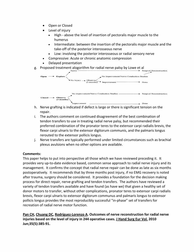
Open or Closed
Level of injury High ‐ above the level of insertion of pectoralis major muscle to the
humerus Intermediate: between the insertion of the pectoralis major muscle and the
take‐off of the posterior interosseous nerve Low: involving the posterior interosseous or radial sensory nerve
Compressive: Acute or chronic anatomic compression Delayed presentation
g. Proposed treatment alogorithm for radial nerve palsy by Lowe et al
h. Nerve grafting is indicated if defect is large or there is significant tension on the
repair. i. The authors comment on continued disagreement of the best combination of
tendon transfers to use in treating radial nerve palsy, but recommended their preferred combination of the pronator teres to the extensor carpi radialis brevis, the flexor carpi ulnaris to the extensor digitorum communis, and the palmaris longus rerouted to the extensor pollicis longus.
j. Nerve transfers are typically performed under limited circumstances such as brachial plexus avulsions when no other options are available.
Comments: This paper helps to put into perspective all those which we have reviewed preceding it. It provides very up‐to‐date evidence based, common sense approach to radial nerve injury and its management. It confirms the concept that radial nerve repair can be done as late as six months postoperatively. It recommends that by three months post injury, if no EMG recovery is noted after trauma, surgery should be considered. It provides a foundation for the decision making process for direct repair, nerve grafting and tendon transfers. The authors have reviewed a variety of tendon transfers available and have found (as have we) that given a healthy set of donor motors to transfer, without other complications, pronator teres to extensor carpi radialis brevis, flexor carpi ulnaris to extensor digitorum communus and palmaris longus to extensor pollicis longus provides the most reproducibly successful “in‐phase” set of transfers for recreation of radial nerve motor function. Pan CH, Chuang DC, Rodríguez‐Lorenzo A. Outcomes of nerve reconstruction for radial nerve injuries based on the level of injury in 244 operative cases. J Hand Surg Eur Vol. 2010 Jun;35(5):385‐91.
Summary: Between 1988 and 2005 Pan et al report a single surgeon series of 244 patients with radial nerve injuries who received nerve repair, neurolysis, or nerve graft. The authors developed a new classification for location of radial nerve injury. Nerve grafting was used most frequently in all groups, and was the only method of reconstruction for an injury at the spiral groove of the humerus. At 21.5 months follow up, isolated posterior interosseous nerve injuries had significantly better outcomes of finger and thumb extension, while wrist extension recovered in at least 80% of the patients irrespective of the level of injury. The radial nerve recovered better if repaired or reconstructed within 5 months of injury. Learning Points:
a. The radial nerve can be injured directly by stretch, division, rupture or avulsion and indirectly with fracture or during manipulation/fixation. Other causes include compression, tumour resection, or local inflammation.
b. 244 patients that developed radial nerve palsy after trauma were identified and divided into 4 groups
Level I: Infraclavicular
Level II: Humerus spiral groove radial nerve injury
Level III: Lateral arm and antecubital fossa radial nerve injury
Level IV: Posterior interosseous radial nerve injury c. The only outcome assessed was motor recovery by measuring strength of extension
of the elbow, wrist, fingers and thumb. Medical Research Council (MRC) grading system was used.
d. The outcome was rated “good” if strength was at least “M3”; the elbow and wrist could extend against gravity, and the finger MCP joints could extend fully with the wrist in maximum extension.
e. The Chi‐squared test was used to compare “good” outcomes for each level of injury (I‐IV) and mechanism of injury.
f. The Student’s t‐test was used to evaluate patient’s age, time to surgery, and nerve graft length between patients with satisfactory (M ≥ 3) and unsatisfactory outcomes (M < 3).
g. Level III injury (lateral arm antecubial fossa) was the most common (104 patients, 42%), followed by Level IV PIN (64 patients, 26%), Level I (Infraclavular) (44 patients, 17%) and Level II (Humerus, spiral groove) (37 patients, 15%).
h. Nerve grafting was most frequently used method in this last group of delayed treatment of patients.
i. Elbow extension was preserved in all patients with Level II, III and IV injuries. Wrist extension was spared in patients with Level IV injury.
j. More patients had good recovery of thumb and finger extension after treatment for Level IV injury (P< 0.01).
k. Patients with open injury had a better “good” outcome rate than those with closed injuries (P< 0.01).
l. Mean delay to surgery in cases with “good” outcomes was approximately 90 days.
m. Mean delay in those with unsuccessful outcomes was 157 days (P< 0.01). n. Short grafts gave better outcomes with a mean graft length of 5.3 cm in the group
with successful outcomes and 9.1 cm in the group with an unsuccessful outcome (P< 0.01).
Comments: This paper demonstrates the effective late management of radial nerve injury either with direct repair or more commonly with late nerve grafting. At each level when nerve grafting was employed, better results occurred when treatment was carried out within 90 days of injury. Less successful outcomes were recognized in patients with nerve grafting after five months of delay following surgery. This study also underscored that short nerve grafts and/or direct repairs did better than long nerve grafts. This paper emphasizes the importance of recognizing a radial nerve trans‐section and if possible, fixing it early. If it is not recognized early, successful outcomes can be achieved with late repair and/or nerve grafting but after five or six months results can be expected to diminish. Sammer DM, Chung KC. Tendon transfers: part I. Principles of transfer and transfers for radial nerve palsy. Plast Reconstr Surg. 2009 May;123(5):169e‐177e. Summary: This article reviews the history of tendon transfer procedures, and describes the principles and biomechanics behind them. It also discusses the anatomy and clinical findings of radial nerve palsy and the tendon transfer procedures used to treat it. Learning Points:
a. First tendon transfer procedures were devised to improve limb function in polio patients.
b. Indications for tendon transfer: acute and chronic nerve injuries that are physically irreparable (i.e. root avulsions), injuries that do not recover after direct nerve repair or grafting, failed nerve transfers and long‐standing irreparable injuries.
c. Eight Principles of successful tendon transfer: 1. Supple joints before transfer‐ The joint that the tendon transfer will move
must have maximum passive ROM before the procedure 2. Soft‐tissue equilibrium‐ Tendon transfer should pass through a healthy bed of
tissue that is free from inflammation, edema, and scar 3. Donor of adequate excursion‐ The excursion or maximum linear movement of
the transferred muscle‐tendon unit should be adequate to achieve the desired hand movement
4. Donor of adequate strength‐ The muscle‐tendon unit to be transferred must be strong enough to achieve the desired movement, but should not be too strong
5. Expendable donor‐ There must be another remaining muscle that can continue to adequately perform the transferred muscle‐tendon unit’s original function
6. Straight line of pull‐ Procedures are most effective if there is a straight line of pull, because direction changes diminish the force that the transferred unit is able to exert on its insertion
7. Synergy‐ Certain muscle groups usually work together to perform a function or movement. Transferring a wrist flexor to restore finger extension is “in‐phase” and adheres to the principle of synergy
8. Single function per transfer‐ A single tendon should be used to restore a single function. Transfer of one muscle‐tendon unit to restore multiple functions will result in compromised strength and movement
d. Four main biomechanical concepts: 1. Muscle‐tendon unit excursion‐ see #3 above 2. Muscle‐tendon unit force‐generating ability‐ The amount of force that a
muscle‐tendon unit can generate is directly proportional to the physiologic cross‐sectional area of the muscle belly
3. The moment arm‐ Determined by how far away the tendon line of action is from the joint axis of rotation. Placement is a fine balance between strength & ROM
4. Tension at which the transfer is set‐ Ideal tension depends on the amount of actin‐myosin overlap that occurs at different muscle lengths. Practically, muscle‐tendon units should be set at a tension as close as possible to preoperative resting tension
e. Main goals of treatment for radial nerve palsy: restoring finger (MCP joint), thumb, and wrist extension.
f. Finger MCP joint extension can be reestablished by transferring the flexor carpi ulnaris, flexor carpi radialis, or flexor digitorum superficialis tendons to the extensor digitorum communis.
g. The palmaris longus or the ring finger flexor digitorum superficialis to the extensor pollicis longus are used most often used to restore thumb extension.
h. The most accepted method for restoring wrist extension after high radial nerve injury is the pronator teres–to– extensor carpi radialis brevis transfer.
i. Post operative care should allow a tension free splint or casting of all tendon transfers.
j. Rehabilitation will focus initially on mobilization of single joints, while keeping tension off the transfer, and progressing to full activity by 12 weeks.
Comments: This paper provides an overview for late management of radial nerve paralysis when primary decompression, repair, and grafting are not possible. Late tendon transfers provide an effective means for recreation of thumb, finger and wrist extension. Although not normal in terms of
motion and strength, these tendon transfers can provide very functional restoration of radial nerve motor function following rehabilitation and muscle reeducation guided by a knowledgeable hand therapist. Multiple potential tendons for transfer are outlined as noted by the authors of this paper. We have found EPL restoration by means of transfer of the Palmaris longus; wrist extension, reconstruction by means of pronator teres to ECRB; and FCU transfer to EDC, when possible, provide the most reliable outcome. Recognizing that these tendons are not always available, alternatives provided in this paper can be used to still achieve a very functional outcome. Conclusion: Radial nerve injury runs a spectrum from the neck to the wrist. It may be associated with mechanical compression, localized trauma, trans‐section or idiopathic dysfunction. Careful history taking and performance of a physical examination in combination with use of discreet three‐dimensional imaging and electro‐diagnosis studies can provide the foundation for appropriate diagnosis and formulation of a concomitantly appropriate treatment plan. We still have a lot to learn about radial nerve dysfunction. Better understanding and clinical management techniques in the future, lies in careful, well thought‐out prospective studies with high levels of evidence to guide us in our care of our patients.
OLECRANON BURSITIS Septic arthritis: Experience in a Community Practice Frances D. Pien Diane Ching Emery Kim Orthopaedics, Sept 1991, Vol 14 (9); pages 981‐984 Summary: This article is a retrospective review of 47 patients with septic bursitis who presented to a private community medical practice and included 34 cases of septic olecranon bursitis. Treatment of septic olecranon bursitis included serial aspirations and antibiotic therapy in a majority of cases. 44% of patients required hospitalization with intravenous antibiotics for severe cases and 18% of patients required operative drainage. All patients were eventually cured without serious complications. Learning Points:
a. The majority of patients presenting with septic olecranon bursitis are male and the etiology of septic bursitis is related to recreational or occupational trauma.
b. Staphylococcus aureus is the predominant organism in septic olecranon bursitis. c. Associated cellulitis is seen in a high percentage of patients with septic olecranon
bursitis. d. Outpatient therapy with oral antibiotic treatment was successful in just over half of
the patients in this series. e. Indications for hospitalization were primarily fever and severe associated cellulitis. f. Bursal fluid findings were variable in septic bursitis with a wide range in WBC/ml.
Comments: This article represents a retrospective review of patients presenting with septic olecranon bursitis. When caught early, this can be treated with serial aspirations and oral antibiotics. In more advanced cases with fever, hospitalization with a course of intravenous antibiotics followed by oral antibiotics is successful. The clinician cannot depend entirely on laboratory evaluation of the bursal fluid to separate septic from non‐septic olecranon bursitis. It is a clinical diagnosis. The indication for serial aspirations vs. surgical drainage is not clear based on this report but repeated aspirations for recurrent fluid was successful in a large number of cases. In most instances, septic olecranon bursitis was relatively benign and responded well to this conservative treatment program. The Clinical Spectrum of Severe Septic Bursitis in Northwestern Spain: A 10‐Year Study Carlos Garcia‐Porrua Miguel A. Gonzalez Dolores Ibanez Maria Garcia‐Pais Journal of Rheumatology, 1999 26‐3; pages 663‐667
Summary: This article represents a retrospective review of all patients diagnosed with septic olecranon bursitis in Lugo, Spain over a 10‐year period from 1987‐1997 and included 35 patients with septic olecranon bursitis. The majority of patients presented with cellulitis, fever, leukocytosis, and had inflammatory bursal fluid aspirates. Two patients presenting late developed osteomyelitis. The vast majority of cases responded to serial aspirations and intravenous antibiotic treatment. Treatment outcomes were excellent except in the cases of osteomyelitis. Learning Points:
a. One of the predisposing factors to development of septic bursitis include occupational trauma to the bursa.
b. The olecranon is the most common site of septic bursitis in this series as in others and most patients are male.
c. Patients treated in the hospital for septic olecranon bursitis tend to be sicker with a higher proportion of cellulitis, fever, and leukocytosis.
d. Staphylococcus aureus is the most common pathogen in septic bursitis although multiple other organisms have been isolated.
e. Promptness in treatment and duration of infection prior to the commencement of treatment were a major determining factor in outcome.
f. Leukocyte count of bursal fluid was below 5000/ml in 28% of the patients with septic olecranon bursitis and is not always a good guide to treatment.
g. Bacteria associated with septic bursitis vary according to occupation with brucellosis seen among cattle farmers, slaughter house workers and veterinary surgeons.
h. Patients requiring inpatient care for septic olecranon bursitis are more commonly immunocomprimsed and positive blood cultures are seen in almost 1/5th of cases.
Comments: This study represents a 10‐year review of 35 patients who required inpatient care for septic olecranon bursitis. These tended to be sicker patients who were often immunocomprimised or intravenous drug abusers. Their clinical course was more severe than outpatient series. There was a higher incidence of cellulitis, fever, positive blood cultures, and leukocytosis. Patients required intravenous antibiotics but were most commonly successfully treated with serial aspirations and did not require surgery. The offending bacteria and recommended antibiotic will vary according to patient occupation and exposure. Antibiotic Therapy of Septic Bursitis: It’s Implication in the Treatment of Septic Arthritis George Ho, Jr. Eugene SU Arthritis and Rheumatism, July 1981, Vol 24(7); pages 905‐911 Summary: This is a prospective study evaluating the effectiveness of antibiotic therapy for septic bursitits of the olecranon, prepatellar and infrapatellar bursae and included 21 patients with olecranon
bursitis. Patients were categorized as severe, moderate, or mild and severe infections or potentially noncompliant patients were treated with intravenous antibiotics and serial aspirations. Patients with mild or moderate infections were treated with outpatient oral antibiotics and serial aspirations. The length of time necessary to achieve bursal fluid culture sterility with antibiotic therapy correlated with the duration of symptoms prior to diagnosis. Patients treated within two weeks of the onset of symptoms achieved bursal fluid sterility within one week of therapy and were cured with five additional days of antibiotics. The authors conclude that serial aspirations and oral or IV antibiotic depending on the severity of the infection is effective in the treatment of septic olecranon bursitis and that early initiation of treatment results in more rapid response of the infection. Learning Points:
a. Occupations predisposing to bursitis were janitorial work, heavy machine operation, carpentry, and other manual labor.
b. The most common organism isolated was Staphylococcus aureus in 88% of patients the majority of which were resistant to Pencillin but sensitive to Oxacillin.
c. Sterility of bursal fluid was achieved most rapidly when patients presented within two weeks of the onset of symptoms.
d. Needle aspiration of bursal fluid with concomitant antibiotic therapy is effective in the management of septic olecranon bursitis.
Comments: This paper reviews a reasonable number of patients with septic olecranon bursitis treated with oral or intravenous antibiotic therapy and serial aspirations. Bursal fluid was cultured serially to determine the point at which it became sterile. Antibiotic therapy for five additional days after achieving bursal fluid sterility was successful in eradicating infection in all cases. Patients with severe bursitis were noted to have sterile bursal fluids at approximately four days compared to patients with moderate infections at 3 days. Patients receiving intravenous antibiotic therapy resulted in sterile bursal fluid cultures in 3 days whereas those on oral antibiotics required 4 days to become sterile. The patients who presented with greater than two weeks of symptoms required 9‐15 days to achieve sterility of bursal fluid. This series recommends individualizing antibiotic treatment according to severity of infection but again, demonstrates that serial aspirations with antibiotic therapy is typically successful in the management of septic olecranon bursitis. Comparison of Nonseptic and Septic Bursitis: Further Observations on the Treatment of Septic Bursitis George Ho, Jr. Alan D. Tice Archives of Internal Medicine, November 1979,Vol 139;pages 1269‐1273 Summary: This paper compares ten patients with septic prepatellar or olecranon bursitis with twenty patients with nonseptic bursitis in the same locations. A high leukocyte count, low bursal‐
serum glucose ratio, and positive gram stain smear of bursal fluid distinguished septic from nonseptic bursitis. A prospective antibiotic program involving a bacteriacidal agent against Penicillin resistant Staph aureus resulted in an average of 12 days for successful therapy. Learning Points:
a. Nonseptic bursitis is typically related to occupational trauma to the bursa but is also associated with inflammatory processes such as gout and rheumatoid arthritis.
b. Aspirated bursal fluid from nonseptic bursae usually appeared serosanguineous but could be grossly bloody whereas septic bursal fluid generally appeared purulent
c. Bursal fluid analysis clearly differentiated septic from nonseptic bursitis with a higher leukocyte count, predominance of polymorphonuclear cells, and low fluid‐serum glucose ratios in the former. A bursal fluid glucose concentration of less than 50% of the corresponding serum level was almost exclusively seen in septic cases. There was no significant difference in levels of compliment or immunoglobulin level in the fluid between septic and nonseptic bursitis.
d. The mean leukocyte count for nonseptic cases was approximately 1500 WBC’s/cubic mm compared to a mean leukocyte count of approximately 109,000 WBS’s/cubic mm in septic cases. The overlap between the two groups was small and in such cases, leukocyte differential usually distinguished septic from nonseptic cases.
e. Gram stains of septic bursal fluid were positive in all septic cases and negative in all nonseptic cases and 90% of cases revealed Staphylococcus aureus strains resistant to Penicillin.
f. Patients that presented within one week of symptoms required 4.4 days to achieve bursal fluid sterility while patients presenting with more than seven days of symptoms required 9.2 days to achieve bursal fluid sterility on an antibiotic regime.
g. Antibiotic levels from bursal fluids indicate that adequately high levels can be achieved in bursal fluid with oral therapy although not as high as perenteral therapy.
h. The average duration of total antibiotic treatment was 12.3 days. Comments: This article presents a head to head comparison of septic and nonseptic bursitis. Several factors in the serum fluid clearly assist the clinician in differentiating septic from nonseptic bursitis and these include the overall leukocyte count, percentage of PMN cells, and fluid‐serum glucose ratio. Compliment levels and immunoglobulins were not helpful in differentiating septic from nonseptic bursitis. Oral antibiotics effective against Penicillin resistant Staphylococcus aureus achieve adequate levels in the bursa to eradicate infection. Serial aspirations appear to be beneficial in eradicating infections. Treatment of Nonseptic Olecranon Bursitis: A Controlled, Blinded Prospective Trial David L. Smith John H McAfee Linda Lucas Kusmum L. Kumar Doug M. Romney
Achives of Internal Medicine, Nov 1989, Vol 149;pages 2527‐2530 Summary: This is a double blind prospective trial for the treatment of forty two patients with nonseptic olecranon bursitis to compare the efficacy of intrabursal steroid preparations with oral anti‐inflammatory agents. Patients were randomized into one of four treatment regimes: Group 1 received methylprednisolone acetate 20 mgs. as an intrabursal injection along with oral Naproxen, Group 2 received the same injection with oral placebo, Group 3 received oral Naproxen, and Group 4 received an oral placebo. The degree of swelling in mm was assessed periodically. Groups that received intrabursal methylprednisolone acetate demonstrated the most rapid decrease in swelling and the least reaccumulation of bursal fluid. The addition of Naproxen did not significantly affect results. Learning Points:
a. Intrabursal methylprenisolone acetate injection results in rapid decrease in bursal swelling and fluid accumulation with results lasting to six months.
b. Methylprednisolone acetate injection into the bursa was safe with no evidence of skin atrophy or secondary septic bursitis.
Comments: This is a blinded prospective trial looking at four different treatment options which include methylprednisolone injection into the bursa with or without oral nonsteroidal anti‐inflammatory treatment. This is a simple well‐designed study that clearly shows the benefit and safety of intrabursal methlyprednisolone acetate treatment for nonseptic bursitis. The authors recommend a 22 guage needle and a lateral injection portal as well as sterile preparation and bandaging of the skin following injection to avoid potential complications. They did not show any added benefit from NSAID medication. Surgical Treatment of Aseptic Olecranon Bursitis Nathaniel J. Stewart James B. Manzanares Bernard F. Norrey JSES, Jan/Feb 1997, Vol 6(1);pages 49‐53 Summary: The authors in this series evaluate the results of surgical bursectomy in 21 cases for aseptic olecranon bursitis treated over a ten year period at the Mayo Clinic with a minimum follow‐up of two years and an average follow‐up of over five years. 94% (15/16) of patients without rheumatoid arthritis had complete and long‐term relief where as only 40% (2/5) of patients with rheumatoid arthritis had such success. The authors conclude that properly performed surgical treatment is effective in patients without rheumatoid arthritis. Learning Points:
a. Septic vs. nonseptic bursitis should be ruled out during the planning of surgical intervention.
b. The majority of patients presenting with aseptic olecranon bursitis will respond to conservative treatment measures.
c. A lateral incision with excision of the bursa as a single structure is recommended to ensure complete removal of all bursal linings.
d. Osteophytes should be removed when present and a meticulous closure with compression dressing is recommended.
e. Operative treatment is indicated only for patients who have failed conservative measures. Chronic cases may be complicated by the presence of a pedunculated fibrous mass which may have a pathologic role.
f. Recurrent symptoms are common in patients with rheumatoid arthritis. Comments: This is a small series of patients who required operative treatment for aseptic olecranon bursitis over a ten year period of time at a busy orthopaedic hospital suggesting that the majority of cases can be managed nonoperatively. The success rate in patients without rheumatoid arthritis was high whereas the failure rate was over 50% in patients with rheumatoid arthritis. Patients with rheumatoid arthritis must be warned of a potential poor outcome following this surgery. There were no significant complications of surgery such as skin necrosis indicating that careful surgical technique is safe for removal of chronically inflamed olecranon bursa. Chronically inflamed patients may have a fibrous band in the bursa noted at the time of surgery. Complications following Resection of the Olecranon Bursa Ilse Degreef Luc De Smet Acta Orthopedica Belg. 2006,Vol 72(4); pages 400‐403 Summary: This is a retrospective review of 37 cases of resection of the olecranon bursa in which wound healing problems were noted in 27% and recurrent bursitis in 22%. One patient required a lateral arm flap. The authors recommend that the risk of wound healing problems and recurrence be taken into account when planning surgical excision. Learning Points:
a. Multiple complications are possible following olecranon bursectomy despite the simplicity of this procedure and include prolonged drainage, recurrent effusions, infection, and necrosis of overlying skin.
b. Because complications after bursectomy and recurrence are relatively frequent, conservative treatment with immobilization is recommended.
c. 3‐4 weeks of postoperative cast immobilization should be considered following olecranon bursectomy.
Comments: This article represents a series of patients with a high complication rate following olecranon bursectomy. 10/37 patients had prolonged drainage and eight patients had recurrent bursal effusions. Postoperative infections occurred with chronic draining sinuses and skin necrosis. This article does emphasize the fact that a relatively simple appearing operation can lead to significant complications. Relative success of nonoperative management must be weighed against the complications of operative treatment. Long‐term Follow‐Up of Corticosteroid Injection for Traumatic Olecranon Bursitis Philip Weinstein Juan Canoso Jeffrey Wohlgethan Annals of the Rheumatic Diseases 1984, Vol 43; pages 44‐46 Summary: This article reviews the results of treatment of 47 patients with traumatic olecranon bursitis comparing twenty‐two patients treated with bursal aspiration alone with 25 patients treated with bursal aspiration followed by intrabursal injection of 20 mgs. of Triamcinolone hexacetonide. The patients treated with bursal aspiration alone had delayed recovery but no complication of therapy. In the 25 patients who received intrabursal corticosteroid injection, there were three infections, five cases of skin atrophy, and chronic local pain in seven cases. The authors recommend a more conservative approach without steroid injection because recovery will eventually occur in the patients treated without corticosteroids. Learning Points:
a. Chronic aseptic olecranon bursitis can be treated adequately with aspiration alone without corticosteroid infusion although resolution of symptoms will be more prolonged compared to the group treated with corticosteroid injection.
b. Corticosteroid bursal injection is effective in the management of chronic traumatic aseptic olecranon bursitis and results in resolution of bursal effusions typically within one to two weeks.
c. Corticosteroid bursal injection is associated with complications such as skin atrophy, delayed infection, and sensitivity and may be dose related.
d. The diagnosis of septic olecranon bursitis must be excluded prior to the use of corticosteroid bursal injection.
Comments: This is a retrospective non‐randomized trial comparing the use of bursal aspiration alone with bursal aspiration followed by corticosteroid injection. The patient selection was not random and treatment was not standardized. There was a rapid recovery usually within one week following corticosteroid injection. There were several complications but the severity and etiology can be questioned. Only three of the five cases of skin atrophy were significant. The infections occurred at one week, two months, and four months post injection and the exact relationship between the more delayed cases and the injection can be questioned. Overall this
article does highlight the fact that corticosteroid bursal injection is not without risk. The exclusion of a septic source of bursitis is also critical prior to the utilization of corticosteroid injections. Prepatellar and Olecranon Arthroscopic Bursectomy Douglas R. Kerr Clinics in Sports Medicine, January 1993,Vol 12(1);pages 137‐142 Summary: This article represents a review of eleven patients who underwent arthroscopic bursectomy; four of whom underwent arthroscopic olecranon bursectomy. Arthroscopic surgery was recommended because of complications associated with open treatment including wound healing problems, chronic tenderness, and decreased sensation about the scars. One patient taking immunosuppressive drugs developed a postoperative complication of infection. One patient with traumatic olecranon bursitis had a poor result and three had a good result. There were no wound healing problems. Learning Points:
a. Arthroscopic treatment for olecranon bursitis is safe and effective without significant wound healing problems.
b. With either open or arthroscopic surgery, care must be taken to avoid excessive bursal wall excision and skin damage.
c. Removal of olecranon osteophytes is not required for a good result in the management of olecranon bursitis.
Comments: Although the use of arthroscopy in the management of olecranon bursitis may appear excessive, this article does emphasize the fact that wound healing problems can occur with open techniques. Surgeons are encouraged to make incisions off the midline and to be careful in undermining the skin during open excision. More aggressive treatment such as osteophyte removal and partial olecranon excision as advocated by Quayle is not necessarily required. Olecranon and Prepatellar Bursitis: Treating Acute, Chronic, and Inflammed Edward McFarland Pomthep Mamanee William Queale Andrew Cosgarea The Physician and Sports Medicine, March 2000, Vol 28(3);pages 40‐52 Summary: This article represents a review of management of elbow and knee bursitis in the athletic and nonathletic population and separates bursitis into three areas of acute, chronic nonspetic, and chronic infected. An anatomical review as well as the pathophysiology and treatment strategies of these three types of bursitis are reviewed.
Learning Points:
a. Acute bursitis tends to be traumatic in origin and injury of surrounding structures must be ruled out through clinical and radiographic evaluation.
b. Aspiration and immobilization may be required in cases of acute bursitis but injection of corticosteroids is typically not recommended.
c. Chronic nonseptic bursitis typically presents with chronic swelling without significant pain or erythema and may respond to nonoperative treatment including antiinflammatory medications, compression, padding, and activity modification.
d. Injection of corticosteroids has been shown to be beneficial in several studies but may be associated with complications such as infection, skin atrophy, and chronic pain.
e. Surgery for chronic bursitis is rarely required for recalcitrant cases but is typically successful.
f. Septic olecranon bursitis can most commonly be separated from the aseptic variety through aspiration and bursal fluid analysis. Staph aureus resistant to Penicillin is most common followed by Strep species.
g. Septic olecranon bursitis is most commonly managed with oral or intravenous antibiotic therapy utilizing a Pecillinase resistant semisynthetic antibiotic with modification depending on culture results.
h. Repeated aspiration is recommended for recurrent fluid accumulations and surgery is rarely indicated.
Comments: This article represents a good review of the literature regarding the management of olecranon bursitis. The differentiation of bursitis into acute, chronic non septic, and septic is a good framework for evaluation and treatment of these patients and helps the clinician developed a strategy for both evaluation and treatment of these patients.