Embed Size (px)
Citation preview

RESEARCH ARTICLE
The apoptotic effect of shikonin on human papillary thyroidcarcinoma cells through mitochondrial pathway
Chibo Liu & Lihui Yin & Jiaqi Chen & Jiayu Chen
Received: 1 August 2013 /Accepted: 18 September 2013 /Published online: 2 October 2013# International Society of Oncology and BioMarkers (ISOBM) 2013
Abstract This study aims to explore the apoptotic function ofshikonin on the papillary thyroid cancer cells and the relatedmechanism. The papillary thyroid cancer cell lines K1 andW3and thyroid follicular epithelial cells NTHY-ORI 3-1 weretreated with different concentrations of shikonin. Cell prolif-eration was tested. Morphological changes of the apoptoticcells were observed by Hoechst 33342 staining. The apoptosisrate of the papillary thyroid cancer cells was measured withflow cytometry. Changes of the cell cycle were explored. Themitochondrial membrane potential changes were analyzedafter JC-1 staining. Bcl-2 family proteins and caspase-3expression with shikonin treatment was analyzed by real-time fluorescence polymerase chain reaction (PCR). Cell pro-liferation of K1 and W3 was inhibited by shikonin, and theinhibition was dose–time dependent. Papillary thyroid carci-noma cells treated by shikonin had no obvious cell cycle arrestbut were observed with the higher apoptosis rate and the
typical apoptotic morphological changes of the cell nucleus.JC-1 staining showed that shikonin reduced the mitochondrialmembrane potential of papillary thyroid carcinoma cells.Real-time PCR results showed that shikonin significantlyincreased Bax and caspase-3 expression and upregulatedBcl-2 expression in a dose-dependent manner in papillarythyroid carcinoma cells. However, the NTHY-ORI 3-1 wasalmost not affected by shikonin treatment. Shikonin can inhibitK1 and W3 cell proliferation in a dose- and time-dependentmanner, enhance Bax levels, reduce anti-apoptotic protein Bcl-2 levels, result in decreasing mitochondrial membrane potentialand activating caspase-3 enzyme, and finally lead to apoptosis.
Keywords Shikonin . Papillary thyroid cancer .
Cell apoptosis . Mitochondria signal pathway
Introduction
Thyroid cancer is a common endocrine cancer, which ac-counts for 1.3–1.5 % of the whole-body malignant tumor.Papillary thyroid carcinoma (PTC) is one of the most commonpathological types, representing 75 to 85 % of thyroid cancer[1], and the rate has been increasing in the recent years.Currently, there is no more effective treatment other thansurgical resection for this cancer. So finding effective naturalmedicine which has anti-papillary thyroid carcinoma effect isof great significance undoubtedly.
Shikonin initially comes from the root of radix arnebiae seulithospermi and has a long medical history in China. In recentseveral years, it has been confirmed to have good antitumoreffects on leukemia cells [2], liver cancer [3], prostate cancer[4], colon cancer [5, 6], melanoma [7], and osteosarcoma [8]and an important role in cancer treatments. Researchesshowed that shikonin could affect the signal transduction, geneexpression, as well as metabolism, proliferation, differentiation
Chibo Liu and Jiayu Chen contributed equally to this work.
C. LiuDepartment of Clinical Laboratory, Taizhou Municipal Hospital,Taizhou, Zhejiang 317000, China
L. YinBlood Center ofWisconsin, 638 N 18th Street, Milwaukee,WI 5323,USA
J. ChenCollege of Life Science, Jilin University, Changchun, Jilin 136000,China
J. ChenSchool of Laboratory Medicine, Wenzhou Medical University,Wenzhou, Zhejiang 325035, China
J. Chen (*)Medical School of Taizhou University, 1139 Shifu Road, JiaojiangDistrict, Taizhou City, Zhejiang Province 318000, Chinae-mail: [email protected]
Tumor Biol. (2014) 35:1791–1798DOI 10.1007/s13277-013-1238-5

of tumor cells, etc. At present, the functions of shikonin onthyroid papillary cancer have not been reported. This studyfocused on the cell apoptotic effect on thyroid papillary cancerinduced by shikonin and its mechanism.
The experimental materials and methods
Cell culture
Papillary thyroid carcinoma cell lines K1 and W3 werepurchased from ATCC (Manassas, VA, USA), and normalthyroid follicular epithelial cell line NTHY-ORI 3-1wasbought from Aidingbao Biological Technology DevelopmentCompany (Shanghai, China). All the cell line identities weremycoplasma free and were cultured in Dulbecco's modifiedEagle's medium (DMEM) supplemented with 10 % heat-inactivated fetal calf serum (FCS) at 37 °C and 5 % CO2.
Shikonin liquids preparation
Ten microgram shikonin (from Sigma) was dissolved withl00 μl DMSO and stored at −20 °C. This stock solutionwas diluted with DMEM, and the cell culture mediumcontaining different concentrations of shikonin was prepared.This medium was filtered and stored in the dark under atemperature of 4 °C.
Cell proliferation assay
CellTiter 96® AQueous Non-Radioactive Cell ProliferationAssay was applied to test cell proliferation. Briefly, the K1,W3, and normal thyroid follicular epithelial cells NTHY-ORI3-1were collected at logarithmic phase, diluted into 5×10⁴
cells/ml with DMEM containing 10% fetal calf serum, seededinto 96-well flat-bottom plates at 100 μl /well, and incubatedat 37 °C with 5 % CO2 for 24 h. Then, cells were cultured in100 μl DMEM medium with different concentrations ofshikonin (0, 1, 2, 4, 8, and 16 μg/ml) and 10 % fetal calfserum, at 37 °C and 5%CO2 for 0, 1, 2, and 3 days. After that,20 μl CellTiter 96® AQueous Non-Radioactive Cell Prolifer-ation Assay Reagent (Promega, USA) was added to each wellfor 1–4 h of additional incubation. The absorption values at490 nm were determined with an automatic enzyme-linkedimmunosorbent assay plate reader (BMGPOLARstar Omega,BMG Labtech, Germany).
Assessment of mitochondrial membrane potential
Mitochondria play an important role in cell apoptosis inducedby many stimulators, and the decline of mitochondrial mem-brane potential (ΔΨm) is an earlier landmark/event of theapoptosis. 5,5′,6,6′-Tetrachloro-1,1′,3,3′-tetraethylbenzimi-
dazolylcarbocyanine iodide (JC-1) is an effective probe forthe detection of mitochondrial membrane potential. It exists inmonomer when its concentration and the mitochondrial mem-brane potential are lower, and the cells show green fluores-cence. On the contrary, the dimer forms when its concentra-tion or the mitochondrial membrane potential is higher, andthe cells show red fluorescence. To measure the mitochondrialmembrane potential for apoptosis analysis, JC-1staining wasperformed with Accuri's JC-1 Mitochondrial Potential AssayKit (Invitrogen, USA) according to the protocol [9].
Nucleus morphological changes observed by fluorescencemicroscopy
Cells in logarithmic growth phase were seeded into the six-well plates at 5×10³ cells/well. After preincubation for 12 hunder normal condition, cells were treated with shikonin (0, 2,and 4 μg/ml) and 10 % fetal calf serum in DMEM. Eachtreatment was tested in triplicate. After 24 h, cells werewashed with phosphate buffer saline (PBS) twice, fixed with1 ml of 4 % paraformaldehyde at 4 °C for 10 min. Then, theplate was washed with PBS for three times, and the cells weredyed for 10 min with Hoechst 33342 at the room temperaturein the dark. Later, the cells were washed with PBS for threetimes and observed under an inverted fluorescence micro-scope immediately. Live cells will show dispersion, uniformfluorescence in nuclei, while dead cells are not dyed byHoechst staining. When apoptosis happens, obvious nuclearmorphological changes, such as blue fluorescent stained com-pact particulates, can be seen in the nucleus or cytoplasm. Thecells with three or more than three fluorescent DNA fragmentsare identified as apoptosis cells.
Flow cytometry analysis of cell apoptosis induced by shikonin
The cells at logarithmic growth phase were seeded into six-well plates at 2–4×10⁵ cells/well, incubated at 37 °C with 5 %CO2 for 24 h. The culture medium was changed with 2 ml ofDMED containing 0 or 4 μg/ml shikonin and 10 % fetal calfserum and was cultured at 37 °C 5 % CO2 for 24 h. Eachtreatment was tested in three wells. The cells were harvestedafter digested with pancreatic enzymes, washed with PBS forthree times, and double stained with phycoerythrin-conjugated annexin V and 7-amino-actinomycin D (7-AAD)according to the manufacturer's instructions (BD Bioscience,San Jose, CA, USA). Viable cells are labeled as aimexin V (−)and 7-AAD (−) and located in the lower left quadrant. Earlyapoptosis cells were labeled as annexin V (+) and 7-AAD (−)in the upper left quadrant. Dead cells labeled as annexin V (−)and 7-AAD (+) are located in the lower right quadrant. Lateapoptosis and necrosis cells labeled as annexin V (+) and 7-AAD (+) are located in the right upper quadrant. Twentythousand events were collected for each sample using the
1792 Tumor Biol. (2014) 35:1791–1798

Becton-Dickinson FACScan (BD Bioscience, San Jose, CA,USA). Data were analyzed using the FlowJo software (TreeStar, San Carlos, CA, USA).
The cell cycle of thyroid papillary cancer cells treatedwith shikonin detected by flow cytometry
The cells at logarithmic growth phase were seeded into six-well plates at 2–4×10⁵ cells/well, incubated at 37 °C with 5 %CO2 for 24 h. The medium was changed with 2 ml DMEDcontaining 0 or 4 μg/ml shikonin and 10 % fetal calf serumand was cultured at 37 °C with 5 % CO2 for 24 h. Eachtreatment was tested in three wells. The cells were harvestedafter digestion with pancreatic enzymes, washed with PBS forthree times, and fixed with ice-cold 70 % ethanol overnight at−20 °C. After centrifugation for 5 min at 1,500 rpm, the cellswere re-suspended in 5 ml fluorescence-activated cell sortingwashing medium (3 % FCS/PBS) and centrifuged again. Cellpellets were re-suspended in 0.5 ml of PI/RNase StainingBuffer (BD Pharmingen™) and incubated at room tempera-ture for 30 min. DNA content in each cell was analyzed usinga flow cytometer (BD Bioscience, San Jose, CA, USA). Datawere analyzed using FlowJo software (Tree Star, San Carlos,CA, USA).
Examination of Bcl-2, Bax, and caspase-3 mRNA expressionlevels with quantitative real-time fluorescence polymerasechain reaction
The CK, W3, and NTHY-ORI 3-1cells were collected aftertreatment with 0 or 4 μg/ml shikonin for 24 h; total RNAwasextracted from the cells using TRIzol™ Reagent (InvitrogenCompany). Briefly, 1 ml TRIzol (Invitrogen, USA) was addedto PBS-washed cells, mixed by pipetting until the solutionswere clear and transparent, incubated in the room temperaturefor 5 min, added with 200 μl chloroform and mixed well,incubated in the room temperature for 3 min, and centrifugedfor 15 min at 12,000 rpm at 4 °C. The supernatant wastransferred into a 1.5-ml centrifuge tube with 500 μlisopropanol, mixed well, incubated for 10 min at the roomtemperature, and centrifuged at 12,000 rpm for 15min at 4 °C.The supernatant was discarded, added with 1 ml 75 % ethanolinto the tubes to clean the RNA sediment, and centrifuged for5 min at 12,000 rpm at 4 °C. The supernatant was discarded,air-dried for 5–10 min, and dissolved the RNA pellet in 50–100μl RNase-free water. RNAwas quantified with NanoDrop(Thermo Fisher), and the integrity was detected by agarose gelelectrophoresis. Then, the RNAs were reversely transcribedwith the SuperScript® III First-Strand Synthesis System(Invitrogen™). One microliter of the RT product was usedfor subsequent quantitative real-time fluorescence polymerasechain reaction (Q-RT-PCR) with the final concentration ofPCR reaction being 2.5×Real Master Mix/20×SYBR
solution mixture with 9 and 2 μl upstream and downstreamprimers (100 nmol/l, produced by Sheng gong Company,Shanghai, China). The ddH2O was added to reach the totalvolume of 20 μl. The threshold cycle (Ct) value of each cellwas recorded, and the data were analyzed by the comparativedelta–delta Ct method. The sequences of the primers werelisted in Table 1.
Statistical analysis
All data were analyzed using SPSS 16.0 software (SPSS, Inc.,Chicago, IL, USA). Data were expressed as mean ± standarddeviation. Kruskal–Wallis test was used for multiple groupcomparisons followed by Wilcoxon rank sum test includingBonferroni adjustment for comparison between two groups.All tests performed were two-sided. P <0.05 was consideredstatistically significant.
Results
Shikonin inhibited thyroid papillary cancer cells proliferation
CellTiter 96® AQueous Non-Radioactive Cell ProliferationAssay was applied to test cell proliferation. The absorptionvalues at 490 nm were determined with an automatic enzyme-linked immunosorbent assay plate reader. The inhibition rateof cells was calculated following the formula: the inhibitionrate = (OD value of shikonin-untreated group − OD value ofshikonin treated group)/OD value of shikonin-untreatedgroup×100 %. The data shown in Table 2 suggested that thethyroid papillary cancer cell proliferation was affected withdifferent concentrations of shikonin, and the inhibition effectwas time and dose dependent. However, no significantinhibition was observed in the normal cell line NTHY-ORI3-1 treated with 1, 2, and 4 μg/ml shikonin compared to thecontrol group (with 0 μg/ml shikonin) (P >0.05), as shown inTable 2. With 8 μg/ml shikonin, proliferation of the NTHY-ORI 3-1 cells was inhibited in a dose- and time-dependentmanner (P <0.05).
Table 1 The primer sequences of Bcl-2, Bax, caspase-3, and GAPDH
Gene name Forward primer Annealingcondition
Bcl-2-F GGGTGGGAGGGAGGAAGAAT 60 °C, 60 sBcl-2-R TTCGCAGAGGCATCACATCG
Bax-F CTCACCGCCTCACTCACCAT 60 °C, 60 sBax-R TGTGTCCCGAAGGAGGTTTATT
Caspase-3-F GAGTAGATGGTTTGAGCCTGAG 60 °C, 60 sCaspase-3-R TGCCTCACCACCTTTAGAAC
GAPDH-F TGAAGGTCGGAGTCAACGG 60 °C, 60 sGAPDH-R CTGGAAGATGGTGATGGGATT
Tumor Biol. (2014) 35:1791–1798 1793

Morphological changes of the cell nucleus undera fluorescence microscope after being treated with shikonin
As shown in Fig. 1, the cell nuclei of CK and W3 dyed byHoechst 33342 are uniform, round, or oval, with uniformity inchromatin distribution. After shikonin treatment, more cellsappeared to be characteristics of apoptosis with the changes ofnuclear morphometry, such as forming round cell karyorrhexisparticles, chromatin condensation, the particle shape distribu-tion, the bright blue nuclear pyknosis, and lobulated nuclearfragmentation. The morphology of the cell nucleus in normalcells NTHY-ORI 3-1 treated by shikonin was not changed asmuch as the thyroid papillary cancer cells, and the typicalapoptotic characteristics were not observed.
Shikonin-induced apoptosis of thyroid papillary cancer cellsanalyzed by flow cytometry
The papillary thyroid cancer cells CK and W3 and the normalcells NTHY-ORI 3-1 were treated with 4 μg/ml shikonin. The
apoptosis rates were examined by flow cytometry after cellswere dyed with 7-AAD and annexin V-PE. Data showed(Fig. 2, Table 3) that early apoptotic cells for the papillarythyroid cancer cells K1 and W3 induced by shikoninaccounted for 53.0 and 29 %, higher than the 0 μg/mlshikonin-treated groups (3.34 and 4.87 %). The percentagesof cells at a later stage of apoptosis and at secondary necrosisstage were increased from 3.24 and 2.89% to 26.7 and 17.0%(P <0.01). The total apoptosis rates for K1 and W3 which aretreated with shikonin reached to 79.7 and 39.9 %, which issignificantly different from the control groups and the normalcell (P <0.01).
The cell cycle changes of papillary thyroid carcinoma cellsexamined by flow cytometry
Papillary thyroid carcinoma cells K1 and W3 were treatedwith 0 or 4 μg/ml of shikonin for 24 h, and the cell cycle wasevaluated by flow cytometry. The data shown in Fig. 3 sug-gested that the cells distributed in each cell cycle stage had noobvious difference from the controls (P >0.05).
Reduced mitochondrial membrane potential by shikonintreatment detected by JC-1 staining
JC-1 is a cationic dye which can selectively enter into mito-chondria and reversibly change color from red to green as themembrane potential decreases [10] at low concentrations. Ifapoptosis induced by shikonin is through the mitochondrialpathway, the mitochondrial membrane integrity will be dam-aged at the early stage of cell apoptosis, the ΔΨm should bereduced, and the JC-1 will exist mainly in a monomeric formin the cells and emit green fluorescence. The data shown inFig. 4 suggested that, compared with 0 μg/ml shikonin-treatedgroup, mitochondria membrane potentials of W3 and K1 cellsafter 4 μg/ml shikonin treatment were decreased, so the cellapoptosis induced by shikonin may be associated with declinein mitochondrial membrane potential.
Table 2 The inhibition rate of K1, W3, and NTHY-ORI 3-1 cells treated with shikonin for 24, 48, or 72 h (x� s , n =3)
Alkannin(μg/ml)
K1 cell line W3 cell line NTHY-ORI 3-1 cell line
24 h 48 h 72 h 24 h 48 h 72 h 24 h 48 h 72 h
0 0 0 0 0 0 0 0 0 0
1 21.01±1.03Δ 37.12±5.43Δ 56.87±4.22Δ 13.98±1.22Δ 24.64±3.71Δ 37.71±6.31Δ 0.54±0.07 0.53±0.04 0.57±0.02
2 42.43±1.32Δ 64.11±7.31Δ 76.13±8.33Δ 21.78±2.45Δ 41.34±5.68Δ 59.37±8.29Δ 0.61±0.06 0.49±0.08 0.58±0.06
4 65.12±2.21Δ 80.23±6.35Δ 90.71±7.45Δ 36.65±2.33Δ 56.52±4.68Δ 73.43±6.22Δ 0.89±0.19 1.10±0.32 1.27±0.58
8 78.32±7.32Δ 89.51±4.55Δ 94.33±2.19Δ 48.41±5.53Δ 61.22±7.14Δ 80.42±7.66Δ 7.39±3.12* 8.37±4.25* 10.66±4.19*
16 89.22±6.71Δ 94.33±3.11Δ 98.69±2.31Δ 55.65±7.87Δ 70.11±8.48Δ 89.55±9.15Δ 10.43±6.14* 17.66±8.76Δ 21.79±8.22Δ
*P<0.05; ΔP<0.01, compared with control group, respectively
The concentration of shikonin
0µg/mL 2µg/mL 4µg/mL
K1
W3
NTHY-ORI 3-1
Fig. 1 The morphology of the cell nucleus in normal cell line NTHY-ORI 3-1 and thyroid papillary cancer cells K1 andW3 treated by shikoninwas observed after Hoechst staining (×400)
1794 Tumor Biol. (2014) 35:1791–1798
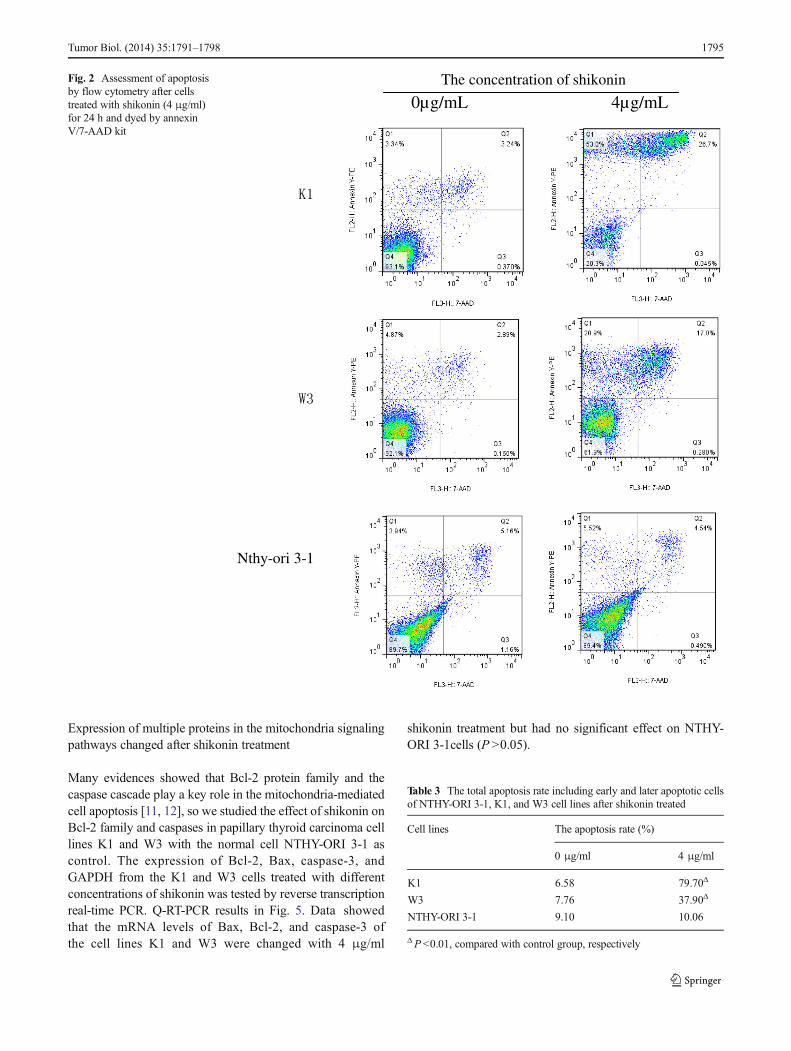
Expression of multiple proteins in the mitochondria signalingpathways changed after shikonin treatment
Many evidences showed that Bcl-2 protein family and thecaspase cascade play a key role in the mitochondria-mediatedcell apoptosis [11, 12], so we studied the effect of shikonin onBcl-2 family and caspases in papillary thyroid carcinoma celllines K1 and W3 with the normal cell NTHY-ORI 3-1 ascontrol. The expression of Bcl-2, Bax, caspase-3, andGAPDH from the K1 and W3 cells treated with differentconcentrations of shikonin was tested by reverse transcriptionreal-time PCR. Q-RT-PCR results in Fig. 5. Data showedthat the mRNA levels of Bax, Bcl-2, and caspase-3 ofthe cell lines K1 and W3 were changed with 4 μg/ml
shikonin treatment but had no significant effect on NTHY-ORI 3-1cells (P >0.05).
Nthy-ori 3-1
The concentration of shikonin
0µg/mL 4µg/mL
Fig. 2 Assessment of apoptosisby flow cytometry after cellstreated with shikonin (4 μg/ml)for 24 h and dyed by annexinV/7-AAD kit
Table 3 The total apoptosis rate including early and later apoptotic cellsof NTHY-ORI 3-1, K1, and W3 cell lines after shikonin treated
Cell lines The apoptosis rate (%)
0 μg/ml 4 μg/ml
K1 6.58 79.70Δ
W3 7.76 37.90Δ
NTHY-ORI 3-1 9.10 10.06
ΔP <0.01, compared with control group, respectively
Tumor Biol. (2014) 35:1791–1798 1795

Discussion
Cell apoptosis is an important mechanism for body develop-ment, cell differentiation, and physiological and pathologicaldeath. It is an actively programmed cell death process tomaintain a stable internal environment. It involves a series of
activation, expression, and regulation of genes. Abnormal cellapoptosis is an important etiology for most malignant tumors,and searching for effective drugs to induce tumor cell apoptosishas become the target in cancer therapy and studies.
Shikonin (molecular formula, C16H1605), extracted fromradix arnebiae seu lithospermi root, is one of the importantcomponents of naphthoquinone compounds. Modern re-searches have shown that shikonin and its derivatives hadanti-inflammatory [11], antineoplastic, antifungal [13],antiviral [14], and wound healing functions [15–17]. Theantitumor effect of shikonin was first reported in 1977 [18],showing that 5–10 mg/kg/day of the shikonin could fightagainst ascites tumor. In 1981 [19], Professor Sankawa U usedX-ray to analyze shikonin once again and further concludedshikonin's antitumor properties. Later, researchers did exten-sive studies on the antitumor function of shikonin [2–8]. Theyfound that shikonin could hinder the growth of tumor cells byaffecting metabolism, proliferation, and differentiation of tumorcells. However, the role and mechanism of shikonin on thyroidpapillary carcinoma has not been reported.
In order to explore the effect of shikonin on thyroid papil-lary cancer cells, this study investigated the proliferationactivity of thyroid papillary cancer cells and normal follicular
G1 = 67. 91%
S = 23. 13%
G2 = 8. 2%
G1 = 66. 85%
S = 24. 73%
G2 = 9. 85%
G1 = 62. 44%
S = 27. 14%
G2 = 7. 02%
G1= 63. 29%
S = 24. 52%
G2 = 6. 91%
The concentration of shikonin
0µg/mL 4µg/mL
K1
W3
Fig. 3 The cell cycles ofpapillary thyroid carcinoma cellsK1 and W3 treated with treatedwith 0 or 4 μg/ml of shikoninfor 24 h
K1
W3
The concentration of shikonin
0µg/mL 4µg/mL
Fig. 4 The mitochondria membrane potentials of W3 and K1 cells treatedwith shikonin. *P<0.01, compared with before treatment of shikonin
1796 Tumor Biol. (2014) 35:1791–1798

epithelial cell after drug treatment. The results showed thatshikonin could inhibit the proliferation of thyroid papillarycancer cell lines K1 and W3, and the proliferation inhibitionwas dose and time dependent. The nuclei morphology ofpapillary thyroid cancer cells was changed significantly aftershikonin treatment, including condensation and marginationof chromatin, karyorrhexis, anisokaryosis, debris and apopto-tic bodies, and other typical irregular apoptotic morphologicalfeatures. The cell cycle and the ratio of apoptosis cells weredetected with flow cytometry after PI or annexin V-PE/7-AAD double staining. The results showed that the apoptosisrate increased significantly after the thyroid papillary cancercells were treated with shikonin for 24 h (P <0.01), withoutchanges in the cell cycles. Instead, the proliferation of normalthyroid follicular epithelial cell was not affected at a lowconcentration of shikonin, neither the cell apoptosis rates ofthe normal cells were increased when the drug concentrationreached to 4 μg/ml. Thus, shikonin has a distinct therapeuticeffect on papillary thyroid cancer cells through inducingcancer cells apoptosis without influencing the normal thyroidepithelial cells.
Mitochondrial apoptosis pathway is also known as anendogenous apoptosis pathway. It is the major target of anti-cancer drugs in inducing tumor cell apoptosis. In this pathway,some stimulators which can result in the DNA damage willactivate the apoptosis-promoting proteins of Bcl-2 family byproteolysis or dephosphorylation. This will downregulate themitochondrial membrane potential. Apoptosis signaling mol-ecules such as cytochrome C will enter into the cytoplasm,activate the caspase, and lead to the characteristic changes ofapoptosis [12]. To clarify if the apoptosis of thyroid papillarycancer cells was induced by shikonin through the mitochon-dria pathway, a series of studies were performed. Data showedthat Bcl-2 and Bax took part in the process of cell apoptosisinduced by shikonin. Shikonin broke the balance of Bcl-2 andBax, leading Bax in a dominant position. Caspase-3 wasupregulated, resulting in cell apoptosis.
To make sure if the function of shikonin on cell growthinhibition and apoptosis induction is tumor cell selective, thisstudy selected the normal human thyroid follicular epithelial
cell line NTHY-ORI 3-1 as a control. Results revealed thatshikonin could not inhibit the growth of NTHY-ORI 3-1 at 1,2, or 4 μg/ml after incubation for 24, 48, or 72 h (P >0.05).The inhibition only appeared at the higher concentration of theshikonin (8 μg/ml, P <0.05), but still significantly lower thanon K1 or W3 cells (P <0.01). Under the fluorescence micro-scope, typical apoptotic cells of NTHY-ORI 3-1 were notfound in each group with 0 or 4 μg/ml shikonin, and thesewere in accordance with the results examined by flow cytom-etry. At the same time, the Q-RT-PCR results showed that themRNA levels of Bcl-2, Bax, and caspase-3 were not changedafter the shikonin was added to the normal cells. All these datarevealed that shikonin had no significant inhibition of cellproliferation and apoptosis induction on the normal cells whenthe dosage was lower than 4 μg/ml.
In conclusion, shikonin can inhibit the growth of thyroidfollicular epithelial cells in dose- and time-dependent manner,and the inhibition is more effective on thyroid carcinoma cells.Shikonin can improve the level of apoptosis precursor proteinBax and downregulate anti-apoptotic protein Bcl-2, andincrease Bax/Bcl-2 ratio, leading to the decrease of themitochondrial membrane potential, activating the caspase-3 cascade, and resulting in apoptosis. This experiment providesa certain reference value for the application of shikonin inclinical therapy of thyroid papillary carcinoma.
Acknowledgments This study was supported by the grants from theZhejiang Natural Science Foundation (Y2100248), Foundation ofDepartment of Science andTechnology of Zhejiang Province (2009C33155),Foundation of Zhejiang Health Department (2009A218), TaizhouScience and Technology Bureau (102KY15), and Zhejiang ProvinceChinese Medicine Study Foundation (2011ZA113).
Conflicts of interest None
References
1. Aschebrook-Kilfoy B, Ward MH, Sabra MM, Devesa SS. Thyroidcancer incidence patterns in the United States by histologic type,1992–2006. Thyroid. 2011;21(2):125–34.
2. Han W, Xie J, Fang Y, Wang Z, Pan H. Nec-1 enhances shikonin-induced apoptosis in leukemia cells by inhibition of RIP-1 andERK1/2. Int J Mol Sci. 2012;13(6):7212–25.
3. Gong K, Li W. Shikonin, a Chinese plant-derived naphthoquinone,induces apoptosis in hepatocellular carcinoma cells through reactiveoxygen species: a potential new treatment for hepatocellular carcinoma.Free Radic Biol Med. 2011;51(12):2259–71.
4. Sasi N, Hwang M, Jaboin J, Csiki I, Lu B. Regulated cell deathpathways: new twists in modulation of Bcl-2 family function [J]. MolCancer Ther. 2009;8(6):1421–9.
5. Hsu P, Huang Y, Tsai M, Wang YJ, Lin JK, Pan MH. Induction ofapoptosis by shikonin through coordinative modulation of the Bcl-2family, p27, and p53, release of cytochrome c and sequential activa-tion of caspases in human colorectal carcinoma cells. J Agric FoodChem. 2004;52(20):6330–7.
0
1
2
3
4
5
6
7
8
NTHY-ORI 3-1 K1 W3
The
rat
io o
f 2- Δ
ΔCt
(exp
erim
ent/
cont
rol )
The cell lines
Bcl-2(0µg/ml)
Bcl-2(4µg/ml)
Bax(0µg/ml)
Bax(4µg/ml)
Caspase-3(0µg/ml)
Caspase-3(4µg/ml)
* *
*
*
*
*
Fig. 5 The effect of shikonin on the expression of Bcl-2, Bax, andcaspase-3 mRNA and NTHY-ORI 3-1, K1, and W3 cell lines (x� s ,n =3)
Tumor Biol. (2014) 35:1791–1798 1797

6. Salvioli S, Ardizzoni A, Franceschi C, Cossarizza A. JC-1, but notDiOC6(3) or rhodamine 123, is a reliable fluorescent probe to assessdelta psi changes in intact cells: implications for studies on mito-chondrial functionality during apoptosis. FEBS Lett. 1997;411(1):77–82.
7. Kretschmer N, Rinner B, Deutsch AJ, Lohberger B, Knausz H,Kunert O, et al. Naphthoquinones from Onosma paniculata inducecell-cycle arrest and apoptosis in melanoma cells. J Nat Prod.2012;75(5):865–9.
8. Chang IC, HuangYJ, Chiang TI, Yeh CW, Hsu LS. Shikonin inducesapoptosis through reactive oxygen species/extracellular signal-regulated kinase pathway in osteosarcoma cells. Biol Pharm Bull.2010;33(5):816–24.
9. Fan Y, Jin S, He J, Shao Z, Yan J, Feng T, et al. Effect of β,β-dimethylacrylshikonin on inhibition of human colorectal cancer cellgrowth in vitro and in vivo. Int J Mol Sci. 2012;13(7):9184–93.
10. Elumalai P, Gunadharini DN, Senthilkumar K, Banudevi S,Arunkumar R, Benson CS, et al. Induction of apoptosis in humanbreast cancer cells by nimbolide through extrinsic and intrinsicpathway. Toxicol Lett. 2012;215(2):131–42.
11. Lu L, Qin A, Huang H, Zhou P, Zhang C, Liu N, et al. Shikoninextracted from medicinal Chinese herbs exerts anti-inflammatoryeffect via proteasome inhibition. Eur J Pharmacol. 2011;658(2–3):242–7.
12. Leibowitz B, Yu J.Mitochondrial signaling in cell death via the Bcl-2family. Cancer Biol Ther. 2010;9(6):417–22.
13. Abe H, Yoshizaki F. In vitro antifungal activity of naphthoquinonederivatives. Biol Pharm Bull. 2002;25(5):669–70.
14. Gao H, Liu L, Qu ZY, Wei FX, Wang SQ, Chen G, et al. Anti-adenovirus activities of shikonin, a component of Chinese herbalmedicine in vitro. Biol Pharm Bull. 2011;34(2):197–202.
15. Andújar I, Ríos JL, Giner RM, Recio MC. Shikonin promotes intes-tinal wound healing in vitro via induction of TGF-β release in IEC-18cells. Eur J Pharm Sci. 2013;49(4):637–41.
16. Kontogiannopoulos KN, Assimopoulou AN, Hatziantoniou S,Karatasos K, Demetzos C, Papageorgiou VP. Chimeric advanceddrug delivery nano systems (chi-aDDnSs) for shikonin combiningdendritic and liposomal technology. Int J Phann. 2012;422(1–2):381–9.
17. Yin SY, Peng AP, Huang LT, Wang YT, Lan CW, Yang NS. Thephytochemical shikonin stimulates epithelial-mesenchymal transition(EMT) in skin wound healing. Evid Based Complement AlternatMed. 2013;2013:262796.
18. SankawaU, Ebizuka Y,Miyazaki T, Isomura Y, OtsukaH. Antitumoractivity of shikonin and its derivatives. Chem Pharm Bull (Tokyo).1977;25(9):2392–5.
19. Sankawa U, Otsuka H, Kataoka Y, Iitaka Y, Hoshi A,Kuretani K. Antitumor activity of shikonin, alkannin and theirderivatives. II. X-ray analysis of cyclo-alkannin leucoacetate,tautomerism of alkannin and cyclo-alkannin and antitumoractivity of alkannin derivatives. Chem Pharm Bull (Tokyo).1981;29(1):116–2.
1798 Tumor Biol. (2014) 35:1791–1798