Embed Size (px)
Citation preview
The acronym ‘laser’ actually describes the physics behind how a laser beam is created. A laser machine releases light-energy whenever the laser medium is stimulated. Stimulation of the laser medium causes one of its electrons to drop from a higher energy state (Q1) to a lower energy state (Q2), releasing light energy – a process referred to as stimulated emission of radiation. The energy released by the laser medium becomes amplified before exiting through a col-limated tube to provide a concentrated source of light energy – the laser beam (Moritz, 2006).
Components of a laserAll lasers consist of three basic components:
• The laser medium (sometimes referred to as a gain medium)
• The pump source
• The optical cavity or optical resonator.
Laser mediumThe laser medium is the ‘active element’ which produces the laser beam. Most elements in the periodic table can be used as media to develop a laser beam. A laser medium can be a gas, dye (in liquid), solid-state element (distributed in a solid crystal or glass matrix), or semiconductor (diode). The medium will determine the wavelength output, which primarily influences the efficacy of the laser at the target site.
Pump sourceThe pump source ‘stimulates’ the lasing medium until light-energy is emitted. Examples of pump sources include:
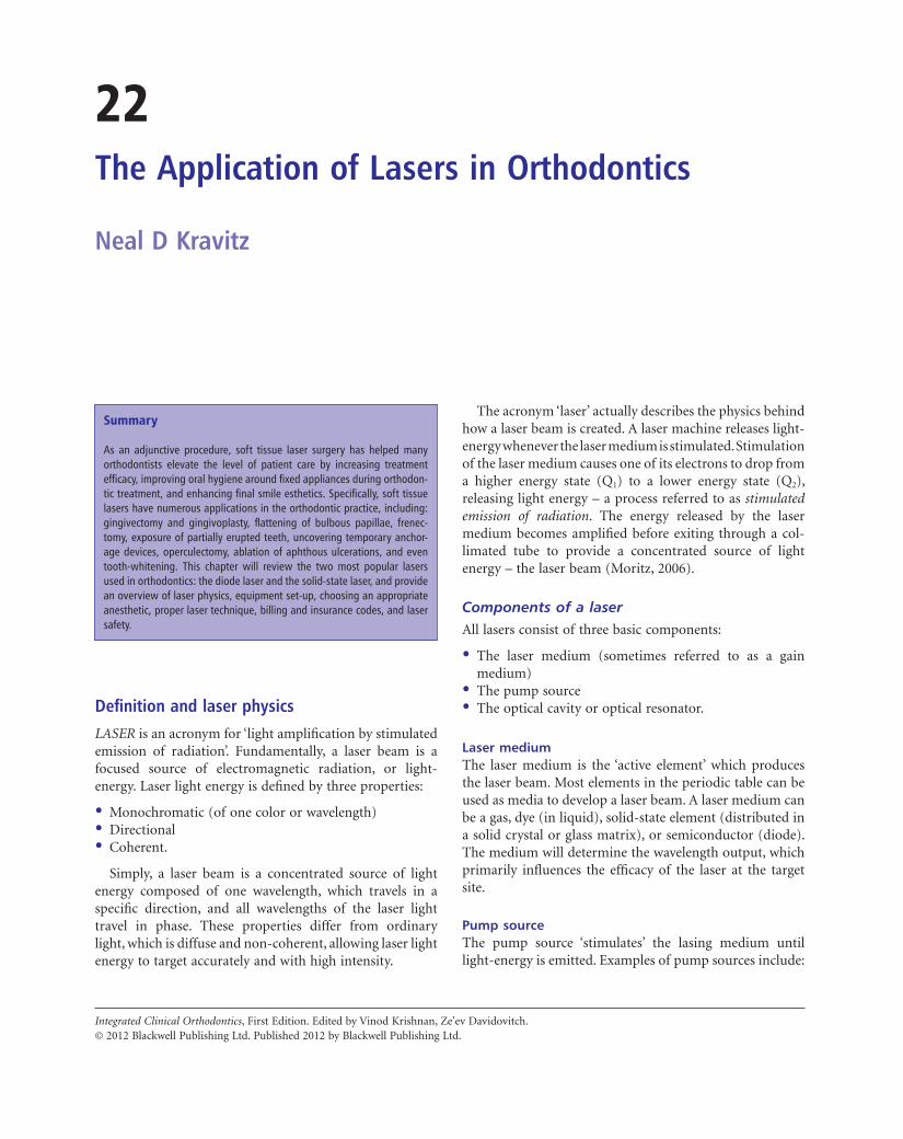
22The Application of Lasers in Orthodontics
Neal D Kravitz
Definition and laser physicsLASER is an acronym for ‘light amplification by stimulated emission of radiation’. Fundamentally, a laser beam is a focused source of electromagnetic radiation, or light-energy. Laser light energy is defined by three properties:
• Monochromatic (of one color or wavelength)
• Directional
• Coherent.
Simply, a laser beam is a concentrated source of light energy composed of one wavelength, which travels in a specific direction, and all wavelengths of the laser light travel in phase. These properties differ from ordinary light, which is diffuse and non-coherent, allowing laser light energy to target accurately and with high intensity.
Integrated Clinical Orthodontics, First Edition. Edited by Vinod Krishnan, Ze’ev Davidovitch.© 2012 Blackwell Publishing Ltd. Published 2012 by Blackwell Publishing Ltd.
Summary
As an adjunctive procedure, soft tissue laser surgery has helped many orthodontists elevate the level of patient care by increasing treatment efficacy, improving oral hygiene around fixed appliances during orthodon-tic treatment, and enhancing final smile esthetics. Specifically, soft tissue lasers have numerous applications in the orthodontic practice, including: gingivectomy and gingivoplasty, flattening of bulbous papillae, frenec-tomy, exposure of partially erupted teeth, uncovering temporary anchor-age devices, operculectomy, ablation of aphthous ulcerations, and even tooth-whitening. This chapter will review the two most popular lasers used in orthodontics: the diode laser and the solid-state laser, and provide an overview of laser physics, equipment set-up, choosing an appropriate anesthetic, proper laser technique, billing and insurance codes, and laser safety.
Lasers in Orthodontics 423
ammonia gas as the active medium that produced micro-wave amplification and the first generation of electromag-netic radiation by stimulated emissions. This device was called the ‘maser’ which is an acronym for microwave amplification by stimulated emission of radiation. In 1958, Arthur Schawlow, Townes’s brother-in-law, proposed the operation of optical and infrared masers, or ‘lasers’ – a term first coined by physicist Gordon Gould in 1957.
In 1960, the first laser to use visible light (using a ruby medium) was developed by physicist Theodor H Maiman, following the theoretical work of Einstein, Townes, and Schawlow. In 1962, Robert Hall developed the first diode or semiconductor laser. The carbon dioxide gas laser was invented by Kumar Patel in 1964, and 4 years later this laser was used to perform the first soft tissue surgery.
In 1985, Paghidiwala, tested the erbium-doped solid state laser (Er:YAG) on dental hard tissue. Nearly a decade later, in 1997, the United States Food and Drug Administration (FDA) approved the Er:YAG solid-state laser for hard tissue surgery. The following year (1998), the first diode laser was approved for soft tissue surgery. More than 25 years since Dr Paghidiwala first tested a laser on dental tissues, dental laser technology has undergone remarkable advancements in design. Contemporary dental laser machinery has become portable, compact, wireless, safer, increasingly affordable, and simpler to operate, allowing more ortho-dontists to incorporate soft tissue laser surgery into their practice.
Laser versus scalpelA laser offers numerous advantages compared with conven-tional scalpel surgery. The most significant advantage is that a laser coagulates capillaries, seals lymphatics, and sterilizes the surgical field during ablation (Sarver and Yanosky, 2005). Separation of tissue is more precise with a laser than a scalpel (Rossman and Cobb, 1995). Additionally, minor aphthous and herpetic ulcerations – which com-monly occur during the early stages of treatment as the patients get accustomed to their orthodontic appliances – can be vaporized. Laser surgery is routinely performed using only light local anesthetic or compound topical anes-thetic. Furthermore, there is markedly less bleeding with laser surgery, particularly for frenal surgery, as well as minimal edema, and no need for sutures or unsightly peri-odontal dressing (Haytac and Ozcelik, 2006). A report sug-gested that laser excisions produce less scar tissue than conventional scalpel surgery (Fisher et al., 1983), although contrary evidence also exists (Buell and Schuller, 1983; Frame, 1985). Post-surgically, patients report less discom-fort, fewer complications related to speaking and chewing, and require less pain medication than do patients treated with conventional scalpel surgery (Haytac and Ozcelik, 2006). The benefits of laser surgery are best summarized by Sarver and Yanosky (2005): ‘[soft tissue lasers] result
electrical discharges, flash-lamps, arc-lamps, or chemical reactions. The type of pump source used depends on the type of laser medium.
Optical cavity or resonatorThe laser optical cavity or resonator amplifies the light-energy. The optical cavity is a compartment of mirrors that contains the laser medium. Light-energy released from the laser medium is reflected by the mirrors back on to itself (referred to as feedback), where it may be amplified by stimulated emission before exciting the cavity. The align-ment of the mirrors with respect to the laser medium will determine the exact operating wavelength of the laser system.
In summary, a laser is a special form of artificial light with specific properties. A laser beam is produced within a laser machine when the pump source stimulates the laser media, releasing light energy which amplifies as it travels through the optical cavity. The amplified light energy released from the machine is what we refer to as the laser beam. When light energy enters the target tissue, it trans-forms into heat – a process known as the photothermal effect – resulting in the vaporization of the target tissue cells.
Thermal ablationUnlike a scalpel which slices, a laser separates tissue by thermal ablation. Thermal ablation is an instantaneous process of absorption, melting, and then vaporization, resulting in decomposition of the tissue. As the target cells absorb the concentrated light-energy of the laser beam, the tissue rapidly rises in temperature. The target cells instantly undergo stages of warming, welding, coagulation, protein denaturization, drying, and vaporization via a micro-explosion known as spallation (Sarver and Yanosky, 2005).
Thermal ablation is dependent on the amount of light energy absorbed, which is determined primarily by the wavelength of the laser. The degree of tissue absorption is influenced by:
• Laser wavelength (measured in nanometers) – a compo-nent of the laser media
• Electrical power of the surgical unit (measured in watts)
• Exposure time
• Composition and thickness of the tissues.
In summary, lasers separate tissue by thermal ablation. The type of laser media produces a specific wavelength, which among other factors, influences the degree of tissue absorption and thus the ‘cutting’ power of the laser.
Historical perspectiveAs early as 1917, American physicist Albert Einstein first proposed the theory of ‘stimulated emission’, the process which made lasers possible. In 1954, Charles Townes at Columbia University demonstrated a working device using
424 Integrated Clinical Orthodontics
and aluminum, whose ability to conduct electricity is between that of conductors and insulators. By doping the laser medium with impurities (dopants), stimulated emis-sion occurs.
The wavelengths produced by diode lasers range between 810 nm and 980 nm. Light energy at these wavelengths is easily absorbed by melanin (soft tissue pigmentation) and hemoglobin, and poorly absorbed by enamel. Therefore, diode lasers are highly effective in soft tissue ablation, hemostasis, and sealing lymphatics, with low risk of damag-ing teeth and bone, making them ideal for soft tissue laser surgery (Kravitz and Kusnoto, 2008). Compared with other laser types, diode lasers are compact, reliable, and have a long operatory lifetime, and are packaged in portable units typically weighing less than 4.5 kg (10 lb). Connecting to the main unit is a thin, pencil-sized hand-piece containing a 200–400 µm fiberoptic tip. Newer models have handpieces that receive single-use, twist-on laser fiberoptic tips, providing a higher potential standard of cleanliness and eliminating time-consuming stripping and cleaving of the fiberoptic tip.
PrimingBefore surgery with a diode laser, the fiberoptic tip must be conditioned or primed. All diode lasers need to have some type of pigment applied to the fiber tip in order to create a sufficient amount of energy for ablation. Priming is the process of concentrating heat energy at the fiberoptic tip (Tracey, 2005). Priming is performed by tapping an
in shorter operative time and faster post-operative recuperation.’
The primary disadvantage of laser surgery is the opera-tory and upkeep expense. Some clinicians have reported additional disadvantages, such as: less tactile sensation, tissue desiccation, and poor wound healing (Baker et al., 2002). Furthermore, laser surgery is primarily excisional and performed without a flap, often resulting in no change in alveolar crest height. As such, there is a tendency for significant tissue rebounding or regrowth after laser surgery.
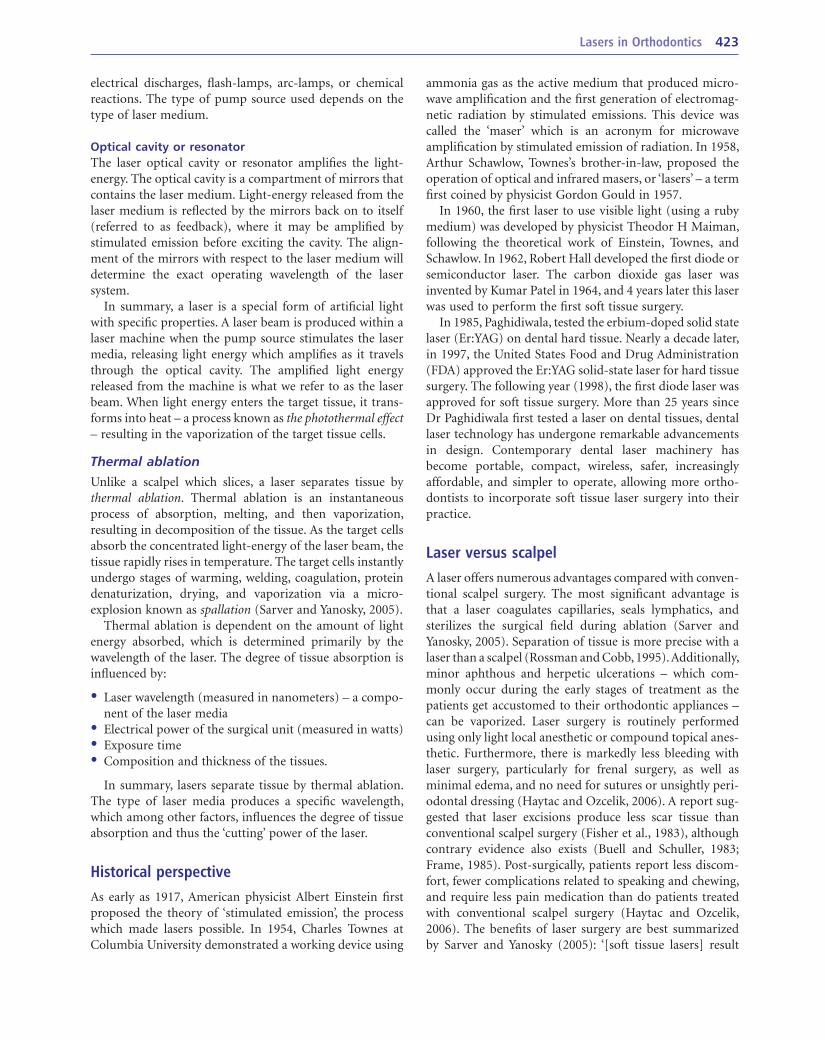
Diode versus solid-state lasersThe two most popular lasers used in dentistry are the diode and the solid-state lasers (Figure 22.1). Diode lasers are almost exclusively used for soft tissue surgery. Solid-state lasers, on the other hand, can be used for both soft and hard tissue surgery, such as tooth preparation, root canal debri-dement, and crown lengthening. The fundamental differ-ence between a diode and a solid-state laser is the laser medium, which generates laser beams of different wave-lengths. As already stated, the laser medium determines the wavelength output, which ultimately influences the efficacy of thermal ablation at the target site.
Diode (semiconductor) lasersDiode lasers convert electrical energy into light energy. Diode lasers are known as semiconductors, as they use a media of gallium and arsenide, and occasionally indium
Figure 22.1 (a) Ezlase 940 diode laser and (b) Waterlase MD Turbo (Er,Cr:YSGG) erbium-doped solid-state laser (Biolase). Images are not to scale.
(a) (b)
Lasers in Orthodontics 425
ablate both hard and soft tissues. Erbium-doped solid-state lasers are routinely used by pediatric dentists and general dentists for caries excavation, and less frequently for endo-dontic and periodontal procedures. When performing soft tissue laser surgery with an erbium-doped solid-state laser, the laser machine is operated at low electrical power to reduce the depth of tissue penetration (Kravitz and Kusnoto, 2008).
For most gingival surgeries, soft tissue excision may require 1.5–2.5 W depending on the tissue thickness. Coagulation with a solid-state laser requires a different setting, generally less than 1.0 W often without water spray. It should be noted that a solid-state laser will begin to ablate hard tissue at approximately 4.0–5.0 W. Solid-state lasers are packaged in larger, more complex rolling units (weigh-ing up to 40 kg or 90 lb). The laser handpiece resembles a high-speed handpiece with removable fiberoptic tips ranging from 400 µm to 750 µm.
During surgery with an erbium-doped solid-state laser, the fiberoptic tip should be held 1 mm away from the tissue (Hadley et al., 2000). Priming is not required as the solid-state lasing medium does not absorb pigmentation. Excision is performed with slow, short back-and-forth strokes. Coagulation is achieved under a different operatory setting, with low wattage and often no water. Tissues appear slightly reddish during excision and chalky white after coagulation. Although a solid-state later can effectively control hemor-rhaging, hemostasis may be easier with a diode, particularly during more invasive soft tissue surgeries such as frenectomies.
Choosing a proper anestheticSoft tissue lasers both coagulate and produce a mild anes-thetic effect during ablation. Accordingly, many clinicians perform soft tissue laser surgery using only light local anesthesia, strong topical anesthesia, or without any anesthesia at all. Local infiltration with 2% lidocaine, 4%
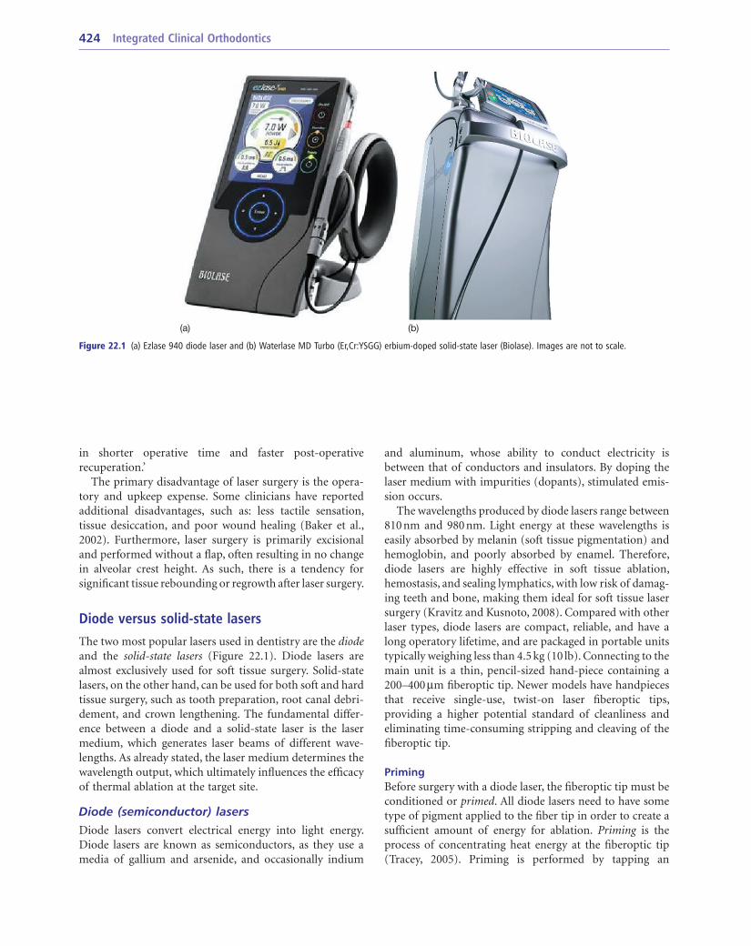
initiated-fiberoptic tip on thick blue articulating paper (Figure 22.2), a felt tip marker, a solid color in a magazine page, or a cork. Essentially, the laser tries to ablate the pig-mented region, creating a super focus of light energy. Failure to properly prime the diode laser may result in less effective tissue ablation.
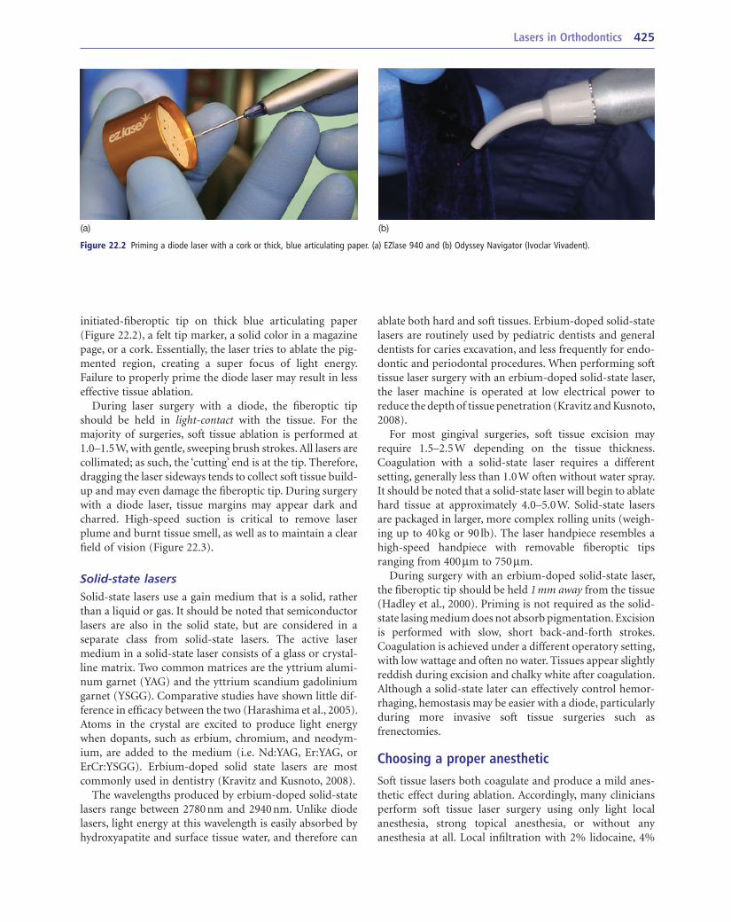
During laser surgery with a diode, the fiberoptic tip should be held in light-contact with the tissue. For the majority of surgeries, soft tissue ablation is performed at 1.0–1.5 W, with gentle, sweeping brush strokes. All lasers are collimated; as such, the ‘cutting’ end is at the tip. Therefore, dragging the laser sideways tends to collect soft tissue build-up and may even damage the fiberoptic tip. During surgery with a diode laser, tissue margins may appear dark and charred. High-speed suction is critical to remove laser plume and burnt tissue smell, as well as to maintain a clear field of vision (Figure 22.3).
Solid-state lasersSolid-state lasers use a gain medium that is a solid, rather than a liquid or gas. It should be noted that semiconductor lasers are also in the solid state, but are considered in a separate class from solid-state lasers. The active laser medium in a solid-state laser consists of a glass or crystal-line matrix. Two common matrices are the yttrium alumi-num garnet (YAG) and the yttrium scandium gadolinium garnet (YSGG). Comparative studies have shown little dif-ference in efficacy between the two (Harashima et al., 2005). Atoms in the crystal are excited to produce light energy when dopants, such as erbium, chromium, and neodym-ium, are added to the medium (i.e. Nd:YAG, Er:YAG, or ErCr:YSGG). Erbium-doped solid state lasers are most commonly used in dentistry (Kravitz and Kusnoto, 2008).
The wavelengths produced by erbium-doped solid-state lasers range between 2780 nm and 2940 nm. Unlike diode lasers, light energy at this wavelength is easily absorbed by hydroxyapatite and surface tissue water, and therefore can
Figure 22.2 Priming a diode laser with a cork or thick, blue articulating paper. (a) EZlase 940 and (b) Odyssey Navigator (Ivoclar Vivadent).
(a) (b)
426 Integrated Clinical Orthodontics
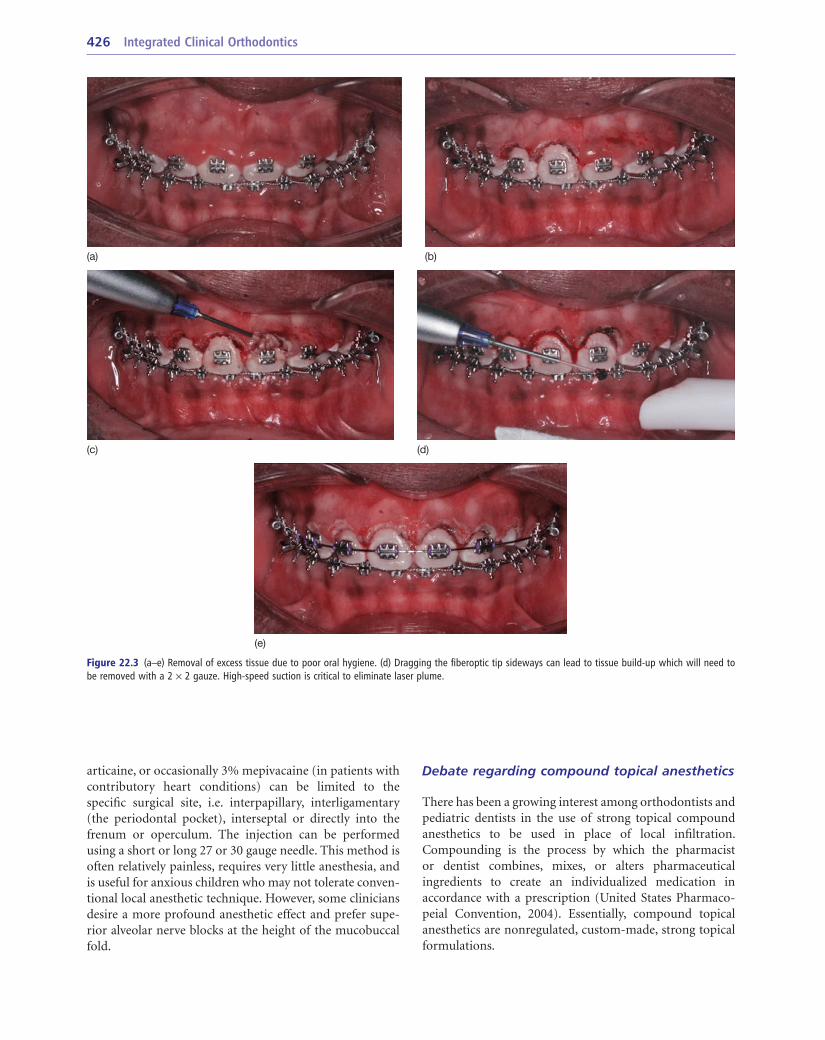
Figure 22.3 (a–e) Removal of excess tissue due to poor oral hygiene. (d) Dragging the fiberoptic tip sideways can lead to tissue build-up which will need to be removed with a 2 × 2 gauze. High-speed suction is critical to eliminate laser plume.
(a) (b)
(c) (d)
(e)
articaine, or occasionally 3% mepivacaine (in patients with contributory heart conditions) can be limited to the specific surgical site, i.e. interpapillary, interligamentary (the periodontal pocket), interseptal or directly into the frenum or operculum. The injection can be performed using a short or long 27 or 30 gauge needle. This method is often relatively painless, requires very little anesthesia, and is useful for anxious children who may not tolerate conven-tional local anesthetic technique. However, some clinicians desire a more profound anesthetic effect and prefer supe-rior alveolar nerve blocks at the height of the mucobuccal fold.
Debate regarding compound topical anesthetics
There has been a growing interest among orthodontists and pediatric dentists in the use of strong topical compound anesthetics to be used in place of local infiltration. Compounding is the process by which the pharmacist or dentist combines, mixes, or alters pharmaceutical ingredients to create an individualized medication in accordance with a prescription (United States Pharmaco-pe ial Convention, 2004). Essentially, compound topical anesthetics are nonregulated, custom-made, strong topical formulations.
Lasers in Orthodontics 427
Doctors who store and repeatedly use the same com-pound formulation on multiple patients may be in viola-tion of state and federal laws. To date, two deaths have been attributed to the lay use of excessive amounts of compound topical anesthetics. Clinicians who are intent on using com-pound topical anesthetics may consider the following protocol.
1. Review medical history to ensure no contributory health conditions.
2. Dry the mucosa with 2 × 2 gauze.3. Apply 0.2 mL (equivalent to one cotton swab head) of
topical anesthetic to the mucosa.4. Let the topical anesthetic remain on tissue for 3–5
minutes while the doctor remains with the patient. Prolonged application can cause tissue-irritation and sloughing.
5. Suction away topical and confirm anesthesia with a peri-odontal probe. Anesthetic effect will last approximately 30 minutes.
Despite the risks, there is arguably a place for doctor-prescribed, doctor-applied compound topical anesthetics for use sparingly on an individualized basis. However, until these drugs become federally regulated, the large-scale pre-production of popular formulations by big pharmacies remains an end-run on manufacturing requirements, and their routine multi-patient use by either orthodontists or other dental specialists remains a questionable therapeutic practice.
Laser machine set-upAll soft tissue lasers have similar machine components and set-up steps. The main components of a dental laser machine include:
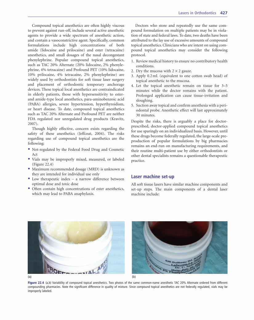
Compound topical anesthetics are often highly viscous to prevent against run-off, include several active anesthetic agents to provide a wide spectrum of anesthetic action, and contain a vasoconstrictive agent. Specifically, common formulations include high concentrations of both amide (lidocaine and prilocaine) and ester (tetracaine) anesthetics, and small dosages of the nasal decongestant phenylephrine. Popular compound topical anesthetics, such as TAC 20% Alternate (20% lidocaine, 2% phenyle-phrine, 4% tetracaine) and Profound PET (10% lidocaine, 10% prilocaine, 4% tetracaine, 2% phenylephrine) are widely used by orthodontists for soft tissue laser surgery and placement of orthodontic temporary anchorage devices. These topical local anesthetics are contraindicated in elderly patients, those with hypersensitivity to ester- and amide-type local anesthetics, para-aminobenzoic acid (PABA) allergies, severe hypertension, hyperthyroidism, or heart disease. To date, compound topical anesthetics such as TAC 20% Alternate and Profound PET are neither FDA regulated nor unregulated drug products (Kravitz, 2007).
Though highly effective, concern exists regarding the safety of these anesthetics (Jeffcoat, 2004). The risks regarding use of compound topical anesthetics are the following:
• Not-regulated by the Federal Food Drug and Cosmetic Act
• Vials may be improperly mixed, measured, or labeled (Figure 22.4)
• Maximum recommended dosage (MRD) is unknown as they are intended for individual use only
• Low therapeutic index – a narrow difference between optimal dose and toxic dose
• Often contain high concentrations of ester anesthetics, which may lead to PABA anaphylaxis.
Figure 22.4 (a,b) Variability of compound topical anesthetics. Two photos of the same common-name anesthetic TAC 20% Alternate ordered from different compounding pharmacies. Note the significant difference in quality of mixture. Since compound topical anesthetics are not federally regulated, vials may be improperly labeled.
(a) (b)
428 Integrated Clinical Orthodontics
(a) (b)
(c) (d)
(e) (f)
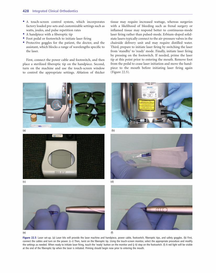
Figure 22.5 Laser set-up. (a) Laser kits will provide the laser machine and handpiece, power cable, footswitch, fiberoptic tips, and safety goggles. (b) First, connect the cables and turn on the power. (c–i) Then, twist on the fiberoptic tip. Using the touch-screen monitor, select the appropriate procedure and modify the settings as needed. When ready to initiate laser firing, touch the ‘ready’ button on the monitor and (j–k) step on the footswitch. (l) A red light will be visible at the end of the fiberoptic tip when the laser is initiated. Priming should begin now prior to entering the mouth.
• A touch-screen control system, which incorporates factory loaded pre-sets and customizable settings such as watts, joules, and pulse repetition rates
• A handpiece with a fiberoptic tip
• Foot pedal or footswitch to initiate laser firing
• Protective goggles for the patient, the doctor, and the assistant, which blocks a range of wavelengths specific to the laser.
First, connect the power cable and footswitch, and then place a sterilized fiberoptic tip on the handpiece. Second, turn on the machine and use the touch-screen window to control the appropriate settings. Ablation of thicker
tissue may require increased wattage, whereas surgeries with a likelihood of bleeding such as frenal surgery or inflamed tissue may respond better to continuous-mode laser firing rather than pulsed-mode. Erbium-doped solid-state lasers typically connect to the air-pressure valves in the chairside delivery unit and may require distilled water. Third, prepare to initiate laser firing by switching the laser from ‘standby’ to ‘ready’ mode. Finally, initiate laser firing by pressing on the footswitch. If needed, prime the laser tip at this point prior to entering the mouth. Remove foot from the pedal to cease laser initiation and move the hand-piece to the mouth before initiating laser firing again (Figure 22.5).

Lasers in Orthodontics 429
interdental tissue prior to drilling of ‘diseased’ bone. In 1912, Henry Percy Pickerill’s famous book, Stomatology in General Practice, further described the operation. By the first half of the nineteenth century, following the landmark research of Ziesel, Nodine, Orban, Kronfeld, Sischer, and Goldman, the etiology of pyorrhea or periodontal disease was no longer thought to be exclusive to bone. Nonetheless, the focus of gingivectomy surgery remained on pocket elimination and not soft tissue esthetics (Kremenak and Squier, 1997; Armitage and Robertson, 2009).
Gingivoplasty is the surgical reshaping and re-contouring of the outer surface of gingival tissue for cosmetic,
Procedures and surgical technique
Gingivectomy and gingivoplasty(Recommend laser settings: diode laser: 1.0–1.5 W; erbium-doped solid-state laser: 1.5–2.5 W)
Gingivectomy is the surgical removal of a portion of gin-gival tissue for improved oral health, functional contour or esthetic appearance (Glossary of Periodontal Terms, 2001). The history of gingivectomy dates back to 1742, when Fauchard proposed a surgical procedure to remove excessive gingival tissue. In 1884, researcher and clinician Robicsek first described the semicircular excision of labial and lingual
(g) (h)
(i) (j)
(k) (l)
Figure 22.5 Continued
430 Integrated Clinical Orthodontics
physiological, or functional purposes, usually done in com-bination with gingivectomy. With conventional scalpel surgery, gingivoplasty corrected the thick, unnatural gingi-val margins left after the gingivectomy procedure. With soft tissue laser surgery, gingivectomy and gingivoplasty are almost always used simultaneously to improve gingival health and enhance smile esthetics.
Smile estheticsEsthetic soft tissue laser surgery is aimed at producing an acceptable gingival display with proper gingival shape and contour, while incorporating the cosmetic principles of proper tooth-size and proportion.
The maxillary anterior teeth should follow two basic principles of cosmetic dentistry. First, the width of maxil-lary incisors should be approximately 80% the height. Second, the widths of the anterior teeth should follow the ‘golden proportion’; that is, the width of the lateral incisor should be two-thirds the width of the central incisor, and the width of the mesial-half of the canine should be two-thirds the width of the lateral incisor (Sarver, 2004) (Figures 22.6, 22.7). While the need for enameloplasty is a result of tooth-shape discrepancies, the extent of incisal reduction is often guided by the amount of overbite and the length of the incisors. In patients with small incisors or large incisal fractures, enameloplasty is enhanced when working in con-junction with soft tissue laser surgery (Kravitz, 2010).
Recommended gingival shape or curvature of the gingi-val margin has the gingival zeniths of the maxillary lateral incisors and mandibular incisors coinciding with the long axes of the teeth. Alternatively, the gingival zeniths of the maxillary central incisors and canines are distal to their long axes (Sarver and Yanosky, 2005). In addition, the gingival height of maxillary central incisors and canines should be 0.5 mm above the maxillary lateral incisors. The maxillary lateral incisors often benefit esthetically from a 0.5 mm gingival-step as well as a 0.5 mm incisal-step (Kravitz, 2010).
Upon animated smile, the patient should reveal 1–4 mm of gingival display – the amount of exposed tissue from the gingival margins of the anterior teeth to the bottom of the upper lip (Kokich et al., 2006). Excessive gingival display may be attributed to multiple factors, such as: vertical max-illary excess, hypermobility of the upper lip, altered passive eruption, gingival hypertrophy, and hyperplasia. Gingival display will reduce slightly over lifetime as the upper lip droops with age.
Patients with severe vertical maxillary excess will benefit mostly from surgical impaction rather than soft tissue laser surgery, while patients with hypermobility of the upper lip, gingival hypertrophy (often a result of poor oral hygiene), or gingival hyperplasia (often induced by medication) respond favorably to tissue removal, as well as adjunctive plastic procedures such as collagen or botulinum toxin in the upper lip. Altered passive eruption almost always requires a surgical flap to reshape the alveolar crest and
place the biologic width along the root of the tooth, rather than the crown.
Biologic widthThe biologic width is the periodontal attachment located between the cementoenamel junction and the apical crest of the alveolar bone. The distance of the biologic width is approximately 2 mm (2.04 mm on average), composed of roughly 1 mm (0.97 mm on average) of epithelial attach-ment (otherwise known as junctional epithelium) above and 1 mm (1.07 mm on average) of connective tissue attach-ment below. Some dentists will include approximately 1 mm of sulcular depth in their definition of the biologic width, due to the challenge of restoring a tooth to the exact coronal edge of the junctional epithelium (Bargiulo et al., 1961; Glossary of Periodontal Terms, 2001; Camargo et al., 2007).
During gingivectomy, if the surgery extends beyond the gingival pocket and into the periodontal attachment, crestal bone will resorb, and a new biologic width will develop lower on the root surface. The biologic width tends to always remain at approximately 2 mm. Violation of the bio-logic width may result in the patient experiencing pro-longed discomfort and gingival inflammation. Although many clinicians warn against interrupting the biologic width during gingival surgery, some crestal resorption may be needed to reduce the effects of gingival rebounding and regrowth.
Tissue reboundingSignificant tissue rebounding or marginal regeneration can occur in the weeks following gingivectomy or gingivo-plasty. Experimental studies have shown that gingi vectomies even extending into alveolar mucosa may regenerate as much as 50% with the formation of new attached marginal gingiva (Wennström, 1983). This can be particu-larly frustrating for both the clinician and patient as a second ‘refinement’ laser surgery may be needed to achieve optimal esthetics. Conventional scalpel surgery with apically positioned flap tends to produce less rebound-ing due to the crestal resorption which occurs after lifting the tissue.
Different tissue biotypes – thick or thin gingival tissue – will respond differently to inflammation, restorative trauma, parafunctional habits, and laser surgery (Fu et al., 2010). The bone sets the tone for gingival architecture. Dense bone with flat topography and minimal ridge atrophy is associated with thick gingival tissue; whereas, thin labial alveolar bone with ridge resorption and fenestrations is associated with thin tissue. Eighty-five percent of individu-als have a thick tissue biotype. Thick gingival tissue is characterized by periodontal health, a large zone of attach-ment, dense fibrotic tissue, and resistance to trauma. Unfortunately, thick tissue biotypes respond poorly to laser surgery, with a high likelihood of tissue rebounding. Thin tissue biotypes are characterized by increased likelihood of
Lasers in Orthodontics 431
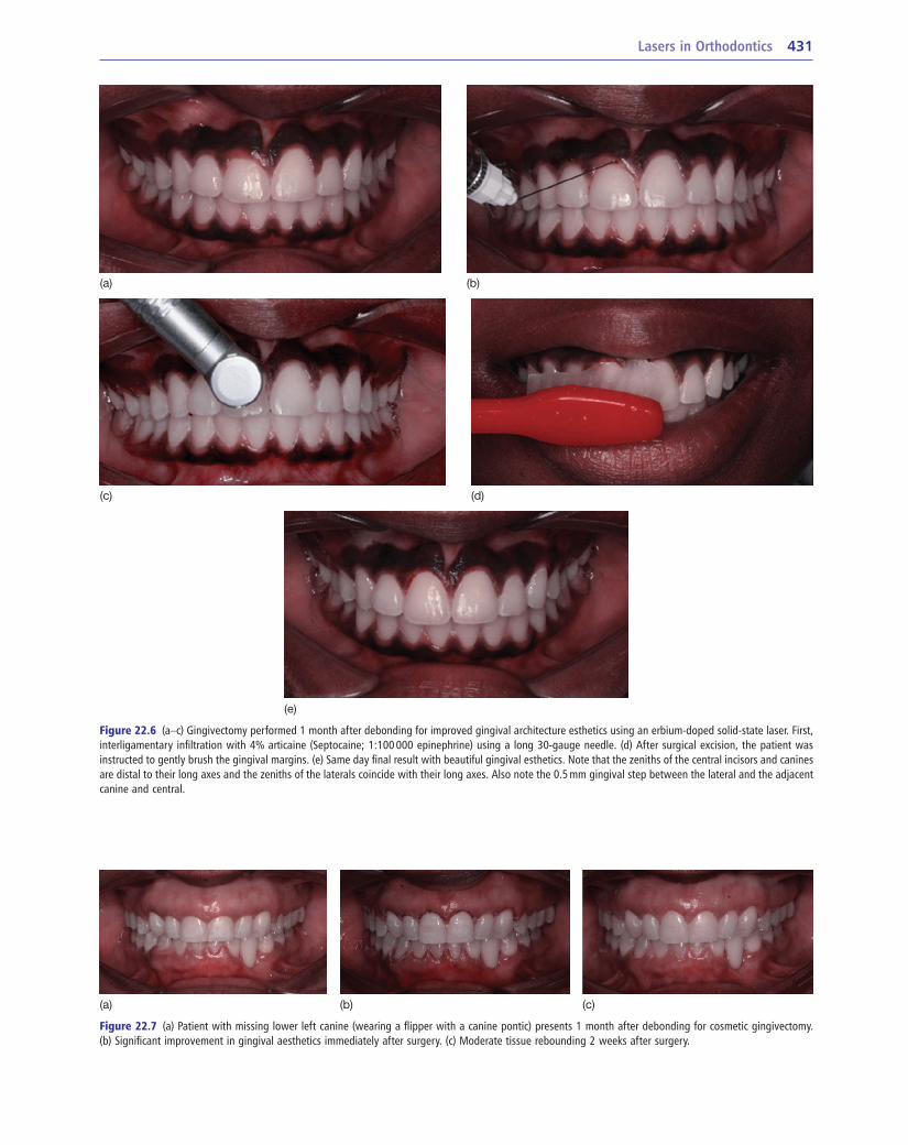
Figure 22.7 (a) Patient with missing lower left canine (wearing a flipper with a canine pontic) presents 1 month after debonding for cosmetic gingivectomy. (b) Significant improvement in gingival aesthetics immediately after surgery. (c) Moderate tissue rebounding 2 weeks after surgery.
(a) (b) (c)
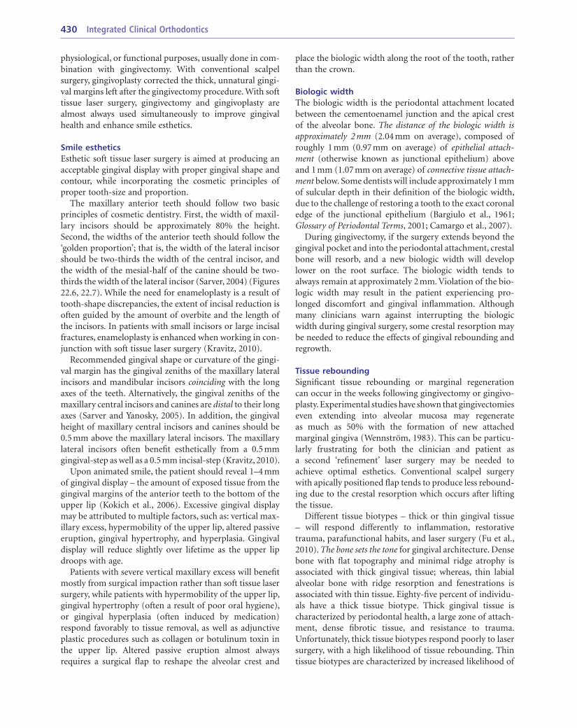
Figure 22.6 (a–c) Gingivectomy performed 1 month after debonding for improved gingival architecture esthetics using an erbium-doped solid-state laser. First, interligamentary infiltration with 4% articaine (Septocaine; 1:100 000 epinephrine) using a long 30-gauge needle. (d) After surgical excision, the patient was instructed to gently brush the gingival margins. (e) Same day final result with beautiful gingival esthetics. Note that the zeniths of the central incisors and canines are distal to their long axes and the zeniths of the laterals coincide with their long axes. Also note the 0.5 mm gingival step between the lateral and the adjacent canine and central.
(a) (b)
(c) (d)
(e)
432 Integrated Clinical Orthodontics
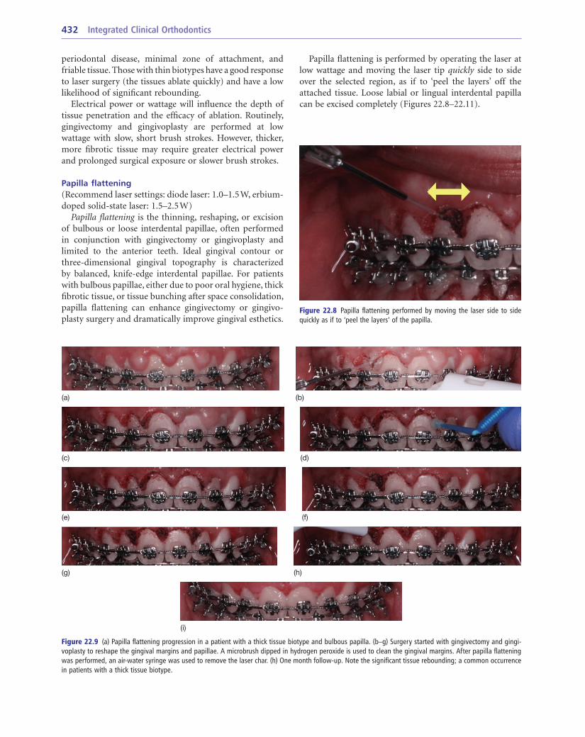
Figure 22.8 Papilla flattening performed by moving the laser side to side quickly as if to ‘peel the layers’ of the papilla.
periodontal disease, minimal zone of attachment, and friable tissue. Those with thin biotypes have a good response to laser surgery (the tissues ablate quickly) and have a low likelihood of significant rebounding.
Electrical power or wattage will influence the depth of tissue penetration and the efficacy of ablation. Routinely, gingivectomy and gingivoplasty are performed at low wattage with slow, short brush strokes. However, thicker, more fibrotic tissue may require greater electrical power and prolonged surgical exposure or slower brush strokes.
Papilla flattening(Recommend laser settings: diode laser: 1.0–1.5 W, erbium-doped solid-state laser: 1.5–2.5 W)
Papilla flattening is the thinning, reshaping, or excision of bulbous or loose interdental papillae, often performed in conjunction with gingivectomy or gingivoplasty and limited to the anterior teeth. Ideal gingival contour or three-dimensional gingival topography is characterized by balanced, knife-edge interdental papillae. For patients with bulbous papillae, either due to poor oral hygiene, thick fibrotic tissue, or tissue bunching after space consolidation, papilla flattening can enhance gingivectomy or gingivo-plasty surgery and dramatically improve gingival esthetics.
Figure 22.9 (a) Papilla flattening progression in a patient with a thick tissue biotype and bulbous papilla. (b–g) Surgery started with gingivectomy and gingi-voplasty to reshape the gingival margins and papillae. A microbrush dipped in hydrogen peroxide is used to clean the gingival margins. After papilla flattening was performed, an air-water syringe was used to remove the laser char. (h) One month follow-up. Note the significant tissue rebounding; a common occurrence in patients with a thick tissue biotype.
(a) (b)
(c) (d)
(e) (f)
(g) (h)
(i)
Papilla flattening is performed by operating the laser at low wattage and moving the laser tip quickly side to side over the selected region, as if to ‘peel the layers’ off the attached tissue. Loose labial or lingual interdental papilla can be excised completely (Figures 22.8–22.11).
Lasers in Orthodontics 433
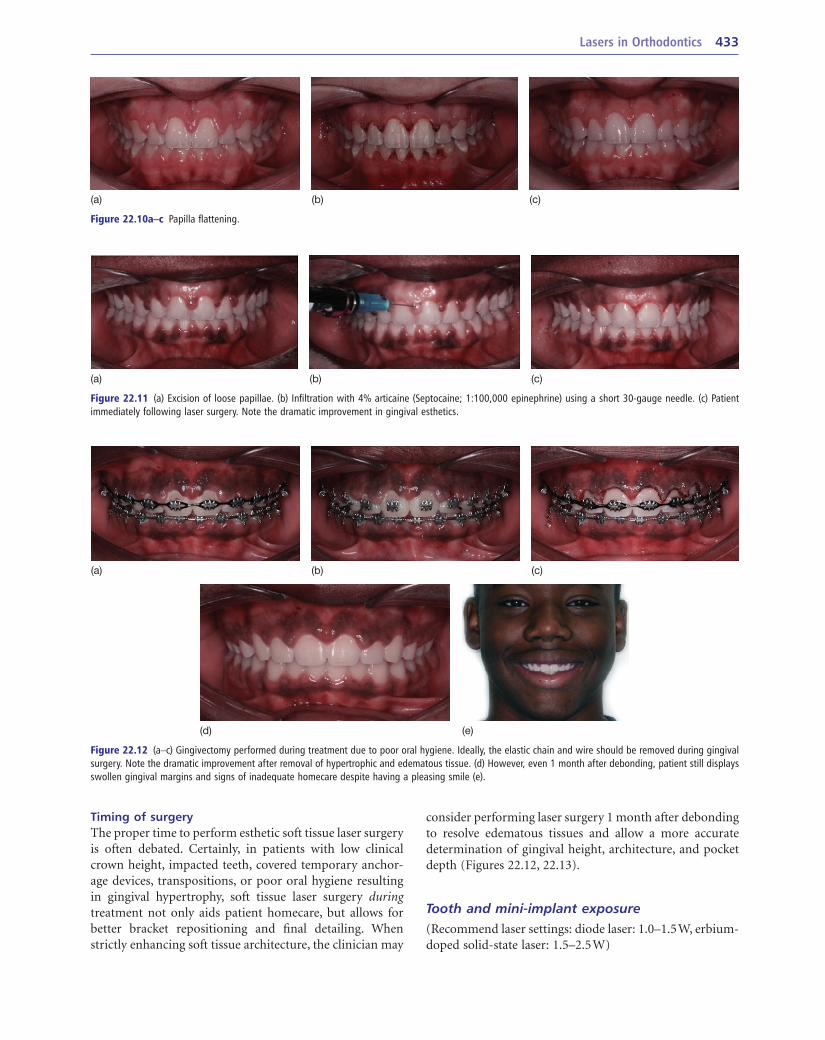
Figure 22.10a–c Papilla flattening.
(a) (b) (c)
Figure 22.11 (a) Excision of loose papillae. (b) Infiltration with 4% articaine (Septocaine; 1:100,000 epinephrine) using a short 30-gauge needle. (c) Patient immediately following laser surgery. Note the dramatic improvement in gingival esthetics.
(a) (b) (c)
Timing of surgeryThe proper time to perform esthetic soft tissue laser surgery is often debated. Certainly, in patients with low clinical crown height, impacted teeth, covered tem porary anchor-age devices, transpositions, or poor oral hygiene resulting in gingival hypertrophy, soft tissue laser surgery during treatment not only aids patient homecare, but allows for better bracket repo sitioning and final detailing. When strictly enhancing soft tissue architecture, the clinician may
Figure 22.12 (a–c) Gingivectomy performed during treatment due to poor oral hygiene. Ideally, the elastic chain and wire should be removed during gingival surgery. Note the dramatic improvement after removal of hypertrophic and edematous tissue. (d) However, even 1 month after debonding, patient still displays swollen gingival margins and signs of inadequate homecare despite having a pleasing smile (e).
(a) (b) (c)
(d) (e)
consider performing laser surgery 1 month after debonding to resolve edematous tissues and allow a more accurate determination of gingival height, architecture, and pocket depth (Figures 22.12, 22.13).
Tooth and mini-implant exposure(Recommend laser settings: diode laser: 1.0–1.5 W, erbium-doped solid-state laser: 1.5–2.5 W)
434 Integrated Clinical Orthodontics
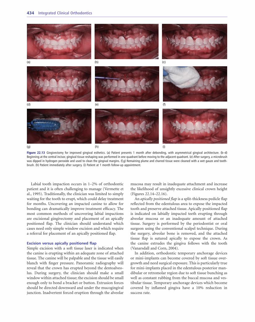
Figure 22.13 Gingivectomy for improved gingival esthetics. (a) Patient presents 1 month after debonding, with asymmetrical gingival architecture. (b–d) Beginning at the central incisor, gingival tissue reshaping was performed in one quadrant before moving to the adjacent quadrant. (e) After surgery, a microbrush was dipped in hydrogen peroxide and used to clean the gingival margins. (f,g) Remaining plume and charred tissue were cleaned with a wet gauze and tooth-brush. (h) Patient immediately after surgery. (i) Patient at 1 month follow-up appointment.
(a) (b) (c)
(d) (e) (f)
(g) (h) (i)
Labial tooth impaction occurs in 1–2% of orthodontic patient and it is often challenging to manage (Vermette et al., 1995). Traditionally, the clinician was limited to simply waiting for the tooth to erupt, which could delay treatment for months. Uncovering an impacted canine to allow for bonding can dramatically improve treatment efficacy. The most common methods of uncovering labial impactions are excisional gingivectomy and placement of an apically positioned flap. The clinician should understand which cases need only simple window excision and which require a referral for placement of an apically positioned flap.
Excision versus apically positioned flapSimple excision with a soft tissue laser is indicated when the canine is erupting within an adequate zone of attached tissue. The canine will be palpable and the tissue will easily blanch with finger pressure. Panoramic radiography will reveal that the crown has erupted beyond the dentoalveo-lus. During surgery, the clinician should make a small window within attached tissue; the excision should be small enough only to bond a bracket or button. Extrusion forces should be directed downward and under the mucogingival junction. Inadvertent forced eruption through the alveolar
mucosa may result in inadequate attachment and increase the likelihood of unsightly excessive clinical crown height (Figures 22.14–22.16).
An apically positioned flap is a split-thickness pedicle flap reflected from the edentulous area to expose the impacted tooth and preserve attached tissue. Apically positioned flap is indicated on labially impacted teeth erupting through alveolar mucosa or an inadequate amount of attached tissue. Surgery is performed by the periodontist or oral surgeon using the conventional scalpel technique. During the surgery, alveolar bone is removed, and the attached tissue flap is sutured apically to expose the crown. As the canine extrudes the gingiva follows with the tooth (Vanarsdall and Corn, 2004).
In addition, orthodontic temporary anchorage devices or mini-implants can become covered by soft tissue over-growth and need surgical exposure. This is particularly true for mini-implants placed in the edentulous posterior man-dibular or retromolar region due to soft tissue bunching as well as constant rubbing from the buccal mucosa and ves-tibular tissue. Temporary anchorage devices which become covered by inflamed gingiva have a 10% reduction in success rate.
Lasers in Orthodontics 435
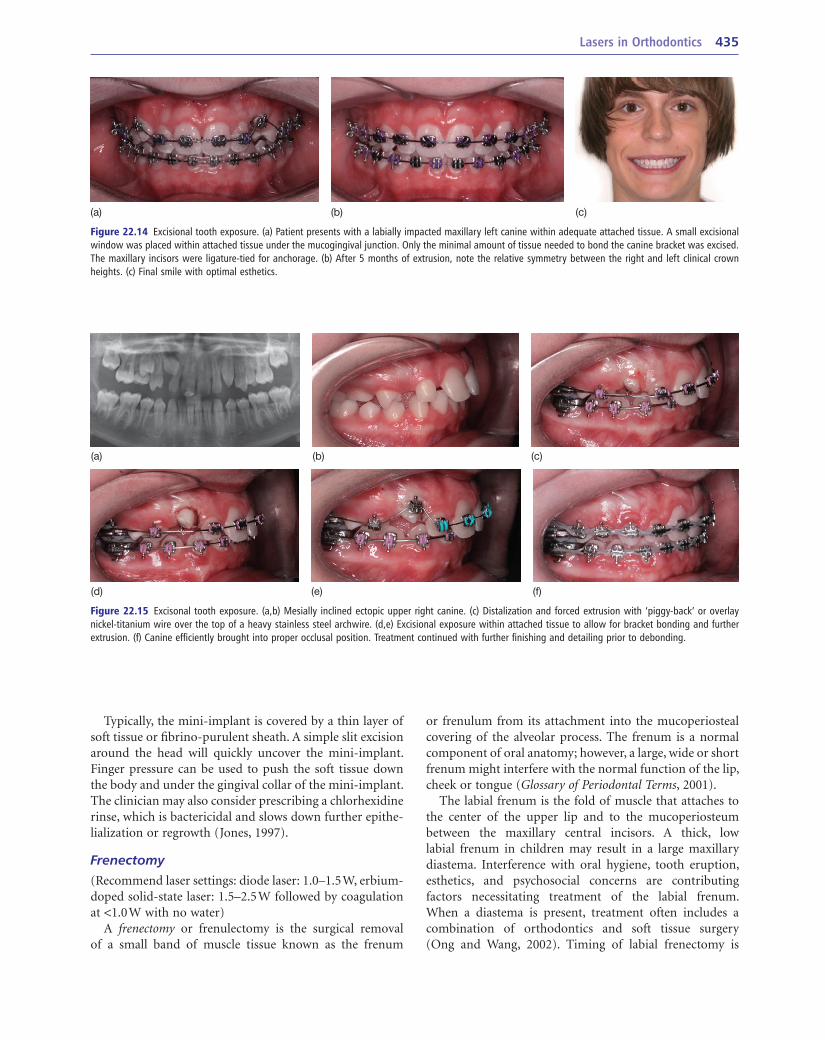
Figure 22.15 Excisonal tooth exposure. (a,b) Mesially inclined ectopic upper right canine. (c) Distalization and forced extrusion with ‘piggy-back’ or overlay nickel-titanium wire over the top of a heavy stainless steel archwire. (d,e) Excisional exposure within attached tissue to allow for bracket bonding and further extrusion. (f) Canine efficiently brought into proper occlusal position. Treatment continued with further finishing and detailing prior to debonding.
(a) (b) (c)
(d) (e) (f)
Figure 22.14 Excisional tooth exposure. (a) Patient presents with a labially impacted maxillary left canine within adequate attached tissue. A small excisional window was placed within attached tissue under the mucogingival junction. Only the minimal amount of tissue needed to bond the canine bracket was excised. The maxillary incisors were ligature-tied for anchorage. (b) After 5 months of extrusion, note the relative symmetry between the right and left clinical crown heights. (c) Final smile with optimal esthetics.
(a) (b) (c)
Typically, the mini-implant is covered by a thin layer of soft tissue or fibrino-purulent sheath. A simple slit excision around the head will quickly uncover the mini-implant. Finger pressure can be used to push the soft tissue down the body and under the gingival collar of the mini-implant. The clinician may also consider prescribing a chlorhexidine rinse, which is bactericidal and slows down further epithe-lialization or regrowth (Jones, 1997).
Frenectomy(Recommend laser settings: diode laser: 1.0–1.5 W, erbium-doped solid-state laser: 1.5–2.5 W followed by coagulation at <1.0 W with no water)
A frenectomy or frenulectomy is the surgical removal of a small band of muscle tissue known as the frenum
or frenulum from its attachment into the mucoperiosteal covering of the alveolar process. The frenum is a normal component of oral anatomy; however, a large, wide or short frenum might interfere with the normal function of the lip, cheek or tongue (Glossary of Periodontal Terms, 2001).
The labial frenum is the fold of muscle that attaches to the center of the upper lip and to the mucoperiosteum between the maxillary central incisors. A thick, low labial frenum in children may result in a large maxillary diastema. Interference with oral hygiene, tooth eruption, esthetics, and psychosocial concerns are contributing factors necessitating treatment of the labial frenum. When a diastema is present, treatment often includes a combination of orthodontics and soft tissue surgery (Ong and Wang, 2002). Timing of labial frenectomy is
436 Integrated Clinical Orthodontics
(a) (b) (c)
(d) (e) (f)
(g) (h) (i)
(j) (k) (l)
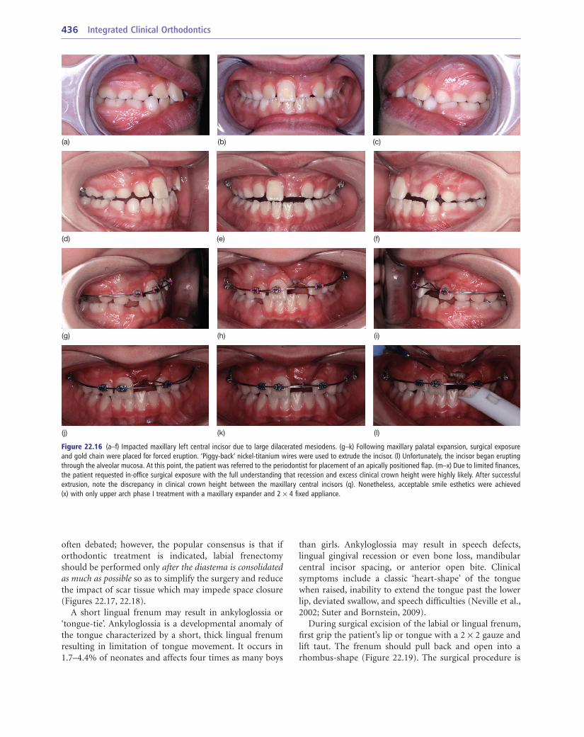
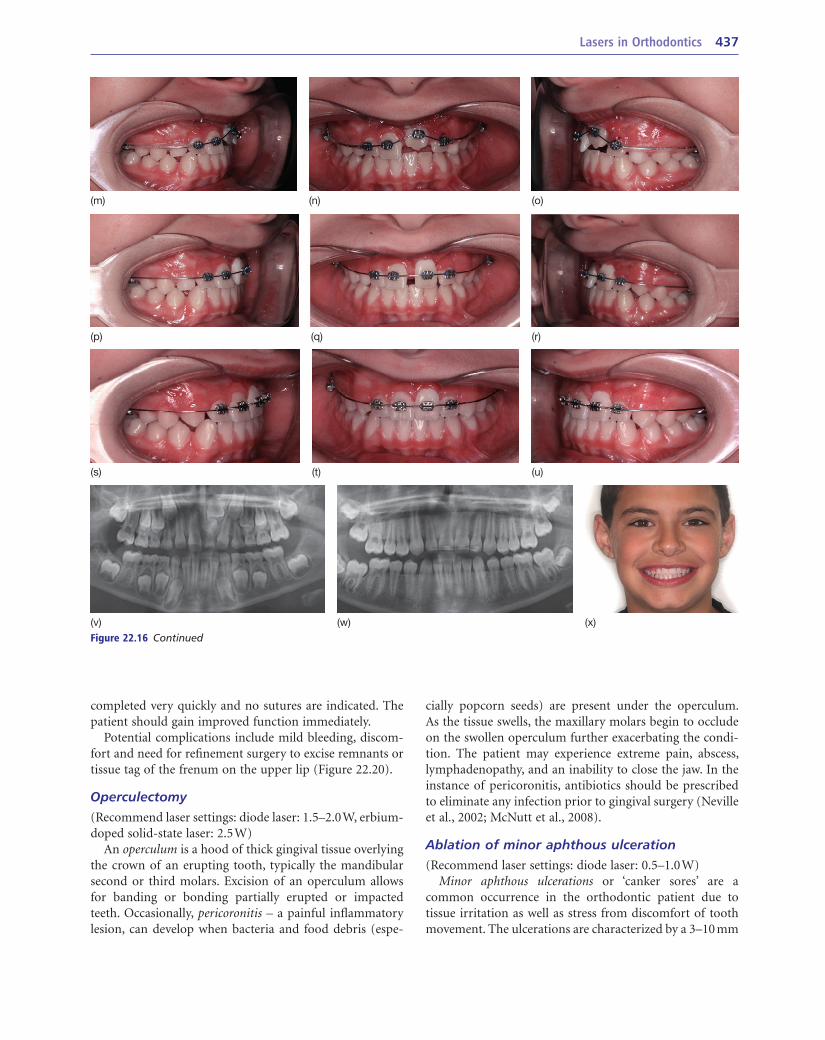
Figure 22.16 (a–f) Impacted maxillary left central incisor due to large dilacerated mesiodens. (g–k) Following maxillary palatal expansion, surgical exposure and gold chain were placed for forced eruption. ‘Piggy-back’ nickel-titanium wires were used to extrude the incisor. (l) Unfortunately, the incisor began erupting through the alveolar mucosa. At this point, the patient was referred to the periodontist for placement of an apically positioned flap. (m–x) Due to limited finances, the patient requested in-office surgical exposure with the full understanding that recession and excess clinical crown height were highly likely. After successful extrusion, note the discrepancy in clinical crown height between the maxillary central incisors (q). Nonetheless, acceptable smile esthetics were achieved (x) with only upper arch phase I treatment with a maxillary expander and 2 × 4 fixed appliance.
than girls. Ankyloglossia may result in speech defects, lingual gingival recession or even bone loss, mandibular central incisor spacing, or anterior open bite. Clinical symptoms include a classic ‘heart-shape’ of the tongue when raised, inability to extend the tongue past the lower lip, deviated swallow, and speech difficulties (Neville et al., 2002; Suter and Bornstein, 2009).
During surgical excision of the labial or lingual frenum, first grip the patient’s lip or tongue with a 2 × 2 gauze and lift taut. The frenum should pull back and open into a rhombus-shape (Figure 22.19). The surgical procedure is
often debated; however, the popular consensus is that if orthodontic treatment is indicated, labial frenectomy should be performed only after the diastema is consolidated as much as possible so as to simplify the surgery and reduce the impact of scar tissue which may impede space closure (Figures 22.17, 22.18).
A short lingual frenum may result in ankyloglossia or ‘tongue-tie’. Ankyloglossia is a developmental anomaly of the tongue characterized by a short, thick lingual frenum resulting in limitation of tongue movement. It occurs in 1.7–4.4% of neonates and affects four times as many boys
Lasers in Orthodontics 437
(m) (n) (o)
(p) (q) (r)
(s) (t) (u)
(v) (w) (x)
Figure 22.16 Continued
completed very quickly and no sutures are indicated. The patient should gain improved function immediately.
Potential complications include mild bleeding, discom-fort and need for refinement surgery to excise remnants or tissue tag of the frenum on the upper lip (Figure 22.20).
Operculectomy(Recommend laser settings: diode laser: 1.5–2.0 W, erbium-doped solid-state laser: 2.5 W)
An operculum is a hood of thick gingival tissue overlying the crown of an erupting tooth, typically the mandibular second or third molars. Excision of an operculum allows for banding or bonding partially erupted or impacted teeth. Occasionally, pericoronitis – a painful inflammatory lesion, can develop when bacteria and food debris (espe-
cially popcorn seeds) are present under the operculum. As the tissue swells, the maxillary molars begin to occlude on the swollen operculum further exacerbating the condi-tion. The patient may experience extreme pain, abscess, lymphadenopathy, and an inability to close the jaw. In the instance of pericoronitis, antibiotics should be prescribed to eliminate any infection prior to gingival surgery (Neville et al., 2002; McNutt et al., 2008).
Ablation of minor aphthous ulceration(Recommend laser settings: diode laser: 0.5–1.0 W)
Minor aphthous ulcerations or ‘canker sores’ are a common occurrence in the orthodontic patient due to tissue irritation as well as stress from discomfort of tooth movement. The ulcerations are characterized by a 3–10 mm
438 Integrated Clinical Orthodontics
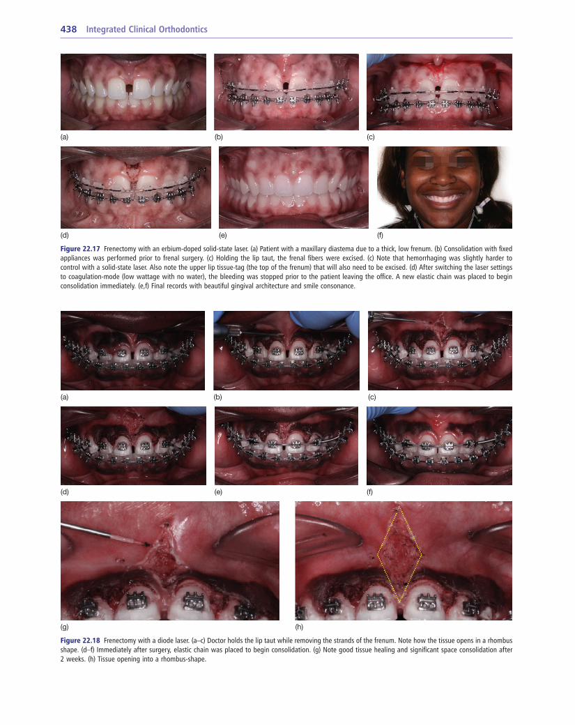
Figure 22.17 Frenectomy with an erbium-doped solid-state laser. (a) Patient with a maxillary diastema due to a thick, low frenum. (b) Consolidation with fixed appliances was performed prior to frenal surgery. (c) Holding the lip taut, the frenal fibers were excised. (c) Note that hemorrhaging was slightly harder to control with a solid-state laser. Also note the upper lip tissue-tag (the top of the frenum) that will also need to be excised. (d) After switching the laser settings to coagulation-mode (low wattage with no water), the bleeding was stopped prior to the patient leaving the office. A new elastic chain was placed to begin consolidation immediately. (e,f) Final records with beautiful gingival architecture and smile consonance.
(a) (b) (c)
(d) (e) (f)
Figure 22.18 Frenectomy with a diode laser. (a–c) Doctor holds the lip taut while removing the strands of the frenum. Note how the tissue opens in a rhombus shape. (d–f) Immediately after surgery, elastic chain was placed to begin consolidation. (g) Note good tissue healing and significant space consolidation after 2 weeks. (h) Tissue opening into a rhombus-shape.
(a) (b) (c)
(d) (e) (f)
(g) (h)
Lasers in Orthodontics 439
procedures with a diode laser, ablation of aphthous ulcera-tions are performed 1–2 mm away from the tissue. The clini-cian should proceed with short, side-to-side brush strokes over the ulcerated area. Typically, anesthesia is not required. Postoperative management can include routine palliative treatment.
Excision of soft tissue lesions(Recommend laser settings: diode laser: 1.0–1.5 W, erbium-doped solid-state laser: 1.5–2.5 W)
Occasionally, the orthodontic patient will present with a soft tissue lesion on the mucosa or tongue due to local irritation or trauma. In most instances, the orthodontist should refer to an oral surgeon or oral medicine specialist for evaluation and biopsy. However, in some instances, simple benign lesions, such as an irritation fibroma, pyogenic granuloma, or mucocele can be treated by the
diameter yellowish-white, removable fibroino-purulent membrane encircled by an erythematous halo, localized almost exclusively on nonkeratinized tissue such as the buccal mucosa, alveolar mucosa, tongue, and lips. The eti-ology of aphthous ulcerations includes trauma, stress, aller-gies (i.e. to nickel in the appliances), nutritional deficiencies, hematologic abnormalities, hormones, infectious agents, systemic conditions (i.e. immunoglobulin A deficiency, neutropenia, celiac disease), or a genetic predisposition. After relieving the etiologic agent, minor ulcerations typi-cally heal without scarring in 7–14 days (Neville et al., 2002).
Several types of treatment are available for management of aphthous ulcerations, including: simply waiting for the mouth to heal, warm salt water rinses, hydrogen peroxide, antifungal compound medications, liquid topical anesthet-ics, and diode laser surgery treatment. Unlike other surgical
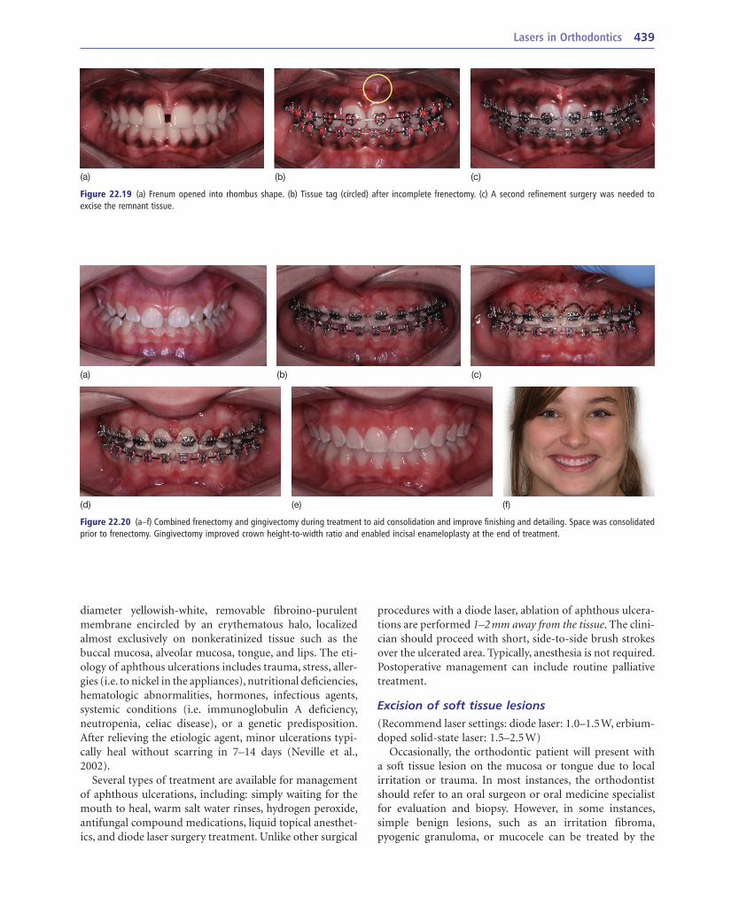
Figure 22.19 (a) Frenum opened into rhombus shape. (b) Tissue tag (circled) after incomplete frenectomy. (c) A second refinement surgery was needed to excise the remnant tissue.
(a) (b) (c)
Figure 22.20 (a–f) Combined frenectomy and gingivectomy during treatment to aid consolidation and improve finishing and detailing. Space was consolidated prior to frenectomy. Gingivectomy improved crown height-to-width ratio and enabled incisal enameloplasty at the end of treatment.
(a) (b) (c)
(d) (e) (f)
440 Integrated Clinical Orthodontics
Laser safetyLaser hazard classificationThe major risk during laser surgery is exposure to laser radiation. Laser safety is regulated according to the American National Standards Institute’s (ANSI) Z136 safety standards in the United States and the International Electrotechnical Commission (IEC) 60825 internationally. ANSI laser safety standards are the basis for Occupational Safety and Health Administration (OSHA) and state occu-pational safety rules. All lasers sold in the United States since 1976 are classified according to their hazard potential, power, and wavelength. Currently, there are seven laser hazard classes (Class 1, 1M, 2, 2M, 3R, 3B, and 4). Lasers used in medical or dental therapeutic use, such as soft tissue lasers, are Class 4 products (Kravitz and Kusnoto, 2008).
Class 4 lasers have an output power >0.5 W. At this power, eye and skin are endangered even at diffuse reflection. As such, a clinician is required to ensure the following safety precautions:
• Creation of a danger zone – typically this entails a des-ignated surgical chair or room with a warning sign indi-cating that a laser is operational
• Presence of a laser safety officer (typically the orthodontist)
• Proper training of users (the orthodontist and staff)
• Consideration of potential fire hazards.
Eye and skin injuryUnquestionably, the greatest specific risk of soft tissue laser surgery is injury to the eye. The severity of injury depends
orthodontist with conservative surgical excision using a soft tissue laser.
• Fibroma (irritation fibroma, fibrous nodule) is a benign, asymptomatic nodular mass of dense fibrous connective tissue covered by squamous epithelium (Neville et al., 2002). A fibroma is the most common abnormal growth in the oral cavity; the most common location is the buccal mucosa, likely a consequence of trauma from cheek biting.
• Pyogenic granuloma is a non-neoplastic, smooth or lobu-lated erythematous mass of granulation tissue. Three-quarters of oral pyogenic granulomas are located on the gingiva (Neville et al., 2002). The greatest precipitating factor may be gingival inflammation from poor oral hygiene around fixed appliances.
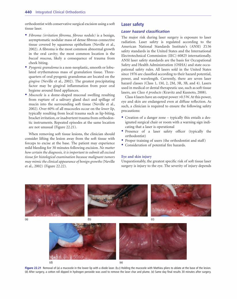
• Mucocele is a dome-shaped mucosal swelling resulting from rupture of a salivary gland duct and spillage of mucin into the surrounding soft tissue (Neville et al., 2002). Over 60% of all mucoceles occur on the lower lip, typically resulting from local trauma such as lip-biting, bracket irritation, or inadvertent trauma from orthodon-tic instruments. Repeated episodes at the same location are not unusual (Figure 22.21).
When removing soft tissue lesions, the clinician should consider lifting the lesion away from the soft tissue with forceps to excise at the base. The patient may experience mild bleeding for 30 minutes following excision. No matter how certain the diagnosis, it is important to submit all excised tissue for histological examination because malignant tumors may mimic the clinical appearance of benign growths (Neville et al., 2002) (Figure 22.22).
Figure 22.21 Removal of (a) a mucocele in the lower lip with a diode laser. (b,c) Holding the mucocele with Mathieu pliers to ablate at the base of the lesion. (d) After surgery, a cotton roll dipped in hydrogen peroxide was used to remove the laser char and plume. (e) Same day final results 30 minutes after surgery.
(a) (b) (c)
(d) (e)
Lasers in Orthodontics 441
includes both diodes and erbium lasers), reaching a maximum penetration at 1000 nm. The arms, hands, and head are the regions of the body most likely to be exposed to laser radiation.
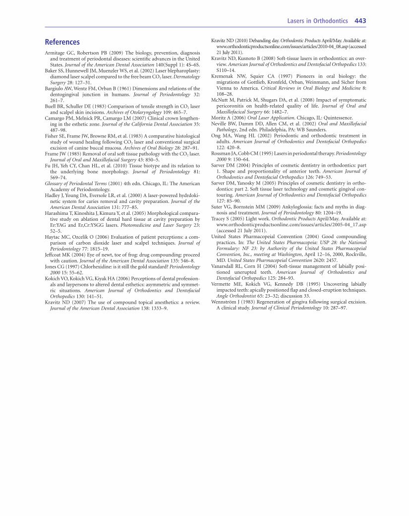
Patient and operator protectionThe patient and clinician should be fully covered and wavelength-specific protective goggles should be worn by the doctor, the assistant, and the patient at all times. It is imperative that the goggles block light at the appropriate wavelength and protect all possible reflective paths to the eyes (Figure 22.23). Therefore, the orange protective goggles
on the laser wavelength, distance from the laser source, and power of the laser machine. The eye is precise at focusing light, and a split-second exposure to laser radiation may be sufficient to cause permanent injury. Retinal damage can occur at 400–1400 nm (this range is known as the retinal hazard region). The major danger is a stray laser beam reflected from a table, jewelry, or belt. Diode lasers risk retinal burns and cataract, whereas solid-state lasers risk corneal burn, aqueous flare-ups, and infrared cataract.
Skin is the largest organ of the body and poses high risk of radiation exposure, regardless of the laser used. Skin can be penetrated at wavelengths of 300–3000 nm (which
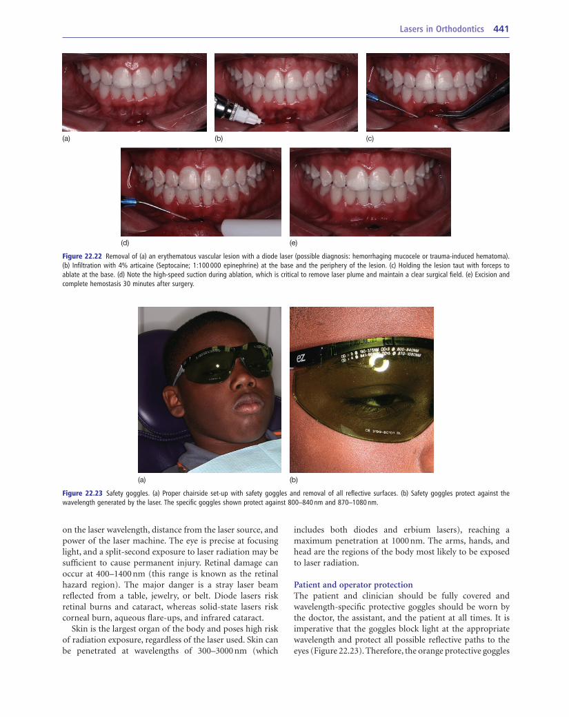
Figure 22.22 Removal of (a) an erythematous vascular lesion with a diode laser (possible diagnosis: hemorrhaging mucocele or trauma-induced hematoma). (b) Infiltration with 4% articaine (Septocaine; 1:100 000 epinephrine) at the base and the periphery of the lesion. (c) Holding the lesion taut with forceps to ablate at the base. (d) Note the high-speed suction during ablation, which is critical to remove laser plume and maintain a clear surgical field. (e) Excision and complete hemostasis 30 minutes after surgery.
(a) (b) (c)
(d) (e)
Figure 22.23 Safety goggles. (a) Proper chairside set-up with safety goggles and removal of all reflective surfaces. (b) Safety goggles protect against the wavelength generated by the laser. The specific goggles shown protect against 800–840 nm and 870–1080 nm.
(a) (b)

442 Integrated Clinical Orthodontics
and gently massage the surgical area with a soft-bristle toothbrush. Bleeding and discomfort are typically minimal, with the exception of a frenal surgery, in which minor bleeding is expected for 24 hours postsurgery. Complete tissue healing will take place after 1–2 weeks, at which point the patient should be seen for a postoperative follow-up.
Billing and insurance codesLaser surgery insurance codes provided by the American Dental Association (ADA) for common soft tissue proce-dures are listed under both specialties of Periodontics (D4000–4999) and Oral Surgery (D7000–7999), and therefore will not affect the patient’s orthodontic benefits. The orthodontist should stay current with annual changes in codes and definitions made to the Current Dental Terminology (CDT) handbook. Insurance claims often require specific information, such as quadrant, probing depths, reason for surgery, and surgical records, provided in a letter submitted along with the insurance claim (Kravitz and Kusnoto, 2008) (Table 22.1).
ConclusionThe use of soft tissue lasers offers many advantages such as improved oral hygiene, practice efficiencies, and esthetic finishing. Clinicians interested in incorporating soft tissue lasers into their practice should obtain proficiency certifica-tion, attend continuing education courses, and recognize the inherent risks associated with laser surgery. As an ortho-dontist committed to providing the best possible service, adjunctive procedures such as soft tissue surgery can dra-matically enhance the overall treatment experience in your office.
used during light curing will not suffice. Patients should remove all facial jewelry and nearby reflective surfaces should be covered or removed. Class 4 laser systems pose a fire hazard if the beam contacts flammable substances, and flame-retardant materials should be available in the office. A discernable danger zone should be created around the surgical bay with a sign reading: Warning: Visible and Invisible Laser Radiation. Avoid Eye or Skin Exposure to Direct Scatter Radiation. Class IV laser product. Such signs are typically provided by the laser manufacturer and are available over the internet (Kravitz and Kusnoto, 2008) (Figure 22.24).
Informed consentSoft tissue laser surgery is currently not listed on the American Association of Orthodontics standard informed consent packet. Until then, clinicians may consider writing their own informed consent. Informed consent may vary depending on the type of laser and the procedure per-formed. Consent for the diode laser may include: the rec-ommended treatment; although rare, the principal risks and complications, including postsurgical infection, swell-ing, bleeding, headache, temporomandibular joint (TMJ) (jaw joint) pain, tooth/gum pain, microcracks in the enamel, pulpal over-heating leading to hyperemia, shrink-age of gum tissues, muscle soreness, soft tissue numbness, postoperative discomfort, and mild bleeding; expected results and need for potential surgical refinement; and nec-essary follow-up care and homecare.
Postsurgical managementImmediately after the procedure, the clinician can run a microbrush or cotton roll dipped in hydrogen peroxide along the gingival margins to remove any charred tissue and laser plume. The patient should be encouraged to rinse
Figure 22.24 Laser safety sign to be displayed when performing surgery.
Table 22.1 Dental codes for common soft tissue procedures
Code Procedure
D4210 Gingivectomy/gingivoplasty – four or more contiguous teeth per quadrant
D4211 Gingivectomy/gingivoplasty – one to three contiguous teeth per quadrant
D7960 Frenectomy
D7971 Operculectomy
D7465 Aphthous ulcer
D7430 Excision of benign tumor – diameter <1.25 cm
D7430 Excision of benign tumor – diameter >1.25 cm
D7286 Biopsy of oral tissue, soft
When submitting a claim, insurance companies will ask for an accompanying letter that explains the need for surgery, the probing depths, the quadrant of surgery, and other pertinent information. Soft tissue surgical codes fall under periodontal and oral surgery insurance coverage.
Lasers in Orthodontics 443
Kravitz ND (2010) Debanding day. Orthodontic Products April/May. Available at: www.orthodonticproductsonline.com/issues/articles/2010-04_08.asp (accessed 21 July 2011).
Kravitz ND, Kusnoto B (2008) Soft-tissue lasers in orthodontics: an over-view. American Journal of Orthodontics and Dentofacial Orthopedics 133: S110–14.
Kremenak NW, Squier CA (1997) Pioneers in oral biology: the migrations of Gottlieb, Kronfeld, Orban, Weinmann, and Sicher from Vienna to America. Critical Reviews in Oral Biology and Medicine 8: 108–28.
McNutt M, Patrick M, Shugars DA, et al. (2008) Impact of symptomatic pericoronitis on health-related quality of life. Journal of Oral and Maxillofacical Surgery 66: 1482–7.
Moritz A (2006) Oral Laser Application. Chicago, IL: Quintessence.Neville BW, Damm DD, Allen CM, et al. (2002) Oral and Maxillofacial
Pathology, 2nd edn. Philadelphia, PA: WB Saunders.Ong MA, Wang HL (2002) Periodontic and orthodontic treatment in
adults. American Journal of Orthodontics and Dentofacial Orthopedics 122: 420–8.
Rossman JA, Cobb CM (1995) Lasers in periodontal therapy. Periodontology 2000 9: 150–64.
Sarver DM (2004) Principles of cosmetic dentistry in orthodontics: part 1. Shape and proportionality of anterior teeth. American Journal of Orthodontics and Dentofacial Orthopedics 126: 749–53.
Sarver DM, Yanosky M (2005) Principles of cosmetic dentistry in ortho-dontics: part 2. Soft tissue laser technology and cosmetic gingival con-touring. American Journal of Orthodontics and Dentofacial Orthopedics 127: 85–90.
Suter VG, Bornstein MM (2009) Ankyloglossia: facts and myths in diag-nosis and treatment. Journal of Periodontology 80: 1204–19.
Tracey S (2005) Light work. Orthodontic Products April/May. Available at: www.orthodonticproductsonline.com/issues/articles/2005-04_17.asp (accessed 21 July 2011).
United States Pharmacopeial Convention (2004) Good compounding practices. In: The United States Pharmacopeia: USP 28: the National Formulary: NF 23: by Authority of the United States Pharmacopeial Convention, Inc., meeting at Washington, April 12–16, 2000, Rockville, MD. United States Pharmacopeial Convention 2620: 2457.
Vanarsdall RL, Corn H (2004) Soft-tissue management of labially posi-tioned unerupted teeth. American Journal of Orthodontics and Dentofacial Orthopedics 125: 284–93.
Vermette ME, Kokich VG, Kennedy DB (1995) Uncovering labially impacted teeth: apically positioned flap and closed-eruption techniques. Angle Orthodontist 65: 23–32; discussion 33.
Wennström J (1983) Regeneration of gingiva following surgical excision. A clinical study. Journal of Clinical Periodontology 10: 287–97.
ReferencesArmitage GC, Robertson PB (2009) The biology, prevention, diagnosis
and treatment of periodontal diseases: scientific advances in the United States. Journal of the American Dental Association 140(Suppl 1): 4S–6S.
Baker SS, Hunnewell JM, Muenzler WS, et al. (2002) Laser blepharoplasty: diamond laser scalpel compared to the free beam CO2 laser. Dermatology Surgery 28: 127–31.
Bargiulo AW, Wentz FM, Orban B (1961) Dimensions and relations of the dentogingival junction in humans. Journal of Periodontology 32: 261–7.
Buell BR, Schuller DE (1983) Comparison of tensile strength in CO2 laser and scalpel skin incisions. Archives of Otolaryngology 109: 465–7.
Camargo PM, Melnick PR, Camargo LM (2007) Clinical crown lengthen-ing in the esthetic zone. Journal of the California Dental Association 35: 487–98.
Fisher SE, Frame JW, Browne RM, et al. (1983) A comparative histological study of wound healing following CO2 laser and conventional surgical excision of canine buccal mucosa. Archives of Oral Biology 28: 287–91.
Frame JW (1985) Removal of oral soft tissue pathology with the CO2 laser. Journal of Oral and Maxillofacial Surgery 43: 850–5.
Fu JH, Yeh CY, Chan HL, et al. (2010) Tissue biotype and its relation to the underlying bone morphology. Journal of Periodontology 81: 569–74.
Glossary of Periodontal Terms (2001) 4th edn. Chicago, IL: The American Academy of Periodontology.
Hadley J, Young DA, Eversole LR, et al. (2000) A laser-powered hydrdoki-netic system for caries removal and cavity preparation. Journal of the American Dental Association 131: 777–85.
Harashima T, Kinoshita J, Kimura Y, et al. (2005) Morphological compara-tive study on ablation of dental hard tissue at cavity preparation by Er:YAG and Er,Cr:YSGG lasers. Photomedicine and Laser Surgery 23: 52–5.
Haytac MC, Ozcelik O (2006) Evaluation of patient perceptions: a com-parison of carbon dioxide laser and scalpel techniques. Journal of Periodontology 77: 1815–19.
Jeffcoat MK (2004) Eye of newt, toe of frog: drug compounding: proceed with caution. Journal of the American Dental Association 135: 546–8.
Jones CG (1997) Chlorhexidine: is it still the gold standard? Periodontology 2000 15: 55–62.
Kokich VO, Kokich VG, Kiyak HA (2006) Perceptions of dental profession-als and laypersons to altered dental esthetics: asymmetric and symmet-ric situations. American Journal of Orthodontics and Dentofacial Orthopedics 130: 141–51.
Kravitz ND (2007) The use of compound topical anesthetics: a review. Journal of the American Dental Association 138: 1333–9.