Embed Size (px)
Citation preview
page 1/4
NanoWizard, CellHesion, BioMAT, NanoTracker, ForceRobot and QI are trademarks or registered trademarks of JPK Instruments AG
© JPK Instruments AG - all rights reserved – www.jpk.com This material shall not be used for an offer in: USA China Japan Europe & other regions
The application of NanoWizard® ULTRA Speed AFM to study dynamics of living KPG7 fibroblasts
Introduction The last three decades have seen the rise of the atomic
force microscope (AFM) as an indispensable tool for high-
resolution structural analysis of specimens ranging from
single molecules [1] to complex biological systems [2].
Unlike other high-resolution imaging techniques, such as
advanced electron microscopy and super-resolution optical
microscopy , AFM remains the only tool that currently
offers premium resolution of the analysed biological
systems (proteins, cells, etc.) while being able to
simultaneously acquire information about the sample’s
mechanical properties at near physiological/native sample
conditions. It also does not demand any sample
modification and does not introduce preparation artefacts.
Studying single macromolecule dynamics and the function
of complex biological systems, such as individual living
cells, requires a tool that can meet the requirements for
both high spatial and temporal resolution [3].
Developments in the last 10-15 years have paved the way
towards the application of ultra-small cantilevers,
piezoactuator-based sample scanners, and optical beam
deflection (OBD) detectors for studying of high-speed
single molecule processes [4–6]. Such high-speed
developments are hardly applicable for living cells due to
the significantly reduced XY-scan (a few micrometres) and
Z-scan size (less than a micrometre) [7]. The very recent
fast-speed developments in tip-scanning AFMs make
possible the successful structural analysis of a multitude of
dynamic processes in cells such as exocytosis, vesicle
transport, cytoskeleton reorganization, cell migration, each
taking place on the timescale of seconds. Structurally
resolving morphological surface changes and these
cellular events is no longer fundamentally limited by the
conventional optical diffraction limit, but rather in
combination with the advantages of optical/fluorescence
detection systems such as observed with correlative
atomic force microscopy [8].
ULTRA Speed imaging of cells in liquid The conventional AFM imaging of live cells in intermittent
contact (and contact) mode is typically rather challenging
due to the rather slow image acquisition times and
relatively slow feedback being unable to cope with the
rather soft and topographically inhomogeneous samples.
The fast-scanning NanoWizard® ULTRA Speed AFM from
JPK Instruments AG can now be applied for studying of
weak and rapidly changing signals, such as dynamic
cellular processes at near physiological liquid conditions.
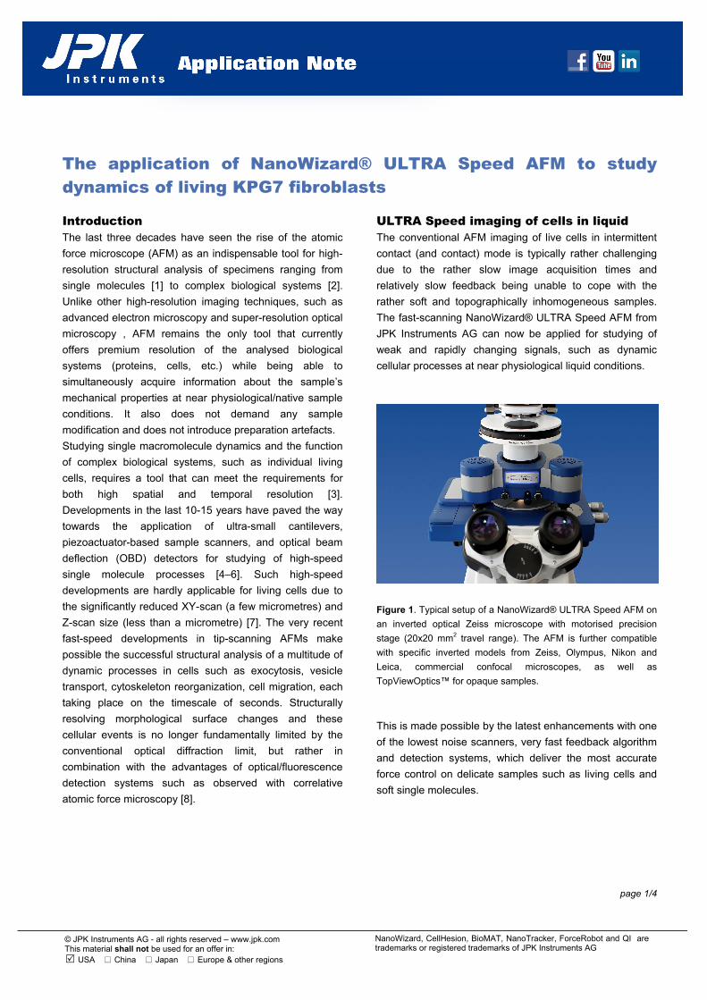
Figure 1. Typical setup of a NanoWizard® ULTRA Speed AFM on
an inverted optical Zeiss microscope with motorised precision
stage (20x20 mm2 travel range). The AFM is further compatible
with specific inverted models from Zeiss, Olympus, Nikon and
Leica, commercial confocal microscopes, as well as
TopViewOptics™ for opaque samples.
This is made possible by the latest enhancements with one
of the lowest noise scanners, very fast feedback algorithm
and detection systems, which deliver the most accurate
force control on delicate samples such as living cells and
soft single molecules.
page 2/4
NanoWizard, CellHesion, BioMAT, NanoTracker, ForceRobot and QI are trademarks or registered trademarks of JPK Instruments AG
© JPK Instruments AG - all rights reserved – www.jpk.com This material shall not be used for an offer in: USA China Japan Europe & other regions
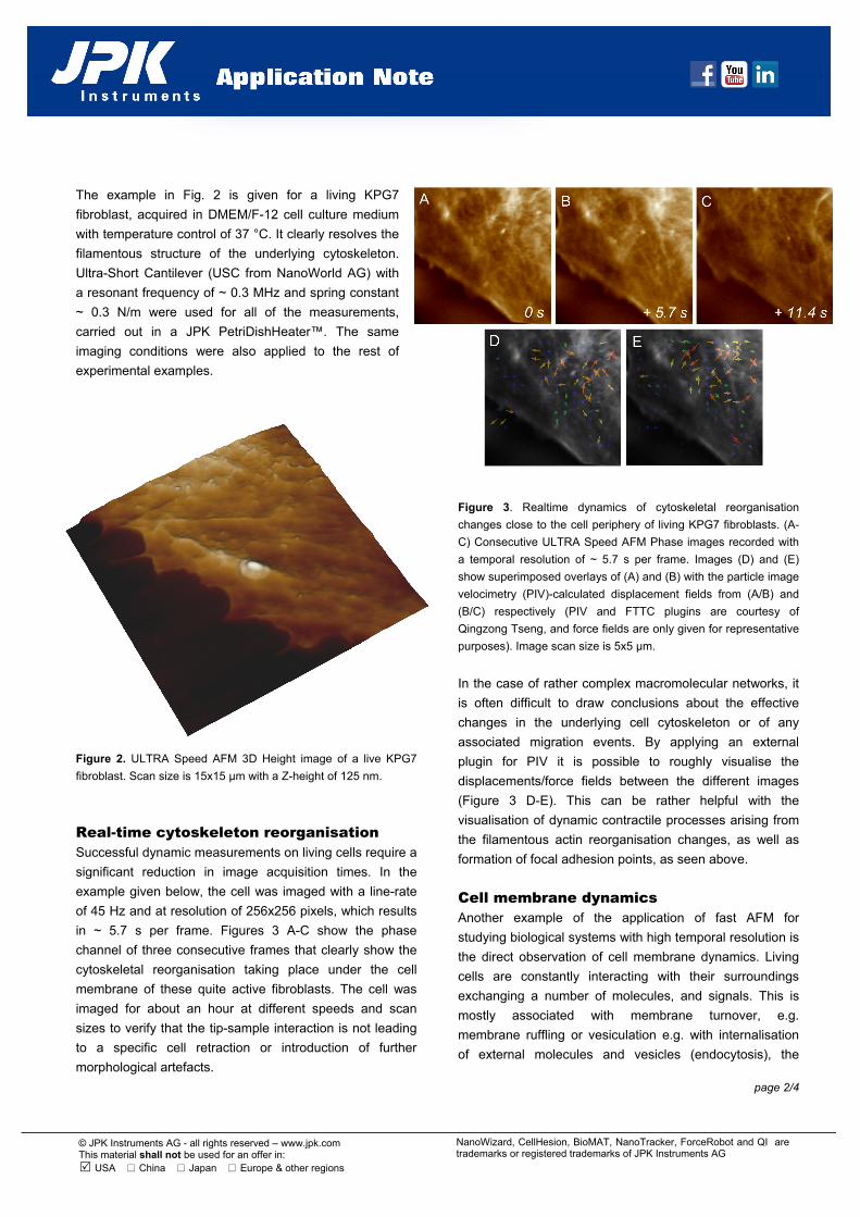
The example in Fig. 2 is given for a living KPG7
fibroblast, acquired in DMEM/F-12 cell culture medium
with temperature control of 37 °C. It clearly resolves the
filamentous structure of the underlying cytoskeleton.
Ultra-Short Cantilever (USC from NanoWorld AG) with
a resonant frequency of ~ 0.3 MHz and spring constant
~ 0.3 N/m were used for all of the measurements,
carried out in a JPK PetriDishHeater™. The same
imaging conditions were also applied to the rest of
experimental examples.
Figure 2. ULTRA Speed AFM 3D Height image of a live KPG7
fibroblast. Scan size is 15x15 µm with a Z-height of 125 nm.
Real-time cytoskeleton reorganisation Successful dynamic measurements on living cells require a
significant reduction in image acquisition times. In the
example given below, the cell was imaged with a line-rate
of 45 Hz and at resolution of 256x256 pixels, which results
in ~ 5.7 s per frame. Figures 3 A-C show the phase
channel of three consecutive frames that clearly show the
cytoskeletal reorganisation taking place under the cell
membrane of these quite active fibroblasts. The cell was
imaged for about an hour at different speeds and scan
sizes to verify that the tip-sample interaction is not leading
to a specific cell retraction or introduction of further
morphological artefacts.
Figure 3. Realtime dynamics of cytoskeletal reorganisation
changes close to the cell periphery of living KPG7 fibroblasts. (A-
C) Consecutive ULTRA Speed AFM Phase images recorded with
a temporal resolution of ~ 5.7 s per frame. Images (D) and (E)
show superimposed overlays of (A) and (B) with the particle image
velocimetry (PIV)-calculated displacement fields from (A/B) and
(B/C) respectively (PIV and FTTC plugins are courtesy of
Qingzong Tseng, and force fields are only given for representative
purposes). Image scan size is 5x5 µm.
In the case of rather complex macromolecular networks, it
is often difficult to draw conclusions about the effective
changes in the underlying cell cytoskeleton or of any
associated migration events. By applying an external
plugin for PIV it is possible to roughly visualise the
displacements/force fields between the different images
(Figure 3 D-E). This can be rather helpful with the
visualisation of dynamic contractile processes arising from
the filamentous actin reorganisation changes, as well as
formation of focal adhesion points, as seen above.
Cell membrane dynamics Another example of the application of fast AFM for
studying biological systems with high temporal resolution is
the direct observation of cell membrane dynamics. Living
cells are constantly interacting with their surroundings
exchanging a number of molecules, and signals. This is
mostly associated with membrane turnover, e.g.
membrane ruffling or vesiculation e.g. with internalisation
of external molecules and vesicles (endocytosis), the
page 3/4
NanoWizard, CellHesion, BioMAT, NanoTracker, ForceRobot and QI are trademarks or registered trademarks of JPK Instruments AG
© JPK Instruments AG - all rights reserved – www.jpk.com This material shall not be used for an offer in: USA China Japan Europe & other regions
release of metabolitic degradation products or the release
of signalling molecules to other cells (exocytosis). The
timescale of most of these processes can depend on the
type of membrane fusion or secretion, and normally ranges
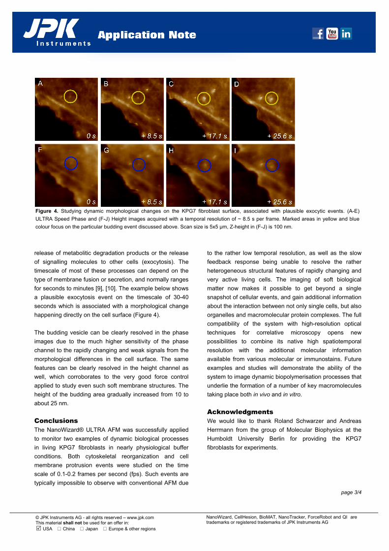
for seconds to minutes [9], [10]. The example below shows
a plausible exocytosis event on the timescale of 30-40
seconds which is associated with a morphological change
happening directly on the cell surface (Figure 4).
The budding vesicle can be clearly resolved in the phase
images due to the much higher sensitivity of the phase
channel to the rapidly changing and weak signals from the
morphological differences in the cell surface. The same
features can be clearly resolved in the height channel as
well, which corroborates to the very good force control
applied to study even such soft membrane structures. The
height of the budding area gradually increased from 10 to
about 25 nm.
Conclusions The NanoWizard® ULTRA AFM was successfully applied
to monitor two examples of dynamic biological processes
in living KPG7 fibroblasts in nearly physiological buffer
conditions. Both cytoskeletal reorganization and cell
membrane protrusion events were studied on the time
scale of 0.1-0.2 frames per second (fps). Such events are
typically impossible to observe with conventional AFM due
to the rather low temporal resolution, as well as the slow
feedback response being unable to resolve the rather
heterogeneous structural features of rapidly changing and
very active living cells. The imaging of soft biological
matter now makes it possible to get beyond a single
snapshot of cellular events, and gain additional information
about the interaction between not only single cells, but also
organelles and macromolecular protein complexes. The full
compatibility of the system with high-resolution optical
techniques for correlative microscopy opens new
possibilities to combine its native high spatiotemporal
resolution with the additional molecular information
available from various molecular or immunostains. Future
examples and studies will demonstrate the ability of the
system to image dynamic biopolymerisation processes that
underlie the formation of a number of key macromolecules
taking place both in vivo and in vitro.
Acknowledgments We would like to thank Roland Schwarzer and Andreas
Herrmann from the group of Molecular Biophysics at the
Humboldt University Berlin for providing the KPG7
fibroblasts for experiments.
Figure 4. Studying dynamic morphological changes on the KPG7 fibroblast surface, associated with plausible exocytic events. (A-E)
ULTRA Speed Phase and (F-J) Height images acquired with a temporal resolution of ~ 8.5 s per frame. Marked areas in yellow and blue
colour focus on the particular budding event discussed above. Scan size is 5x5 µm, Z-height in (F-J) is 100 nm.
page 4/4
NanoWizard, CellHesion, BioMAT, NanoTracker, ForceRobot and QI are trademarks or registered trademarks of JPK Instruments AG
© JPK Instruments AG - all rights reserved – www.jpk.com This material shall not be used for an offer in: USA China Japan Europe & other regions
References [1] D. J. Muller, “AFM: a nanotool in membrane biology.,” Biochemistry, vol. 47, no. 31, pp. 7986–7998, Aug. 2008. [2] A. Berquand, C. Roduit, S. Kasas, A. Holloschi, L. Ponce, and M. Hafner, “Atomic Force Microscopy Imaging of Living Cells,” Microscopy Today, p. 8, 2010. [3] R. Tomer, K. Khairy, F. Amat, and P. J. Keller, “Quantitative high-speed imaging of entire developing embryos with simultaneous multiview light-sheet microscopy.,” Nat Methods, vol. 9, no. 7, pp. 755–763, Jul. 2012. [4] T. Ando, N. Kodera, E. Takai, D. Maruyama, K. Saito, and A. Toda, “A high-speed atomic force microscope for studying biological macromolecules.,” Proc Natl Acad Sci U S A, vol. 98, no. 22, pp. 12468–12472, Oct. 2001. [5] L. Bozec A. Ulcinas D. J. Engledew M. Antognozzi M. A. Horton L. M. Picco and M. J. Miles, “Breaking the speed limit with atomic force microscopy,” Nanotechnology, vol. 18, 2007. [6] M. B. Viani, L. I. Pietrasanta, J. B. Thompson, A. Chand, I. C. Gebeshuber, J. H. Kindt, M. Richter, H. G. Hansma, and P. K. Hansma, “Probing protein-protein interactions in real time.,” Nat Struct Biol, vol. 7, no. 8, pp. 644–647, Aug. 2000. [7] T. Ando, “High-speed atomic force microscopy coming of age,” Nanotechnology, vol. 23, no. 6, p. 062001, 2012. [8] A. Monserrate, S. Casado, and C. Flors, “Correlative Atomic Force Microscopy and Localization-Based Super-Resolution Microscopy: Revealing Labelling and Image Reconstruction Artefacts.,” Chemphyschem, Nov. 2013. [9] J. Klingauf, E. T. Kavalali, and R. W. Tsien, “Kinetics and regulation of fast endocytosis at hippocampal synapses.,” Nature, vol. 394, no. 6693, pp. 581–585, Aug. 1998. [10] S. W. Schneider, K. C. Sritharan, J. P. Geibel, H. Oberleithner, and B. P. Jena, “Surface dynamics in living acinar cells imaged by atomic force microscopy: identification of plasma membrane structures involved in exocytosis.,” Proc Natl Acad Sci U S A, vol. 94, no. 1, pp. 316–321, Jan. 1997.