Embed Size (px)
Citation preview
The Arabidopsis Chromatin-ModifyingNuclear siRNA Pathway Involvesa Nucleolar RNA Processing CenterOlga Pontes,1 Carey Fei Li,2 Pedro Costa Nunes,1,4 Jeremy Haag,1 Thomas Ream,1 Alexa Vitins,1
Steven E. Jacobsen,2,3 and Craig S. Pikaard1,*1Biology Department, Washington University, 1 Brookings Drive, St. Louis, MO 63130, USA2Department of Molecular, Cell and Developmental Biology3Howard Hughes Medical Institute
University of California, Los Angeles, Los Angeles, CA 90095, USA4Seccao de Genetica, Centro de Botanica Aplicada a Agricultura, Instituto Superior de Agronomia, Tapada da Ajuda,1349-017 Lisboa, Portugal
*Contact: [email protected]
DOI 10.1016/j.cell.2006.05.031
SUMMARY
In Arabidopsis thaliana, small interfering RNAs(siRNAs) direct cytosine methylation at endoge-nous DNA repeats in a pathway involving twoforms of nuclear RNA polymerase IV (Pol IVaand Pol IVb), RNA-DEPENDENT RNA POLY-MERASE 2 (RDR2), DICER-LIKE 3 (DCL3), AR-GONAUTE4 (AGO4), the chromatin remodelerDRD1, and the de novo cytosine methyltransfer-ase DRM2. We show that RDR2, DCL3, AGO4,and NRPD1b (the largest subunit of Pol IVb)colocalize with siRNAs within the nucleolus.By contrast, Pol IVa and DRD1 are external tothe nucleolus and colocalize with endogenousrepeat loci. Mutation-induced loss of pathwayproteins causes downstream proteins to mis-localize, revealing their order of action. Pol IVaacts first, and its localization is RNA dependent,suggesting an RNA template. We hypothesizethat maintenance of the heterochromatic stateinvolves locus-specific Pol IVa transcription fol-lowed by siRNA production and assembly ofAGO4- and NRPD1b-containing silencing com-plexes within nucleolar processing centers.
INTRODUCTION
In diverse eukaryotes, small interfering RNAs (siRNAs)
regulate processes that include mRNA degradation, viral
suppression, centromere function, and silencing of retro-
transposons and endogenous DNA repeats (Almeida
and Allshire, 2005; Baulcombe, 2004; Grewal and Rice,
2004; Tomari and Zamore, 2005). siRNAs are generated
by Dicer endonuclease cleavage of double-stranded
RNAs (dsRNAs), whose production in Neurospora, C. ele-
gans, S. pombe, and plants involves one or more RNA-de-
pendent RNA polymerases (RdRPs) (Baulcombe, 2004;
Wassenegger and Krczal, 2006). Following dicing of
dsRNAs into �20–25 bp duplexes (Bernstein et al.,
2001; Hannon, 2002), one RNA strand is loaded into effec-
tor complexes that carry out the silencing functions. A de-
fining feature of these effector complexes is the inclusion
of an Argonaute (AGO) family protein (Carmell et al., 2002;
Sontheimer and Carthew, 2004). In RNA-slicing effector
complexes, the AGO-associated siRNA base pairs with
its target, thereby positioning the target RNA for endonu-
cleolytic cleavage (Song et al., 2004). Within effector com-
plexes that direct chromatin modifications (Grewal and
Rice, 2004; Verdel et al., 2004; Volpe et al., 2002; Wasse-
negger, 2005), the mechanisms by which siRNAs guide
target modifications are not yet understood.
In Arabidopsis thaliana, silencing at endogenous repeat
loci involves histone H3K9 methylation and RNA-directed
DNA methylation that is correlated with the production of
homologous siRNAs (Cao et al., 2003; Lippman et al.,
2003; Xie et al., 2004; Zilberman et al., 2004). Key players
in this chromatin-modifying nuclear siRNA pathway in-
clude DICER-LIKE 3 (DCL3), ARGONAUTE4 (AGO4), RNA-
DEPENDENT RNA POLYMERASE 2 (RDR2), and two
forms of nuclear RNA polymerase IV (Pol IV). The largest
and second largest subunits of Pol IV are similar to the cat-
alytic b and b0 subunits of E. coli DNA-dependent RNA
polymerase and to the corresponding subunits of eukary-
otic nuclear RNA polymerases I, II, and III (see Onodera
et al., 2005 and references therein). Two genes encode
distinct Pol IV largest subunits, and two genes encode
Pol IV second largest subunits. Both of the largest-subunit
genes (NRPD1a and NRPD1b) are expressed, but only
one of the second-largest-subunit genes (NRPD2a) is
functional (Herr et al., 2005; Onodera et al., 2005; Pontier
et al., 2005). As a result, there are two genetically nonre-
dundant forms of Pol IV, namely Pol IVa and Pol IVb,
Cell 126, 79–92, July 14, 2006 ª2006 Elsevier Inc. 79
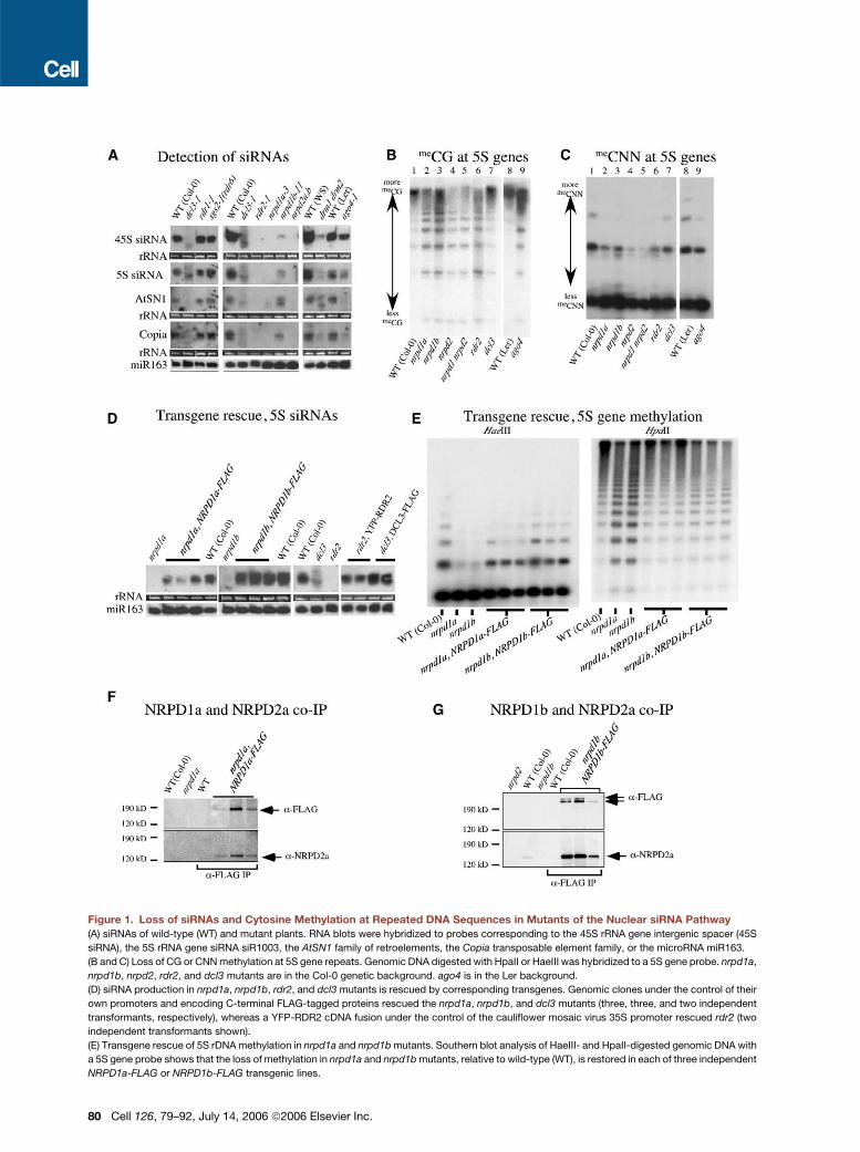
Figure 1. Loss of siRNAs and Cytosine Methylation at Repeated DNA Sequences in Mutants of the Nuclear siRNA Pathway
(A) siRNAs of wild-type (WT) and mutant plants. RNA blots were hybridized to probes corresponding to the 45S rRNA gene intergenic spacer (45S
siRNA), the 5S rRNA gene siRNA siR1003, the AtSN1 family of retroelements, the Copia transposable element family, or the microRNA miR163.
(B and C) Loss of CG or CNN methylation at 5S gene repeats. Genomic DNA digested with HpaII or HaeIII was hybridized to a 5S gene probe. nrpd1a,
nrpd1b, nrpd2, rdr2, and dcl3 mutants are in the Col-0 genetic background. ago4 is in the Ler background.
(D) siRNA production in nrpd1a, nrpd1b, rdr2, and dcl3 mutants is rescued by corresponding transgenes. Genomic clones under the control of their
own promoters and encoding C-terminal FLAG-tagged proteins rescued the nrpd1a, nrpd1b, and dcl3 mutants (three, three, and two independent
transformants, respectively), whereas a YFP-RDR2 cDNA fusion under the control of the cauliflower mosaic virus 35S promoter rescued rdr2 (two
independent transformants shown).
(E) Transgene rescue of 5S rDNA methylation in nrpd1a and nrpd1b mutants. Southern blot analysis of HaeIII- and HpaII-digested genomic DNA with
a 5S gene probe shows that the loss of methylation in nrpd1a and nrpd1b mutants, relative to wild-type (WT), is restored in each of three independent
NRPD1a-FLAG or NRPD1b-FLAG transgenic lines.
80 Cell 126, 79–92, July 14, 2006 ª2006 Elsevier Inc.

designated according to which largest subunit is used.
Disruption of Pol IV, RDR2, DCL3, or AGO4 genes causes
decreased cytosine methylation and siRNA accumulation
at endogenous repeats, including 5S ribosomal RNA
genes and transposable elements (Herr et al., 2005;
Kanno et al., 2005; Onodera et al., 2005; Pontier et al.,
2005; Xie et al., 2004). However, the order in which these
proteins act in the biogenesis of nuclear siRNAs is unclear.
Using RNA fluorescence in situ hybridization (RNA-
FISH) together with protein immunolocalization, we pres-
ent evidence for siRNA processing centers associated
with the nucleolus. Within these centers, siRNAs colocal-
ize with a significant portion of the RDR2, DCL3, AGO4,
and NRPD1b protein pools. The two subunits of Pol IVa,
however, do not localize to the processing centers but co-
localize with chromosomal loci that are both sources and
targets of siRNAs. A portion of the NRPD1b pool also co-
localizes with target loci, as does the SWI2/SNF2 chroma-
tin-remodeling ATPase family member DRD1, a protein
required for RNA-directed DNA methylation that acts
downstream of siRNA production (Kanno et al., 2004).
Based on cytological, biochemical, and genetic evidence,
we present a spatial and temporal model for nuclear
siRNA biogenesis.
RESULTS
Loss of siRNAs and Cytosine Methylation
in Nuclear siRNA Pathway Mutants
In A. thaliana, siRNAs homologous to repeated gene fam-
ilies are readily detected on RNA blots, as shown for
siRNAs corresponding to the intergenic spacers of 45S
or 5S rRNA genes or siRNAs corresponding to AtSN1 or
Copia transposable-element families (Figure 1A). Collec-
tively, these endogenous repeats represent genes tran-
scribed by RNA polymerase I (45S rRNA genes), RNA
polymerase II (Copia elements), and RNA polymerase III
(5S genes, AtSN1 elements). The siRNAs are essentially
eliminated upon mutation of the Pol IVa largest subunit,
NRPD1a, or upon mutation of the second subunit of
both Pol IVa and Pol IVb, NRPD2 (note that the nrpd2a-2
nrpd2b-1 double mutant [Onodera et al., 2005] is abbrevi-
ated as nrpd2 throughout this paper). siRNAs are also
eliminated in rdr2 mutants. By contrast, siRNAs are re-
duced in abundance, but not eliminated, in nrpd1b or
ago4 mutants. A smear of alternatively sized small RNAs
is generated in a dcl3 mutant (Figure 1A) and is probably
explained by the action of alternative Dicers (Gasciolli
et al., 2005). The abundance of siRNAs is also greatly re-
duced in the drm1 drm2 mutant, indicating that de novo
cytosine methylation plays a role in nuclear siRNA accu-
mulation.
Loss of endogenous siRNAs correlates with loss of cy-
tosine methylation at corresponding DNA sequences. For
instance, 5S gene repeats are heavily methylated at CG
motifs, making them resistant to digestion by the methyl-
ation-sensitive restriction endonuclease HpaII in wild-type
A. thaliana (Figure 1B, lanes 1 and 8). CG methylation at
HpaII sites is decreased to a similar extent in rdr2, ago4,
nrpd1a, nrpd1b, and nrpd2 mutants, resulting in more hy-
bridization signal in digested bands nearer the bottom of
Southern blots (Figure 1B). Methylation is least affected
in a dcl3 mutant, presumably because other Dicers par-
tially compensate (Gasciolli et al., 2005).
CNN methylation is a hallmark of RNA-directed DNA
methylation, which is accomplished by the de novo cyto-
sine methyltransferase DRM2 (Cao et al., 2003). At 5S
gene loci, sensitivity to digestion by HaeIII reports on
CNN methylation. 5S genes are more sensitive to HaeIII
digestion in rdr2, nrpd1a, nrpd1b, and nrpd2 mutants
compared to wild-type plants (Figure 1C). Mutation of
DCL3 has a lesser effect on CNN methylation, again sug-
gesting partial compensation by other Dicers. Collectively,
the data of Figures 1A–1C indicate that the loss of endog-
enous repeat siRNAs correlates with the loss of both CG
and CNN methylation, implicating RNA-directed DNA
methylation (Aufsatz et al., 2002; Cao et al., 2003).
To facilitate cytological and biochemical studies, we de-
veloped transgenic lines that express functional, epitope-
tagged versions of the proteins involved in the nuclear
siRNA pathway. Genomic-clone transgenes expressing
NRPD1a, NRPD1b, or DCL3 bearing C-terminal FLAG epi-
tope tags all rescued their corresponding mutations and
restored siRNA production, as did a YFP-RDR2 fusion en-
gineered using a full-length RDR2 cDNA (Figure 1D). The
NRPD1a and NRPD1b transgenes also restored cytosine
methylation at 5S gene repeats (Figure 1E). Collectively,
these results indicate that the recombinant proteins retain
their biological functions.
The Alternative Pol IV Largest Subunits, NRPD1a
and NRPD1b, Physically Interact with NRPD2
Genetic evidence suggests that the Pol IV second largest
subunit NRPD2 interacts with NRPD1a or NRPD1b within
Pol IVa or Pol IVb, respectively (Herr et al., 2005; Kanno
et al., 2005; Onodera et al., 2005; Pontier et al., 2005).
To obtain biochemical evidence for such interactions,
we exploited transgenic plants expressing FLAG-tagged
NRPD1a or NRPD1b and an anti-NRPD2 antibody
(Onodera et al., 2005) to ask whether NRPD2 associates
with the alternative largest subunits in vivo. Indeed,
NRPD2 coimmunoprecipitates with both NRPD1a-
FLAG and NRPD1b-FLAG in multiple independent trans-
genic plants (Figures 1F and 1G). The quantity of
(F) Physical interaction between Pol IVa subunits NRPD1a and NRPD2 detected by coimmunoprecipitation. Proteins from multiple independent
NRPD1a-FLAG transgenic lines were immunoprecipitated using anti-FLAG antibody, then subjected to SDS-PAGE and electroblotting. Membranes
were sequentially analyzed to detect the FLAG epitope (top) and NRPD2 (bottom).
(G) Physical interaction between NRPD1b and NRPD2. The experiment was performed as for (F) using multiple independent NRPD1b-FLAG trans-
genic lines.
Cell 126, 79–92, July 14, 2006 ª2006 Elsevier Inc. 81

Figure 2. Nuclear Localization of siRNAs
(A) RNA-FISH using the same probe sequences used for the RNA blots of Figure 1A was performed in wild-type, nuclease-treated, or mutant nuclei as
indicated. As a control, a probe that detects the 45S rRNA precursor transcripts was also used. Nuclei were counterstained with DAPI (blue). Size bars
represent 5 mm in all panels.
(B) Different siRNAs colocalize within the nucleolus. Simultaneous detection of RNA target pairs was performed using two-color FISH. Three-dimen-
sional projections of five to seven optical sections obtained by multiphoton microscopy are shown. The red or green color of the lettering corresponds
to the color of the signal for the indicated probes. Nuclei were counterstained with DAPI (false colored gray in these images). Thirty-five nuclei were
observed for each probe combination. In all nuclei examined, at least 50% of the green and red pixels overlapped in the digital images to yield yellow
signals.
(C) Two-color FISH using the 45S siRNA probe (red) and miR163 probe (green). Nuclei were counterstained with DAPI (blue). A localization pattern like
that shown was observed in all 155 nuclei examined.
coimmunoprecipitated NRPD2 is proportional to the
abundance of NRPD1a or NRPD1b in the different lines,
as expected of subunits with fixed stoichiometries.
siRNAs Are Concentrated within the Nucleolus
It is not known where endogenous siRNAs are generated
or processed within the cell. So, to detect siRNAs or their
precursors, we employed RNA fluorescence in situ hybrid-
ization (RNA-FISH) with digoxigenin- or biotin-labeled
probes (Figure 2A) identical in sequence to those used
for siRNA blot hybridization (see Figure 1A). With all siRNA
probes, an intense hybridization signal was observed
within the nucleolus, which is the region of the nucleus
not stained appreciably by the fluorescent DNA binding
dye DAPI. This was true of leaf mesophyll cells at inter-
phase, as shown throughout this paper, and in root meri-
82 Cell 126, 79–92, July 14, 2006 ª2006 Elsevier Inc.
stem cells (O.P., unpublished data). In the case of the
AtSN1 probe, a diffuse signal was also observed through-
out the nucleoplasm. The nucleolar dots detected with
siRNA probes occupy a small portion of the nucleolus
when compared to the 45S pre-rRNA precursor tran-
scripts that are generated by RNA polymerase I and pro-
cessed in the nucleolus (Figure 2A, bottom row).
Hybridization signals detected using different siRNA
probes colocalized, as shown using two-color RNA-
FISH with probes specific for 45S siRNAs corresponding
to opposite DNA strands (45S siRNA and 45S siRNA*) or
5S siRNAs (Figure 2B). These siRNA probe signals are
spatially distinct from the signals obtained using a miRNA
probe (Figure 2C). Collectively, these data indicate that
nuclear siRNA hybridization signals localize within a dis-
crete compartment of the nucleolus, smaller than the

volume occupied by 45S pre-rRNA and distinct from sites
where miRNA or their precursors are concentrated.
As shown in Figure 2A, siRNA and pre-rRNA hybridiza-
tion signals are eliminated if nuclei are treated with ribonu-
clease A (RNase A) prior to extensive washing and probe
hybridization but are not affected by DNase I treatment.
These tests suggest that the hybridization signals result
from the RNA probes’ annealing to RNA targets. Impor-
tantly, the nucleolar dot signals are absent in nrpd2,
nrpd1a, rdr2, dcl3, or ago4 mutants, and, typically, no sig-
nal is observed elsewhere (although low-intensity, dis-
persed signals occurred infrequently; see Table S1 in the
Supplemental Data available with this article online for
quantitative data). The exception is nrpd1b, for which dis-
persal of the nucleolar dot (as shown in Figure 2A) is more
common than complete loss of signal (see Table S1). In
general, these observations are consistent with the RNA
blot hybridization data (Figure 1A). Importantly, 45S pre-
rRNAs are unaffected by the siRNA pathway mutations,
as expected.
The loss of hybridization signals in the mutants, includ-
ing dcl3 and ago4, which should act downstream of siRNA
precursor formation, suggests that we are detecting
siRNAs in the nucleolar dots rather than precursors. Per-
haps the latter escape detection because they are dis-
persed throughout the nucleus and not concentrated in
one location. However, the AtSN1 signals, external to
the nucleolus, that persist in the mutants might be precur-
sor RNAs.
Nucleolar siRNA Processing Centers
The detection of nuclear siRNAs prompted us to ask
where the proteins of the nuclear siRNA pathway are lo-
cated. NRPD1a, NRPD1b, RDR2, DCL3, and AGO4 were
immunolocalized in transgenic nuclei by virtue of their epi-
tope or YFP tags, whereas native NRPD2 was localized
using an anti-peptide antibody (Figure 3A, top row).
NRPD1a and NRPD2, the known subunits of Pol IVa,
showed similar, punctate localization patterns; signifi-
cantly, neither protein associates with the nucleolus. By
contrast, FLAG-tagged NRPD1b, the largest subunit of
Pol IVb, localizes within a nucleolar dot in addition to
puncta external to the nucleolus (see also Li et al., 2006
[this issue of Cell] and Table S2). RDR2, DCL3, and
AGO4 also display prominent nucleolar dot signals in ad-
dition to puncta or diffuse signals outside the nucleolus.
RDR2 signals are distinctive in that a ring or crescent at
the perimeter of the nucleolus is typically observed in ad-
dition to the nucleolar dot, and this is true for both epitope-
tagged and native RDR2. Control experiments showed
that no immunolocalization signals were detected in trans-
genic nuclei if primary antibodies were omitted; likewise,
no signals were detected in wild-type nuclei using anti-
FLAG, anti-Myc, or anti-YFP antibodies (see Figure S1).
Nucleolar dot signals can be observed at the center or
the periphery of the nucleolus, consistent with data of Li
et al. (2006) showing that AGO4 colocalizes with markers
of nucleolar accessory bodies, or Cajal bodies (Cioce and
Lamond, 2005). Cajal bodies are dynamic nuclear organ-
elles that can move in and out of nucleoli (Boudonck
et al., 1999) and are implicated in the assembly of RNA-
protein complexes, including snRNPs and snoRNPs
(Cioce and Lamond, 2005). Therefore, what we call nucle-
olar dots throughout this paper are likely to be Cajal bod-
ies or related entities (see Li et al., 2006).
Treating nuclei with RNase A prior to antibody incuba-
tion caused a complete loss of signal for all of the proteins
in the majority of nuclei examined, suggesting that the pro-
teins are not retained in RNA-depleted nuclei (Figure 3A).
However, a minority of the nuclei continued to show wild-
type protein localization patterns, albeit at reduced inten-
sity, suggesting that not all nuclei are equally accessible to
RNase treatment (see Table S2). Further analysis showed
that, whereas NRPD2, NRPD1a, and NRPD1b signals are
lost from RNase A-treated nuclei, the proteins are not lost
from DNase I-treated nuclei, although NRPD1b and
NRPD2 are partially mislocalized (Figure 3B and
Figure S2, green signals). Conversely, the signals for the
second largest subunit of DNA-dependent RNA polymer-
ase II are lost upon DNase, but not RNase, treatment
(Figure 3B, red signals). Collectively, these observations
suggest that Pol IV interacts with RNA rather than DNA
templates, unlike Pol II.
Using anti-epitope antibodies that detect transgene-
encoded recombinant proteins, in combination with anti-
peptide antibodies recognizing the native proteins, we si-
multaneously localized pairs of proteins using two-color
immunofluorescence (Figure 3C; Table S3). The native
proteins and the recombinant proteins were found to dis-
play the same localization patterns, indicating that the
anti-peptide antibodies are specific for their targets and
that the epitope tags do not disrupt recombinant protein
localization. NRPD1a and NRPD2, the subunits of Pol
IVa, colocalize precisely, resulting in yellow signals
(Figure 3C, top row; note that differences in intensity of
the green and red signals influence the apparent extent
of overlap). Slightly more than half of the NRPD1b foci ex-
ternal to the nucleolus colocalize with the NRPD1a/
NRPD2 foci (Figure 3C, second row from top), suggesting
that Pol IVb occurs at approximately half of the Pol IVa
foci. However, the remaining NRPD1b foci are spatially
distinct from NRPD2 (and NRPD1a). A conclusion from
the latter observation is that the Pol IVb largest subunit
can exist apart from the second largest subunit, both ex-
ternal to the nucleolus and within the nucleolus, where
no NRPD2 is detectable.
External to the nucleolus, NRPD1a, NRPD2, and
NRPD1b do not colocalize with RDR2, DCL3, or AGO4.
However, the portion of the NRPD1b pool that is nucleolus
associated colocalizes with RDR2, DCL3, and AGO4
within the nucleolar dot (Figure 3C).
We next asked whether the nucleolar dots previously
detected by RNA-FISH (Figure 2) correspond to the
same nucleolar dots where NRPD1b, RDR2, DCL3, and
AGO4 colocalize (Figure 3). To address this question,
we performed protein immunolocalization followed by
Cell 126, 79–92, July 14, 2006 ª2006 Elsevier Inc. 83

84 Cell 126, 79–92, July 14, 2006 ª2006 Elsevier Inc.
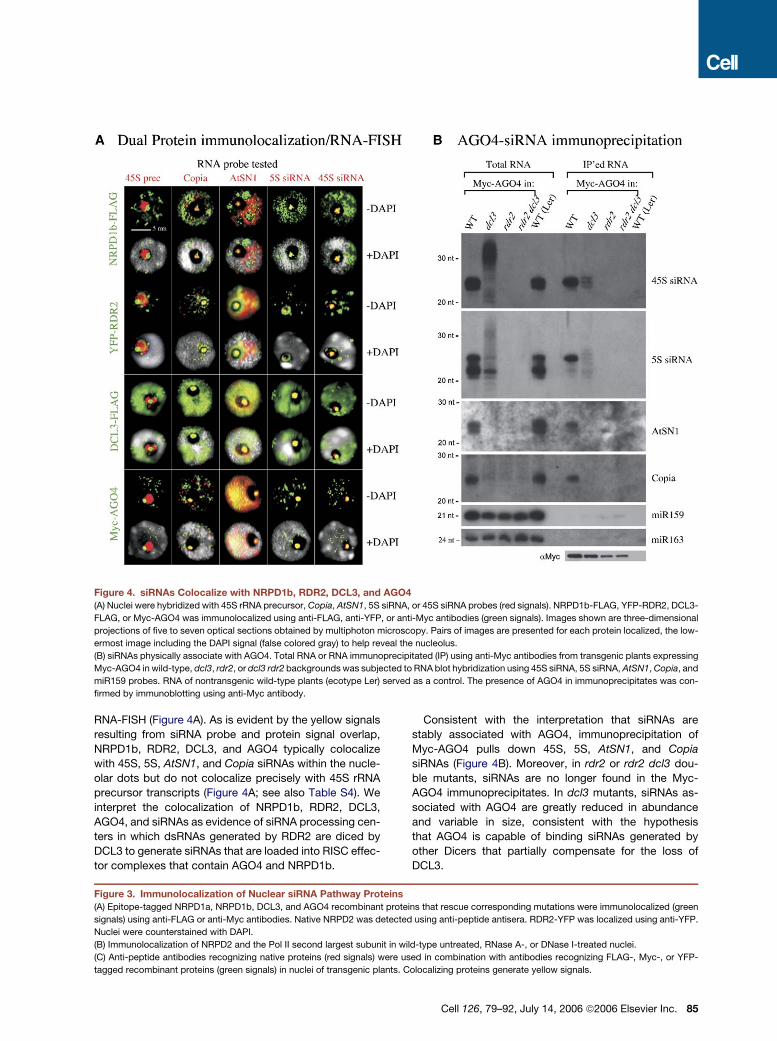
Figure 4. siRNAs Colocalize with NRPD1b, RDR2, DCL3, and AGO4
(A) Nuclei were hybridized with 45S rRNA precursor, Copia, AtSN1, 5S siRNA, or 45S siRNA probes (red signals). NRPD1b-FLAG, YFP-RDR2, DCL3-
FLAG, or Myc-AGO4 was immunolocalized using anti-FLAG, anti-YFP, or anti-Myc antibodies (green signals). Images shown are three-dimensional
projections of five to seven optical sections obtained by multiphoton microscopy. Pairs of images are presented for each protein localized, the low-
ermost image including the DAPI signal (false colored gray) to help reveal the nucleolus.
(B) siRNAs physically associate with AGO4. Total RNA or RNA immunoprecipitated (IP) using anti-Myc antibodies from transgenic plants expressing
Myc-AGO4 in wild-type, dcl3, rdr2, or dcl3 rdr2 backgrounds was subjected to RNA blot hybridization using 45S siRNA, 5S siRNA, AtSN1, Copia, and
miR159 probes. RNA of nontransgenic wild-type plants (ecotype Ler) served as a control. The presence of AGO4 in immunoprecipitates was con-
firmed by immunoblotting using anti-Myc antibody.
RNA-FISH (Figure 4A). As is evident by the yellow signals
resulting from siRNA probe and protein signal overlap,
NRPD1b, RDR2, DCL3, and AGO4 typically colocalize
with 45S, 5S, AtSN1, and Copia siRNAs within the nucle-
olar dots but do not colocalize precisely with 45S rRNA
precursor transcripts (Figure 4A; see also Table S4). We
interpret the colocalization of NRPD1b, RDR2, DCL3,
AGO4, and siRNAs as evidence of siRNA processing cen-
ters in which dsRNAs generated by RDR2 are diced by
DCL3 to generate siRNAs that are loaded into RISC effec-
tor complexes that contain AGO4 and NRPD1b.
Consistent with the interpretation that siRNAs are
stably associated with AGO4, immunoprecipitation of
Myc-AGO4 pulls down 45S, 5S, AtSN1, and Copia
siRNAs (Figure 4B). Moreover, in rdr2 or rdr2 dcl3 dou-
ble mutants, siRNAs are no longer found in the Myc-
AGO4 immunoprecipitates. In dcl3 mutants, siRNAs as-
sociated with AGO4 are greatly reduced in abundance
and variable in size, consistent with the hypothesis
that AGO4 is capable of binding siRNAs generated by
other Dicers that partially compensate for the loss of
DCL3.
Figure 3. Immunolocalization of Nuclear siRNA Pathway Proteins
(A) Epitope-tagged NRPD1a, NRPD1b, DCL3, and AGO4 recombinant proteins that rescue corresponding mutations were immunolocalized (green
signals) using anti-FLAG or anti-Myc antibodies. Native NRPD2 was detected using anti-peptide antisera. RDR2-YFP was localized using anti-YFP.
Nuclei were counterstained with DAPI.
(B) Immunolocalization of NRPD2 and the Pol II second largest subunit in wild-type untreated, RNase A-, or DNase I-treated nuclei.
(C) Anti-peptide antibodies recognizing native proteins (red signals) were used in combination with antibodies recognizing FLAG-, Myc-, or YFP-
tagged recombinant proteins (green signals) in nuclei of transgenic plants. Colocalizing proteins generate yellow signals.
Cell 126, 79–92, July 14, 2006 ª2006 Elsevier Inc. 85
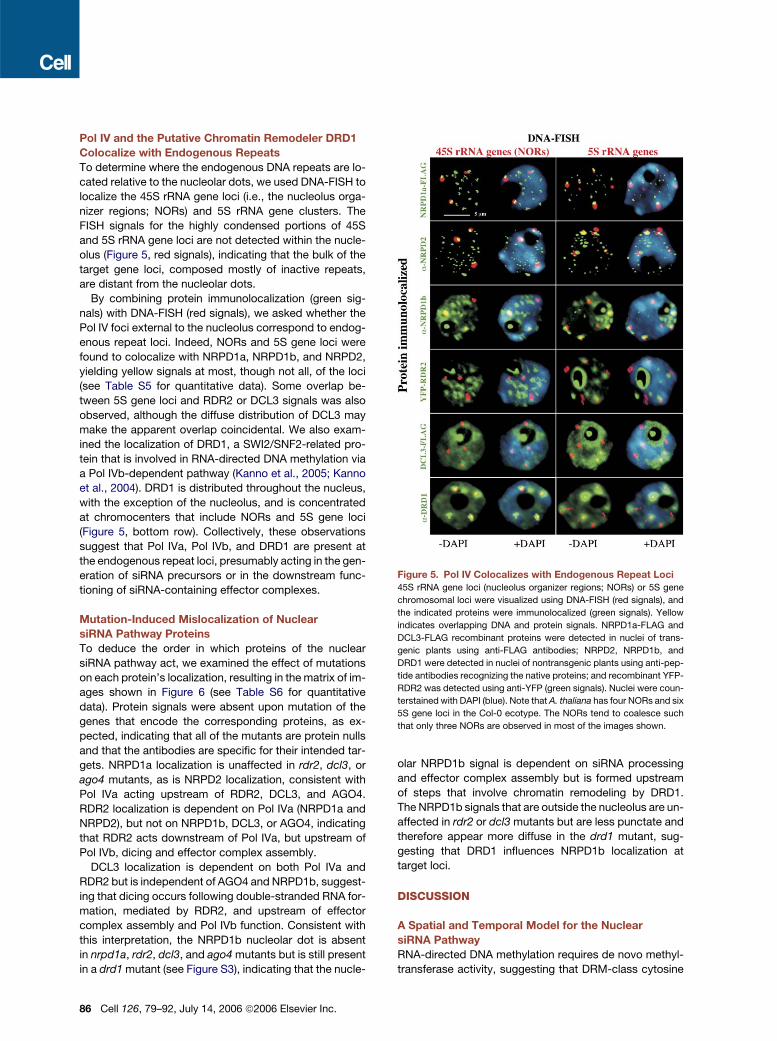
Pol IV and the Putative Chromatin Remodeler DRD1
Colocalize with Endogenous Repeats
To determine where the endogenous DNA repeats are lo-
cated relative to the nucleolar dots, we used DNA-FISH to
localize the 45S rRNA gene loci (i.e., the nucleolus orga-
nizer regions; NORs) and 5S rRNA gene clusters. The
FISH signals for the highly condensed portions of 45S
and 5S rRNA gene loci are not detected within the nucle-
olus (Figure 5, red signals), indicating that the bulk of the
target gene loci, composed mostly of inactive repeats,
are distant from the nucleolar dots.
By combining protein immunolocalization (green sig-
nals) with DNA-FISH (red signals), we asked whether the
Pol IV foci external to the nucleolus correspond to endog-
enous repeat loci. Indeed, NORs and 5S gene loci were
found to colocalize with NRPD1a, NRPD1b, and NRPD2,
yielding yellow signals at most, though not all, of the loci
(see Table S5 for quantitative data). Some overlap be-
tween 5S gene loci and RDR2 or DCL3 signals was also
observed, although the diffuse distribution of DCL3 may
make the apparent overlap coincidental. We also exam-
ined the localization of DRD1, a SWI2/SNF2-related pro-
tein that is involved in RNA-directed DNA methylation via
a Pol IVb-dependent pathway (Kanno et al., 2005; Kanno
et al., 2004). DRD1 is distributed throughout the nucleus,
with the exception of the nucleolus, and is concentrated
at chromocenters that include NORs and 5S gene loci
(Figure 5, bottom row). Collectively, these observations
suggest that Pol IVa, Pol IVb, and DRD1 are present at
the endogenous repeat loci, presumably acting in the gen-
eration of siRNA precursors or in the downstream func-
tioning of siRNA-containing effector complexes.
Mutation-Induced Mislocalization of Nuclear
siRNA Pathway Proteins
To deduce the order in which proteins of the nuclear
siRNA pathway act, we examined the effect of mutations
on each protein’s localization, resulting in the matrix of im-
ages shown in Figure 6 (see Table S6 for quantitative
data). Protein signals were absent upon mutation of the
genes that encode the corresponding proteins, as ex-
pected, indicating that all of the mutants are protein nulls
and that the antibodies are specific for their intended tar-
gets. NRPD1a localization is unaffected in rdr2, dcl3, or
ago4 mutants, as is NRPD2 localization, consistent with
Pol IVa acting upstream of RDR2, DCL3, and AGO4.
RDR2 localization is dependent on Pol IVa (NRPD1a and
NRPD2), but not on NRPD1b, DCL3, or AGO4, indicating
that RDR2 acts downstream of Pol IVa, but upstream of
Pol IVb, dicing and effector complex assembly.
DCL3 localization is dependent on both Pol IVa and
RDR2 but is independent of AGO4 and NRPD1b, suggest-
ing that dicing occurs following double-stranded RNA for-
mation, mediated by RDR2, and upstream of effector
complex assembly and Pol IVb function. Consistent with
this interpretation, the NRPD1b nucleolar dot is absent
in nrpd1a, rdr2, dcl3, and ago4 mutants but is still present
in a drd1 mutant (see Figure S3), indicating that the nucle-
86 Cell 126, 79–92, July 14, 2006 ª2006 Elsevier Inc.
olar NRPD1b signal is dependent on siRNA processing
and effector complex assembly but is formed upstream
of steps that involve chromatin remodeling by DRD1.
The NRPD1b signals that are outside the nucleolus are un-
affected in rdr2 or dcl3 mutants but are less punctate and
therefore appear more diffuse in the drd1 mutant, sug-
gesting that DRD1 influences NRPD1b localization at
target loci.
DISCUSSION
A Spatial and Temporal Model for the Nuclear
siRNA Pathway
RNA-directed DNA methylation requires de novo methyl-
transferase activity, suggesting that DRM-class cytosine
Figure 5. Pol IV Colocalizes with Endogenous Repeat Loci
45S rRNA gene loci (nucleolus organizer regions; NORs) or 5S gene
chromosomal loci were visualized using DNA-FISH (red signals), and
the indicated proteins were immunolocalized (green signals). Yellow
indicates overlapping DNA and protein signals. NRPD1a-FLAG and
DCL3-FLAG recombinant proteins were detected in nuclei of trans-
genic plants using anti-FLAG antibodies; NRPD2, NRPD1b, and
DRD1 were detected in nuclei of nontransgenic plants using anti-pep-
tide antibodies recognizing the native proteins; and recombinant YFP-
RDR2 was detected using anti-YFP (green signals). Nuclei were coun-
terstained with DAPI (blue). Note that A. thaliana has four NORs and six
5S gene loci in the Col-0 ecotype. The NORs tend to coalesce such
that only three NORs are observed in most of the images shown.

Figure 6. Effects of Mutations on the Localization of Proteins Involved in Nuclear siRNA Biogenesis
The figure shows a matrix of images in which NRPD1a, NRPD2, NRPD1b, RDR2, and DCL3 were immunolocalized using anti-peptide antibodies rec-
ognizing the native proteins (green signals) in multiple genetic backgrounds as indicated along the vertical axis. Nuclei were counterstained with DAPI
(blue).
methyltransferases (probably DRM2 only, because DRM1
is not expressed appreciably) act downstream of siRNA
production (Cao et al., 2003). However, endogenous nu-
clear siRNAs fail to accumulate in drm mutants (Xie
et al., 2004; Zilberman et al., 2004), suggesting that
DRM2 also acts upstream of siRNA production (see also
Figure 1A). Our model attempts to address this apparent
paradox (Figure 7). Based on a study in Neurospora sug-
gesting that methylation impedes RNA polymerase elon-
gation (Rountree and Selker, 1997), we propose that tran-
scripts trailing from polymerases that are stalled or slowed
by DRM-mediated methylation (Figure 7, upper left) are
sensed as aberrant and, directly or indirectly, become
templates for Pol IVa. In this model, Pol IVa is spatially
tethered to the DNA by virtue of the RNA template. This
aspect of the model accounts for the colocalization of
Pol IVa subunits with endogenous repeat loci and their
loss in RNase A-treated nuclei. We place Pol IVa first in
the pathway because Pol IVa is located directly at the
endogenous repeat loci and because mutation of either
Pol IVa subunit (NRPD1a or NRPD2) eliminates siRNA pro-
duction. By contrast, mutation of NRPD1b, the largest
subunit of Pol IVb, which also colocalizes with the endog-
enous repeat loci, does not eliminate siRNA production
but does affect RNA-directed cytosine methylation, sug-
gesting that Pol IVb acts late in the pathway (Kanno
et al., 2005; Pontier et al., 2005; Vaucheret, 2005; see
also Figures 1A–1C). The fact that siRNA accumulation
is reduced in nrpd1b mutants (see Figure 1A) may be
due to the destabilization of the NRPD2 pool upon loss
of NRPD1b (see Figure 1G, Figure 6 and Pontier et al.,
2005). Loss of NRPD2 would indirectly deplete Pol IVa
activity by depriving NRPD1a of its partner catalytic sub-
unit. Alternatively, decreased Pol IVb-dependent cytosine
methylation might decrease the incidence of aberrant
transcript production at endogenous repeat loci, thereby
depleting the pool of Pol IVa templates. These alternative
explanations are not mutually exclusive.
Cell 126, 79–92, July 14, 2006 ª2006 Elsevier Inc. 87

Figure 7. A Spatial and Temporal Model for Nuclear siRNA BiogenesisSubunits of Pol IVa (abbreviated 1a and 2) colocalize with endogenous repeat loci but are mislocalized upon RNase A treatment, suggesting that Pol
IVa transcribes RNA templates whose spatial distribution is influenced by DNA. We propose that cytosine methylation by DRM induces the production
of aberrant RNAs, possibly by impeding polymerase elongation, which Pol IVa then uses as templates. Pol IVa transcripts then move, by an unknown
mechanism, to the nucleolus, where RDR2, DCL3, and AGO4 are located. In the siRNA processing center, the largest subunit of Pol IVb, NRPD1b,
joins the AGO4-containing RISC complex and acquires the NRPD2 subunit to become functional Pol IVb only upon leaving the nucleolus. Formation of
Pol IVb is required for the stability of the NRPD2 pool despite the fact that NRPD2 colocalizes more precisely with NRPD1a than with NRPD1b, sug-
gesting that NRPD2 subunits exchange between Pol IVa and b. AGO4, Pol IVb, and DRD1 then play unspecified roles in guiding heterochromatic
modifications at the endogenous repeats, including de novo cytosine methylation by DRM. Methylation-dependent production of aberrant RNAs
results in a positive feedback loop for maintaining heterochromatin at the DNA repeats.
Like Pol IVa, RDR2 is required for endogenous siRNA
production. RDR2 is mislocalized in an nrpd1a mutant,
whereas the converse is not true (see Figure 6), indicating
that RDR2 acts downstream of Pol IVa. RDR2 is not abun-
dant at the endogenous repeats but is concentrated in the
nucleolus. Collectively, these observations suggest that
Pol IVa generates precursor RNAs at the endogenous re-
peats and that these transcripts then move to the nucleo-
lus, where their complements are generated by RDR2
transcription. Annealing of these RNAs would produce
dsRNAs that are then diced by DCL3 and loaded into an
AGO4-containing effector complex, or RISC (RNA-in-
duced silencing complex), within the siRNA processing
center. The observation that Pol IVa subunits and RDR2
are not mislocalized in dcl3 or ago4 mutants is consistent
with Pol IVa and RDR2 acting upstream of DCL3 and
88 Cell 126, 79–92, July 14, 2006 ª2006 Elsevier Inc.
AGO4. Likewise, the absence of siRNAs associated with
AGO4 in rdr2 mutants, the atypical sizes of siRNAs asso-
ciated with AGO4 in dcl3 mutants, and the mislocalization
of AGO4 in rdr2 or dcl3 mutants (see also Li et al., 2006)
indicate that AGO4 acts downstream of RDR2 and DCL3.
Two observations suggest that Pol IVb acts down-
stream of AGO4-RISC assembly. First, the largest subunit
of Pol IVb, NRPD1b, colocalizes with the nucleolar dot, but
only if siRNAs are being produced and assembled into ef-
fector complexes; the nucleolar NRPD1b signal is absent
in nrpd1a, rdr2, dcl3, or ago4 mutants. Second, the
NRPD2 subunit is never observed within the nucleolus
yet is presumably essential for Pol IVb function based on
the genetic screen of Kanno et al. that recovered nine mu-
tant alleles of NRPD1b and 12 alleles of NRPD2a but no al-
leles of NRPD1a (Kanno et al., 2005). The genetic evidence
strongly predicts that NRPD1b is nonfunctional in the ab-
sence of the second largest subunit. We propose that
NRPD1b associates with AGO4-RISC, which is supported
by our immunolocalization data and the finding that
NRPD1b can be coimmunoprecipitated in association
with AGO4 (Li et al., 2006). Upon leaving the nucleolus
as a subunit of AGO4-RISC, we deduce that NRPD1b
can then associate with NRPD2, forming functional Pol
IVb. Consistent with this hypothesis, NRPD2 coimmuno-
precipitates with AGO4 (J.H. and C.S.P., unpublished
data) as well as with NRPD1b (see Figure 1G).
How AGO4-RISC-Pol IVb complexes mediate their ef-
fects on chromatin modification at target loci is unclear.
One possibility is that AGO4-RISC directs Pol IVb to its tar-
get sites. Alternatively, AGO4 might transfer the siRNA to
Pol IVb when the NRPD2 subunit joins the NRPD1b sub-
unit, after the AGO4-RISC-NRPD1b complex leaves the
nucleolus. The siRNA, or a Pol IVb transcript primed by
the siRNA, might then be used to conduct a homology
search for target sequences, aided by DRD1 (Kanno
et al., 2004), a member of the SWI2/SNF2-related family
of chromatin-remodeling ATPases that is within a subfam-
ily most closely related to yeast RAD54. In double-strand
DNA break repair, RAD54 is required for helping broken
DNA ends conduct a homology search and invade homol-
ogous duplex DNA of a sister chromosome, thereby facil-
itating repair by homologous recombination (Krogh and
Symington, 2004). A partnership between Pol IVb and
DRD1 could account for their presence at the target loci,
the observation that NRPD1b and DRD1 are both essen-
tial for cytosine methylation but not siRNA production
(Kanno et al., 2004, 2005), and the partial mislocalization
of NRPD1b in a drd1-6 mutant (see Figure S2). Moreover,
RNA polymerases and chromatin-remodeling ATPases
are nucleotide triphosphate-hydrolyzing molecular mo-
tors that can be envisioned working together, with proces-
sive movement of the polymerase possibly providing
directionality to subsequent chromatin modifications. Re-
sulting de novo DNA methylation by DRM2, which is pre-
dicted to contribute to aberrant RNA production, would
provide for positive feedback in our model (Figure 7).
As touched upon previously, our observation that
NRPD2 signals are severely reduced in nrpd1b, more so
than in the nrpd1a mutant (see Figure 1G and Figure 6),
is consistent with previously published immunoblot data
(Pontier et al., 2005). Nonetheless, it is surprising given
the nearly perfect colocalization of NRPD2 with NRPD1a,
as opposed to only �50% overlap of NRPD2 with
NRPD1b (see Figure 3C). Based on these data, one might
expect NRPD1a to be most important for NRPD2 stability.
To reconcile these findings, we propose that NRPD2 must
be able to exchange between Pol IVb and Pol IVa (Fig-
ure 7), with NRPD1b interactions somehow more impor-
tant for the overall stability of the NRPD2 pool.
The idea that incomplete, or otherwise aberrant, tran-
scripts can induce transcriptional silencing at endogenous
repeats may have parallels with the silencing of nonpro-
ductive human immunoglobulin genes. In this phenome-
non, genes whose transcripts contain premature stop co-
dons following V-D-J recombination are transcriptionally
silenced (Buhler et al., 2005), indicating a link between
nonsense-mediated decay (NMD) and chromatin modifi-
cation. In Arabidopsis, proteins of the exon-joining com-
plex and NMD pathways were identified within the nucle-
olar proteome, and some were shown to localize as
nucleolar dots (Pendle et al., 2005). Whether these pro-
teins colocalize with the siRNA processing centers is un-
clear at present.
The nucleolus is best known as the site of 45S pre-rRNA
transcription and ribosome assembly. However, small-
RNA-directed pre-rRNA cleavage, methylation, and pseu-
douridylation; biogenesis of signal-recognition particle
and telomerase small RNAs; tRNA processing by RNase
P; and some pre-mRNA processing also take place within
the nucleolus (Bertrand et al., 1998; Filipowicz and Poga-
cic, 2002; Kiss, 2002; Pederson, 1998). Our findings sug-
gest that processing of endogenous nuclear siRNAs, and
possibly RISC storage or sequestration, are additional nu-
cleolar functions to be explored.
EXPERIMENTAL PROCEDURES
Mutant Plant Strains
Arabidopsis rdr2-1 and dcl3-1 were provided by Jim Carrington, sgs2-
1 (alias sde1; rdr6) was provided by Herve Vaucheret, and drd1-6 was
provided by Tatsuo Kanno and Marjori Matzke. drm2-1, ago4-1, and
nrpd1b-11 (SALK_029919) were obtained from the Arabidopsis Bio-
logical Resource Center. nrpd1a and nrpd2 mutants were described
previously (Onodera et al., 2005).
Generation of Transgenic Lines
Full-length genomic sequences including promoters were amplified by
PCR from A. thaliana Col-0 DNA using Pfu polymerase (Stratagene)
and cloned into pENTR/D-TOPO (Invitrogen). NRPD1a primers were
50-CACCGGTGTCTCACATTCCAAAGTCCCC-30 (forward) and 50-
CGGGTTTTCGGAGAAACCACC-30 (reverse). NRPD1b primers were
50-CACCGCGTACTACAAACGGAAACGGTCA-30 and 50-TGTCTGCG
TCTGGGACGG-30. Genomic DCL3 was amplified from BAC clone
T15B3 using 50-CACCCCGACCGAAATCCTCATGACCTAA-30 and 50-
CTTTTGTATTATGACGATCTTGCGGCGC-30; the CACC added to for-
ward primers allowed directional cloning into the entry vector. Reverse
primers eliminated stop codons to allow epitope-tag fusion. Genes
were recombined into pEarleyGate 302 (Earley et al., 2006) to add C-
terminal FLAG epitopes. RDR2 coding sequences were amplified by
RT-PCR using Pfx Platinum DNA polymerase (Invitrogen) and primers
50-CACCATGGTGTCAGAGACGACGAC-30 and 50-GGGCAATCAAAT
GGATACAAGTCC-30. PCR products captured in pENTR/D-TOPO
were recombined into pEarleyGate 104 (Earley et al., 2006), fusing
RDR2 sequences C-terminal to YFP expressed from a CaMV 35S pro-
moter. Transformation of constructs into corresponding homozygous
mutants was by the floral dip method (Clough and Bent, 1998).
Southern Blotting and Small-RNA Blot Hybridization
Genomic DNA (250 ng) digested with HaeIII or HpaII was subjected to
agarose gel electrophoresis, blotted to nylon membranes, and hybrid-
ized to a 5S gene probe as described previously (Onodera et al., 2005).
Generation of RNA probes labeled with [a-32P]CTP and small-RNA blot
hybridization were also as described previously (Onodera et al., 2005).
Specific oligodeoxynucleotides used in T7 polymerase reactions
(CCTGTCTC hybridized to the T7 promoter adaptor) were as
follows: 45S siRNA: 50-CAATGTCTGTTGGTGCCAAGAGGGAAAAG
Cell 126, 79–92, July 14, 2006 ª2006 Elsevier Inc. 89

GGCCCTGTCTC-30; 45S prec: 50-AGTCCGTGGGGAACCCCCTTTT
TCGGTTCGCCCCTGTCTC-30; 5S siRNA: 50-AGACCGTGAGGCCAA
ACTTGGCATCCTGTCTC-30; Copia: 50-TTATTGGAACCCGGTTAGG
ACCTGTCTC-30, and miR163: 50-TTGAAGAGGACTTGGAACTTCG
ATCCTGTCTC-30.
Antibodies
Rabbit antibodies raised against NRPD2 and Pol II second-largest-
subunit peptides were described previously (Onodera et al., 2005).
Chicken antibodies recognizing DCL3, NRPD1a, NRPD1b, or RDR2
were generated against peptides conjugated to keyhole limpet hemo-
cyanin. Peptides were as follows: DCL3: SLEPEKMEEGGGSNC;
NRPD1a: EELQVPVGTLTSIGC; NRPD1b: MEEESTSEILDGEIC;
RDR2: ETTTNRSTVKISNVC; DRD1: NKNVHKRKQNQVDDGC. Immu-
nolocalization was performed using 1:200 dilutions of antisera, except
that NRPD1b antiserum was diluted 1:500. FLAG-tagged proteins
were detected using mouse monoclonal anti-FLAG antibody (Sigma-
Aldrich) diluted 1:400. RDR2-YFP was detected using mouse anti-
GFP/YFP (BD Biosciences) diluted 1:500.
Immunolocalization
Leaves from 28-day-old plants were harvested and nuclei were ex-
tracted as described previously (Onodera et al., 2005). After postfixa-
tion in 4% paraformaldehyde/PBS (phosphate-buffered saline),
washes in PBS, and blocking at 37ºC, slides were exposed overnight
to primary antisera in PBS and 0.5% blocking reagent (Roche). After
washes in PBS, slides were incubated at 37ºC with anti-mouse-FITC
diluted 1:100 (Sigma), goat anti-chicken Alexa 488 diluted 1:300 (Mo-
lecular Probes), or goat anti-chicken Alexa 543 diluted 1:400 (Molecu-
lar Probes). Nuclei were counterstained with 1 mg/ml DAPI (Sigma) in
Vectashield (Vector Laboratories).
Immunoprecipitation and Immunoblotting
of Epitope-Tagged Proteins
Pol IV immunoprecipitation was performed using protein extracted
from 2.0 g of tissue according to Baumberger and Baulcombe
(2005), except that homogenates were filtered through two layers of
Miracloth and subjected to centrifugation at 16,000 3 g for 15 min at
4ºC prior to incubation with anti-FLAG M2 affinity gel (Sigma). Proteins
eluted in 23 SDS-PAGE loading buffer at 100ºC for 2 min were frac-
tionated on 7.5% Tris-glycine SDS-polyacrylamide gels (Cambrex)
and electroblotted to PVDF membranes (Millipore). Membranes incu-
bated with peroxidase-linked anti-FLAG M2 antibody diluted 1:2000
(Sigma) were visualized using chemiluminescence detection (Amer-
sham). Membranes were then stripped using 25 mM glycine-HCl (pH
2.0), 1% (w/v) SDS for 30 min with agitation, followed by two 10 min
washes in Tris-buffered saline, 0.05% (v/v) Tween 20. NRPD2 immu-
noblotting was as described in Onodera et al. (2005).
For coimmunoprecipitation of AGO4 and siRNAs, flowers (0.7 g) fro-
zen in liquid nitrogen were homogenized in 2 ml of IP buffer (50 mM
Tris-Cl [pH 7.5], 150 mM NaCl, 5 mM MgCl2, 10% glycerol, 0.1%
NP-40) containing fresh DTT (2 mM), PMSF (1 mM), pepstatin (0.7
mg/ml), MG132 (10 mg/ml), and Complete protease inhibitor cocktail
(Roche). Following centrifugation, lysates precleared with Protein G-
agarose beads (Pierce) for 1 hr at 4ºC were incubated with anti-Myc
(Upstate) diluted 1:250 for 3 hr at 4ºC. Antibody-antigen complexes
were captured on Protein G-agarose (60 ml) at 4ºC for 2 hr and washed
four times with IP buffer. For siRNA detection, beads were treated with
Proteinase K and extracted sequentially with TE containing 1.5%,
0.5%, or 0.1% SDS. Pooled supernatants extracted with phenol:
chloroform (1:1) followed by chloroform were ethanol precipitated. To-
tal siRNAs and RNA blots were prepared and hybridized as previously
described (Mette et al., 2000; Zilberman et al., 2003). DNA probes were
used to detect 5S siRNAs, 45S siRNAs, miR157, and miR163; RNA
probes were used to detect AtSN1 and Copia siRNAs. Probe se-
quences were as follows: 5S siRNA: 50-ATGCCAAGTTTGGCCTC
ACGGTCT-30; 45S siRNA: 50-GTCTGTTGGTGCCAAGAGGGAAAAG
90 Cell 126, 79–92, July 14, 2006 ª2006 Elsevier Inc.
GGCTAAT-30; AtSN1: 50-ACCAACGTGTTGTTGGCCCAGTGGTAAA
TCTCTCAGATAGAGG-30; Copia: 50-TTATTGGAACCCGGTTAGGA-
30; miR159: 50-TAGAGCTCCCTTCAATCCAAA-30; miR163: 50-ATCGA
AGTTGGAAGTCCTCTTCAA-30.
RNA and DNA In Situ Hybridization
RNA probes were labeled by in vitro T7 polymerase (Ambion) transcrip-
tion with digoxigenin-11-UTP or biotin-16-UTP RNA labeling mix
(Roche). RNA in situ hybridization was carried out at 42ºC overnight
using a probe solution containing 1 mg RNA probe, 5 mg yeast tRNA
(Roche), 50% dextran sulfate, 100 mM PIPES [pH 8.0], 10 mM
EDTA, and 3 M NaCl as described previously (Highett et al., 1993).
Slides were washed sequentially in 23 SSC, 50% formamide, 50ºC
followed by 13 SSC, 50% formamide, 50ºC, then 13 SSC 20ºC, and
finally TBS at 20ºC. Where applicable, nuclei were incubated at
37ºC for 30 min in a solution of RNase-free DNase I (0.015 U/ml) or in
a solution of RNase A (100 mg/ml, Roche). Nuclease reactions were
stopped in 10 mM EDTA (pH 7.5) for 2 min followed by three washes
in 0.13 SSC.
DNA-FISH using 5S or 45S rRNA gene probes labeled with biotin-
dUTP or digoxigenin-dUTP was performed as described (Pontes
et al., 2003). Digoxigenin-labeled probes were detected using mouse
anti-digoxigenin antibody (1:250, Roche) followed by rabbit anti-
mouse antibody conjugated to Alexa 488 (Molecular Probes). Biotin-
labeled probes were detected using goat anti-biotin conjugated with
avidin (1:200, Vector Laboratories) followed by streptavidin-Alexa
543 (Molecular Probes). DNA was counterstained with DAPI (1 mg/ml)
in Vectashield (Vector Laboratories). For dual protein/nucleic acid
localization experiments, slides were first subjected to immunofluores-
cence, then postfixed in 4% formaldehyde/PBS followed by RNA- or
DNA-FISH.
Microscopy
Nuclei were routinely examined using a Nikon Eclipse E800i epifluores-
cence microscope, with images collected using a Photometrics Cool-
snap ES Mono digital camera. The images were pseudocolored,
merged, and processed using Adobe Photoshop (Adobe Systems).
Multiphoton optical-section stacks were collected using a Zeiss LSM
510 Meta microscope. Single optical sections using 403 averaging
were acquired by simultaneous scanning to avoid artifactual shift be-
tween two optical channels. The 488 nm line of an argon laser was
used for detection of FITC FLAG-tagged proteins, and the 543 nm
line of a helium-neon laser was used for detection of Alexa 543 siRNA
signals. For the detection of DAPI, either a 715 or 750 nm multiphoton
tuned titanium-sapphire laser was used. Projections of 3D data stacks
were composed using Imaris 4.1 software from Bitplane (http://www.
bitplane.com).
Supplemental Data
Supplemental Data include three figures and six tables and can be
found with this article online at http://www.cell.com/cgi/content/full/
126/1/79/DC1/.
ACKNOWLEDGMENTS
O.P. performed all microscopy, P.C.N. generated siRNA blots, T.R.
performed DNA methylation assays, and J.H. generated epitope-
tagged Pol IV lines and coIP data. YFP-RDR2 and FLAG-DCL3 lines
were generated by O.P. and A.V., respectively; C.S.P. wrote the man-
uscript. C.F.L. and S.E.J. generated the Myc-AGO4 transgenic line and
contributed Figure 4B. O.P. and P.C.N. were supported by fellowships
SFRH/BPD/17508/2004 and SFRH/BD/6520/2001, respectively, from
the Fundacao para a Ciencia e Tecnologia (Portugal). Pikaard lab work
was supported by NIH grants R01GM60380 and R01GM077590 and
the Monsanto Company/Washington University Biology Research
Agreement. C.F.L. was supported by Ruth L. Kirschstein National Re-
search Service Award GM07185. S.E.J. is a Howard Hughes Medical
Institute Investigator. Work in the Jacobsen laboratory was supported
by NIH grant GM60398. Any opinions, findings, and conclusions ex-
pressed in this material are those of the authors and do not necessarily
reflect the views of NIH, HHMI, or the Monsanto Company. We thank
Howard Berg (Donald Danforth Plant Science Center) for multiphoton
microscopy training, Tom Juehne and Keming Song (Sigma-Aldrich
Company) for Pol IV antibody production, Dr. Wanda Viegas (Instituto
Superior de Agronomia, Portugal) for comentoring P.C.N., and Eric Ri-
chards (Washington University) for suggestions to improve the manu-
script.
Received: March 1, 2006
Revised: March 31, 2006
Accepted: May 17, 2006
Published: July 13, 2006
REFERENCES
Almeida, R., and Allshire, R.C. (2005). RNA silencing and genome reg-
ulation. Trends Cell Biol. 15, 251–258.
Aufsatz, W., Mette, M.F., van der Winden, J., Matzke, A.J., and
Matzke, M. (2002). RNA-directed DNA methylation in Arabidopsis.
Proc. Natl. Acad. Sci. USA 99, 16499–16506.
Baulcombe, D. (2004). RNA silencing in plants. Nature 431, 356–363.
Baumberger, N., and Baulcombe, D.C. (2005). Arabidopsis ARGO-
NAUTE1 is an RNA Slicer that selectively recruits microRNAs and short
interfering RNAs. Proc. Natl. Acad. Sci. USA 102, 11928–11933.
Bernstein, E., Caudy, A.A., Hammond, S.M., and Hannon, G.J. (2001).
Role for a bidentate ribonuclease in the initiation step of RNA interfer-
ence. Nature 409, 363–366.
Bertrand, E., Houser-Scott, F., Kendall, A., Singer, R.H., and Engelke,
D.R. (1998). Nucleolar localization of early tRNA processing. Genes
Dev. 12, 2463–2468.
Boudonck, K., Dolan, L., and Shaw, P.J. (1999). The movement of
coiled bodies visualized in living plant cells by the green fluorescent
protein. Mol. Biol. Cell 10, 2297–2307.
Buhler, M., Mohn, F., Stalder, L., and Muhlemann, O. (2005). Transcrip-
tional silencing of nonsense codon-containing immunoglobulin mini-
genes. Mol. Cell 18, 307–317.
Cao, X., Aufsatz, W., Zilberman, D., Mette, M.F., Huang, M.S., Matzke,
M., and Jacobsen, S.E. (2003). Role of the DRM and CMT3 methyl-
transferases in RNA-directed DNA methylation. Curr. Biol. 13, 2212–
2217.
Carmell, M.A., Xuan, Z., Zhang, M.Q., and Hannon, G.J. (2002). The Ar-
gonaute family: tentacles that reach into RNAi, developmental control,
stem cell maintenance, and tumorigenesis. Genes Dev. 16, 2733–
2742.
Cioce, M., and Lamond, A.I. (2005). Cajal bodies: a long history of dis-
covery. Annu. Rev. Cell Dev. Biol. 21, 105–131.
Clough, S.J., and Bent, A.F. (1998). Floral dip: a simplified method for
Agrobacterium-mediated transformation of Arabidopsis thaliana. Plant
J. 16, 735–743.
Earley, K.W., Haag, J.R., Pontes, O., Opper, K., Juehne, T., Song, K.,
and Pikaard, C.S. (2006). Gateway-compatible vectors for plant func-
tional genomics and proteomics. Plant J. 45, 616–629.
Filipowicz, W., and Pogacic, V. (2002). Biogenesis of small nucleolar
ribonucleoproteins. Curr. Opin. Cell Biol. 14, 319–327.
Gasciolli, V., Mallory, A.C., Bartel, D.P., and Vaucheret, H. (2005). Par-
tially redundant functions of Arabidopsis DICER-like enzymes and
a role for DCL4 in producing trans-acting siRNAs. Curr. Biol. 15,
1494–1500.
Grewal, S.I., and Rice, J.C. (2004). Regulation of heterochromatin by
histone methylation and small RNAs. Curr. Opin. Cell Biol. 16, 230–
238.
Hannon, G.J. (2002). RNA interference. Nature 418, 244–251.
Herr, A.J., Jensen, M.B., Dalmay, T., and Baulcombe, D.C. (2005).
RNA polymerase IV directs silencing of endogenous DNA. Science
308, 118–120.
Highett, M.I., Beven, A.F., and Shaw, P.J. (1993). Localization of 5 S
genes and transcripts in Pisum sativum nuclei. J. Cell Sci. 105,
1151–1158.
Kanno, T., Mette, M.F., Kreil, D.P., Aufsatz, W., Matzke, M., and
Matzke, A.J. (2004). Involvement of putative SNF2 chromatin remodel-
ing protein DRD1 in RNA-directed DNA methylation. Curr. Biol. 14,
801–805.
Kanno, T., Huettel, B., Mette, M.F., Aufsatz, W., Jaligot, E., Daxinger,
L., Kreil, D.P., Matzke, M., and Matzke, A.J. (2005). Atypical RNA poly-
merase subunits required for RNA-directed DNA methylation. Nat.
Genet. 37, 761–765.
Kiss, T. (2002). Small nucleolar RNAs: an abundant group of noncoding
RNAs with diverse cellular functions. Cell 109, 145–148.
Krogh, B.O., and Symington, L.S. (2004). Recombination proteins in
yeast. Annu. Rev. Genet. 38, 233–271.
Li, C.F., Pontes, O., El-Shami, M., Henderson, I.R., Bernatavichute,
Y.V., Chan, S.W.-L., Lagrange, T., Pikaard, C.S., and Jacobsen, S.E.
(2006). An ARGONAUTE4-containing nuclear processing center colo-
calized with Cajal bodies in Arabidopsis thaliana. Cell 126, this issue,
93–106.
Lippman, Z., May, B., Yordan, C., Singer, T., and Martienssen, R.
(2003). Distinct mechanisms determine transposon inheritance and
methylation via small interfering RNA and histone modification. PLoS
Biol. 1, E67.
Mette, M.F., Aufsatz, W., van der Winden, J., Matzke, M.A., and
Matzke, A.J. (2000). Transcriptional silencing and promoter methyla-
tion triggered by double-stranded RNA. EMBO J. 19, 5194–5201.
Onodera, Y., Haag, J.R., Ream, T., Costa Nunes, P., Pontes, O., and
Pikaard, C.S. (2005). Plant nuclear RNA polymerase IV mediates siRNA
and DNA methylation-dependent heterochromatin formation. Cell 120,
613–622.
Pederson, T. (1998). The plurifunctional nucleolus. Nucleic Acids Res.
26, 3871–3876.
Pendle, A.F., Clark, G.P., Boon, R., Lewandowska, D., Lam, Y.W., An-
dersen, J., Mann, M., Lamond, A.I., Brown, J.W., and Shaw, P.J.
(2005). Proteomic analysis of the Arabidopsis nucleolus suggests
novel nucleolar functions. Mol. Biol. Cell 16, 260–269.
Pontes, O., Lawrence, R.J., Neves, N., Silva, M., Lee, J.H., Chen, Z.J.,
Viegas, W., and Pikaard, C.S. (2003). Natural variation in nucleolar
dominance reveals the relationship between nucleolus organizer chro-
matin topology and rRNA gene transcription in Arabidopsis. Proc. Natl.
Acad. Sci. USA 100, 11418–11423.
Pontier, D., Yahubyan, G., Vega, D., Bulski, A., Saez-Vasquez, J., Ha-
kimi, M.A., Lerbs-Mache, S., Colot, V., and Lagrange, T. (2005). Rein-
forcement of silencing at transposons and highly repeated sequences
requires the concerted action of two distinct RNA polymerases IV in
Arabidopsis. Genes Dev. 19, 2030–2040.
Rountree, M.R., and Selker, E.U. (1997). DNA methylation inhibits elon-
gation but not initiation of transcription in Neurospora crassa. Genes
Dev. 11, 2383–2395.
Song, J.J., Smith, S.K., Hannon, G.J., and Joshua-Tor, L. (2004). Crys-
tal structure of Argonaute and its implications for RISC slicer activity.
Science 305, 1434–1437.
Sontheimer, E.J., and Carthew, R.W. (2004). Molecular biology. Argo-
naute journeys into the heart of RISC. Science 305, 1409–1410.
Tomari, Y., and Zamore, P.D. (2005). Perspective: machines for RNAi.
Genes Dev. 19, 517–529.
Vaucheret, H. (2005). RNA polymerase IV and transcriptional silencing.
Nat. Genet. 37, 659–660.
Cell 126, 79–92, July 14, 2006 ª2006 Elsevier Inc. 91
Verdel, A., Jia, S., Gerber, S., Sugiyama, T., Gygi, S., Grewal, S.I., and
Moazed, D. (2004). RNAi-mediated targeting of heterochromatin by
the RITS complex. Science 303, 672–676.
Volpe, T.A., Kidner, C., Hall, I.M., Teng, G., Grewal, S.I., and Martiens-
sen, R.A. (2002). Regulation of heterochromatic silencing and histone
H3 lysine-9 methylation by RNAi. Science 297, 1833–1837.
Wassenegger, M. (2005). The role of the RNAi machinery in hetero-
chromatin formation. Cell 122, 13–16.
Wassenegger, M., and Krczal, G. (2006). Nomenclature and functions
of RNA-directed RNA polymerases. Trends Plant Sci. 11, 142–151.
92 Cell 126, 79–92, July 14, 2006 ª2006 Elsevier Inc.
Xie, Z., Johansen, L.K., Gustafson, A.M., Kasschau, K.D., Lellis, A.D.,
Zilberman, D., Jacobsen, S.E., and Carrington, J.C. (2004). Genetic
and functional diversification of small RNA pathways in plants. PLoS
Biol. 2, E104.
Zilberman, D., Cao, X., and Jacobsen, S.E. (2003). ARGONAUTE4
control of locus-specific siRNA accumulation and DNA and histone
methylation. Science 299, 716–719.
Zilberman, D., Cao, X., Johansen, L.K., Xie, Z., Carrington, J.C., and
Jacobsen, S.E. (2004). Role of Arabidopsis ARGONAUTE4 in RNA-
directed DNA methylation triggered by inverted repeats. Curr. Biol.
14, 1214–1220.


![Interacting Genomic Landscapes of REC8-Cohesin ...Interacting Genomic Landscapes of REC8-Cohesin, Chromatin, and Meiotic Recombination in Arabidopsis[CC-BY] Christophe Lambing,a Andrew](https://img.pdfslide.net/doc/110x75/60dbfd21ae2a635f0e07e13a/interacting-genomic-landscapes-of-rec8-cohesin-interacting-genomic-landscapes.jpg)