Embed Size (px)
Citation preview
The Auxin-Regulated AP2/EREBP Gene PUCHI Is Requiredfor Morphogenesis in the Early Lateral RootPrimordium of Arabidopsis W
Atsuko Hirota, Takehide Kato, Hidehiro Fukaki,1 Mitsuhiro Aida, and Masao Tasaka2
Graduate School of Biological Sciences, Nara Institute of Science and Technology, Nara 630-0192, Japan
Organ primordia develop from founder cells into organs due to coordinated patterns of cell division. How patterned cell
division is regulated during organ formation, however, is not well understood. Here, we show that the PUCHI gene, which
encodes a putative APETALA2/ethylene-responsive element binding protein transcription factor, is required for the co-
ordinated pattern of cell divisions during lateral root formation in Arabidopsis thaliana. Recessive mutations in PUCHI dis-
turbed cell division patterns in the lateral root primordium, resulting in swelling of the proximal region of lateral roots. PUCHI
expression was initially detected in all of the cells in early lateral root primordia, and later it was restricted to the proximal
region of the primordia. Stable expression of PUCHI required auxin-responsive elements in its promoter region, and exo-
genous auxin increased the level of PUCHI mRNA accumulation. These results suggest that PUCHI acts downstream of
auxin signaling and that this gene contributes to lateral root morphogenesis through affecting the pattern of cell divisions
during the early stages of primordium development.
INTRODUCTION
Plasticity and adaptability are important life strategies in plants.
Plant architecture is largely dependent on the formation of new
organs after germination both in the shoot and the root. For
example, lateral roots (LRs) are continuously formed from the
primary root in the postembryonic root system. LRs are not
produced directly from the parental root meristem, but instead
develop from inner cells of a more mature part of the parental
root.
Previous studies in Arabidopsis thaliana have detailed the de-
velopment of the lateral root primordium (LRP) from pericycle
cells (Malamy and Benfey, 1997; Casimiro et al., 2001; Dubrovsky
et al., 2001). Initially, one or two mature pericycle cells adjacent
to the xylem poles divide asymmetrically to form daughter
cells, which are shorter than the flanking undivided pericycle
cells. These daughter cells proliferate further with a largely fixed
pattern of cell divisions to form the LR meristem, which has
almost the same structure as that of the primary root meristem.
Many studies have shown that the plant hormone auxin is a key
factor that controls LR formation (Smet et al., 2006; Fukaki et al.,
2007). For example, application of exogenous auxin increases
the number of LRs, whereas auxin transport inhibitors decrease
their number (Blakely et al., 1988; Laskowski et al., 1995; Reed
et al., 1998; Casimiro et al., 2001). Molecular genetic studies
using Arabidopsis have identified auxin-dependent signaling
processes that are important for LR initiation. Dominant or semi-
dominant mutations in AUXIN/INDOLE-3-ACETIC ACID (Aux/IAA)
genes, such as AXR5/IAA1, SHY2/IAA3, SLR/IAA14, MSG2/
IAA19, and IAA28, result in a reduced number of LRs (Tian and
Reed, 1999; Rogg et al., 2001; Fukaki et al., 2002; Tatematsu
et al., 2004; Yang et al., 2004). This class of mutations causes
stabilization of the corresponding Aux/IAA proteins, which are
transcriptional repressors of auxin-responsive gene expression,
and results in an inhibition of auxin signaling. For example, the
slr-1 mutant, which carries a point mutation that causes stabi-
lization of the IAA14 protein, fails to produce LRs due to the in-
hibition of the initial cell divisions associated with LR initiation
(Fukaki et al., 2002). Another class of genes involved in LR for-
mation includes AUXIN RESPONSE FACTOR7 (ARF7) and ARF19.
These genes encode transcriptional activators that bind to the
auxin-responsive element (AuxRE), a cis-regulatory sequence for
auxin-responsive genes (Okushima et al., 2005; Wilmoth et al.,
2005). Similar to the slr-1 mutant, LRs rarely form in the arf7 arf19
double mutant, indicating that these genes are redundantly re-
quired for LR initiation. The SLR/IAA14, ARF7, and ARF19 genes
are expressed in a broad region of the root, including the pericycle,
and the SLR/IAA14 protein physically interacts with ARF7 and
ARF19 to block their activity (Fukaki et al., 2005). This interaction is
thought to be important for the regulation of target gene activation
that is required for LR initiation (Okushima et al., 2007).
Polar auxin transport, which is required for the asymmetric
distribution of auxin, is also involved in LRP development. Treat-
ment of wild-type roots with auxin transport inhibitors blocks
LR initiation, whereas subsequent application of the auxin
1-naphthalene acetic acid (NAA) results in homogenous prolif-
eration of all of the pericycle cells and formation of a highly in-
creased number of LRs (Casimiro et al., 2001; Himanen et al.,
2002). Similarly, multiple mutant combinations of PIN family genes
1 Current address: Department of Biology, Graduate School of Science,Kobe University, Rokkodai 1-1, 657-8501 Kobe, Japan.2 Address correspondence to [email protected] author responsible for distribution of materials integral to thefindings presented in this article in accordance with the policy describedin the Instructions for Authors (www.plantcell.org) is: Masao Tasaka([email protected]).W Online version contains Web-only data.www.plantcell.org/cgi/doi/10.1105/tpc.107.050674
The Plant Cell, Vol. 19: 2156–2168, July 2007, www.plantcell.org ª 2007 American Society of Plant Biologists
(PINs), which encode auxin efflux carrier proteins, or in weak
mutant alleles of the GNOM gene, which is required for the
coordinated polar localization of PIN proteins, show homoge-
neous proliferation of pericycle cells in response to exogenous
NAA and subsequent formation of LRPs with highly disorganized
morphology (Benkova et al., 2003; Geldner et al., 2004). The
auxin transport pathway mediated by the PIN and GNOM pro-
teins is thus thought to be crucial for LR initiation and subsequent
primordium development. In addition, it has been reported that
the auxin influx carrier AUX1 also affects LR initiation (Marchant
et al., 2002).
Although it has been shown that LR initiation involves auxin
accumulation controlled by the auxin transport system and
the auxin signaling pathway mediated by the ARF and Aux/IAA
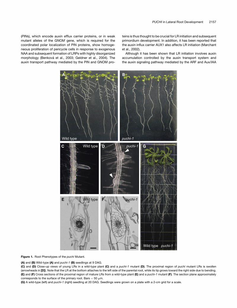
Figure 1. Root Phenotypes of the puchi Mutant.
(A) and (B) Wild-type (A) and puchi-1 (B) seedlings at 9 DAG.
(C) and (D) Close-up views of young LRs in a wild-type plant (C) and a puchi-1 mutant (D). The proximal region of puchi mutant LRs is swollen
(arrowheads in [D]). Note that the LR at the bottom attaches to the left side of the parental root, while its tip grows toward the right side due to bending.
(E) and (F) Cross sections of the proximal region of mature LRs from a wild-type plant (E) and a puchi-1 mutant (F). The section plane approximately
corresponds to the surface of the primary root. Bars ¼ 50 mm.
(G) A wild-type (left) and puchi-1 (right) seedling at 20 DAG. Seedlings were grown on a plate with a 2-cm grid for a scale.
PUCHI in Lateral Root Development 2157

Figure 2. LRP Development in the puchi-1 Mutant.
(A) to (F) Nomarski images of wild-type ([A] to [C]) and puchi-1 ([D] to [F]) LRPs in cleared primary roots at 7 DAG. LRPs in (A) and (B) are at stages II and
III, respectively, whereas (C) represents the LRPs that have formed more than four cell layers. (D) to (F) show mutant LRPs that have formed the same
number of layers as those in (A) to (C), respectively. Bars ¼ 50 mm.
(G) The number of short cells in the outermost layer along the radial axis at early stage II, where one or two central cells have just undergone periclinal
divisions (red line in the schematic diagram).
(H) The number of cells in the outermost layer of MOL at early stage III, where one to three outermost cells have undergone periclinal divisions (red line in
left schematic diagram), and the early stage IV, where one to three innermost cells have undergone periclinal divisions (red line in right schematic
diagram). Blue lines indicate the MOL border.
(I) Width along the innermost cell layer of MOL in LRPs at early stages III and IV. Blue lines indicate the MOL border, and arrows indicate the width
of MOL.
2158 The Plant Cell
proteins, the molecular mechanisms that control the subsequent
development of the primordium remain largely unknown. Here,
we identified the novel Arabidopsis gene PUCHI, which en-
codes a putative transcription factor of the APETALA2/ethylene-
responsive element binding protein (AP2/EREBP) family. The
puchi mutation disturbs cell division pattern during early LRP
development and results in expansion of the proximal region of
LRs. Reporter gene analyses indicate that PUCHI is expressed in
the early stages of LRP development, initially in all of the pri-
mordium cells and later in the proximal region of the LR. Stable
expression of PUCHI in the LRP requires the AuxREs in its pro-
moter region and is upregulated in roots in response to ex-
ogenous auxin in an AuxRE-dependent manner. These results
indicate that PUCHI is involved in the control of cell division pat-
terns during LRP development and may act downstream of auxin
signaling.
RESULTS
The puchi Mutation Affects Flower and LR Development
The puchi-1 mutant was initially identified by a subtle shoot
phenotype, which is not described in detail in this article (see
Methods). Besides this, the puchi-1 mutant showed clearly
recognizable phenotypes in the root. At 9 d after germination
(9 DAG), visible LRs in the puchi-1 mutant were much shorter
than those of wild-type plants (Figures 1A and 1B). Mutant LRs
that emerged out of the primary root surface, however, grew
normally, and the root system in older plants was indistinguish-
able from that of wild-type plants (Figure 1G). Moreover, the
proximal region of each LR in the puchi-1 mutant was signifi-
cantly swollen and often bent (Figures 1C and 1D). In cross
sections of mature LRs, the number of cells in this region was
increased in puchi-1, especially along the apical-basal axis of the
parental root (Figures 1E and 1F). In contrast with LRs, growth
and morphology of the primary root of puchi was normal (Figures
1A and 1B; data not shown).
To genetically test whether the phenotypes were caused by a
single mutation, we crossed the puchi-1 mutant with a wild-type
plant (Columbia [Col]) and examined the phenotypes in the shoot
and root in subsequent generations. All F1 plants were indistin-
guishable from wild-type plants (n¼ 20). In the F2 generation, the
shoot and root phenotypes cosegregated with a 3:1 ratio (25.6%
mutant, n ¼ 164), indicating that puchi-1 was a recessive muta-
tion in a single locus. Hereafter, we focus on the characterization
of LRP development in puchi-1.
LRP Development in the puchi-1 Mutant
We examined LRP development in the puchi-1 mutant in detail.
In the wild type, the initiation of a LR starts with anticlinal cell
divisions of pericycle cells, resulting in an array of cells that are
shorter than the flanking undivided cells (stage I; Malamy and
Benfey, 1997; Dubrovsky et al., 2001). Subsequently, some of
these short cells undergo periclinal divisions to form two cell
layers (stage II; Figure 2A). Additional cell expansion and peri-
clinal divisions result in LRP with three layers (stage III; Figure 2B)
and then four layers (stage IV). From stages II to IV, anticlinal
cell divisions in each layer occur and increase cell number along
the radial axis of the LRP. Patterned cell divisions and expansion
continue, resulting in the dome-shaped LRP (Figure 2C).
LRPs of the puchi-1 mutant were indistinguishable from that
of the wild type both in terms of the overall cellular organization
(Figures 2A and 2D) and the number of cells along the radial axis
(Figure 2G) up to early stage II, where a few short pericycle cells
at the center had undergone periclinal cell divisions. The puchi-1
mutant LRP started to deviate from the wild type from late stage II
to stage III, where additional periclinal division began to form the
third cell layer (Figures 2B and 2E). In these stages, the mutant
LRP forms more cells than the wild-type LRP along the radial axis
due to extra anticlinal divisions. To quantitatively compare the
cell number, we defined an area consisting of more than one cell
layer (MOL area; delimited by blue lines in Figures 2H to 2J) as a
region that will presumably give rise to most part of the LR. We
then counted the number of cells in the outermost layer of MOL
along the radial axis. Cell number in the outermost layer of puchi-1
MOL was significantly increased compared with the wild type
in primordia forming the third cell layer (Figure 2H; 6.5 6 0.2 SE,
n ¼ 13 in the wild type; 8.6 6 0.5 SE, n ¼ 13 in puchi-1) or in
the primordia forming the fourth layer (Figure 2H; 7.9 6 0.4, n¼ 13
in the wild type; 10.3 6 0.4, n¼ 13 in puchi-1), indicating that the
frequency of anticlinal cell divisions relative to periclinal ones is
increased in the puchi-1 mutant during the early stage of LRP
development.
As the primordium development continued, the mutant LRP
became flatter than that of the wild type (Figures 2C and 2F). The
width at the most proximal region in the MOL was not signifi-
cantly different between the wild type and puchi-1 LRP with three
and four cell layers (Figure 2I; early stage III, 104.1 mm 6 4.4 SE in
the wild type [n ¼ 13] and 102.0 mm 6 6.4 SE in puchi-1 [n ¼ 13];
Figure 2. (continued).
(J) Width along the innermost cell layer of MOL and MTL in LRPs with more than four cell layers and before emergence. Blue and green lines indicate the
borders of MOL and MTL, respectively. Blue and green arrows indicate the width of MOL and MTL, respectively.
(K) and (L) Expression of ProCycB1;1:CycB1;1(NT)-GUS in stage IV LRPs of wild-type (K) and of puchi-1 (L) at the corresponding stage at 9 DAG. GUS
staining was for 12 h. Arrow indicates GUS staining in a cell next to the MOL border. Bars ¼ 50 mm.
(M) Schematic diagram of the LRP. The cells next to the MOL border (blue line) are indicated in gray.
Table 1. Frequency of ProCycB1;1:CycB1;1(NT)-GUS Positive Cells
GUS Positivea Total LRP Numbersb
Wild type 0 24
puchi-1 8 25
a Number of LRPs in which either or both of the cells next to the MOL
border (Figure 2M, gray cells) are stained with GUS.b LRPs with three or four cell layers.
PUCHI in Lateral Root Development 2159

early stage IV, 103.1 mm 6 6.6 SE in the wild type [n ¼ 16] and
112.8 mm 6 5.1 SE in puchi-1 [n ¼ 16]). However, the MOL of
puchi-1 became ;15% wider than that of the wild type in later
stages (Figure 2J; 129.2 mm 6 5.3 SE in the wild type [n ¼ 26]
and 149.0 mm 6 4.4 SE in puchi-1 [n¼ 28]). In addition, the width
of the area with more than two layers (MTL: delimited by green
lines in Figure 2J) was also increased by ;31% (Figure 2J; 76.9
mm 6 2.8 SE in the wild type [n ¼ 26] and 100.3 mm 6 4.1 SE in
puchi-1 [n ¼ 28]). These results are consistent with the flatter
shape of the puchi-1 mutant LRPs.
The increased width of the primordium observed in the puchi
mutant LRPs may indicate that cell divisions occur in a wider
area in the mutant primordium than in the wild type. To test this
possibility, we examined expression of the ProCycB1;1:CycB1;
1(NT)-GUS reporter, which specifically marks cells in the late
G2 and M phases (Colon-Carmona et al., 1999). The intensity
and pattern of b-glucuronidase (GUS) staining appeared normal
in puchi-1 LRPs, suggesting that the mutation did not affect
overall frequency or duration of G2/M (Figures 2K and 2L).
However, the frequency of staining in cells at the periphery was
significantly higher in puchi-1 mutant primordia than in the wild
type (Figures 2K and 2L, Table 1; P < 0.01, Fisher’s exact test).
These results indicate that PUCHI restricts the area of cell
proliferation.
Later, when the LR meristem emerges out of the parental root,
the puchi-1 mutant LR forms extra tissue consisting of highly
enlarged cells at the periphery of the most proximal region, whereas
no such tissue is observed in the corresponding region of wild-
type LR (Figures 3A and 3B). The formation of such extra tissue is
consistent with a wider area of cell divisions in earlier stages.
We next examined expression of the radial pattern markers
ProSHR:GUS and ProSCR:green fluorescent protein (GFP) in the
expanded region of puchi-1 mutant LRs. Expression of ProSHR:
GUS, which was detected in the stele of the wild-type root
(Helariutta et al., 2000), was more expanded toward the periph-
ery than that in the wild type (Figures 3C and 3D). Similarly,
expression of ProSCR:GFP, which was normally detected in the
endodermis (Di Laurenzio et al., 1996), was expanded in puchi-1
(Figures 3E and 3F). In addition, although ProSCR:GFP expres-
sion was restricted to a single cell layer in the proximal region of
wild-type LRs (Figure 3E), the GFP signal was detected in more
than one cell layer, including the primordia surface, in puchi LRs
(Figure 3F, arrowheads). Besides these abnormalities, the rela-
tive position of the ProSHR:GUS and ProSCR:GFP expression
domains was essentially the same as that of the wild type in that
the SCR domain surrounds the SHR domain. This observation
suggests that the radial pattern in the proximal region of the
puchi mutant LR was largely maintained.
Although the puchi mutation strongly affected cell division
patterns during early stages of LRP development, the mutant
developed an LR meristem similar to that of the wild-type LRP
when the primordium emerged out of the parental root, except
that the cellular organization and expression pattern of ProSCR:
GFP around the quiescent center (QC) was slightly disturbed (see
Supplemental Figures 1A to 1F online). These subtle phenotypes,
however, were eventually rescued in mature LRs (see Supple-
mental Figures 1G and 1H online), which was consistent with
normal growth of mature LRs (Figure 1G). These results indicate
Figure 3. Morphology and the Radial Pattern in a Young LR.
(A) and (B) Nomarski images of a young LR in the wild type (A) and
puchi-1 (B) with similar length. The LR of puchi-1 contains ectopic tissue
(arrows), resulting in the expansion of the organ.
(C) and (D) ProSHR:GUS expression in a young LR from a wild-type plant
(C) and a puchi-1 mutant (D).
(E) and (F) ProSCR:GFP expression in a young LR from a wild-type plant
(E) and a puchi-1 mutant (F). Arrowheads indicate surface cells ectop-
ically expressing GFP.
Bars ¼ 50 mm.
2160 The Plant Cell

Figure 4. Molecular Characterization of PUCHI.
(A) Location of the puchi mutations. The PUCHI locus was mapped between two polymorphic markers located on the F20L16 and T1A4 BACs
on chromosome 5. The number of recombination events obtained between each marker and PUCHI among the F2 plants is indicated. The yellow box
PUCHI in Lateral Root Development 2161

that PUCHI is strictly required for proper morphogenesis in early
stages of LRP development but not for the establishment or
maintenance of the LR meristem.
Cloning of the PUCHI Gene
To clone the PUCHI gene, we mapped the mutated locus in
puchi-1 based on the expanded LR phenotype. The mutation
was located in a region between two polymorphic markers on the
BAC clones F20L16 and T1A4 from chromosome 5 (Figure 4A).
Among the 26 predicted genes in this region of the puchi-1
mutant, we found a 28-bp deletion that caused a frame shift in
the predicted open reading frame (ORF) of the annotated gene
At5g18560 (Figure 4A). We then obtained a TILLING line that
carried a nonsense mutation in the same gene (line 172F1;
Henikoff et al., 2004). Homozygous mutants isolated from this
line showed identical phenotypes to those of the puchi-1 mutant
both in the shoot and the root. In addition, a crossing experiment
showed that the two mutants were allelic (data not shown). We
thus designated the mutation in 172F1 as puchi-2 (Figure 4A).
Next, we cloned a 6.7-kb genomic fragment that spanned the
3.9-kb upstream and 1.7-kb downstream sequences of the
At5g18560 coding region and transformed it into the puchi-1
mutant. Most transgenic plants from the T1 generation showed
wild-type phenotypes both in the shoot and the root (five of
six; Figure 4E; data not shown). We therefore concluded that
At5g18560 was the PUCHI gene.
The PUCHI gene has a single exon and encodes a protein
containing 348 amino acids with a putative nuclear localization
signal and an AP2 DNA binding domain in the N-terminal half of
the protein (Figure 4B). The PUCHI protein has been classified as
a member of the AP2/EREBP family, which are plant-specific
transcription factors (Riechmann and Meyerowitz, 1998; Alonso
et al., 2003). Phylogenic studies have indicated that PUCHI
belongs to the same subgroup as the Arabidopsis LEAFY PET-
IOLE (LEP), DORNROSCHEN/ENHANCER OF SHOOT REGEN-
ERATION1 (DRN/ESR1), and DRN-LIKE (DRL/ESR2) proteins,
which have been reported to affect shoot development or
embryo patterning (van der Graaff et al., 2000; Banno et al.,
2001; Alonso et al., 2003; Kirch et al., 2003; Chandler et al., 2007).
The closest homolog of PUCHI in Arabidopsis is LEP, which
shares 95% amino acid identity in the AP2 domain (Figures 4C
and 4D). The sequence of the 39 region of the PUCHI ORF,
however, did not show any significant similarities with any other
ORFs of the Arabidopsis genome, suggesting that PUCHI is
a unique gene in this species. The AP2 domain of PUCHI is
highly homologous with that of the maize (Zea mays) protein
BRANCHED SILKLESS1 (BD1; Chuck et al., 2002) and the rice
(Oryza sativa) protein FRIZZY PANICLE (FZP; Komatsu et al.,
2003; Figures 4C and 4D), both of which affect the inflorescence
architecture.
To investigate the subcellular localization of the PUCHI protein,
we expressed a PUCHI-GFP fusion protein under the control of
the cauliflower mosaic virus 35S promoter. Pro35S:PUCHI-GFP
rescued the LR phenotype of the puchi-1 mutant (data not shown),
indicating that the fusion protein was functional. A strong fluo-
rescent signal was detected in the nuclei of root epidermal cells,
whereas a weaker signal was observed in the cytoplasm (Figure 4F),
which was consistent with the proposed function of PUCHI as a
transcription factor.
Expression Analysis of PUCHI
We next investigated the pattern of PUCHI expression. In situ
hybridizations in root tissue using a PUCHI-specific probe failed
to produce a detectable signal (data not shown), suggesting that
the expression level of PUCHI is low. We therefore used reporter
genes to analyze PUCHI expression. We inserted the coding se-
quence of GFP into the 6.7-kb genomic fragment so that it was
fused in frame to the 59 end of the PUCHI ORF (genomic GFP-
PUCHI). When this construct was introduced into the puchi-1
background, the root phenotype was rescued in all of the T1
plants (n¼ 21), indicating that the construct was able to drive the
expression of the GFP-PUCHI fusion protein in cells that require
PUCHI function for normal LR development.
In cells of the rescued transgenic plants, the GFP-PUCHI
signal was detected mainly in the nucleus, although a weak sig-
nal was observed in the cytoplasm. At a low magnification, GFP-
positive cells were observed in the pericycle layer of the primary
root (Figure 5A). These signals marked all of the short pericycle
cells in LRPs at stage I (Figure 5B) but not the flanking pericycle
cells. The GFP signal was detected in all of the LRP cells until
the primordia formed the fourth cell layer (Figure 5C). After this
stage, the signal was excluded from the tip of each primordium,
whereas it remained at the proximal region (Figure 5D). Thus, the
expression of the GFP-PUCHI fusion protein driven by a native
Figure 4. (continued).
indicates the predicted ORF, whereas the green boxes denote the AuxREs. The numbers below the ORF indicate the relative nucleotide positions from
the start codon (ATG).
(B) The structure of the predicted PUCHI protein. PUCHI has a putative nuclear localization signal (NLS; blue bar) and an AP2 DNA binding domain (red
box) in the N-terminal half of the protein.
(C) Alignment of the predicted amino acid sequences of the AP2 domains from PUCHI, LEP, BD1, and FZP. Amino acids conserved among PUCHI,
BD1, and FZP but not in LEP are indicated in blue, whereas the amino acid conserved only between PUCHI and LEP is denoted in orange.
(D) A phylogenic tree of PUCHI and related Arabidopsis AP2/EREBP family members (black), maize BD1, and rice FZP (blue) constructed based on the
amino acid sequences of the AP2 domains. Among the Arabidopsis AP2/EREBP proteins, PUCHI belongs to the same subgroup as LEP and DRN/
ESR1, which are involved in the development of shoot organs.
(E) The LR of a puchi-1 mutant transformed with the 6.7-kb genomic fragment containing PUCHI.
(F) Subcellular localization of the PUCHI-GFP fusion protein expressed under the control of the 35S promoter.
Bar ¼ 50 mm.
2162 The Plant Cell

cis-regulatory region of PUCHI marked the early stages of LRP
formation, which was consistent with the function of PUCHI
deduced from the mutant phenotype.
PUCHI Acts Downstream of Auxin Signaling
Local auxin gradient is important for LRP initiation (Benkova
et al., 2003). To investigate whether PUCHI influences the pattern
of auxin accumulation in the early LRP, we examined expression
of the auxin-responsive DR5 reporter (ProDR5:GUS; Sabatini
et al., 1999; Benkova et al., 2003). In the wild type, the strongest
level of GUS expression was detected at the central region, and
the expression level decreased gradually toward the periphery
of the LRP (Figures 6A to 6C; Benkova et al., 2003). When we ob-
served puchi-1 mutant LRPs, the expression pattern of ProDR5:
GUS was essentially the same as that of the wild type at least
from stage I to the formation of the fourth cell layer (Figures 6D
to 6F), indicating that PUCHI did not affect the pattern of auxin
Figure 5. Expression Analysis of GFP-PUCHI in the puchi-1 Mutant.
(A) Expression of genomic GFP-PUCHI in the primary root. Bar ¼ 100 mm.
(B) to (D) Expression of genomic GFP-PUCHI in LRPs at stage I (B), stage IV (C), and stage VI (D). Bars ¼ 20 mm.
Figure 6. Expression Patterns of ProDR5:GUS in the LRP.
(A) to (C) Expression of ProDR5:GUS in wild-type LRPs at stage I (A), stage II (B), and stage IV (C).
(D) to (F) Expression of ProDR5:GUS in puchi LRPs at stages corresponding to (A) to (C), respectively. GUS staining was for 2 h.
Bars ¼ 50 mm.
PUCHI in Lateral Root Development 2163

distribution or the primary transcriptional response to auxin in
early stages of LRP development.
Next, we investigated whether the expression of PUCHI is
controlled by auxin. To this end, 7-DAG wild-type seedlings were
treated with 1 mM NAA for 90 min, and the level of PUCHI mRNA
was analyzed using quantitative RT-PCR analysis. The results
showed a clear induction of PUCHI gene expression after a
90-min treatment with NAA (Figure 7A).
AuxREs containing the TGTCxC motif have been identified in a
number of promoters of primary auxin response genes, and ARF
proteins bind to AuxREs to regulate the transcription of these
genes (Ulmasov et al., 1999). We identified three AuxREs in the
Figure 7. Effects of Auxin on PUCHI Expression.
(A) Results of quantitative RT-PCR analysis showing the expression level of PUCHI in 7-DAG wild-type seedlings incubated in liquid Murashige and
Skoog (MS) medium supplemented with (þNAA) or without (mock) 1 mM NAA for 90 min. PUCHI expression is significantly induced by NAA. The levels of
PUCHI expression were normalized to b-TUBULIN. Data shown are the average of five biological replicates, with error bars representing SE.
(B) and (C) Expression of ProPUCHI2.5:GUS (B) and ProPUCHI2.5m3:GUS (C). GUS staining was performed for 3 h (B) or 72 h (C).
(D) to (K) Effects of auxin treatment. 7-DAG seedlings carrying ProPUCHI2.5:GUS ([D], [E], [H], and [I]) or ProPUCHI2.5m3:GUS ([F], [G], [J], and [K])
were placed onto MS medium containing 5 mM NAA ([E], [G], [I], and [K]) or onto mock MS medium ([D], [F], [H], and [J]) for 12 h. Young LRs are shown
in (D) to (G). The distal region of the primary roots is shown in (H) to (K).
Bars ¼ 50 mm in (B) to (G) and 100 mm in (H) to (K).
2164 The Plant Cell

region upstream of the PUCHI ORF (two of them are inverted
GxGACA sequences) and another one in the region downstreamof
the PUCHI ORF (Figure 4A), raising the possibility that PUCHI
transcription is regulated by auxin through these AuxREs. To
test this possibility, we fused the 2.5-kb promoter region that
contains all three upstream AuxREs to the GUS reporter gene
(ProPUCHI2.5:GUS). All of the T1 transgenic plants carrying this
reporter showed similar GUS activity in the LRPs (eight of eight). In
these plants, GUS staining was detected at the periphery of LRPs
at stage II (see Supplemental Figure 2A online). Shortly thereafter,
the domain of GUS expression formed a ring that marked the
proximal region of the LRP (see Supplemental Figures 2B and 2C
online; Figure 7B). This expression pattern was similar to that ob-
served in genomic GFP-PUCHI lines, except that the ProPUCHI2.5:
GUS lines lacked GUS activity in the central part of the LRP at early
stages of development. In addition to the expression in the LRP,
ProPUCHI2.5:GUS lines occasionally show weak and patchy ex-
pression in a distal region of the primary root (data not shown).
We then introduced single nucleotide substitutions into all
three AuxREs simultaneously so that the sequences changed
from TGTCxC to TATCxC (ProPUCHI2.5m3:GUS) and trans-
formed this construct into wild-type plants. We failed to detect
GUS activity in the LRPs of more than half of the transgenic
plants in the T1 generation (7 of 10; data not shown). Among the
other three T1 plants, one line showed weak and patchy staining
within the expression domain of the reporter gene with the wild-
type promoter (cf. Figures 7B and 7C). The two other lines
showed staining patterns that were identical to that of the wild-
type reporter (data not shown). These results demonstrate that at
least one of the three upstream AuxREs is required for the stable
activity of the PUCHI promoter. The three transgenic lines with
detectable GUS activity did not show altered spatial patterns of
staining (Figure 7C), suggesting that the spatial control of PUCHI
expression does not require these three AuxREs.
We next examined the effects of exogenous auxin on expres-
sion driven by the PUCHI promoter. When 5 mM NAA was applied
to ProPUCHI2.5:GUS lines, GUS activity was significantly in-
duced in the distal region of the primary root (Figures 7H and 7I),
indicating that the elevated auxin level activated the PUCHI pro-
moter in this region. By contrast, the pattern and intensity of GUS
activity in LRPs were not affected by NAA application (Figures 7D
and 7E). These results suggest that the activity of the promoter in
cells within LRPs was saturated and not responsive to the ap-
plication of exogenous auxin. We then examined the effects of the
AuxREs on the auxin responsiveness of the PUCHI promoter
using the ProPUCHI2.5m3:GUS line with weak basal GUS activ-
ity. No induction was observed in the distal part of the primary root
(Figures 7J and 7K) or in the LRPs (Figures 7F and 7G), indicating
that the induction of the PUCHI promoter activity by exogenous
auxin requires at least one of the three upstream AuxREs.
DISCUSSION
The PUCHI Gene Encodes a Putative AP2/EREBP
Transcription Factor That Controls LR Development
We identified the PUCHI gene as a novel regulator of LRP
development in Arabidopsis. The predicted PUCHI protein is a
member of the AP2/EREBP family of transcription factors and
belongs to a subfamily that includes the Arabidopsis LEP, DRN/
ESR1, and DRL/ESR2 proteins, which affect leaf petiole forma-
tion, the maintenance of shoot meristem, or embryo patterning
(van der Graaff et al., 2000; Kirch et al., 2003; Ikeda et al., 2006;
Chandler et al., 2007). No phenotypes associated with LR forma-
tion, however, have been reported for these genes. The PUCHI
protein is also highly homologous with the maize BD1 protein and
the rice FZP protein, which have been shown to function in the
establishment of floral meristem identity. Because the puchi mu-
tant developed normal flowers except for the ectopic pin-shaped
protrusions, PUCHI seems to have a function that is different
from those of BD1 and FZP. This notion is supported by the fact
that the abnormalities in the underground portions of bd1 and fzp
mutants have not been reported and by the observation that BD1
is not expressed in the root (Chuck et al., 2002). Therefore, our anal-
ysis has identified a novel factor that controls LR development.
PUCHI Is Required for Proper Pattern of Cell Divisions in
Early Stages of LRP Development
After the first round of anticlinal divisions, pericycle cells that will
eventually form the LRP divide periclinally to adapt their growth
orientation to the new axis. As LRP development proceeds, cell
divisions become restricted to the central part of the primordium,
resulting in a dome-shaped structure. The increase in cell num-
ber and the width along the radial axis and the frequency of
ProCycB1;1:CycB1;1(NT)-GUS in the puchi-1 mutant indicate
that PUCHI is required for restricting the zone of cell proliferation
in the LRP at early stages.
Figure 8. Model for the PUCHI-Dependent Auxin Signaling in Early LRP
Development.
Auxin promotes the transcription of PUCHI, possibly through affecting
Aux/IAA and ARF protein functions. PUCHI then controls the cell division
for LRP development as a transcriptional regulator.
PUCHI in Lateral Root Development 2165

Although the defect in overall shape of the puchi mutant LRPs
was restricted to the peripheral region, excess of anticlinal cell
divisions did occur in the central region of the puchi mutant LRP,
showing that PUCHI is also required for the proper cell division
pattern in the entire region of the early primordium. Consistently,
the expression of genomic GFP-PUCHI was initially detected
in all of the primordium cells, and this construct was able to fully
complement the LR phenotype. These results indicate that PUCHI
controls overall morphology of the LRP in combination with other
position-specific factors.
Expression of genomic GFP-PUCHI becomes restricted to the
proximal region after the primordium have formed the fourth cell
layer, indicating that PUCHI function is continuously required for
morphogenesis of this region in later stages of LRP formation. On
the other hand, the exclusion of genomic GFP-PUCHI expres-
sion from the distal region is consistent with the fact that PUCHI
is not strictly required for the establishment or maintenance of
the LR meristem. Interestingly, the puchi mutant displays a subtle
and transient defect in the cellular organization around the QC in
the young LR meristem. Whether this phenotype reflects direct
involvement of PUCHI in QC organization or a secondary effect
of defects in early cell division pattern remains to be determined.
PUCHI Expression Is Controlled by Auxin Signaling
We showed that expression of PUCHI is induced by exogenous
auxin. In addition, AuxREs in the upstream promoter sequence are
required for PUCHI expression in LRPs as well as for the induc-
tion of expression in the distal root region in response to auxin
treatment. These results suggest that the expression of PUCHI is
regulated by auxin through ARF transcription factors during
the early stages of LRP development (Figure 8). Among the known
ARF proteins, ARF7 and ARF19 are key regulators of LR initiation,
and their activity is negatively regulated by the IAA protein SLR/
IAA14 (Fukaki et al., 2005; Okushima et al., 2005). In addition,
ectopic expression of a stabilized mutant IAA14 protein in early
LRPs results in the formation of disorganized primordia, suggest-
ing that the normal auxin response mediated by Aux/IAA signaling
is required for proper patterning of LRP (Fukaki et al., 2005).
Microarray analyses have indicated that the induction of PUCHI
expression by auxin does not occur in the slr-1 or arf7 arf19 mutant
background (Okushima et al., 2005; Vanneste et al., 2005). Al-
though it is not known whether the ARF7 and ARF19 proteins
are involved not only in LR initiation but also in subsequent
morphogenesis of the LRP, it is possible that PUCHI expression
may be directly regulated by these ARF proteins. Alternatively, ex-
pression of PUCHI may be regulated by other unknown ARF pro-
teins that are activated by auxin during early LRP development.
METHODS
Plant Materials and Growth Conditions
Arabidopsis thaliana accession Col was used as the wild-type strain. The
puchi-1 mutant was isolated from a T-DNA insertion line (SALK_046393;
Alonso et al., 2003) based on a subtle phenotype in the shoot, namely, the
formation of small pin-shaped protrusions at the base of pedicels (see
Supplemental Figure 3 online). The mutant name puchi was derived from
a Japanese mimetic word describing a small round object or projection.
Detailed analysis of the shoot phenotype in puchi is in progress. The
T-DNA insertion, which was not linked to the puchi phenotype, was re-
moved before detailed analysis by backcrossing the mutant three times
to Col. The puchi-2 mutant (172F1) was obtained from the collection of
Arabidopsis mutants obtained using the Targeting Induced Local Lesions
in Genomes (TILLING) method (Henikoff et al., 2004). Seeds of the ProDR5:
GUS, ProCycB1;1:CycB1;1(NT)-GUS, and ProSHR:GUS lines were kindly
provided by T. Guilfoyle (University of Missouri), Peter Doerner (University
of Edinburgh), and Philip N. Benfey (Duke University), respectively. The
ProSCR:GFP line has been described previously (pspt 3-6; Saito et al.,
2005). For analysis of seedling phenotypes, seeds were surface sterilized
and sown on the MS plates as described by Fukaki et al. (1996). After
incubation for at least 2 d at 48C in darkness, plates were incubated in a
growth chamber at 238C under constant white light.
Auxin Treatments
Auxin treatment for quantitative RT-PCR analysis was performed by
preincubating seedlings in the liquid MS medium for 30 min and then
incubating them in the medium supplemented with 0.001% DMSO and
1 mM NAA for 90 min. For auxin treatment of ProPUCHI2.5:GUS and
ProPUCHI2.5m3:GUS plants, seedlings were transferred onto MS plates
supplemented with 0.05% DMSO and 5 mM NAA and incubated for 12 h.
Both treatments were performed at 238C under constant white light. For
mock treatments, medium without NAA but with an equivalent amount of
DMSO was used.
Map-Based Cloning of PUCHI
The puchi-1 mutant was crossed to the accession Landsberg erecta, and
F2 seedlings with the puchi LR phenotype were examined for recombi-
nation events between the mutation and PCR-based polymorphic mark-
ers. The genomic sequence of the PUCHI locus was amplified by PCR
using ExTaq DNA polymerase (TaKaRa). The resulting PCR products
were directly sequenced using a BigDye Terminator v3.1 cycle sequenc-
ing kit and an ABI PRISM 3100 sequencer (Applied Biosystems).
Complementation of the puchi Mutant
For complementation analysis, the 6.7-kb PUCHI genomic fragment,
including 3.9 kb of upstream sequence and 1.7 kb of downstream se-
quence, was amplified with PCR from the T28N17 BAC clone, subcloned
into pUC19 (gPUCHI6.7), and then inserted into pBIN19AN, a binary
vector modified from pBIN19. This construct was transformed into the
Agrobacterium tumefaciens strain GV3101 (pMP90) and was then intro-
duced into puchi-1 plants. T1 plants were selected for resistance to kana-
mycin (30 mg/mL).
Plasmid Construction for Expression Analysis of PUCHI
A full-length cDNA sequence representing PUCHI has been reported by
Genoscope (GenBank accession number BX832365). For Pro35S:
PUCHI-GFP, the PUCHI ORF deduced from the cDNA sequence was
introduced into the pDONR221 vector using the Gateway BP Clonase
enzyme mix (Invitrogen) and transferred from the pDONR221 vector to the
pGWB5 vector (GWB vectors are kind gifts from Tsuyoshi Nakagawa)
using Gateway LR recombination reactions (Invitrogen). The 2.5-kb 59
sequence, including the region immediately upstream of the start codon
of PUCHI, was amplified from the T28N17 BAC using the PCR primers
pPUCHI-XbaI_F (59-GGCTCTAGATTTAGAACTCTATGTAACATCCGG-39)
and pPUCHI-XbaI_R (59-GGCTCTAGAGATGATGAAGAAATGGTTTTT-
TTG-39), digested with XbaI, and subcloned into pBluescript II. The
2166 The Plant Cell

promoter fragment was then inserted upstream of the GUS gene in the
binary vector pBI101 to generate ProPUCHI2.5:GUS. The mutated pro-
moter fragment for ProPUCHI2.5m3:GUS was made on a 4.4-kb PUCHI
genomic fragment, including 2.5 kb of upstream sequence and 0.9 kb of
downstream sequence, which was subcloned into pBluescript II (gPU-
CHI4.4) using a three-step approach as follows (see Supplemental Figures
4A to 4C online). In Step I, the point mutation was introduced into the AuxRE
closest to the start codon in gPUCHI4.4 using the QuikChange protocol
(Wang and Malcolm, 1999) and the following mutagenic primer pair for the
PCR: pPUCHI156_F (59-CAAGCTCATTaTCTCTCTATTTATAAC-39) and
pPUCHI156_R (59-GTTATAAATAGAGAGAtAATGAGCTTG-39). Next (Step
II), a 500-bp PCR fragment that contained the point mutation in the two
other AuxREs was amplified from the wild-type 2.5-kb promoter using the
following mutagenic primer pair: pPUCHI1020_F (59-CAAGGTGAtAAA-
ATTGTTCTCATTTC-39) and pPUCHI528_R (59-GCAAAATCTGGAATaT-
CACAGTAACC-39). Finally (Step III), the triple mutant 2.5-kb promoter
was generated using the mutated product from Step I as a template and
the 500-bp PCR fragment as a mutagenic mega primer pair according to
the method described by Kirsch and Joly (1998). The mutant 2.5-kb frag-
ment was then inserted upstream of the GUS gene in the binary vector
pBI101 (ProPUCHI2.5m3:GUS). These constructs were transformed into
Col by Agrobacterium-mediated transformation. For genomic GFP-PUCHI,
PCR amplification of the 6.7-kb PUCHI genomic fragment, including pUC19,
was performed using the QuikChange protocol and the following primer pair:
PUCHI N_F (59-ATGTCAACCTCCAAAACCCTAGACCATAATAAACC-39)
and PUCHI N_R (59-GATGATGAAGAAATGGTTTTTTTGAAAGGAGG-
TTTC-39). A GFP-encoding fragment excised from pUC19_gggGFPgggp
using SmaI (Morita et al., 2002) was ligated with this product. The ligated
fragment was then inserted into pBIN19AN, and the construct was trans-
formed into the puchi-1 mutant by Agrobacterium-mediated transformation.
RNA Isolation and Quantitative PCR
Total RNA was isolated from plant tissues using the RNeasy kit (Qiagen).
First-strand cDNA was synthesized from 2 mg of total RNA with an
oligo(dT)24 primer and SuperScriptII reverse transcriptase (Invitrogen).
Transcripts were quantified by real-time PCR analyses using 1/60th of the
resulting cDNA as template. b-TUBULIN was used as an internal standard
(Lorrain et al., 2004). Real-time PCR was performed with the LightCycler
system (Roche) with SYBR Premix Ex Taq (TaKaRa). The following pri-
mers were used for the amplification of the gene-specific region of each
gene: PUCHI LCL (59-ACGGCTCGTTATCTTCTTCACT-39) and PUCHI LCR
(59-TGGACTTATTATGTTCTTCGCTTG-39); and b-TUBULIN_F (59-GAG-
GGAGCCATTGACAACATCTT-39) and b-TUBULIN_R (59-GCGAACAGT-
TCACAGCTATGTTCA-39).
Microscopy
For whole-mount visualization, the seedlings were cleared and mounted
according to Malamy and Benfey (1997) and observed under the Eclipse
E800 Nomarski microscope (Nikon). For GUS staining, tissues were
incubated in 50 mM sodium phosphate, pH 7.2, 0.5 mM ferricyanide, 0.5
mM ferrocyanide, and 2 mM 5-bromo-4-chloro-3-indolyl-b-D-glucuronic
acid at 378C. After incubation, samples were cleared and observed under
a Nomarski microscope as described above. For detecting the expres-
sion of GFP-PUCHI, root samples were counterstained with 50 mg/mL of
propidium iodide (Sigma-Aldrich), and fluorescence images were ob-
tained using the FV1000 confocal laser scanning microscope (Olympus).
GFP fluorescence was detected with the 490- to 540-nm spectral settings
for emission and 488 nm for excitation. The fluorescence of propidium io-
dide was detected with the 560- to 660-nm spectral settings for emission
and 543 nm for excitation. Scanning electron microscopy was preformed
as described previously (Aida et al., 1999).
Accession Number
The Genoscope GenBank accession number and Arabidopsis Genome
Initiative code for PUCHI are BX832365 and At5g18560, respectively.
Supplemental Data
The following materials are available in the online version of this article.
Supplemental Figure 1. The Structure of the LR Meristem in the
puchi-1 Mutant.
Supplemental Figure 2. The Expression of ProPUCHI2.5:GUS in
Wild-Type LRPs.
Supplemental Figure 3. Flower Phenotypes of the puchi Mutant.
Supplemental Figure 4. A Schematic Diagram of the Construction of
ProPUCHI2.5m3:GUS.
ACKNOWLEDGMENTS
We thank the ABRC for providing mutant seeds and BAC clones. We
also thank T. Guilfoyle, P. Doerner, P.N. Benfey, and T. Nakagawa for
providing materials and N. Fujihara for technical assistance. This work
was partly supported by a Grant-in-Aid for Scientific Research on
Priority Areas (14036222) to M.T., a Grant-in-Aid for Young Scientists
(12740439) to M.A. and (17770035) to T.K., and a Grant-in-Aid for
Scientific Research on Priority Areas (17027019) to H.F.
Received January 25, 2007; revised June 9, 2007; accepted June 22,
2007; published July 13, 2007.
REFERENCES
Aida, M., Ishida, T., and Tasaka, M. (1999). Shoot apical meristem and
cotyledon formation during Arabidopsis embryogenesis: Interaction
among the CUP-SHAPED COTYLEDON and SHOOT MERISTEMLESS
genes. Development 126: 1563–1570.
Alonso, J.M., et al. (2003). Genome-wide insertional mutagenesis of
Arabidopsis thaliana. Science 301: 653–657.
Banno, H., Ikeda, Y., Niu, Q.W., and Chua, N.H. (2001). Overexpres-
sion of Arabidopsis ESR1 induces initiation of shoot regeneration.
Plant Cell 13: 2609–2618.
Benkova, E., Michniewicz, M., Sauer, M., Teichmann, T., Seifertova,
D., Jurgens, G., and Friml, J. (2003). Local, efflux-dependent auxin
gradients as a common module for plant organ formation. Cell 115:
591–602.
Blakely, L.M., Blakely, R.M., Colowit, P.M., and Elliott, D.S. (1988).
Experimental studies on lateral root formation in radish seedling roots:
II. Analysis of the dose–response to exogenous auxin. Plant Physiol.
87: 414–419.
Casimiro, I., Marchant, A., Bhalerao, R.P., Beeckman, T., Dhooge,
S., Swarup, R., Graham, N., Inze, D., Sandberg, G., Casero, P.J.,
and Bennett, M. (2001). Auxin transport promotes Arabidopsis lateral
root initiation. Plant Cell 13: 843–852.
Chandler, J.W., Cole, M., Flier, A., Grewe, B., and Werr, W. (2007). The
AP2 transcription factors DORNROSCHEN and DORNROSCHEN-LIKE
redundantly control Arabidopsis embryo patterning via interaction with
PHAVOLUTA. Development 134: 1653–1662.
Chuck, G., Muszynski, M., Kellogg, E., Hake, S., and Schmidt, R.J.
(2002). The control of spikelet meristem identity by the branched
silkless1 gene in maize. Science 298: 1238–1241.
PUCHI in Lateral Root Development 2167

Colon-Carmona, A., You, R., Haimovitch-Gal, T., and Doerner, P.
(1999). Spatio-temporal analysis of mitotic activity with a labile cyclin-
GUS fusion protein. Plant J. 20: 503–508.
Di Laurenzio, L., Wysocka-Diller, J., Malamy, J.E., Pysh, L., Helariutta,
Y., Freshour, G., Hahn, M.G., Feldmann, K.A., and Benfey, P.N.
(1996). The SCARECROW gene regulates an asymmetric cell division
that is essential for generating the radial organization of the Arabi-
dopsis root. Cell 86: 423–433.
Dubrovsky, J.G., Rost, T.L., Colon-Carmona, A., and Doerner, P.
(2001). Early primordium morphogenesis during lateral root initiation in
Arabidopsis thaliana. Planta 214: 30–36.
Fukaki, H., Fujisawa, H., and Tasaka, M. (1996). SGR1, SGR2, SGR3:
Novel genetic loci involved in shoot gravitropism in Arabidopsis
thaliana. Plant Physiol. 110: 945–955.
Fukaki, H., Nakao, Y., Okushima, Y., Theologis, A., and Tasaka, M.
(2005). Tissue-specific expression of stabilized SOLITARY-ROOT/
IAA14 alters lateral root development in Arabidopsis. Plant J. 44:
382–395.
Fukaki, H., Okushima, Y., and Tasaka, M. (2007). Auxin-mediated
lateral root formation in higher plants. Int. Rev. Cytol. 256: 111–137.
Fukaki, H., Tameda, S., Masuda, H., and Tasaka, M. (2002). Lateral
root formation is blocked by a gain-of-function mutation in the
SOLITARY-ROOT/IAA14 gene of Arabidopsis. Plant J. 29: 153–168.
Geldner, N., Richter, S., Vieten, A., Marquardt, S., Torres-Ruiz, R.A.,
Mayer, U., and Jurgens, G. (2004). Partial loss-of-function alleles
reveal a role for GNOM in auxin transport-related, post-embryonic
development of Arabidopsis. Development 131: 389–400.
Helariutta, Y., Fukaki, H., Wysocka-Diller, J., Nakajima, K., Jung, J.,
Sena, G., Hauser, M.T., and Benfey, P.N. (2000). The SHORT-ROOT
gene controls radial patterning of the Arabidopsis root through radial
signaling. Cell 101: 555–567.
Henikoff, S., Till, B.J., and Comai, L. (2004). TILLING. Traditional
mutagenesis meets functional genomics. Plant Physiol. 135: 630–636.
Himanen, K., Boucheron, E., Vanneste, S., de Almeida Engler, J.,
Inze, D., and Beeckman, T. (2002). Auxin-mediated cell cycle acti-
vation during early lateral root initiation. Plant Cell 14: 2339–2351.
Ikeda, Y., Banno, H., Niu, Q.W., Howell, S.H., and Chua, N.H. (2006).
The ENHANCER OF SHOOT REGENERATION 2 gene in Arabidopsis
regulates CUP-SHAPED COTYLEDON 1 at the transcriptional level and
controls cotyledon development. Plant Cell Physiol. 47: 1443–1456.
Kirch, T., Simon, R., Grunewald, M., and Werr, W. (2003). The
DORNROSCHEN/ENHANCER OF SHOOT REGENERATION1 gene
of Arabidopsis acts in the control of meristem cell fate and lateral
organ development. Plant Cell 15: 694–705.
Kirsch, R.D., and Joly, E. (1998). An improved PCR-mutagenesis
strategy for two-site mutagenesis or sequence swapping between
related genes. Nucleic Acids Res. 26: 1848–1850.
Komatsu, M., Chujo, A., Nagato, Y., Shimamoto, K., and Kyozuka, J.
(2003). FRIZZY PANICLE is required to prevent the formation of
axillary meristems and to establish floral meristem identity in rice
spikelets. Development 130: 3841–3850.
Laskowski, M.J., Williams, M.E., Nusbaum, H.C., and Sussex, I.M.
(1995). Formation of lateral root meristems is a two-stage process.
Development 121: 3303–3310.
Lorrain, S., Lin, B., Auriac, M.C., Kroj, T., Saindrenan, P., Nicole, M.,
Balague, C., and Roby, D. (2004). VASCULAR ASSOCIATED DEATH1,
a novel GRAM domain-containing protein, is a regulator of cell death
and defense responses in vascular tissues. Plant Cell 16: 2217–2232.
Malamy, J.E., and Benfey, P.N. (1997). Organization and cell differen-
tiation in lateral roots of Arabidopsis thaliana. Development 124: 33–44.
Marchant, A., Bhalerao, R., Casimiro, I., Eklof, J., Casero, P.J.,
Bennett, M., and Sandberg, G. (2002). AUX1 promotes lateral root
formation by facilitating indole-3-acetic acid distribution between sink
and source tissues in the Arabidopsis seedling. Plant Cell 14: 589–597.
Morita, M.T., Kato, T., Nagafusa, K., Saito, C., Ueda, T., Nakano, A.,
and Tasaka, M. (2002). Involvement of the vacuoles of the endoder-
mis in the early process of shoot gravitropism in Arabidopsis. Plant
Cell 14: 47–56.
Okushima, Y., Fukaki, H., Onoda, M., Theologis, A., and Tasaka, M.
(2007). ARF7 and ARF19 regulate lateral root formation via direct
activation of LBD/ASL genes in Arabidopsis. Plant Cell 19: 118–130.
Okushima, Y., et al. (2005). Functional genomic analysis of the AUXIN
RESPONSE FACTOR gene family members in Arabidopsis thaliana:
Unique and overlapping functions of ARF7 and ARF19. Plant Cell 17:
444–463.
Reed, R.C., Brady, S.R., and Muday, G.K. (1998). Inhibition of auxin
movement from the shoot into the root inhibits lateral root develop-
ment in Arabidopsis. Plant Physiol. 118: 1369–1378.
Riechmann, J.L., and Meyerowitz, E.M. (1998). The AP2/EREBP family
of plant transcription factors. Biol. Chem. 379: 633–646.
Rogg,L.E.,Lasswell, J., andBartel,B. (2001). A gain-of-function mutation
in IAA28 suppresses lateral root development. Plant Cell 13: 465–480.
Sabatini, S., Beis, D., Wolkenfelt, H., Murfett, J., Guilfoyle, T.,
Malamy, J., Benfey, P., Leyser, O., Bechtold, N., Weisbeek, P.,
and Scheres, B. (1999). An auxin-dependent distal organizer of
pattern and polarity in the Arabidopsis root. Cell 99: 463–472.
Saito, C., Morita, M.T., Kato, T., and Tasaka, M. (2005). Amyloplasts
and vacuolar membrane dynamics in the living graviperceptive cell of
the Arabidopsis inflorescence stem. Plant Cell 17: 548–558.
Smet, I.D., Vanneste, S., Inze, D., and Beeckman, T. (2006). Lateral root
initiation or the birth of a new meristem. Plant Mol. Biol. 60: 871–887.
Tatematsu, K., Kumagai, S., Muto, H., Sato, A., Watahiki, M.K.,
Harper, R.M., Liscum, E., and Yamamoto, K.T. (2004). MASSUGU2
encodes Aux/IAA19, an auxin-regulated protein that functions to-
gether with the transcriptional activator NPH4/ARF7 to regulate dif-
ferential growth responses of hypocotyl and formation of lateral roots
in Arabidopsis thaliana. Plant Cell 16: 379–393.
Tian, Q., and Reed, J.W. (1999). Control of auxin-regulated root
development by the Arabidopsis thaliana SHY2/IAA3 gene. Develop-
ment 126: 711–721.
Ulmasov, T., Hagen, G., and Guilfoyle, T.J. (1999). Activation and
repression of transcription by auxin-response factors. Proc. Natl.
Acad. Sci. USA 96: 5844–5849.
van der Graaff, E., Dulk-Ras, A.D., Hooykaas, P.J., and Keller, B. (2000).
Activation tagging of the LEAFY PETIOLE gene affects leaf petiole
development in Arabidopsis thaliana. Development 127: 4971–4980.
Vanneste, S., et al. (2005). Cell cycle progression in the pericycle is not
sufficient for SOLITARY ROOT/IAA14-mediated lateral root initiation
in Arabidopsis thaliana. Plant Cell 17: 3035–3050.
Wang, W., and Malcolm, B.A. (1999). Two-stage PCR protocol allowing
introduction of multiple mutations, deletions and insertions using
QuikChange site-directed mutagenesis. Biotechniques 26: 680–682.
Wilmoth, J.C., Wang, S., Tiwari, S.B., Joshi, A.D., Hagen, G.,
Guilfoyle, T.J., Alonso, J.M., Ecker, J.R., and Reed, J.W. (2005).
NPH4/ARF7 and ARF19 promote leaf expansion and auxin-induced
lateral root formation. Plant J. 43: 118–130.
Yang, X., Lee, S., So, J.H., Dharmasiri, S., Dharmasiri, N., Ge, L.,
Jensen, C., Hangarter, R., Hobbie, L., and Estelle, M. (2004). The
IAA1 protein is encoded by AXR5 and is a substrate of SCF (TIR1).
Plant J. 40: 772–782.
2168 The Plant Cell

DOI 10.1105/tpc.107.050674; originally published online July 13, 2007; 2007;19;2156-2168Plant Cell
Atsuko Hirota, Takehide Kato, Hidehiro Fukaki, Mitsuhiro Aida and Masao TasakaArabidopsisLateral Root Primordium of
Is Required for Morphogenesis in the EarlyPUCHIThe Auxin-Regulated AP2/EREBP Gene
This information is current as of July 17, 2018
Supplemental Data /content/suppl/2007/06/26/tpc.107.050674.DC1.html
References /content/19/7/2156.full.html#ref-list-1
This article cites 44 articles, 28 of which can be accessed free at:
Permissions https://www.copyright.com/ccc/openurl.do?sid=pd_hw1532298X&issn=1532298X&WT.mc_id=pd_hw1532298X
eTOCs http://www.plantcell.org/cgi/alerts/ctmain
Sign up for eTOCs at:
CiteTrack Alerts http://www.plantcell.org/cgi/alerts/ctmain
Sign up for CiteTrack Alerts at:
Subscription Information http://www.aspb.org/publications/subscriptions.cfm
is available at:Plant Physiology and The Plant CellSubscription Information for
ADVANCING THE SCIENCE OF PLANT BIOLOGY © American Society of Plant Biologists

![Topographic Department Takehide HISHIYAMA地名のローマ字表記 1 $ é ` o Q < æ K O f | !ª 9Ô z9ß9Õ 0¡"í Ç"í ] é ` o Q < ß à ã Ä | æ&Ô û 9Ô z9ß9Õ Ö þ! G](https://img.pdfslide.net/doc/110x75/5fb7a4fa9d15457e3225bccf/topographic-department-takehide-hishiyama-oefffee-1-o.jpg)