Embed Size (px)
Citation preview
University of Groningen
The BACELL factory and therapeutic proteinsWesters, Lidia
IMPORTANT NOTE: You are advised to consult the publisher's version (publisher's PDF) if you wish to cite fromit. Please check the document version below.
Document VersionPublisher's PDF, also known as Version of record
Publication date:2008
Link to publication in University of Groningen/UMCG research database
Citation for published version (APA):Westers, L. (2008). The BACELL factory and therapeutic proteins s.n.
CopyrightOther than for strictly personal use, it is not permitted to download or to forward/distribute the text or part of it without the consent of theauthor(s) and/or copyright holder(s), unless the work is under an open content license (like Creative Commons).
Take-down policyIf you believe that this document breaches copyright please contact us providing details, and we will remove access to the work immediatelyand investigate your claim.
Downloaded from the University of Groningen/UMCG research database (Pure): http://www.rug.nl/research/portal. For technical reasons thenumber of authors shown on this cover page is limited to 10 maximum.
Download date: 07-04-2018

CHAPTER 3
SECRETION OF FUNCTIONAL HUMAN INTERLEUKIN-3 FROM BACILLUS SUBTILIS
Lidia Westers, Dolf Swaving Dijkstra, Helga Westers, Jan Maarten van Dijl, and Wim J. Quax
This chapter was published in Journal of Biotechnology (2006) 123: 211-224.

The BACELL factory and therapeutic proteins
34
SUMMARY
The Gram-positive bacterium Bacillus subtilis is well-known for its huge capacity to produce secreted bacterial enzymes. Nevertheless , the secretion of pharmaceutically interesting recombinant proteins by this organism is frequently inefficient. This paper documents for the f irst time on the optimisation of B. subtilis for the production of human interleukin-3 (hIL-3), a four-helix bundle cytokine, which stimulates the pr oliferation and differentiation of a broad range of blood cells. By developing a host-vector system on the basis of the multiple protease-deficient B. subtilis strain WB700 and a multicopy plasmid containing two tandemly positioned strong promoters plus an efficient signal sequence, the hIL-3 protein was efficiently produced and secreted into the growth medi um. As verified by SDS-PAGE, mass spectrometry and cross-linking experiments with a thiol-specific reagent, intact and properly folded hIL-3 was purified from the B. subtilis growth medium. Bioactivity tests showed that the isolated hIL-3 was able to specifically induce proliferation of the hIL-3-dependent leukaemia cell line MO7e. Using the eightfold protease-deficient strain WB800 the hIL-3 accumulation in the growth me dium was increased to levels up to 100 mg/l.
35
CHAPTER 3 Secretion of functional hIL-3 from Bacillus subtilis
INTRODUCTION
The four-helix bundle cytokine interleukin-3 is a colony-stimulating factor, which has a proliferative effect on multiple haematopoietic progenitor cells. Human interleukin-3 (hIL-3) became available only after cDNA cloning (Yang et al., 1986; Dorssers et al., 1987) and has been widely studied to treat bone marrow failure, haematological malignancies or to support engraftment after bone marrow transplantation (Eder et al., 1997). For clinical use of hIL-3, various procedures have been developed to obtain purified recombinant hIL-3 including renaturation of Escherichia coli refractile bodies and CHO cells. Although natural hIL-3 is glycosylated, it was shown that recombinant non-glycosylated hIL-3 produced in E. coli and glycosylated hIL-3 produced by Jurkat cells have very similar bioactivity (Zenke et al., 1991; Mangi and Newland, 1999). Poor yields prompted researchers to test several other organisms for secretion and production of recombinant hIL-3 (van Leen et al., 1991), but often problems as insolubility (Davis et al., 1999) or degradation of produced hIL-3 (van Leen et al., 1991) were encountered. Using secretion vectors in a proprietary Bacillus licheniformis host, active hIL-3 could be purified in high yield from the growth medium without further need for refolding or modification (van Leen et al., 1991). Notably, the hIL-3 produced with B. licheniformis was engineered to lack four C-terminal residues, which are dispensable for full biological activity (Dorssers and van Leen, 1996). The removal of these residues was found to preclude partial C-terminal cleavage of hIL-3 by unidentified proteases of B. licheniformis, but as can be judged from Fig. 3 of van Leen et al. (1991) there is still residual proteolytic degradation occurring. In order to systematically study the secretion and degradation of hIL-3 the poorly characterised host B. licheniformis is not very suitable. For recombinant protein expression the use of Bacillus subtilis is well-established. Compared to other micro-organisms the advantages of using this Gram-positive bacterium are the ability to secrete functional extracellular proteins directly into the culture medium, the lack of pathogenicity, and the absence of lipopolysaccharides (endotoxins) from the cell wall (Simonen and Palva, 1993). Furthermore, since the genome sequence has been resolved (Kunst et al., 1997), vital information concerning its survival and secretion mechanisms has been gathered. With this growing availability of information on genomics and proteomics of B. subtilis, difficulties in the production of heterologous proteins can now be systematically addressed and overcome (Harwood, 1992; Kobayashi et al., 2003).

The BACELL factory and therapeutic proteins
36
The present studies were aimed at establishing a stable production system for hIL-3, based on B. subtilis, by testing a number of active promoter and signal sequences in various combinations. We decided to test the constitutively active P43 promoter (Wang and Doi, 1984) and the strong nap promoter (Dröge et al., 2001). For easy purification on a large scale, secretion of the recombinant hIL-3 into the culture medium is preferred. To facilitate optimal secretion of hIL-3 by B. subtilis, three signal peptides were tested. These are the modified AmyL (Lat, B. licheniformis α-amylase) (Quax et al., 1993), Pel (Tjalsma et al., 2000; Antelmann et al., 2001) and SacB (Ye et al., 1999) signal peptides. The AmyL signal peptide was previously modified to remove a promiscuous second maturation site (Quax et al., 1993). Notably, B. subtilis secretes at least nine distinct proteases (Antelmann et al., 2001), which have the potential to degrade heterologous proteins (Chapter 2, Westers et al., 2004b). Therefore, we investigated the application potential of four different protease-deficient strains for hIL-3 production.
EXPERIMENTAL PROCEDURES
Bacterial strains , plasmids and media The bacterial strains and plasmids that were used in this study are listed in Table 1. E. coli DH5α was used for construction of plasmids and was cultured in Luria Bertani broth (1.0 % Bacto tryptone, 0.5 % Bacto yeast extract and 0.5 % NaCl). B. subtilis strains were cultured in 2x TY, medium extra rich (MXR), or medium super rich (MSR). 2x TY contained 1.6 % Bacto tryptone, 1.0 % Bacto yeast extract, 1.0 % NaCl, and 20 mM potassium phosphate buffer, pH 7.0. MXR medium that was used for overexpression of lipase (Lesuisse et al., 1993) contained 2.4 % Bacto yeast extract, 1.2 % casein hydrolysate, 0.4 % Arabic gum, 0.4 % glycerol, 0.17 M KH2PO4 and 0.72 M K2HPO4. MSR medium contained 2.5 % Bacto yeast extract, 1.5 % Bacto tryptone, 0.3 % K2HPO4 and 1.0 % glucose. If appropriate, trace elements were added from a 1000x stock solution (2 M MgCl2, 0.7 M CaCl2, 50 mM MnCl2, 5 mM FeCl3, 1 mM ZnCl2, and 2 mM thiamine). Antibiotics were used at the following concentrations: ampicillin (Ap), 100 µg/ml (E. coli); erythromycin (Em), 2.5 µg/ml (B. subtilis); hygromycin (Hyg), 100 µg/ml (B. subtilis); kanamycin (Km) 30 µg/ml (B. subtilis/E. coli).
Table 1. Strains and plasmids
Strains Genotype/relevant properties a Source/reference E. coli DH5α
F− Φ80dlacZ∆M15 endA1 recA1 gyrA96 thi-1 hsdR17(rK
− mK−) supE44 relA1 deoR ∆(lacZYA-
argF) U169
Invitrogen (USA)
Top10F’
B. subtilis
F’ {lacIq Tn10 (TetR)} mcrA ∆(mrr-hsdRMS-mcrBC) Φ80lacZ∆M15 ∆lacX74 deoR recA1 araD139 ∆(ara-leu) 7697 galU galK rpsL endA1 nupG
Invitrogen (USA)
168 trpC2 Kunst et al. (1997) DB104 his nprE aprE Kawamura and Doi (1984)

37
CHAPTER 3 Secretion of functional hIL-3 from Bacillus subtilis
Table 1. continued
Strains Genotype/relevant properties a Source/reference WB600 trpC2 nprE aprE epr bpr mpr nprB; Emr Wu et al. (1991) WB700 trpC2 nprE aprE epr bpr mpr nprB vpr; Emr Ye et al. (1999) WB800 trpC2 nprE aprE epr bpr mpr nprB vpr wprA;
Hygr Murashima et al. (2002)
KS408 IwprA
trpC2 amyE; xylose-inducible amyL; wprA::pMutin2; Emr Cmr
Stephenson and Harwood (1998)
Plasmids Relevant properties a Source/reference pUC18 Plac, ColE1, Apr Norrander et al. (1983) pLatIL3 Vector for production and secretion of hIL-3 by
means of a modified AmyL signal peptide; Kmr Quax et al. (1993); Dorssers and van Leen (1996)
pMA5 pUB110 derivative, ColE1, repB, Kmr, Apr, PhpaII Dartois et al. (1994) pMAthai pMA5 derivative, containing the B. subtilis Thai I-8
nap gene, downstream of the HpaII and nap promoter
Dröge et al. (2001)
pNapLatIL3 pMAthai derivative, containing the hIL-3 gene with the lat signal sequence, downstream of the HpaII and nap promoter
This study
pNapPelIL3 pMAthai derivative, containing the hIL-3 gene with the pel signal sequence, downstream of the HpaII and nap promoter
This study
pNapSacBIL3
pMAthai derivative, containing the hIL-3 gene with the sacB signal sequence, downstream of the HpaII and nap promoter
This study
pP43LatIL3
pMA5 derivative, containing the hIL-3 gene with the lat signal sequence, downstream of the HpaII and P43 promoter
This study
pP43PelIL3 pMA5 derivative, containing the hIL-3 gene with the pel signal sequence, downstream of the HpaII and P43 promoter
This study
pP43SacBIL3 pMA5 derivative, containing the hIL-3 gene with the sacB signal sequence, downstream of the HpaII and P43 promoter
This study
aKmr, kanamycin resistance marker; Emr, erythromycin resistance marker; Apr, ampicillin resistance marker; Hygr, hygromycin resistance marker.
Primers and DNA techniques Procedures for DNA purification, restriction, ligation, agarose gel electrophoresis and transformation of competent E. coli cells were carried out as described by Sambrook et al. (1989). Restriction endonucleases were obtained from Invitrogen Life Technologies (UK), DNA polymerases were obtained from Roche Diagnostics (Germany) and Stratagene (USA). The primers that were used for construction of the plasmids were from Invitrogen Life Technologies (UK) and are listed in Table 2. Amplified DNA fragments were purified with the Qiaquick PCR Purification Kit (Qiagen, Germany) or from gel using the Qiaquick Gel Extraction Kit (Qiagen, Germany).
Construction of the expression plasmids for product ion of hIL -3 To investigate the expression and secretion of hIL-3 by B. subtilis, the influence of different promoters and signal sequences (Fig. 1) was examined. For this purpose, a series of plasmids was constructed based on the pUB110 derived expression vector pMA5, which replicates in B. subtilis

The BACELL factory and therapeutic proteins
38
and E. coli (Bruckner et al., 1984; Zyprian and Matzura, 1986; Dartois et al., 1994). For convenient cloning downstream the nap promoter sequence (Pnap) in the pMA5-derived plasmid pMAthai (Dröge et al., 2001), an NdeI site was introduced at the start codon of the nap gene via the PCR-based QuikChange Site-Directed Mutagenesis method (Stratagene, USA) using the primers pMAthaiNdeIFor and pMAthaiNdeIRev. A second NdeI site present in the pMA series of plasmids was removed by the same method, using the primers ∆NdeIFor and ∆NdeIRev. For cloning of the P43 promoter, the upstream region of the cdd gene of B. subtilis 168, was amplified by PCR, using the primers P43ProF and P43ProR. A fragment containing the optimised B. licheniformis α-amylase signal sequence (amyL-SASA), followed by the hIL-3 gene was PCR-amplified from the pLatIL3 plasmid using the primers LatNdeIF and IL3R129. The B. subtilis signal sequences of pectate lyase (pel) and levansucrase (sacB) were PCR-amplified from the B. subtilis 168 genome using the primers PelNdeIF and PelIL3R, or SacBF and SacBIL3R, respectively. These two signal sequences were fused to the DNA sequence of the 129 amino acids variant of hIL-3 via the Splicing by Overlap Extension (SOE) method (Horton et al., 1989) after amplifying the hIL-3 part with the primers PelIL3F or SacBIL3F and IL3R129. The resulting fragments latIL3, pelIL3 and sacBIL3 were cleaved with NdeI and HindIII and ligated into the NdeI and HindIII double digested pMAthai vector. Alternatively, these fragments were cleaved with NdeI, ligated to the NdeI digested P43 fragment, and re-amplified by PCR with the P43ProF and IL3R129 primers. Subsequently, the resulting fragments were cleaved with SphI and HindIII and ligated into the SphI and HindIII double digested pMA5 vector. Finally, the pBR322 origin of replication of the resulting plasmids was removed by BamHI digestion (pMAthai-derivatives) or SstI digestion (pMA5-derivatives). The subsequent self-ligation of the fragments with the promoter, signal sequence and hIL-3 combinations resulted in the plasmids pNapLatIL3, pNapPelIL3, pNapSacBIL3, pP43LatIL3, pP43PelIL3 and pP43SacBIL3.
Optimisation of the production of hIL-3 B. subtilis was transformed as described by Kunst and Rapoport (1995). For production of hIL-3, fresh transformants were picked and grown in broth using broad-necked flasks filled to 20 % of the flask volume for optimal aeration. The flasks were incubated at 37 ºC for 24 h under constant agitation (300 rpm). After 7 h of growth in MSR medium, 0.1 % citrate was added. The cells were removed by centrifugation for 30 min at 4500 rpm and the growth medium fractions were stored at −20 ºC.
Table 2. Primers a 5’����3’
pMAthaiNdeFor GGGAGGGGCATTCATATGTCAAACCATTCa pMAthaiNdeRev GAATGGTTTGACATATGAATGCCCCTCCC ∆NdeFor GGAGCGATTTACACATGAGTTATGCAG ∆NdeRev CTGCATAACTCATGTGTAAATCGCTCC P43ProF GGGCGCATGCACTTTTAAATACAGCCATTG P43ProR CCGCCCATATGTACATTCCTCTCTTACC latNdeIF GGGAGGAGACATATGAAACAACAAAAACGG IL3R129 CCACCCCAAGCTTCTAGCTCAAAGTCG pelNdeIF GCCCGGCCATATGAAAAAAGTGATGTTAG pelIL3R CATGGGAGCTGCGTTCGCGCCTGCTGGAG pelIL3F CGAACGCAGCTCCCATGACCCAGACAACG sacBF GGGGTATACAGCATATGAACATCAAAAAG sacBIL3R CATGGGAGCCGCAAACGCTTGAGTTG sacBIL3F GCGTTTGCGGCTCCCATGACCCAGAC aIntroduced restriction sites are depicted in bold, point mutations are underlined.

39
CHAPTER 3 Secretion of functional hIL-3 from Bacillus subtilis
Purification of produced hIL-3 The purification procedure was performed as described by Persoon and van Leen (1990) with some modifications. For hydrophobic interaction chromatography, solid NH4Ac was added to growth medium fractions to a final concentration of 1.5 M (pH 7.2). Precipitated protein was removed by centrifugation for 15 min at 4500 rpm in 50 ml Greiner tubes. The supernatant was applied to a 20 ml Toyopearl Butyl-650C column (Supelco, USA), which was equilibrated with 50 mM (NH4)HCO3, 1.5 M NH4Ac (pH 7.2) using a Duo Flow system (Bio-Rad, USA). After a washing step with 50 mM (NH4)HCO3, 1.5 M NH4Ac (pH 7.2), hIL-3 eluted in two peaks (A280) during an isocratic flow of 50 mM (NH4)HCO3 (pH 7.3) and a following isocratic flow of demineralised water. The flow rate was 2ml/min and the pressure 47 psi during the whole procedure. The hIL-3 containing fractions eluted at 50 mM (NH4)HCO3 were freeze-dried overnight, dissolved in 5 mM (NH4)HCO3 (pH 7.8) and
Figure 1. Schematic representation of hIL-3 overproduction plasmids for B. subtilis All constructs are based on the hIL-3 gene present in pLatIL3, which carries the gene for a hybrid Lat-hIL-3 precursor under the transcriptional control of the HpaII promoter (PhpaII). The pMA5-based hIL-3 over-production plasmids carry cassettes for the overproduction of hIL-3 under the transcriptional control of the HpaII and P43 promoters placed in tandem. The pMAthai-based hIL-3 overproduction plasmids carry cassettes for the overproduction of hIL-3 under the transcriptional control of the HpaII and nap (Pnap) promoters placed in tandem. The hIL-3 overproduction cassettes in pMA5- or pMAthai-based plasmids include: latIL3 from pLatIL3, pelIL3 (encodes a Pel-hIL-3 hybrid precursor) and sacBIL3 (encodes a SacB-hIL-3 hybrid precursor). Restriction sites used for cloning are indicated.
The BACELL factory and therapeutic proteins
40
applied to a 10 ml Q-Sepharose column (GE Healthcare, USA). The hIL-3 fractions, which were obtained by elution with demineralised water were brought to pH 7.8 and directly applied to a Q-Sepharose column. Finally, upon washing the Q-Sepharose column with 5 mM (NH4)HCO3 (pH 7.8), pure hIL-3 was collected from the flow through fraction. Protein concentration determinations were performed according to Bradford (1976).
Western blotting and immunodetection Samples were subjected to reducing SDS-PAGE according to Laemmli (1970), using 18 % Ready Gels (Bio-Rad, USA). Alternatively, 4–12 or 12 % NuPage Novex Bis–Tris Gels in combination with, respectively, MES or MOPS SDS Running Buffer (Invitrogen, USA) were used. The Low-Range RainbowTM Molecular Weight Marker (GE Healthcare, USA) or SeeBlue Plus2 Pre-Stained Standard (Invitrogen, USA) were used to determine the apparent molecular weight of separated proteins. After electrophoresis the gels were stained with Coomassie Brilliant Blue (Neuhoff et al., 1988) or blotted to a Protran nitrocellulose transfer membrane (Schleicher and Schuell, USA) as described by Kyhse-Andersen (1984). For a dot blot, 10 µl samples of culture supernatant were spotted on the membrane, after which the membrane was air-dried. Membranes were blocked for 1 h or overnight using 5 % fat-free milk (Nutricia, The Netherlands), 0.05 % Tween 20, 150 mM NaCl, 10 mM Tris–HCl pH 8.0 (MTBST), then incubated overnight with a 1:5000 dilution of a rabbit polyclonal anti-hIL3 antibody (Sigma–Aldrich, USA) in MTBST. After washing in 0.05 % Tween 20, 150 mM NaCl, 10 mM Tris–HCl pH 8.0 (TBST), membranes were incubated for 1 h with alkaline phosphatase or horseradish peroxidase-conjugated goat anti-rabbit IgG secondary antibody (Biosource, USA) diluted 1:10,000 in TBST. Finally, the presence of hIL-3 was visualised using alkaline phosphatase detection or chemiluminescent detection (Sambrook et al., 1989).
Detection of free thiol groups in hIL-3 Samples of 10 µM hIL-3 were incubated in the presence or absence of a 10-fold molar excess of dithiothreitol (DTT), after which 25 mM 4-acetamido-4’-maleimidylstilbene-2,2’-disulfonic acid (AMS) (Molecular Probes, USA) was used for crosslinking to free thiol groups (30 min incubation at 37 ºC). For subsequent visualisation of AMS crosslinked to hIL-3 by SDS-PAGE, a non-reducing sample buffer was used.
Analysis of purified hIL-3 using MALDI-TOF Samples of 30 µM hIL-3 in 5 mM (NH4)HCO3 (pH 7.8) were diluted three times in 0.1 % TFA and mixed 1:1 with matrix solution consisting of 20 mg/ml α-cyano-4-hydroxy cinnamic acid (Sigma– Aldrich, USA) in a 70/30 (v/v) solution of acetonitril and 0.1 % TFA. A reference protein set containing cytochrome c (12,361 Da) and myoglobin (16,952 Da) in the same matrix was used following the same procedure. Besides the singly charged ions of both reference proteins, the doubly charged ion of cytochrome c (at m/z 6181) was used for calibration. Spectra were recorded on a TofSpec-E MALDI mass spectrometer (Micromass Inc., UK). Where mentioned in the text, molecular weight is described as average molecular mass.
Bioactivity assay for purified hIL-3 The bioactivity of purified hIL-3 was determined by measuring the hIL-3-dependent stimulation of the 3H-thymidine (3H-TdR) uptake by the human leukaemia cell line MO7e. Cells (50 × 103) were cultured in 150 µl RPMI 1640 medium with 100 U penicillin, 100 U streptomycin, and 1 % FBS (Foetal Bovine Serum). The cells were incubated with or without hIL-3 in a 96-well round-bottomed microtiter plate in triplicate. After 4 days of culturing (37 ºC and 5 % CO2), 6 h prior to cell harvest,

41
CHAPTER 3 Secretion of functional hIL-3 from Bacillus subtilis
Figure 2. Production of hIL-3 at different time points during growth After different time points of growth, cells were separated from the growth medium by centrifugation. Aliquots of growth medium fractions were analysed by dot blotting, and immunodetection, using hIL-3-specific antibodies. The strains and hIL-3 over-production plasmids that were used are indicated.
0.1 µCi 3H-TdR with a specific activity of 2 Ci/mmol was added to each well. Radioactivity uptake was determined by liquid scintillation counting. Values are expressed as desintegrations per second (DPS).
RESULTS
Optimisation of the expression of hIL-3 in B. subtilis
Since the production of neither the authentic hIL-3, nor hIL-3 lacking its four C- terminal residues by B. subtilis strain 168 could be observed (data not shown), we performed initial expression experiments with the aprE nprE double mutant DB104. Furthermore, to allow a comparison of the hIL-3 produced by B. subtilis with hIL-3 produced by B. licheniformis, we used engineered hIL-3 that lacked its four C-terminal residues. As a first approach towards the optimisation of hIL-3 production, B. subtilis DB104 was transformed with pLatIL3 and production of hIL- 3 by cells grown in 2x TY, 1x MXR or 1x MSR medium was followed in time. Upon growth in 1x MXR the highest optical densities (OD600 nm of 12) were obtained. Nevertheless, the highest yields of extracellular protein (400 mg/l) were obtained upon growth in 1x MSR. Even under these optimised conditions for growth and protein secretion, the production of hIL-3 was below 100 µg/l. Since this production level is not commercially relevant, two series of vectors were constructed for optimisation of hIL-3 production: the pNapLatIL3, pNapPelIL3 and pNapSacBIL3 series, and the pP43LatIL3, pP43PelIL3 and pP43SacBIL3 series. The pNap plasmids carry signal sequence-hIL-3 fusions under the transcriptional control of the strong nap promoter and the pP43 plasmids carry these fusions under the control of the strong P43 promoter. These plasmids were used to transform B. subtilis strain DB104 or the sixfold protease-deficient strain B. subtilis WB600. Next, the production of hIL-3 was studied at different time intervals of growth on 1x MSR medium. Dot blot analysis with specific hIL-3 antibodies revealed that the highest levels of
The BACELL factory and therapeutic proteins
42
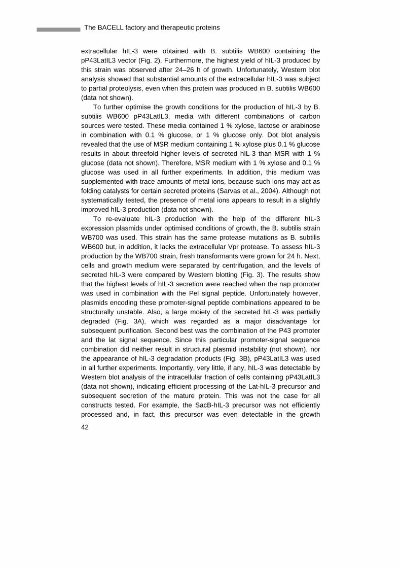
extracellular hIL-3 were obtained with B. subtilis WB600 containing the pP43LatIL3 vector (Fig. 2). Furthermore, the highest yield of hIL-3 produced by this strain was observed after 24–26 h of growth. Unfortunately, Western blot analysis showed that substantial amounts of the extracellular hIL-3 was subject to partial proteolysis, even when this protein was produced in B. subtilis WB600 (data not shown). To further optimise the growth conditions for the production of hIL-3 by B. subtilis WB600 pP43LatIL3, media with different combinations of carbon sources were tested. These media contained 1 % xylose, lactose or arabinose in combination with 0.1 % glucose, or 1 % glucose only. Dot blot analysis revealed that the use of MSR medium containing 1 % xylose plus 0.1 % glucose results in about threefold higher levels of secreted hIL-3 than MSR with 1 % glucose (data not shown). Therefore, MSR medium with 1 % xylose and 0.1 % glucose was used in all further experiments. In addition, this medium was supplemented with trace amounts of metal ions, because such ions may act as folding catalysts for certain secreted proteins (Sarvas et al., 2004). Although not systematically tested, the presence of metal ions appears to result in a slightly improved hIL-3 production (data not shown). To re-evaluate hIL-3 production with the help of the different hIL-3 expression plasmids under optimised conditions of growth, the B. subtilis strain WB700 was used. This strain has the same protease mutations as B. subtilis WB600 but, in addition, it lacks the extracellular Vpr protease. To assess hIL-3 production by the WB700 strain, fresh transformants were grown for 24 h. Next, cells and growth medium were separated by centrifugation, and the levels of secreted hIL-3 were compared by Western blotting (Fig. 3). The results show that the highest levels of hIL-3 secretion were reached when the nap promoter was used in combination with the Pel signal peptide. Unfortunately however, plasmids encoding these promoter-signal peptide combinations appeared to be structurally unstable. Also, a large moiety of the secreted hIL-3 was partially degraded (Fig. 3A), which was regarded as a major disadvantage for subsequent purification. Second best was the combination of the P43 promoter and the lat signal sequence. Since this particular promoter-signal sequence combination did neither result in structural plasmid instability (not shown), nor the appearance of hIL-3 degradation products (Fig. 3B), pP43LatIL3 was used in all further experiments. Importantly, very little, if any, hIL-3 was detectable by Western blot analysis of the intracellular fraction of cells containing pP43LatIL3 (data not shown), indicating efficient processing of the Lat-hIL-3 precursor and subsequent secretion of the mature protein. This was not the case for all constructs tested. For example, the SacB-hIL-3 precursor was not efficiently processed and, in fact, this precursor was even detectable in the growth

43
CHAPTER 3 Secretion of functional hIL-3 from Bacillus subtilis
Figure 3. Western blot analysis of hIL -3 production by B. subtilis WB700 Cells of B. subtilis WB700 transformed with pNapLatIL3, pNapPelIL3, pNapSacBIL3, pLatIL3 or pP43LatIL3 were grown at 37 ºC in MSR medium. After 24 h of growth, cells were separated from the growth medium by centrifugation. From the growth medium fractions 10 µl aliquots were analysed by SDS-PAGE, Western blotting, and immunodetection, using hIL-3-specific antibodies. Panel A, chemiluminescent detection of hIL-3-specific antibodies with horseradish peroxidase-conjugated goat anti-rabbit IgG secondary antibody. The SacB-hIL-3 precursor protein that is detected in the growth medium of cells containing pNapSacBIL3 is marked with “p”. The position of mature hIL-3 is marked with an arrow, and the positions of degradation products of hIL-3 are marked with “d”. Panel B, detection of hIL-3-specific antibodies with alkaline phosphatase conjugated goat anti-rabbit IgG. The position of mature hIL-3 is marked with an arrow.
Figure 4. Western blot analysis of hIL -3 production by different protease-deficient strains Cells of different B. subtilis strains transformed with pP43LatIL3 were grown at 37 ºC in MSR medium. After 24 h of growth, cells were separated from the growth medium by centrifugation. From the growth medium fractions 10 µl aliquots were analysed by SDS-PAGE, Western blotting, and immunodetection, using hIL-3- specific antibodies and alkaline phosphatase-conjugated goat anti-rabbit IgG. The strains that were used are indicated. As a control, 50 ng of purified hIL-3 was loaded.
medium of cells containing pNapSacBIL3, which may occur due to lysis of cells (Fig. 3A).
Finally, the influence of the wall protease A on the production of hIL-3 was tested using B. subtilis WB800. This strain has an inactivated wprA gene and lacks the proteases that are also absent from to the WB700 strain. As shown in Fig. 4, the use of B. subtilis WB800 in combination with the pP43LatIL3 plasmid resulted in significantly elevated levels of hIL-3 production, as compared to the WB600 and WB700 strains containing the same plasmid. As judged by densitometric analyses of Coomassie Brilliant Blue stained SDS-PAA gels, B. subtilis WB800 pP43LatIL3 produced about 100 mg of hIL-3 per liter (data not shown). Importantly, the absence of WprA alone from B. subtilis is not sufficient to obtain hIL-3 production levels that are detectable by Western blotting, as shown with the wprA mutant B. subtilis strain KS408 IwprA (Fig. 4).

The BACELL factory and therapeutic proteins
44
Figure 5. Mass spectrom etric analysis of purified hIL -3 (A) Mass spectrum of hIL-3 purified from the growth medium of B. subtilis WB700 pP43LatIL3. The peak with an m/z of 14,606 relates to mature hIL-3. The peak with an m/z of 13,700 corresponds with an N-terminal hIL-3 degradation product of 120 amino acid residues starting at Lys10. (B) Mass spectrum of hIL-3 purified from the growth medium of B. licheniformis. Note that the hIL-3 purified from the B. licheniformis medium was produced with the original AmyL signal peptide, resulting in a minor hIL-3 fraction with an additional N-terminal alanine residue. This results in an extra peak of at m/z +71 amu. The peaks with m/z values lower than 14,600 relate to unidentified proteins.
Purification and analyses of hIL-3 produced in B. subtilis WB700
hIL-3 was purified to near homogeneity from the culture broth of B. subtilis WB700 containing the pP43LatIL3 construct. After the first chromatographic step (hydrophobic interaction chromatography) two elution peaks (A280) containing hIL-3 were detectable: one at the end of the first gradient (50 mM NH4Ac) and a second during elution with demineralised water. Biochemically the material from both peaks is indistinguishable. The protein fractions, which emerged during elution with demineralised water, were directly applied to a Q- Sepharose column. The peak fraction at 50 mM NH4Ac was first freeze-dried and dissolved in 5 mM (NH4)HCO3, before it was applied to the Q-Sepharose column. As shown by SDS-PAGE and Western blotting, fractions obtained upon Q-Sepharose chromatography of the samples derived from the first chromatographic step contained hIL-3-specific protein bands in the range of about 13–14.5 kDa. The mass spectrum of the purified hIL-3 showed a prominent peak at m/z 14606.1 (Fig. 5A). This is in good agreement with the

45
CHAPTER 3 Secretion of functional hIL-3 from Bacillus subtilis
Figure 6. AMS labeling of free thiol groups in purified hIL -3 hIL-3 purified from the growth medium of B. subtilis WB800 pP43LatIL3 was incubated with 25 mM AMS in the absence or presence of 0.1 mM DTT. Subsequently, the AMS treated hIL-3 was analysed by SDS-PAGE under non-reducing conditions. hIL-3 that was neither treated with AMS, nor DTT was used as a control. The positions of AMS-hIL-3 and non-cross-linked hIL-3 are marked.
mass spectrum observed for hIL-3 that was previously produced in B. licheniformis (m/z 14594.6) (van Leen et al., 1991) and that was used as a reference in the present experiment (Fig. 5B). Notably, the reference material reveals an additional peak at m/z 14665.4, which relates to an alternative maturation site in the AmyL signal peptide that is absent from the Lat signal peptide. Maturation at the second site results in the presence of an additional N-terminal alanine residue in hIL-3, explaining the 71 Da increment (Bonekamp and van Tilborg, 1998). Besides these main peaks, some fractions contained products with masses of about m/z 14,100 and m/z 13,700. The smallest fragment was analysed by N-terminal amino acid sequencing. The results showed that this fragment corresponds with an N-terminal hIL-3 degradation product of 120 amino acid residues starting at Lys10. Consistent with the mass spectrometric analysis, the theoretical mass of this degradation product is 13705.7 Da. hIL-3 is known to contain one intramolecular disulphide bond. To verify the correct formation of this bond, AMS labeling experiments were performed. Notably, AMS will only cross-react with free thiol groups, thereby causing a reduced mobility of a cross-linked protein in SDS-PAGE. As shown by SDS- PAGE, AMS did not cross-react with hIL-3 purified from the growth medium of B. subtilis. In fact, incubation of hIL-3 with AMS resulted in a reduced mobility on SDS-PAGE only when hIL-3 was reduced with DTT prior to the incubation with AMS. This implies that there are no free thiol groups present in the purified protein (Fig. 6). Since hIL-3 contains only two cysteine residues, the observed lack of AMS cross-linking is a clear indication that the disulphide bond in hIL-3 is properly formed by B. subtilis.
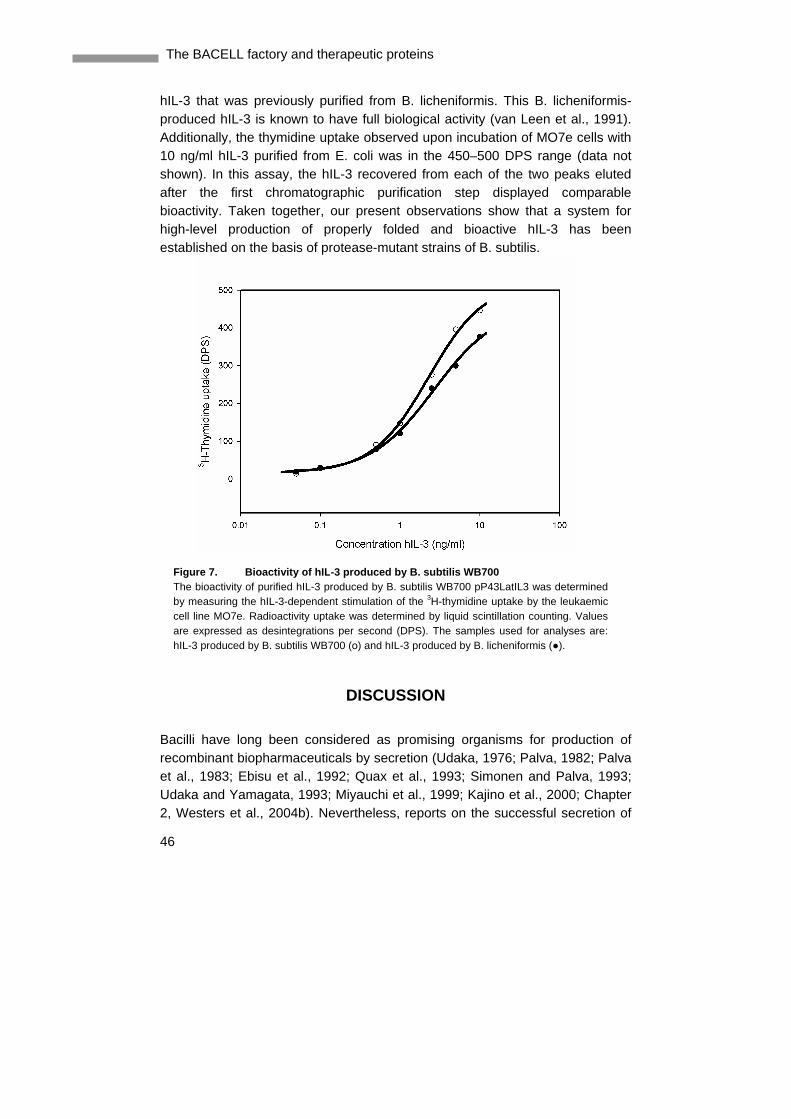
Bioactivity of hIL-3 produced in B. subtilis WB700
The bioactivity of hIL-3 purified from the growth medium of B. subtilis WB700 was tested using the hIL-3-dependent leukaemia cell line MO7e and a thymidine uptake assay. As shown in Fig. 7, the thymidine uptake curve displayed by cells that were stimulated with hIL-3 produced in B. subtilis compared very well with the curve displayed by cells that were stimulated with
The BACELL factory and therapeutic proteins
46
Figure 7. Bioactivi ty of hIL -3 produced by B. subtilis WB700 The bioactivity of purified hIL-3 produced by B. subtilis WB700 pP43LatIL3 was determined by measuring the hIL-3-dependent stimulation of the 3H-thymidine uptake by the leukaemic cell line MO7e. Radioactivity uptake was determined by liquid scintillation counting. Values are expressed as desintegrations per second (DPS). The samples used for analyses are: hIL-3 produced by B. subtilis WB700 (ο) and hIL-3 produced by B. licheniformis (●).
hIL-3 that was previously purified from B. licheniformis. This B. licheniformis-produced hIL-3 is known to have full biological activity (van Leen et al., 1991). Additionally, the thymidine uptake observed upon incubation of MO7e cells with 10 ng/ml hIL-3 purified from E. coli was in the 450–500 DPS range (data not shown). In this assay, the hIL-3 recovered from each of the two peaks eluted after the first chromatographic purification step displayed comparable bioactivity. Taken together, our present observations show that a system for high-level production of properly folded and bioactive hIL-3 has been established on the basis of protease-mutant strains of B. subtilis.
DISCUSSION
Bacilli have long been considered as promising organisms for production of recombinant biopharmaceuticals by secretion (Udaka, 1976; Palva, 1982; Palva et al., 1983; Ebisu et al., 1992; Quax et al., 1993; Simonen and Palva, 1993; Udaka and Yamagata, 1993; Miyauchi et al., 1999; Kajino et al., 2000; Chapter 2, Westers et al., 2004b). Nevertheless, reports on the successful secretion of

47
CHAPTER 3 Secretion of functional hIL-3 from Bacillus subtilis
human proteins from B. subtilis are very scarce. Obstacles encountered include plasmid instability, proteolytic degradation of products and formation of intracellular inclusion bodies. In our study, we have been able to show the secretion of correctly folded and fully biologically active hIL-3 from B. subtilis. Using a variety of expression signals and mutants available for this well-characterised organism, we have performed a systematic study of the parameters important for optimising secretion from Bacillus. Especially, the large knowledge about the secretion machinery, secreted proteins and the corresponding signal peptides is a very useful resource in optimising B. subtilis as cell factory of extracellular proteins (Tjalsma et al., 2000 and 2004). This information is mostly lacking for other Bacillus species that are used for bioproduction of secretory proteins, such as Bacillus amyloliquefaciens, Bacillus brevis and B. licheniformis. Although it was shown for B. brevis that a low copy number plasmid can lead to high-level production of a heterologous protein (Ebisu et al., 1996), for this study plasmids were constructed based on the high copy number plasmid pMA5. This plasmid was previously shown to be well suitable for high-level production of B. subtilis lipase A (Lesuisse et al., 1993). As an additional starting point for optimised hIL-3 production, we selected the well-characterised P43 and the nap promoters, which are known to direct high-level gene expression in B. subtilis (Ye et al., 1999; Dröge et al., 2001). Taking advantage of the tools available for a well-known cell factory, such as B. subtilis, we established a test matrix for an optimal hIL-3 production system. In order to facilitate product recovery and to avoid a cell breakage step, we chose to secrete hIL-3 in the growth medium by linking a signal sequence to the hIL-3 gene. Use of the modified AmyL signal peptide (Lat) resulted in a reproducibly high secretion of hIL-3 from the cells. The signal peptide was removed correctly during secretion as demonstrated by mass spectrometric analysis. This is in contrast to the 20 % miscleavage of the authentic AmyL signal peptide from hIL-3 precursor in B. licheniformis (Bonekamp and van Tilborg, 1998). Remarkably, the SacB signal peptide did not result in productive secretion of hIL-3, although efficient secretion of staphylokinase under guidance of this signal peptide has been reported (Ye et al., 1999). The Pel signal peptide incidentally gave rise to a high-level of secreted product in the medium. However, the plasmid instability that was observed when the pel signal sequence was used in combination with the nap promoter suggests that high-level production of the Pel-hIL-3 precursor may be detrimental for the cells. This would result in a selective growth advantage of cells that have lost the ability to produce this precursor. Additionally, the occurrence of a corresponding degradation product may be an indication that the production of Pel-hIL-3 at high-levels elicits a secretion stress response, as previously observed upon
The BACELL factory and therapeutic proteins
48
high-level production of Bacillus α-amylases (Hyyryläinen et al., 2000; Darmon et al., 2002). Such a secretion stress response would result in the production of HtrA-like proteases at elevated levels and, in turn, this could result in increased product degradation. Unfortunately, it is presently not possible to predict which signal peptide will be optimal for the secretion of a particular heterologous protein such as hIL-3. Possibly, this relates to the fact that the signal peptide does not only serve as a targeting signal, but also seems to have a role in preventing pretranslocational folding of secretory proteins (Park et al., 1988). Moreover, for efficient export, the optimal junction between a particular signal peptide and a heterologous protein is likely to depend on the nature of the heterologous protein. The use of B. subtilis strains with increasing numbers of mutated protease genes resulted in a stepwise improvement of hIL-3 accumulation in the medium. After purification of hIL-3 from the culture medium of the B. subtilis WB700 strain and analyses of the fractions, still some degradation products were detected. N-terminal sequencing of the smallest degradation product that was detected by MALDI-TOF of some fractions revealed that degradation took place at the N-terminus of the protein. A major increase in hIL-3 level was observed upon using B. subtilis WB800, which lacks WprA, a cell wall protease implicated in the degradation of slowly folding proteins in the cell wall. However, WprA is not the only protease degrading hIL-3 as can be inferred from the absence of hIL-3 in the supernatant of strain KS408 IwprA, which lacks only WprA. These observations demonstrate that hIL-3 is prone to proteolysis by multiple cell wall- and extracellular proteases of B. subtilis. This suggests that proteolysis of exported hIL-3 molecules can occur at all stages of the secretion process, starting immediately after translocation across the membrane and continuing in the cell wall environment and growth medium. The hIL-3 purified from the supernatant shows full biological activity in the cell proliferation assay using the MO7e cell line, which is known to be hIL-3- dependent (Avanzi et al., 1990). Since B. subtilis is a non-glycosylating production host, this observation confirms the view that non-glycosylated hIL-3 is fully active (Zenke et al., 1991; Mangi and Newland, 1999). Furthermore, our observations suggest that hIL-3 produced by B. subtilis is properly folded. Consistent with this idea, we were able to show that the two cysteine residues in hIL-3 produced by B. subtilis were oxidised, most likely, due to the formation of an intramolecular disulphide bond. To date, it is not known how this disulphide bond is formed, but it is conceivable that the BdbCD system is involved in this process. Our previous studies have shown that both BdbC and BdbD are required for the folding of exported proteins containing disulphide bonds (Bolhuis et al., 1999b; Meima et al., 2002).
49
CHAPTER 3 Secretion of functional hIL-3 from Bacillus subtilis
In conclusion, our present studies have shown that adaptations to the expression and secretion signals used are needed for optimal production of hIL-3. For a larger scale production of hIL-3, the most optimal production system both with regard to stability and yield was found to be the B. subtilis WB800 strain in combination with the pP43LatIL3 vector, including the modified AmyL signal sequence. After 24 h of culturing, this strain gave a yield of 100 mg per liter hIL-3. This should be sufficient for the further development of a commercial production process. We anticipate that our results will also be applicable for the production of other heterologous proteins, especially four-helix bundle cytokines related to hIL-3 that do not require glycosylation for full bioactivity.
ACKNOWLEDGEMENTS
We wish to thank Sui-Lam Wong for his kind gift of the B. subtilis WB600, WB700 and WB800 strains, Mathieu Platteel for determining the sequences of the different DNA vectors, Fridolin van der Lecq for determining the N-termini of the hIL-3 fragments, Mariet Esselink for performing the bioactivity assays, and Albert Kiewiet for determining and analysing the mass spectra. The B. licheniformis-broth containing hIL-3 was a kind gift from DSM N.V. L.W. was supported by the Senter projects TSGE2035 and CSI4011, J.M.v.D. and H.W. were supported in part by European Union (EU) Grants QLK3-CT-1999-00413/00917, LSHC-CT-2004-503468 and LSHG-CT-2004-005257 and W.J.Q. was supported by the EU project BIO4-CT98-0250 and QLK3-CT-2001-00498.
50
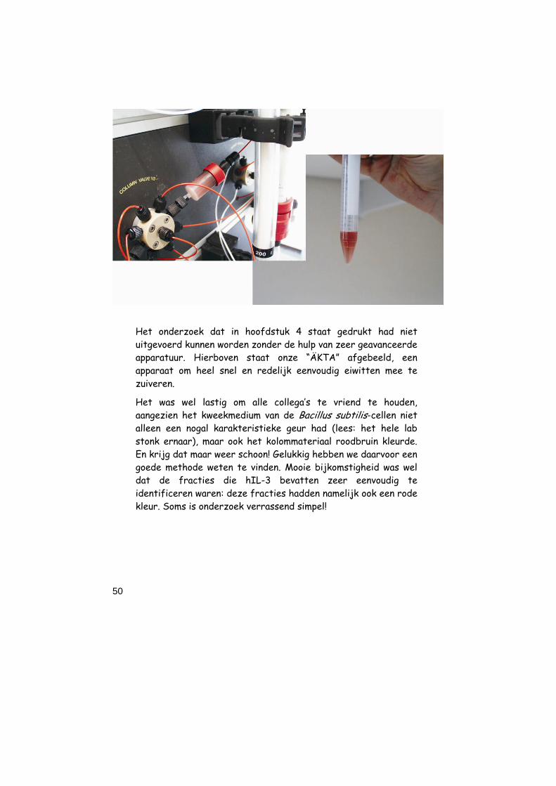
Het onderzoek dat in hoofdstuk 4 staat gedrukt had niet uitgevoerd kunnen worden zonder de hulp van zeer geavanceerde apparatuur. Hierboven staat onze “ÄKTA” afgebeeld, een apparaat om heel snel en redelijk eenvoudig eiwitten mee te zuiveren.
Het was wel lastig om alle collega’s te vriend te houden, aangezien het kweekmedium van de Bacillus subtilis-cellen niet alleen een nogal karakteristieke geur had (lees: het hele lab stonk ernaar), maar ook het kolommateriaal roodbruin kleurde. En krijg dat maar weer schoon! Gelukkig hebben we daarvoor een goede methode weten te vinden. Mooie bijkomstigheid was wel dat de fracties die hIL-3 bevatten zeer eenvoudig te identificeren waren: deze fracties hadden namelijk ook een rode kleur. Soms is onderzoek verrassend simpel!