Embed Size (px)
Citation preview
The Basal Ganglia in Parkinson’s Disease:Current Concepts and Unexplained
ObservationsJose A. Obeso, PhD, MD,1,2 Concepcio Marin, PhD, MD,2,3 C. Rodriguez-Oroz, PhD, MD,1,2 Javier Blesa,1,2
B. Benitez-Temino, PhD,1 Juan Mena-Segovia, PhD, MD,4 Manuel Rodrıguez, PhD, MD,2,5 andC. Warren Olanow, MD, FRCPC6
The pathophysiology of Parkinson’s disease is reviewed in light of recent advances in the understanding of the functionalorganization of the basal ganglia (BG). Current emphasis is placed on the parallel interactions between corticostriatal andcorticosubthalamic afferents on the one hand, and internal feedback circuits modulating BG output through the globus palliduspars interna and substantia nigra pars reticulata on the other. In the normal BG network, the globus pallidus pars externaemerges as a main regulatory station of output activity. In the parkinsonian state, dopamine depletion shifts the BG towardinhibiting cortically generated movements by increasing the gain in the globus pallidus pars externa-subthalamic nucleus-globuspallidus pars interna network and reducing activity in “direct” cortico-putaminal-globus pallidus pars interna projections. Stan-dard pharmacological treatments do not mimic the normal physiology of the dopaminergic system and, therefore, fail to restorea functional balance between corticostriatal afferents in the so-called direct and indirect pathways, leading to the development ofmotor complications. This review emphasizes the concept that the BG can no longer be understood as a “go-through” stationin the control of movement, behavior, and emotions. The growing understanding of the complexity of the normal BG and thechanges induced by DA depletion should guide the development of more efficacious therapies for Parkinson’s disease.
Ann Neurol 2008;64 (suppl):S30–S46
The hallmark feature of Parkinson’s disease (PD) is de-generation of dopamine neurons in the substantia nigrapars compacta (SNc) and a consequent striatal dopa-mine deficiency. This dopamine deficiency leads to acascade of functional changes in basal ganglia circuitry,which are ultimately responsible for the developmentof the cardinal features of PD.1–3 The essential patho-
physiological characteristic of the PD state is increasedneuronal firing activity in the output nuclei of thebasal ganglia (globus pallidus pars interna [GPi] andsubstantia nigra pars reticulata [SNr]) leading to exces-sive inhibition of thalamocortical and brainstem motorsystems. This was initially proposed to arise as a con-sequence of increased firing in basal ganglia output
From the 1Departments of Neurology, Neurophysiology and Neu-rosurgery, Clinica Universitaria and Medical School, NeuroscienceCentre, Center for Applied Medical Research, University of Na-varra, Pamplona; 2Centro de Investigacion Biomedica en Red sobreEnfermedades Neurodegenerativas (CIBERNED); 3Laboratori deNeurologia Experimental, Fundacio Clınic-Hospital Clınic, Institutd’Investigacions Biomediques August Pi i Sunyer (IDIBAPS), Hos-pital Clınic, Barcelona, Spain; 4Medical Research Centre Anatomi-cal Neuropharmacology Unit, University of Oxford, Oxford, UnitedKingdom; 5Department of Physiology, Medical School, Universidadde La Laguna, Tenerife, Spain; and 6Department of Neurology,Mount Sinai School of Medicine, New York, NY.
Received Mar 3, 2008, and in revised form Jun 30. Accepted forpublication Jul 11, 2008.
Potential conflict of interest: This article is part of a supplementsponsored by Boehringer Ingelheim (BI). J.A.O. has served once inthe past three years as a consultant in an advisory board meeting ofBI and GlaxoSmithKline, and on a single occasion for NovartisPharmaceutica. C.M., C.R.-O., J.B., B.B.-T., J.M.-S., and M.R. de-clare that they have no financial relationship with BI. J.M.-S. wasfunded by the Medical Research Council UK and the Parkinson’sDisease Society of the UK. M. R. has no financial or consultingrelationships with pharmaceutical enterprises. C.W. O. has served asa consultant to BI, Novartis, Teva, Merck Serono, and Ceregene.
Published online in Wiley InterScience (www.interscience.wiley.com).DOI: 10.1002/ana.21481
Address correspondence to Dr Obeso, Clinica Universitaria,Avenida Pio XII, 36, Pamplona, 31008, Spain.E-mail: [email protected]
S30 © 2008 American Neurological AssociationPublished by Wiley-Liss, Inc., through Wiley Subscription Services
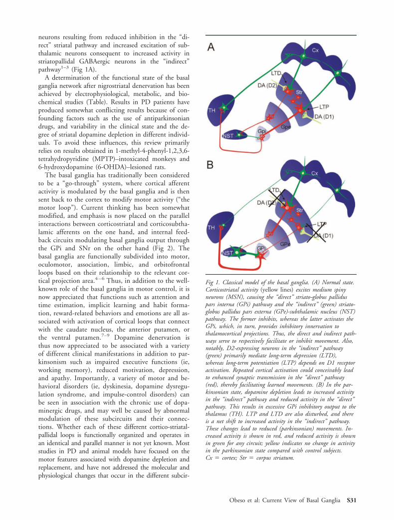
neurons resulting from reduced inhibition in the “di-rect” striatal pathway and increased excitation of sub-thalamic neurons consequent to increased activity instriatopallidal GABAergic neurons in the “indirect”pathway1–3 (Fig 1A).
A determination of the functional state of the basalganglia network after nigrostriatal denervation has beenachieved by electrophysiological, metabolic, and bio-chemical studies (Table). Results in PD patients haveproduced somewhat conflicting results because of con-founding factors such as the use of antiparkinsoniandrugs, and variability in the clinical state and the de-gree of striatal dopamine depletion in different individ-uals. To avoid these influences, this review primarilyrelies on results obtained in 1-methyl-4-phenyl-1,2,3,6-tetrahydropyridine (MPTP)–intoxicated monkeys and6-hydroxydopamine (6-OHDA)–lesioned rats.
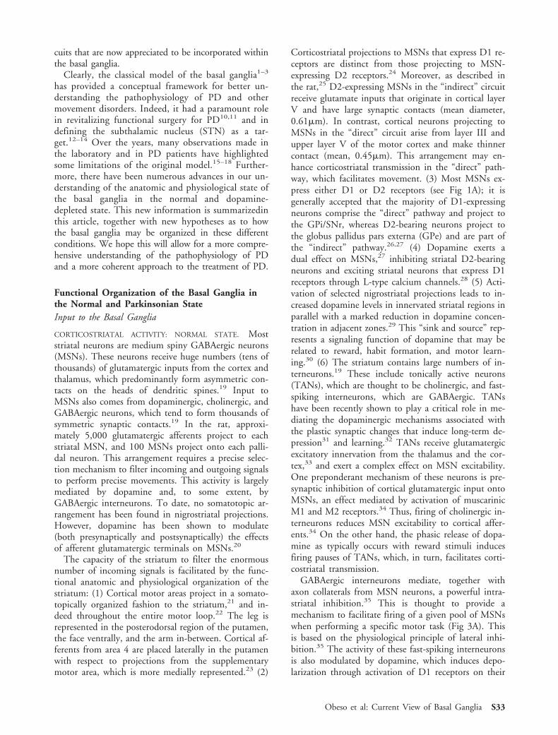
The basal ganglia has traditionally been consideredto be a “go-through” system, where cortical afferentactivity is modulated by the basal ganglia and is thensent back to the cortex to modify motor activity (“themotor loop”). Current thinking has been somewhatmodified, and emphasis is now placed on the parallelinteractions between corticostriatal and corticosubtha-lamic afferents on the one hand, and internal feed-back circuits modulating basal ganglia output throughthe GPi and SNr on the other hand (Fig 2). Thebasal ganglia are functionally subdivided into motor,oculomotor, association, limbic, and orbitofrontalloops based on their relationship to the relevant cor-tical projection area.4 – 6 Thus, in addition to the well-known role of the basal ganglia in motor control, it isnow appreciated that functions such as attention andtime estimation, implicit learning and habit forma-tion, reward-related behaviors and emotions are all as-sociated with activation of cortical loops that connectwith the caudate nucleus, the anterior putamen, orthe ventral putamen.7–9 Dopamine denervation isthus now appreciated to be associated with a varietyof different clinical manifestations in addition to par-kinsonism such as impaired executive functions (ie,working memory), reduced motivation, depression,and apathy. Importantly, a variety of motor and be-havioral disorders (ie, dyskinesia, dopamine dysregu-lation syndrome, and impulse-control disorders) canbe seen in association with the chronic use of dopa-minergic drugs, and may well be caused by abnormalmodulation of these subcircuits and their connec-tions. Whether each of these different cortico-striatal-pallidal loops is functionally organized and operates inan identical and parallel manner is not yet known. Moststudies in PD and animal models have focused on themotor features associated with dopamine depletion andreplacement, and have not addressed the molecular andphysiological changes that occur in the different subcir-
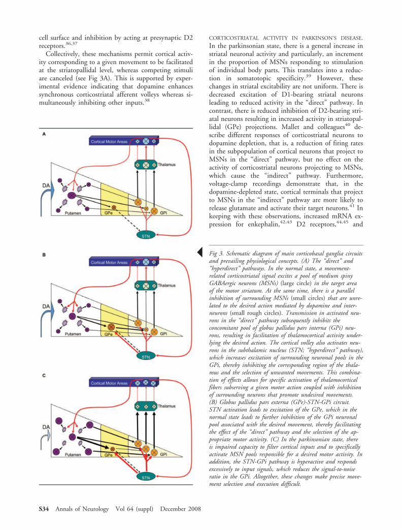
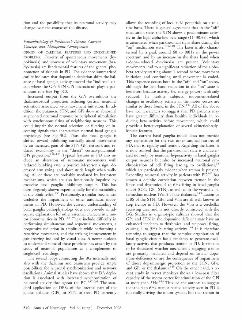
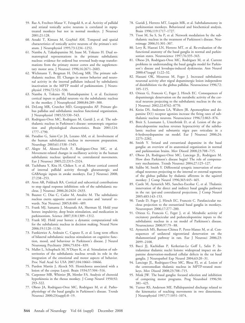
Fig 1. Classical model of the basal ganglia. (A) Normal state.Corticostriatal activity (yellow lines) excites medium spinyneurons (MSN), causing the “direct” striato-globus palliduspars interna (GPi) pathway and the “indirect” (green) striato-globus pallidus pars externa (GPe)-subthalamic nucleus (NST)pathway. The former inhibits, whereas the latter activates theGPi, which, in turn, provides inhibitory innervation tothalamocortical projections. Thus, the direct and indirect path-ways serve to respectively facilitate or inhibit movement. Also,notably, D2-expressing neurons in the “indirect” pathway(green) primarily mediate long-term depression (LTD),whereas long-term potentiation (LTP) depends on D1 receptoractivation. Repeated cortical activation could conceivably leadto enhanced synaptic transmission in the “direct” pathway(red), thereby facilitating learned movements. (B) In the par-kinsonian state, dopamine depletion leads to increased activityin the “indirect” pathway and reduced activity in the “direct”pathway. This results in excessive GPi inhibitory output to thethalamus (TH). LTP and LTD are also disturbed, and thereis a net shift to increased activity in the “indirect” pathway.These changes lead to reduced (parkinsonian) movements. In-creased activity is shown in red, and reduced activity is shownin green for any circuit; yellow indicates no change in activityin the parkinsonian state compared with control subjects.Cx ! cortex; Str ! corpus striatum.
Obeso et al: Current View of Basal Ganglia S31
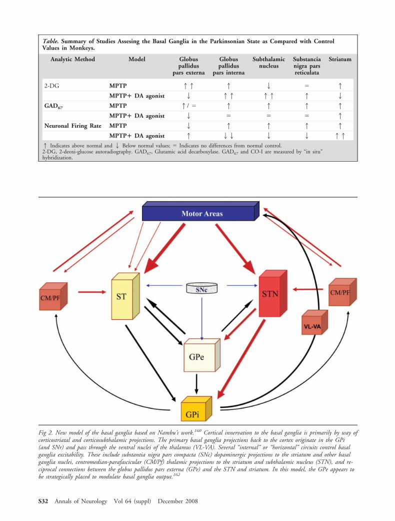
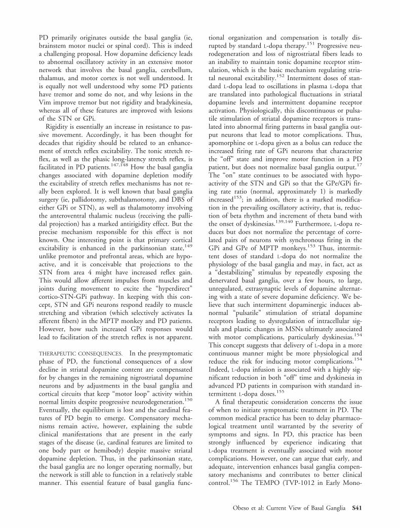
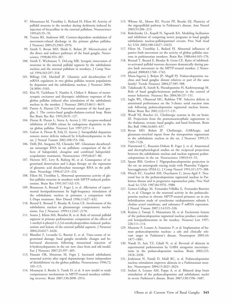
Fig 2. New model of the basal ganglia based on Nambu’s work.160 Cortical innervation to the basal ganglia is primarily by way ofcorticostriatal and corticosubthalamic projections. The primary basal ganglia projections back to the cortex originate in the GPi(and SNr) and pass through the ventral nuclei of the thalamus (VL-VA). Several “internal” or “horizontal” circuits control basalganglia excitability. These include substantia nigra pars compacta (SNc) dopaminergic projections to the striatum and other basalganglia nuclei, centromedian-parafascicular (CM/Pf) thalamic projections to the striatum and subthalamic nucleus (STN), and re-ciprocal connections between the globus pallidus pars externa (GPe) and the STN and striatum. In this model, the GPe appears tobe strategically placed to modulate basal ganglia output.162
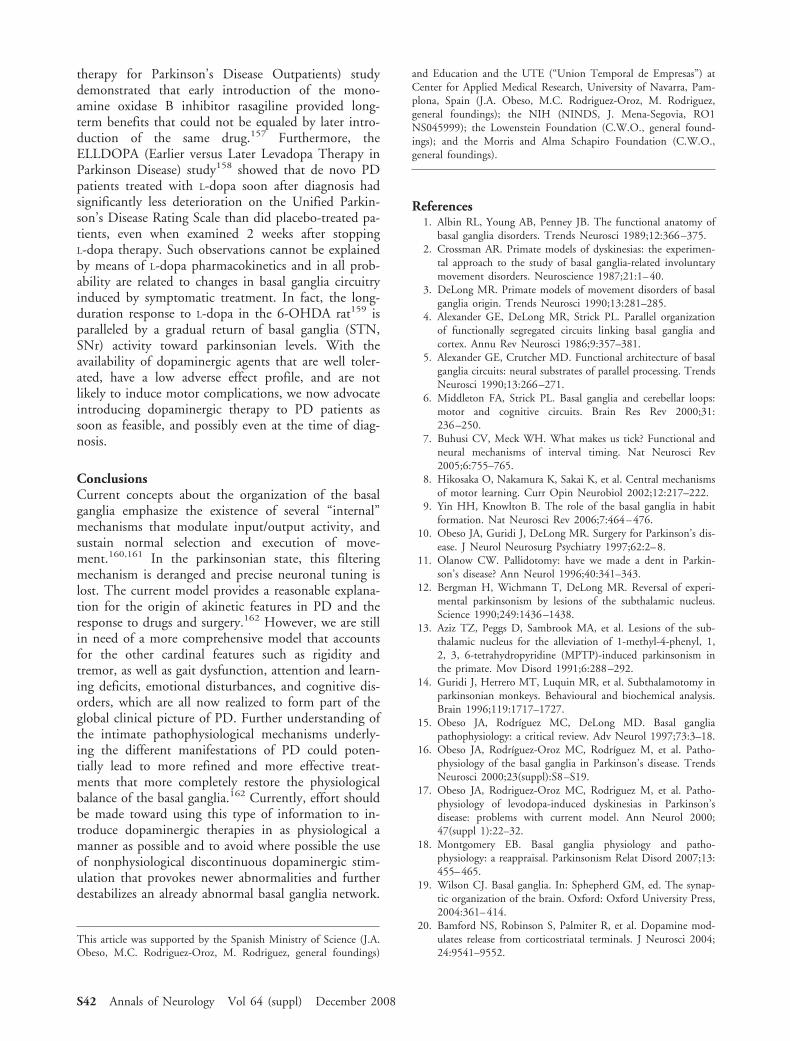
Table. Summary of Studies Assesing the Basal Ganglia in the Parkinsonian State as Compared with ControlValues in Monkeys.
Analytic Method Model Globuspallidus
pars externa
Globuspallidus
pars interna
Subthalamicnucleus
Substancianigra parsreticulata
Striatum
2-DG MPTP 11 1 2 ! 1
MPTP! DA agonist 2 11 11 1 2
GAD67 MPTP 1/ ! 1 1 1 1
MPTP! DA agonist 2 ! ! ! 1
Neuronal Firing Rate MPTP 2 1 1 1 1
MPTP! DA agonist 1 22 2 2 11
1 Indicates above normal and 2 Below normal values; ! Indicates no differences from normal control.2-DG, 2-deoxi-glucose autoradiography. GAD67, Glutamic acid decarboxylase. GAD67 and CO-I are measured by “in situ”hybridization.
S32 Annals of Neurology Vol 64 (suppl) December 2008
cuits that are now appreciated to be incorporated withinthe basal ganglia.
Clearly, the classical model of the basal ganglia1–3
has provided a conceptual framework for better un-derstanding the pathophysiology of PD and othermovement disorders. Indeed, it had a paramount rolein revitalizing functional surgery for PD10,11 and indefining the subthalamic nucleus (STN) as a tar-get.12–14 Over the years, many observations made inthe laboratory and in PD patients have highlightedsome limitations of the original model.15–18 Further-more, there have been numerous advances in our un-derstanding of the anatomic and physiological state ofthe basal ganglia in the normal and dopamine-depleted state. This new information is summarizedinthis article, together with new hypotheses as to howthe basal ganglia may be organized in these differentconditions. We hope this will allow for a more compre-hensive understanding of the pathophysiology of PDand a more coherent approach to the treatment of PD.
Functional Organization of the Basal Ganglia inthe Normal and Parkinsonian StateInput to the Basal Ganglia
CORTICOSTRIATAL ACTIVITY: NORMAL STATE. Moststriatal neurons are medium spiny GABAergic neurons(MSNs). These neurons receive huge numbers (tens ofthousands) of glutamatergic inputs from the cortex andthalamus, which predominantly form asymmetric con-tacts on the heads of dendritic spines.19 Input toMSNs also comes from dopaminergic, cholinergic, andGABAergic neurons, which tend to form thousands ofsymmetric synaptic contacts.19 In the rat, approxi-mately 5,000 glutamatergic afferents project to eachstriatal MSN, and 100 MSNs project onto each palli-dal neuron. This arrangement requires a precise selec-tion mechanism to filter incoming and outgoing signalsto perform precise movements. This activity is largelymediated by dopamine and, to some extent, byGABAergic interneurons. To date, no somatotopic ar-rangement has been found in nigrostriatal projections.However, dopamine has been shown to modulate(both presynaptically and postsynaptically) the effectsof afferent glutamatergic terminals on MSNs.20
The capacity of the striatum to filter the enormousnumber of incoming signals is facilitated by the func-tional anatomic and physiological organization of thestriatum: (1) Cortical motor areas project in a somato-topically organized fashion to the striatum,21 and in-deed throughout the entire motor loop.22 The leg isrepresented in the posterodorsal region of the putamen,the face ventrally, and the arm in-between. Cortical af-ferents from area 4 are placed laterally in the putamenwith respect to projections from the supplementarymotor area, which is more medially represented.23 (2)
Corticostriatal projections to MSNs that express D1 re-ceptors are distinct from those projecting to MSN-expressing D2 receptors.24 Moreover, as described inthe rat,25 D2-expressing MSNs in the “indirect” circuitreceive glutamate inputs that originate in cortical layerV and have large synaptic contacts (mean diameter,0.61"m). In contrast, cortical neurons projecting toMSNs in the “direct” circuit arise from layer III andupper layer V of the motor cortex and make thinnercontact (mean, 0.45"m). This arrangement may en-hance corticostriatal transmission in the “direct” path-way, which facilitates movement. (3) Most MSNs ex-press either D1 or D2 receptors (see Fig 1A); it isgenerally accepted that the majority of D1-expressingneurons comprise the “direct” pathway and project tothe GPi/SNr, whereas D2-bearing neurons project tothe globus pallidus pars externa (GPe) and are part ofthe “indirect” pathway.26,27 (4) Dopamine exerts adual effect on MSNs,27 inhibiting striatal D2-bearingneurons and exciting striatal neurons that express D1receptors through L-type calcium channels.28 (5) Acti-vation of selected nigrostriatal projections leads to in-creased dopamine levels in innervated striatal regions inparallel with a marked reduction in dopamine concen-tration in adjacent zones.29 This “sink and source” rep-resents a signaling function of dopamine that may berelated to reward, habit formation, and motor learn-ing.30 (6) The striatum contains large numbers of in-terneurons.19 These include tonically active neurons(TANs), which are thought to be cholinergic, and fast-spiking interneurons, which are GABAergic. TANshave been recently shown to play a critical role in me-diating the dopaminergic mechanisms associated withthe plastic synaptic changes that induce long-term de-pression31 and learning.32 TANs receive glutamatergicexcitatory innervation from the thalamus and the cor-tex,33 and exert a complex effect on MSN excitability.One preponderant mechanism of these neurons is pre-synaptic inhibition of cortical glutamatergic input ontoMSNs, an effect mediated by activation of muscarinicM1 and M2 receptors.34 Thus, firing of cholinergic in-terneurons reduces MSN excitability to cortical affer-ents.34 On the other hand, the phasic release of dopa-mine as typically occurs with reward stimuli inducesfiring pauses of TANs, which, in turn, facilitates corti-costriatal transmission.
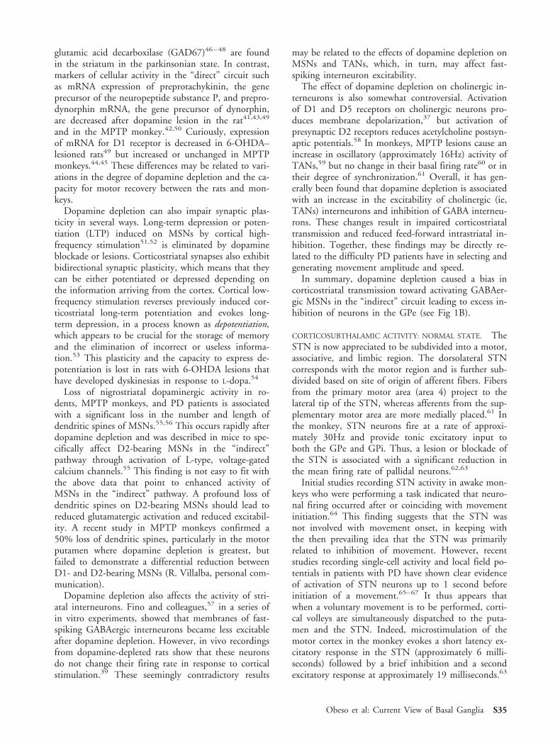
GABAergic interneurons mediate, together withaxon collaterals from MSN neurons, a powerful intra-striatal inhibition.35 This is thought to provide amechanism to facilitate firing of a given pool of MSNswhen performing a specific motor task (Fig 3A). Thisis based on the physiological principle of lateral inhi-bition.35 The activity of these fast-spiking interneuronsis also modulated by dopamine, which induces depo-larization through activation of D1 receptors on their
Obeso et al: Current View of Basal Ganglia S33
cell surface and inhibition by acting at presynaptic D2receptors.36,37
Collectively, these mechanisms permit cortical activ-ity corresponding to a given movement to be facilitatedat the striatopallidal level, whereas competing stimuliare canceled (see Fig 3A). This is supported by exper-imental evidence indicating that dopamine enhancessynchronous corticostriatal afferent volleys whereas si-multaneously inhibiting other inputs.38
CORTICOSTRIATAL ACTIVITY IN PARKINSON’S DISEASE.
In the parkinsonian state, there is a general increase instriatal neuronal activity and particularly, an incrementin the proportion of MSNs responding to stimulationof individual body parts. This translates into a reduc-tion in somatotopic specificity.39 However, thesechanges in striatal excitability are not uniform. There isdecreased excitation of D1-bearing striatal neuronsleading to reduced activity in the “direct” pathway. Incontrast, there is reduced inhibition of D2-bearing stri-atal neurons resulting in increased activity in striatopal-lidal (GPe) projections. Mallet and colleagues40 de-scribe different responses of corticostriatal neurons todopamine depletion, that is, a reduction of firing ratesin the subpopulation of cortical neurons that project toMSNs in the “direct” pathway, but no effect on theactivity of corticostriatal neurons projecting to MSNs,which cause the “indirect” pathway. Furthermore,voltage-clamp recordings demonstrate that, in thedopamine-depleted state, cortical terminals that projectto MSNs in the “indirect” pathway are more likely torelease glutamate and activate their target neurons.41 Inkeeping with these observations, increased mRNA ex-pression for enkephalin,42,43 D2 receptors,44,45 and
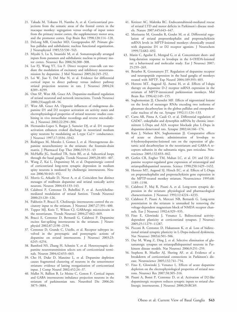
! Fig 3. Schematic diagram of main corticobasal ganglia circuitsand prevailing physiological concepts. (A) The “direct” and“hyperdirect” pathways. In the normal state, a movement-related corticostriatal signal excites a pool of medium spinyGABAergic neurons (MSNs) (large circle) in the target areaof the motor striatum. At the same time, there is a parallelinhibition of surrounding MSNs (small circles) that are unre-lated to the desired action mediated by dopamine and inter-neurons (small rough circles). Transmission in activated neu-rons in the “direct” pathway subsequently inhibits theconcomitant pool of globus pallidus pars interna (GPi) neu-rons, resulting in facilitation of thalamocortical activity under-lying the desired action. The cortical volley also activates neu-rons in the subthalamic nucleus (STN; “hyperdirect” pathway),which increases excitation of surrounding neuronal pools in theGPi, thereby inhibiting the corresponding region of the thala-mus and the selection of unwanted movements. This combina-tion of effects allows for specific activation of thalamocorticalfibers subserving a given motor action coupled with inhibitionof surrounding neurons that promote undesired movements.(B) Globus pallidus pars externa (GPe)-STN-GPi circuit.STN activation leads to excitation of the GPe, which in thenormal state leads to further inhibition of the GPi neuronalpool associated with the desired movement, thereby facilitatingthe effect of the “direct” pathway and the selection of the ap-propriate motor activity. (C) In the parkinsonian state, thereis impaired capacity to filter cortical inputs and to specificallyactivate MSN pools responsible for a desired motor activity. Inaddition, the STN-GPi pathway is hyperactive and respondsexcessively to input signals, which reduces the signal-to-noiseratio in the GPi. Altogether, these changes make precise move-ment selection and execution difficult.
S34 Annals of Neurology Vol 64 (suppl) December 2008
glutamic acid decarboxilase (GAD67)46–48 are foundin the striatum in the parkinsonian state. In contrast,markers of cellular activity in the “direct” circuit suchas mRNA expression of preprotachykinin, the geneprecursor of the neuropeptide substance P, and prepro-dynorphin mRNA, the gene precursor of dynorphin,are decreased after dopamine lesion in the rat41,43,49
and in the MPTP monkey.42,50 Curiously, expressionof mRNA for D1 receptor is decreased in 6-OHDA–lesioned rats49 but increased or unchanged in MPTPmonkeys.44,45 These differences may be related to vari-ations in the degree of dopamine depletion and the ca-pacity for motor recovery between the rats and mon-keys.
Dopamine depletion can also impair synaptic plas-ticity in several ways. Long-term depression or poten-tiation (LTP) induced on MSNs by cortical high-frequency stimulation51,52 is eliminated by dopamineblockade or lesions. Corticostriatal synapses also exhibitbidirectional synaptic plasticity, which means that theycan be either potentiated or depressed depending onthe information arriving from the cortex. Cortical low-frequency stimulation reverses previously induced cor-ticostriatal long-term potentiation and evokes long-term depression, in a process known as depotentiation,which appears to be crucial for the storage of memoryand the elimination of incorrect or useless informa-tion.53 This plasticity and the capacity to express de-potentiation is lost in rats with 6-OHDA lesions thathave developed dyskinesias in response to L-dopa.54
Loss of nigrostriatal dopaminergic activity in ro-dents, MPTP monkeys, and PD patients is associatedwith a significant loss in the number and length ofdendritic spines of MSNs.55,56 This occurs rapidly afterdopamine depletion and was described in mice to spe-cifically affect D2-bearing MSNs in the “indirect”pathway through activation of L-type, voltage-gatedcalcium channels.55 This finding is not easy to fit withthe above data that point to enhanced activity ofMSNs in the “indirect” pathway. A profound loss ofdendritic spines on D2-bearing MSNs should lead toreduced glutamatergic activation and reduced excitabil-ity. A recent study in MPTP monkeys confirmed a50% loss of dendritic spines, particularly in the motorputamen where dopamine depletion is greatest, butfailed to demonstrate a differential reduction betweenD1- and D2-bearing MSNs (R. Villalba, personal com-munication).
Dopamine depletion also affects the activity of stri-atal interneurons. Fino and colleagues,57 in a series ofin vitro experiments, showed that membranes of fast-spiking GABAergic interneurons became less excitableafter dopamine depletion. However, in vivo recordingsfrom dopamine-depleted rats show that these neuronsdo not change their firing rate in response to corticalstimulation.39 These seemingly contradictory results
may be related to the effects of dopamine depletion onMSNs and TANs, which, in turn, may affect fast-spiking interneuron excitability.
The effect of dopamine depletion on cholinergic in-terneurons is also somewhat controversial. Activationof D1 and D5 receptors on cholinergic neurons pro-duces membrane depolarization,37 but activation ofpresynaptic D2 receptors reduces acetylcholine postsyn-aptic potentials.58 In monkeys, MPTP lesions cause anincrease in oscillatory (approximately 16Hz) activity ofTANs,59 but no change in their basal firing rate60 or intheir degree of synchronization.61 Overall, it has gen-erally been found that dopamine depletion is associatedwith an increase in the excitability of cholinergic (ie,TANs) interneurons and inhibition of GABA interneu-rons. These changes result in impaired corticostriataltransmission and reduced feed-forward intrastriatal in-hibition. Together, these findings may be directly re-lated to the difficulty PD patients have in selecting andgenerating movement amplitude and speed.
In summary, dopamine depletion caused a bias incorticostriatal transmission toward activating GABAer-gic MSNs in the “indirect” circuit leading to excess in-hibition of neurons in the GPe (see Fig 1B).
CORTICOSUBTHALAMIC ACTIVITY: NORMAL STATE. TheSTN is now appreciated to be subdivided into a motor,associative, and limbic region. The dorsolateral STNcorresponds with the motor region and is further sub-divided based on site of origin of afferent fibers. Fibersfrom the primary motor area (area 4) project to thelateral tip of the STN, whereas afferents from the sup-plementary motor area are more medially placed.61 Inthe monkey, STN neurons fire at a rate of approxi-mately 30Hz and provide tonic excitatory input toboth the GPe and GPi. Thus, a lesion or blockade ofthe STN is associated with a significant reduction inthe mean firing rate of pallidal neurons.62,63
Initial studies recording STN activity in awake mon-keys who were performing a task indicated that neuro-nal firing occurred after or coinciding with movementinitiation.64 This finding suggests that the STN wasnot involved with movement onset, in keeping withthe then prevailing idea that the STN was primarilyrelated to inhibition of movement. However, recentstudies recording single-cell activity and local field po-tentials in patients with PD have shown clear evidenceof activation of STN neurons up to 1 second beforeinitiation of a movement.65–67 It thus appears thatwhen a voluntary movement is to be performed, corti-cal volleys are simultaneously dispatched to the puta-men and the STN. Indeed, microstimulation of themotor cortex in the monkey evokes a short latency ex-citatory response in the STN (approximately 6 milli-seconds) followed by a brief inhibition and a secondexcitatory response at approximately 19 milliseconds.63
Obeso et al: Current View of Basal Ganglia S35
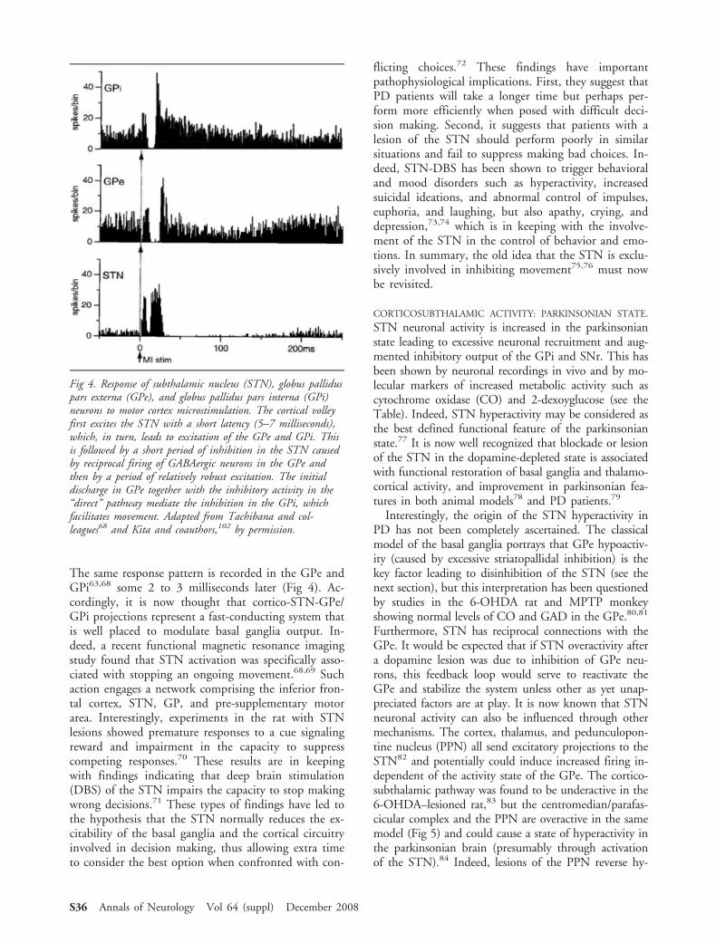
The same response pattern is recorded in the GPe andGPi63,68 some 2 to 3 milliseconds later (Fig 4). Ac-cordingly, it is now thought that cortico-STN-GPe/GPi projections represent a fast-conducting system thatis well placed to modulate basal ganglia output. In-deed, a recent functional magnetic resonance imagingstudy found that STN activation was specifically asso-ciated with stopping an ongoing movement.68,69 Suchaction engages a network comprising the inferior fron-tal cortex, STN, GP, and pre-supplementary motorarea. Interestingly, experiments in the rat with STNlesions showed premature responses to a cue signalingreward and impairment in the capacity to suppresscompeting responses.70 These results are in keepingwith findings indicating that deep brain stimulation(DBS) of the STN impairs the capacity to stop makingwrong decisions.71 These types of findings have led tothe hypothesis that the STN normally reduces the ex-citability of the basal ganglia and the cortical circuitryinvolved in decision making, thus allowing extra timeto consider the best option when confronted with con-
flicting choices.72 These findings have importantpathophysiological implications. First, they suggest thatPD patients will take a longer time but perhaps per-form more efficiently when posed with difficult deci-sion making. Second, it suggests that patients with alesion of the STN should perform poorly in similarsituations and fail to suppress making bad choices. In-deed, STN-DBS has been shown to trigger behavioraland mood disorders such as hyperactivity, increasedsuicidal ideations, and abnormal control of impulses,euphoria, and laughing, but also apathy, crying, anddepression,73,74 which is in keeping with the involve-ment of the STN in the control of behavior and emo-tions. In summary, the old idea that the STN is exclu-sively involved in inhibiting movement75,76 must nowbe revisited.
CORTICOSUBTHALAMIC ACTIVITY: PARKINSONIAN STATE.
STN neuronal activity is increased in the parkinsonianstate leading to excessive neuronal recruitment and aug-mented inhibitory output of the GPi and SNr. This hasbeen shown by neuronal recordings in vivo and by mo-lecular markers of increased metabolic activity such ascytochrome oxidase (CO) and 2-dexoyglucose (see theTable). Indeed, STN hyperactivity may be considered asthe best defined functional feature of the parkinsonianstate.77 It is now well recognized that blockade or lesionof the STN in the dopamine-depleted state is associatedwith functional restoration of basal ganglia and thalamo-cortical activity, and improvement in parkinsonian fea-tures in both animal models78 and PD patients.79
Interestingly, the origin of the STN hyperactivity inPD has not been completely ascertained. The classicalmodel of the basal ganglia portrays that GPe hypoactiv-ity (caused by excessive striatopallidal inhibition) is thekey factor leading to disinhibition of the STN (see thenext section), but this interpretation has been questionedby studies in the 6-OHDA rat and MPTP monkeyshowing normal levels of CO and GAD in the GPe.80,81
Furthermore, STN has reciprocal connections with theGPe. It would be expected that if STN overactivity aftera dopamine lesion was due to inhibition of GPe neu-rons, this feedback loop would serve to reactivate theGPe and stabilize the system unless other as yet unap-preciated factors are at play. It is now known that STNneuronal activity can also be influenced through othermechanisms. The cortex, thalamus, and pedunculopon-tine nucleus (PPN) all send excitatory projections to theSTN82 and potentially could induce increased firing in-dependent of the activity state of the GPe. The cortico-subthalamic pathway was found to be underactive in the6-OHDA–lesioned rat,83 but the centromedian/parafas-cicular complex and the PPN are overactive in the samemodel (Fig 5) and could cause a state of hyperactivity inthe parkinsonian brain (presumably through activationof the STN).84 Indeed, lesions of the PPN reverse hy-
Fig 4. Response of subthalamic nucleus (STN), globus palliduspars externa (GPe), and globus pallidus pars interna (GPi)neurons to motor cortex microstimulation. The cortical volleyfirst excites the STN with a short latency (5–7 milliseconds),which, in turn, leads to excitation of the GPe and GPi. Thisis followed by a short period of inhibition in the STN causedby reciprocal firing of GABAergic neurons in the GPe andthen by a period of relatively robust excitation. The initialdischarge in GPe together with the inhibitory activity in the“direct” pathway mediate the inhibition in the GPi, whichfacilitates movement. Adapted from Tachibana and col-leagues68 and Kita and coauthors,102 by permission.
S36 Annals of Neurology Vol 64 (suppl) December 2008
peractivity in the STN in 6-OHDA–lesioned rodents.85
Dopamine depletion also has a direct effect on STN ac-tivity.84,86 Local interruption of dopamine terminals di-rected to the STN increase its activity, indicating a di-rect modulatory effect of dopamine on the STN.84 Wehave postulated that early denervation of the STN inPD may act as a compensatory mechanism that main-tains the basal ganglia output within normal limits de-spite a progressive loss of striatal dopamine,87 thus po-tentially explaining why clinical features do not emergeuntil there is a 70 to 80% reduction in striatal dopaminelevels.
THALAMOSTRIATAL AND THALAMOSUBTHALAMIC PRO-
JECTIONS IN THE NORMAL AND PARKINSONIAN STATES.
The centromedian-parafascicular (CM-Pf) is an impor-tant source of glutamatergic innervation of the basalganglia, with major projections to the striatum,88,89
STN,90 globus pallidus, and substantia nigra.90 TheCM-Pf forms two “internal” or “transverse” loopswithin the basal ganglia (see Fig 2), which suggests ithas a modulatory effect on the striatum and STN.
In rats with 6-OHDA lesions, Pf neurons that in-nervate the STN and the striatum are hyperactive,91,92
and Pf ablation reverses the increase in STN metabolicactivity associated with the parkinsonian state.93 How-
ever, we found neither antiparkinsonian benefit noramelioration of L-dopa–induced dyskinesias after chem-ical ablation of the CM nucleus in MPTP-treatedmonkeys.94 This casts doubts on the likelihood thatthis intralaminar nucleus plays an important role in thepathophysiology of PD. It is still possible, though, thatthe CM is involved in more complex aspects of move-ment and behavioral control.
Output of the Basal GangliaThe main output of the basal ganglia for movementcontrol of the face and limbs stems from the GPi/SNr, which project to the thalamocortical and brain-stem motor regions. Considerable evidence indicatesthat the excitability and the firing pattern of GPi/SNrneurons play a key role in determining normal func-tion, parkinsonism, and motor complications such asdyskinesia. The excitability and firing pattern of GPi/SNr neurons is influenced and controlled by severalkey projections: (1) the striatopallidal circuit (“direct”pathway), (2) the cortico-STN-GPi/SNr projection,(3) dopaminergic and serotoninergic afferents, and (4)the GPe-GPi/SNr projection. Movement facilitationis associated with a pause or a reduction in neuronalfiring rate in the GPi (or the SNr), which, in turn,facilitates thalamocortical activation.1,3 This has classi-
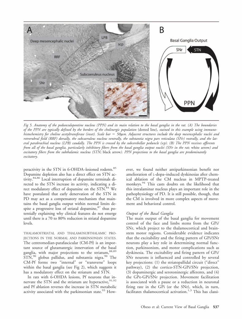
Fig 5. Anatomy of the pedunculopontine nucleus (PPN) and its main relation to the basal ganglia in the rat. (A) The boundariesof the PPN are typically defined by the borders of the cholinergic population (dotted line), stained in this example using immuno-histochemistry for choline acetyltransferase (inset). Scale bar ! 50!m. Adjacent structures include the deep mesencephalic nuclei andretrorubral field (RRF) dorsally, the subcaeruleus nucleus ventrally, the substantia nigra pars reticulata (SNr) rostrally, and the lat-eral parabrachial nucleus (LPB) caudally. The PPN is crossed by the subcerebellar peduncle (scp). (B) The PPN receives afferentsfrom all of the basal ganglia, particularly inhibitory fibers from the basal ganglia output nuclei (SNr in the rat; white arrow) andexcitatory fibers from the subthalamic nucleus (STN; black arrow). PPN projections to the basal ganglia are predominantlyexcitatory.
Obeso et al: Current View of Basal Ganglia S37
cally been interpreted as being mediated by the “direct”pathway (see Fig 1), which caused the inhibition,whereas activation of the “indirect” pathway leads tothalamic inhibition and stops unwanted movements95
(see Figs 1A and 3A). Recording of neuronal activity inmonkeys indeed shows changes in firing rate in GPeand GPi in keeping with this general assumption,96
but also shows that individual neighboring neurons canexpress a decrease, increase, or both in firing frequencydepending on the characteristics of the task.97,98 Thiscan be taken to indicate that activation of cortico-putaminal-GPi activity (“direct” pathway) is not neces-sarily the predominant or the only mechanism involvedin facilitating movement. Rather, facilitation of move-ment is now known to principally involve GPe andSTN connections with the GPi.
GLOBUS PALLIDUS PARS EXTERNA-SUBTHALAMIC NUCLEUS-
GLOBUS PALLIDUS PARS INTERNA-THALAMIC NETWORK:
NORMAL STATE. The GPe, STN, and GPi form ahighly organized network that exhibits a precise ana-tomic correspondence.99 For example, reciprocally in-terconnected areas of the GPe and STN converge onthe same region of the GPi. The GPe projection to theGPi is important and accounts for about half of GPiafferents.100 Importantly, about 40% of GPe neuronsalso project to the striatum where they predominantlysynapse with interneurons.101 The GPe is thereforeperfectly placed to exert a powerful impact on basalganglia information processing (see Fig 2).
In the normal monkey, Kita and coauthors102 showthat 10Hz stimulation of the STN induced strong ex-citation of the GPe and a shorter latency brief excita-tion of the GPi, which is followed by an inhibitoryresponse (see Fig 4). This inhibition is mediated by theGPe (local blockade of GABA afferents onto the GPieliminate it) and predominates over the initial short-latency STN-GPi excitatory response. This impliesthat, after cortical activation, the incoming excitatoryvolley onto the STN predominantly leads to inhibitionof the GPi via activation of the inhibitory GPe disyn-aptic connection with the GPi (see Fig 3B).
Cortical activity associated with movement initiationleads to parallel activation of two afferent loops to thebasal ganglia, that is, the striatum and the STN (seeFig 2). Subsequently, the GPe receives an inhibitoryvolley from GABAergic-enkephalin MSNs in the indi-rect pathway and excitation via STN projections. Bothprojections onto the GPe are disynaptic, and it is likelythat the cortically derived signals arrive close in time.Simultaneously, the GPi receives a disynaptic short-duration excitation via the cortico-STN-GPi projectionand inhibition from the GABAergic-substantiaP-dynorphin “direct” pathway (see Fig 2). Under theseseries of events, the third synaptic component of thecircuit, the GPe-GPi inhibitory synapse, can modulate
GPi neuronal excitability and directly influence its out-put effect on thalamocortical projections. This organi-zation is ideal for assisting in the performance of motoractivities such as repetitive, sequential, and switchingmovements, which require precise spatial and temporalselection of different muscle groups.
The GPe also has connections with the reticular nu-cleus of the thalamus, the SNc, and the PPN.103 It isperhaps also important to appreciate that both the GPeand GPi receive dopaminergic innervation by a nigro-pallidal projection, as well as from collaterals of nigro-striatal axons.103 Indeed, D2-expressing neurons arefound in about 40 to 50% of all pallidal neurons,104
and local administration of dopamine into the GP (inthe rat) modifies the neuronal firing rate.105 Thus, do-pamine exerts a direct physiological effect on pallidalactivity that may be physiologically relevant and rele-vant to the pharmacological effects of dopaminergicdrugs in PD.
GLOBUS PALLIDUS PARS EXTERNA-SUBTHALAMIC NUCLEUS-
GLOBUS PALLIDUS PARS INTERNA-THALAMIC NETWORK:
PARKINSONIAN STATE. The functional state of the GPein the parkinsonian state has been a matter of discus-sion106,107 (see the Table). Studies have reported a lowfiring rate in the GPe of parkinsonian animals,108 nochange,109 or slight increase at the time of onset ofparkinsonian signs.110 More recently, a detailedstudy111 in the MPTP monkey showed that GPe firingis significantly lower than normal and the GPe/GPidischarge rate is well below 1 (the normal ratio). Inaddition, GABA release (measured by microdialysis)was reduced in the STN and GAD (measured by im-munoradiography) was increased in the GPe, in keep-ing with previous findings.105,106 This study110 sup-ports the original model contention that the GPe isfunctionally hypoactive in the parkinsonian state lead-ing to reduced inhibition of the STN and GPi. How-ever, it also demonstrates a dissociation between neu-ronal electrophysiological activity and GAD expressionin the GPe, which had been at the base of previousquestioning of the functional state and role of this nu-cleus in PD.80,87 It is possible that these discrepanciesare related to changes in GPe excitability and activitythroughout the evolution of PD.87 This idea is sup-ported by a recent study of the time course of CO-Iactivity after a unilateral intrastriatal injection of6-OHDA in the rat.112 This approach induces a slowerand less complete SNc degeneration than the classicalapproach where the toxin is injected directly into thenigrostriatal fascicle. Although CO-I activity was ulti-mately increased in all basal ganglia nuclei examined(GP, STN, SNr, and the entopeduncular nucleus !GPi in primates), the temporal pattern varied amongthe different nuclei throughout the evolution of dam-age. In the GP, an immediate decrease in CO-I activity
S38 Annals of Neurology Vol 64 (suppl) December 2008
was observed at day 1, followed by a modest (#10%)increase that peaked (#25%) by the fourth week afterlesion developed. The STN showed a moderate in-crease in CO-I activity at day 1 and remained increasedthroughout the 4 weeks after lesion developed. Theoutput nuclei (SNr and entopeduncular nucleus, EP)exhibited a triphasic metabolic response with a signifi-cant increase at day 1, reduction at 3 and 7 days afterinjection, and a permanent increase after the thirdweek. Basal ganglia output was, therefore, kept withinnormal limits during the initial 2 weeks probablythrough compensatory mechanisms in the GPe-STN-GPi circuitry. It has been shown that GPe activity (orGP in the rat) remains within normal limits during theinitial (presymptomatic) period of nigrostriatal dopa-mine loss113 and maintains basal ganglia output withinnormal limits,87 which likely accounts for why animalsare asymptomatic during this stage. In addition, dopa-mine innervation of the GPi is higher in MPTP mon-keys who exhibit clinical recovery after MPTP,114 andF-dopa positron emission tomography shows increaseduptake in the GPi in the early but not advanced stagesof PD.115 Thus, the basal ganglia network adapts tothe functional effects of dopamine depletion in severalways beyond the nigrostriatal system, and changes arenot concatenated in an orderly manner as was sug-gested by the original pathophysiological model.
In summary, the GPe-STN-GPi network has the ca-pacity to modulate basal ganglia output to a greaterextent than is widely appreciated, and it may compen-sate for the functional consequences of striatal dopa-mine loss and the initial changes in the “indirect” and“direct” pathways. Once the dopaminergic deficit sur-passes the threshold for the available compensatorymechanisms, the GPe-STN-GPi network becomes hy-peractive and loses its normal physiological tuning. Itmay be said that the symptomatic parkinsonian state ischaracterized by a shift in the internal excitability ofthe BG (see Fig 3C). Here, overactivity in the excita-tory STN-GPi pathway predominates and overwhelmsthe inhibitory effects of GPi afferents from the GPe(which is hypoactive) and the “direct” pathway.116
This leads to excessive firing of the GPi in response toafferent signals117 and a decreased signal-to-noise ra-tio,118 thereby impeding the normal facilitation of adesired movement.
BASAL GANGLIA-BRAINSTEM ACTIVITY: NORMAL AND PAR-
KINSON’S DISEASE STATES. The basal ganglia projectsto the brainstem and, therefore, is in a position to in-fluence excitability of several brainstem centers andfunctions. These include the control of saccadic move-ment of the eyes, the blink reflex and startle reaction,up-righting, postural reflexes, and locomotion. Gaitand balance are important sources of disability in PDand are generally not adequately controlled with avail-
able therapies. Among the brainstem centers that arelinked to the basal ganglia, the PPN has attracted par-ticular attention because of its relation to sleep disor-ders and gait dysfunction seen in PD.
The PPN is composed of a heterogeneous popula-tion of neurons with long-range axonal projections thattarget primarily the basal ganglia119 (see Fig 5). ThePPN is implicated in a wide variety of functions, suchas locomotion, inhibition of motor activity duringREM sleep, and possibly attention.120,121 By means ofa mixture of cholinergic, glutamatergic, and GABAer-gic fibers, PPN neurons project preferentially to twomain targets in the basal ganglia, the STN and theSNr/GPi.122,123 PPN also projects to the SNc and theventral tegmental area. In turn, PPN neurons receivedense glutamatergic and GABAergic projections arisingfrom the STN and SNr/GPi, respectively.123,124, 125
A role of the PPN in PD has been suspected sincethe late 1980s. In a postmortem study, Hirsch and col-leagues126 identified a decrease in the number of cho-linergic neurons in the PPN in PD patients. Recently,a reduction in CO-I activity (measured by in situ hy-bridization) of acetylcholine neurons in the PPN ofMPTP monkeys has also been reported,127 but this wasnot associated with cell loss. These findings could beexplained by increased GABAergic input from the SNrand GPi. On the other hand, in the parkinsonian state,the PPN receives an increased glutamatergic input orig-inating from the STN that should drive PPN neuronsto increase their basal firing rate, which, in turn, couldfurther increase STN firing. This dual excitatory con-nection between the PPN and STN could form a pos-itive feed-forward loop that could perpetuate abnormalbasal ganglia hyperactivity (see Fig 5). Theoretically,one would expect that reducing the activity in one ofthem could lead to a decrease in the activity of theothers. Indeed, lesions of the PPN have been reportedto induce parkinsonism in monkeys.128
Low-frequency DBS in the PPN has been recentlyproposed as a novel target for the surgical treatment forPD.129 This is based on the idea that the massive out-flow from the GPi in PD has overinhibited the PPN.Indeed, one study in the MPTP monkey modelshowed improvement in akinesia after local infusion ofa GABA antagonist into the PPN,130 and low stimu-lation of the PPN led to improvement of parkinsonianfeatures in MPTP monkeys.131 A pilot study of DBSof the PPN has shown promising results particularlywith respect to locomotion that could not be obtainedwith stimulation of the STN.132 Recently, it has alsobeen shown that stimulation of the PPN improvesH-reflex excitability in PD patients133 arguing in favorof a direct effect on spinal cord physiology. Despite allthese clues, the functional state of the PPN in the par-kinsonian state is not well defined, and this may provedifficult to define given its heterogeneous cell popula-
Obeso et al: Current View of Basal Ganglia S39
tion and the possibility that its neuronal activity maychange over the course of the disease.
Pathophysiology of Parkinson’s Disease: CurrentConcepts and Therapeutic ConsequencesORIGIN OF CARDINAL FEATURES AND UNEXPLAINED
PROBLEMS. Poverty of spontaneous movements (hy-pokinesia) and slowness of voluntary movement (bra-dykinesia) are fundamental features of the general phe-nomenon of akinesia in PD. The evidence summarizedearlier indicates that dopamine depletion shifts the bal-ance of basal ganglia activity toward the “indirect” cir-cuit where the GPe-STN-GPi microcircuit plays a par-amount role (see Fig 3C).
Increased output from the GPi overinhibits thethalamocortical projection reducing cortical neuronalactivation associated with movement initiation. In ad-dition, the putamen, STN, and GPi show an abnormalaugmented neuronal response to peripheral stimulationwith synchronous firing of neighboring neurons. Thiscould impair the normal selection or filtering of in-coming signals that characterizes normal basal gangliaphysiology (see Fig 3C). Thus, the basal ganglia isshifted toward inhibiting cortically aided movementsby an increased gain of the STN-GPi network and re-duced excitability in the “direct” cortico-putaminal-GPi projection.116,134 Typical features in PD also in-clude an alteration of automatic movements withreduced blinking rate, a positive Meyerson’s sign, de-creased arm swing, and short stride length when walk-ing. All of these are probably mediated by brainstemmechanisms, which are also functionally impaired byexcessive basal ganglia inhibitory outputs. This hasbeen elegantly shown experimentally for the excitabilityof the blink reflex.135 Presumably, a similar mechanismunderlies the impairment of other automatic move-ments in PD. However, the current understanding ofbasal ganglia pathophysiology does not provide an ad-equate explanation for other essential characteristic mo-tor abnormalities in PD.136 These include difficulty inperforming simultaneous and sequential movements, aprogressive reduction in amplitude while performing arepetitive movement, and the striking improvement ingait freezing induced by visual cues. A newer outlookto understand some of these problems has arisen by thestudy of neuronal populations as a complement tosingle-cell recordings.
The several loops connecting the BG internally andalso with the thalamus and brainstem provide amplepossibilities for neuronal synchronization and networkoscillations. Animal studies have shown that DA deple-tion is associated with increased synchronization ofneuronal activity throughout the BG.137,138 The stan-dard application of DBSs of the internal part of theglobus pallidus (GPi) or STN to treat PD currently
allows the recording of local field potentials on a rou-tine basis. There is general agreement that in the “off”medication state, the STN shows a predominant activ-ity in the high alpha-low beta range (11–30Hz), whichis attenuated when parkinsonian signs abate during the“on” medication state.139,140 The latter is also charac-terized by a peak around 60 to 80Hz in the powerspectrum and by an increase in the theta band whenL-dopa–induced dyskinesias are present. Voluntarymovements lead to a significant reduction of the alpha-beta activity starting about 1 second before movementinitiation and continuing until movement is ended.This sequence occurs both in the “off” and “on” states,although the beta band reduction in the “on” state isless overt because activity (ie, energy power) is alreadyreduced. In healthy subjects, movement-relatedchanges in oscillatory activity in the motor cortex aresimilar to those found in the STN.141 All of the abovehave led researchers to suggest that PD patients mayhave greater difficulty than healthy individuals in re-ducing beta activity before movement, which couldprovide a better explanation of several akinetic/brady-kinetic features.
The current basal ganglia model does not provideany explanation for the two other cardinal features ofPD, that is, rigidity and tremor. Regarding the latter, itis now realized that the parkinsonian state is character-ized not only by neuronal hyperactivity in basal gangliaoutput neurons but also by increased neuronal syn-chronization of cell firing leading to oscillations,18
which are particularly evident when tremor is present.Recording neuronal activity in patients with PD142 hasshown a definite correlation between tremor in thelimbs and rhythmical 4 to 6Hz firing in basal ganglianuclei (GPe, GPi, STN), as well as in the ventralis in-termedius nucleus (Vim) of the thalamus.143 Lesion orDBS of the STN, GPi, and Vim are all well known tostop tremor in PD. However, the Vim is a cerebellarreceiving area and is not directly connected with theBG. Studies in organotypic cultures showed that theGPe and STN in the dopamine deficient state have anenhanced tendency to rhythmical and reciprocal firing,causing 4 to 5Hz bursting activity.144 It is thereforetempting to suggest that the complex organization ofbasal ganglia circuits has a tendency to generate oscil-latory activity that produces tremor in PD. It remainsto be elucidated whether mechanisms engaging tremorare primarily mediated and depend on striatal dopa-mine deficiency or are the consequence of impairmentof direct dopaminergic projection to the STN, GPe,and GPi or the thalamus.145 On the other hand, a re-cent study in vervet monkeys shows a low-pass filtercapacity of the motor cortex for stimulation of the GPiat more than 5Hz.146 This led the authors to suggestthat the 4 to 6Hz tremor-related activity seen in PD isnot really driving the motor system, but that tremor in
S40 Annals of Neurology Vol 64 (suppl) December 2008
PD primarily originates outside the basal ganglia (ie,brainstem motor nuclei or spinal cord). This is indeeda challenging proposal. How dopamine deficiency leadsto abnormal oscillatory activity in an extensive motornetwork that involves the basal ganglia, cerebellum,thalamus, and motor cortex is not well understood. Itis equally not well understood why some PD patientshave tremor and some do not, and why lesions in theVim improve tremor but not rigidity and bradykinesia,whereas all of these features are improved with lesionsof the STN or GPi.
Rigidity is essentially an increase in resistance to pas-sive movement. Accordingly, it has been thought fordecades that rigidity should be related to an enhance-ment of stretch reflex excitability. The tonic stretch re-flex, as well as the phasic long-latency stretch reflex, isfacilitated in PD patients.147,148 How the basal gangliachanges associated with dopamine depletion modifythe excitability of stretch reflex mechanisms has not re-ally been explored. It is well known that basal gangliasurgery (ie, pallidotomy, subthalamotomy, and DBS ofeither GPi or STN), as well as thalamotomy involvingthe anteroventral thalamic nucleus (receiving the palli-dal projection) has a marked antirigidity effect. But theprecise mechanism responsible for this effect is notknown. One interesting point is that primary corticalexcitability is enhanced in the parkinsonian state,149
unlike premotor and prefrontal areas, which are hypo-active, and it is conceivable that projections to theSTN from area 4 might have increased reflex gain.This would allow afferent impulses from muscles andjoints during movement to excite the “hyperdirect”cortico-STN-GPi pathway. In keeping with this con-cept, STN and GPi neurons respond readily to musclestretching and vibration (which selectively activates Iaafferent fibers) in the MPTP monkey and PD patients.However, how such increased GPi responses wouldlead to facilitation of the stretch reflex is not apparent.
THERAPEUTIC CONSEQUENCES. In the presymptomaticphase of PD, the functional consequences of a slowdecline in striatal dopamine content are compensatedfor by changes in the remaining nigrostriatal dopamineneurons and by adjustments in the basal ganglia andcortical circuits that keep “motor loop” activity withinnormal limits despite progressive neurodegeneration.150
Eventually, the equilibrium is lost and the cardinal fea-tures of PD begin to emerge. Compensatory mecha-nisms remain active, however, explaining the subtleclinical manifestations that are present in the earlystages of the disease (ie, cardinal features are limited toone body part or hemibody) despite massive striataldopamine depletion. Thus, in the parkinsonian state,the basal ganglia are no longer operating normally, butthe network is still able to function in a relatively stablemanner. This essential feature of basal ganglia func-
tional organization and compensation is totally dis-rupted by standard L-dopa therapy.151 Progressive neu-rodegeneration and loss of nigrostriatal fibers leads toan inability to maintain tonic dopamine receptor stim-ulation, which is the basic mechanism regulating stria-tal neuronal excitability.152 Intermittent doses of stan-dard L-dopa lead to oscillations in plasma L-dopa thatare translated into pathological fluctuations in striataldopamine levels and intermittent dopamine receptoractivation. Physiologically, this discontinuous or pulsa-tile stimulation of striatal dopamine receptors is trans-lated into abnormal firing patterns in basal ganglia out-put neurons that lead to motor complications. Thus,apomorphine or L-dopa given as a bolus can reduce theincreased firing rate of GPi neurons that characterizethe “off” state and improve motor function in a PDpatient, but does not normalize basal ganglia output.17
The “on” state continues to be associated with hypo-activity of the STN and GPi so that the GPe/GPi fir-ing rate ratio (normal, approximately 1) is markedlyincreased153; in addition, there is a marked modifica-tion in the prevailing oscillatory activity, that is, reduc-tion of beta rhythm and increment of theta band withthe onset of dyskinesias.139,140 Furthermore, L-dopa re-duces but does not normalize the percentage of corre-lated pairs of neurons with synchronous firing in theGPi and GPe of MPTP monkeys.153 Thus, intermit-tent doses of standard L-dopa do not normalize thephysiology of the basal ganglia and may, in fact, act asa “destabilizing” stimulus by repeatedly exposing thedenervated basal ganglia, over a few hours, to large,unregulated, extrasynaptic levels of dopamine alternat-ing with a state of severe dopamine deficiency. We be-lieve that such intermittent dopaminergic induces ab-normal “pulsatile” stimulation of striatal dopaminereceptors leading to dysregulation of intracellular sig-nals and plastic changes in MSNs ultimately associatedwith motor complications, particularly dyskinesias.154
This concept suggests that delivery of L-dopa in a morecontinuous manner might be more physiological andreduce the risk for inducing motor complications.154
Indeed, L-dopa infusion is associated with a highly sig-nificant reduction in both “off” time and dyskinesia inadvanced PD patients in comparison with standard in-termittent L-dopa doses.155
A final therapeutic consideration concerns the issueof when to initiate symptomatic treatment in PD. Thecommon medical practice has been to delay pharmaco-logical treatment until warranted by the severity ofsymptoms and signs. In PD, this practice has beenstrongly influenced by experience indicating thatL-dopa treatment is eventually associated with motorcomplications. However, one can argue that early, andadequate, intervention enhances basal ganglia compen-satory mechanisms and contributes to better clinicalcontrol.156 The TEMPO (TVP-1012 in Early Mono-
Obeso et al: Current View of Basal Ganglia S41
therapy for Parkinson’s Disease Outpatients) studydemonstrated that early introduction of the mono-amine oxidase B inhibitor rasagiline provided long-term benefits that could not be equaled by later intro-duction of the same drug.157 Furthermore, theELLDOPA (Earlier versus Later Levadopa Therapy inParkinson Disease) study158 showed that de novo PDpatients treated with L-dopa soon after diagnosis hadsignificantly less deterioration on the Unified Parkin-son’s Disease Rating Scale than did placebo-treated pa-tients, even when examined 2 weeks after stoppingL-dopa therapy. Such observations cannot be explainedby means of L-dopa pharmacokinetics and in all prob-ability are related to changes in basal ganglia circuitryinduced by symptomatic treatment. In fact, the long-duration response to L-dopa in the 6-OHDA rat159 isparalleled by a gradual return of basal ganglia (STN,SNr) activity toward parkinsonian levels. With theavailability of dopaminergic agents that are well toler-ated, have a low adverse effect profile, and are notlikely to induce motor complications, we now advocateintroducing dopaminergic therapy to PD patients assoon as feasible, and possibly even at the time of diag-nosis.
ConclusionsCurrent concepts about the organization of the basalganglia emphasize the existence of several “internal”mechanisms that modulate input/output activity, andsustain normal selection and execution of move-ment.160,161 In the parkinsonian state, this filteringmechanism is deranged and precise neuronal tuning islost. The current model provides a reasonable explana-tion for the origin of akinetic features in PD and theresponse to drugs and surgery.162 However, we are stillin need of a more comprehensive model that accountsfor the other cardinal features such as rigidity andtremor, as well as gait dysfunction, attention and learn-ing deficits, emotional disturbances, and cognitive dis-orders, which are all now realized to form part of theglobal clinical picture of PD. Further understanding ofthe intimate pathophysiological mechanisms underly-ing the different manifestations of PD could poten-tially lead to more refined and more effective treat-ments that more completely restore the physiologicalbalance of the basal ganglia.162 Currently, effort shouldbe made toward using this type of information to in-troduce dopaminergic therapies in as physiological amanner as possible and to avoid where possible the useof nonphysiological discontinuous dopaminergic stim-ulation that provokes newer abnormalities and furtherdestabilizes an already abnormal basal ganglia network.
This article was supported by the Spanish Ministry of Science (J.A.Obeso, M.C. Rodriguez-Oroz, M. Rodriguez, general foundings)
and Education and the UTE (“Union Temporal de Empresas”) atCenter for Applied Medical Research, University of Navarra, Pam-plona, Spain (J.A. Obeso, M.C. Rodriguez-Oroz, M. Rodriguez,general foundings); the NIH (NINDS, J. Mena-Segovia, RO1NS045999); the Lowenstein Foundation (C.W.O., general found-ings); and the Morris and Alma Schapiro Foundation (C.W.O.,general foundings).
References1. Albin RL, Young AB, Penney JB. The functional anatomy of
basal ganglia disorders. Trends Neurosci 1989;12:366–375.2. Crossman AR. Primate models of dyskinesias: the experimen-
tal approach to the study of basal ganglia-related involuntarymovement disorders. Neuroscience 1987;21:1–40.
3. DeLong MR. Primate models of movement disorders of basalganglia origin. Trends Neurosci 1990;13:281–285.
4. Alexander GE, DeLong MR, Strick PL. Parallel organizationof functionally segregated circuits linking basal ganglia andcortex. Annu Rev Neurosci 1986;9:357–381.
5. Alexander GE, Crutcher MD. Functional architecture of basalganglia circuits: neural substrates of parallel processing. TrendsNeurosci 1990;13:266–271.
6. Middleton FA, Strick PL. Basal ganglia and cerebellar loops:motor and cognitive circuits. Brain Res Rev 2000;31:236–250.
7. Buhusi CV, Meck WH. What makes us tick? Functional andneural mechanisms of interval timing. Nat Neurosci Rev2005;6:755–765.
8. Hikosaka O, Nakamura K, Sakai K, et al. Central mechanismsof motor learning. Curr Opin Neurobiol 2002;12:217–222.
9. Yin HH, Knowlton B. The role of the basal ganglia in habitformation. Nat Neurosci Rev 2006;7:464–476.
10. Obeso JA, Guridi J, DeLong MR. Surgery for Parkinson’s dis-ease. J Neurol Neurosurg Psychiatry 1997;62:2–8.
11. Olanow CW. Pallidotomy: have we made a dent in Parkin-son’s disease? Ann Neurol 1996;40:341–343.
12. Bergman H, Wichmann T, DeLong MR. Reversal of experi-mental parkinsonism by lesions of the subthalamic nucleus.Science 1990;249:1436–1438.
13. Aziz TZ, Peggs D, Sambrook MA, et al. Lesions of the sub-thalamic nucleus for the alleviation of 1-methyl-4-phenyl, 1,2, 3, 6-tetrahydropyridine (MPTP)-induced parkinsonism inthe primate. Mov Disord 1991;6:288–292.
14. Guridi J, Herrero MT, Luquin MR, et al. Subthalamotomy inparkinsonian monkeys. Behavioural and biochemical analysis.Brain 1996;119:1717–1727.
15. Obeso JA, Rodrıguez MC, DeLong MD. Basal gangliapathophysiology: a critical review. Adv Neurol 1997;73:3–18.
16. Obeso JA, Rodrıguez-Oroz MC, Rodrıguez M, et al. Patho-physiology of the basal ganglia in Parkinson’s disease. TrendsNeurosci 2000;23(suppl):S8–S19.
17. Obeso JA, Rodriguez-Oroz MC, Rodriguez M, et al. Patho-physiology of levodopa-induced dyskinesias in Parkinson’sdisease: problems with current model. Ann Neurol 2000;47(suppl 1):22–32.
18. Montgomery EB. Basal ganglia physiology and patho-physiology: a reappraisal. Parkinsonism Relat Disord 2007;13:455–465.
19. Wilson CJ. Basal ganglia. In: Sphepherd GM, ed. The synap-tic organization of the brain. Oxford: Oxford University Press,2004:361–414.
20. Bamford NS, Robinson S, Palmiter R, et al. Dopamine mod-ulates release from corticostriatal terminals. J Neurosci 2004;24:9541–9552.
S42 Annals of Neurology Vol 64 (suppl) December 2008
21. Takada M, Tokuno H, Nambu A, et al. Corticostriatal pro-jections from the somatic areas of the frontal cortex in themacaque monkey: segregation versus overlap of input zonesfrom the primary motor cortex, the supplementary motor area,and the promotor cortex. Exp Brain Res 1998;120:114–128.
22. DeLong MR, Crutcher MD, Georgopoulos AP. Primate glo-bus pallidus and subthalamic nucleus functional organization.J Neurophysiol 1985;53:530–543.
23. Miyahi S, Lu X, Imanishi M, et al. Somatotopically arrangedinputs from putamen and subthalamic nucleus to primary mo-tor cortex. Neurosci Res 2006;56:300–308.
24. Lee FJ, Wang YT, Liu F. Direct receptor cross-talk can me-diate the modulation of excitatory and inhibitory neurotrans-mission by dopamine. J Mol Neurosci 2005;26:245–252.
25. Lei W, Jiao Y, Del Mar N, et al. Evidence for differentialcortical input to direct pathway versus indirect pathwaystriatal projection neurons in rats. J Neurosci 2004;24:8289–8299.
26. Onn SP, West AR, Grace AA. Dopamine-mediated regulationof striatal neuronal and network interactions. Trends Neurosci2000;23(suppl):48–56.
27. West AR, Grace AA. Opposite influences of endogenous do-pamine D1 and D2 receptor activation on activity states andelectrophysiological properties of striatal neurons: studies com-bining in vivo intracellular recordings and reverse microdialy-sis. J Neurosci 2002;22:294–304.
28. Hernandez-Lopez S, Bargas J, Sumeier DJ, et al. D1 receptoractivation enhances evoked discharge in neostriatal mediumspiny neurons by modulating an L-type Ca2# conductance.J Neurosci 1997;17:3334–3342.
29. Rodriguez M, Morales I, Gomez I, et al. Heterogeneous do-pamine neurochemistry in the striatum: the fountain-drainmatrix. J Pharmacol Exp Ther 2006;319:31–43
30. McHaffie JG, Stanford TR, Stein BE, et al. Subcortical loopsthrough the basal ganglia. Trends Neurosci 2005;28:401–407.
31. Wang Z, Kai L, Dopaminey M, et al. Dopaminergic controlof corticostriatal long-term synaptic depression in mediumspiny neurons is mediated by cholinergic interneurons. Neu-ron 2006;50:443–452.
32. Morris G, Arkadir D, Nevet A, et al. Coincident but distinctmessages of midbrain dopamine and striatal tonically activeneurons. Neuron 2004;43:133–143.
33. Calabresi P, Centonze D, Bubellini P, et al. Acetylcholine-mediated modulation of striatal funtion. Trends Neurosci2000;23:120–126.
34. Pakhotin P, Bracci E. Cholinergic interneurons control the ex-citatory input to the striatum. J Neurosci 2007;27:391–400.
35. Tepper MJ, Koos T, Wilson CJ. GABAergic microcircuits inthe neostriatum. Trends Neurosci 2004;27:662–669.
36. Bracci E, Centonze D, Bernardi G, Calabresi P. Dopamineexcites fast-spiking interneurons in the striatum. J Neuro-physiol 2002;87:2190–2194.
37. Centonze D, Grande C, Usiello, et al. Receptor subtypes in-volved in the presynaptic and postsynaptic actions ofdopamine on striatal interneurons. J Neurosci 2003;23:6245–6254.
38. Bamford NS, Zhang H, Schmitz Y, et al. Heterosynaptic do-pamine neurotransmission selects sets of corticostriatal termi-nals. Neuron 2004;42:653–663.
39. Cho H, Duke D, Manzino L, et al. Dopamine depletioncauses fragmented clustering of neurons in the sensorimotorstriatum: evidence of lasting reorganization of corticostriatalinput. J Comp Neurol 2002;452:24–37.
40. Mallet N, Ballion B, Le Moine C, Gonon F. Cortical inputsand GABA interneurons imbalance projection neurons in thestriatum of parkinsonian rats. Neurobiol Dis 2006;26:3875–3884.
41. Kreitzer AC, Malenka RC. Endocannabinoid-mediated rescueof striatal LTD and motor deficits in Parkinson’s disease mod-els. Nature 2007;445:643–647.
42. Morissette M, Grondin R, Goulet M, et al. Differential regu-lation of striatal preproenkephalin and preprotachykininmRNA levels in MPTP-lesioned monkeys chronically treatedwith dopamine D1 or D2 receptor agonists. J Neurochem1999;72:682–692.
43. Marin C, Aguilar E, Mengod G, et al. Concomitant short- andlong-duration response to levodopa in the 6-OHDA-lesionedrat: a behavioural and molecular study. Eur J Neurosci 2007;25:259–269.
44. Betarbet R, Greenamyre JT. Regulation of dopamine receptorand neuropeptide expression in the basal ganglia of monkeystreated with MPTP. Exp Neurol 2004;189:393–403.
45. Herrero MT, Augood SJ, Asensi H, et al. Effects of l-dopatherapy on dopamine D-2 receptor mRNA expression in thestriatum of MPTP-intoxicated parkinsonian monkeys. MolBrain Res 1996;42:149–155.
46. Soghomonian JJ, Chesselet MF. Effects of nigrostriatal lesionson the levels of messenger RNAs encoding two isoforms ofglutamate decarboxylase in the globus pallidus and entopedun-cular nucleus of the rat. Synapse 1992;11:124–133.
47. Carta AR, Pinna A, Cauli O, et al. Differential regulation ofGAD67, enkephalin and dynorphin mRNAs by chronic inter-mittent L-Dopa and A2A receptor blockade plus L-Dopa indopamine-denervated rats. Synapse 2002;44:166–174.
48. Katz J, Nielsen KN, Soghomonian JJ. Comparative effectsof acute or chronic administration of levodopa to6-hydroxydopamine-lesioned rats on the expression of glu-tamic acid decarboxylase in the neostriatum and GABA-A re-ceptors subunits in the substantia nigra, pars reticulata. Neu-roscience 2005;132:833–842.
49. Gerfen CR, Engber TM, Mahan LC, et al. D1 and D2 do-pamine receptor-regulated gene expression of striatonigral andstriatopallidopaminel neurons. Science 1990;250:1429–1432.
50. Herrero MT, Augood SJ, Hirsch EC, et al. Effects of L-Dopaon prepronekephalin and preprotachykinin gene expression inthe MPTP-treated monkey striatum. Neuroscience 1995;68:1189–1198.
51. Calabresi P, Maj R, Pisani A, et al. Long-term synaptic de-pression in the striatum: physiological and pharmacologicalcharacterization. J Neurosci 1992;12:4224–4233.
52. Calabresi P, Pisani A, Mercuri NB, Bernardi G. Long-termpotentiation in the striatum is unmasked by removing thevoltage-dependent magnesium block of NMDA receptor chan-nels. Eur J Neurosci 1992;4:929–935.
53. Fino E, Glowinski J, Venance L. Bidirectional activity-dependent plasticity at corticostriatal synapses. J Neurosci2005;25:11279–11287.
54. Picconi B, Centonze D, Hakansson K, et al. Loss of bidirec-tional striatal synaptic plasticity in L-Dopa-induced dyskinesia.Nat Neurosci 2003;6:501–506.
55. Day M, Wang Z, Ding J, et al. Selective elimination of glu-tamatergic synapses on striatopallidopaminel neurons in Par-kinson disease models. Nat Neurosci 2006;9:251–259.
56. Stephens B, Mueller AJ, Shering AF, et al. Evidence of abreakdown of corticostriatal connections in Parkinson’s dis-ease. Neuroscience 2005;132:741–754.
57. Fino E, Glowinski J, Venance L. Effects of acute dopaminedepletion on the electrophysiological properties of striatal neu-rons. Neurosci Res 2007;58:305–316.
58. Pisani A, Bonsi P, Centonze D, et al. Activation of D2-likedopaminergic receptors reduces synaptic inputs to striatal cho-linergic interneurons. J Neurosci 2000;20:RC69.
Obeso et al: Current View of Basal Ganglia S43
59. Raz A, Frechter-Mazar V, Feingold A, et al. Activity of pallidaland striatal tonically active neurons is correlated in mptp-treated monkeys but not in normal monkeys. J Neurosci2001;21:128.
60. Aosaki T, Kimura M, Graybiel AM. Temporal and spatialcharacteristics of tonically active neurons of the primate’s stri-atum. J Neurophysiol 1995;73:1234–1252.
61. Nambu A, Takadopamine M, Inase M, Tokuno H. Dual so-matotopical representations in the primate subthalamicnucleus: evidence for ordered but reversed body-map transfor-mations from the primary motor cortex and the supplemen-tary motor area. J Neurosci 1996;16:2671–2683.
62. Wichmann T, Bergman H, DeLong MR. The primate sub-thalamic nucleus. III. Changes in motor behavior and neuro-nal activity in the internal pallidum induced by subthalamicinactivation in the MPTP model of parkinsonism. J Neuro-physiol 1994;72:521–530.
63. Nambu A, Tokuno H, Hamadopamine I, et al. Excitatorycortical inputs to pallidal neurons via the subthalamic nucleusin the monkey. J Neurophysiol 2000;84:289–300.
64. DeLong MR, Crutcher MD, Georgopoulos AP. Primate glo-bus pallidus and subthalamic nucleus functional organization.J Neurophysiol 1985;53:530–543.
65. Rodriguez-Oroz MC, Rodriguez M, Guridi J, et al. The sub-thalamic nucleus in Parkinson’s disease: somatotopic organiza-tion and physiological characteristics. Brain 2001;124:1777–1790.
66. Paradiso G, Saint-Cyr JA, Lozano AM, et al. Involvement ofthe human subthalamic nucleus in movement preparation.Neurology 2003;61:1538–1545.
67. Alegre M, Alonso-Frech F, Rodriguez-Oroz MC, et al.Movement-related changes in oscillatory activity in the humansubthalamic nucleus: ipsilateral vs. contralateral movements.Eur J Neurosci 2005;22:2315–2324.
68. Tachibana Y, Kita H, Chiken S, et al. Motor cortical controlof internal pallidal activity through glutamatergic andGABAergic inputs in awake monkeys. Eur J Neurosci 2008;27:238–253.
69. Aron AR, Poldrack RA. Cortical and subcortical contributionsto stop signal response inhibition: role of the subthalamic nu-cleus. J Neurosci 2006;26:2424–2433.
70. Baunez C, Dias C, Cador M, Amalric M. The subthalamicnucleus exerts opposite control on cocaine and ‘natural’ re-wards. Nat Neurosci 2005;8:484–489.
71. Frank MJ, Samanta J, Moustafa AA, Sherman SJ. Hold yourhorses: impulsivity, deep brain stimulation, and medication inparkinsonism. Science 2007;318:1309–1312.
72. Frank MJ. Hold your horses: a dynamic computational rolefor the subthalamic nucleus in decision making. Neural Netw2006;19:1120–1136.
73. Funkiewiez A, Ardouin C, Caputo E, et al. Long term effectsof bilateral subthalamic nucleus stimulation on cognitive func-tion, mood, and behaviour in Parkinson’s disease. J NeurolNeurosurg Psychiatry 2004;75:834–839.
74. Mallet L, Schupbach M, N’Diaye K, et al. Stimulation of sub-territories of the subthalamic nucleus reveals its role in theintegration of the emotional and motor aspects of behavior.Proc Natl Acad Sci USA 2007;104:10661–10666.
75. Purdon Martin J, Alcock NS. Hemichorea associated with alesion of the corpus Luysii. Brain 1934;57:504–516.
76. Carpenter MB, Whittier JR, Mettler FA. Analysis of choreoidhyperkinesia in the rhesus monkey. J Comp Neurol 1950;92:293–322.
77. Obeso JA, Rodriguez-Oroz MC, Rodriguez M, et al. Patho-physiology of the basal ganglia in Parkinson’s disease. TrendsNeurosci 2000;23(suppl):8–19.
78. Guridi J, Herrero MT, Luquin MR, et al. Subthalamotomy inparkinsonian monkeys. Behavioural and biochemical analysis.Brain 1996;119:1717–1727.
79. Trost M, Su S, Su P, et al. Network modulation by the sub-thalamic nucleus in the treatment of Parkinson’s disease. Neu-roimage 2006;31:301–307.
80. Levy R, Hazrati LN, Herrero MT, et al. Re-evaluation of thefunctional anatomy of the basal ganglia in normal and parkin-sonian states. Neuroscience 1997;76:335–343.
81. Obeso JA, Rodrıguez-Oroz MC, Rodrıguez M, et al. Currentproblems in understanding the basal ganglia model for Parkin-son$s disease and levodopa-induced dyskinesias. Ann Neurol2000;47(suppl 1):22–32.
82. Hassani OK, Mouroux M, Feger J. Increased subthalamicneuronal activity after nigral dopaminergic lesion independentof disinhibition via the globus pallidus. Neuroscience 1996;72:105–115.
83. Orieux G, Francois C, Feger J, Hirsch EC. Consequences ofdopaminergic denervation on the metabolic activity of the cor-tical neurons projecting to the subthalamic nucleus in the rat.J Neurosci 2002;22:8762–8770.
84. Kreiss DS, Anderson LA, Walters JR. Apomorphine and do-pamine D(1) receptor agonists increase the firing rates of sub-thalamic nucleus neurons. Neuroscience 1996;72:863–876.
85. Breit S, Lessmann L, Unterbrink D, et al. Lesion of the pe-dunculopontine nucleus reverses hyperactivity of the subtha-lamic nucleus and substantia nigra pars reticulata in a6-hydroxydopamine rat model. Eur J Neurosci 2006;24:2275–2282.
86. Smith Y. Striatal and extrastriatal dopamine in the basalganglia: an overview of its anatomical organization in normaland parkinsonian brains. Mov Disord 2008;23:708–715.
87. Obeso JA, Rodriguez-Oroz MC, Lanciego JL, Rodriguez M.How does Parkinson’s disease begin? The role of compensa-tory mechanisms. Trends Neurosci 2004;27:125–127.
88. Sidibe M, Smith Y. Differential synaptic innervation of striat-ofugal neurones projecting to the internal or external segmentsof the globus pallidus by thalamic afferents in the squirrelmonkey. J Comp Neurol 1996;365:445–465.
89. Castle M, Aymerich MS, Sanchez-Escobar C, et al. Thalamicinnervation of the direct and indirect basal ganglia pathwaysin the rat: ipsi-and contralateral projections. J Comp Neurol2005;483:143–153.
90. Tande D, Feger J, Hirsch EC, Francois C. Parafascicular nu-cleus projection to the extrastriatal basal ganglia in monkeys.Neuroreport 2006;17:277–280.
91. Orieux G, Francois C, Feger J, et al. Metabolic activity ofexcitatory parafascicular and pedunculopontine inputs to thesubthalamic nucleus in a rat model of Parkinson’s disease.Neuroscience 2000;97:79–88.
92. Aymerich MS, Barroso-Chinea P, Perez-Manso M, et al. Con-sequences of unilateral nigrostriatal denervation on thethalamostriatal pathway in rats. Eur J Neurosci 2006;23:2099–2108.
93. Bacci JJ, Kachidian P, Kerkerian-Le Goff L, Salin P. In-tralaminar thalamic nuclei lesions: widespread impact on do-pamine denervation-mediated cellular defects in the rat basalganglia. J Neuropathol Exp Neurol 2004;63:20–31.
94. Lanciego JL, Rodriguez-Oroz MC, Blesa FJ, et al. Lesion ofthe centromedian thalamic nucleus in MPTP-treated mon-keys. Mov Disord 2008;23:708–715.
95. Mink JW. The basal ganglia: focused selection and inhibitionof competing motor programs. Prog Neurobiol 1996;50:381–425.
96. Turner RS, Anderson ME. Pallidopaminel discharge related tothe kinematics of reaching movements in two dimensions.J Neurophysiol 1997;77:1051–1074.
S44 Annals of Neurology Vol 64 (suppl) December 2008
97. Matsumura M, Tremblay L, Richard H, Filion M. Activity ofpallidal neurons in the monkey during dyskinesia induced byinjection of bicuculline in the external pallidum. Neuroscience1995;65:59–70.
98. Turner RS, Anderson ME. Context-dependent modulation ofmovement-related discharge in the primate globus pallidus.J Neurosci 2005;25:2965–2976.
99. Smith Y, Bevan MD, Shink E, Bolam JP. Microcircuitry ofthe direct and indirect pathways of the basal ganglia. Neuro-science 1998;86:353–387.
100. Smith Y, Wichmann T, DeLong MR. Synaptic innervation ofneurones in the internal pallidal segment by the subthalamicnucleus and the external pallidum in monkeys. J Comp Neu-rol 1994;343:297–318.
101. Billings LM, Marshall JF. Glutamic acid decarboxylase 67mRNA regulation in two globus pallidus neuron populationsby dopamine and the subthalamic nucleus. J Neurosci 2004;24:3094–3103.
102. Kita H, Tachibana Y, Nambu A, Chiken S. Balance of mono-synaptic excitatory and disynaptic inhibitory responses of theglobus pallidus induced after stimulation of the subthalamicnucleus in the monkey. J Neurosci 2005;25:8611–8619.
103. Parent A, Hazrati LN. Functional anatomy of the basal gan-glia. I. The cortico-basal ganglia-thalamo-cortical loop. BrainRes Brain Res Rev 1995;20:91–127.
104. Floran B, Floran L, Sierra A, Aceves J. D2 receptor-mediatedinhibition of GABA release by endogenous dopamine in therat globus pallidus. Neurosci Lett 1997;237:1–4.
105. Galvan A, Floran B, Erlij D, Aceves J. Intrapallidal dopaminerestores motor deficits induced by 6-hydroxydopamine in therat. J Neural Transm 2001;108:153–166.
106. Delfs JM, Anegawa NJ, Chesselet MF. Glutamate decarboxyl-ase messenger RNA in rat pallidum: comparison of the ef-fects of haloperidol, clozapine and combined haloperidol-scopolamine treatments. Neuroscience 1995;66:67–80.
107. Herrero MT, Levy R, Ruberg M, et al. Consequence of ni-grostriatal denervation and L-dopa therapy on the expressionof glutamic acid decarboxylase messenger RNA in the palli-dum. Neurology 1996;47:219–224.
108. Filion M, Tremblay L. Abnormal spontaneous activity of glo-bus pallidus neurons in monkeys with MPTP-induced parkin-sonism. Brain Res 1991;547:142–151.
109. Benazzouz A, Boraud T, Feger J, et al. Alleviation of experi-mental hemiparkinsonism by high-frequency stimulation ofthe subthalamic nucleus in primates: a comparison withL-Dopa treatment. Mov Disord 1996;11:627–632.
110. Bezard E, Boraud T, Bioulac B, Gross CE. Involvement of thesubthalamic nucleus in glutamatergic compensatory mecha-nisms. Eur J Neurosci 1999;11:2167–2170.
111. Soares J, Kliem MA, Betarbet R, et al. Role of external pallidalsegment in primate parkinsonism: comparison of the effects of1-methyl-4-phenyl-1,2,3,6-tetrahydropyridine-induced parkin-sonism and lesions of the external pallidal segment. J Neurosci2004;24:6417–6426.
112. Blandini F, Levandis G, Bazzini E, et al. Time-course of ni-grostriatal damage, basal ganglia metabolic changes and be-havioural alterations following intrastriatal injection of6-hydroxydopamine in the rat: new clues from and old model.Eur J Neurosci 2007;25:397–405.
113. Hassani OK, Mouroux M, Feger J. Increased subthalamicneuronal activity after nigral dopaminergic lesion independentof disinhibition via the globus pallidus. Neuroscience 1996;72:105–115.
114. Mounayar S, Boulet S, Tande D, et al. A new model to studycompensatory mechanisms in MPTP-treated monkeys exhibit-ing recovery. Brain 2007;130:2898–2914.
115. Whone AL, Moore RY, Piccini PP, Brooks DJ. Plasticity ofthe nigropallidal pathway in Parkinson’s disease. Ann Neurol2003;53:206–213.
116. Rubchinsky LL, Kopell N, Sigvardt KA. Modeling facilitationand inhibition of competing motor programs in basal gangliasubthalamic nucleus-pallidopaminel circuits. Proc Natl AcadSci USA 2003;100:14427–14432.
117. Filion M, Tremblay L, Bedard PJ. Abnormal influences ofpassive limb movement on the activity of globus pallidus neu-rons in parkinsonian monkeys. Brain Res 1988;444:165–176.
118. Boraud T, Bezard E, Bioulac B, Gross CE. Ratio of inhibited-to-activated pallidal neurons decreases dramatically during pas-sive limb movement in the MPTP-treated monkey. J Neuro-physiol 2000;83:1760–1763.
119. Mena-Segovia J, Bolam JP, Magill PJ. Pedunculopontine nu-cleus and basal ganglia: distant relatives or part of the samefamily? Trends Neurosci 2004;27:585–588.
120. Takakusaki K, Saitoh K, Haradopamine H, Kashiwayanagi M.Role of basal ganglia-brainstem pathways in the control ofmotor behaviors. Neurosci Res 2004;50:137–151.
121. Inglis WL, Olmstead MC, Robbins TW. Selective deficits inattentional performance on the 5-choice serial reaction timetask following pedunculopontine tegmental nucleus lesions.Behav Brain Res 2001;123:117–131.
122. Woolf NJ, Butcher LL. Cholinergic systems in the rat brain:III. Projections from the pontomesencephalic tegmentum tothe thalamus, tectum, basal ganglia, and basal forebrain. BrainRes Bull 1986;16:603–637.
123. Bevan MD, Bolam JP. Cholinergic, GABAergic, andglutamate-enriched inputs from the mesopontine tegmentumto the subthalamic nucleus in the rat. J Neurosci 1995;15:7105–7120.
124. Hammond C, Rouzaire-Dubois B, Feger J, et al. Anatomicaland electrophysiological studies on the reciprocal projectionsbetween the subthalamic nucleus and nucleus tegmenti pedun-culopontinus in the rat. Neuroscience 1983;9:41–52.
125. Spann BM, Grofova I. Nigropedunculopontine projection inthe rat: an anterograde tracing study with Phaseolus Vulgaris-leucoagglutinin (PHA-L). J Comp Neurol 1991;311:375–388.
126. Hirsch EC, Graybiel AM, Duyckaerts C, Javoy-Agid F. Neu-ronal loss in the pedunculopontine tegmental nucleus in Par-kinson disease and in progressive supranuclear palsy. Proc NatlAcad Sci USA 1987;84:5976–5980.
127. Gomez-Gallego M, Fernandez-Villalba E, Fernandez-BarreiroA, et al. Changes in the neuronal activity in the pedunculo-pontine nucleus in chronic MPTP-treated primates: an in situhybridization study of cytochrome oxidopaminese subunit I,choline acetyl transferase, and substance P mRNA expression.J Neural Transm 2007;114:319–326.
128. Kojima J, Yamaji Y, Matsumura M, et al. Excitotoxic lesionsof the pedunculopontine tegmental nucleus produce contralat-eral hemiparkinsonism in the monkey. Neurosci Lett 1997;226:111–114.
129. Mazzone P, Lozano A, Stanzione P, et al. Implantation of hu-man pedunculopontine nucleus: a safe and clinically rele-vant target in Parkinson’s disease. Neuroreport 2005;16:1877–1881.
130. Nandi D, Aziz TZ, Giladi N, et al. Reversal of akinesia inexperimental parkinsonism by GABA antagonist microinjec-tions in the pedunculopontine nucleus. Brain 2002;125:2418–2430.
131. Jenkinson N, Nandi D, Miall RC, et al. Pedunculopontinenucleus stimulation improves akinesia in a Parkinsonian mon-key. Neuroreport 2004;15:2621–2624.
132. Stefani A, Lozano AM, Peppe A, et al. Bilateral deep brainstimulation of the pedunculopontine and subthalamic nucleiin severe Parkinson’s disease. Brain 2007;130:1596–1607.
Obeso et al: Current View of Basal Ganglia S45
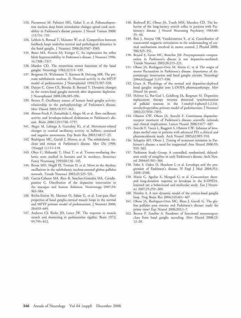
133. Pierantozzi M, Palmieri MG, Galati S, et al. Pedunculopon-tine nucleus deep brain stimulation changes spinal cord excit-ability in Parkinson’s disease patients. J Neural Transm 2008;115:731–735.
134. Leblois A, Boraud T, Meissner W, et al. Competition betweenfeedback loops underlies normal and pathological dynamics inthe basal ganglia. J Neurosci 2006;26:3567–3583.
135. Basso MA, Powers AS, Evinger C. An explanation for reflexblink hyperexcitability in Parkinson’s disease. J Neurosci 1996;16:7308–7317.
136. Marden CD. The mysterious motor functions of the basalganglia. Neurology 1982;32:514–539.
137. Bergman H, Wichmann T, Karmon B, DeLong MR. The pri-mate subthalamic nucleus. II. Neuronal activity in the MPTPmodel of parkinsonism. J Neurophysiol 1994;72:507–520.
138. Dejean C, Gross CE, Bioulac B, Boraud T. Dynamic changesin the cortex-basal ganglia network after dopamine depletion.J Neurophysiol 2008;100:385–396.
139. Brown P. Oscillatory nature of human basal ganglia activity:relationship to the pathophysiology of Parkinson’s disease.Mov Disord 2003;18:357–363.
140. Alonso-Frech F, Zamarbide I, Alegre M, et al. Slow oscillatoryactivity and levodopa-induced dyskinesias in Parkinson’s dis-ease. Brain 2006;129:1748–1757.
141. Alegre M, Labarga A, Gurtubay IG, et al. Movement-relatedchanges in cortical oscillatory activity in ballistic, sustainedand negative movements. Exp Brain Res 2003;148:17–25.
142. Rodrıguez MC, Guridi J, Mewes, et al. The subthalamic nu-cleus and tremor in Parkinson’s disease. Mov Dis 1998;13(suppl 1):111–118.
143. Ohye C, Shibazaki T, Hirai T, et al. Tremor-mediating tha-lamic zone studied in humans and in monkeys. StereotactFunct Neurosurg 1993;60:136–145.
144. Bevan MD, Magill PJ, Terman D, et al. Move to the rhythm:oscillations in the subthalamic nucleus-external globus pallidusnetwork. Trends Neurosci 2002;25:525–531.
145. Garcia-Cabezas MA, Rico B, Sanchez-Gonzalez MA, Cavado-pamine C. Distribution of the dopamine innervation inthe macaque and human thalamus. Neuroimage 2007;34:965–984.
146. Rivlin-Etzion M, Marmor O, Saban G, et al. Low-pass filterproperties of basal ganglia cortical muscle loops in the normaland MPTP primate model of parkinsonism. J Neurosci 2008;28:633–649.
147. Andrews CJ, Burke JD, Lance JW. The response to musclestretch and shortening in parkinsonian rigidity. Brain 1972;95:795–812.
148. Rothwell JC, Obeso JA, Traub MM, Marsden CD. The be-havior of the long-latency stretch reflex in patients with Par-kinson’s disease. J Neurol Neurosurg Psychiatry 1983;46:35–44.
149. Reis J, Swayne OB, Vandermeeren Y, et al. Contribution oftranscranial magnetic stimulation to the understanding of cor-tical mechanisms involved in motor control. J Physiol 2008;586:325–351.
150. Bezard E, Gross MC, Brotchie JM. Presymptomatic compen-sation in Parkinson’s disease is not dopamine-mediated.Trends Neurosci 2003;26:215–221.
151. Obeso JA, Rodriguez-Oroz M, Marin C, et al The origin ofmotor fluctuations in Parkinson’s disease: importance of do-paminergic innervation and basal ganglia circuits. Neurology2004;62(suppl 1):S17–S30.
152. Grace A. Physiology of the normal and dopamine-depletedbasal ganglia: insights into L-DOPA pharmacotherapy. MovDisord (in press).
153. Heimer G, Bar-Gad I, Goldberg JA, Bergman H. Dopaminereplacement therapy reverses abnormal synchronizationof pallidal neurons in the 1-methyl-4-phenyl-1,2,3,6-tetrahydropyridine primate model of parkinsonism. J Neurosci2002;22:7850–7855.
154. Olanow CW, Obeso JA, Stocchi F. Continuous dopamine-receptor treatment of Parkinson’s disease: scientific rationaleand clinical implications. Lancet Neurol 2006;5:677–687.
155. Stocchi F, Vacca L, Ruggieri S, Olanow CW. Infusion of levo-dopa methyl ester in patients with advanced PD: a clinical andpharmacokinetic study. Arch Neurol 2005;62:905–910.
156. Schapira AH, Obeso J. Timing of treatment initiation in Par-kinson’s disease: a need for reappraisal? Ann Neurol 2006;59:559–562.
157. Parkinson Study Group. A controlled, randomized, delayed-start study of rasagiline in early Parkinson’s disease. Arch Neu-rol 2004;61:561–566.
158. Fahn S, Oakes D, Shoulson I, et al. Levodopa and the pro-gression of Parkinson’s disease. N Engl J Med 2004;351:2498–2508.
159. Marin C, Aguilar E, Mengod G, et al. Concomitant short-and long-duration response to levodopa in the 6-OHDA-lesioned rat: a behavioural and molecular study. Eur J Neuro-sci 2007;25:259–269.
160. Nambu A. A new dynamic model of the cortico-basal ganglialoop. Prog Brain Res 2004;143:461–467
161. Obeso JA, Rodriguez-Oroz MC, Blesa J, Guridi G. The glo-bus pallidus pars externa and Parkinson’s disease: ready forprime time? Exp Neurol 2006;202:1–7.
162. Brown P, Eusebio A. Paradoxes of functional neurosurgery:clues from basal ganglia recording. Mov Disord 2008;23:12–20.
S46 Annals of Neurology Vol 64 (suppl) December 2008