Embed Size (px)
Citation preview

INTRODUCTION
The differentiation of a neuroendocrine phenotype involves theselection and production of a specific peptide hormone. Forboth endocrine and neuroendocrine cells, recent geneticanalyses in mice have revealed transcriptional hierarchiescontrolling such developmental events. The process typicallyinvolves multiple stages of gene activation and extinction, andit represents the actions of multiple regulatory cascades. Forexample, the proliferation and specification of hypothalamicneurosecretory neurons that produce vasopressin, oxytocinand corticotrophin-releasing factor are controlled by earlyexpression of Orthopedia(Acampora et al., 1999) and Sim1(Michaud et al., 1998). These effects are mediated largely byinduction or maintenance of a secondary factor, Brn2, whichdirectly regulates neuropeptide gene expression (Schonemannet al., 1995; Nakai et al., 1995). Likewise, in pituitary and inpancreatic endocrine cells, tissue-specific (e.g. Ptx1/2) andsecondary cell type-specific transcription factors (e.g.neurogenin3) promote patterned expression of peptidehormones (Sheng and Westphal, 1999).
Neuroendocrine cell differentiation also involves theintegrated assembly of cellular machinery needed to producelarge amounts of secretory peptides. Such mechanismscoordinate several events associated with the regulatedsecretory pathway (Arvan and Castle, 1998): the ability to
synthesize, process, sort, traffic and accumulate dense-coresecretory granules and their contents. Neurons differ greatlyand reproducibly in the amount of secretory peptides that theyproduce, and in their elaboration of the secretory pathway. Forexample, mammalian motoneurons produce low levels ofneuropeptides such CGRP or galanin (Streit et al., 1989),and at the ultrastructural level, their terminals contain manysmall, clear vesicles, but very few large, dense-core (peptide-containing) granules (Hall and Sanes, 1993). By contrast,hypothalamic neurosecretory neurons produce large amountsof vasopressin, oxytocin, or corticotrophin-releasing factor,and they contain correspondingly large numbers of dense-coresecretory granules (Burbach et al., 2001). Cells also transientlymodify their levels of secretory activity following injury (e.g.Blake-Bruzzini et al., 1997) or stimulation (e.g. Herman et al.,1991). The mechanisms underlying these differences in levelsof secretory activity are unknown.
Because the amplified expression of the secretory pathwayis a stable and cell-specific feature of neuroendocrine cells, wehypothesize the existence of genetic factors that control thisphenotype. Identifying such factors will facilitate a detailed,mechanistic analysis of neuroendocrine cell organization andphysiology. Such an analysis will be crucial to a generalunderstanding of neuroendocrine cell biology and will supportefforts to produce a program of neuroendocrine differentiationfrom stem cells in vitro. We describe a Drosophila bHLH gene,
1771Development 130, 1771-1781 © 2003 The Company of Biologists Ltddoi:10.1242/dev.00404
Neuroendocrine cells are specialized to produce, maintainand release large stores of secretory peptides. We show thatthe Drosophila dimmed/Mist1bHLH gene confers such apro-secretory phenotype on neuroendocrine cells. dimmedis expressed selectively in central and peripheralneuroendocrine cells. In dimmedmutants, these cellssurvive, and adopt normal cell fates and morphology.However, they display greatly diminished levels of secretorypeptide mRNAs, and of diverse peptides and proteinsdestined for regulated secretion. Secretory peptide levelsare lowered even in the presence of artificially highsecretory peptide mRNA levels. In addition, overexpression
of dimmed in a wild-type background produces acomplimentary phenotype: an increase in secretory peptidelevels by neuroendocrine cells, and an increase in thenumber of cells displaying a neuroendocrine phenotype.We propose that dimmedencodes an integral component ofa novel mechanism by which diverse neuroendocrinelineages differentiate and maintain the pro-secretory state.
Supplemental data available online
Key words: Neuroendocrine, Drosophila, bHLH, Neuropeptide,Differentiation, Regulated secretion
SUMMARY
The bHLH protein Dimmed controls neuroendocrine cell differentiation in
Drosophila
Randall S. Hewes 1,2,*, Dongkook Park 1, Sebastien A. Gauthier 2, Anneliese M. Schaefer 1 andPaul H. Taghert 1,†
1Department of Anatomy and Neurobiology, Washington University School of Medicine, Saint Louis, MO 63110, USA2Department of Zoology, University of Oklahoma, Norman, OK 73019, USA*Present address: Department of Zoology, University of Oklahoma, 730 Van Vleet Oval, Norman, OK 73019, USA†Author for correspondence (e-mail: [email protected])
Accepted 9 January 2003
1772
dimmed (dimm), with an expression pattern that correspondsprecisely to the neuronal and endocrine cells that accumulatelarge amounts of secretory peptides. We present both loss-of-function and gain-of-function analyses to argue that dimmconfers a pro-secretory phenotype within these diverse cells,and that its actions appear confined to that aspect of cellulardifferentiation. Thus, we propose a novel and generalmechanism, of which dimm is an essential component, for theamplification of the regulated secretory pathway by dedicatedsecretory cells.
MATERIALS AND METHODS
StrainsFlies were cultured at 22-25°C on a standard cornmeal-yeast-agarmedium. The molecular and genetic characterization of c929,R6 andRev8 has been described previously (Hewes et al., 2000).Rev4 andRev18 are X-ray revertants of P{PZ}l(2)k0510606311(M. Horner andC. Thummel, personal communication).y– w– revertants ofKG02598(e.g.dimmS2a) were obtained by transposase-mediated excision, andeach line was characterized by PCR with primers flanking the originalinsertion site. Except as noted, all other strains are describedelsewhere (Lindsley and Zimm, 1992; FlyBase, 1999) and wereobtained from the Bloomington stock center, the BDGP GeneDisruption Project and other sources.
Scoring of dimm larvaeEggs were collected on apple juice-agar plates supplemented withyeast paste. Larvae were collected from the plates, and heterozygotes(y* w* and balanced over CyO-y+) were distinguished by mouthpartcolor. After scoring, size-matched pairs of y– and y+ larvae weredissected and stained in parallel.
ImmunostainingImmunostaining was performed as described previously (Benvenisteet al., 1998). Briefly, tissues were fixed in 4% paraformaldehyde(PFA), Bouin’s, or 4% paraformaldehyde/7% picric acid (PFA-PA).Polyclonal or monoclonal primary antisera were used (overnight at4°C) to detect the following proteins: β-gal (1:1000, PFA-PA;Promega); PHM (1:750 pre-absorbed to PHM–/– larvae, Bouin’s)(Jiang et al., 2000); -RFa (‘PT2’)(1:2000, PFA-PA) (Taghert, 1999);FMRF (1:2000, PFA-PA) (Chin et al., 1990); corazonin (1:500, TexasRed-conjugated; PFA-PA) (Veenstra, 1994); LK (1:500, PFA-PA)(Nässel and Lundquist, 1991); CCAP (1:400, PFA-PA) (Ewer andTruman, 1996); PDH and PAP (each 1:2000, PFA-PA) (Renn et al.,1999); MM (1:800, PFA-PA) (O’Brien and Taghert, 1998); dopadecarboxylase (affinity purified 1:100, PFA) (Scholnick et al., 1991);Furin-1 (1:1000, Bouin’s) (Jiang et al., 2000); and Myc (1:500, PFA-PA; a gift from Y.-N. Jan; Sigma). Secondaries used were goatCy3, FITC, Texas Red or ALEXA 488 conjugates (JacksonImmunoResearch) at a 1:500 dilution. Confocal z-series projectionswere obtained using an Olympus Fluoview microscope.
RACEA cDNA library was made from RNA of y w adult heads usingcommercial reagents (Clontech). 5′RACE was performed accordingto manufacturer’s recommendations using CG8667-specific primers.
dimm RNAiRNAi was performed based on the methods of Kennerdell andCarthew (Kennerdell and Carthew, 1998) and Clemens et al. (Clemenset al., 2000). The template for RNA synthesis was generated by PCR,using primers containing a T7 promoter sequence (5′-GAATTA-ATACGACTCACTATAGGGAGA-3′) at the 5′ends and P1 DNA(DS00532) as the PCR template. The gene specific primers 5′-
CAGATTCCAGTTCGCAAAGCGAT-3′ and 5′-GGGCTCGTCG-AAATTATCATTGATA-3′ amplified a 951 bp segment of openreading frame in exon 3, including the entire bHLH domain.Transcription and analysis of the double-stranded RNA (dsRNA) wereperformed as described (Clemens et al., 2000). dsRNA (3 µM) wasinjected into syncytial blastoderm embryos (Canton-S) ~75% alongthe anteroposterior axis. For the mock controls, all steps wereperformed in parallel, except that the P1 DNA was omitted from thePCR reaction. Mock- and RNA-injected larvae were dissected duringor within 6 hours after hatching.
UAS-dimm transgeneThe predicted coding region of CG8667was amplified by PCR usingcDNA generated from y wadult head RNA (Clontech). The primers5′-CAGATCTCGACGATTTTTGTTCAGCCAT-3′(5′ UTR) and 5′-TGCGGCCGCAGAAACTCTCGAAAGGGCT-3′ (end of the ORF)were used to construct a 1236 bp fragment that was cloned intopBSK+and then transferred to pP{UAST-Myc}at the BglII and NotIsites. Transgenic lines containing P{UAS-dimm::Myc}insertions werecreated using standard techniques (Benveniste and Taghert, 1999).UAS-dimm::Myc2-A-3, Rev8/CyO, Act-GFPflies were crossed to c127-Gal4, UAS-GFP; Rev4/CyO, Act-GFP, and first instar larvae werescored for Act-GFP- and c127-specific GFP labeling patterns.
mRNA in situ hybridizationWhole-mount in situ hybridization (Tautz and Pfeifle, 1989) wasperformed using single-stranded, digoxigenin-labeled RNA or DNAprobes (Patel, 1996) prepared from P1 or cDNA templates.
Staining quantificationCells were imaged on a Zeiss Axioplan fitted with a SPOT CCDcamera and software (Diagnostic Instruments) or in confocal z-seriesscans. Exposure settings were adjusted to optimize detection withoutsaturating the signal. For a given neuron, identical settings were usedfor all preparations and genotypes. Mean pixel luminosity for the areacovering the soma (S) was measured for each neuron using AdobePhotoshop. An adjacent area was sampled to measure the backgroundsignal (B). The intensity index=(S–B)/B. Cells not visible were scored0. Cells that were obscured or lost due to tissue damage were notanalyzed. Brightfield images were inverted before quantification. CNSsize was measured as an additional control – in each case, mean brainlobe diameters were not significantly different between genotypes(data not shown). Statistics were performed using the NCSS-2000Statistical Analysis System or StatView (MANOVAs; Games-Howell). Variances are reported as ±s.e.m.
RESULTS
c929 is broadly expressed in peptidergic cellsThe c929 P-element insertion was isolated in a P{Gal4}enhancer detection screen for genes expressed in the Tvneuroendocrine neurons (O’Brien and Taghert, 1998). Inaddition to the Tv neurons, c929 drove reporter geneexpression (GFP or β-galactosidase) in ~200 neurons scatteredthroughout the larval CNS and in neuroendocrine projectionsto the ring gland, the dorsal neurohemal organs and thetransverse nerves (Fig. 1A). Outside the CNS, this patternincluded at least three classes of endocrine cells: intrinsic cellsof the corpora cardiaca (Fig. 1A), 10-20 midgut cells (data notshown) and the peritracheal myomodulin-immunoreactivecells. The latter appear homologous to the endocrine Inka cells(O’Brien and Taghert, 1998). c929 reporter expression alsoappeared in several other tissues, including peptidergic PNSneurons (LBD neurons; D. Allan and S. Thor, personal
R. S. Hewes and others

1773Neuroendocrine cell differentiation
communication), fat body, epithelial cells and salivaryglands (data not shown).
To determine whether c929-positive neurons expressneuropeptides, we performed double-label experimentsfor the c929reporter and for the peptide biosyntheticenzyme, peptidylglycine-α-hydroxylating mono-oxygenase (PHM). In Drosophila, PHM is a marker formost peptidergic cells. It is required for neuropeptideamidation (Jiang et al., 2000), which is a highly specificmodification of secretory peptides (Eipper et al., 1993);greater than 90% of all known or predicted Drosophilapeptide transmitters are amidated (Hewes and Taghert,2001). Most if not all c929-positive CNS neurons wereimmunostained very strongly by PHM antibodies (n=8specimens; Fig. 1B,E).
Conversely, most neurons displaying strong PHMimmunostaining were also c929positive, while mostweakly PHM-positive neurons were not c929positive(data not shown). In addition, PHM was expressed inall three c929-positive endocrine cell types and in theLBD peripheral neurons (O’Brien and Taghert,1998) (data not shown). Thus, in the larval CNSand in several peripheral tissues, c929primarilylabels neuroendocrine cells as its expressionwas highly correlated with the production oflarge amounts of amidating enzyme, amidatedneuropeptides and peptide hormones.
To assess the degree of heterogeneity amongc929-positive cells, we compared the expressionpattern of c929 with a variety of otherpeptidergic cell markers. This population of cellswas chemically diverse. For example, sevenbilateral pairs of c929-positive neurons weredouble-labeled with the PT2 antiserum (Fig.1C,E). PT2 is a marker for -RFamide containingneuropeptides, which include the products of atleast three Drosophilagenes (Taghert, 1999).Additional subsets of c929-positive neuronswere immunostained with antisera directedagainst a variety of neuropeptides. Theseincluded the Drosophila FMRF propeptide (n=8specimens), cockroach corazonin (n=5), cricketleucokinin-1 (LK), crustacean cardioactivepeptide (CCAP; n=4), crustacean beta-PDH(n=4) and Aplysiamyomodulin (MM; Fig.1D,E). Finally, a distinct subset of 34c929-positive neurons (see below) wasimmunopositive for an additional, putativeDrosophilapeptide biosynthetic enzyme (n=10;P.H.T. and M. Han, unpublished) Furin 1 (De Bieet al., 1995). Based on their positions, cellularmorphologies, and immunostaining with theabove markers, the cells within the c929patternrepresent more than 26 distinct classes ofpeptidergic neurons and endocrine cells. Noc929-positive neurons were stained with anantiserum to dopa decarboxylase (n=8), anenzyme required for synthesis of the biogenicamines, serotonin and dopamine (Hirsh, 1989).
No single transmitter system we tested wasentirely c929positive. For example, among the
Fig. 1.The c929reporter gene is expressed in ~200 peptidergic CNS neurons.(A) Confocal z-series of c929reporter (c929-Gal4, P{UAS-lacZ}Bg4-1-2) expressionin the larval CNS. Expression is detected in ~200 neurons, in neuroendocrine releasesites (asterisks) and in intrinsic cells of the endocrine corpora cardiaca (CC) (n>50).(B,B′) Double-labeling for c929reporter (B) and the peptide biosynthetic enzyme,PHM (B′). Yellow cells in the overlay (B″) were positive for both markers (n=8).(C) Double-labeling (C″) for c929reporter (C; P{UAS-GFP}D1) (Yeh et al., 1995)and neuropeptides ending in the epitope, RF-amide (PT2 antiserum; αRFa, C′)(n=10). (D) Double-labeling (D″) for c929reporter (D) and neuropeptides related toMM (D′ ) (n=10). (E) Schematic illustration of the expression patterns for the c929reporter, the PHM enzyme, and MM- or RFa-immunoreactive neuropeptides.Although generally c929negative at this stage (mid-third larval instar), the MP1 andVA neurons express the c929reporter gene for one or more brief periods duringdevelopment. Scale bars: 50 µm.

1774
17 Fmrf cell types (Benveniste and Taghert, 1999), only the Tvneurons were c929-positive. However, in third instar larvaethere were somec929-negative neurons, such as thepeptidergic MP1s and VAs, which displayed weak and/ortransient c929 reporter expression during other stages ofdevelopment (e.g. Fig. 5G). Thus, our identification of c929-positive peptidergic neurons is likely to be an underestimate ofthe total population of peptidergic cells that express thereporter gene.
dimm is required for maintenance of neurosecretoryprotein levelsTo test for roles of a putative ‘c929’ gene in the developmentand/or function of peptidergic cells, we generated deletionsflanking the c929 insertion site (Fig. 2A). These deletionscaused recessive lethality, owing to disruption of at least oneessential gene, cryptocephal(crc) (Hewes et al., 2000).However, many homozygous mutant animals survived intothe larval stages, when we could examine the fates of CNSpeptidergic neurons.
By immunostaining the mutant animals for PHM, wedetected a novel phenotype: R6/Rev8 trans-heterozygousanimals contain small deficiencies around the c929insertionsite that are ~12 and ~ 35 kb respectively (Fig. 2A). Trans-heterozygous larvae displayed marked reductions in PHM
protein levels in all strongly c929-positive CNS neurons (Fig.2B). c929-negative neurons were unaffected in R6/Rev8 larvae,and weakly or transiently c929-positive neurons, such as theVAs, showed smaller reductions in PHM immunostaining (Fig.2B). The mutant phenotype was detectable at the time of larvalhatching and throughout all larval stages. By contrast,heterozygous R6 or Rev8/+ larvae were essentially wild type,although these alleles displayed mild haploinsufficiency (n=24;data not shown) with other markers (see below). These resultsdemonstrate a requirement for ~10 kb of DNA flanking thec929 insertion site for the normal expression and/ormaintenance of PHM in c929-positive CNS neurons. Wenamed the affected gene dimmedto reflect the diminishedstaining.
We used six additional neurosecretory markers in dimmmutant larvae, and found that all six displayed moderateto severe reductions in immunostaining in spatial patternscorresponding to the c929reporter pattern. The affected proteinsincluded several known or presumed neuropeptides – MM (Fig.2C), LK (n>25), the FMRF propeptide (n>12) and several PT2positive neuropeptides (n>50) – and the putative neuropeptidebiosynthetic enzyme Furin 1 (see below). All c929-positiveneurons displayed the mutant phenotype for at least one marker,PHM (Fig. 2B); many showed reduced immunostaining withmultiple markers. For example, the Tv neurons had reduced
R. S. Hewes and others
Fig. 2.dimmis required forexpression of normal levels ofpeptide transmitters. (A) The39C4-D1 region ofchromosome 2L showing thelocations of the c929 andKG02598 (‘2598’)P elementinsertions (triangles), theCG18362, cryptocephal(crc)and dimm(CG8667,Mistr)genes, and local deletions(wavy lines; bars indicatebreakpoint uncertainties).TW1(not shown) deletes38A7-B1 to 39C3, ending ~17kb distal to the c929insertion(Hewes et al., 2000). Rev18deletes chromosomal bands39A3-7 to 39D3-5 (A.Carpenter, personalcommunication). Rev4deletes39C1-4 (Hewes et al., 2000)and is a molecular null for thedimmgene (D. Eberl, personalcommunication; data notshown). Introns smaller than70 bp are not shown. Asegment of DNA extending toward the telomere (wild type) was deleted without affecting neuropeptide levels in the CNS. A second region(dimm5′) was required for the maintenance of normal neuropeptide levels and probably contains dimm5′ enhancer elements. (B) The dimmmutant phenotype for a peptide biosynthetic enzyme, PHM. Reduction in PHM immunostaining in the CNS of a third instar dimm–/– larva (B′),compared with a dimm+/– sibling control (B) (n=28).(C) The dimmmutant phenotype for neuropeptides related to MM. Reduction in MMimmunostaining in the CNS of a first instar dimm–/– larva (C′), compared with a dimm+/– sibling control (C) (n>50). (D) Phenocopy of thedimm–/– phenotype (MM immunostaining) by injection of double-stranded CG8667RNA into wild-type embryos (D′), compared with a mock(saline-injected) control (D) (n=7). Asterisk, weak signal in midline S2 and/or S1a neurons. (E,E′) The dimmmutant phenotype in theendocrine Inka cells. Anti-MM immunostaining of an Inka cell in a third instar dimm–/+ larva Inka cell immunostaining [E; compare withO’Brien and Taghert (O’Brien and Taghert, 1998)] was no longer detectable in a dimm–/– larva (E′). The apparent increase in background in E′is due to the incidence of air pockets in the tracheae of this specimen. The precise location of the Inka cell is variable and not certain in (E′);however, it is normally within this image field. Scale bars: 50 µm.

1775Neuroendocrine cell differentiation
levels of PHM, the FMRF propeptide, –RFamide peptides andFurin 1 (Fig. 2B, see Fig. 3B, see Fig. 6B). Thus, in a large anddiverse population of CNS peptidergic neurons, dimmregulateslevels of a broad array of secretory proteins.
As the three classes of c929-positive endocrine cells alsolikely secrete peptide hormones, we also tested them for effectsof the dimmmutations. The ring gland (n=15) and trachealendocrine cells (n>50) displayed severe reductions in peptideimmunostaining for PHM and/or MM in dimm–/– mutants (Fig.2E; data not shown); the gut endocrine cells were not tested.Taken together, these results suggest a crucial role for dimmincontrolling bioactive peptide levels in diverse neuronal andendocrine secretory cells.
dimm encodes a basic helix-loop-helix proteinUsing chromosomal deletions, we genetically mapped thedimm gene. We performed peptide immunostaining on Rev8homozygotes (n=15) and on hemizygotes (n>50) bearing onecopy of R6 (or Rev8) over one of several independently deriveddeficiencies of the entire 39C4-D1 region of chromosome2L (e.g. Rev4). In each case, the effects on peptideimmunostaining were comparable, although the reduction inMM staining in larvae homozygous for Rev4, a null allele(Fig. 2A), was more pronounced than in R6/Rev8 trans-heterozygotes (n=12; data not shown). Thus, R6and Rev8arehypomorphic alleles. Normal peptide immunostaining wasrestored in male Rev8 homozygotes (n=6) bearing a duplicationof chromosome bands 35A-40 [Tp(2;Y)J54], consistent withthe location of dimmin 39C4-D1.
In contrast to R6/Rev8mutants, larvae with disruptions inthe crc gene (c929 homozygotes, n=6; crc1/R6 trans-heterozygotes, n>40; R2 homozygotes, n=5), or deletions ofDNA extending up to 200-300 kb towards the telomere(TW1/Rev18trans-heterozygotes, n=7) displayed wild-typeneuropeptide levels (see Table S1 at http://dev.biologists.org/supplemental/). Thus, dimm is not crc, nor is it any other genelocated distal to the site of the c929 insertion.
The closest gene proximal to c929is CG8667(Mistr), foundwithin 25 kb (Fig. 2A). It encodes a basic helix-loop-helix(bHLH) protein that is a member of the Atonal subfamily oftranscription factors (Moore et al., 2000). Its bHLH domaindisplays 79% identity with the mouse Mist1 protein (Pin etal., 1999). In Rev8 homozygous embryos, CG8667 mRNAexpression was markedly reduced, but not eliminated (n>50;data not shown), consistent with the identification of Rev8asa hypomorphic dimm allele. After 5′RACE identification ofthe 5′ end of CG8667, we identified a P-element insertion(dimmKG02598) located 111 bp upstream (Fig. 2A).dimmKG02598 displays homozygous lethality (see Table S2 athttp://dev.biologists.org/supplemental/), and represents asevere hypomorphic dimmallele, becauseCG8667 mRNAexpression appeared low or undetectable in dimmKG02598
homozygous mutant embryos (Fig. 3A). HatchlingdimmKG02598/Rev4 larvae displayed reduced immunostainingfor PT2-positive neuropeptides (n>15; Fig. 3B). Normal PT2immunostaining was restored (n>15; Fig. 3B) after preciseexcision of the dimmKG02598 P element. Consistent with theconclusion that dimmand crcare separate genes,KG02598was
Fig. 3.The KG02598 P-element insertion reduces CG8667expression and is a strong dimmmutant allele. (A) Photomontages ofCG8667insitu hybridization in homozygous stage 16 and 17 embryonic CNS: normal staining levels were observed in 7/15 specimens from the stockdimmKG02598/CyO-y+ (left); reduced staining levels were observed in the remaining eight specimens (right). In the presumed homozygotes,residual staining was seen only in unpaired cell bodies in caudal abdominal neuromeres that normally stain strongly (arrow). Normal staininglevels were also found in 15 out of 15 comparable specimens from the cross dimmKG02598/CyO-y+ × Canton S(not shown). (B) Reduction inimmunostaining with the PT2 antiserum in the CNS of a first instar dimmKG02598/Rev4 larva (B′), compared with a dimm+/– sibling control (B)and a dimmS2a/Rev4 control (B′′). dimmS2ais a precise excision of the KG02598 P-element. (C). Photomontages of reduced Fmrf in situhybridization in the CNS of a first instar dimmKG02598/Rev4larva (C′) compared with a dimm+/– sibling control (C). Scale bars: 50 µm.
1776
lethal when trans-heterozygous with Rev4, but not with crc1
(see Table S2 at http://dev.biologists.org/supplemental/). ThedimmKG02598mutation also reduces levels of secretory peptidemRNAs in the Tv neuroendocrine cells, which display highlevels of Fmrf mRNA expression (Schneider et al., 1993):when assessed using in situ hybridization, the mean numberof Fmrf-positive Tv neurons per CNS was 5.57 in dimmheterozygotes (n=7; Fig. 3C) and 2.33 in dimm hemizygotes(n=9; Fig. 3C; P<0.01). These combined data indicate that inthe absence of dimm, there is a reduction in levels of bothsecretory peptide mRNAs and secretory peptides.
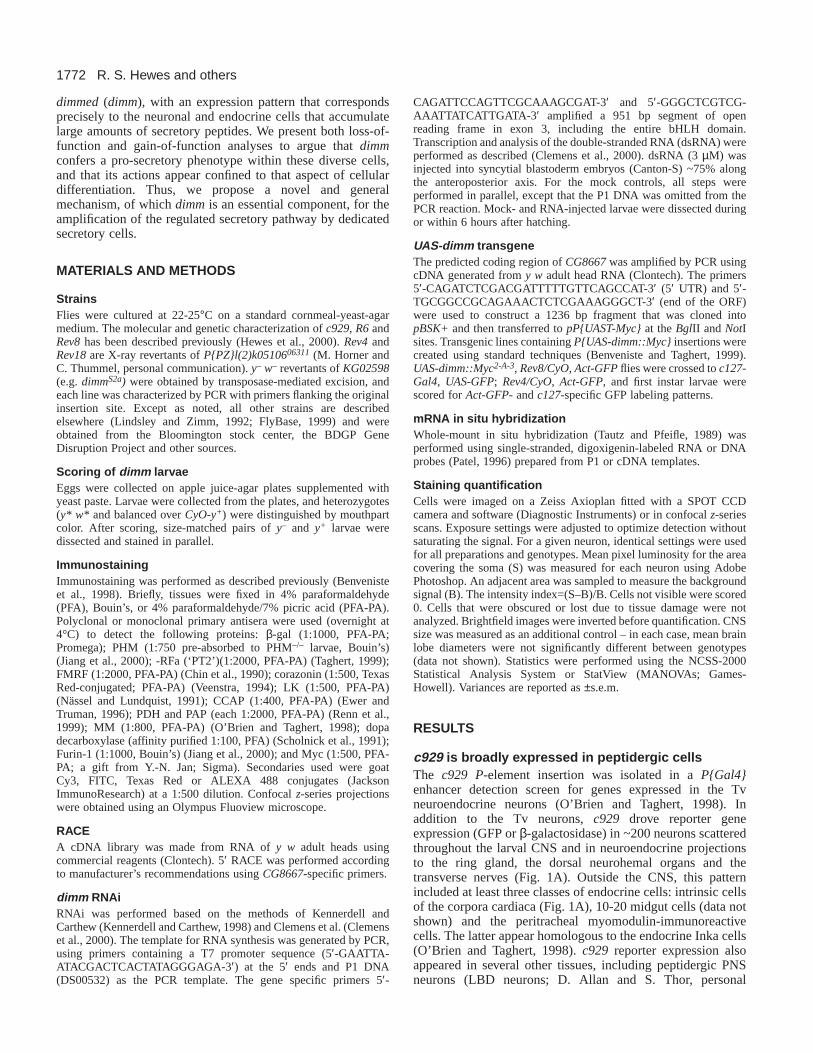
To examine further the effect of disruptions in CG8667expression, we performed RNAi analysis and observed reducedlevels of MM immunostaining in hatchling stage larvae (Fig.2D). The reduction in MM immunostaining was comparablewith the phenotype in null dimm–/– mutants (Fig. 2C). Weobtained the same results using two additional antisera, PT2and anti-LK (n=5 and n=6; data not shown). We also tested theability of a UAS-dimm::Myc transgene to restore neuropeptidelevels in dimm–/– animals. We used the c127-Gal4line to drivedimm::Myc expression in a small set of ventral CNS neurons,which included the 14 LK-positive cells in abdominalneuromeres (Fig. 4A). Expression of dimm::Myc selectivelyrestored normal levels of LK immunostaining in Rev8/Rev4
animals (n=19; Fig. 4C), but not in the absence of the Gal4driver (n=17; Fig. 4B). The rescue displayed cell specificity:the FMRF-positive MP2 neurons did not express UAS-GFP byc127-Gal4, and they were not rescued (n=10; data not shown).Together, these results support the hypothesis that dimmis theDrosophila Mist1ortholog, CG8667.
We performed a gain-of-function analysis by driving UAS-dimm::mycin an otherwise wild-type background. When mis-expressed using a pan-neuronal elav-GAL4 driver, mostembryos died (data not shown). This suggested that the effectsof dimmon shaping neuronal properties can be widespread. Topermit a more restricted analysis, we used ap-Gal4(Fig. 4E-G), a P{Gal4} reporter inserted in the apterous(ap) gene(O’Keefe et al., 1998). When overexpressed in a subset of brainneurons, dimmincreased the brightness of LK immunostainingin the cell body and processes of the LK-positive Br1 neuron(Fig. 3F,G). dimmoverexpression did not produce widespreadLK misexpression, but it reproducibly increased the number ofLK-positive neurons from one (in animals lacking the ap-Gal4element, n=18 hemispheres) to two (n=22 hemispheres). Theadditional LK-positive neuron was always adjacent to thenormal one. Thus, dimm can alter the properties of normalneuroendocrine cells, and it can affect the number of cellsdisplaying a neuroendocrine phenotype.
R. S. Hewes and others
Fig. 4.Expression of wild-type CG8667restores neuropeptide levels in dimmmutants (left) and increases normal neuropeptide levels in wild-type animals (right). (A) LK immunostaining of the A1-A7 neurons in the ventral nerve cord in animals heterozygous for the Rev8or Rev4deficiencies. The P{UAS-dimm::Myc}transgene, which contains the entire predicted CG8667ORF, was present on the Rev8chromosome.(B) Markedly reduced LK immunostaining in dimm–/– (Rev8/Rev4) animals. (C) Staining levels in A1-A7 were restored to normal in Rev8/Rev4animals by inclusion of the c127-Gal4element. The A1-7 LK-positive neurons were all GFP positive at this stage (C′). (D) Mean pixel intensity(intensity index) for four pairs of LK-positive neurons in the three genotypes (NS, heterozygous versus rescue; P<0.01, homozygous versusrescue). (E) LK immunostaining of the Br1 neuron in the lateral brain of an animal wild type for dimmand containing one copy of the UAS-dimm::myctransgene. (F) LK-immunostaining in Br1 (soma and arbor) is markedly increased, and a neighboring neuron Br2 becomes LKpositive, when UAS-dimm-Mycis driven by ap-GAL4. Br1 was identified based on the retained shape of its axonal arbor. (F′) Anti-Mycimmunostaining of the specimen in F reveals that Br1 and Br2 neuron are both ap-GAL4positive. Scale bar, 50 µm in C; 20 µm in F. (G) Meanpixel intensity (intensity index) for the Br1 LK-positive neuron in the two genotypes (P<0.001).

1777Neuroendocrine cell differentiation
CG8667 is specifically expressed in peptidergicneurons and endocrine cellsCG8667 mRNAs were ubiquitous in pre-cellular blastodermembryos (Moore et al., 2000; data not shown) and later wereexpressed in the developing nervous system (Moore et al.,2000). Presumed zygotic CG8667 expression was first visibleas nascent transcripts scattered throughout the CNS in stage 12embryos. Cytoplasmic CG8667 hybridization was visible inmany of these cells beginning around stage 14 (Fig. 5A), wasstrong by stage 16 (Fig. 5B) and persisted in stage 17 embryos(Fig. 5C) and in hatchling larvae less than 24 hour old (Fig.5G).
The pattern of CNS CG8667in situ hybridization resembledthe c929 reporter pattern (Fig. 5A-C,G). Based on theirpositions and morphologies, more than 12 separate types ofCG8667-expressing neurons were putatively identified as c929positive. These included dorsal chain neurons (e.g. d3-d5), T1-3v, LP1, MP1, MP2, SP1, T1-3vb and VA, as well as several
bilateral clusters of neurons: large, midline protocerebral braincells (MC), lateral protocerebral brain cells (LC), ventralsubesophageal neurons (SE) and lateral abdominal neurons(neuromeres N1, N4 and N5).
We also observed expression of the c929 reporter andCG8667 in strikingly similar patterns in peripheral tissues(Fig. 5). These sites included the LBD neurons and severalendocrine tissues: intrinsic cells of the corpora cardiaca, Inkacells and a few midgut cells. Numerically, all peripheral celltypes were equally represented, except that there were fewerCG8667-expressing gut cells in embryos than c929-positivegut cells of larvae. CG8667was not expressed in any otherlocation, except for a few unidentified non-CNS cellsscattered throughout the anterior and lateral regions (stages12-15). Thus, in CNS, PNS and endocrine tissues, expressionof the c929reporter closely mirrored CG8667 expression.These expression analyses support the genetic mapping,genetic identification and RNAi data. Thus, from this point
onwards we refer to CG8667asdimmed.
dimm mutant cells surviveand arborize normallyWe next determined whethersecretory cells survived anddifferentiated in dimm–/– mutantanimals. In larvae homozygousfor the null allele, Rev4,someof the affected cells displayedlow residual immunostaining forsecretory proteins (e.g. Fig.2B,C). Thus, some dimm-expressing cells survived inmutant larvae and were at least
Fig. 5.dimmmRNA is expressed in ac929-like pattern of differentiatingand post-embryonic peptidergic cells.(A-C) Photomontages of dimm insitu hybridization in embryonic CNS(A, stage 14; B, stage 16; C, stage17). (D-F) Photomontages of dimmin situ hybridization in the Inka cells(D, stage 17), the peptidergic LBDneurons (LBD), which are adjacentto muscle 8 (m8) (E, stage 15), and agut cell (mg) at the base of theanterior region of the midgut (F,stage 17). The ventral midline (D-E)is towards the left and anterior isupwards. dv, dorsal vessel; tr,tracheae. (G) Matching patterns ofdimmexpression revealed by in situhybridization (G, photomontage) andthe c929reporter (G′, confocalimage) in the brain lobes of hatchling(first instar) larvae. The LC cells arepresent but out of focus in G(compare with C). The intrinsic cellsof the corpora cardiaca (CC) wereremoved during dissection of thebrain in G′. Scale bars: 50 µm in A-G; 20 µm in insets in A-C.

1778
partially differentiated. Others displayed a complete loss ofpeptide immunostaining, and their status was unclear.
In order to determine the fates of the latter cells, we usedGal4/UASmosaics to express ectopic, non-secretory proteinsin dimm mutant neurons. We studied 34 CNS neurons that co-expressed three different markers: the c929 reporter, theputative peptide biosynthetic enzyme Furin 1, and ap-Gal4(Fig. 6A; P.H.T. and M. Han, unpublished). In dimm–/– larvalCNS, all 34 neurons displayed strongly reduced, and oftenundetectable, immunostaining for Furin 1 (Fig. 6B). Usingap-Gal4 to drive heterologous expression of a tau::Mycfusion protein, we found that all 34 of these neurons werepresent and displayed normal morphology in the dimm–/–
larvae (Fig. 6C). In addition, the intensity of anti-Mycimmunostaining was not affected (Fig. 6D). We obtainedidentical results using green fluorescent protein (GFP) tomark the cells (n=6; data not shown). Thus, dimm mutantneurons displayed multiple differentiated features andsynthesized non-secretory proteins at normal levelsthroughout larval development.
We also examined the effects of dimmon the terminal arborof the LK-positive neurons. These cells displayed reducedsoma LK immunostaining in dimm–/– CNS (Fig. 4B). Eachneuron had a single efferent axon that projected across theposterior muscle 8 surface and terminated dorsally near atracheal branch. In third instar dimm–/– larvae, these axons alsodisplayed reduced LK immunostaining. However, a sufficientnumber of immunoreactive boutons remained to indicate anormal axonal expanse (see Fig. S1 at http://dev.biologists.org/
supplemental/). Thus, the effects of dimmon this LK neuronappear limited to expression of the transmitter phenotype.
dimm affects levels of proteins destined for bothregulated and constitutive secretionOur earlier measures of the dimmedphenotype were restrictedto analysis of proteins abundant in the regulated secretorypathway. We also tested for an effect of dimmon constitutivelysecreted proteins. With ap-Gal4, we directed expression of aCD8::GFP fusion protein (UAS-CD8::GFP) to a subset ofdimm-expressing neurons. CD8 is an integral membraneprotein that is targeted to the plasma membrane in Drosophilacells (Zito et al., 1997). In dimm–/– mutant larvae, all 34 ap-Gal4 (Furin-1) neurons expressed CD8::GFP and displayednormal neuritic projections. However, CD8::GFP levels weresignificantly lower in c929-positive neurons in the dimm–/–
background (see Fig. S2 at http://dev.biologists.org/supplemental/). This effect was more subtle than the effects onlevels of regulated secretory proteins. However, it suggests thatdimm influences both regulated and constitutive secretoryactivity in neuroendocrine cells.
dimm regulates multiple elements of the secretorypathwayBecause ap-dependent expression of transgenes was unaffectedby dimm (Fig. 6C), we were able to uncouple neuropeptidetranscription from potential effects of dimmon secretoryactivity. Thus, when ap-Gal4 drove ectopic expression ofthe pdf neuropeptide gene, ectopic pdf mRNA levels were
R. S. Hewes and others
Fig. 6.dimm–/– mutantneurons differentiate,survive, and expressnon-secretory proteinsat normal levels.(A) Thirty-four neurons(black circles) in theventral nerve cord thatco-express the c929reporter, Furin 1 andap-Gal4 (apGal4).Additional ventralneurons (white circles)express ap-Gal4but donot express the othertwo markers. (B) Thedimmmutantphenotype for the 34Furin 1 neurons.Reduction in Furin 1immunostaining in theCNS of a third instardimm–/– (R6/Rev8)larva (B′), comparedwith a dimm+/– (R6 orRev8/+) sibling control(B) (n>50).Arrowheads indicate Tv and Tvb neurons. (C) Ectopic expression of the non-secretory fusion protein tau::Myc in the 34 Furin 1-positiveneurons. In dimm–/– mutant CNS (third instar; R6, ap-Gal4/Rev8; UAS-tau::Myc/+), all 34 Furin 1 neurons displayed anti-Myc immunostaining(C′) that was comparable with that in the dimm+/– sibling control (C; R6, ap-Gal4/+; UAS-tau::Myc/+). In both genotypes, each of the dorsalchain neurons (d1-d11) displayed projections that extended toward the midline (vertical arrows) and then ran longitudinally (horizontal arrows).We also observed normal immunostained projections in each abdominal nerve (arrowheads). (D) Mean pixel intensity (intensity index) forsoma Furin 1 immunostaining in five pairs of dorsal chain neurons in seven dimm+/– and 13 dimm–/– CNSs. *P<0.05. Scale bars: 50 µm.
1779Neuroendocrine cell differentiation
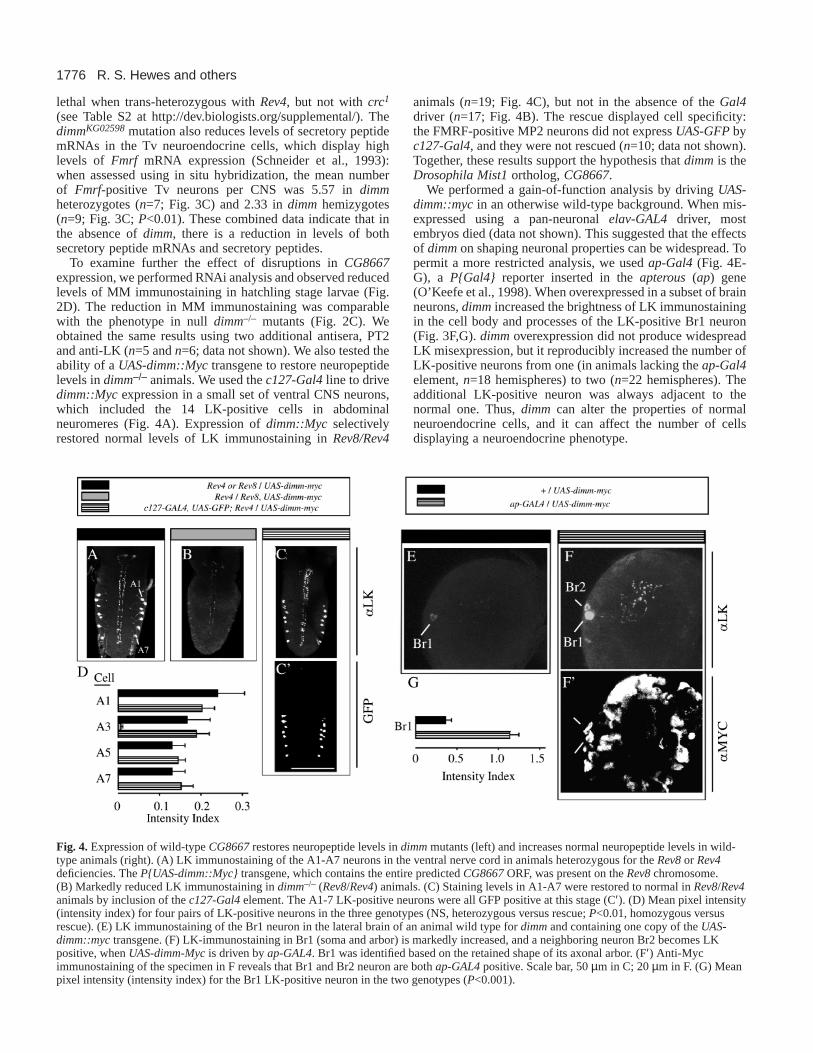
unaffected in dimm–/– larvae (Fig. 7A,B).By contrast, ectopic PDF protein levelswere severely reduced. We performedimmunostaining for two peptide epitopes of theproPDF precursor (Renn et al., 1999): PAP(Fig. 7C,F) and PDF (n=20; data not shown).All 34 (c929-positive) neurons displayedseverely reduced immunostaining for bothPDF-related epitopes. Additional ventralabdominal neurons served as internal controls.These included 44 neurons that also displayedectopic pdf expression driven by ap-Gal4, anda set of approximately eight native pdf neurons(not ap-positive). All of the internal controlcells were c929 negative, and PAP/PDFimmunostaining in these neurons wasunaffected in dimm–/– larvae (Fig. 7C-F). Thus,dimm was required within c929-positiveneurons for the maintenance of ectopic PDFneuropeptide levels, but not of ectopic pdfmRNA.
DISCUSSION
Dimm is the first example of a dedicated pro-secretory factor. Dimm is necessary to conferneuroendocrine features onto peptidergicneurons that, in its absence, survive withnormal neuronal properties. In addition, Dimmoverexpression produces supra-normal levelsof neuropeptide expression in peptidergicneurons and the appearance of additionalcells with neuroendocrine features. Fromthis genetic analysis, we suggest thatneuroendocrine cell differentiation includestwo interrelated, but separate sets ofinstructions. The first specifies the identity ofthe neuropeptide(s) or peptide hormone(s) tobe expressed, while the second, which involvesDimm, specifies the level of regulatedsecretory activity.
The bHLH domain of the predicted Dimmprotein showed the highest degree of sequenceidentity with the mouse, rat and human Mist1proteins. These proteins may be orthologs(Moore et al., 2000). Interestingly, mouseMist1 is present in many adult peripheraltissues, but within these tissues it is found onlyin serous exocrine cells (Pin et al., 2000). Therestriction of mouse Mist1 expression todedicated secretory cells suggests that dimmand mouse Mist1 may both control levelsof secretory activity, and so may performevolutionarily conserved functions. Othermembers of the Atonal family are expressedin both differentiating and terminallydifferentiated cells (e.g. NeuroD) (Morrow etal., 1999). Several mammalian Atonal familybHLH proteins have previously beenimplicated in earlier stages of endocrine cell
Fig. 7.dimmcontrols levels of ectopic neuropeptide (proPDF) but not levels ofectopic pro-pdfmRNA. (A) Brightfield photomontages of pdfmRNA in situhybridization in the dorsal chain neurons of dimm+/– (A; R6, ap-Gal4/+; UAS-pdf/+)and dimm–/– (A′ ; R6, ap-Gal4/Rev8; UAS-pdf/+) third instar stage CNS. Heterologousexpression of the pdf neuropeptide gene, which encodes the PAP neuropeptide, wasdirected to these neurons using UAS-pdf (Renn et al., 1999) and ap-GAL4. (B) Meanpixel intensity (intensity index) for pdfmRNA in situ hybridization in the somata ofselected pairs of c929-positive dorsal neurons (dimm+/–, n=11-12; dimm–/–, n=8-9).(C) Confocal z-series images of dorsal PAP (proPDF) immunostaining in the CNSfrom dimm–/+ (C) and dimm–/– (C′) third instar stage larvae. Reduced staining (C′) wasobserved in the dorsal chain neurons (d1-11) and the dorsal neurohemal organs(asterisks), which contain the neuroendocrine terminals of the Tv neuron (Benvenisteand Taghert, 1999). Note that staining levels in c929-negative cells were unaffected:these included PN (natively pdf-positive), and v7 and v8 (ectopic, ap-Gal4-dependentexpression). (D) Mean pixel intensity (intensity index) for PAP immunostaining in thesomata of selected pairs of dorsal neurons and in the PN neurons (dimm+/–, n=6;dimm–/–, n=5). (E) Confocal z-series images of ventral PAP immunostaining in T2v(arrows) and in the ventral chain neurons, including v2a and v8, of a dimm–/+ thirdinstar stage CNS (E; same CNS as C) and a dimm–/– CNS (E′; same CNS as C′). TheTv neurons (arrows) express the c929 reporter gene (Fig. 1C,E) and display the dimmmutant phenotype. By contrast, the ventral chain neurons do not express the reportergene (data not shown), and they do not display the dimm mutant phenotype. (F) Meanpixel intensity (intensity index) for PAP immunostaining in the somata of selectedpairs of ventral neurons (dimm+/–, n=6; dimm–/–, n=5). The brightest cell in each Tvcluster was assumed to be Tvb (P.H.T. and M. Han, unpublished). *P<0.05, **P<0.01,***P<0.001. Scale bars: 50 µm.

1780
development, including cell lineage commitment (e.g. Yanget al., 2001) and endocrine cell differentiation (Sheng andWestphal, 1999).
In Drosophila, dimmperforms a novel, pro-secretory functionin a diverse population of peptidergic CNS and PNS neurons andendocrine cells. In its absence, peptidergic cells complete manyaspects of their differentiation – some express low levels ofappropriate peptide transmitters. However, they uniformly fail todisplay normal amplified levels of secretory activity, which is acharacteristic and fundamental property of peptidergic secretorycells (Arvan and Castle, 1998). How such cells acquire andmaintain this capacity is largely unknown. We have shown thatit is under the control of specific genetic mechanisms, asrevealed by animals deficient in expression of the dimmgene.These experiments indicate that dimmplays a fundamental rolein the differentiation of neuroendocrine lineages.
We propose a working model in which Dimm directlyregulates transcription of genes required for production of aneuroendocrine phenotype – genes encoding neuropeptides,peptide hormones and peptide biosynthetic enzymes.Consistent with this model, we found that dimm reduces thenormally high levels of Fmrf neuropeptide mRNA in specificneuroendocrine cells. In addition, Dimm also may regulateexpression of proteins (e.g. transcription factors, or structuralor regulative proteins of dense core granules) that are importantfor the function and amplification of the secretory pathway[e.g., as suggested by Kim et al. (Kim et al., 2001)]. Dimmfunctions after cell fate determination and during the earlydifferentiation of these neurons – in dimm mutants, affectedpeptidergic neurons are present, arborize normally and oftenexpress low levels of appropriate neuropeptides.
Some secretory proteins form dense aggregations(‘progranules’) in the trans-Golgi network prior to their uptakeinto immature secretory granules. Similarly, condensation ofsecretory proteins during subsequent granule maturation maybe required for their retention in maturing granules (Arvan andCastle, 1998). Therefore, direct reductions in the levels of asmall number of target secretory proteins in dimm mutantcells may lead to a secondary disruption in aggregation orcondensation of other proteins. In turn, these effects couldlead to loss of most secretory proteins by mis-routing anddegradation. This may account for our observation thatsecretory peptide levels could be reduced in a dimm mutantbackground, despite the artificial elevation of the cognatesecretory peptide mRNA (Fig. 7).
Does dimmalso regulate the constitutive secretory pathway?Although constitutive secretion was quantitatively affected byloss of dimmfunction, mutant neurons maintained their normalcellular morphology. These observations suggest that Dimm hasonly moderate effects on the constitutive secretory pathway.Given the physical interactions between cargoes destined for theregulated and constitutive pathways (Arvan and Castle, 1998),the reduction in constitutive secretion may reflect an indirecteffect of disruptions in the regulated pathway.
We favor the view that during development and maturity,dimm expression is a crucial determinant of high secretoryprotein expression in neuroendocrine cells. This hypothesiswas supported by the gain-of-function analysis.Overexpression of dimmin a wild-type background producedhigher levels of LK expression in the normally LK-positive Br1neuroendocrine neuron. It also increased the number of cells
that display the specific LK neuroendocrine phenotype, butonly within the immediate proximity of Br1. In this case, dimmoverexpression was driven by a promoter (ap-GAL4) that isonly expressed in postmitotic neurons. Therefore, it appearslikely that the additional LK immunoreactive neurons representcells that normally express LK but at levels that are too low tobe detected. In addition, the limited number of ectopicleukokinin cells is likely a function of the specific GAL4driverused (apis only expressed in a subset of cells), and the markerassayed (LK is only expressed in ~20 out of 10,000 neurons).Although the complete extent of the effects of dimm, whenoverexpressed, is not yet known it is likely to be large, as UAS-dimmproduces large-scale embryonic lethality when driven bythe pan-neuronal elav-GAL4(D. P., unpublished).
Accordingly, we propose that dimmpromotes diverseneuroendocrine cell fates in different cellular locales, dependingon local cellular context and identity. We observed dimmexpression soon after cells cease dividing, and in its absence,most of these cells were deficient in ‘transmitter expression’.Thus, Dimm appears to function like NeuroD proteins, whichare also members of the Atonal family and which act as celldifferentiation factors (Hassan and Bellen, 2000).
Analysis within the identified, neuroendocrine Tv neuronsmay be especially informative to reveal further details of themechanisms of dimm action. Four regulatory factors havenow been defined that affect FMRF neuropeptide levels in Tvneurons. Loss-of-function ap(Benveniste et al., 1998), Chip(Van Meyel et al., 2000) and dimm(this report) alleles alldecrease Tv-specific FMRF expression, but do not influenceTv survival or morphology. Likewise, the squeeze(sqz) genehelps regulate Tv-specific FMRF levels (S. Thor, personalcommunication). Within Tv neurons, ap, Chip, dimmand sqzmay function in a linear pathway to regulate Fmrfgeneexpression, akin to the sequential actions of the bHLH proteinMASH1 and the Phox2 homeoproteins in neurons of the locuscoeruleus (Pattyn et al., 2000). Alternatively, they may workin parallel fashion, akin to the synergistic interactionsbetween the bHLH NeuroD1 and the LIM homeoproteinsLmx1.1 and Lmx1.2 to control insulin expression (Ohneda etal., 2000). As a first step, we have shown that ap promoterfunction is independent of dimm. Further work will permitdescription of the molecular pathways controlling qualitativeand quantitative aspects of neuroendocrine cell differentiationin vivo.
We thank Weihua Li and Aloka Amarakone for technical assistance,and Hans Agricola, Doug Allan, Hugo Bellen, Gabrielle Boulianne,Adelaide Carpenter, Heinrich Dircksen, Chris Doe, Dan Eberl, JohnEwer, Jeff Hall, Mike Horner, Yuh-Nung Jan, Iris Lindberg, DickNässel, Jae Park, Anton Roebroek, Steve Scholnick, Amy Sheehan,John Thomas, Stephan Thor, Carl Thummel, Jan Veenstra, KlaudeWeiss and Andrew Zelhof for information, DNA, antibodies or flystocks. We thank Lou Muglia, Jim Skeath and Stefan Thor forcomments on the manuscript, the Bloomington Stock Center for flystocks, and the BDGP for DNA sequence. This work was supportedby an American Cancer Society Postdoctoral Fellowship PF4212(R.S.H.) and by a grant NS21749 from the NIH (P.H.T.).
REFERENCES
Acampora, D., Postiglione, M. P., Avantaggiato, V., di Bonito, M.,Vaccarino, F. M., Michaud, J. and Simeone, A. (1999). Progressive
R. S. Hewes and others

1781Neuroendocrine cell differentiation
impairment of developing neuroendocrine cell lineages in the hypothalamusof mice lacking the Orthopediagene. Genes Dev. 13, 2787-2800.
Arvan, P. and Castle, D. (1998). Sorting and storage during secretory granulebiogenesis: looking backward and looking forward. Biochem. J.332, 593-610.
Benveniste, R. J. and Taghert, P. H. (1999). Cell type-specific regulatorysequences control expression of the Drosophila FMRF-NH2 neuropeptidegene.J. Neurobiol.38, 507-520.
Benveniste, R. J., Thor, S., Thomas, J. B. and Taghert, P. H. (1998). Celltype-specific regulation of the Drosophila FMRF-NH2 neuropeptide gene byApterous, a LIM homeodomain transcription factor. Development125,4757-4765.
Blake-Bruzzini, K. M., Borke, R. C., Anders, J. J. and Potts, J. D. (1997).Calcitonin gene-related peptide and alpha-CGRPmRNA expression incranial motoneurons after hypoglossal nerve injury during postnataldevelopment. J. Neurocytol.26, 163-179.
Burbach, J. H. P., Luckman, S. M., Murphy, D. and Gainer, H.(2001).Gene regulation in the magnocellular hypothalmo-neurohypophysealsystem. Physiol. Rev. 81, 1197-1267.
Clemens, J. C., Worby, C. A., Simonson-Leff, N., Muda, M., Maehama,T., Hemmings, B. A. and Dixon, J. E. (2000). Use of double-stranded RNAinterference in Drosophilacell lines to dissect signal transduction pathways.Proc. Natl. Acad. Sci. USA97, 6499-6503.
Chin, A., Reynolds, E. and Scheller, R. H. (1990). Organization andexpression of the Drosophila FMRFamide-related prohormone gene. DNACell Biol. 9, 263-271.
De Bie, I., Savaria, D., Roebroek, A. J., Day, R., Lazure, C., van de Ven,W. J. and Seidah, N. G. (1995). Processing specificity and biosynthesis ofthe Drosophila melanogasterconvertases dfurin1, dfurin1-CRR, dfurin1-X,and dfurin2. J. Biol. Chem. 270, 1020-1028.
Eipper, B. A., Stoffers, D. A. and Mains, R. E. (1993). Biosynthesis ofneuropeptides: alpha-amidation. Annu. Rev. Neurosci. 15, 57-85.
Ewer, J. and Truman, J. W. (1996). Increases in cyclic 3′,5′-guanosinemonophosphate (cGMP) occur at ecdysis in an evolutionarily conservedcrustacean cardioactive peptide-immunoreactive insect neuronal network. J.Comp. Neurol. 370, 330-341.
FlyBase(1999). The FlyBase database of the Drosophilagenome projects andcommunity literature. The FlyBase Consortium. Nucleic Acids Res. 27, 85-88.
Hall, Z. W. and Sanes, J. R. (1993). Synaptic structure and development: theneuromuscular junction. Cell Suppl.72, 99-121.
Hassan, B. A. and Bellen, H. J. (2000). Doing the MATH: is the mouse agood model for fly development? Genes Dev. 14, 1852-1865.
Herman, J. P., Schaefer, M. K., Watson, S. J. and Sherman, T. J. (1991).In situ hybridization analysis of arginine vasopressin gene transcriptionusing intron-specific probes. Mol. Endocrinol. 5,1447-1456.
Hewes, R. S. and Taghert, P. H. (2001). Neuropeptides and neuropeptidereceptors in the Drosophila melanogastergenome. Genome Res. 11, 1126-1142.
Hewes, R. S., Schaefer, A. and Taghert, P. H. (2000). The cryptocephalgene(ATF4) encodes multiple basic-leucine zipper proteins controlling moltingand metamorphosis in Drosophila. Genetics155, 1711-1723.
Hirsh, J. (1989). Molecular genetics of dopa decarboxylase and biogenicamines in Drosophila. Dev. Genet.10, 232-238.
Jiang, N., Kolhekar, A. S., Jacobs, P. S., Mains, R. E., Eipper, B. A. andTaghert, P. H. (2000). PHM is required for normal developmentaltransitions and for biosynthesis of secretory peptides in Drosophila. Dev.Biol. 226, 118-136.
Kennerdell, J. R. and Carthew, R. W. (1998). Use of dsRNA-mediatedgenetic interference to demonstrate that frizzledand frizzled 2act in theWingless pathway. Cell95, 1017-1026.
Kim, T., Tao-Cheng, J., Eiden, L. E. and Loh. Y. P. (2001). ChromograninA, an ‘On/Off’ switch controlling dense-core secretory granule biogenesis.Cell 106, 499-509.
Lindsley, D. L. and Zimm, G. G. (1992). The genome of Drosophilamelanogaster.San Diego, CA: Academic Press.
Michaud, J. L., Rosenquist, T., May, N. R. and Fan, C. M. (1998).Development of neuroendocrine lineages requires the bHLH-PAStranscription factor SIM1. Genes Dev. 12, 3264-3275.
Moore, A. W., Barbel, S., Jan, L. Y. and Jan, Y. N. (2000). A genome-widesurvey of basic helix-loop-helix factors in Drosophila. Proc. Natl. Acad. Sci.USA 97, 10436-10441.
Morrow, E. M., Furukawa, T., Lee, J. E. and Cepko, C. L. (1999). NeuroDregulates multiple functions in the developing neural retina in rodent.Development126, 23-36.
Nakai, S., Kawano, H., Yudate, T., Nishi, M., Kuno, J., Nagata, A., Jishage,K., Hamada, H., Fujii, H. and Kawamura, K. (1995). The POU domaintranscription factor Brn-2 is required for the determination of specificneuronal lineages in the hypothalamus of the mouse. Genes Dev. 9, 3109-3121.
Nässel, D. R. and Lundquist, C. T. (1991). Insect tachykinin-like peptide:distribution of leucokinin immunoreactive neurons in the cockroach andblowfly brains. Neurosci. Lett. 130, 225-228.
O’Brien, M. A. and Taghert, P. H. (1998). A peritracheal neuropeptidesystem in insects: release of myomodulin-like peptides at ecdysis. J. Exp.Biol. 201, 193-209.
O’Keefe, D. D., Thor, S. and Thomas, J. B. (1998). Function and specificityof LIM domains in Drosophila nervous system and wing development.Development125, 3915-3923.
Ohneda, K., Ee, H. and German, M. (2000). Regulation of insulin genetranscription. Semin. Cell Dev. Biol.11, 227-233.
Patel, N. H. (1996). In situ hybridization to whole mount Drosophilaembryos.In A Laboratory Guide to RNA(ed. P. A. Krieg), pp. 357-370. New York:Wiley-Liss.
Pattyn, A., Goridis, C. and Brunet, J. F. (2000). Specification of the centralnoradrenergic phenotype by the homeobox gene Phox2b. Mol. Cell.Neurosci.15, 235-243.
Pin, C. L., Bonvissuto, A. C. and Konieczny, S. F. (2000). Mist1 expressionis a common link among serous exocrine cells exhibiting regulatedexocytosis. Anat. Rec. 259, 157-167.
Pin, C. L., Lemercier, C. and Konieczny, S. F. (1999). Cloning of the murineMist1 gene and assignment to mouse chromosome band 5G2-5G3.Cytogenet. Cell. Genet. 86, 219-222.
Renn, S. C., Park, J. H., Rosbash, M., Hall, J. C. and Taghert, P. H. (1999).A pdf neuropeptide gene mutation and ablation of PDF neurons each causesevere abnormalities of behavioral circadian rhythms in Drosophila. Cell99,791-802.
Schneider, L. E., Sun, E. T., Garland, D. J. and Taghert, P. H. (1993). Animmunocytochemical study of the FMRFamide neuropeptide gene productsin Drosophila. J. Comp. Neurol. 337, 446-460.
Scholnick, S. B., Caruso, P. A., Klemencic, J., Mastick, G. S., Mauro, C.and Rotenberg, M. (1991). Mutations within the Ddc promoter alter itsneuron-specific pattern of expression. Dev. Biol.146, 423-437.
Schonemann, M. D., Ryan, A. K., McEvilly, R. J., O’Connell, S. M., Arias,C. A., Kalla, K. A., Li, P., Sawchenko, P. E. and Rosenfeld, M. G. (1995).Development and survival of the endocrine hypothalamus and posteriorpituitary gland requires the neuronal POU domain factor Brn-2. Genes Dev.9, 3122-3135.
Sheng, H. Z. and Westphal, H. (1999). Early steps in pituitary organogenesis.Trends Genet. 15, 236-240.
Streit, W. J., Dumouli, F. L., Raivich, G. and Kreutzberg, G. W. (1989).Calcitonin gene-related peptide increases in rat facial motoneurons afterperipheral nerve transections. Neurosci. Lett.101, 143-148.
Taghert, P. H. (1999). FMRFamide neuropeptides and neuropeptide-associated enzymes in Drosophila. Microsc. Res. Tech.45, 80-95.
Tautz, D. and Pfeifle, C. (1989). A non-radioactive in situ hybridizationmethod for the localization of specific RNAs in Drosophilaembryos revealstranslational control of the segmentation gene hunchback. Chromosoma98,81-85.
van Meyel, D. J., O’Keefe, D. D., Thor, S., Jurata, L. W., Gill, G. N.and Thomas, J. B. (2000). Chip is an essential cofactor for apterous inthe regulation of axon guidance in Drosophila. Development127, 1823-1831.
Veenstra, J. A. (1994). Isolation and structure of the Drosophila corazoningene. Biochem. Biophys. Res. Commun.204, 292-296.
Yang, Q., Bermingham, N. A., Finegold, M. J. and Zoghbi, H. Y. (2001)Requirement of Math1for secretory cell lineage commitment in the mouseintestine. Science294, 2155-2158.
Yeh, E., Gustafson, K. and Boulianne, G. L. (1995). Green fluorescentprotein as a vital marker and reporter of gene expression in Drosophila.Proc. Natl. Acad. Sci. USA92, 7036-7040.
Zito, K., Fetter, R. D., Goodman, C. S. and Isacoff, E. Y. (1997). Synapticclustering of Fasciclin II and Shaker: essential targeting sequences and roleof Dlg. Neuron19, 1007-1016.