Embed Size (px)
Citation preview
RESEARCH ARTICLE
The biomechanics of tree frogs climbing curved surfaces:a gripping problemIain D. C. Hill1, Benzheng Dong2, W. Jon P. Barnes1,*, Aihong Ji2 and Thomas Endlein3
ABSTRACTThe adhesive mechanisms of climbing animals have become animportant research topic because of their biomimetic implications.Weexamined the climbing abilities of hylid tree frogs on vertical cylindersof differing diameter and surface roughness to investigate the relativeroles of adduction forces (gripping) and adhesion. Tree frogs adhereusing their toe pads and subarticular tubercles, the adhesive jointbeing fluid-filled. Our hypothesis was that on an effectively flat surface(adduction forces on the largest 120 mm diameter cylinder wereinsufficient to allow climbing), adhesion would effectively be the onlymeans by which tree frogs could climb, but on the 44 and 13 mmdiameter cylinders, frogs could additionally utilise adduction forces bygripping the cylinder either with their limbs outstretched or bygrasping around the cylinder with their digits, respectively. Thefrogs’ performance would also depend on whether the surfaces weresmooth (easy to adhere to) or rough (relatively non-adhesive). Ourfindings showed that climbing performance was highest on thenarrowest smooth cylinder. Frogs climbed faster, frequently using a‘walking trot’ gait rather than the ‘lateral sequencewalk’ used on othercylinders. Using an optical technique to visualise substrate contactduring climbing on smooth surfaces, we also observed an increasingengagement of the subarticular tubercles on the narrower cylinders.Finally, on the rough substrate, frogs were unable to climb the largestdiameter cylinder, but were able to climb the narrowest one slowly.These results support our hypotheses and have relevance for thedesign of climbing robots.
KEY WORDS: Adhesion, Adduction, Litoria caerulea, Osteopilusseptentrionalis
INTRODUCTIONThe ability to climb is common in animals, being utilised byamphibians, reptiles, birds and mammals, as well as manyarthropods. It offers many advantages, including access to safetyfrom ground-dwelling predators, faster travel in dense vegetationregions and access to food (including prey) (Hildebrand andGoslow, 2001). Arboreal animals possess a wide range ofmorphological adaptations to assist in the behaviour of climbing.These may include long arms for reaching and pulling, and hind legsadapted for jumping from branch to branch. Additionally, they needsome way of remaining attached to substrates they climb. Claws do
this rather well, either by interlocking with surface irregularities orby digging into the surface if it is soft enough. However, severalanimal groups have developed mechanisms of adhesion (e.g.geckos, tree frogs and many arthropods) for the same purpose(though geckos and insects may possess claws as well). Theseadhesive mechanisms may be used alone, or in conjunction with theproduction of adduction forces, which, aided by friction, are used togrip structures such as stems and branches (Cartmill, 1985).
The adhesive mechanisms of climbing animals have been widelystudied in recent years, especially those of geckos, insects and treefrogs. This is to a large extent due to their relevance to biomimetics.Firstly, as discussed by Barnes (2007), this is because they haveremarkable powers of adhesion to a wide variety of surfaces. Even alarge gecko can run across a ceiling (Autumn and Peattie, 2002), anda tree frog jumping from branch to branch does not fall so long as asingle toe pad makes good contact with the tree (Bijma et al., 2016).Additionally, ants can carry over 100 times their own weight whilewalking upside-down (Federle and Endlein, 2004). Secondly, theadhesive mechanisms are reversible (geckos can walk at over 10steps per second) and detachment is effortless (Autumn, 2007).Thirdly, animal adhesive pads can also have self-cleaning propertiesand thus do not get fouled (Crawford et al., 2012; Hansen andAutumn, 2005; Hu et al., 2012). It is also appropriate to add that, asgecko feet are non-adhesive in the default state because they have avery low contact fraction (<6.6% of surface), they do not stick toeverything they touch (Autumn and Hansen, 2006). Suchcombinations of properties are not found in more traditionaladhesives, but are now being developed in many laboratories,inspired by the natural world (Xia, 2016).
Turning specifically to frogs (Amphibia; Anura), there isextensive work on the biomechanics of frog jumping (e.g. Astleyand Roberts, 2014; Astley et al., 2015), but much less on climbing,the speciality of tree frogs. However, two papers are of particularinterest (Manzano et al., 2008; Herrel et al., 2013), as they providestrong evidence that the forelimbs of at least some tree frog speciescan generate gripping forces. These gripping forces are used both inthe manipulation of food and in locomotion, particularly horizontalwalking and climbing along narrow substrates. The species mostadept at these behaviours are members of the FamilyPhyllomedusidae (Duellman et al., 2016), which are able totraverse rods down to a diameter of 1 mm. Appropriately, thesefrogs are known as monkey frogs, as they tend to move from place toplace by climbing rather than jumping as do most other tree frogs.Manzano et al. (2008) have shown that they are able to employ bothpower and precision grips, as defined by Napier (1956). Roughlyspeaking, a power grip describes how one holds a hammer, while anexample of a precision grip would be holding a pencil.
Tree frogs adhere by means of a wet adhesive joint, a low-viscosity fluid being secreted by the epithelium of the toe pads,located ventrally on each digit (Federle et al., 2006). The toe padepithelium consists of squamous epithelial cells, surrounded byReceived 15 August 2017; Accepted 12 January 2018
1Centre for Cell Engineering, University of Glasgow, Joseph Black Building,University Avenue, Glasgow G12 8QQ, UK. 2Institute of Bioinspired Structure andSurface Engineering, Nanjing University of Aeronautics and Astronautics, 29 YudaoStreet, Nanjing 210016, China. 3Max Planck Institute for Intelligent Systems,Heisenbergstraβe 3, 70569 Stuttgart, Germany.
*Author for correspondence ([email protected])
W.J.P.B., 0000-0002-1359-5803; T.E., 0000-0002-6358-5523
1
© 2018. Published by The Company of Biologists Ltd | Journal of Experimental Biology (2018) 221, jeb168179. doi:10.1242/jeb.168179
Journal
ofEx
perim
entalB
iology
channels that serve to distribute the pad fluid over the surface of thepad (Green, 1979; Emerson and Diehl, 1980; Smith et al., 2006a).At the nanoscale, these epithelial cells are covered in a dense arrayof nanopillars (Scholz et al., 2009). The main adhesive force isthought to be capillarity, though a contribution from viscosity-dependant hydrodynamic forces is likely, but as yet unproven(Hanna and Barnes, 1991; Barnes et al., 2006; Persson, 2007; Buttet al., 2010; Drotlef et al., 2012; Endlein et al., 2013). The ability togenerate high friction forces is probably more important than theability to generate adhesive ones (Endlein et al., 2013), as frogs arenormally active on surfaces with slopes varying between horizontaland 90 deg, though they can support themselves on overhangingsurfaces, and small frogs can hang on when completely upside-down (Barnes et al., 2006; Smith et al., 2006b). Friction forces arehigher than should be possible from a fluid joint (Federle et al.,2006), so it is likely that that there is actual contact between pad andsubstrate. Indeed, the tips of the nanopillars appear, frominterference reflection microscopy, to be making actual contactwith the underlying surface (Federle et al., 2006). Subarticulartubercles, located more proximally on each digit, have a structuresimilar to that of the toe pads, but are smaller and anatomically lessspecialised; for example, the channels that separate the cells are bothshallower and narrower (Green, 1979; Endlein et al., 2017).Studies of adhesion have largely been carried out on flat surfaces,
specifically because, on such surfaces, there is no possiblecontribution from adduction forces (Cartmill, 1985). However, theworld of tree frogs seldom consists of flat, smooth surfaces, butrather curved ones such as branches and twigs, where, as discussedabove, there is good evidence for gripping by adduction. This paperis aimed at bridging this gap, studying the relative roles of adhesionand gripping on surfaces varying in both curvature and roughness,the roughness being chosen to be a surface where the frogs generateminimal adhesive forces. We compared the climbing abilities of twospecies of hylid frog, Litoria caerulea and Osteopilusseptentrionalis, on cylinders of different diameter, the largesteffectively acting like a flat surface in relation to the size of the frogs.The intermediate cylinder was chosen so that it would be possiblefor frogs to generate adduction forces across the body, i.e. a ‘bearhug’, whereas the narrowest was such that toes would be able towrap around the cylinder. Where climbing was observed, wemeasured the frogs’ velocity as well as the gait used. Additionally,as the smooth surfaces were transparent, we used the technique offrustrated total internal reflection (FTIR) (Betts et al., 1980; Endleinet al., 2013; Eason et al., 2015) to observe the structures used inclimbing the different cylinders, enabling us to observe when thesub-articular tubercles, located more proximally on each toe, wereused in addition to the toe pads.
MATERIALS AND METHODSStudy animalsWhite’s tree frogs [Litoria caerulea (White 1790)] and Cuban treefrogs [Osteopilus septentrionalis (A.M.C. Duméril & Bibron1841)] were obtained from commercial suppliers (L. caeruleafrom Partick Aquatics and Reptiles, Glasgow, UK; O.septentrionalis from Coast to Coast Exotics, Darlington, UK).Litoria caerulea (n=4) had snout–vent lengths (SVLs) in the rangeof 48–60 mm, and SVLs ofO. septentrionalis (n=4) ranged from 48to 71 mm. All frogs were housed in custom-built vivaria(295×460×765 mm, width×length×height), illuminated with fullspectrum terrarium lamps (Repti Glo 2.0 Compact, www.exo-terra.com) on a 12 h:12 h light:dark cycle. Ambient room temperaturewas controlled and coupled with heat mats to provide a temperature of
23±2°C; additionally, a Honeywell Ultrastar humidifier (Honeywell,Bracknell, Berkshire, UK) provided a relative humidity ofapproximately 70%. The frogs were provided with houseplants(Ficus sp.) to climb on and dishes of Cu-free fresh water. They werefed with house crickets (ca. three per week) dusted with Nutrobal, acalcium balancer and multivitamin supplement (Vetark Professional,Hants, UK). Prior to testing, each individual frog was identified andweighed, and the length of its forelimbs, hindlimbs, front span andSVLwasmeasured to the nearest millimetre using a set ofmechanicalcallipers (see Table S1 for the actual measurements). Before eachsession of experiments, the frogs involved were removed from thevivaria and checked for any debris or shedding skin that could affecttheir ability to climb and/or adhere. The use of individual frogs wasrandomised and, prior to testing, each frog was left in a separatelabelled container for 30 min to allow it to be as calm as possible fordata collection. All experiments were non-invasive and accordedwithcurrent laws on animal experimentation in the UK.
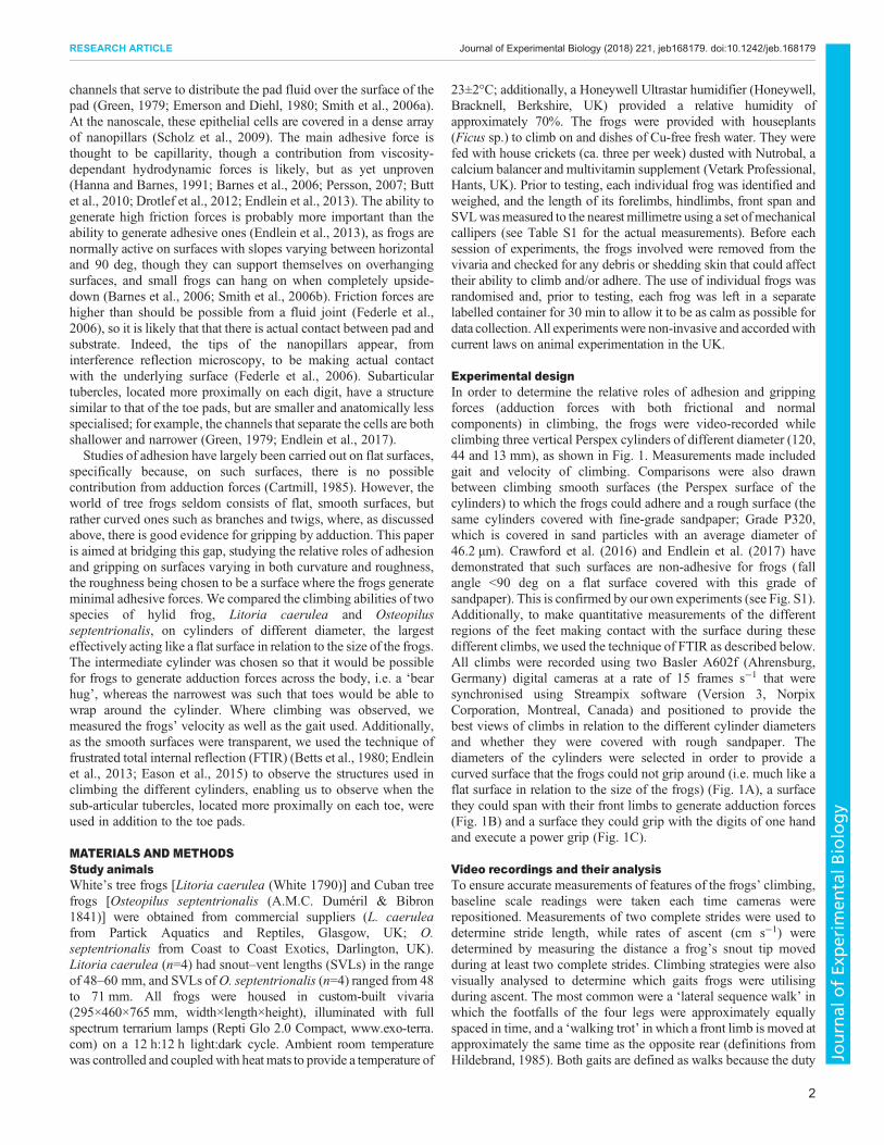
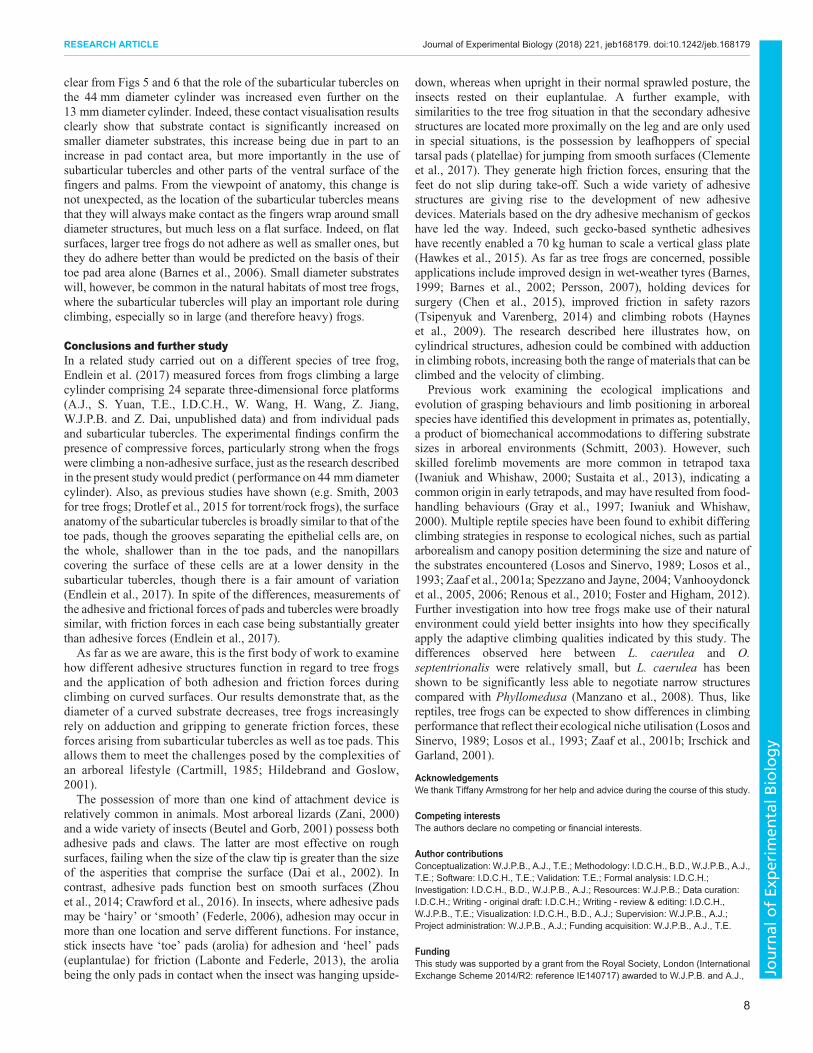
Experimental designIn order to determine the relative roles of adhesion and grippingforces (adduction forces with both frictional and normalcomponents) in climbing, the frogs were video-recorded whileclimbing three vertical Perspex cylinders of different diameter (120,44 and 13 mm), as shown in Fig. 1. Measurements made includedgait and velocity of climbing. Comparisons were also drawnbetween climbing smooth surfaces (the Perspex surface of thecylinders) to which the frogs could adhere and a rough surface (thesame cylinders covered with fine-grade sandpaper; Grade P320,which is covered in sand particles with an average diameter of46.2 µm). Crawford et al. (2016) and Endlein et al. (2017) havedemonstrated that such surfaces are non-adhesive for frogs (fallangle <90 deg on a flat surface covered with this grade ofsandpaper). This is confirmed by our own experiments (see Fig. S1).Additionally, to make quantitative measurements of the differentregions of the feet making contact with the surface during thesedifferent climbs, we used the technique of FTIR as described below.All climbs were recorded using two Basler A602f (Ahrensburg,Germany) digital cameras at a rate of 15 frames s−1 that weresynchronised using Streampix software (Version 3, NorpixCorporation, Montreal, Canada) and positioned to provide thebest views of climbs in relation to the different cylinder diametersand whether they were covered with rough sandpaper. Thediameters of the cylinders were selected in order to provide acurved surface that the frogs could not grip around (i.e. much like aflat surface in relation to the size of the frogs) (Fig. 1A), a surfacethey could span with their front limbs to generate adduction forces(Fig. 1B) and a surface they could grip with the digits of one handand execute a power grip (Fig. 1C).
Video recordings and their analysisTo ensure accurate measurements of features of the frogs’ climbing,baseline scale readings were taken each time cameras wererepositioned. Measurements of two complete strides were used todetermine stride length, while rates of ascent (cm s−1) weredetermined by measuring the distance a frog’s snout tip movedduring at least two complete strides. Climbing strategies were alsovisually analysed to determine which gaits frogs were utilisingduring ascent. The most common were a ‘lateral sequence walk’ inwhich the footfalls of the four legs were approximately equallyspaced in time, and a ‘walking trot’ in which a front limb is moved atapproximately the same time as the opposite rear (definitions fromHildebrand, 1985). Both gaits are defined as walks because the duty
2
RESEARCH ARTICLE Journal of Experimental Biology (2018) 221, jeb168179. doi:10.1242/jeb.168179
Journal
ofEx
perim
entalB
iology
factor (relative duration of ‘on-ground’ period) was more than 0.5(∼0.8 in most cases). In addition, a ‘slip’ gait was defined as aclimb during which the legs frequently slipped on the surface (ca.70% of steps), the frog finding stable purchase on the substratedifficult, and ‘other’ as patterns of footfall that were irregular, withfew or no slips.In a random order, assigned at the beginning of each session,
individual frogs were encouraged to climb the three differentdiameters, with a successful climb defined as the animal completingat least two full strides uninterrupted by periods of rest. Frogs weregiven regular breaks between climbs and experiments werediscontinued for the day with individual frogs if they showed anyunwillingness to climb. The aim was to obtain a minimum of fivesuccessful climbs for each individual frog on each cylinder that thefrogs were able to climb.
Measurement of contact areaTo visualise the frog’s areas of contact with the substrate, custom-built LED arrays (Tru Opto, ultra-bright, narrow-angle 5 mm, RapidElectronics, Colchester, Essex, UK) were constructed to fit the topand bottom of each cylinder, arranged so that the light was directedinwards into the Perspex material. Because of total internalreflection, the light was ‘trapped’ within the Perspex and only‘escaped’, producing a bright spot, when something (e.g. water)with a significantly higher refractive index than air (nwater=1.33compared with nair=1.0) touched the surface. This technique, FTIR,has been developed in our laboratory (see Endlein et al., 2013) fromthe ‘cat-walk’ of Betts et al. (1980) and is used here to visualise theareas of the toe pads (nfluid≈1.33) and other structures that makecontact with the cylinders during tree frog climbing. The areas ofthese bright spots were measured using custom-made MATLAB(MathWorks, Natick, MA, USA) scripts. In this way, the totalcontact area of individual limbs could be measured and comparisonsmade between individuals, species and the same frog on thedifferent diameter surfaces. A visual analysis of images generatedfrom these videos was also used to determine the extent to whichother limb structures (particularly the subarticular tubercles) were
being used in addition to the toe pads. Although such an analysiswas possible for climbs on the two largest diameter cylinders (44and 120 mm), climbs on the 13 mm cylinder proved impossible toanalyse quantitatively, as frogs climb this cylinder by wrapping theirdigits around it. Thus, the bright spots of contact may be obscured byother limb parts or be located on parts of the cylinder that are edge-onto the camera. Thus, as shown in Fig. S2, the area of a circle of 1 mmdiameter, as measured from the video image, will depend on itslocation on the cylinder. Centrally placed circles can be accuratelymeasured, but more laterally placed ones have areas that progressivelydecline as you move laterally in either direction. Although this effectis similar for both the 120 and 44 mm cylinders, the figure shows that,taking into account the leg span of the frogs, these measurementerrors will occur more commonly on the smaller 44 mm diametercylinder (and even more so on the smallest 13 mm diameter cylinder;data not shown). Given that the areas of contact were often not circles(or other simple geometric shapes for which a correction factor couldbe simply calculated), correction factors were not a practical option.Instead, in most cases (e.g. Table 1), relative contacts betweendifferent structures (e.g. toe pads and subarticular tubercles) weremade on a percentage basis. Pads and tubercles are located close toeach other, but the foot may be placed anywhere on the back of thecylinder. Thus, although measurements of the actual area of contactare subject to the errors described above, comparisons of the relativeareas of contact of adjoining structures will be much more accurate.One further point is worth making, as many of our experimentscompare performance on cylinders of different diameter. Asmeasurements on the smaller diameter cylinders are more likely tounderestimate the actual area of contact, our finding that contact isincreased on such cylinders occurred in spite of the effects of cylindercurvature described above.
Statistical analysisIn addition to the standard packages provided in R version 3.1.1(http://www.R-project.org/), the following R packages were used:lme4, ggplot2, RVAideMemoire, gridExtra, Rmisc, caret, car andlmerTest.
A B C
50 mm 50 mm 50 mm
50 mm50 mm 50 mm
Fig. 1. Images of the same individual Litoriacaerulea on three diameters of Perspexcylinder viewed from above (top images)and the side (bottom images). Cylinderdiameters represented are (A) 120 mm, (B)44 mm and (C) 13 mm.
3
RESEARCH ARTICLE Journal of Experimental Biology (2018) 221, jeb168179. doi:10.1242/jeb.168179
Journal
ofEx
perim
entalB
iology
Rate of ascentThe available data were inputted into a linear mixed-effect model(LME) that used rate of ascent (ROA) as the response variable. Theexplanatory variables were the ‘diameter’ and ‘surface texture’ ofthe substrate, ‘weight’ and ‘species’ of frog climbing and aninteraction between ‘weight’ and ‘species’. The individual frog andtrial number were included as ‘random effects’ to compensate forthe fact that we ran a small number of frogs repeatedly during theclimbs. A stepwise down model selection was used by comparingAkaike information criterion values to determine the model that bestfitted the data.
GaitGait was determined using successful climbs that had at least twosteps, categorized by the following: (1) lateral sequence walk; (2)walking trot; (3) slip; and (4) other (see above for definitions). Todetermine whether gait varied with diameter on the smooth surface,a Kruskal–Wallis test was used. Owing to a lack of climbs, this wasnot completed on the rough surface.
Area of contact (subarticular tubercle and other areas of foot contactin relation to toe pad contact)To standardise the relative contribution of contact areas ofsubarticular tubercles and other areas of the foot for each step,their proportion of total area (in %) was used as the responsevariable. Contact was measured once during each step at the pointthe next limb to move (or set of limbs in the case of ‘trots’) came outof contact with the surface. This enabled the examination of thepercentage of contact of tubercles (which could not be separatedfrom other structures of the hands/feet in some instances). An LMEwith the fixed explanatory variables of the ‘diameter’ of thesubstrate, the ‘species’ of frog and an interaction between ‘species’and ‘diameter’was conducted. This included the individual frog andstep as ‘random effects’. The same method of model selection asdescribed previously was applied to determine the model of best fit.General linear models (GLMs) were used to examine changes in thecontact area (mm2) of these areas of the foot in response to diameterof the substrate.
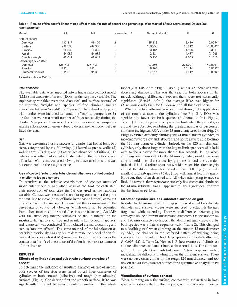
RESULTSEffects of cylinder size and substrate surface on rates ofascentTo determine the influence of substrate diameter on rate of ascent,both species of tree frog were tested on all three diameters ofcylinder on both smooth (adhesive) and rough (non-adhesive)surfaces (Fig. 2). Considering first the smooth surface, ROA wassignificantly different between cylinder diameters in the whole
model (P<0.005, d.f.=2; Fig. 2, Table 1), with ROA increasing withdecreasing diameter. This was the case for both species in themodel. Although differences between them were not statisticallysignificant (P>0.05, d.f.=1), the average ROA was higher forO. septentrionalis than for L. caerulea on all three cylinders.
When effective adhesion was inhibited through the applicationof the rough surface to the cylinders (see Fig. S1), ROA wassignificantly lower for both species (P<0.0001, d.f.=1; Fig. 2,Table 1). Indeed, frogs were only able to climb when they could griparound the substrate, exhibiting the greatest number of successfulclimbs at the highest ROA on the 13 mm diameter cylinder (Fig. 2).Frogs exhibited difficulty climbing the 44 mm diameter cylinder, asmovements were slow and laboured, and no frogs were able to climbthe 120 mm diameter cylinder. Indeed, on the 120 mm diametercylinder, only those frogs with the largest limb span were able holdonto to the substrate for more than a few seconds, falling whenclimbing was attempted. On the 44 mm cylinder, most frogs wereable to hold onto the surface by gripping around the cylinder.Indeed, all had a forelimb span that would have enabled them to griparound the 44 mm diameter cylinder from 188 deg (frog withsmallest forelimb span) to 246 deg (frog with largest forelimb span).However, they often detached and fell when attempting to move alimb. As a result, there were comparatively few successful climbs onthe 44 mm substrate, and all appeared to take a great deal of effortfor the frogs to perform.
Effect of cylinder size and substrate surface on gaitIn order to determine how climbing gait was affected by substratediameter and surface, videos were analysed to establish the gaitfrogs used while ascending. There were differences between gaitsemployed on the different surfaces and diameters. On the smooth 44and 120 mm diameter cylinders, the dominant gait employed byboth species was a ‘lateral sequence walk’ (Table 2). This changedto a ‘walking trot’ when climbing on the smooth 13 mm diametercylinder, the changes in the preferred pattern of walking beingsignificantly different for both frog species (Kruskal–Wallis test,P=0.003, d.f.=2; Table 2). Movies 1–5 show examples of climbs onall three diameters and under both surface conditions. The dominantgait on the rough 13 mm substrate was a ‘lateral sequence walk’,indicating the difficulty in climbing on the different surface. Therewere no successful climbs on the rough 120 mm diameter and toofew on the 44 mm diameter surface for a quantitative analysis to bepossible.
Visualisation of surface contactWhen climbing on a flat surface, contact with the surface in bothspecies was dominated by the toe pads, with subarticular tubercles
Table 1. Results of the best-fit linear mixed-effect model for rate of ascent and percentage of contact of Litoria caerulea and Osteopilusseptentrionalis
Model SS MS Numerator d.f. Denominator d.f. F P
Rate of ascentDiameter 132.817 66.409 2 135.135 5.419 0.0054*Surface 289.366 289.366 1 136.253 23.612 <0.0001*Species 18.336 18.336 1 3.184 1.496 0.304Weight 54.982 54.982 1 3.13 4.487 0.1206Species:Weight 49.811 49.811 1 3.195 4.065 0.1316
Percentage of contactDiameter 22774.2 22774.2 1 97.208 231.007 <0.0001*Species 1983 1983 1 6.302 20.114 0.0037*Diameter:Species 691.3 691.3 1 97.211 7.012 0.0094*
Asterisks indicate P<0.05.
4
RESEARCH ARTICLE Journal of Experimental Biology (2018) 221, jeb168179. doi:10.1242/jeb.168179
Journal
ofEx
perim
entalB
iology
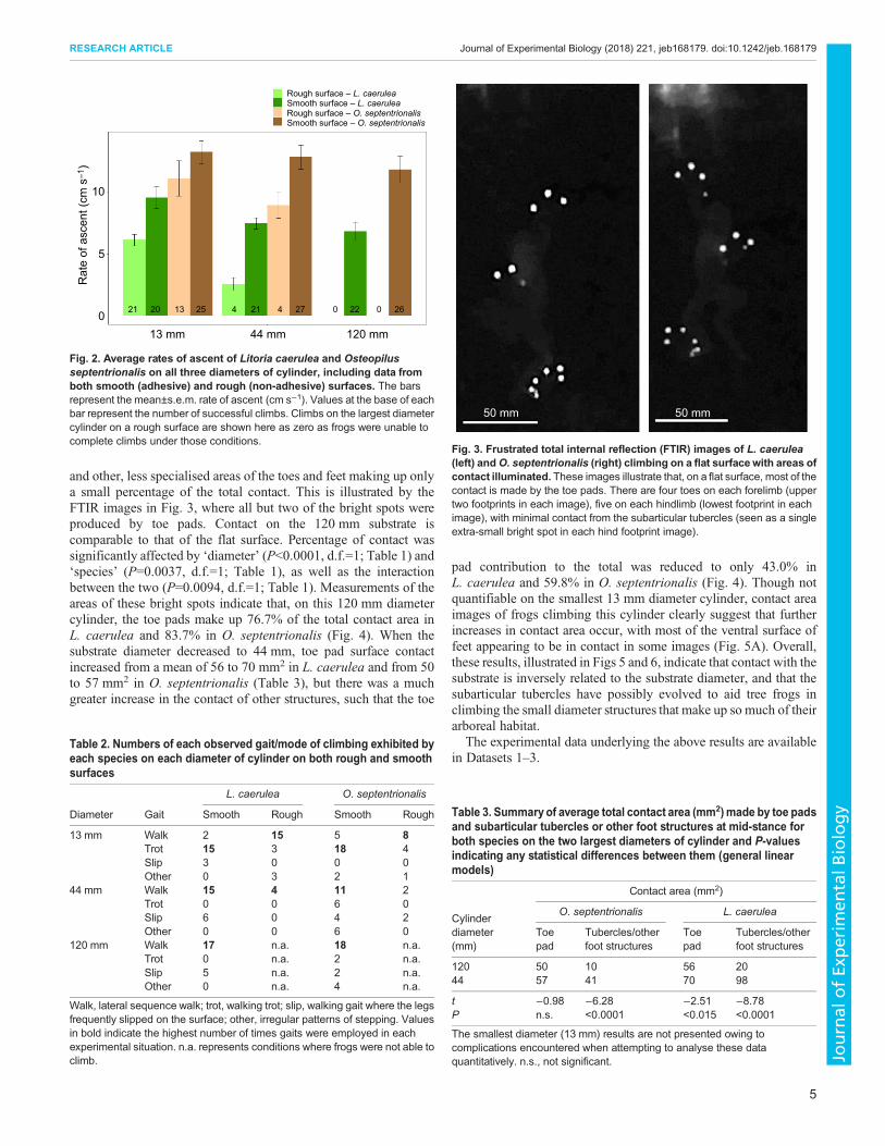
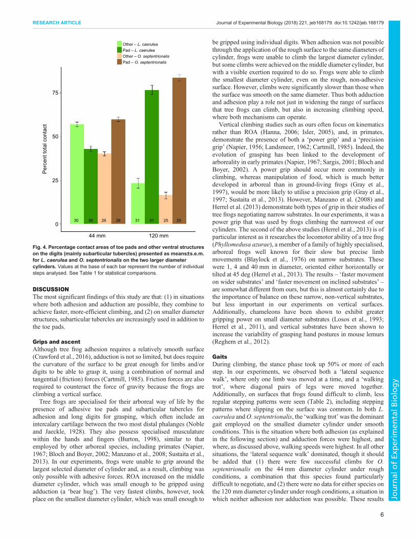
and other, less specialised areas of the toes and feet making up onlya small percentage of the total contact. This is illustrated by theFTIR images in Fig. 3, where all but two of the bright spots wereproduced by toe pads. Contact on the 120 mm substrate iscomparable to that of the flat surface. Percentage of contact wassignificantly affected by ‘diameter’ (P<0.0001, d.f.=1; Table 1) and‘species’ (P=0.0037, d.f.=1; Table 1), as well as the interactionbetween the two (P=0.0094, d.f.=1; Table 1). Measurements of theareas of these bright spots indicate that, on this 120 mm diametercylinder, the toe pads make up 76.7% of the total contact area inL. caerulea and 83.7% in O. septentrionalis (Fig. 4). When thesubstrate diameter decreased to 44 mm, toe pad surface contactincreased from a mean of 56 to 70 mm2 in L. caerulea and from 50to 57 mm2 in O. septentrionalis (Table 3), but there was a muchgreater increase in the contact of other structures, such that the toe
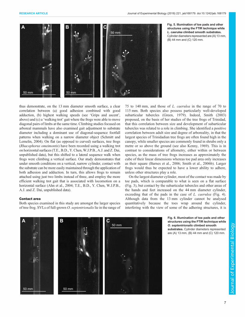
pad contribution to the total was reduced to only 43.0% inL. caerulea and 59.8% in O. septentrionalis (Fig. 4). Though notquantifiable on the smallest 13 mm diameter cylinder, contact areaimages of frogs climbing this cylinder clearly suggest that furtherincreases in contact area occur, with most of the ventral surface offeet appearing to be in contact in some images (Fig. 5A). Overall,these results, illustrated in Figs 5 and 6, indicate that contact with thesubstrate is inversely related to the substrate diameter, and that thesubarticular tubercles have possibly evolved to aid tree frogs inclimbing the small diameter structures that make up so much of theirarboreal habitat.
The experimental data underlying the above results are availablein Datasets 1–3.
Rough surface – L. caeruleaSmooth surface – L. caeruleaRough surface – O. septentrionalisSmooth surface – O. septentrionalis
Rat
e of
asc
ent (
cm s
–1)
21013 mm 44 mm 120 mm
5
10
20 13 25 4 21 4 27 0 022 26
Fig. 2. Average rates of ascent of Litoria caerulea and Osteopilusseptentrionalis on all three diameters of cylinder, including data fromboth smooth (adhesive) and rough (non-adhesive) surfaces. The barsrepresent the mean±s.e.m. rate of ascent (cm s−1). Values at the base of eachbar represent the number of successful climbs. Climbs on the largest diametercylinder on a rough surface are shown here as zero as frogs were unable tocomplete climbs under those conditions.
Table 2. Numbers of each observed gait/mode of climbing exhibited byeach species on each diameter of cylinder on both rough and smoothsurfaces
L. caerulea O. septentrionalis
Diameter Gait Smooth Rough Smooth Rough
13 mm Walk 2 15 5 8Trot 15 3 18 4Slip 3 0 0 0Other 0 3 2 1
44 mm Walk 15 4 11 2Trot 0 0 6 0Slip 6 0 4 2Other 0 0 6 0
120 mm Walk 17 n.a. 18 n.a.Trot 0 n.a. 2 n.a.Slip 5 n.a. 2 n.a.Other 0 n.a. 4 n.a.
Walk, lateral sequence walk; trot, walking trot; slip, walking gait where the legsfrequently slipped on the surface; other, irregular patterns of stepping. Valuesin bold indicate the highest number of times gaits were employed in eachexperimental situation. n.a. represents conditions where frogs were not able toclimb.
50 mm 50 mm
Fig. 3. Frustrated total internal reflection (FTIR) images of L. caerulea(left) andO. septentrionalis (right) climbing on a flat surface with areas ofcontact illuminated. These images illustrate that, on a flat surface, most of thecontact is made by the toe pads. There are four toes on each forelimb (uppertwo footprints in each image), five on each hindlimb (lowest footprint in eachimage), with minimal contact from the subarticular tubercles (seen as a singleextra-small bright spot in each hind footprint image).
Table 3. Summary of average total contact area (mm2)made by toe padsand subarticular tubercles or other foot structures at mid-stance forboth species on the two largest diameters of cylinder and P-valuesindicating any statistical differences between them (general linearmodels)
Contact area (mm2)
Cylinderdiameter(mm)
O. septentrionalis L. caerulea
Toepad
Tubercles/otherfoot structures
Toepad
Tubercles/otherfoot structures
120 50 10 56 2044 57 41 70 98
t −0.98 −6.28 −2.51 −8.78P n.s. <0.0001 <0.015 <0.0001
The smallest diameter (13 mm) results are not presented owing tocomplications encountered when attempting to analyse these dataquantitatively. n.s., not significant.
5
RESEARCH ARTICLE Journal of Experimental Biology (2018) 221, jeb168179. doi:10.1242/jeb.168179
Journal
ofEx
perim
entalB
iology
DISCUSSIONThe most significant findings of this study are that: (1) in situationswhere both adhesion and adduction are possible, they combine toachieve faster, more-efficient climbing, and (2) on smaller diameterstructures, subarticular tubercles are increasingly used in addition tothe toe pads.
Grips and ascentAlthough tree frog adhesion requires a relatively smooth surface(Crawford et al., 2016), adduction is not so limited, but does requirethe curvature of the surface to be great enough for limbs and/ordigits to be able to grasp it, using a combination of normal andtangential (friction) forces (Cartmill, 1985). Friction forces are alsorequired to counteract the force of gravity because the frogs areclimbing a vertical surface.Tree frogs are specialised for their arboreal way of life by the
presence of adhesive toe pads and subarticular tubercles foradhesion and long digits for grasping, which often include anintercalary cartilage between the two most distal phalanges (Nobleand Jaeckle, 1928). They also possess specialised musculaturewithin the hands and fingers (Burton, 1998), similar to thatemployed by other arboreal species, including primates (Napier,1967; Bloch and Boyer, 2002; Manzano et al., 2008; Sustaita et al.,2013). In our experiments, frogs were unable to grip around thelargest selected diameter of cylinder and, as a result, climbing wasonly possible with adhesive forces. ROA increased on the middlediameter cylinder, which was small enough to be gripped usingadduction (a ‘bear hug’). The very fastest climbs, however, tookplace on the smallest diameter cylinder, which was small enough to
be gripped using individual digits. When adhesion was not possiblethrough the application of the rough surface to the same diameters ofcylinder, frogs were unable to climb the largest diameter cylinder,but some climbs were achieved on the middle diameter cylinder, butwith a visible exertion required to do so. Frogs were able to climbthe smallest diameter cylinder, even on the rough, non-adhesivesurface. However, climbs were significantly slower than those whenthe surface was smooth on the same diameter. Thus both adductionand adhesion play a role not just in widening the range of surfacesthat tree frogs can climb, but also in increasing climbing speed,where both mechanisms can operate.
Vertical climbing studies such as ours often focus on kinematicsrather than ROA (Hanna, 2006; Isler, 2005), and, in primates,demonstrate the presence of both a ‘power grip’ and a ‘precisiongrip’ (Napier, 1956; Landsmeer, 1962; Cartmill, 1985). Indeed, theevolution of grasping has been linked to the development ofarboreality in early primates (Napier, 1967; Sargis, 2001; Bloch andBoyer, 2002). A power grip should occur more commonly inclimbing, whereas manipulation of food, which is much betterdeveloped in arboreal than in ground-living frogs (Gray et al.,1997), would be more likely to utilise a precision grip (Gray et al.,1997; Sustaita et al., 2013). However, Manzano et al. (2008) andHerrel et al. (2013) demonstrate both types of grip in their studies oftree frogs negotiating narrow substrates. In our experiments, it was apower grip that was used by frogs climbing the narrowest of ourcylinders. The second of the above studies (Herrel et al., 2013) is ofparticular interest as it researches the locomotor ability of a tree frog(Phyllomedusa azurae), a member of a family of highly specialised,arboreal frogs well known for their slow but precise limbmovements (Blaylock et al., 1976) on narrow substrates. Thesewere 1, 4 and 40 mm in diameter, oriented either horizontally ortilted at 45 deg (Herrel et al., 2013). The results – ‘faster movementon wider substrates’ and ‘faster movement on inclined substrates’ –are somewhat different from ours, but this is almost certainly due tothe importance of balance on these narrow, non-vertical substrates,but less important in our experiments on vertical surfaces.Additionally, chameleons have been shown to exhibit greatergripping power on small diameter substrates (Losos et al., 1993;Herrel et al., 2011), and vertical substrates have been shown toincrease the variability of grasping hand postures in mouse lemurs(Reghem et al., 2012).
GaitsDuring climbing, the stance phase took up 50% or more of eachstep. In our experiments, we observed both a ‘lateral sequencewalk’, where only one limb was moved at a time, and a ‘walkingtrot’, where diagonal pairs of legs were moved together.Additionally, on surfaces that frogs found difficult to climb, lessregular stepping patterns were seen (Table 2), including steppingpatterns where slipping on the surface was common. In both L.caerulea andO. septentrionalis, the ‘walking trot’was the dominantgait employed on the smallest diameter cylinder under smoothconditions. This is the situation where both adhesion (as explainedin the following section) and adduction forces were highest, andwhere, as discussed above, walking speeds were highest. In all othersituations, the ‘lateral sequence walk’ dominated, though it shouldbe added that (1) there were few successful climbs for O.septentrionalis on the 44 mm diameter cylinder under roughconditions, a combination that this species found particularlydifficult to negotiate, and (2) there were no data for either species onthe 120 mm diameter cylinder under rough conditions, a situation inwhich neither adhesion nor adduction was possible. These results
Other – L. caerulea Pad – L. caerulea Other – O. septentrionalis Pad – O. septentrionalis
30 30 26 26 31 31 25 25 0
44 mm
Per
cent
tota
l con
tact
120 mm
25
50
75
Fig. 4. Percentage contact areas of toe pads and other ventral structureson the digits (mainly subarticular tubercles) presented as means±s.e.m.for L. caerulea and O. septentrionalis on the two larger diametercylinders. Values at the base of each bar represent the number of individualsteps analysed. See Table 1 for statistical comparisons.
6
RESEARCH ARTICLE Journal of Experimental Biology (2018) 221, jeb168179. doi:10.1242/jeb.168179
Journal
ofEx
perim
entalB
iology
thus demonstrate, on the 13 mm diameter smooth surface, a clearcorrelation between (a) good adhesion combined with goodadduction, (b) highest walking speeds (see ‘Grips and ascent’,above) and (c) a ‘walking trot’ gait where the frogs were able to movediagonal pairs of limbs at the same time. Climbing studies focused onarboreal mammals have also examined gait adjustment to substratediameter including a dominant use of diagonal-sequence footfallpatterns when walking on a narrow diameter object (Schmitt andLemelin, 2004). On flat (as opposed to curved) surfaces, tree frogs(Rhacophorus omeimontis) have been recorded using a walking troton horizontal surfaces (T.E., B.D., Y. Chen,W.J.P.B., A.J. and Z.Dai,unpublished data), but this shifted to a lateral sequence walk whenfrogs were climbing a vertical surface. Our study demonstrates thatunder smooth conditions on a vertical, narrow cylinder, contact withthe substrate can bemore easily maintained through the application ofboth adhesion and adduction. In turn, this allows frogs to remainattached using just two limbs instead of three, and employ the moreefficient walking trot gait that is associated with locomotion on ahorizontal surface (Ahn et al., 2004; T.E., B.D., Y. Chen, W.J.P.B.,A.J. and Z. Dai, unpublished data).
Contact areaBoth species examined in this study are amongst the larger speciesof tree frog. SVLs of full-grownO. septentrionalis lie in the range of
75 to 140 mm, and those of L. caerulea in the range of 70 to115 mm. Both species also possess particularly well-developedsubarticular tubercles (Green, 1979). Indeed, Smith (2003)proposed, on the basis of her studies of the tree frogs of Trinidad,that this correlation between size and development of subarticulartubercles was related to a role in climbing. She identified a positivecorrelation between adult size and degree of arboreality, in that thelargest species of Trinidadian tree frogs are often found high in thecanopy, while smaller species are commonly found in shrubs only ametre or so above the ground (see also Kenny, 1969). This is incontrast to considerations of allometry, either within or betweenspecies, as the mass of tree frogs increases as approximately thecube of their linear dimensions whereas toe pad area only increasesas their square (Barnes et al., 2006; Smith et al., 2006b). Largerfrogs would thus be expected to have a lower ability to adhere,unless other structures play a role.
On the largest diameter cylinder, most of the contact was made bytoe pads, which is comparable to what is seen on a flat surface(Fig. 3), but contact by the subarticular tubercles and other areas ofthe hands and feet increased on the 44 mm diameter cylinder,exceeding that of the pads in the case of L. caerulea (Fig. 4).Although data from the 13 mm cylinder cannot be analysedquantitatively because the toes wrap around the cylinder,interfering with the view of some of the adhering structures, it is
50 mm 50 mm
50 mmA B C Fig. 5. Illumination of toe pads and other
structures using the FTIR technique whileL. caerulea climbed smooth substrates.Cylinder diameters represented are (A) 13 mm,(B) 44 mm and (C) 120 mm.
50 mm
A B C
50 mm
50 mm
Fig. 6. Illumination of toe pads and otherstructures using the FTIR technique whileO. septentrionalis climbed smoothsubstrates. Cylinder diameters representedare (A) 13 mm, (B) 44 mm and (C) 120 mm.
7
RESEARCH ARTICLE Journal of Experimental Biology (2018) 221, jeb168179. doi:10.1242/jeb.168179
Journal
ofEx
perim
entalB
iology
clear from Figs 5 and 6 that the role of the subarticular tubercles onthe 44 mm diameter cylinder was increased even further on the13 mm diameter cylinder. Indeed, these contact visualisation resultsclearly show that substrate contact is significantly increased onsmaller diameter substrates, this increase being due in part to anincrease in pad contact area, but more importantly in the use ofsubarticular tubercles and other parts of the ventral surface of thefingers and palms. From the viewpoint of anatomy, this change isnot unexpected, as the location of the subarticular tubercles meansthat they will always make contact as the fingers wrap around smalldiameter structures, but much less on a flat surface. Indeed, on flatsurfaces, larger tree frogs do not adhere as well as smaller ones, butthey do adhere better than would be predicted on the basis of theirtoe pad area alone (Barnes et al., 2006). Small diameter substrateswill, however, be common in the natural habitats of most tree frogs,where the subarticular tubercles will play an important role duringclimbing, especially so in large (and therefore heavy) frogs.
Conclusions and further studyIn a related study carried out on a different species of tree frog,Endlein et al. (2017) measured forces from frogs climbing a largecylinder comprising 24 separate three-dimensional force platforms(A.J., S. Yuan, T.E., I.D.C.H., W. Wang, H. Wang, Z. Jiang,W.J.P.B. and Z. Dai, unpublished data) and from individual padsand subarticular tubercles. The experimental findings confirm thepresence of compressive forces, particularly strong when the frogswere climbing a non-adhesive surface, just as the research describedin the present study would predict (performance on 44 mm diametercylinder). Also, as previous studies have shown (e.g. Smith, 2003for tree frogs; Drotlef et al., 2015 for torrent/rock frogs), the surfaceanatomy of the subarticular tubercles is broadly similar to that of thetoe pads, though the grooves separating the epithelial cells are, onthe whole, shallower than in the toe pads, and the nanopillarscovering the surface of these cells are at a lower density in thesubarticular tubercles, though there is a fair amount of variation(Endlein et al., 2017). In spite of the differences, measurements ofthe adhesive and frictional forces of pads and tubercles were broadlysimilar, with friction forces in each case being substantially greaterthan adhesive forces (Endlein et al., 2017).As far as we are aware, this is the first body of work to examine
how different adhesive structures function in regard to tree frogsand the application of both adhesion and friction forces duringclimbing on curved surfaces. Our results demonstrate that, as thediameter of a curved substrate decreases, tree frogs increasinglyrely on adduction and gripping to generate friction forces, theseforces arising from subarticular tubercles as well as toe pads. Thisallows them to meet the challenges posed by the complexities ofan arboreal lifestyle (Cartmill, 1985; Hildebrand and Goslow,2001).The possession of more than one kind of attachment device is
relatively common in animals. Most arboreal lizards (Zani, 2000)and a wide variety of insects (Beutel and Gorb, 2001) possess bothadhesive pads and claws. The latter are most effective on roughsurfaces, failing when the size of the claw tip is greater than the sizeof the asperities that comprise the surface (Dai et al., 2002). Incontrast, adhesive pads function best on smooth surfaces (Zhouet al., 2014; Crawford et al., 2016). In insects, where adhesive padsmay be ‘hairy’ or ‘smooth’ (Federle, 2006), adhesion may occur inmore than one location and serve different functions. For instance,stick insects have ‘toe’ pads (arolia) for adhesion and ‘heel’ pads(euplantulae) for friction (Labonte and Federle, 2013), the aroliabeing the only pads in contact when the insect was hanging upside-
down, whereas when upright in their normal sprawled posture, theinsects rested on their euplantulae. A further example, withsimilarities to the tree frog situation in that the secondary adhesivestructures are located more proximally on the leg and are only usedin special situations, is the possession by leafhoppers of specialtarsal pads (platellae) for jumping from smooth surfaces (Clementeet al., 2017). They generate high friction forces, ensuring that thefeet do not slip during take-off. Such a wide variety of adhesivestructures are giving rise to the development of new adhesivedevices. Materials based on the dry adhesive mechanism of geckoshave led the way. Indeed, such gecko-based synthetic adhesiveshave recently enabled a 70 kg human to scale a vertical glass plate(Hawkes et al., 2015). As far as tree frogs are concerned, possibleapplications include improved design in wet-weather tyres (Barnes,1999; Barnes et al., 2002; Persson, 2007), holding devices forsurgery (Chen et al., 2015), improved friction in safety razors(Tsipenyuk and Varenberg, 2014) and climbing robots (Hayneset al., 2009). The research described here illustrates how, oncylindrical structures, adhesion could be combined with adductionin climbing robots, increasing both the range of materials that can beclimbed and the velocity of climbing.
Previous work examining the ecological implications andevolution of grasping behaviours and limb positioning in arborealspecies have identified this development in primates as, potentially,a product of biomechanical accommodations to differing substratesizes in arboreal environments (Schmitt, 2003). However, suchskilled forelimb movements are more common in tetrapod taxa(Iwaniuk and Whishaw, 2000; Sustaita et al., 2013), indicating acommon origin in early tetrapods, and may have resulted from food-handling behaviours (Gray et al., 1997; Iwaniuk and Whishaw,2000). Multiple reptile species have been found to exhibit differingclimbing strategies in response to ecological niches, such as partialarborealism and canopy position determining the size and nature ofthe substrates encountered (Losos and Sinervo, 1989; Losos et al.,1993; Zaaf et al., 2001a; Spezzano and Jayne, 2004; Vanhooydoncket al., 2005, 2006; Renous et al., 2010; Foster and Higham, 2012).Further investigation into how tree frogs make use of their naturalenvironment could yield better insights into how they specificallyapply the adaptive climbing qualities indicated by this study. Thedifferences observed here between L. caerulea and O.septentrionalis were relatively small, but L. caerulea has beenshown to be significantly less able to negotiate narrow structurescompared with Phyllomedusa (Manzano et al., 2008). Thus, likereptiles, tree frogs can be expected to show differences in climbingperformance that reflect their ecological niche utilisation (Losos andSinervo, 1989; Losos et al., 1993; Zaaf et al., 2001b; Irschick andGarland, 2001).
AcknowledgementsWe thank Tiffany Armstrong for her help and advice during the course of this study.
Competing interestsThe authors declare no competing or financial interests.
Author contributionsConceptualization: W.J.P.B., A.J., T.E.; Methodology: I.D.C.H., B.D., W.J.P.B., A.J.,T.E.; Software: I.D.C.H., T.E.; Validation: T.E.; Formal analysis: I.D.C.H.;Investigation: I.D.C.H., B.D., W.J.P.B., A.J.; Resources: W.J.P.B.; Data curation:I.D.C.H.; Writing - original draft: I.D.C.H.; Writing - review & editing: I.D.C.H.,W.J.P.B., T.E.; Visualization: I.D.C.H., B.D., A.J.; Supervision: W.J.P.B., A.J.;Project administration: W.J.P.B., A.J.; Funding acquisition: W.J.P.B., A.J., T.E.
FundingThis study was supported by a grant from the Royal Society, London (InternationalExchange Scheme 2014/R2: reference IE140717) awarded to W.J.P.B. and A.J.,
8
RESEARCH ARTICLE Journal of Experimental Biology (2018) 221, jeb168179. doi:10.1242/jeb.168179
Journal
ofEx
perim
entalB
iology
and by grants from the National Natural Science Foundation of China (grant no.51375232) and the Natural Science Foundation of Jiangsu Province (grant no.BK20141410) awarded to A.J.
Supplementary informationSupplementary information available online athttp://jeb.biologists.org/lookup/doi/10.1242/jeb.168179.supplemental
ReferencesAhn, A. N., Furrow, E. and Biewener, A. A. (2004). Walking and running in the red-legged running frog, Kassina maculata. J. Exp. Biol. 207, 399-410.
Astley, H. C. and Roberts, T. J. (2014). The mechanics of elastic loading and recoilin anuran jumping. J. Exp. Biol. 217, 4372-4378.
Astley, H. C., Haruta, A. and Roberts, T. J. (2015). Robust jumping performanceand elastic energy recovery from compliant perches in tree frogs. J. Exp. Biol. 218,3360-3363.
Autumn, K. (2007). Gecko adhesion: structure, function, and applications. Mater.Res. Soc. Bull. 32, 473-478.
Autumn, K. and Hansen, W. (2006). Ultrahydrophobicity indicates a non-adhesivestate in gecko setae. J. Comp. Physiol. A 192, 1205-1212.
Autumn, K. and Peattie, A. M. (2002). Mechanisms of adhesion in geckos. Integr.Comp. Biol. 42, 1081-1090.
Barnes, W. J. P. (1999). Tree frogs and tire technology. In Tire TechnologyInternational. March ‘99 (ed. A. Baker and J. Lawson), pp. 42-47. Dorking, UK: UK& International Press.
Barnes, W. J. P. (2007). Biomimetic solutions to sticky problems. Science 318,203-204.
Barnes, W. J. P., Smith, J., Oines, C. and Mundl, R. (2002). Bionics and wet grip.In Tire Technology International. December ‘02 (ed. A. Baker and J. Lawson),pp. 56-60. Dorking, UK: UK & International Press.
Barnes, W. J. P., Oines, C. and Smith, J. M. (2006). Whole animal measurementsof shear and adhesive forces in adult tree frogs: insights into underlyingmechanisms of adhesion obtained from studying the effects of size and scale.J. Comp. Physiol. A 192, 1179-1191.
Betts, R. P., Duckworth, T., Austin, I. G., Crocker, S. P. and Moore, S. (1980).Critical light reflection at a plastic/glass interface and its application to footpressure measurements. J. Med. Eng. Technol. 4, 136-142.
Beutel, R. G. andGorb, S. N. (2001). Ultrastructure of attachment specializations ofhexapods (Arthropoda): evolutionary patterns inferred from a revised ordinalphylogeny. J. Zool. Syst. Evol. Res. 39, 177-207.
Bijma, N. N., Gorb, S. N. and Kleinteich, T. (2016). Landing on branches in the frogTrachycephalus resinifictrix (Anura: Hylidae). J. Comp. Physiol. A 202, 267-276.
Blaylock, L., Ruibal, R. and Platt-Aloia, K. (1976). Skin structure and wipingbehaviour of Phyllomedusinae frogs. Copeia 2, 283-295.
Bloch, J. I. and Boyer, D. M. (2002). Grasping primate origins. Science 298,1606-1610.
Burton, T. C. (1998). Are the distal extensor muscles of the fingers of anurans anadaptation to arboreality? J. Herpetol. 32, 611-617.
Butt, H.-J., Barnes, W. J. P., del Campo, A., Kappl, M. and Schonfeld, F. (2010).Capillary forces between soft, elastic spheres. Soft Matter 6, 5930-5936.
Cartmill, M. (1985). Climbing. In Functional Vertebrate Morphology (ed. M.Hildebrand, D. M. Bramble, K. F. Liem and D. B. Wake), pp. 73-88. Cambridge,MA: Belknap Press.
Chen, H., Zhang, L., Zhang, D., Zhang, P. and Han, Z. (2015). Bio-inspired surfacefor surgical graspers based on the strong wet friction of tree frog toe pads. ACSAppl. Mater. Interfaces 7, 13987-13995.
Clemente, C. J., Goetzke, H. H., Bullock, J. M. R., Sutton, G. P. Burrows, M. andFederle, W. (2017). Jumping without slipping: leafhoppers (Hemiptera:Cicadellidae) possess special tarsal structures for jumping from smoothsurfaces. J. R. Soc. Interface 14, 20170022.
Crawford, N., Endlein, T. and Barnes, W. J. P. (2012). Self-cleaning in tree frog toepads; a mechanism for recovering from contamination without the need forgrooming. J. Exp. Biol. 215, 3965-3972.
Crawford, N., Endlein, T., Pham, J. T., Riehle, M. and Barnes, W. J. P. (2016).When the going gets rough – studying the effect of surface roughness on theadhesive abilities of tree frogs. Beilstein J. Nanotech. 7, 2116-2131.
Dai, Z., Gorb, S. N. and Schwarz, U. (2002). Roughness-dependent friction force ofthe tarsal claw system in the beetle Pachnoda marginata (Coleoptera,Scarabaeidae). J. Exp. Biol. 205, 2479-2488.
Drotlef, D.-M., Stepien, l., Kappl, M., Barnes, W. J. P., Butt, H.-J. and del Campo,A. (2012). Insights into the adhesive mechanisms of tree frogs using artificialmimics. Adv. Funct. Mater. 9, 1094.
Drotlef, D. M., Appel, E., Peisker, H., Dening, K., del Campo, A., Gorb, S. N. andBarnes, W. J. P. (2015). Morphological studies of the toe pads of the rock frog,Staurois parvus, (family Ranidae) and their relevance to the development of newbiomimetically inspired reversible adhesives. Interface Focus 5, 20140036.
Duellman, W. E., Marion, A. B. and Hedges, S. B. (2016). Phylogenetics,classification, and biogeography of the tree frogs (Amphibia: Anura: Arboranae).Zootaxa 4104, 1-109.
Eason, E. V., Hawkes, E. W., Windheim, M., Christensen, D. L., Libby, T. andCutkosky, M. R. (2015). Stress distribution and contact area measurements of agecko toe using a high resolution tactile sensor. Bioinspir. Biomim. 10, 016013.
Emerson, S. B. and Diehl, D. (1980). Toe pad morphology and mechanisms ofsticking in tree frogs. Biol. J. Linn. Soc. 13, 199-216.
Endlein, T., Barnes, W. J. P., Samuel, D. S., Crawford, N. A., Biaw, A. B. andGrafe, U. (2013). Sticking under wet conditions: the remarkable attachmentabilities of the torrent frog, Staurois guttatus. PLoS One 9, e73810.
Endlein, T., Ji, A., Yuan, S., Hill, I. D. C., Wang, H., Barnes, W. J. P., Dai, Z. andSitti, M. (2017). The use of clamping grips by tree frogs for climbing curvedsurfaces. Proc. Roy. Soc. Lond. B 284, 20162867.
Federle, W. (2006). Why are so many adhesive pads hairy? J. Exp. Biol. 209,2611-2621.
Federle, W. and Endlein, T. (2004). Locomotion and adhesion: dynamic control ofadhesive surface contact in ants. Arthropod Struct. Dev. 33, 67-75.
Federle, W., Barnes, W. J. P., Baumgartner, W., Dreschler, P. and Smith, J. M.(2006). Wet but not slippery – boundary friction in tree frog adhesive toe pads.J. Roy. Soc. Interface 3, 689-697.
Foster, K. L. and Higham, T. E. (2012). How forelimb and hindlimb functionchanges with incline and perch diameter in the green anole, Anolis carolinensis.J. Exp. Biol. 215, 2288-2300.
Gray, L. A., O’Reilly, J. C. and Nishikawa, K. C. (1997). Evolution of forelimbmovement patterns for prey manipulation in anurans. J. Exp. Zool. 277, 417-424.
Green, D. M. (1979). Treefrog toe pads: comparative surface morphology usingscanning electron microscopy. Can. J. Zool. 57, 2033-2046.
Hanna, G. andBarnes,W. J. P. (1991). Adhesion and detachment of the toe pads oftree frogs. J. Exp. Biol. 155, 103-125.
Hanna, J. B. (2006). Kinematics of vertical climbing in lorises and Cheirogaleusmedius. J. Hum. Evol. 50, 469-478.
Hansen, W. and Autumn, K. (2005). Evidence for self-cleaning in gecko setae.Proc. Nat. Acad. Sci. USA 102, 103-125.
Hawkes, E. W., Eason, E. V., Christensen, D. L. and Cutkosky, M. R. (2015).Human climbing with efficiently ascaled gecko-inspired dry adhesives. J. Roy.Soc. Interface 12, 20140675.
Haynes, G. C., Khripin, A., Lynch, G., Amory, J., Saunders, A., Rizzi, A. A. andKoditschek, D. E. (2009). Rapid pole climbing with a quadrupedal robot. In IEEEInternational Conference on Robotics and Automation, 2009, pp. 2767-2772.Kobe, Japan: IEEE.
Herrel, A., Measey, G. J., Vanhooydonck, B. and Tolley, K. A. (2011). Functionalconsequences of morphological differentiation between populations of the Capedwarf chameleon (Bradypodion pumilum). Biol. J. Linn. Soc. 104, 692-700.
Herrel, A. Perrenoud, M., Decamps, T., Abdala, V., Manzano, A. and Pouydebat,E. (2013). The effect of substrate diameter and incline on locomotion in anarboreal frog. J. Exp. Biol. 216, 3599-3605.
Hildebrand, M. (1985). Walking and running. In Functional Vertebrate Morphology(ed. M. Hildebrand, D. M. Bramble, K. F. Liem and D. B. Wake), pp. 38-57.Cambridge, MA: Belknap Press.
Hildebrand, M. and Goslow, G. (2001). Analysis of Vertebrate Structure, 5th edn.New York: John Wiley and Sons.
Hu, S., Lopez, S., Niewiarowski, P. H. and Xia, Z. (2012). Dynamic self-cleaning ingecko setae via digital hyperextension. J. R. Soc. Interface 9, 2781-2790.
Irschick, D. J. and Garland, T.Jr (2001). Integrating function and ecology in studiesof adaptation: investigations of locomotor capacity as amodel system. Annu. Rev.Ecol. Syst. 32, 367-396.
Isler, K. (2005). 3D-kinematics of vertical climbing in hominoids. Am. J. Phys.Anthropol. 126, 66-81.
Iwaniuk, A. N. and Whishaw, I. Q. (2000). On the origin of skilled forelimbmovements. Trends Neurosci. 23, 372-376.
Kenny, J. S. (1969). The Amphibia of Trinidad. Stud. Fauna Curacao Carib. Isl. 103,1-77.
Labonte, D. and Federle, W. (2013). Functionally different pads on the same footallow control of attachment: stick insects have load-sensitive “heel” pads forfriction and shear-sensitive “toe” pads for adhesion. PLoS ONE 8, e81943.
Landsmeer, J. M. F. (1962). Power grip and precision handling. Ann. Rheum. Dis.21, 164-170.
Losos, J. B. and Sinervo, B. (1989). The effects of morphology and perch diameteron sprint performance of Anolis lizards. J. Exp. Biol. 145, 23-30.
Losos, J. B., Walton, B. M. andBennett, A. F. (1993). Trade-offs between sprintingand clinging ability in Kenyan chameleons. Funct. Ecol. 7, 281-286.
Manzano, A. S., Abdala, V. and Herrel, A. (2008). Morphology and function of theforelimb in arboreal frogs: specializations for grasping ability? J. Anat. 213,296-307.
Napier, J. R. (1956). The prehensile movements of the human hand. J. Bone JointSurg. 38B, 902-913.
Napier, J. R. (1967). Evolutionary aspects of primate locomotion. Am. J. Phys.Anthropol. 27, 333-342.
Noble, G. K. and Jaeckle, M. E. (1928). The digital pads of the tree frogs. A study ofthe phylogenesis of an adaptive structure. J. Morph. 45, 259-292.
Persson, B. N. J. (2007). Wet adhesion with application to tree frog adhesive toepads and tires. J. Phys. Condens. Mat. 19, 376110.
9
RESEARCH ARTICLE Journal of Experimental Biology (2018) 221, jeb168179. doi:10.1242/jeb.168179
Journal
ofEx
perim
entalB
iology
Reghem, E., Byron, C., Bels, V. and Pouydebat, E. (2012). Hand posture in thegrey mouse lemur during arboreal locomotion on narrow branches. J. Zool. 288,76-81.
Renous, S., Hofling, E. and da Rocha, P. L. B. (2010). Effect of substrate on thelocomotion behaviour of the South American iguanian lizard Polychrusacutirostris. Ital. J. Zool. 77, 216-226.
Sargis, E. J. (2001). The grasping behaviour, locomotion and substrate use of thetree shrews Tupaia minor and T. tana (Mammalia, Scandentia). J. Zool., Lond.253, 485-490.
Schmitt, D. (2003). Substrate size and primate forelimb mechanics: implications forunderstanding the evolution of primate locomotion. Int. J. Primatol. 24, 1023-1036.
Schmitt, D. and Lemelin, P. (2004). Locomotor mechanics of the slender loris(Loris tardigradus). J. Hum. Evol. 47, 85-94.
Scholz, I., Barnes, W. J. P., Smith, J. M. and Baumgartner, W. (2009).Ultrastructure and physical properties of an adhesive surface, the toe padepithelium of the tree frog, Litoria caerulea White. J. Exp. Biol. 212, 155-162.
Smith, J. M. (2003).Effect of allometric growth and toe padmorphology on adhesionin hylid tree frogs. PhD thesis, University of Glasgow.
Smith, J. M., Barnes, W. J. P., Downie, J. R. and Ruxton, G. D. (2006a). Structuralcorrelates of increased adhesive efficiency in the toe pads of hylid tree frogs.J. Comp. Physiol. 192, 1193-1204.
Smith, J. M., Barnes, W. J. P., Downie, J. R. and Ruxton, G. D. (2006b). Adhesionand allometry from metamorphosis to maturation in hylid tree frogs: a stickyproblem. J. Zool. 270, 372-383.
Spezzano, L. C. and Jayne, B. C. (2004). The effects of surface diameter andincline on the hindlimb kinematics of an arboreal lizard (Anolis sagrei). J. Exp. Biol.207, 2115-2131.
Sustaita, D., Pouydebat, E., Manzano, A., Abdala, V. Hertel, F. and Herrel, A.(2013). Getting a grip on tetrapod grasping: form, function, and evolution. Biol.Rev. 88, 380-405.
Tsipenyuk, A. and Varenberg, M. (2014). Use of biomimetic surface texture infriction against lubricated skin. J.R. Soc Interface 11, 20140113.
Vanhooydonck, B., Andronescu, A., Herrel, A. and Irshick, D. J. (2005). Effectsof substrate structure on speed and acceleration capacity in climbing geckos.Biol. J. Linn. Soc. 85, 385-393.
Vanhooydonck, B., Herrel, A. and Irschick, D. J. (2006). Out on a limb: thedifferential effect of substrate diameter on acceleration capacity in Anolis lizards.J. Exp. Biol. 209, 4515-4523.
Xia, Z. (2016). Biomimetic Principles and Design of Advanced EngineeringMaterials. New York: John Wiley & Sons.
Zaaf, A., Van Damme, R., Herrel, A. and Aerts, P. (2001a). Limb jointkinematics during vertical climbing and level running in a specialist climber:Gekko gecko Linneus, 1758 (Lacertilia: Gekkonidae). Belg. J. Zool. 131,173-182.
Zaaf, A., Van Damme, R., Herrel, A. and Aerts, P. (2001b). Spatio-temporal gaitcharacteristics of level and vertical locomotion in a ground-dwelling and a climbinggecko. J. Exp. Biol. 204, 1233-1246.
Zani, P. A. (2000). The comparative evolution of lizard claw and toe morphology andclinging performance. J. Evolution. Biol. 13, 316-325.
Zhou, Y., Robinson, A., Steiner, U. and Federle, W. (2014). Insect adhesion onrough surfaces: analysis of adhesive contact of smooth and hairy pads ontransparent microstructured substrates. J. R. Soc. Interface 11, 20140499.
10
RESEARCH ARTICLE Journal of Experimental Biology (2018) 221, jeb168179. doi:10.1242/jeb.168179
Journal
ofEx
perim
entalB
iology