Embed Size (px)
Citation preview

Depressive disorders affect ~20% of people in the United States within their lifetime1, and roughly one-half of patients do not fully respond to available treatments2. The behavioural symptoms of depression are extensive, covering emotional, motivational, cognitive and physio-logical domains. Large subsets of patients with these disorders exhibit deficits in several aspects of reward, as defined by responses to positive emotional stimuli such as food, sex and social interaction. Prominent among these reward deficits is anhedonia. Depression is highly comorbid with anxiety, and it has been estimated that over 20% of individuals with a mood or anxiety disorder also fulfil criteria for drug addiction and, conversely, that 30–40% of individuals suffering from addictive disorders have a comorbid mood or anxiety disorder3. These con-siderations suggest a large degree of overlap among the brain regions affected in depression and those affected in drug addiction.
Indeed, increasing evidence in humans and animals suggests that mood disorders and drug addiction are associated with major disruptions within the brain’s reward circuitry4, which normally serves to guide our attention towards and consumption of natural rewards and ensure our survival. We begin this Review by pro-viding a brief overview of reward circuits in the brain and then summarize findings from studies in humans, which suggest that structural and functional changes in this reward circuitry are associated with reward-related behavioural impairments in mood disorders. We then focus on the more extensive rodent and non-human pri-mate literature to formulate a circuit-level understanding
of reward deficits in these disorders, highlighting the molecular and neurophysiological mechanisms that drive these circuit abnormalities. We end by discussing future directions for research, particularly the oppor-tunities for the development of novel therapeutics that target these reward-based deficits.
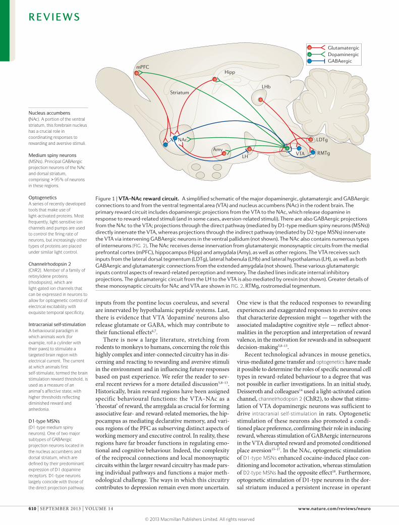
The brain’s reward circuitryThe best characterized reward circuit in the brain com-prises dopaminergic neurons in the ventral tegmental area (VTA) that project to the nucleus accumbens (NAc), which is part of the ventral striatum. The principal neu-rons of the NAc are GABAergic medium spiny neurons (MSNs). This VTA–NAc circuit is crucial for the recog-nition of rewards in the environment and for initiating their consumption5, but these regions respond to aver-sive stimuli as well (see below for further discussion). VTA dopaminergic neurons also innervate several regions of the prefrontal cortex (PFC), central amyg-dala, basolateral amygdala (BLA) and hippocampus, as well as other areas (FIG. 1). All of these so-called ‘brain reward regions’ are inter-connected in complex ways: for example, the NAc receives dense glutamatergic innervation from the PFC, amygdala and hippocam-pus; and the PFC, amygdala and hippocampus form reciprocal glutamatergic connections with one another. The functional output of each of these regions is mod-ulated by several types of GABAergic interneurons and, in the NAc, by cholinergic interneurons as well. Moreover, each of these regions receives serotonergic inputs from midbrain raphe nuclei and noradrenergic
Fishberg Department of Neuroscience and Friedman Brain Institute, Icahn School of Medicine at Mount Sinai, New York, New York 10029, USA.Correspondence to S.J.R. e-mail: [email protected]:10.1038/nrn3381 Published online 14 August 2013
RewardA positive emotional stimulus. In psychological terms, a reward is reinforcing — it promotes repeated responding to obtain the same stimulus.
AnhedoniaLoss of the ability to experience pleasure from normally rewarding stimuli, such as food, sex and social interactions.
Ventral tegmental area(VTA). A ventral midbrain site containing dopaminergic neurons that are an essential component of the brain’s reward circuitry.
The brain reward circuitry in mood disordersScott J. Russo and Eric J. Nestler
Abstract | Mood disorders are common and debilitating conditions characterized in part by profound deficits in reward-related behavioural domains. A recent literature has identified important structural and functional alterations within the brain’s reward circuitry — particularly in the ventral tegmental area–nucleus accumbens pathway — that are associated with symptoms such as anhedonia and aberrant reward-associated perception and memory. This Review synthesizes recent data from human and rodent studies from which emerges a circuit-level framework for understanding reward deficits in depression. We also discuss some of the molecular and cellular underpinnings of this framework, ranging from adaptations in glutamatergic synapses and neurotrophic factors to transcriptional and epigenetic mechanisms.
R E V I E W S
NATURE REVIEWS | NEUROSCIENCE VOLUME 14 | SEPTEMBER 2013 | 609
© 2013 Macmillan Publishers Limited. All rights reserved
Nature Reviews | Neuroscience
LHb
GABAergicDopaminergicGlutamatergic
mPFCHipp
RMTgVTA
LDTgNAc
AmyLH
Striatum
Nucleus accumbens(NAc). A portion of the ventral striatum, this forebrain nucleus has a crucial role in coordinating responses to rewarding and aversive stimuli.
Medium spiny neurons(MSNs). Principal GABAergic projection neurons of the NAc and dorsal striatum, comprising >95% of neurons in these regions.
OptogeneticsA series of recently developed tools that make use of light-activated proteins. Most frequently, light-sensitive ion channels and pumps are used to control the firing rate of neurons, but increasingly other types of proteins are placed under similar light control.
Channelrhodopsin 2(ChR2). Member of a family of retinylidene proteins (rhodopsins), which are light-gated ion channels that can be expressed in neurons to allow for optogenetic control of electrical excitability with exquisite temporal specificity.
Intracranial self-stimulationA behavioural paradigm in which animals work (for example, roll a cylinder with their paws) to stimulate a targeted brain region with electrical current. The current at which animals first self-stimulate, termed the brain stimulation reward threshold, is used as a measure of an animal’s affective state, with higher thresholds reflecting diminished reward and anhedonia.
D1-type MSNs(D1-type medium spiny neurons). One of two major subtypes of GABAergic projection neurons located in the nucleus accumbens and dorsal striatum, which are defined by their predominant expression of D1 dopamine receptors. D1-type neurons largely coincide with those of the direct projection pathway.
inputs from the pontine locus coeruleus, and several are innervated by hypothalamic peptide systems. Last, there is evidence that VTA ‘dopamine’ neurons also release glutamate or GABA, which may contribute to their functional effects6,7.
There is now a large literature, stretching from rodents to monkeys to humans, concerning the role this highly complex and inter-connected circuitry has in dis-cerning and reacting to rewarding and aversive stimuli in the environment and in influencing future responses based on past experience. We refer the reader to sev-eral recent reviews for a more detailed discussion5,8–13. Historically, brain reward regions have been assigned specific behavioural functions: the VTA–NAc as a ‘rheostat’ of reward, the amygdala as crucial for forming associative fear- and reward-related memories, the hip-pocampus as mediating declarative memory, and vari-ous regions of the PFC as subserving distinct aspects of working memory and executive control. In reality, these regions have far broader functions in regulating emo-tional and cognitive behaviour. Indeed, the complexity of the reciprocal connections and local monosynaptic circuits within the larger reward circuitry has made pars-ing individual pathways and functions a major meth-odological challenge. The ways in which this circuitry contributes to depression remain even more uncertain.
One view is that the reduced responses to rewarding experiences and exaggerated responses to aversive ones that characterize depression might — together with the associated maladaptive cognitive style — reflect abnor-malities in the perception and interpretation of reward valence, in the motivation for rewards and in subsequent decision-making5,8–13.
Recent technological advances in mouse genetics, virus-mediated gene transfer and optogenetics have made it possible to determine the roles of specific neuronal cell types in reward-related behaviour to a degree that was not possible in earlier investigations. In an initial study, Deisseroth and colleagues14 used a light-activated cation channel, channelrhodopsin 2 (ChR2), to show that stimu-lation of VTA dopaminergic neurons was sufficient to drive intracranial self-stimulation in rats. Optogenetic stimulation of these neurons also promoted a condi-tioned place preference, confirming their role in inducing reward, whereas stimulation of GABAergic interneurons in the VTA disrupted reward and promoted conditioned place aversion15–17. In the NAc, optogenetic stimulation of D1-type MSNs enhanced cocaine-induced place con-ditioning and locomotor activation, whereas stimulation of D2-type MSNs had the opposite effect18. Furthermore, optogenetic stimulation of D1-type neurons in the dor-sal striatum induced a persistent increase in operant
Figure 1 | VTA–NAc reward circuit. A simplified schematic of the major dopaminergic, glutamatergic and GABAergic connections to and from the ventral tegmental area (VTA) and nucleus accumbens (NAc) in the rodent brain. The primary reward circuit includes dopaminergic projections from the VTA to the NAc, which release dopamine in response to reward-related stimuli (and in some cases, aversion-related stimuli). There are also GABAergic projections from the NAc to the VTA; projections through the direct pathway (mediated by D1-type medium spiny neurons (MSNs)) directly innervate the VTA, whereas projections through the indirect pathway (mediated by D2-type MSNs) innervate the VTA via intervening GABAergic neurons in the ventral pallidum (not shown). The NAc also contains numerous types of interneurons (FIG. 2). The NAc receives dense innervation from glutamatergic monosynaptic circuits from the medial prefrontal cortex (mPFC), hippocampus (Hipp) and amygdala (Amy), as well as other regions. The VTA receives such inputs from the lateral dorsal tegmentum (LDTg), lateral habenula (LHb) and lateral hypothalamus (LH), as well as both GABAergic and glutamatergic connections from the extended amygdala (not shown). These various glutamatergic inputs control aspects of reward-related perception and memory. The dashed lines indicate internal inhibitory projections. The glutamatergic circuit from the LH to the VTA is also mediated by orexin (not shown). Greater details of these monosynaptic circuits for NAc and VTA are shown in FIG. 2. RTMg, rostromedial tegmentum.
R E V I E W S
610 | SEPTEMBER 2013 | VOLUME 14 www.nature.com/reviews/neuro
© 2013 Macmillan Publishers Limited. All rights reserved

From LDTg
Nature Reviews | Neuroscience
SOMinterneuron
CALB2 interneuron
PV interneuron
FrommPFC
From BLA
From extended amygdala
From VTA
From hippocampus
D1 or D2MSN
From LHbFrom mPFC
From NAc
From lateral hypothalamus
FromRMTg
–
–
–
a Nucleus accumbens
b Ventral tegmental area
+
+
+/–
++
+
++
+
+
–
–
–
DA neuron
GlutamatergicDopaminergicGABAergicCholinergicOrexinergic
ChAT interneuron
GABAergic interneuron
+
+
lever pressing, whereas stimulation of D2-type neurons induced a transient decrease19. This and related studies support the view that the dorsal striatum is also a crucial site for integrating behavioural responses to both positive reinforcement and punishment20.
A recent study demonstrated the potent ability of optogenetically activated cholinergic interneurons in the NAc to promote the rewarding effects of cocaine21, even though these interneurons comprise <1% of all neurons in this region. Perhaps surprisingly, inhibition of the cholinergic interneurons by a small and previ-ously unappreciated GABAergic projection from the VTA enhanced stimulus–outcome learning22. Further work is needed to clarify these findings, but they are interesting in light of pharmacological studies in rodents and monkeys showing that cholinergic agonists are rewarding through their actions within the VTA–NAc circuit8,23. Circuit-specific tools are also being used to study the influence of distinct glutamatergic projec-tions to the NAc (FIG. 2a) on complex behaviour24–27. As just one example, optogenetic activation of NAc gluta-matergic terminals that derive from the BLA and PFC was reinforcing, although the firing frequency neces-sary to support reinforcement was different between the two regions24,27.
Recent evidence also points to a role for lateral habenula (LHb) and rostromedial tegmental nucleus (RMTg) neurons in mediating aversion. Specifically, a subset of GABAergic neurons in the RMTg28 — those that project to VTA dopaminergic neurons — receive excitatory projections from LHb neurons that specifi-cally respond to aversive stimuli28 (FIG. 2b). Optogenetic stimulation of LHb glutamatergic terminals in the RMTg inhibited firing of VTA dopaminergic neurons, inducing a greater degree of behavioural avoidance in response to an unpredictable footshock28. VTA dopaminergic neu-rons themselves are far more heterogeneous than was previously believed: distinct subsets of VTA dopamin-ergic neurons respond differently to rewarding versus aversive stimuli29,30, and there are topographical (ante-rior versus posterior) differences in the influence of VTA dopaminergic and GABAergic neurons in regulating reward31,32.
Together, these studies illustrate how genetic, viral vector and optogenetic tools are leading to the formula-tion of fundamentally new hypotheses regarding reward circuitry function. Such work highlights the cell- and circuit-specificity of neural pathways and neurotrans-mitter systems that control the VTA and NAc. It is clear that many regions that are not classically defined as ‘reward structures’ feed onto this circuitry and con-tribute to the processing of both positive and negative emotional information to guide complex behaviour. With this in mind, we can begin to formulate a detailed circuit-level understanding of reward-related deficits in mood disorders.
Brain imaging studies in humansThe brain imaging literature with regard to depres-sion is extensive and has been reviewed elsewhere in greater detail33. Here, we highlight only those major
Figure 2 | Local microcircuitry of the NAc and VTA. a | A close-up view detailing the presynaptic inputs onto D1- and D2-type GABAergic medium spiny neurons (MSNs) and onto several types of interneurons within the nucleus accumbens (NAc). These interneurons include GABAergic interneurons that express calretinin (CALB2), parvalbumin (PV), somatostatin (SOM) or calbindin (CALB1; not shown) and large cholinergic interneurons that express choline acetyltransferase (ChAT). Glutamatergic neurons from the medial prefrontal cortex (mPFC), hippocampus and basolateral amygdala (BLA) release glutamate onto spine synapses to provide excitatory signals to GABAergic MSN projection neurons. These excitatory inputs also synapse directly onto the GABAergic and cholinergic interneurons that modulate MSNs (not shown). D1- and D2-type MSNs also receive signals from dopamine (DA) through shaft or spine neck synapses. The figure does not depict possible differences in glutamatergic innervation of D1-type versus D2-type MSNs, which are only now beginning to be explored. b | A close-up view detailing the presynaptic inputs onto ventral tegmental area (VTA) DA neurons and local GABAergic interneurons in the VTA. Glutamatergic neurons from the mPFC and lateral dorsal tegmentum (LDTg) synapse directly onto VTA DA neurons, whereas the extended amygdala sends both glutamatergic and GABAergic projections. By contrast, glutamatergic neurons from the lateral habenula (LHb) synapse directly onto inhibitory GABAergic neurons in the rostromedial tegmentum (RMTg) or VTA proper, which then inhibit DA neurons and promote aversion. DA neurons receive direct excitatory inputs from peptidergic (for example, orexinergic) or glutamatergic neurons in the lateral hypothalamus, which increase DA release and promote reward. Although GABAergic projections from the NAc to the VTA are shown as direct, in fact, roughly half of these projections are indirect, occurring via GABAergic neurons in ventral pallidum (not shown). Moreover, although GABAergic projections from the NAc to the VTA are shown innervating VTA DA neurons, these projections also innervate VTA GABA neurons (not shown).
R E V I E W S
NATURE REVIEWS | NEUROSCIENCE VOLUME 14 | SEPTEMBER 2013 | 611
© 2013 Macmillan Publishers Limited. All rights reserved

D2-type MSNs(D2-type medium spiny neurons). One of two major subtypes of GABAergic projection neurons located in the nucleus accumbens and dorsal striatum, which are defined by their predominant expression of D2 dopamine receptors. D2 type-neurons largely coincide with those of the indirect projection pathway.
Excitatory synapsesSynapses at which the release of glutamate from presynaptic nerve terminals activates glutamate receptors located on dendritic spines on postsynaptic neurons, which increases the probability of an action potential in that postsynaptic neuron.
findings that inform our understanding of reward mechanisms in depression. In general, volumetric structural and metabolic activity changes have been identified throughout the reward system in mood disorders. The results are complicated, and often, for each positive finding, opposite or null findings appear in the literature, emphasizing that results should be interpreted with caution. Such variable findings are not surprising, given the considerable heterogeneity among patients with depression. For example, many of the studies that identify volume reductions in reward regions based on grey matter loss are biased towards older individuals, and it is possible that grey matter loss accompanies depression symptoms mainly in these populations but may not be a general biomarker of depression. Keeping this in mind, we review struc-tural and functional changes reported in brain regions that make direct monosynaptic connections with the VTA–NAc circuit. Findings from post-mortem human brains that inform the imaging studies are discussed as well (TABLE 1).
Nucleus accumbens. Numerous studies have shown no change in NAc volume in major depression34,35, although several studies of mainly elderly patients reported a loss of NAc volume36,37. Activity of the NAc (and the ventral striatum as a whole) is consistently reduced in major depression38,39. This is thought to reflect a loss of reward function that drives symptoms such as anhedonia; however, the interpretation of these data is complicated. First, it is estimated that >98% of neurons in the NAc are GABAergic, such that a decrease in blood-oxygen-level-dependent (BOLD) activity or cerebral blood flow might reflect a decrease in the inhibitory output of this region. Second, the effect of an increase or decrease in the GABAergic output of the NAc on reward behaviour is complex, with opposite effects observed for D1-type versus D2-type MSNs in mice, as stated above18. These imaging studies of the NAc from patients with mood dis-orders need to be complemented with neuropathologi-cal examination of post-mortem brain tissue (TABLE 1). Recent evidence suggests that genes that promote syn-aptic remodelling are regulated in the NAc of depressed subjects40, although detailed neuroanatomical studies of NAc structure are not available.
Basolateral amygdala. Although several studies iden-tified volume loss in BLA grey matter in patients with mood disorders upon post-mortem examination41,42, many showed no change34,43,44. There is greater consen-sus regarding functional changes. Most functional MRI (fMRI) studies have shown the BLA to be hyperactive in a range of mood disorders, both at baseline38,45,46 and in response to stimuli such as sad faces or negative words47,48, possibly reflecting the increased negative emo-tional and cognitive state often seen in these conditions. It remains unclear whether this signal reflects increased activity of glutamatergic projection neurons, GABAergic interneurons, glial cells or other cells, but rodent studies have shown that chronic stress may induce depression- or anxiety-like behavioural responses by increasing the excitatory tone on glutamatergic neurons49.
Prefrontal cortex. Studies of the PFC have largely con-centrated on the orbitofrontal cortex (OFC) and medial PFC (mPFC), including the anterior cingulate cortex. Patients with major depression have a smaller cortical volume — including reduced white matter volume — in the OFC and mPFC50–53. In the ventrolateral and dorso-medial PFC, depressed subjects show smaller BOLD sig-nal changes measured by fMRI during a reversal-learning task54, although it should be noted that Brodmann area 25 (also known as the subgenual anterior cingulate cortex) shows increased activity in depression55. The changes in size and activity of the PFC are thought to result, in part, from the loss of glial cells or the neuronal atrophy that is evident in post-mortem tissue56,57. These findings support the hypothesis that mood disorders are characterized in part by a loss of excitatory cortical control over subcortical reward-related structures such as the NAc and amygdala, leading to aberrant process-ing of rewarding and aversive events. A crucial question, however, as with the amygdala, is what cellular element in the OFC and mPFC is responsible for the reduced cor-tical activity seen in fMRI studies. Interestingly, a recent study in depressed humans indicates dramatic reduc-tions in excitatory synapses in the mPFC, supporting the hypothesis that a decreased BOLD signal reflects a loss of excitatory tone in this region58. Further studies are needed to understand the functional consequences of the reported loss of glial cell density56,57. One possibility is
Table 1 | Comparison of brain imaging and post-mortem studies in human depression
Brain region Human imaging results Human post-mortem analysis
Nucleus accumbens ↓ Volume ↓ BOLD signal during reward-related task
↓ Expression of synaptic remodelling gene RAC1
Ventral tegmental area NA NA
Hippocampus ↓ Volume ↓ BOLD signal during positive word-encoding task
↓ Synapse density ↓ Glial cell density
Basolateral amygdala ↓ Volume ↑ Resting-state BOLD signal
↓ Grey matter ↓ Glial cell density
Medial prefrontal cortex ↓ Volume ↓ BOLD signal during reversal-learning task
↓ White matter ↓ Dendritic branching ↓ Glial cell density
BOLD, blood-oxygen-level-dependent; NA, not assessed.
R E V I E W S
612 | SEPTEMBER 2013 | VOLUME 14 www.nature.com/reviews/neuro
© 2013 Macmillan Publishers Limited. All rights reserved

Dendritic spinesSmall protrusions from a dendrite that are typically associated with synaptic input from glutamatergic axon terminals at the spine’s head, but which may receive other inputs along their sides or necks.
Postsynaptic densityA specialization on excitatory dendritic spines, originally identified by electron-micros-copy, which contains glutamate receptors and many associated scaffolding and trafficking proteins that are crucial for excitatory synaptic transmission.
Glutamate receptorsReceptors for the major excitatory neurotransmitter in the brain, comprised of ionotropic and metabotropic (G protein-coupled) classes. Ionotropic glutamate receptors are named for specific agonists, AMPA,NMDA and kainate.
Deep brain stimulationA method that involves implantation of an electrode for stimulation of specific brain areas to treat symptoms of neurological and psychiatric diseases. It is used in the treatment of Parkinson’s disease, tremor, dystonia, obsessive-compulsive disorder and depression.
that they contribute to abnormal glutamatergic transmis-sion. It should also be emphasized that monkey studies are particularly important for discerning the influence of distinct PFC regions in mood and motivation, given the relatively rudimentary PFC in rodents.
Hippocampus. Owing to its dense reciprocal connec-tions (direct and indirect) with the VTA and NAc, the hippocampus is thought to strengthen memory encod-ing based on the valence of a stimulus. It has been sug-gested that people with mood or anxiety disorders have memory encoding errors that result in exaggerated or misinterpreted experiences of the event59. Many studies have found reduced hippocampal volume in depression and other stress-related disorders60, although again this seems to be found mostly in studies of middle-aged or elderly subjects. Histological analysis of post-mortem brains from depressed humans suggest that reduced volume is due, in part, to synaptic and glial loss61.
Studies of hippocampal activity in depression are limited and often conflicting. A recent study showed that depression is associated with a reduced BOLD sig-nal in the right hippocampus during a positive word-encoding task62. These findings led the authors to speculate that, in mood and anxiety disorders, the hip-pocampus does not properly encode rewarding memo-ries and that this may make an individual insensitive to positive information.
Although the literature summarized above has con-tributed to the formulation of a systems-level under-standing of depression, there are still areas of controversy, and major questions remain unaddressed. For example, the VTA and LHb, which are well established as areas that control responses to rewarding and aversive stimuli, are relatively small and have not yet been functionally or structurally characterized in human depression. In general, post-mortem neuroanatomical analyses in sup-port of fMRI data have been limited to the hippocampus and mPFC. Moreover, as we have highlighted, currently available brain imaging methods in humans do not have the resolution to differentiate cell types and cannot dis-tinguish between changes in excitation or inhibition. As discussed below, the ability to genetically target specific cell populations within monosynaptic reward circuits in rodents makes it possible to test the functional relevance of specific cells and circuits in controlling depression-like behaviour. Such advances may one day enable the development of new imaging methods for diagnostic (biomarker) screens for these syndromes.
Effects of stress in rodent modelsBecause stress can be a precipitating factor in the onset and severity of depression and anxiety63, investigators have used a wide array of rodent stress models to induce depression- and anxiety-like symptoms and then exam-ined the underlying neural and molecular mechanisms involved. Just as depression and anxiety symptoms co-occur in many patients, all chronic stress models in rodents involve a mixture of abnormalities in both behavioural domains. An overview of stress models is provided in BOX 1. Here, we focus on the structural
and functional plasticity of the brain’s reward circuitry in these models. A brief description of the behavioural symptoms (or ‘endophenotypes’) most relevant to reward circuit function is also provided in BOX 1.
Structural and functional synaptic plasticity of reward circuits. Most of the literature on stress-induced struc-tural and functional plasticity in rodent stress models has focused on the hippocampus and PFC, with more recent attention given to the NAc and amygdala. A large majority of this work has addressed structural plasticity of dendrites and dendritic spines, and the associated elec-trophysiological changes at excitatory synapses, within these reward regions (see below). Analysis of stress-induced changes at inhibitory synapses has received far less attention and will be crucial to understand the net functional change within a particular circuit. With this caveat in mind, we describe here the emerging picture of the effects of chronic stress on excitatory synaptic plasticity of the reward circuitry in rodents. Wherever possible, we frame these data in the context of findings from human imaging and post-mortem studies.
Stress-induced structural plasticity of neurons was first identified by McEwen’s group in hippocampal pyramidal neurons45. They found that exposure to chronic restraint stress causes dendritic atrophy in both CA1 and CA3 regions of the hippocampus64. Ultrastructural analy-sis of glutamatergic synapses in CA1 revealed that the size of the postsynaptic density — an area that contains many glutamate receptors and scaffolding proteins required for signalling — was increased despite atrophy of the neu-rons’ dendrites65. This suggested that the neurons had become less complex but that their remaining synapses were larger and more mature. These findings are consist-ent with the reduction in hippocampal volume observed in humans with stress-related disorders33. Moreover, the results suggest that at least part of this reduction can be attributed to plasticity of excitatory neurons.
The PFC also shows a general atrophy of dendrites and loss of spines in response to chronic restraint and unpredictable stress64,66,67. In rodents, atrophy and loss of spines occurs predominantly on pyramidal glutamater-gic neurons in both the prelimbic (PL) and infralimbic (IL) regions of the mPFC; however, subpopulations of IL neurons that project to the BLA seem to be resistant to restraint stress-induced changes67. The finding that cortical plasticity is cell- and projection region-specific underscores the complexity of interpreting brain imag-ing studies of the human PFC. Conversely, a major limi-tation of rodent studies, as noted above, is the lack of clarity regarding the rodent homologues of the human PFC. For example, the PL and IL together are consid-ered to be homologues of the human subgenual anterior cingulate cortex (Brodmann area 25) — which medi-ates antidepressant responses in humans upon deep brain stimulation68 — but recent studies suggest that PL and IL cortices subserve different behavioural functions from one another69. Nonetheless, data from human imaging and rodent stress studies support the idea that a loss of excitatory tone within the mPFC in stress-related dis-orders corresponds to decreased BOLD activity during
R E V I E W S
NATURE REVIEWS | NEUROSCIENCE VOLUME 14 | SEPTEMBER 2013 | 613
© 2013 Macmillan Publishers Limited. All rights reserved

cortex-driven reward tasks. This scheme is also consist-ent with the idea that depression involves a reduction in top-down cortical control over subcortical limbic structures70. However, this idea is rather simplistic and contrary to the increased activity of subgenual anterior cingulate cortex seen in many depressed patients68.
Several recent animal studies have investigated whether chronic stress alters excitatory synaptic plas-ticity in the amygdala71. Most studies suggest that stress causes cell type- and amygdala nucleus-specific increases in spine formation and dendritic hypertrophy. For example, chronic restraint stress increases dendritic arborization and spine density selectively in BLA spiny glutamatergic neurons49,72. Interestingly, unlike in the hip-pocampus, these changes are enduring and correlate with
sustained anxiety-like behaviour. This could explain why syndromes such as post-traumatic stress disorder persist even after the precipitating threat has long receded. These structural findings are also in line with functional imag-ing studies in humans, which suggest that the amygdala is hyperactive in depression and anxiety33.
With regard to the NAc, chronic social defeat stress in mice (BOX 1) has been shown to increase spine density on MSNs73,74. Accordingly, stress increases the frequency of AMPA-mediated mini-excitatory postsynaptic currents (mEPSCs), indicating a greater number of functional glutamatergic synapses73,75. It is difficult to interpret these rodent data within the context of human studies. Patients with depression generally show decreased BOLD fMRI responses in the NAc, and deep brain stimulation of this
Box 1 | Animals models of depression and measures of anhedonia
Models of depressionIt is impossible to fully model depression in an animal because, first, depression in humans is not one illness but a highly heterogeneous syndrome; second, key symptoms of human depression (that is, guilt, suicidality and sadness) cannot be assessed (and may not exist) in animals; and third, the biology underlying the many types of human depression remains poorly understood. However, it is clear that exposure to stress increases the risk for depression in humans63, and consequently, most rodent depression models rely on environmental stressors to induce depression-like symptoms that can be studied mechanistically (see below).
One of the best established models is the chronic social defeat stress model, in which experimental mice are subjected repeatedly to physical and non-physical cues from more aggressive mice80,91,110,149,150. Much like humans, not all C57BL/6J mice develop a depression-like syndrome: in approximately two-thirds of mice, chronic social defeat stress induces social avoidance, anhedonia (reduced interest in sucrose, high-fat food and sex), a metabolic syndrome and anxiety-like behaviours, and these mice are therefore termed ‘susceptible’. The remaining one-third only exhibit the anxiety-like symptoms after chronic social defeat stress, and these mice are termed ‘resilient’ (REF. 80). A similar behavioural profile has recently been demonstrated in mice who witness other mice being subjected to social defeat stress150.
The chronic ‘mild’ stress paradigm, sometimes referred to as chronic variable stress (CVS) or chronic unpredictable stress (CUS), is another established depression model151. The stressors involved are typically physical in nature — they can include restraint, tail suspension, disruption of the light–dark cycle, cage tilt, food or water restriction, changing of cagemates, temperature reductions, exposure to soiled or wet bedding and footshocks — and are given in an unpredictable order. Animals exposed to these regimens generally develop anhedonia-like symptoms, deficits in grooming and compromised immune function151.
Unlike chronic social defeat stress in C57BL/6J mice, which works in male mice only, CUS can be used to study stress responses in females, a critical under-studied area of basic depression research and a high priority for future work115. Importantly, a recent report demonstrated effects of social defeat stress in females of a different rodent species152.
Learned helplessness is another commonly used measure of depression in rodents. In this task, an animal is exposed to an inescapable stress, commonly a footshock153. When subsequently presented with an active avoidance task, in which an operant response is now required to terminate the stress, some previously stressed animals fail to learn the active avoidance test compared with controls. Still another paradigm is prolonged social isolation during adulthood, which also results in a mixture of anhedonia- and anxiety-like symptoms124.
Measuring anhedoniaAppetitive tasks are generally used to measure anhedonia, a core symptom of depression (for a review, see REF. 154), in laboratory animals. Initial first-pass screens for anhedonia often involve sucrose preference tests, in which a rodent can choose between a sucrose solution and water. Many variations of the procedure are employed, but in a commonly used version of this test, rats or mice are given access to a bottle of 1% sucrose and a bottle of water for 1–2 hours155 or overnight80. The percent sucrose intake is used as a measure of natural reward. An animal’s degree of social interaction may also be related to anhedonia, as rodents are social animals and find these interactions highly rewarding. Various chronic stress models (for example, chronic social defeat stress, prolonged social isolation and CUS; see above) and several genetic manipulations cause decreased sucrose preference and social avoidance91,115,124, whereas chronic treatment with standard antidepressants can reverse these deficits.
Sexual activity has also been used to assess anhedonia-like behaviour — it is considered a measure of natural reward156. This measure also has face validity, given that many depressed humans exhibit sexual dysfunction or lack of interest. Another measure of reward function is intracranial self-stimulation154,157. In this model, animals volitionally work (for example, by rolling a cylinder with their paws) to stimulate an electrode that is implanted directly into one of several brain areas, most often the medial forebrain bundle, which contains dense dopaminergic fibres emanating from the VTA. The minimum current that is necessary to support an operant response is termed the brain stimulation reward threshold. Chronic stress increases reward thresholds, which is thought to reflect a reward deficit (that is, anhedonia), whereas antidepressant treatments and drugs of abuse have the opposite effect154,158.
R E V I E W S
614 | SEPTEMBER 2013 | VOLUME 14 www.nature.com/reviews/neuro
© 2013 Macmillan Publishers Limited. All rights reserved

EpigeneticA mechanism of a stable change in gene expression that does not involve changes in DNA sequence. A small subset of epigenetic changes can be transmitted to subsequent generations.
ResilienceThe ability to maintain normal physiological and behavioural function in the face of severe stress.
SusceptibilityThe vulnerability to succumb to the deleterious effects of stress.
and nearby regions has antidepressant effects76. These contrasting findings may reflect the fact that, as noted earlier, the NAc consists mainly (>98%) of GABAergic neurons or the possibility that deep brain stimulation stimulates fibres of passages that synapse outside the NAc. Moreover, although stress increases excitatory tone in the NAc, it seems to induce an even larger decrease in inhibitory tone, which is driven by a stress-induced loss of inhibitory synapses77. This supports the hypothesis that decreased NAc BOLD responses in patients with depression could actually reflect a decrease in inhibition that may be normalized by deep brain stimulation.
Future studies are needed to clarify these findings and to identify the specific monosynaptic pathways that are influenced by these changes in excitatory and inhibi-tory synapses. For example, is the increased excitatory tone on NAc MSNs associated with chronic stress73,75,78 related to PFC, amygdala or hippocampal inputs, and how does stress influence the integration of these excita-tory inputs in the NAc79? Furthermore, the two main subtypes of GABAergic output neurons from the NAc (that is, D1-type and D2-type MSNs) have opposite effects on reward behaviour1, emphasizing the need for further delineation of cell type-specific effects of stress. Far more must also be learned about the strength of innervating presynaptic nerve terminals, the func-tional state of the new dendritic spines and the underly-ing molecular mechanisms involved in stress-induced structural plasticity. Our work has identified a subset of transcriptional and epigenetic targets induced by chronic social defeat stress that selectively increase immature (for example, stubby) spines on NAc MSNs, however, it is not yet clear whether this reflects plasticity of D1-type versus D2-type MSNs73.
Functional studies of monosynaptic reward circuits. Recent work has started to investigate the role of dis-crete monosynaptic connections within the brain’s reward circuitry in depression- and anxiety-like behav-iour in rodents. Although a definitive circuit perspec-tive of mood and anxiety cannot yet be obtained from
these studies, it is clear that specific glutamatergic and dopaminergic systems exert distinct and, in some cases opposite, functional effects.
Chronic social defeat stress increases phasic fir-ing of VTA dopaminergic neurons in susceptible mice only80,81, and this effect is specific to dopamine neurons that innervate the NAc82. Indeed, dopamine neurons that innervate the mPFC show reduced firing after chronic stress82. Silencing all VTA neurons (regardless of projec-tion region or cell type) by locally overexpressing a K+
channel subunit prevents social defeat stress-induced social avoidance and anhedonia (that is, it renders ani-mals resilient), whereas exciting all VTA neurons by overexpressing a dominant negative mutant K+ channel has the opposite effect80. Although these early studies suggested the involvement of hyperexcitable VTA dopa-mine neurons in stress-induced behavioural pathol-ogy, greater cell, circuit and temporal specificity of the manipulations was needed to confirm this idea. Recent experiments involving the expression of ChR2 or halor-hodopsin (NpHR) in VTA dopamine neurons project-ing to the NAc versus the mPFC showed that increased phasic (burst) firing of dopamine neurons projecting to the NAc, but not those projecting to the mPFC, medi-ates the increased social avoidance and anhedonia that characterizes susceptible mice82. Preventing this firing rate increase optogenetically increased resilience to sub-sequent stress and also produced antidepressant-like responses in previously stressed animals. By contrast, optogenetic suppression of VTA neurons projecting to the mPFC, which mimics the effect of stress82, promoted susceptibility. These findings are interesting in light of further evidence that VTA-to-PFC neurons control behavioural responses to pain-related information29. BOX 2 details increasing evidence that the VTA and its targets in the reward circuitry have a crucial role in the perception of pain as well as in opiate-induced analgesia.
The finding that VTA–NAc dopamine neuron firing is increased in susceptible mice seems to be at odds with the general notion that such activation is associated with reward and with preliminary findings from deep brain stimulation in humans showing that stimulation of the VTA (or the medial forebrain bundle) has anti-depressant effects (T. Schlaepfer, personal communica-tion). However, we know that distinct subpopulations of VTA dopamine neurons, with at least partly distinct inputs and outputs, respond differently to rewarding and aversive stimuli28,29,83.
Additional studies have highlighted the complexity of VTA dopamine neurons and their possible bidirec-tional role in stress. For example, chronic restraint stress and social defeat stress both increase VTA dopamine neuron firing, whereas chronic cold stress decreases the neurons’ activity84. Cold stress is a mild stressor compared to restraint or social defeat, suggesting that the intensity of stress exposure influences the animal’s physiological responses to the stressor. Indeed, a recent study showed that optogenetic activation of VTA dopa-mine neurons that project to the NAc has antidepressant effects in several behavioural domains in mice exposed to chronic unpredictable stress85, which contrasts with
Box 2 | The VTA–NAc reward circuit in pain and analgesia
There is growing evidence in humans and rodents that reward circuits are important in pain responses. Imaging studies in humans have shown that greater functional connectivity between the nucleus accumbens (NAc) and the prefrontal cortex predicts pain persistence in patients with chronic back pain159. Interestingly, the degree of dopamine release in the NAc is associated with the anticipated and subjectively perceived effectiveness of a placebo and reductions in continuous pain ratings160. These studies raise the possibility that opiate pain medications might promote pain relief partly by changing the perception of analgesia through augmentation of dopamine- and opioid-dependent signalling in the ventral tegmental area (VTA)–NAc circuit.
In mice, chronic neuropathic pain models, in which the sciatic nerve is ligated, inhibit dopamine signalling in the VTA and dopamine release in the NAc through a μ-opioid receptor-dependent mechanism161. In addition, a still smaller number of recent studies have identified signalling cascades in the NAc that regulate opiate-mediated analgesia as well as analgesic tolerance to repeated opiate administration. For example, regulator of G protein signalling 4 (RGS4) promotes analgesia, whereas ΔFOSB reduces sensitivity to analgesia and promotes a greater degree of tolerance162–164. These studies highlight the delicate balance between pleasure and pain systems and may explain the high comorbidity of mood disorders and chronic pain syndromes.
R E V I E W S
NATURE REVIEWS | NEUROSCIENCE VOLUME 14 | SEPTEMBER 2013 | 615
© 2013 Macmillan Publishers Limited. All rights reserved

Brain-derived neurotrophic factor(BDNF). The major neurotrophin (nerve growth factor) expressed in the brain.
TRKBA tyrosine kinase receptor, located at the plasma membrane, which mediates the actions of brain-derived neurotrophic factor.
Cyclic AMP-responsive element-binding protein(CREB). A transcription factor that can be activated by cyclic AMP, Ca2+ and brain-derived neurotrophic factor–TRKB-induced signalling cascades.
the finding that VTA–NAc dopamine neuron firing is increased in mice that are susceptible to chronic social defeat stress82. The discrepant findings from these vari-ous studies underscore the need for further research, particularly to better relate different rodent models to human disorders.
As noted above, it has been proposed that motiva-tional disorders, such as addiction and depression, are associated with reduced glutamatergic (PFC) trans-mission in the NAc5,86. An initial study87 showed that optogenetic activation of the mPFC (PL and IL) in mice had antidepressant-like effects in the social defeat para-digm. However, in this study, ChR2 was expressed in both glutamate- and GABA-containing cells in the PL and IL regions, and in the glutamate cells that project to the amygdala, VTA and other regions in addition to the NAc. When ChR2 was expressed specifically in PFC glutamatergic pyramidal neurons, stimulation of the terminals of these neurons in the NAc exerted antide-pressant-like actions, whereas stimulation of thalamic terminals in the NAc produced the opposite effects88. Interestingly, the NAc is a major projection region of the subgenual anterior cingulate cortex, the PFC area that is targeted in deep brain stimulation studies in humans68. Although these findings suggest that stimulation of the mPFC–NAc monosynaptic circuit is antidepressant, it remains unclear whether this is due to a pro-reward response. For example, rodents will not self-stimulate projections from the mPFC to the NAc, and optogenetic stimulation of PFC glutamatergic terminals in the NAc does not promote greater sucrose intake27,88. Together, these studies in rodents and humans highlight the importance of defining the precise glutamatergic inputs to the NAc that control emotional behaviour.
There is also evidence that cholinergic interneurons in the NAc control stress-related behaviours. A recent study showed that toxin-mediated silencing of NAc cho-linergic neurons promotes depression-like behavioural responses89. The anticholinergic drug scopolamine has shown promise for its rapid antidepressant properties in humans90, and most tricyclic antidepressants have anti-cholinergic activity, although it is unclear whether this is related to any important therapeutic effects. Although these clinical data suggest that anticholinergic agents administered systemically may act as antidepressants, it is not known which brain loci mediate their effects. The rewarding action of cholinergic agonists administered directly into the NAc8,23 and the conflicting influence of cholinergic interneurons on reward-related measures cited earlier22 emphasize the need for further research to define the contribution of cholinergic mechanisms act-ing in distinct brain reward circuits to depression-like behaviour and antidepressant responses.
In summary, newly developed experimental approaches are allowing basic scientists to begin to for-mulate a circuit-level understanding of depression-like behaviour with far greater precision than was previously possible. To eventually translate the sometimes conflicting observations to human conditions, it will be important to apply these methods to non-human primates and more ethologically valid depression models.
Molecular mechanisms in the VTA–NAc circuitIt is generally thought that experience-dependent plas-ticity within these many discrete monosynaptic reward circuits shapes the ways in which an individual adapts (or maladapts) to changes in the environment. It is pos-sible that the molecular mechanisms underlying this plasticity become ‘overwhelmed’ in response to severe stress in a subset of individuals and consequently pro-mote pathological behaviours. Although the field is still in its early stages, substantial progress has been made in understanding how molecular changes within spe-cific cell types of brain reward regions control adaptive and maladaptive plasticity — mediating resilience and susceptibility (as defined in BOX 1), respectively — in response to chronic stress (FIGS 3,4). In the subsequent sections, we discuss some of the best-established molec-ular mechanisms within the VTA–NAc circuit and their relevance to depression-like behaviour. Importantly, an increasing number of these mechanisms have been validated in post-mortem human brain tissue. A major goal of current research is to understand how these diverse molecular mechanisms underlie the synapse- and circuit-level changes seen in animal models and depressed humans.
BDNF–TRKB. Chronic social defeat stress increases brain-derived neurotrophic factor (BDNF) signalling from the VTA to the NAc, where increased activation of the receptor tyrosine kinase TRKB (also known as NTRK2) promotes greater susceptibility to the deleterious effects of stress80,91. This induction of BDNF signalling is medi-ated by the stress-induced increase in burst firing of VTA dopamine neurons discussed above80,81. Conversely, blockade of BDNF–TRKB signalling, or of downstream effectors such as extracellular signal-regulated kinase (ERK; also known as MAPK), promotes resilience; that is, it makes animals less susceptible to stress. BDNF–TRKB–ERK signalling exerts a similar pro-depression-like effect in the VTA itself 92–94, where another downstream target of BDNF–TRKB, the serine/threonine kinase AKT, is also pro-depressant95. Several of these stress-induced changes in the BDNF pathway have been documented in the VTA and NAc of depressed humans80,95. A key aim now is to identify the targets downstream of the BDNF–TRKB–ERK and BDNF–TRKB–AKT signalling cascades that mediate these pathological responses. One target is the transcription factor cyclic AMP-responsive element-binding protein (CREB), which is discussed in greater detail below (FIG. 4).
The fact that stress-induced activation of the BDNF signalling pathway in the VTA–NAc reward circuit promotes susceptibility is in direct contrast to stress-induced suppression of this pathway in the hippocampus and PFC — in these areas, activation of this pathway has antidepressant-like actions96. However, it is not unreasonable to assume that a particular protein can have different effects on behaviour depending on the neuronal cell type and larger neural circuits involved. Nevertheless, the discovery of BDNF signalling as a pro-depressant mechanism in the VTA–NAc circuit is interesting with respect to the discussion of dopamine
R E V I E W S
616 | SEPTEMBER 2013 | VOLUME 14 www.nature.com/reviews/neuro
© 2013 Macmillan Publishers Limited. All rights reserved
mPFC neuron
NAc neuron
Resilient
Susceptible
Increased glutamatergic transmission
Reduced AMPA-mediated response
IncreasedAMPA-mediated response
Reducedglutamatergic transmission
ΔFOSB-mediated GluA2 expression
From nucleus
From nucleus
From nucleus
Resilient
Susceptible
Increased firing
NAc neuron VTA neuron
Normal BDNF signalling
Increased BDNF signalling
TRKB receptor
Dopamine receptor
GluA2-containing AMPA receptor
GluA2-lacking AMPA receptor Glutamate BDNF or
dopamineK+ channel
mPFC neuron NAc neuron
VTA neuron
Nature Reviews | Neuroscience
Reduced Rac1 and Gria2 transcription
A
HAT HAT
PM
Rac1 and Gria2 transcription
A A
AA M
Resilient Susceptible
P
HDACHDAC
M
M M
M
M M MHMT HMT
ATFTF
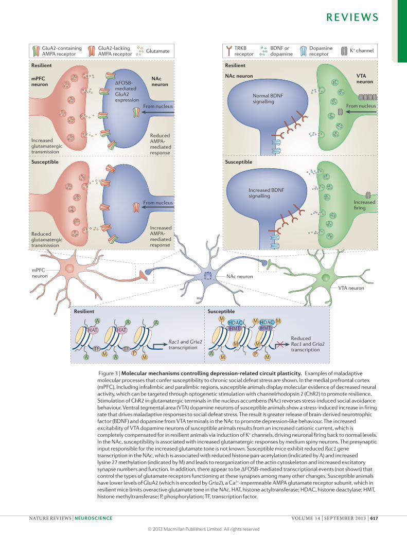
Figure 3 | Molecular mechanisms controlling depression-related circuit plasticity. Examples of maladaptive molecular processes that confer susceptibility to chronic social defeat stress are shown. In the medial prefrontal cortex (mPFC), including infralimbic and paralimbic regions, susceptible animals display molecular evidence of decreased neural activity, which can be targeted through optogenetic stimulation with channelrhodopsin 2 (ChR2) to promote resilience. Stimulation of ChR2 in glutamatergic terminals in the nucleus accumbens (NAc) reverses stress-induced social avoidance behaviour. Ventral tegmental area (VTA) dopamine neurons of susceptible animals show a stress-induced increase in firing rate that drives maladaptive responses to social defeat stress. The result is greater release of brain-derived neurotrophic factor (BDNF) and dopamine from VTA terminals in the NAc to promote depression-like behaviour. The increased excitability of VTA dopamine neurons of susceptible animals results from an increased cationic current, which is completely compensated for in resilient animals via induction of K+ channels, driving neuronal firing back to normal levels. In the NAc, susceptibility is associated with increased glutamatergic responses by medium spiny neurons. The presynaptic input responsible for the increased glutamate tone is not known. Susceptible mice exhibit reduced Rac1 gene transcription in the NAc, which is associated with reduced histone pan-acetylation (indicated by A) and increased lysine 27 methylation (indicated by M) and leads to reorganization of the actin cytoskeleton and increased excitatory synapse numbers and function. In addition, there appear to be ΔFOSB-mediated transcriptional events (not shown) that control the types of glutamate receptors functioning at these synapses among many other changes. Susceptible animals have lower levels of GluA2 (which is encoded by Gria2), a Ca2+-impermeable AMPA glutamate receptor subunit, which in resilient mice limits overactive glutamate tone in the NAc. HAT, histone actyltransferase; HDAC, histone deactylase; HMT, histone methyltransferase; P, phosphorylation; TF, transcription factor.
R E V I E W S
NATURE REVIEWS | NEUROSCIENCE VOLUME 14 | SEPTEMBER 2013 | 617
© 2013 Macmillan Publishers Limited. All rights reserved

Nature Reviews | Neuroscience
TRKB p75NTRBDNF
Cortical orVTA projection
IκBα
IKK
NF-κBCREB
RAS GDP
Nucleus
a Normal (no stress)
PP
RAS GDP
MEK1 or MEK2
ERK1 or ERK2
P
PP P
PCREB
IKKIKK
IKK
NF-κB IκBα
b Susceptible (chronic stress)
GluR
DAR
nAChR
MEK1 or MEK2
ERK1 or ERK2
MM
G9aGLP
G9aGLP
G9aGLP
G9aGLP
MM
MM
MM
PCREB
PNF-κB
Pro-susceptibletarget genes
MM
G9aGLP
AA
AA
AA
InterleukinsA group of cytokines that were first known for their role in immune and inflammatory responses but more recently have been found to regulate neural function.
systems and depression-like behaviour outlined in pre-vious sections. Thus, in most brain regions, including the VTA97, BDNF is thought to act in part by promoting plasticity at glutamatergic synapses. Assuming this is also the case in the NAc, we speculate that under con-ditions of severe stress, the VTA–NAc reward circuit undergoes a strong and inflexible form of learning that is mediated by abnormal glutamatergic plasticity, which under less severe conditions may be adaptive. The advanced molecular and optogenetic tools reviewed earlier make it possible to directly test this hypothesis. The opposing behavioural effects of BDNF–TRKB path-way activation in the VTA–NAc versus cortical regions suggests that this pathway may not be a suitable target for novel antidepressant treatments, unless it were pos-sible to target this pathway selectively in a particular brain area or circuit.
Cytokines and nuclear factor‑κB. Increasing attention has focused on the role of pro-inflammatory cytokines in depression98. For example, there is an increased preva-lence of mood and anxiety disorders in patients suffering from illnesses with strong immune and inflammatory features, such as multiple sclerosis and lupus erythe-matosus98. Conversely, patients with depression are at increased risk for inflammation-related conditions, such as cardiovascular disease and stroke99. Moreover, admin-istration of the pro-inflammatory cytokine interferon-α (IFNα) induces depression symptoms in humans with hepatitis C100 and in normal rodents101. Analysis of peripheral inflammation markers consistently identifies increases in interleukins interleukin-6 and interleukin-1β and tumour necrosis factor-α (TNFα) in patients with mood disorders102. Altered central levels of these factors have been identified as well. Rodent studies have largely
Figure 4 | Enhanced vulnerability to stress via priming of BDNF signalling in the NAc. Increased vulnerability to the depression-like effects of chronic social defeat stress occurs in part via priming of brain-derived neurotrophic factor (BDNF) signalling in the nucleus accumbens (NAc). Under control conditions (part a), BDNF activation of receptor tyrosine kinase TRKB signalling is limited. However, after some prior stimulus that increases susceptibility (for example, repeated stress or chronic cocaine exposure) (part b), BDNF–TRKB signalling is increased in the NAc, causing enhanced phosphorylation (indicated by P) and activity of several downstream signalling mediators, including cyclic AMP-responsive element-binding protein (CREB). This maladaptive response occurs not only through increased BDNF release into the NAc from the ventral tegmental area (VTA) but also through epigenetic modifications that further prime BDNF signalling cascades. For example, chronic stress increases Ras expression in the NAc of susceptible animals by decreasing G9a binding at the H-Ras1 gene promoter, causing reduced levels of repressive H3K9me2 (dimethyl-Lys9-histone H3, a major form of repressive histone methylation (indicated by M)). Ras also appears to be a target for CREB, creating a pathological feedforward loop that promotes CREB activation and Ras expression as well as depression-like behaviour (not shown). The figure also shows the induction of another pathway downstream of BDNF, including nuclear factor-κB (NF-κB) and inhibitor of NF-κB kinase (IKK) — possibly downstream of the neurotrophin receptor p75NTR (also known as NGFR) — in the NAc after chronic social defeat stress in susceptible animals. A, acetylation; DAR, dopamine receptor; ERK, extracellular signal-regulated kinase; IκBα, inhibitor of NF-κB-α; MEK, ERK/MAPK kinase.
R E V I E W S
618 | SEPTEMBER 2013 | VOLUME 14 www.nature.com/reviews/neuro
© 2013 Macmillan Publishers Limited. All rights reserved

Nuclear factor-κB(NF-κB). A transcription factor first characterized for its regulation of immune and inflammatory responses but more recently has been implicated in controlling neural function.
RAC1A small G protein (GTPase) that, in the nervous system, plays a critical part in regulating dendritic spine outgrowth.
MelanocortinFirst characterized for its regulation of melanocytes, melanocortin is also a peptide neurotransmitter secreted by hypothalamic neurons, where it exerts potent anorexogenic effects. In addition, it is implicated in the regulation of mood via actions on the brain’s reward circuitry.
OrexinAlso known as hypocretin, this peptide neurotransmitter is secreted by neurons in the lateral hypothalamus to promote wakefulness and attention. It also promotes reward by direct projections to ventral tegmental area dopamine neurons.
LeptinA peptide hormone secreted by adipocytes. One of the major anorexigenic peptides known, leptin suppresses feeding behaviour through actions on hypothalamus. It has also been implicated in regulation of mood.
GhrelinAn orexigenic peptide hormone secreted by the stomach epithelium after periods of fasting, which acts in hypothalamus and perhaps other brain regions to stimulate appetite. It has been implicated in mood regulation as well.
RNA-seqA high-throughput method to sequence whole-genome cDNA in order to obtain quantitative measures of all expressed RNAs in a tissue.
ChromatinThe mixture of DNA and proteins that comprise the cell nucleus.
confirmed that interleukin-6 and interleukin-1β, acting in the hippocampus or the NAc, increase depression-like behavioural responses to chronic stress103,104. A central question in the field is whether cytokines derived from the periphery versus those locally synthesized in the brain (by neurons or glia) are primarily responsible for the pro-susceptibility effects of these molecules.
The signalling cascades downstream of cytokines that mediate these effects are beginning to be delineated. Nuclear factor-κB (NF-κB) is a transcription factor best known for its role in peripheral immune and inflamma-tory responses, but it is also a well-established downstream target of interleukin-6, interleukin-1β and TNFα, in both the brain and peripheral tissues. Recent evidence suggests that the NF-κB signalling pathway regulates the reward circuitry in depression and addiction models73,105–107. For example, in the hippocampus, NF-κB activation is required for the stress-induced impairment of neurogen-esis and induction of anhedonia (for example, decreased sucrose preference)105. In the NAc, chronic social defeat stress increases levels of inhibitor of NF-κB kinase (IKK), which then increases downstream NF-κB signalling by phosphorylating inhibitor of NF-κB (IκB) and trigger-ing its dissociation from NF-κB73 (FIG. 4). NF-κB activa-tion mediates the formation of new immature excitatory spine structures on NAc dendrites73. Such induction of NF-κB and new spines occurs in susceptible animals but is not seen in resilient individuals73. Interestingly, similar molecular and structural changes are induced in the NAc by chronic cocaine administration107. Direct inhibition of IKK in the NAc prunes these new synapses and reverses the associated depression- and addiction-like pheno-types73,107. Current studies focus on the intracellular sig-nalling pathways through which cytokines regulate NF-κB activity in the context of stress- and addiction-related pathology, and also aim to identify the transcriptional targets of NF-κB that mediate these effects. One target may be RAC1, a small G protein that has recently been implicated in stress- and cocaine-triggered induction of immature spines in this brain region (see below)40,108. These studies illustrate how inflammatory signalling path-ways influence glutamatergic neurotransmission in NAc circuits to influence depression-like behaviour.
Metabolic mechanisms. The past decade has revealed that the VTA–NAc reward circuit is strongly influenced by several peptides that are known for their role in con-trolling food intake and peripheral metabolism and that are produced in the periphery or in the hypothalamus. This is not surprising, because these factors, which presumably reflect physiological measures of hunger or satiety, interface with brain systems that control motivational drive and reward. Thus, hypothalamic neurons expressing peptides such as melanocortin, mel-anin-concentrating hormone or orexin (also known as hypocretin) send dense projections to the NAc or VTA4. Likewise, peripheral peptides, such as leptin (derived from fat) or ghrelin (derived from stomach epithelium), control the activity of the VTA–NAc circuit most likely indirectly, through their effects in the hypothalamus, although direct effects have also been implicated109.
High levels of comorbidity between depression and obesity support an interplay between feeding systems and the brain’s reward circuitry.
Several of these feeding peptides have been shown to regulate depression-like behaviour in animal models by influencing the VTA–NAc circuit. In one study, chronic stress-mediated downregulation of pro-opiomelanocor-tin (POMC) signalling was shown to promote resilience (reduce stress susceptibility), but this was accompanied by obesity and related peripheral metabolic derange-ments110. Chronic stress induces this downregulation of POMC in the hypothalamic arcuate nucleus via increased sympathetic tone and β3-adrenergic receptor activation and decreased blood leptin levels. POMC downregula-tion in turn decreased melanocortin signalling to the NAc and other target regions, causing increased feeding and antidepressant-like behavioural effects110. Consistent with this model, a recent study showed that chronic unpredict-able stress activated melanocortin 4 receptor signalling in D1-type MSNs of the NAc, which decreased the strength of excitatory synapses on these neurons — an effect that was associated with anhedonia111.
In addition, chronic social defeat stress induces periph-eral ghrelin secretion, as well as activation of central orex-inergic neurons in the lateral hypothalamus of resilient mice only, thus contributing to the absence of depression-like behaviour112,113. The site of action of these peptides remains unknown but might involve ghrelin-induced activation of orexinergic neurons and subsequent orexin-induced activation of several reward-related regions, par-ticularly the VTA. Meanwhile, leptin has been implicated in the regulation of depression-like behaviour at the level of the hippocampus114.
These are examples of just some of the prominent feeding peptides that deserve attention in the field of depression. Of particular note is that perturbation of these feeding mechanisms may produce very different effects in different subtypes of depression. It is conceiv-able that individuals in whom depression is characterized by reduced activity and weight gain respond differently to such perturbations than individuals who exhibit increased activity, anxiety and weight loss. It is also possible that different perturbations in feeding peptides are associated with (or may underlie) different subtypes of depression.
Transcriptional and epigenetic mechanisms. Genome-wide methods have increasingly been used to obtain an unbiased view of molecular changes in the VTA and NAc that relate to susceptibility versus those that relate to resilience or to antidepressant responses in animal models of depression80,91,115,116. Earlier studies focused on gene expression microarrays, whereas more recent stud-ies have used deep sequencing of expressed RNAs (RNA-seq), which better captures alternative splice variants as well as non-coding RNAs. There is also growing use of genome-wide chromatin assays, including chromatin immunoprecipitation (ChIP) followed by promoter chips (ChIP–chip), ChIP followed by deep sequencing (ChIP–seq) and several methods to study DNA methylation. The rationale for studying epigenetic end points is, first, that they can reveal the precise molecular mechanisms
R E V I E W S
NATURE REVIEWS | NEUROSCIENCE VOLUME 14 | SEPTEMBER 2013 | 619
© 2013 Macmillan Publishers Limited. All rights reserved

Chromatin immunoprecipitation(ChIP). A method that enables the identification of histone modifications or transcriptional regulatory proteins at a given gene promoter. DNA is crosslinked to nearby proteins by light fixation, the material is sheared, then immunoprecipitated with an antibody to a particular protein of interest, and genes in the final immunoprecipitate are quantified by the polymerase chain reaction.
ChIP–chip(Chromatin immunoprecipita-tion followed by promoter chips). A method that enables a global analysis of genes associated with a particular histone modification or transcriptional regulatory protein. Immunoprecipitated chromatin is analysed on a microarray gene chip, enriched in promoter regions.
ChIP–seq(Chromatin immunoprecipita-tion followed by deep sequencing). A method that allows for global identification of histone modifications or transcriptional regulatory proteins. ChIP is coupled to high-throughput sequencing to obtain analysis across the entire genome, and in this sense differs from ChIP–chip.
ΔFOSBA FOS family transcription factor that, once induced, is particularly long-lived in the brain owing to its stability.
Serum response factor(SRF). A transcription factor, which, in conjunction with another factor termed ELK1, binds to serum response elements within certain genes to regulate their expression.
β-cateninA transcription factor that is activated by the WNT–Frizzled–Dishevelled signalling cascade. It appears to mediate resilience to stress at the level of the nucleus accumbens.
Transcription factorsProteins that bind to specific DNA sequences (called response elements) within responsive genes and thereby increase or decrease the rate at which those genes are transcribed.
by which stress regulates gene expression and, sec-ond, that they represent plausible ‘molecular scars’ at particular genes that stably alter the genes’ inducibil-ity in response to subsequent stress. According to the ‘molecular scar’ hypothesis, stress-induced chromatin modifications across a person’s lifetime contribute to an individual’s inherent vulnerability or resistance to stress-related disorders117–119. Studies of chromatin regulation in stress models therefore promise to reveal new insight into the neurobiological mechanisms involved. To date, most attention has been given to the NAc because of its large size; it is now important to carry out analogous studies of the VTA and other brain reward regions.
The best characterized transcriptional mecha-nism controlling depression-like behaviour in the NAc involves CREB (FIG. 4). In contrast to its antidepressant-like effects in the hippocampus, CREB activity in the NAc promotes depression-like behaviour (that is, it increases stress susceptibility) but concomitantly induces anxio-lytic-like responses in numerous behavioural assays120. This has been demonstrated in studies using viral vec-tors and inducible bitransgenic mice in which CREB or a dominant negative mutant CREB (mCREB) is targeted to the NAc120, in studies using constitutive CREB knockout mice121 and, more recently, in studies using local knock-out of CREB from the NAc122. Gene expression microar-ray analyses of mouse NAc in which CREB or mCREB were overexpressed and ChIP–chip studies of the NAc of wild-type mice exposed to chronic stress have revealed a host of CREB target genes that are likely to mediate these depressant and anxiolytic effects116,123,124. Some prominent targets that are upregulated by CREB are specific K+ channel subunits, glutamate receptor subu-nits, dynorphin and other neuropeptides. The induction of dynorphin in D1-type NAc MSNs induces depres-sion-like behaviour, and this is mediated by activation of κ-opioid receptors on VTA dopamine neurons125,126. Another target that is upregulated in the VTA by CREB is BDNF and several components of BDNF signal-ling cascades122. As these cascades themselves activate CREB, this could establish a feedforward mechanism that underlies susceptibility to chronic stress. CREB is known to increase the intrinsic excitability of NAc MSNs and to promote glutamatergic plasticity127,128, which could be the basis for working hypotheses to relate CREB’s transcriptional effects to circuit-level changes that promote depression. NF-κB is another pro-depression transcription factor in the NAc; it will be important to use genome-wide methods to define its gene targets in stress models as well as to study possible interactions with CREB.
In contrast to CREB and NF-κB, which control sus-ceptibility, studies have identified ΔFOSB (a FOS fam-ily transcription factor), serum response factor (SRF) and β-catenin (which acts downstream of WNT signalling cascades) as mediating resilience. All three factors are preferentially induced by chronic stress in the NAc of resilient mice, where they have been shown to prevent depression-like behaviour129–131. ΔFOSB and SRF also induce antidepressant-like responses in previously sus-ceptible animals. Several candidate target genes through
which ΔFOSB promotes resilience have been identified. An example is the GluA2 AMPA glutamate receptor subunit, which is induced by ΔFOSB in the NAc and reduces glutamatergic tone130, thereby opposing the enhanced glutamatergic innervation of these neurons that seems to promote susceptibility (see above and FIG. 3). One target of SRF is ΔFOSB itself: stress-induced increases in ΔFOSB in the NAc are mediated by this factor129. These data highlight the need for genome-wide ChIP–seq assays to comprehensively define the transcriptional targets of ΔFOSB, SRF and β-catenin that mediate resilience and, in some cases, antidepressant action.
Transcription factors regulate gene expression in con-cert with complex changes in chromatin structure, which involve post-translational modifications of histones and DNA and the recruitment of hundreds of co-activators or co-repressors to regulated genes132. Chronic stress models have been shown to alter the expression levels of several chromatin-modifying enzymes in the NAc, including specific histone deacetylases (HDACs), histone methyltransferases (HMTs) and DNA methyltransferases (DNMTs), and some of these changes are associated with altered global levels of the corresponding modifi-cation (for example, total tissue levels of an acetylated or methylated histone) in this brain region122,133–135. Similar types of modifications have been demonstrated for other reward regions, particularly in the hippocam-pus136–140. Moreover, several of the modifications found in animal models have been validated in post-mortem human brains122,141. Importantly, some of these enzymes and modifications, for example, through local intra-NAc inhibition of certain HDACs or DNMTs or activation of certain HMTs40,122,133,141, have been directly associated with antidepressant-like effects in animal models, sug-gesting potential paths for novel drug discovery efforts.
Recent studies have begun to use genome-wide assays to map the genetic loci in the NAc that are influenced by these chromatin marks in chronic stress models. Some of the early implications are interesting: there is substantial overlap between the chromatin changes seen in resilience and those induced by chronic antidepressant treatment in susceptible animals116. This suggests that one mechanism by which antidepressants work is to induce, in vulner-able individuals, some of the same genomic changes in the NAc that occur naturally in inherently more resilient individuals. A corollary of this observation is that the development of new antidepressants could be based not only on approaches aimed at preventing the deleterious effects of stress but also on approaches aimed at induc-ing resilience. It is expected that, as these genome-wide efforts continue, many novel targets of epigenetic regula-tion will be identified. One example, alluded to earlier, is the small G protein RAC1, a critical regulator of the actin cytoskeleton, which is reduced in NAc in both depression and addiction models. This suppression of RAC1 is cru-cial for stress- and cocaine-induced growth of immature dendritic spines on NAc MSNs and for attendant depres-sion- and addiction-like behaviour40,108. Recent work has shown that the gene encoding RAC1 displays stable epigenetic modifications selectively in this brain region, both in rodent stress models and in depressed humans40.
R E V I E W S
620 | SEPTEMBER 2013 | VOLUME 14 www.nature.com/reviews/neuro
© 2013 Macmillan Publishers Limited. All rights reserved

It will now be important to define the precise signalling pathway by which reduced RAC1 activity, and presum-ably many other mediators, control excitatory synapses on NAc MSNs and which specific monosynaptic connec-tions are affected.
Conclusions and future directionsResearch over the past decade has shown that the reward circuitry has a role in at least some of the symptoms of mood disorders. Human imaging studies have identi-fied gross abnormalities in reward circuit structure and function related to anhedonia and reward-related per-ception and memory deficits. Novel genetic, viral and optogenetic tools in rodents are now enabling the field to precisely define the very complex network of cells and synapses within the reward circuitry that control specific symptoms associated with depression. Such work has confirmed an important role for dopaminer-gic and glutamatergic circuits within the VTA and NAc. However, much remains to be investigated. We need to more precisely define specific cells within these discrete monosynaptic circuits that control a diverse range of reward-related behavioural deficits. We also need to determine the molecular mechanisms within each cell type that control these complex adaptive processes. A recent study142 performed cell-specific molecular pro-filing using a transgenic mouse expressing a bacterial artificial chromosome-translating ribosomal-associated protein (bacTRAP) specifically within layer 5 pyrami-dal neurons in regions of the frontal cortex. They found that a cell-specific molecular profile regulated by the
serotonergic co-activator protein p11 (also known as S100A10) in these cells is necessary for antidepressant responses. p11 acting selectively in cholinergic interneu-rons of the NAc mediates a similar antidepressant-like response89. Future studies using bacTRAP and related technology, along with newer methods for cell type-specific genome-wide profiling of epigenetic modifica-tions143, in combination with well-established rodent stress models will uncover fundamentally new informa-tion about the cell type-specific molecular mechanisms driving reward circuit plasticity in stress susceptibility versus resilience and in antidepressant responses.
Studies to date have demonstrated a perhaps unex-pectedly complex relationship between the brain’s reward circuitry and mood disorders. One might have assumed that ‘more reward’ equals ‘less depression’: for example, that a protein acting in a certain brain struc-ture that increases cocaine’s rewarding effects would exert an antidepressant-like effect based on the assump-tion that the protein also boosts natural reward. To put it another way: one might expect that the influence of a protein in depression models can be predicted from its effects in addiction models and vice versa. However, this is clearly not the case: there is no predictable relation-ship between the effects of a given protein in the NAc in depression models versus drug addiction models (TABLE 2); this is despite the considerable comorbidity between depression and addiction syndromes stated earlier. One complicating factor is that addiction prob-ably involves adaptations that impair brain reward and others that promote reward-related memories5,10,11.
Histone deacetylases(HDACs). Enzymes that catalyse the deacetylation of histone amino-terminal tails.
Histone methyltransferases(HMTs). Enzymes that catalyse the methylation of histone amino-terminal tails.
DNA methyltransferases(DNMTs). Enzymes that catalyse the methylation of cytosine nucleotides, in CpG sequences, in DNA.
Table 2 | Examples of molecular actions in the NAc in depression versus addiction models
Protein class Protein Effect in depression models Effect in addiction models
Transcription factor CREB ↑ Susceptibility ↓ Drug reward
NF-κB ↑ Susceptibility ↑ Drug reward
ΔFOSB ↓ Susceptibility ↑ Drug reward
SRF ↓ Susceptibility No effect
β-catenin* ↓ Susceptibility NA
Signalling BDNF–TRKB ↑ Susceptibility ↑ Cocaine reward or ↓ cocaine reward‡
↓ Morphine reward
GluA2 ↓ Susceptibility ↑ Drug reward
RGS4 ↓ Susceptibility§ ↓ Drug reward
RAC1 ↓ Susceptibility ↓ Drug reward
Dynorphin ↑ Susceptibility ↓ Drug reward
Epigenetic HDAC (class I) inhibition
↓ Susceptibility ↑ Drug reward||
HDAC5¶ ↓ Susceptibility ↓ Drug reward
G9a inhibition ↑ Susceptibility ↑ Drug reward
DNMT inhibition ↓ Susceptibility ↑ Drug reward
BDNF, brain-derived neurotrophic factor; CREB, cyclic AMP-responsive element-binding protein; DNMT, DNA methyltransferase; HDAC, histone deactylase; NA, not assessed; NF-κB, nuclear factor-κB; RGS4, regulator of G protein signalling 4; SRF, serum response factor. *The influence of β-catenin is derived from studies of upstream proteins (for example, Dishevelled and glycogen synthase kinase 3β)131. ‡The influence of BDNF–receptor tyrosine kinase TRKB signalling in the nucleus accumbens on cocaine reward is opposite in D1-type versus D2-type medium spiny neurons, although the net effect is to promote reward18. §Based on REF. 164. ||Although short-term inhibition drives increased reward, longer-term inhibition does the opposite165. ¶A class II HDAC.
R E V I E W S
NATURE REVIEWS | NEUROSCIENCE VOLUME 14 | SEPTEMBER 2013 | 621
© 2013 Macmillan Publishers Limited. All rights reserved

The heterogeneity of cell types within a given reward structure is another likely explanation for the differ-ent effects observed for a protein in stress models ver-sus drug models. For example, in the NAc, TRKB has opposing roles in reward-related behaviours in D1- ver-sus D2-type MSNs18. However, a difference in cell types is not the only explanation. For instance, CREB appears to have very different target genes in the NAc in depres-sion versus addiction models, even within the same cell types116,122–124. This suggests that the stimulus itself (that is, stress or drugs) can engage different intracellular pathways, thereby regulating chromatin structure and gene expression in their own unique ways. Future stud-ies using cell-selective molecular profiling and viral gene transfer approaches can shed light on these complex stimulus-specific effects on reward-related behaviour.
An important caveat regarding the work described in this article is that most of it was done solely in male rodents, despite the fact that, in humans, females are twice as likely to suffer from a mood disorder than are males144, and rodent models consistently show that females display greater depression-like behaviour after experiencing chronic stress115,145. It has been suggested that organizational differences in the development of reward-related neural circuits might predispose women to depression146. In addition, the direct actions of cir-culating gonadal hormones on the reward circuitry might change a female’s sensitivity to stress across the oestrous cycle147. Studies in rodents have indeed shown that ovarian hormones alter brain stimulation reward thresholds (BOX 1), which means that during certain phases of the oestrous cycle, females are more prone to anhedonia148. This should be an extremely high prior-ity for future studies; we must determine the extent to which the mechanisms discussed above apply to female depression models.
Last, despite the fact that we are experiencing a revo-lution in the field of basic psychiatric neuroscience, drug development efforts have arguably led to only two mech-anistically distinct antidepressant medications (ketamine and scopolamine) over the past half century, and their use for depression has still not been approved by the US Food and Drug Administration (FDA). The difficulty partly comes from the fact that patients are diagnosed and treated on the basis of clusters of behavioural symptoms that are heterogeneous across disorders and are likely to be caused by numerous and divergent biological factors. A clear example of this challenge is the fact that, even within a disorder like depression, two patients can exhibit entirely non-overlapping clusters of symptoms, but their official diagnosis and treatment are identical. Another aspect of this challenge is that the distinction between depression and anxiety is poorly defined, with >50% of patients with one disorder showing symptoms of the other. Psychiatry desperately needs a diagnostic system that is based on the underlying genetic and neurobiologi-cal factors that define subtypes of these broad syndromes. An intermediate step would be, if possible, to identify biomarker signatures that accompany specific domains of behavioural abnormalities and predict distinct treat-ment responses. The identification of new biomarker targets based on bona fide disease mechanisms could vastly improve depression treatment for subsequent generations. For example, identification of sex-specific depression mechanisms might help us to develop special-ized gender-based medicine. A greater understanding of the circuits controlling reward-related behaviour might identify new surgical targets for deep brain stimulation as well. Ultimately this information can lead to new treat-ments with improved efficacy and fewer side effects, as well as providing relief to patients that are resistant to all currently available therapies.
1. Kessler, R. C., Chiu, W. T., Demler, O., Merikangas, K. R. & Walters, E. E. Prevalence, severity, and comorbidity of 12-month DSM-IV disorders in the National Comorbidity Survey Replication. Arch. Gen. Psychiatry 62, 617–627 (2005).
2. Culpepper, L. Why do you need to move beyond first-line therapy for major depression? J. Clin. Psychiatry 71 (Suppl. 1), 4–9 (2010).
3. Conway, K. P., Compton, W., Stinson, F. S. & Grant, B. F. Lifetime comorbidity of DSM-IV mood and anxiety disorders and specific drug use disorders: results from the National Epidemiologic Survey on alcohol and related conditions. J. Clin. Psychiatry 67, 247–257 (2006).
4. Nestler, E. J. & Carlezon, W. A. Jr. The mesolimbic dopamine reward circuit in depression. Biol. Psychiatry 59, 1151–1159 (2006).
5. Koob, G. F. & Le Moal, M. Addiction and the brain antireward system. Annu. Rev. Psychol. 59, 29–53 (2008).
6. Hnasko, T. S., Hjelmstad, G. O., Fields, H. L. & Edwards, R. H. Ventral tegmental area glutamate neurons: electrophysiological properties and projections. J. Neurosci. 32, 15076–15085 (2012).This study provides compelling evidence that a subset of VTA projection neurons use glutamate as a neurotransmitter.
7. Tritsch, N. X., Ding, J. B. & Sabatini, B. L. Dopaminergic neurons inhibit striatal output through non-canonical release of GABA. Nature 490, 262–266 (2012).
8. Ikemoto, S. & Wise, R. A. Rewarding effects of the cholinergic agents carbachol and neostigmine in the posterior ventral tegmental area. J. Neurosci. 22, 9895–9904 (2002).
9. Schultz, W. Potential vulnerabilities of neuronal reward, risk, and decision mechanisms to addictive drugs. Neuron 69, 603–617 (2011).This important review summarizes the complex role of dopaminergic systems in controlling reward.
10. Berridge, K. C. & Robinson, T. E. What is the role of dopamine in reward: hedonic impact, reward learning, or incentive salience? Brain Res. Brain Res. Rev. 28, 309–369 (1998).
11. Blakemore, S. J. & Robbins, T. W. Decision-making in the adolescent brain. Nature Neurosci. 15, 1184–1191 (2012).
12. Sharp, C., Monterosso, J. & Montague, P. R. Neuroeconomics: a bridge for translational research. Biol. Psychiatry 72, 87–92 (2012).
13. Sesack, S. R. & Grace, A. A. Cortico-basal ganglia reward network: microcircuitry. Neuropsychopharmacology 35, 27–47 (2010).
14. Witten, I. B. et al. Recombinase-driver rat lines: tools, techniques, and optogenetic application to dopamine-mediated reinforcement. Neuron 72, 721–733 (2011).This paper describes the use of optogenetics to show that light stimulation of VTA dopamine neurons expressing ChR2 causes mice to self-stimulate those neurons, confirming that VTA dopamine neuron activity is indeed reinforcing.
15. Tan, K. R. et al. GABA neurons of the VTA drive conditioned place aversion. Neuron 73, 1173–1183 (2012).
16. Tsai, H. C. et al. Phasic firing in dopaminergic neurons is sufficient for behavioral conditioning. Science 324, 1080–1084 (2009).
17. van Zessen, R., Phillips, J. L., Budygin, E. A. & Stuber, G. D. Activation of VTA GABA neurons
disrupts reward consumption. Neuron 73, 1184–1194 (2012).
18. Lobo, M. K. et al. Cell type-specific loss of BDNF signaling mimics optogenetic control of cocaine reward. Science 330, 385–390 (2010).
19. Kravitz, A. V., Tye, L. D. & Kreitzer, A. C. Distinct roles for direct and indirect pathway striatal neurons in reinforcement. Nature Neurosci. 15, 816–818 (2012).
20. Everitt, B. J. & Robbins, T. W. Neural systems of reinforcement for drug addiction: from actions to habits to compulsion. Nature Neurosci. 8, 1481–1489 (2005).
21. Witten, I. B. et al. Cholinergic interneurons control local circuit activity and cocaine conditioning. Science 330, 1677–1681 (2010).
22. Brown, M. T. et al. Ventral tegmental area GABA projections pause accumbal cholinergic interneurons to enhance associative learning. Nature 492, 452–456 (2012).This paper identifies a GABAergic projection from the VTA that selectively innervates cholinergic interneurons in the NAc. Despite being a relatively minor (~2%) component of all VTA projections, this pathway has a robust behavioural effect.
23. Yeomans, J. S., Kofman, O. & McFarlane, V. Cholinergic involvement in lateral hypothalamic rewarding brain stimulation. Brain Res. 329, 19–26 (1985).
24. Britt, J. P. et al. Synaptic and behavioral profile of multiple glutamatergic inputs to the nucleus accumbens. Neuron 76, 790–803 (2012).
25. Chen, B. T. et al. Rescuing cocaine-induced prefrontal cortex hypoactivity prevents compulsive cocaine seeking. Nature 496, 359–362 (2013).
R E V I E W S
622 | SEPTEMBER 2013 | VOLUME 14 www.nature.com/reviews/neuro
© 2013 Macmillan Publishers Limited. All rights reserved
26. Pascoli, V., Turiault, M. & Luscher, C. Reversal of cocaine-evoked synaptic potentiation resets drug-induced adaptive behaviour. Nature 481, 71–75 (2012).
27. Stuber, G. D. et al. Excitatory transmission from the amygdala to nucleus accumbens facilitates reward seeking. Nature 475, 377–380 (2011).
28. Stamatakis, A. M. & Stuber, G. D. Activation of lateral habenula inputs to the ventral midbrain promotes behavioral avoidance. Nature Neurosci. 15, 1105–1107 (2012).
29. Lammel, S., Ion, D. I., Roeper, J. & Malenka, R. C. Projection-specific modulation of dopamine neuron synapses by aversive and rewarding stimuli. Neuron 70, 855–862 (2012).
30. Lammel, S. et al. Input-specific control of reward and aversion in the ventral tegmental area. Nature 491, 212–217 (2012).
31. Bolanos, C. A. et al. Phospholipase Cγ in distinct regions of the ventral tegmental area differentially modulates mood-related behaviors. J. Neurosci. 23, 7569–7576 (2003).
32. Olson, V. G. et al. Regulation of drug reward by cAMP response element-binding protein: evidence for two functionally distinct subregions of the ventral tegmental area. J. Neurosci. 25, 5553–5562 (2005).
33. Savitz, J. & Drevets, W. C. Bipolar and major depressive disorder: neuroimaging the developmental-degenerative divide. Neurosci. Biobehav. Rev. 33, 699–771 (2009).
34. Bremner, J. D. et al. Hippocampal volume reduction in major depression. Am. J. Psychiatry 157, 115–118 (2000).
35. Hannestad, J. et al. White matter lesion volumes and caudate volumes in late-life depression. Int. J. Geriatr. Psychiatry 21, 1193–1198 (2006).
36. Husain, M. M. et al. A magnetic resonance imaging study of putamen nuclei in major depression. Psychiatry Res. 40, 95–99 (1991).
37. Krishnan, K. R. et al. Magnetic resonance imaging of the caudate nuclei in depression. Preliminary observations. Arch. Gen. Psychiatry 49, 553–557 (1992).
38. Drevets, W. C. et al. A functional anatomical study of unipolar depression. J. Neurosci. 12, 3628–3641 (1992).
39. Mayberg, H. S. et al. Regional metabolic effects of fluoxetine in major depression: serial changes and relationship to clinical response. Biol. Psychiatry 48, 830–843 (2000).
40. Golden, S. A. et al. Epigenetic regulation of RAC1 induces synaptic remodeling in stress disorders and depression. Nature Med. 19, 337–344 (2013).This paper identifies an epigenetic mechanism in the NAc of depressed humans that reduces the expression of the small GTPase RAC1. It further shows that stress reduces Rac1 expression through a similar mechanism in mice and is necessary and sufficient to induce depression-like changes in behaviour and excitatory synaptic plasticity.
41. Hickie, I. B. et al. Serotonin transporter gene status predicts caudate nucleus but not amygdala or hippocampal volumes in older persons with major depression. J. Affect Disord. 98, 137–142 (2007).
42. von Gunten, A., Fox, N. C., Cipolotti, L. & Ron, M. A. A volumetric study of hippocampus and amygdala in depressed patients with subjective memory problems. J. Neuropsychiatry Clin. Neurosci. 12, 493–498 (2000).
43. Frodl, T. et al. Hippocampal and amygdala changes in patients with major depressive disorder and healthy controls during a 1-year follow-up. J. Clin. Psychiatry 65, 492–499 (2004).
44. Sheline, Y. I., Gado, M. H. & Price, J. L. Amygdala core nuclei volumes are decreased in recurrent major depression. Neuroreport 9, 2023–2028 (1998).
45. McEwen, B. S. Stress and hippocampal plasticity. Annu. Rev. Neurosci. 22, 105–122 (1999).
46. Sheline, Y. I. et al. Increased amygdala response to masked emotional faces in depressed subjects resolves with antidepressant treatment: an fMRI study. Biol. Psychiatry 50, 651–658 (2001).
47. Fu, C. H. et al. Attenuation of the neural response to sad faces in major depression by antidepressant treatment: a prospective, event-related functional magnetic resonance imaging study. Arch. Gen. Psychiatry 61, 877–889 (2004).
48. Siegle, G. J., Thompson, W., Carter, C. S., Steinhauer, S. R. & Thase, M. E. Increased amygdala and decreased dorsolateral prefrontal BOLD
responses in unipolar depression: related and independent features. Biol. Psychiatry 61, 198–209 (2007).
49. Vyas, A., Pillai, A. G. & Chattarji, S. Recovery after chronic stress fails to reverse amygdaloid neuronal hypertrophy and enhanced anxiety-like behavior. Neuroscience 128, 667–673 (2004).
50. Caetano, S. C. et al. Smaller cingulate volumes in unipolar depressed patients. Biol. Psychiatry 59, 702–706 (2006).
51. Drevets, W. C. et al. Subgenual prefrontal cortex abnormalities in mood disorders. Nature 386, 824–827 (1997).
52. Lai, T., Payne, M. E., Byrum, C. E., Steffens, D. C. & Krishnan, K. R. Reduction of orbital frontal cortex volume in geriatric depression. Biol. Psychiatry 48, 971–975 (2000).
53. Steffens, D. C., McQuoid, D. R., Welsh-Bohmer, K. A. & Krishnan, K. R. Left orbital frontal cortex volume and performance on the Benton Visual Retention Test in older depressives and controls. Neuropsychopharmacology 28, 2179–2183 (2003).
54. Taylor Tavares, J. V. et al. Neural basis of abnormal response to negative feedback in unmedicated mood disorders. Neuroimage 42, 1118–1126 (2008).
55. Mayberg, H. S. Targeted electrode-based modulation of neural circuits for depression. J. Clin. Invest. 119, 717–725 (2009).
56. Rajkowska, G. Postmortem studies in mood disorders indicate altered numbers of neurons and glial cells. Biol. Psychiatry 48, 766–777 (2000).
57. Uranova, N. A., Vostrikov, V. M., Orlovskaya, D. D. & Rachmanova, V. I. Oligodendroglial density in the prefrontal cortex in schizophrenia and mood disorders: a study from the Stanley Neuropathology Consortium. Schizophr Res. 67, 269–275 (2004).
58. Kang, H. J. et al. Decreased expression of synapse-related genes and loss of synapses in major depressive disorder. Nature Med. 18, 1413–1417 (2012).This study uses microarray analysis to demonstrate downregulation of numerous synapse-related genes in the dorsolateral PFC of depressed humans.
59. Campbell, S. & MacQueen, G. The role of the hippocampus in the pathophysiology of major depression. J. Psychiatry Neurosci. 29, 417–426 (2004).
60. Campbell, S., Marriott, M., Nahmias, C. & MacQueen, G. M. Lower hippocampal volume in patients suffering from depression: a meta-analysis. Am. J. Psychiatry 161, 598–607 (2004).
61. Czeh, B. & Lucassen, P. J. What causes the hippocampal volume decrease in depression? Are neurogenesis, glial changes and apoptosis implicated? Eur. Arch. Psychiatry Clin. Neurosci. 257, 250–260 (2007).
62. van Tol, M. J. et al. Functional magnetic resonance imaging correlates of emotional word encoding and recognition in depression and anxiety disorders. Biol. Psychiatry 71, 593–602 (2012).
63. Kessler, R. C. The effects of stressful life events on depression. Annu. Rev. Psychol. 48, 191–214 (1997).
64. Magarinos, A. M. & McEwen, B. S. Stress-induced atrophy of apical dendrites of hippocampal CA3c neurons: involvement of glucocorticoid secretion and excitatory amino acid receptors. Neuroscience 69, 89–98 (1995).
65. Donohue, H. S. et al. Chronic restraint stress induces changes in synapse morphology in stratum lacunosum-moleculare CA1 rat hippocampus: a stereological and three-dimensional ultrastructural study. Neuroscience 140, 597–606 (2006).
66. Liu, R. J. et al. Brain-derived neurotrophic factor Val66Met allele impairs basal and ketamine-stimulated synaptogenesis in prefrontal cortex. Biol. Psychiatry 71, 996–1005 (2011).
67. Shansky, R. M., Hamo, C., Hof, P. R., McEwen, B. S. & Morrison, J. H. Stress-induced dendritic remodeling in the prefrontal cortex is circuit specific. Cereb. Cortex 19, 2479–2484 (2009).
68. Johansen-Berg, H. et al. Anatomical connectivity of the subgenual cingulate region targeted with deep brain stimulation for treatment-resistant depression. Cereb. Cortex 18, 1374–1383 (2008).
69. Sierra-Mercado, D., Padilla-Coreano, N. & Quirk, G. J. Dissociable roles of prelimbic and infralimbic cortices, ventral hippocampus, and basolateral amygdala in the expression and extinction of conditioned fear. Neuropsychopharmacology 36, 529–538 (2011).
70. Disner, S. G., Beevers, C. G., Haigh, E. A. & Beck, A. T. Neural mechanisms of the cognitive model of
depression. Nature Rev. Neurosci. 12, 467–477 (2011).
71. Leuner, B. & Shors, T. J. Stress, anxiety, and dendritic spines: what are the connections? Neuroscience http://dx.doi.org/10.1016/j.neuroscience.2012.04.021 (2012).
72. Mitra, R., Jadhav, S., McEwen, B. S., Vyas, A. & Chattarji, S. Stress duration modulates the spatiotemporal patterns of spine formation in the basolateral amygdala. Proc. Natl Acad. Sci. USA 102, 9371–9376 (2005).
73. Christoffel, D. J. et al. IκB kinase regulates social defeat stress-induced synaptic and behavioral plasticity. J. Neurosci. 31, 314–321 (2011).
74. Muhammad, A., Carroll, C. & Kolb, B. Stress during development alters dendritic morphology in the nucleus accumbens and prefrontal cortex. Neuroscience 216, 103–109 (2012).
75. Campioni, M. R., Xu, M. & McGehee, D. S. Stress-induced changes in nucleus accumbens glutamate synaptic plasticity. J. Neurophysiol. 101, 3192–3198 (2009).
76. Schlaepfer, T. E. et al. Deep brain stimulation to reward circuitry alleviates anhedonia in refractory major depression. Neuropsychopharmacology 33, 368–377 (2008).This study shows the potential for deep brain stimulation of reward structures, such as the NAc, in depression treatment.
77. Heshmati, M. et al. Inhibitory synaptic control of depression-like behavior in a nucleus accumbens microcircuit. Soc. Neurosci. Abstr. 605.20 (2012).
78. Gill, K. M. & Grace, A. A. Differential effects of acute and repeated stress on hippocampus and amygdala inputs to the nucleus accumbens shell. Int. J Neuropsychopharmacol. http://dx.doi.org/10.1017/S1461145713000618 (2013).
79. Belujon, P., Patton, M. H. & Grace, A. A. Role of the prefrontal cortex in altered hippocampal-accumbens synaptic plasticity in a developmental animal model of Schizophrenia. Cereb. Cortex http://dx.doi.org/10.1093/cercor/bhs380 (2012).
80. Krishnan, V. et al. Molecular adaptations underlying susceptibility and resistance to social defeat in brain reward regions. Cell 131, 391–404 (2007).
81. Cao, J. L. et al. Mesolimbic dopamine neurons in the brain reward circuit mediate susceptibility to social defeat and antidepressant action. J. Neurosci. 30, 16453–16458 (2010).
82. Chaudhury, D. et al. Rapid regulation of depression-related behaviours by control of midbrain dopamine neurons. Nature 493, 532–536 (2013).This study uses circuit-specific optogenetics to confirm that increased phasic firing of VTA dopamine neurons projecting to the NAc has a role in in mediating stress susceptibility.
83. Brischoux, F., Chakraborty, S., Brierley, D. I. & Ungless, M. A. Phasic excitation of dopamine neurons in ventral VTA by noxious stimuli. Proc. Natl Acad. Sci. USA 106, 4894–4899 (2009).
84. Valenti, O., Gill, K. M. & Grace, A. A. Different stressors produce excitation or inhibition of mesolimbic dopamine neuron activity: response alteration by stress pre-exposure. Eur. J. Neurosci. 35, 1312–1321 (2012).
85. Tye, K. M. et al. Dopamine neurons modulate neural encoding and expression of depression-related behaviour. Nature 493, 537–541 (2013).Along with reference 82, this paper highlights the complexity of understanding reward function in depression. Although chronic social defeat stress induces dopamine firing to promote depression-like behaviour (reference 82), these authors find that chronic mild stress decreases dopamine firing to promote depression-like behaviour.
86. Kalivas, P. W. The glutamate homeostasis hypothesis of addiction. Nature Rev. Neurosci. 10, 561–572 (2009).
87. Covington, H. E. et al. Antidepressant effect of optogenetic stimulation of the medial prefrontal cortex. J. Neurosci. 30, 16082–16090 (2011).
88. Christoffel, D. J. et al. Glutamatergic microcircuits regulate stress-induced alterations in social behavior. Soc. Neurosci. Abstr. 605.21 (2012).
89. Warner-Schmidt, J. L. et al. Cholinergic interneurons in the nucleus accumbens regulate depression-like behavior. Proc. Natl Acad. Sci. USA 109, 11360–11365 (2012).
90. Zarate, C. A. Jr, Mathews, D. C. & Furey, M. L. Human biomarkers of rapid antidepressant effects. Biol. Psychiatry 73, 1142–1155 (2013).
R E V I E W S
NATURE REVIEWS | NEUROSCIENCE VOLUME 14 | SEPTEMBER 2013 | 623
© 2013 Macmillan Publishers Limited. All rights reserved
91. Berton, O. et al. Essential role of BDNF in the mesolimbic dopamine pathway in social defeat stress. Science 311, 864–868 (2006).
92. Eisch, A. J. et al. Brain-derived neurotrophic factor in the ventral midbrain–nucleus accumbens pathway: a role in depression. Biol. Psychiatry 54, 994–1005 (2003).
93. Iniguez, S. D. et al. Extracellular signal-regulated kinase-2 within the ventral tegmental area regulates responses to stress. J. Neurosci. 30, 7652–7663 (2010).
94. Warren, B. L. et al. Juvenile administration of concomitant methylphenidate and fluoxetine alters behavioral reactivity to reward- and mood-related stimuli and disrupts ventral tegmental area gene expression in adulthood. J. Neurosci. 31, 10347–10358 (2011).
95. Krishnan, V. et al. AKT signaling within the ventral tegmental area regulates cellular and behavioral responses to stressful stimuli. Biol. Psychiatry 64, 691–700 (2008).
96. Duman, R. S. & Monteggia, L. M. A neurotrophic model for stress-related mood disorders. Biol. Psychiatry 59, 1116–1127 (2006).
97. Pu, L., Liu, Q. S. & Poo, M. M. BDNF-dependent synaptic sensitization in midbrain dopamine neurons after cocaine withdrawal. Nature Neurosci. 9, 605–607 (2006).
98. Miller, A. H. Neuroendocrine and immune system interactions in stress and depression. Psychiatr. Clin. North Am. 21, 443–463 (1998).
99. Anisman, H. & Hayley, S. Inflammatory factors contribute to depression and its comorbid conditions. Sci. Signal 5, pe45 (2012).
100. Maes, M. et al. Treatment with interferon-alpha (IFNα) of hepatitis C patients induces lower serum dipeptidyl peptidase IV activity, which is related to IFNα-induced depressive and anxiety symptoms and immune activation. Mol. Psychiatry 6, 475–480 (2001).
101. Hayley, S., Scharf, J. & Anisman, H. Central administration of murine interferon-α induces depressive-like behavioral, brain cytokine and neurochemical alterations in mice: a mini-review and original experiments. Brain Behav. Immun. 31, 115–127 (2013).
102. Dowlati, Y. et al. A meta-analysis of cytokines in major depression. Biol. Psychiatry 67, 446–457 (2010).
103. Koo, J. W. & Duman, R. S. IL-1β is an essential mediator of the antineurogenic and anhedonic effects of stress. Proc. Natl Acad. Sci. USA 105, 751–756 (2008).
104. Hodes, G. E. et al. Innate peripheral immune responses predispose mice to susceptibility to repeated social defeat stress. Soc. Neurosci. Abstr. 12.10 (2012).
105. Koo, J. W., Russo, S. J., Ferguson, D., Nestler, E. J. & Duman, R. S. Nuclear factor-κB is a critical mediator of stress-impaired neurogenesis and depressive behavior. Proc. Natl Acad. Sci. USA 107, 2669–2674 (2010).
106. Christoffel, D. J. et al. Effects of inhibitor of κB kinase activity in the nucleus accumbens on emotional behavior. Neuropsychopharmacology 37, 2615–2623 (2012).
107. Russo, S. J. et al. Nuclear factor κB signaling regulates neuronal morphology and cocaine reward. J. Neurosci. 29, 3529–3537 (2009).
108. Dietz, D. M. et al. Rac1 is essential in cocaine-induced structural plasticity of nucleus accumbens neurons. Nature Neurosci. 15, 891–896 (2012).
109. Narayanan, N. S., Guarnieri, D. J. & DiLeone, R. J. Metabolic hormones, dopamine circuits, and feeding. Front. Neuroendocrinol. 31, 104–112 (2010).
110. Chuang, J. C. et al. A β3-adrenergic–leptin–melanocortin circuit regulates behavioral and metabolic changes induced by chronic stress. Biol. Psychiatry 67, 1075–1082 (2011).
111. Lim, B. K., Huang, K. W., Grueter, B. A., Rothwell, P. E. & Malenka, R. C. Anhedonia requires MC4R-mediated synaptic adaptations in nucleus accumbens. Nature 487, 183–189 (2012).This study provides evidence for a model in which chronic stress-induced increases in melanocortin signalling to the NAc, acting via melanocortin 4 receptors, selectively attenuate the strength of glutamatergic synapses on D1-type MSNs.
112. Lutter, M. et al. Orexin signaling mediates the antidepressant-like effect of calorie restriction. J. Neurosci. 28, 3071–3075 (2008).
113. Lutter, M. et al. The orexigenic hormone ghrelin defends against depressive symptoms of chronic stress. Nature Neurosci. 11, 752–753 (2008).
114. Guo, M. et al. Forebrain glutamatergic neurons mediate leptin action on depression-like behaviors and synaptic depression. Transl. Psychiatry 2, e83 (2012).
115. LaPlant, Q. et al. Role of nuclear factor κB in ovarian hormone-mediated stress hypersensitivity in female mice. Biol. Psychiatry 65, 874–880 (2009).
116. Wilkinson, M. B. et al. Imipramine treatment and resiliency exhibit similar chromatin regulation in the mouse nucleus accumbens in depression models. J. Neurosci. 29, 7820–7832 (2009).
117. Peter, C. J. & Akbarian, S. Balancing histone methylation activities in psychiatric disorders. Trends Mol. Med. 17, 372–379 (2011).
118. Sun, H., Kennedy, P. J. & Nestler, E. J. Epigenetics of the depressed brain: role of histone acetylation and methylation. Neuropsychopharmacology 38, 124–137 (2012).
119. Zhang, T. Y. & Meaney, M. J. Epigenetics and the environmental regulation of the genome and its function. Annu. Rev. Psychol. 61, 439–466 (2010).
120. Carlezon, W. A. Jr, Duman, R. S. & Nestler, E. J. The many faces of CREB. Trends Neurosci. 28, 436–445 (2005).
121. Blendy, J. A. The role of CREB in depression and antidepressant treatment. Biol. Psychiatry 59, 1144–1150 (2006).
122. Covington, H. E. et al. A role for repressive histone methylation in cocaine-induced vulnerability to stress. Neuron 71, 656–670 (2011).One of the first studies to establish an epigenetic-based mechanism for stress susceptibility versus resilience in the NAc. The authors show that chronic social defeat stress downregulates the HMTs G9a (also known as EHMT2) and GLP (also known as EHMT1) in the NAc, which promotes susceptibility via induction of BDNF–CREB signalling in this brain region.
123. McClung, C. A. & Nestler, E. J. Regulation of gene expression and cocaine reward by CREB and ΔFosB. Nature Neurosci. 6, 1208–1215 (2003).
124. Wallace, D. L. et al. CREB regulation of nucleus accumbens excitability mediates social isolation-induced behavioral deficits. Nature Neurosci. 12, 200–209 (2009).
125. Bruchas, M. R., Land, B. B. & Chavkin, C. The dynorphin/kappa opioid system as a modulator of stress-induced and pro-addictive behaviors. Brain Res. 1314, 44–55 (2010).
126. Chartoff, E. H. et al. Desipramine reduces stress-activated dynorphin expression and CREB phosphorylation in NAc tissue. Mol. Pharmacol. 75, 704–712 (2009).
127. Dong, Y. et al. CREB modulates excitability of nucleus accumbens neurons. Nature Neurosci. 9, 475–477 (2006).
128. Brown, T. E. et al. A silent synapse-based mechanism for cocaine-induced locomotor sensitization. J. Neurosci. 31, 8163–8174 (2011).
129. Vialou, V. et al. Serum response factor promotes resilience to chronic social stress through the induction of ΔFosB. J. Neurosci. 30, 14585–14592 (2010).
130. Vialou, V. et al. ΔFosB in brain reward circuits mediates resilience to stress and antidepressant responses. Nature Neurosci. 13, 745–752 (2010).
131. Wilkinson, M. B. et al. A novel role of the WNT–dishevelled–GSK3β signaling cascade in the mouse nucleus accumbens in a social defeat model of depression. J. Neurosci. 31, 9084–9092 (2011).
132. Borrelli, E., Nestler, E. J., Allis, C. D. & Sassone-Corsi, P. Decoding the epigenetic language of neuronal plasticity. Neuron 60, 961–974 (2008).
133. LaPlant, Q. et al. Dnmt3a regulates emotional behavior and spine plasticity in the nucleus accumbens. Nature Neurosci. 13, 1137–1143 (2009).
134. Renthal, W. et al. Histone deacetylase 5 epigenetically controls behavioral adaptations to chronic emotional stimuli. Neuron 56, 517–529 (2007).
135. Uchida, S. et al. Epigenetic status of Gdnf in the ventral striatum determines susceptibility and adaptation to daily stressful events. Neuron 69, 359–372 (2011).
136. Chandramohan, Y., Droste, S. K., Arthur, J. S. & Reul, J. M. The forced swimming-induced behavioural immobility response involves histone H3 phospho-acetylation and c-Fos induction in dentate gyrus granule neurons via activation of the N-methyl-D-aspartate/extracellular signal-regulated kinase/mitogen- and stress-activated kinase signalling pathway. Eur. J. Neurosci. 27, 2701–2713 (2008).
137. Covington, H. E., Vialou, V. F., LaPlant, Q., Ohnishi, Y. N. & Nestler, E. J. Hippocampal-dependent antidepressant-like activity of histone deacetylase inhibition. Neurosci. Lett. 493, 122–126 (2011).
138. Hunter, R. G., McCarthy, K. J., Milne, T. A., Pfaff, D. W. & McEwen, B. S. Regulation of hippocampal H3 histone methylation by acute and chronic stress. Proc. Natl Acad. Sci. USA 106, 20912–20917 (2009).
139. Tsankova, N. M. et al. Sustained hippocampal chromatin regulation in a mouse model of depression and antidepressant action. Nature Neurosci. 9, 519–525 (2006).
140. Jiang, Y. et al. Setdb1 histone methyltransferase regulates mood-related behaviors and expression of the NMDA receptor subunit NR2B. J. Neurosci. 30, 7152–7167 (2010).
141. Covington, H. E. et al. Antidepressant actions of histone deacetylase inhibitors. J. Neurosci. 29, 11451–11460 (2009).
142. Schmidt, E. F. et al. Identification of the cortical neurons that mediate antidepressant responses. Cell 149, 1152–1163 (2012).The authors demonstrate that p11, a protein that promotes the activity of certain serotonin and several other G protein-coupled receptors, is required for antidepressant-like responses. This action is mediated not only by p11 action in the PFC but also in the NAc, as the authors demonstrated previously.
143. Mellen, M., Ayata, P., Dewell, S., Kriaucionis, S. & Heintz, N. MeCP2 binds to 5hmC enriched within active genes and accessible chromatin in the nervous system. Cell 151, 1417–1430 (2012).
144. Deecher, D., Andree, T. H., Sloan, D. & Schechter, L. E. From menarche to menopause: exploring the underlying biology of depression in women experiencing hormonal changes. Psychoneuroendocrinology 33, 3–17 (2008).
145. Joeyen-Waldorf, J., Edgar, N. & Sibille, E. The roles of sex and serotonin transporter levels in age- and stress-related emotionality in mice. Brain Res. 1286, 84–93 (2009).
146. Blehar, M. C. Women’s mental health research: the emergence of a biomedical field. Annu. Rev. Clin. Psychol. 2, 135–160 (2006).
147. Young, E. & Korszun, A. Sex, trauma, stress hormones and depression. Mol. Psychiatry 15, 23–28 (2010).
148. Bless, E. P., McGinnis, K. A., Mitchell, A. L., Hartwell, A. & Mitchell, J. B. The effects of gonadal steroids on brain stimulation reward in female rats. Behav. Brain Res. 82, 235–244 (1997).
149. Golden, S. A., Covington, H. E., Berton, O. & Russo, S. J. A standardized protocol for repeated social defeat stress in mice. Nature Protoc. 6, 1183–1191 (2011).
150. Warren, B. L. et al. Neurobiological sequelae of witnessing stressful events in adult mice. Biol. Psychiatry 73, 7–14 (2012).
151. Hill, M. N., Hellemans, K. G., Verma, P., Gorzalka, B. B. & Weinberg, J. Neurobiology of chronic mild stress: parallels to major depression. Neurosci. Biobehav. Rev. 36, 2085–2117 (2012).
152. Trainor, B. C. et al. Sex differences in social interaction behavior following social defeat stress in the monogamous California mouse (Peromyscus californicus). PLoS ONE 6, e17405 (2012).This important study establishes the utility of the chronic social defeat stress paradigm in female mice, using a different mouse species. Interestingly, the authors show some similar molecular abnormalities in the female NAc (increased CREB activity) as shown by other groups in males.
153. Maier, S. F. & Watkins, L. R. Stressor controllability and learned helplessness: the roles of the dorsal raphe nucleus, serotonin, and corticotropin-releasing factor. Neurosci. Biobehav. Rev. 29, 829–841 (2005).
154. Der-Avakian, A. & Markou, A. The neurobiology of anhedonia and other reward-related deficits. Trends Neurosci. 35, 68–77 (2012).
155. Dalla, C., Pitychoutis, P. M., Kokras, N. & Papadopoulou-Daifoti, Z. Sex differences in animal models of depression and antidepressant response. Basic Clin. Pharmacol. Toxicol. 106, 226–233 (2010).
156. Willner, P. Chronic mild stress (CMS) revisited: consistency and behavioural-neurobiological concordance in the effects of CMS. Neuropsychobiology 52, 90–110 (2005).
157. Carlezon, W. A. Jr & Chartoff, E. H. Intracranial self-stimulation (ICSS) in rodents to study the neurobiology of motivation. Nature Protoc. 2, 2987–2995 (2007).
R E V I E W S
624 | SEPTEMBER 2013 | VOLUME 14 www.nature.com/reviews/neuro
© 2013 Macmillan Publishers Limited. All rights reserved

158. Chartoff, E. et al. Blockade of kappa opioid receptors attenuates the development of depressive-like behaviors induced by cocaine withdrawal in rats. Neuropharmacology 62, 167–176 (2012).
159. Baliki, M. N. et al. Corticostriatal functional connectivity predicts transition to chronic back pain. Nature Neurosci. 15, 1117–1119 (2012).
160. Scott, D. J. et al. Placebo and nocebo effects are defined by opposite opioid and dopaminergic responses. Arch. Gen. Psychiatry 65, 220–231 (2008).
161. Niikura, K., Narita, M., Butelman, E. R., Kreek, M. J. & Suzuki, T. Neuropathic and chronic pain stimuli downregulate central μ-opioid and dopaminergic transmission. Trends Pharmacol. Sci. 31, 299–305 (2010).
162. Zachariou, V. et al. An essential role for ΔFosB in the nucleus accumbens in morphine action. Nature Neurosci. 9, 205–211 (2006).
163. Han, M. H. et al. Brain region specific actions of regulator of G protein signaling 4 oppose morphine reward and dependence but promote analgesia. Biol. Psychiatry 67, 761–769 (2010).
164. Stratinaki, M. et al. Regulator of G protein signaling is a crucial modulator of antidepressant drug action in depression and neuropathic pain models. Proc. Natl Acad. Sci. USA 110, 8254–8259 (2013).This study demonstrates that the G protein regulatory protein RGS4, acting in the NAc, promotes therapeutic-like responses to standard monoamine-dependent antidepressants. By
contrast, RGS4 opposes the antidepressant-like actions of non-monoamine-based drugs such as ketamine.
165. Kennedy, P. J. et al. Class I HDAC inhibition blocks cocaine-induced plasticity by targeted changes in histone methylation. Nature Neurosci. 16, 434–440 (2013).
Acknowledgments Preparation of this review was supported by grants from the US National Institute of Mental Health: R01 MH090264 (S.J.R.); and R01 MH51399 and P50 MH96890 (E.J.N.).
Competing interests statementThe authors declare no competing financial interests.
R E V I E W S
NATURE REVIEWS | NEUROSCIENCE VOLUME 14 | SEPTEMBER 2013 | 625
© 2013 Macmillan Publishers Limited. All rights reserved