Embed Size (px)
Citation preview
THE BRITISH JOURNALOF
OPHTHALMOLOGY
APRIL, 1933
COMMUNICATIONS
CONCERNING UNUSUAL ULCERS OF THE CORNEAAND THEIR TREATMENT*
BY
DR. ADALBERT FUCHS
VIENNA, AUSTRIA
Translated by BERTHA KLIEN-MONCREIFF, M.D., Assistant.Professor of
Ophthalmology, Rush Medical College, Chicago
WHILE the ophthalmologist practising in industrial districts andcounty towns has many opportunities to see and treat cornealulcers, the physician whose private practice is limited to the citywithout a large proportion of industrial workers and farmers findshimself in this situation less often. The latter sees mostly thosemarginal infiltrates of the cornea which are commonly called"catarrhal ulcers." These may appear in the course of a con-junctivitis or as a complication of acne rosacea, eczema of the skin,sycosis barbae; most often, however, they are an isolated affectionof the cornea without conjunctival irritation. Verhoeff believesthat these marginal infiltrates, which recur relatively often, area form of acne rosacea of the cornea without simultaneous acnerosacea of the skin.t They heal rapidly under treatment with
*From the Department of Ophthalmology of the Allgemeine Poliklinik in Vienna.Director: Professor Dr. A. Fuchs.
tI know, however, patients who suffer from recurrence of such corneal ulcersfor years without ever having a skin lesion.
on 28 May 2019 by guest. P
rotected by copyright.http://bjo.bm
j.com/
Br J O
phthalmol: first published as 10.1136/bjo.17.4.193 on 1 A
pril 1933. Dow
nloaded from

194 THE BRITISH JOURNAL OF OPHTHALMOLOGY
nydriatics, hot applications and collargol, eventually ray treatment,but it is very difficult to prevent their recurrence.The second type of keratitis relatively often seen in the city is
the herpetic form. The keratitis dendritica, usually not yetulcerated, and recognizable only by the slightly elevated epithe-lium; the keratitis punctata superficialis, and the herpetic ulcer.Other corneal ulcers, including the serpent ulcer, are relativelyrare in the city. Sometimes one encounters rather unusual typesof ulcers, and of this kind I shall report a few instances. Theyare worth description, partly on account of the clinical symptoms,and partly for the success of the therapy. Most of those casesbelong to my private clientele.
Kerato-Mycosis Fascicularis
There are but few reports in the literature of this verv unusualtype of keratomycosis, which I have seen only once in my life.
Case I. History :-A female, aged 26 years, entered the firsteye clinic in Vienna in 1919 complaining of an affection of herright eye, which she had first noticed four weeks ago. She didnot have much pain, and could not recall any preceding trauma.She never had a disease of her eyes before.
Findings: R. E. slight ciliary injection. In the lower halfof the pupillary area of the cornea there was a snowy-white,somewhat irregularly outlined area of pinhead size, which appeareddry and finely granulated under the loupe. A winding sulcusled to this area, comring, not as one would expect, from the nearestlimbus, but transversely from the temporal and lower limbus(Fig. 1). Within this sulcus superficial vessels were followingclosely its winding course. One vessel only, which came fromthe lower limbus, was running over apparently normal cornea,joining the other vessels on their wav to the white focus in themiddle of the sulcus.The white crumbly mass was removed with a spud and the
eye bandaged. After a few days the irritation of the eye haddisappeared, and the corneal lesion was healed. Unfortunatelyno cultures were made from the mass, but it was embedded andsectioned, and the microscopic examination showed it to be com-posed of mycotic threads. In this case we had to deal with avery rare but typical form of keratomycosis whose nmost strikingfeature is its similarity to the keratitis fascicularis of eczematousorigin. That this is a typical picture of keratomycosis I concludefrom a short note in de Schweinitz' text-book. Axenfeld alsodescribed briefly three similar cases.*
* Bacteriologie des Auges, Fischer, p. 285, 1902. A case of Martin is mentionedthere also (Arch. f. Augenheilk., p. 177, 1904). Similar cases were described bySerra (Ateneo farm., Vol. I, p. 549, 1929) and by Denti (Lett. oftal., Vol. VII,p. 227, 1930).
on 28 May 2019 by guest. P
rotected by copyright.http://bjo.bm
j.com/
Br J O
phthalmol: first published as 10.1136/bjo.17.4.193 on 1 A
pril 1933. Dow
nloaded from

UNUSUAL UTLCERS OF THE CORNEA
My father could Acft remember having seen a case like this one.Professor de Schweinitz, whom I asked for details of his cases,could not furnish any. I should propose the name of "Keratomy-cosis fascicularis" for this condition.The colony of the fungi probably wanders over the cornea,
otherwise no real sulcus would rewlt. Whether this form ofkeratomycosis is caused by a special variety of fungus or by aspecial condition of the cornea is unknown. Possibly the fungisettle down eccentrically first, and there one side of the colonyis more exposed to corneal defence. Thle fact that only few casesof eczematous keratitis develop a fascicular lesion would speakfor a special condition of the cornea inasmuch as it wouldbe difficult to assume a special variety of the virus in eczematouskeratitis.*
There are children suffering from keratitis fascicularis who.show the characteristic linear scar of a previous keratitis fascicularisin the same or in the other eye.
It is remarkable that in my case the scar has a winding course,and that the simple removal of the mycotic concretion withoutcauterization brought about complete healing of the lesion.
Ulcus Maranticum
History:-A female patient, aged 73 years, had been treatedfor five weeks in Roumania.t Five weeks ago an ulcer with aprogressive margin and a hypopyon had formed in the right eye.The patient suffered great pain. An extraction of a cataract hadbeen performed in this eye 20 years ago. With the slit-lamp onecould still see vitreous in the anterior chamber, and incarceratedin the post-operative scar. Hence an intra-ocular extension of theinflammatory process was to be feared.
After cauterization of the ulcer with the thermo-cautery thecondition had improved, but suddenly the hypopyon had re-appeared and the ulcer had become infiltrated again although thelacrimal sac had been irrigated and found to be free of infection.
After a second cauterization similar symptoms had followed.Severe pain deprived the patient of sleep and weakened herconsiderably. During the second attack also part of the post-operative scar had become inflamed and a small flake of pus wasapplied to the posterior surface of the cornea near the scar. Alate infection was suspected, but this attack also subsided after
*Axenfeld found in keratitis fascicularis pathogenic pneumococci in great number,which he considered as a secondary infection. (Zentral. f. z. Oihthal., Vol. XXII.p. 871).
t I wish to thank Dr. Mandicevska for her kindness in sending me this patient.
195
on 28 May 2019 by guest. P
rotected by copyright.http://bjo.bm
j.com/
Br J O
phthalmol: first published as 10.1136/bjo.17.4.193 on 1 A
pril 1933. Dow
nloaded from

THE BRITISH JOURNAL OF OPHTHALMOLOGY
cauterization and subconjunctival injections of a solution ofcorrosive chloride of mercury. In the meantime the corneal ulcerhad become very large and the cornea within it quite thin. Inview of the severe pain, the old age of the patient and good visualacuity of the other eye an enucleation was considered.When the old and weak patient was brought to my office the
right eye showed marked ciliary injection. A circular, superficialloss of substance involved the entire temporal half of the cornea,the floor of the ulcer being dull but not infiltrated. The remainderof the cornea was dull also, appeared very dry and not verysensitive to touch. The amount of lacrimal fluid was diminished.The anterior chamber was deep, the post-operative limbal scarmarkedly injected. At the bottom of the anterior chamber therewas a flake of pus. The intra-ocular tension was normal.
Because of the general debility of the patient, the diminishedcorneal sensitivity and the dry appearance of the ulcer, which waslarge, indolent, and showed little tendency to heal, I made thediagnosis of a previously infiltrated marantic ulcer. I orderedheat and an atropine-collargol ointment, and gave her a fullteaspoon of ventreamon three times daily. For five days hercondition remained the same, the ciliary injection was marked,the cornea, especially the ulcerated area, very dull. The pain wasless severe. \Ve were afraid of another relapse as it had takenplace already twice.
After the lapse of five days, however, the condition improvedsurprisingly rapidly, the cornea became bright, the ulcer clean andepithelialized within a few days. Ten days after the onset of thetreatment the floor of the ulcer began' to show signs of cicatrization.The general condition of the patient had become worse. Whilepreviously her- weakness had been ascribed to the suffering ofsevere pain and the excessive weeping following her husband'sdeath, which deprived her of her night's rest, one had to lookfor another cause now.The general examination did not reveal any other pathological
conditions but a weakness of the myocardium. The urine hadbeen found normal. The constant feeling of thirst directed theattention to the blood sugar, which was found as high as 232'0 mg.per cent. (hunger) i.e., almost twice the normal amount. Insulintreatment was started but the blood sugar remained high, mountedto 274'0 mg. per cent. a short time afterwards and one per cent.sugar appeared in the urine. During the next few weeks the bloodsugar became lower again and the patient was sent home at theend of the third week. The eye had been pale for one weekalready and the loss of substance in the cornea had been filledup almost completely.*
* The patient died a few months later.
196
on 28 May 2019 by guest. P
rotected by copyright.http://bjo.bm
j.com/
Br J O
phthalmol: first published as 10.1136/bjo.17.4.193 on 1 A
pril 1933. Dow
nloaded from

UNUSUAL ULCERS OF THE CORNEA
This must be considered a case of a marantic ulcer, with repeatedpurulent infections. The dryness and insensibility of the cornea,the deficiency of lacrimal secretion, the general weakness and theincreased blood sugar content support this view.An interesting featture of this case is the repeated purulent
infiltration of the loss of substance with imminent intra-ocularextension of the infection along the incarcerated vitreous. Thelacrimal sac, first thought to be the source of infection, was normal.What then was the cause of the repeated infection of the corneal
ulcer? Possibly there was a disturbance in the secretion of thetears, as suggested by the dry appearance of the cornea.The character of the lacrimal fluid seems to play a part in the
development of keratomalacia. Besides the nutritional disturbancechiefly of the epithelium caused by the avitaminosis, an infectionleads to the rapid destruction of the cornea in this disease. Imention here the "praexerosis" of Pillat, the initial stage ofkeratomalacia, with a normal content of bacteria in the conjunctivalsac, but already severe changes of the corneal epithelium whichare found also far away from the corneal ulcers. The rapid necrosischaracteristic of keratomalacia is caused by the complete absenceoi corneal resistance and probably also by the lack of thebactericidal lysozyme, which Fleming discovered in the lacrimalfluid, which seems to be greatly reduced in keratomalacia.
In rats which were fed with insufficient amounts of vitamin A,Marshal Findlay* could prevent keratomalacia better by instil-lation of real tears than by irrigation with Locke's solution.Findlay sees the cause of this in the lowered lysozyme content inthe tears of the undernourished animals. If the tears are heatedto 700 C., which procedure lessens the lysozyme content, theyprove less effective in preventing keratomalacia than the freshtears of normal animals. Physiological solution of sodium chloridehad no preventive effect.
Hallauer1 recently examined the lysozyme content of the tearfluid of normal and inflamed eyes in 120 clinical cases. Accordingto him the lysozyme content is diminished in general diseases.One could assume that in our patient the nutrition of the cornea
had suffered as a consequence of the general weakness, and that amarantic loss of substance had resulted. Through lack of lysozymewhich normally keeps down pathogenic bacteria in the conjunctivalsac, these germs may have gained virulence, and may haverepeatedly infected the ulcer. This hypothesis is supported bythe surprisingly rapid healing of the ulcer after ventreamontherapy.Ventreamon2 is prepared from the stomach of healthy animals,* Brit. JI. exp. Path., Vol. VI, p. 16, 1925. Treacher Collins, Trans. Ophthal.
Soc. U.K., Vol. L, p. 226.
197
on 28 May 2019 by guest. P
rotected by copyright.http://bjo.bm
j.com/
Br J O
phthalmol: first published as 10.1136/bjo.17.4.193 on 1 A
pril 1933. Dow
nloaded from

THE BRITISH JOURNAL OF OPHTHALMOLOGY
the wall of the stomach being dried at a low temperature. Thispreparation was made primarily for the treatment of perniciousanaemia.
After Castle,3 Locke, and Townsend4 had found that duringdigestion of meat an anti-anaemic substance is formed in thestomach, Sharp, Sturgis and Isaacs5 were successful in thetreatment of pernicious anaemia with carefully dried stomachsof pigs. Rosenow6 had similar success in treating this diseasewitlh ventreamion. Treatment with this preparation proved assuccessful as treatment with liver; the lower cost, however, andthe smaller quantities to be taken seem to make it preferable tothe latter. Liver was known for a long time as a popular remedyand later on became a scientific therapeuticum in hemeralopia,which is an avitaminosis; furthermore, cod-liver-oil plays animportant part in the treatment of keratomalacia, and maranticulcers have a close relationship to both of the above mentioneddiseases. Therefore I tlhought of putting my patient on a liverdiet. Lack of appetite, however, frustrated this plan. Then Ithought of ventreamon, whose action in pernicious anaemia issimilar to that of liver. From the rapid improvement of the eyecondition I concluded that probably ventreamon brought aboutthe deciding turn in the course of the disease, especially as alsoin hemeralopia the influence of the liver therapy becomes apparentwithin a few days, and in keratomalacia cod-liver-oil acts sur-prisingly quickly if it is given in time, and the infection is nottoo virulent.
Hemeralopia, keratomalacia, and marantic ulcers are closelyrelated, the latter being probably a mild form of keratomalaciain the adult. In people suffering from diseases of the liver onefinds occasionally marantic ulcers combined with hemeralopia.*
Ulcus Rodens
Case I. History:-A labourer, aged 38 vears, had a severemarg,inal ulcer of the left cornea a year ago, which was diagnosedin the hospital as a catarrhal ulcer. It had subsided only after
*The treatment of essential hemeralopia with ventreamon would be very inter-esting theoretically Unfortunately I have no opportunily to study such cases inVienina, as hemeralopia seems to have disappeared entirely during the recent years,although they were quite frequient years ago. Therefore, I could not continue mysuccessful and theoretically interesting experiments of treating hemeralopia andkeratomalacia with ultra-violet rays Essential hemeralopia is very common in theorient-India, China, Japan-and there it should be e.tsy to study the influence ofvarious food-fractions, organ-preparations and ray treatments. It would probablybe of the greatest importance as to the knom ledge of avitaminosis, and nutritionaldisturbances of adults Also the possibility of the activating of vitamins by irradi-ation could be studied there.
198
on 28 May 2019 by guest. P
rotected by copyright.http://bjo.bm
j.com/
Br J O
phthalmol: first published as 10.1136/bjo.17.4.193 on 1 A
pril 1933. Dow
nloaded from

UNUSUAL ULCERS OF THE CORNEA
several cauterizations. When I saw him he had quite a painfululcer of the right cornea. This ulcer had been cauterized severaltimes during the past week, and subconjunctival injections ofcorrosive chloride of mercury had been given.
Findings: R. E. Moderate conjunctival secretion, considerableciliary injection. In the inner upper quadrant of the cornea therewas a marginal bean-shaped.loss of substance, which was infiltratedin two places (Fig. 2). The iris was not much affected, the pupilwas dilated by atropine. No hypopyon. Fundus normal. Vision:5/12.
FIG. 1. FIG. 2. FIG. 4.
FIG. 5. FIG. 9. FIG. 10. FIG. 11.
L. E. There was a marginal scar in the inner upper quadrantof the cornea, otherwise the eye was normal. Vision: - 3'50 D. cyl.ax. 160°=5/5. Remarkable is the high astigmatism, which wasevidently caused by the ulcer, as its axis corresponded approxi-mately to the longitudinal axis of the corneal scar.
In spite of collargol ointment and hot applications the centralpart of the uicer in the right eye became more and more infiltrated.A hypopyon, however, did not develop. The central marginof the ulcer was distinctly undermined, as shown with a probe,a fact which led my father and myself to the diagnosis of rodentulcer.A small puncture was made within the peripheral not infiltrated
parts of the ulcer, and re-opened with a spatula the next day. Thiswas repeated five times. On the 14th day of the treatment theulcer was clean, and already covered by epithelium. A marked,oblique astigmatism had developed, which I tried to check witha pressure bandage, as there was no doubt that it was causedby softening of the cornea, a sort of ectasia ex ulcere. The visionthen was 5/24 with - 1-0 D. sph. - 5 50 D. cyl. 200
199
on 28 May 2019 by guest. P
rotected by copyright.http://bjo.bm
j.com/
Br J O
phthalmol: first published as 10.1136/bjo.17.4.193 on 1 A
pril 1933. Dow
nloaded from

THE BRITISH JOURNAL OF OPHTHALMOLOGY
The pressure bandage caused great discomfort and had to bediscontinued. After another week the vision was 5/8 with +2-0D. sph. - 5;5 D. cyl. 200 Apparently the albumin content of thechamber fluid had decreased, and so the refractive power becamelower.The case is interesting on account of the bilateral oblique
astigmatism. In the right eye it could be observed with theophthalmometer while in formation, and also in the left eye itwas unquestionably due to softening of the cornea, although theulcer had remained peripheral, and had not been deep.Out of the multitude of therapeutic suggestions the paracentesis
as the treatment of rodent ulcer has been emphasized again andagain. Nesic,7 assured by the success of the puncture, cauterizedthe most infiltrated part of the ulcer until the thermocauteryreached the anterior chamber and was stopped by a spatula, whichhad been introduced through a keratome section. He kept thefistula open for 15 days and had a striking success. Wibo8 treateda case of ulcus rodens in a similar way. He cauterized the ulcerover a keratome in the anterior chamber. The fistula remainedopen for six weeks, and an adherent leucoma resulted.This kind of treatment with fistulae seems to me too radical,
mainly as an anterior synechia can scarcely be avoided. This isan undesired result as was pointed out in the discussion of Wibo'spaper. Wibo's case shows how long the fistula after cauterizationmay stay open. In a case of Nesic the fistula became 4 mm. wide.A trephine after Sondermann did not seem advisable in my caseas the ulcer had not yet reached the pupillary area, and the rodentulcer is a slowly progressing not very toxic process.The often repeated punctures seem to me the most advisable
first procedure in such cases. Should they remain without successa sclerectomy after Lagrange could be tried. Wibo9 used it success-fully in a severe and very obstinate case of ulcus rodens, andTerson recommends it also.
Case II. History:-A Serbian gentleman, aged 55 years,contracted an inflammation of the right eye while staying in thecountry, and was treated by the local physician with a powder,later with hydrargyrum oxycyanatum and atropine. He consultedme after the vision had become so poor that objects could beseen only as through a mist.
Findings: Ihe left eye was normal. The right eye was dullred injected. Ihe cornea was dull and showed a yellow ring,somewhat wider, and slightlv elevated on the nasal side (Fig. 3).This yellow ring was undermined. The centre of the cornea wastransparent, the periphery exhibited a pannus-like vascularization.A true ulceration was not present. The intra-ocular tension was
200
on 28 May 2019 by guest. P
rotected by copyright.http://bjo.bm
j.com/
Br J O
phthalmol: first published as 10.1136/bjo.17.4.193 on 1 A
pril 1933. Dow
nloaded from

UNUSUAL ULCERS OF THE CORNEA
normal. Right vision: Counting fingers at one-half metre.Wassermann Reaction was negative, although the patient had aprevious luetic infection.On account of the undermined margin the lesion was interpreted
as a rodent ulcer. There could be no doubt that the yellow ringhad been situated formerly more peripherally. This view wasconfirmed by the patient's own observation.
In order to prevent the, progression of the ulcer the patient wastreated with punctures of the anterior chamber. Fourteen weremade in the nasal portion of the cornea, peripheral to the widestpart of the ring. The punctures were performed every secondor third day, and local treatment consisting in atropine, heat,mercury and collargol ointment was continued. After four weeksthe ring was still unchanged, hence I tried two milk injections.They had no favourable influence. Although the ring-shapedinfiltration seemed to clear up, the clear centre of the cornea becameslightly cloudy after the injections. After two months the infiltra-tion was absorbed, the cornea epithelialized, and the peripheralvessels in the cornea collapsed and became almost invisible.A year later I saw the patient again. The cornea showed dense
maculae, the centre was relatively transparent. The vision thenwas: counting fingers at several metres, and Jaeger Ill with astrong plus lens.
Also in this much more severe case of a rodent ulcer the oftenrepeated punctures prevented the loss of the eye. The ulcer didnot progress any more. This case shows, furthermore, that milkinjections are contraindicated in infiltrations of the cornea, whichis especially true when ulcerations are present, because rapidsoftening of the infiltrated parts may lead to a considerable impair-ment of the condition.
Keratitis DendriticaCase L. Historv: A male patient, aged 41 years, had contracted
a very painful inflammation of the right eye following an attackof influenza.
Findings: The left eye was normal. The centre of the rightcornea was occupied by an irregularly outlined, lobulated ulcer(Fig. 4). The lower third of the anterior chamber was filled witha hypopyon. The pupil was dilated by atropine. The tear sacwas normal.By cauterization with the copper stick (an old, reliable method)
most of the corneal epithelium was removed. Iodoform ointment,heat, and atropine were ordered. I had ordered a powder torelieve the pain, as usual, but the pain was so violent, that thepowder was vomited.
201
on 28 May 2019 by guest. P
rotected by copyright.http://bjo.bm
j.com/
Br J O
phthalmol: first published as 10.1136/bjo.17.4.193 on 1 A
pril 1933. Dow
nloaded from

THE BRITISH JOURNAL OF OPHTHALMOLOGY
I did not have to repeat the cauterization. The hypopyondisappeared after ten days, and the ulcer became regressive,although the eye still remained painful and injected. Under localtreatment of atropine, heat and 10 per cent. of iodoform ointment,-there was a gradual healing by cicatrization.
Six weeks later the patient returned complaining of rednessand pain in the same eye of two days dUration.The eye was markedly injected. At the margin of the irregular,
and somewhat uneven scar within the pupillary area a pinheadsized white ulcer, which- I took for a small serpent ulcer, waspresent. During the following few hours a low hypopyon formed(Fig. 5). I cauterized the small ulcer with the electrocautery.Next morning the hypopyon had disappeared, and the ulcer wasclean.
I saw the patient again after one year. As usual in such severecases of herpes, dense maculae corneae occupied the pupillary area.The vision was 5/60 and may be improved by an iridectomy.
Doubtless this was a herpetic ulcer, complicated later on bysecondary infection with pus germs. Such an infection usuallyis limited to a small circumscribed area, hence, resembles a serpentulcer; but exceptionally (instead of a single spot) the entire herpeticefflorescence becomes infiltrated. It is quite unusual for thesecondary infection to occur so long after the first ulceration.Cauterization with the copper stick again proved to be a successfultreatment of the primary herpetic lesion.
Already in my father's text-book,'0 long ago, cuprum sulfuricumwas mentioned for cauterization of herpetic keratitis but it seemsto have been almost forgotten. The disadvantages are the severepain for one to two hours after the application, furthermore, theresulting dense scar. Hence its use is limited to ulcers whichremain progressive in spite of energetic use of heat, atropine,iodoform, ultra-violet rays, or even Bucky rays.IAlso very extensiveherpetic ulcers, and keratitis dendritica can be arrested immediatelyby its application. It is advisable to give atropine-cocaine oint-ment, and a bandage after the cauterization.When cauterizing it is important to touch not only the parts,
which stain with fluorescein, but to remove also the unstained,slightly raised parts of the corneal epithelium, if it is easilyremovable. The copper stick has to be sharpened as finely asa very fine, sharp pencil. It is effective also in those cases ofdendritic keratitis in which not a real loss of substance is present,but only linear, irregular elevations of the epithelium. If suchcorneae are stained with fluorescein only a faint colour (if any)appears at first, but after a minute the stain begins to appear, quitecontrary to ulcers in which the stain appears immediately afterthe application of fluorescein.
202
on 28 May 2019 by guest. P
rotected by copyright.http://bjo.bm
j.com/
Br J O
phthalmol: first published as 10.1136/bjo.17.4.193 on 1 A
pril 1933. Dow
nloaded from

FIG. 3.
I-
FIG. 6. (X 170)
C
.... ....
on 28 May 2019 by guest. P
rotected by copyright.http://bjo.bm
j.com/
Br J O
phthalmol: first published as 10.1136/bjo.17.4.193 on 1 A
pril 1933. Dow
nloaded from

7. ()..
FIG. 7. (X600)
I)
I I
FIG. 8. (X60)
on 28 May 2019 by guest. P
rotected by copyright.http://bjo.bm
j.com/
Br J O
phthalmol: first published as 10.1136/bjo.17.4.193 on 1 A
pril 1933. Dow
nloaded from

UNUSUAL ULCERS OF THE CONEA
This difficulty in staining, and the necessity of cauterizingalso the easily removable parts of the epithelium will be readilyunderstood after studying the anatomical conditions.
Fig. 6 shows a section through a keratitis dendritica in a humaneye. The cornea of an old woman's eye, which had to be removedon account of a carcinoma-of the lids, had been inoculated withthe contents of a vesicle of a herpes labialis four days before theenucleation. Two days after the inoculation, a shallow, dendritic,slightly opaque progressive loss of substance had developed.The histological examination revealed Bowman's membrane
deprived of epithelium in some places, in others the epitheliumconsisted of a thin layer of flat epithelial cells (P), while still inothers an abrupt transition to thick layers of proliferated epitheliumcould be demonstrated. These latter plates represent the clinicallyvisible raised lines of dendritic keratitis. The epithelial layerprevents the lines from being stained with fluorescein. At (C) thereare slits in the thick layer of epithelium. Even if they are partlyartefacts by fixation or sectioning, they show that the connectionof the epithelial cells is looser here than in a normal epithelium.This explains also, why in some instances the raised lines stainwith fluorescein only after one or two minutes.The proliferation of the epithelial cells is not to be considered
a process of repair, as we see it at the borders of other cornealulcers, but the thickened epithelium is diseased and severely de-generated, as the degeneration of the nuclei, observed with thehigh power lens, proves (Fig. 7). The epithelium is very irregular,this alone would not be so unusual at the border of.the defect. T'heimportant 'feature is the degeneration of the nuclei. The bluebasophilic substance, which composes the entire nucleus, isretracted, forming a fine marginal membrane, while the centreof the nuclei is occupied by reddish, eosinophil, homogeneousoblong bodies (herpes corpuscles). This differentiation of thechromatin has to be interpreted as nuclear degeneration.
In cauterizing,with the copper stick one has to remove also thesedegenerated epithelial layers. One removes the epithelium where-ever it is easy to do so, leaving the remaining epithelium untouchedand sponges the cauterized parts with a piece of cotton, moistenedwith a solution of sodium chloride. The next dav the opacitieswithin the cauterized area will appear more intense and theneighbouring parts of the cornea are also swollen. A furtherprogression of the lesion is not to be feared, but the healing processis very slow.
Because of the dense scars left after the cauterization with copper,I use this treatment only when the usual treatment does not stopthe progression of the keratitis. In those cases, however, in whichthe keratitis is limited to the peripheral parts of the cornea I use
203
on 28 May 2019 by guest. P
rotected by copyright.http://bjo.bm
j.com/
Br J O
phthalmol: first published as 10.1136/bjo.17.4.193 on 1 A
pril 1933. Dow
nloaded from

THE BRITISH JOURNAL OF OPHTHALMOLOGY
the copper stick without hesitation. I was not convinced of afavourable or deciding influence of the various ray treatments;even with Bucky rays I could not shorten the course of healing inkeratitis dendritica.
WAThen, in the later stage of the keratitis a secondary infectionwith a virulent germ takes place, as in my first case, cauterizationwith the thermocautery is to be preferred.
Case If. History:-A patient,* aged about 48 years, hadsuffered severe pain from a highly inflamed eye for a few days.
Findings: The most striking finding was a hypopyon whichfilled three-fourths of the anterior chamber. The visible portionof the iris appeared dull and hyperaemic. By inspection of thecorneal reflext and with fluorescein a very shallow, scarcely-visible,dendritic loss of substance was found, which was diagnosed asdendritic keratitis with purulent infiltration. We cauterized theinfiltrated parts with the electro-cautery and gave collargol oint-ment and a bandage. The lacrimal sac had been found normal.The next day the cauterized parts were infiltrated again, the
condition. by no means improved. I proposed a trephine afterSondermann, whiclh was done with a 1 mm. trephine in the centreof the densest infiltration within the pupillary area. The trephinewent easily through the entire thickness of the cornea but did notbring about a complete perforati -n. Descemet's membrane eviden lyhad not been cut through. Finally, Descemet's membrane wasincised obliquely with a discission knife, but scarcely any aqueousescaped. The operation was unsatisfactory on account of theresistance of Descemet's membrane, and it seemed that there waslittle hope of saving this eye. I had to leave the city the next day,and on my return after five days my friend showed me the patientagain and gave the following surprising account of the events:The morning after the operation the eye had been found less
irritated, the hypopyon had disappeared entirely, the anteriorchamber was not formed yet and the infiltration of the cornea hadnot progressed. The latter disappeared with surprising rapidityduring the next few days and when I saw the eye again onlyslight ciliary injection was present, the anterior chamber hadreformed, and at the former site of the infiltration I saw an epithe-lialized uneven area which looked like a cleansed ulcer. Soonafterwards the patient left the hospital and could not be observedfurther.
Later on an iridectomy would have been advisable because ofthe central location of the scar and a possible increase of the intra-
* This patient was sent to me by Dr. S. Gazepis of Athens, whom I wish to thankhere.
t The best and easiest way to diagnose dendritic keratitis, is to inspect thecorneal reflex image with the loupe fro"ii the side. Incipient cases are often easierrecognized that way than with the slit-lamp, which gives a less general view.
204
on 28 May 2019 by guest. P
rotected by copyright.http://bjo.bm
j.com/
Br J O
phthalmol: first published as 10.1136/bjo.17.4.193 on 1 A
pril 1933. Dow
nloaded from

UNUSUAL ULCERS OF THE CORNEA
ocular tension. This eye which had seemed lost was saved contraryto all expectations.Undoubtedly here we had to deal with a "Wild gewordenen
Herpes" as we call it, namely a grave secondary infection whichhas the character of a serpent ulcer, but its herpetic origin couldbe recognized by its dendritic irregular outlines. The malignancvof this ulcer was indicated by the height of the hypopyon and bythe fact, that a thorough thermo-cauterization had proved to bewithout benefit. The healing of this lesion was accomplished ina sovereign manner by the central trephine of the cornea afterSondermann.Sondermann" proposed a trephine opening in the centre of the
ulcer, preferably in the centre of the pupillary area in malignantcorneal ulcers, especially the serpent ulcer. At first he used a1 mm. trephine, later on a 1 and O'5 mm., mechanically workingtrephine. First he cauterized the ulcers and immediately afterwardstrephined. By this method he accomplished almost instant re-gression of the purulent process.
Seidler reconmmended the trephine after Sondermann, but hesoon omitted the preceding cauterization. He emphasized theresulting good vision and the absence of secondary glaucoma.Lindner tried this method, but gave it up again, as in one casethe anterior chamber did not form for three weeks and the finalresult was an increase of the intra-ocular tension due to anteriorroot synechiae.
I have used the trephine after Sondermann in serpent ulcersoften and with very satisfactory results. The advantage is thealmost instant arrest of progressive processes; the infiltration isnot aways smaller the next day, on the contrary, it often appearsmore swollen, but the hypopyon is gone, the eye is less irritated,and usually the pain has disappeared. After a few days theinfiltrate is absorbed, the eye recovers quicklv; the resulting scarsare smaller than those after cautery, hence the optical result is betterthan with other types of treatment.The main advantage of trephining, however, consists in the
rapid healing of the lesions. Apparentlv toxic substances areremoved from the cornea and anterior chamber, hence the ciliaryinjection diminishes rapidly and the irritation of the eyes disappearssooner than after the two other methods of treatment, and this isundoubtedly of the greatest importance to these patients whousually belong to the working class. The necrosis of the iris and ofthe corneal lamellae, caused by the toxins in serpent ulcer, isapparently either prevented by the trephine or the tissues arestimulated to a rapid reorganization.The method certainly has some dangers and this may be the
reason why it has been given so little publicity during the last
205
on 28 May 2019 by guest. P
rotected by copyright.http://bjo.bm
j.com/
Br J O
phthalmol: first published as 10.1136/bjo.17.4.193 on 1 A
pril 1933. Dow
nloaded from

THE BRITISH JOURNAL OF OPHTHALMOLOGY
few years. Sallmann'2 reported a case of serpent ulcer, whichwas trephined at another clinic. During the operation the lenswas injured, and panophthalmitis followed. Such accidents mayhave happened elsewhere also, and this is probably the reasonwhy the method has been abandoned. A badly performed operation,however, does not disprove the value of the operation, and I shallexplain in detail later why in some cases the lens may have beeninjured. With a certain skill trauma to the lens can be avoided,and I should not call the operation a dangerous one.*Another complication is the delay in the formation of the anterior
chamber, which was pointed out by Lindner. It is certain thatan increase of tension may occur in these eyes just as it mayhappen occasionally that the pupillary margin prolapses into thetrephine hole. On the other hand we know that we have to doan iridectomy in almost every case of serpent ulcer treated by othermethods, in order to relieve the increased tension which followedthe perforation and the formation of anterior synechiae.How long shall one keep the trephine hole open and how can
a delay in formation of the anterior chamber be prevented?We know from histological examination that the increase of
tension in such cases is due to a firm adhesion of ligamentumpectinatum and iris periphery, which develops the more easilyas these eyes are more or less inflamed. We know, furthermore,from experience, that an increase of tension takes place onlyseldom, if the chamber is formed within ten days, and almostalways, if the chamber needs longer than ten days to form, whetherit is an eye with a corneal fistula, or with an operation for glaucoma,or with a corneal section after a cataract extraction. That thedangerous time begins just after ten days is due to the fact,that the fibroblasts need, as the anatomical investigation shows,just ten days to form. Hence in treating the serpent ulcer itseems advisable to keep up the drainage of the anterior chamberfor a few days in order to remove the toxins, afterwards, however,to propagate the reformation of the anterior chamber, if possible.After trephining I succeeded in doing so by putting xeroform onthe trephine hole. So a plug is formed within the wound canaland a slight decrease in outflow is sufficient to give rise to theformation of a fibrin coagulum which closes the hole; this takesplace the more readily as in these cases the aqueous contains morethan the normal amount of albumin, hence more easily coagulatingsubstances.Sallmann opines that the successes of Sondermann may be
ascribed, perhaps, to a low virulence of the germs in the ulcerswhich he treated. On the other hand, Sallmann, like Seidler,
* Naturally in trephining for ulcus serpens where glaucoma is present, the suddendiminution of the intra-ocular pressure may cause spontaneous rupture of the lenscapsule and so panophthalmitis.
206'
on 28 May 2019 by guest. P
rotected by copyright.http://bjo.bm
j.com/
Br J O
phthalmol: first published as 10.1136/bjo.17.4.193 on 1 A
pril 1933. Dow
nloaded from

UNUSUAL ULCERS OF THE CORNEA
reports two cases, in which cauterization with zinc and the thermo-cautery proved to be useless, while trephining had the desiredeffect in spite of the large size of the ulcers.My above mentioned case of infected herpes shows also, that
trephining is superior to cauterizing,* and I made up for myselfthe following scheme of treatment:
I cauterize a beginning serpent ulcer with the electro-cautery,if there is a considerable part of the pupillary area still unaffected;if only a small part of the pupillary area is unaffected by the ulcerI trephine with the 1 mm. trephine; in case the ulcer is large andperhaps near perforation I use the 1 and 0 5 mm. trephine. Thetrephining should be done in the centre of the pupillary area ifpossible and within the progressive margin of the ulcer, whichoften coincides with the former. Should the progressive marginbe near the pupillary margin I make the trephine so that the irisdoes not prolapse into the wound, as the chief aim of this treatment,namely drainage, would be annihilated by prolapsed iris. Anteriorsynechiae niay, furthermore, give rise to later complications. Whenthe ulcer occupies the centre of the cornea to a large extent, Imake the trephine in its centre.Why does injury to the lens occur during the trephining, as in
Sallmann's case, and probably not infrequently in other cases?1 should like to explain it as follows:
In cases of serpent ulcer there is frequently a deep cornealinfiltration, as shown in Fig. 8, which may lead early to formationof a posterior abscess, as the following case illustrates:History:-The left eye of a woman, aged 55 years, had become
blind one year ago. Three weeks ago the left eye became inflamedand painful.
In the centre of the dull cornea there was a yellow disc, whichwas distinctly seen in spite of the very high hypopyon.
Histological Findings:-(Nr. 1,650.) The floor of the serpentulcer (U) was swollen and infiltrated with leucocytes. The marginwas not much infiltrated (right) and the epithelium extended some-what over the floor of the ulcer (E). The deep lamellae of thecornea had lost their nuclei and appeared homogeneous. Descemet'smembrane (D) was separated from the stroma by a purulent mass,but its structure and thickness, were not yet affected. In theanterior chamber there was a purulent exudate, the hypopyon (H).-The clinicallv visible yellow disc was caused by the posteriorabscess and not by the ulcer itself as the latter contained relativelyfew pus cells compared with the sharply outlined posterior abscess.
* There are, of course, corneal ulcers with such virulent germs that no therapywill influence them. Then also trephining is 'useless. The corneal ulcer caused bythe bacillus pyocyaneous may belong to this group; in the animal experimentsconducted. by Safar, trephining was useless in these ulcers. Zeitschr. f. Augen-heilk., Vol. LXI, p. 26.
207
on 28 May 2019 by guest. P
rotected by copyright.http://bjo.bm
j.com/
Br J O
phthalmol: first published as 10.1136/bjo.17.4.193 on 1 A
pril 1933. Dow
nloaded from

THE BRITISH JOURNAL OF OPHTHALMOLOGY
The yellow colour of the posterior abscess was also moresaturated than the colour of the hypopyon as it consisted only ofleucocytes, while the hypopyon consisted to a great extent of fibrin.
If one should attempt to trephine the cornea in such a case,one might push Descemet's membrane backward, detaching itwithout perforating it, exactly as it happened in my case describedabove. Not knowing of these pathological conditions and findingno chamber fluid escaping after hiaving apparently gone throughthe cornea with the trephine one might be inclined to push thetrephine deeper. Then, of course, one would press Descemet'smembrane against the lens and while perforating Descemet'smembrane one would also cut a nice hole in the anterior lenscapsule and the lens. The fate of the eye is determined by theperforation of the lens capsule; within the avascular substance ofthe lens the germs grow without resistance and panophthalmitisbegins. If one, however, knows the pathological conditions andproceeds as my friend did, the danger is eliminated. One maypush a sharp needle obliquely through Descemet's membrane,allowing the chamber fluid to escape through the small opening.At first it may seem strange to create a direct connection between
anterior chamber and iris on one hand, and the highly infectiousmargin of the ulcer on the other hand by a wide trephine hole.But the streamning fluid washes the anterior chamber, and probablybrings about a reversal of the corneal lymph stream; hence thetoxins do not diffuse toward the marginal vessel loops but towardthe trephine opening, and the inflammation subsides rapidly.
Case III. History:-The left eye of a boy, aged 9 years, hadbecome inflamed after an attack of influenza three weeks ago. Henever had an inflammation of the eye before.Findings:-There was a moderate mixed injection of the globe.
The cornea was dull. A grey, dendritic ulcer, which was fork-like,split up at both ends, extended transversety across the cornea.The chamber fluid was slightly cloudy. The iris was hyperaemic,the pupil was dilated (atropine).
After four days of consecutive treatment (heat, atropine and 10per cent. iodoform ointment) which did not improve the condition,the ulcer was cauterized with a sharply pointed copper stick.During the following days the ulcer appeared more opaque andlarger (as is usual after this kind of treatment) but did not' progressand cicatrization set in soon. The upper margin of the ulcer hadalready reached the upper limbus.One week after the cauterization I had to leave the city and
when I saw the boy three weeks later, the appearance of the eyehad changed as follows (Fig. 9): While the central portion ofthe ulcer was healed, two yellowish infiltrates had appeared oneither side of the original dendritic ulcer. These infiltrates did
208
on 28 May 2019 by guest. P
rotected by copyright.http://bjo.bm
j.com/
Br J O
phthalmol: first published as 10.1136/bjo.17.4.193 on 1 A
pril 1933. Dow
nloaded from
UNUSUAL ULCERS OF THE CORNEA
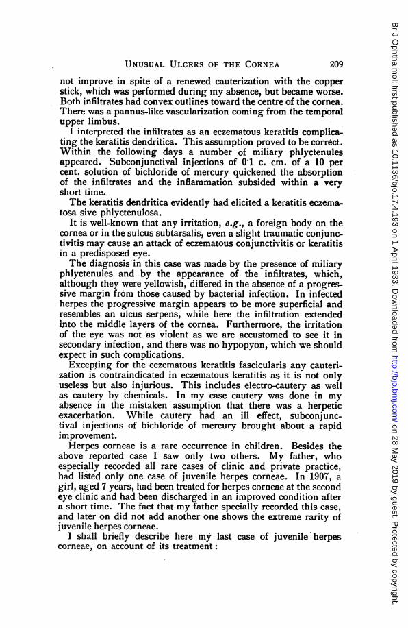
not improve in spite of a renewed cauterization with the copperstick, which was performed during my absence, but became worse.Both infiltrates had convex outlines toward the centre of the cornea.There was a pannus-like vascularization coming from the temporalupper limbus.
I interpreted the infiltrates as an eczematous keratitis complica&ting the keratitis dendritica. This assumption proved to be correct.Within the following days a number of miliary phlyctenulesappeared. Subconjunctival injections of 0 1 c. cm. of a 10 percent. solution of bichloride of mercury quickened the absorptionof the infiltrates and the inflammation subsided within a veryshort time.The keratitis dendritica evidently had elicited a keratitis eczema-
tosa sive phlyctenulosa.It is well-known that any irritation, e.g., a foreign body on the
cornea or in the sulcus subtarsalis, even a slight traumatic conjunc-tivitis may cause an attack of eczematous conjunctivitis or keratitisin a predisposed eye.The diagnosis in this case was made by the presence of miliary
phlyctenules and by the appearance of the infiltrates, which,although they were yellowish, differed in the absence of a progres-sive margin from those caused by bacterial infection. In infectedherpes the progressive margin appears to be more superficial andresembles an ulcus serpens, while here the infiltration extendedinto the middle layers of the cornea. Furthermore, the irritationof the eye was not as violent as we are accustomed to see it insecondary infection, and there was no hypopyon, which we shouldexpect in such complications.
Excepting for the eczematous keratitis fascicularis any cauteri-zation is contraindicated in eczematous keratitis as it is not onlyuseless but also injurious. This includes electro-cautery as wellas cautery by chemicals. In my case cautery was done in myabsence in the mistaken assumption that there was a herpeticexacerbation. While cautery had an ill effect, subconjunc-tival injections of bichloride of mercury brought about a rapidimprovement.Herpes corneae is a rare occurrence in children. Besides the
above reported case I saw only two others. My father, whoespecially recorded all rare cases of clinic and private practice,had listed only one case of juvenile herpes corneae. In 1907, agirl, aged 7 years, had been treated for herpes corneae at the secondeye clinic and had been discharged in an improved condition aftera short time. The fact that my father specially recorded this case,and later on did not add another one shows the extreme rarity ofjuvenile herpes corneae.
I shall briefly describe here my last case of juvenile herpescorneae, on account of its treatment:
209
on 28 May 2019 by guest. P
rotected by copyright.http://bjo.bm
j.com/
Br J O
phthalmol: first published as 10.1136/bjo.17.4.193 on 1 A
pril 1933. Dow
nloaded from

THE BRITISH JOURNAL OF OPHTHALMOLOGY
History:-The left eye of a girl, aged 8 years, had becomeinflamed two weeks ago. No illness had preceded.Findings-:-The left eye showed a typical keratitis dendritica
(Fig. 10) and a herpetic ulcer in the nasal and upper quadrant.The first eye clinic kindly administered two treatments with
Bucky rays in two weeks. During this time the lesions had progressedconsiderably, were extremely painful, and at the end of the secondweek occupied most of the pupillary area (Fig. 11). ImmediatelyI employed the old and reliable method of cauterization with thecopper stick, and within a few weeks the lesion was healed, leaving,however, a scar.
In this case the herpetic keratitis progressed relatively fast, andonly the treatment with the copper stick arrested the process.
REFERENCES1. Hallauer.-Arch. f. Anat., Vol. CIII, p. 199.2. Ventreamon.-Manufactured by Degewop, A. G., Berlin.3. Castle.-Amer. Ji. Med. Soc., p. 748, 1929.4. Locke and Townsend.-Amer. Ji. Med. Sc., p. 764, 1929.5. Sharp, Sturgis and Isaacs.-Ji. Amer. Med. Ass., p. 749. 1929.6. Rosenow.-Klinische Wochenschrift, p. 652, 1930.7. Nesic.-Serb. Arch. f. d. ges. Med., Ig. XXVIII, ref. Jahresber. f. Ofhth.,
Vol. XVII, p. 397.8. Wibo.--Bull. de la soc. belge dXojhtal., Ur. LIII, p. 38, ref. Jahresber. f.
Ophth., Vol. XVIII, p. 322, 1927.9. Bull. de la soc. belge d'oihtal., p.g. ref. Jahresber. f. O15hth., Vol.
XX, p. 354, 1928.10. E. Fuchs' textbook, 12 Aufl., p. 60, English translation by Duane, p. 67.11. Sondermann.-Klsn. .Monatsbl. f. Augenheilk., Vol. LIX, p. 759, and
Vol. LXXII, p. 460.12. Sallmann.-Zeitschrift f. Augenheilk., Vol. LX, p. 284.
THE EFFECT OF DIET ON THE NATURE OFTHE OCULAR LESIONS PRODUCED
BY NAPHTHALENE*BY
MARGHERITA COTONIo BOURNEFROM THE DEPARTMENT OF PHYSIOLOGY AND BIOCHEMISTRY,
UNIVERSITY COLLEGE, LONDON
SINCE Bouchard, in 1886, observed that the administration ofnaphthalene to rabbits caused cataract, a large number of oplhthal-mologists have been attracted to the possibilities of this substanceas a means of producing cataract experimentally. Certainly, theknowledge of a means by which cataract could be produced at willin a laboratory animal would seem to provide an excellent oppor-tunity for attack on the problem of the aetiology of cataract.* This research was carried out for the Committee on the Physiology of Vision of
the Medical Research Council.
210
on 28 May 2019 by guest. P
rotected by copyright.http://bjo.bm
j.com/
Br J O
phthalmol: first published as 10.1136/bjo.17.4.193 on 1 A
pril 1933. Dow
nloaded from