Embed Size (px)
Citation preview
ANRV324-CB23-23 ARI 24 August 2007 20:16
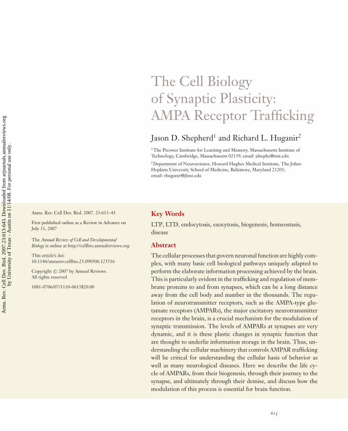
The Cell Biologyof Synaptic Plasticity:AMPA Receptor TraffickingJason D. Shepherd1 and Richard L. Huganir2
1The Picower Institute for Learning and Memory, Massachusetts Institute ofTechnology, Cambridge, Massachusetts 02139; email: [email protected] of Neuroscience, Howard Hughes Medical Institute, The JohnsHopkins University School of Medicine, Baltimore, Maryland 21205;email: [email protected]
Annu. Rev. Cell Dev. Biol. 2007. 23:613–43
First published online as a Review in Advance onJuly 31, 2007
The Annual Review of Cell and DevelopmentalBiology is online at http://cellbio.annualreviews.org
This article’s doi:10.1146/annurev.cellbio.23.090506.123516
Copyright c© 2007 by Annual Reviews.All rights reserved
1081-0706/07/1110-0613$20.00
Key Words
LTP, LTD, endocytosis, exocytosis, biogenesis, homeostasis,disease
AbstractThe cellular processes that govern neuronal function are highly com-plex, with many basic cell biological pathways uniquely adapted toperform the elaborate information processing achieved by the brain.This is particularly evident in the trafficking and regulation of mem-brane proteins to and from synapses, which can be a long distanceaway from the cell body and number in the thousands. The regu-lation of neurotransmitter receptors, such as the AMPA-type glu-tamate receptors (AMPARs), the major excitatory neurotransmitterreceptors in the brain, is a crucial mechanism for the modulation ofsynaptic transmission. The levels of AMPARs at synapses are verydynamic, and it is these plastic changes in synaptic function thatare thought to underlie information storage in the brain. Thus, un-derstanding the cellular machinery that controls AMPAR traffickingwill be critical for understanding the cellular basis of behavior aswell as many neurological diseases. Here we describe the life cy-cle of AMPARs, from their biogenesis, through their journey to thesynapse, and ultimately through their demise, and discuss how themodulation of this process is essential for brain function.
613
Ann
u. R
ev. C
ell D
ev. B
iol.
2007
.23:
613-
643.
Dow
nloa
ded
from
arj
ourn
als.
annu
alre
view
s.or
gby
Uni
vers
ity o
f T
exas
- A
ustin
on
11/1
4/08
. For
per
sona
l use
onl
y.
ANRV324-CB23-23 ARI 24 August 2007 20:16
AMPAR:alpha-amino-3-hydroxy-5-methyl-4-isoxazolepropionicacid receptor
Contents
INTRODUCTION. . . . . . . . . . . . . . . . . 614AMPA RECEPTOR
BIOGENESIS . . . . . . . . . . . . . . . . . . . 614Structure and Composition . . . . . . . 614Transcriptional and Translational
Regulation . . . . . . . . . . . . . . . . . . . . 615Biosynthesis in the Endoplasmic
Reticulum. . . . . . . . . . . . . . . . . . . . . 617Biosynthesis in the Golgi . . . . . . . . . 621
AMPA RECEPTORTRAFFICKING . . . . . . . . . . . . . . . . . 621Vesicular/Cytoskeletal
Trafficking . . . . . . . . . . . . . . . . . . . . 622Exocytosis . . . . . . . . . . . . . . . . . . . . . . . 622Synaptic Targeting and Membrane
Diffusion . . . . . . . . . . . . . . . . . . . . . 624Endocytosis . . . . . . . . . . . . . . . . . . . . . . 625Recycling . . . . . . . . . . . . . . . . . . . . . . . . 626Degradation . . . . . . . . . . . . . . . . . . . . . 626
AMPA RECEPTORS INSYNAPTIC PLASTICITY . . . . . . . 627Long-Term Potentiation . . . . . . . . . . 627Long-Term Depression . . . . . . . . . . . 630Homeostatic Scaling . . . . . . . . . . . . . . 631
AMPA RECEPTORS ANDDISEASE . . . . . . . . . . . . . . . . . . . . . . . . 631Alzheimer’s Disease . . . . . . . . . . . . . . . 632
FUTURE DIRECTIONS. . . . . . . . . . . 632
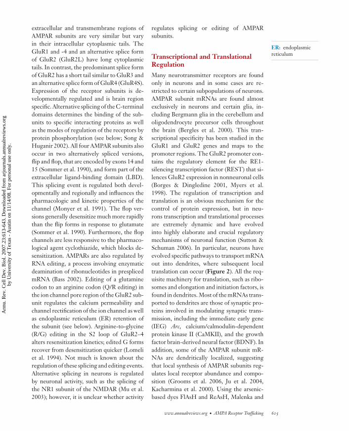
INTRODUCTION
All brain processes, such as the ability to learnand remember and those involved in ouremotions and intelligence, consciousness, andall human behavior, are possible because ofthe incredibly complex connectivity betweenneurons in the brain. Moreover, the abilityto process information and learn in responseto experience is due to continual changes inneuronal communication. Thus, it is not sur-prising that the molecular and cellular mech-anisms that regulate neuronal connectivityare highly complex and exquisitely regulated.In many ways the cellular processes found in
neurons are unique; neurons have adapted andevolved common cell biological pathways tofit their needs. This is particularly evident inthe processes that regulate synaptic function.Single neurons have thousands of synapsesthat can act as their own autonomous compu-tational units. Synaptic transmission at indi-vidual synapses is regulated mostly by changesin neurotransmitter release or in neurotrans-mitter receptor function. Glutamate recep-tors mediate most excitatory neurotransmis-sion and have been intensively investigated.The study of the AMPA-type glutamate re-ceptors (AMPARs), in particular, has proveduseful in elucidating many of the cell biolog-ical processes involved in synaptic function.AMPARs are ligand-gated ion channels andare the main mediators of excitatory neuro-transmission in the brain. Other glutamate re-ceptors such as the NMDA and metabotropicreceptors (mGluRs) also play importantroles in neuronal function. NMDA receptors(NMDARs) are voltage- and ligand-gatedchannels that are calcium permeable and areimportant regulators of synaptic plasticity.mGluRs are G protein–coupled receptorsthat act through diverse signaling pathwaysto modulate neurotransmission. However, itis arguably the AMPARs that have providedthe most insight into the role of membranetrafficking mechanisms in synaptic plasticity.
Here we review recent progress in theregulation of AMPAR trafficking. We high-light how neurons have adapted commoncell biological processes to regulate AMPARs,which ultimately has important implicationsfor brain function.
AMPA RECEPTOR BIOGENESIS
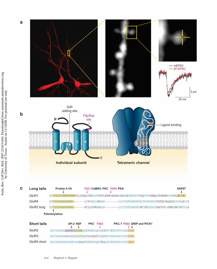
Structure and Composition
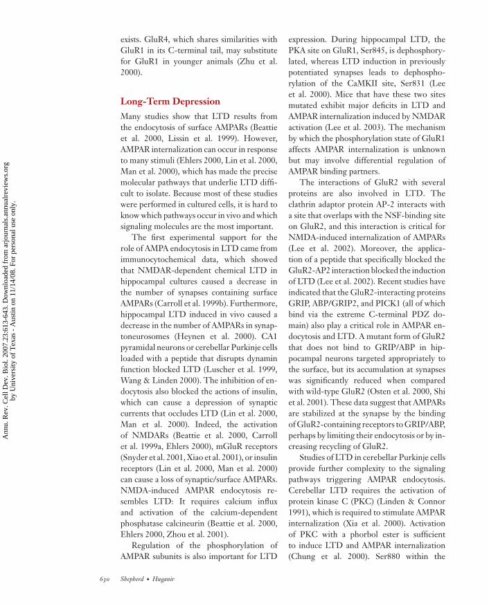
The AMPARs consist of four closely relatedgenes that encode the four subunits GluR1–4or A–D (Figure 1). These subunits combinein different stoichiometries to form ionchannels with distinct functional proper-ties (Hollmann & Heinemann 1994). The
614 Shepherd · Huganir
Ann
u. R
ev. C
ell D
ev. B
iol.
2007
.23:
613-
643.
Dow
nloa
ded
from
arj
ourn
als.
annu
alre
view
s.or
gby
Uni
vers
ity o
f T
exas
- A
ustin
on
11/1
4/08
. For
per
sona
l use
onl
y.
ANRV324-CB23-23 ARI 24 August 2007 20:16
extracellular and transmembrane regions ofAMPAR subunits are very similar but varyin their intracellular cytoplasmic tails. TheGluR1 and -4 and an alternative splice formof GluR2 (GluR2L) have long cytoplasmictails. In contrast, the predominant splice formof GluR2 has a short tail similar to GluR3 andan alternative splice form of GluR4 (GluR4S).Expression of the receptor subunits is de-velopmentally regulated and is brain regionspecific. Alternative splicing of the C-terminaldomains determines the binding of the sub-units to specific interacting proteins as wellas the modes of regulation of the receptors byprotein phosphorylation (see below; Song &Huganir 2002). All four AMPAR subunits alsooccur in two alternatively spliced versions,flip and flop, that are encoded by exons 14 and15 (Sommer et al. 1990), and form part of theextracellular ligand-binding domain (LBD).This splicing event is regulated both devel-opmentally and regionally and influences thepharmacologic and kinetic properties of thechannel (Monyer et al. 1991). The flop ver-sions generally desensitize much more rapidlythan the flip forms in response to glutamate(Sommer et al. 1990). Furthermore, the flopchannels are less responsive to the pharmaco-logical agent cyclothiazide, which blocks de-sensitization. AMPARs are also regulated byRNA editing, a process involving enzymaticdeamination of ribonucleotides in presplicedmRNA (Bass 2002). Editing of a glutaminecodon to an arginine codon (Q/R editing) inthe ion channel pore region of the GluR2 sub-unit regulates the calcium permeability andchannel rectification of the ion channel as wellas endoplasmic reticulum (ER) retention ofthe subunit (see below). Arginine-to-glycine(R/G) editing in the S2 loop of GluR2–4alters resensitization kinetics; edited G formsrecover from desensitization quicker (Lomeliet al. 1994). Not much is known about theregulation of these splicing and editing events.Alternative splicing in neurons is regulatedby neuronal activity, such as the splicing ofthe NR1 subunit of the NMDAR (Mu et al.2003); however, it is unclear whether activity
ER: endoplasmicreticulum
regulates splicing or editing of AMPARsubunits.
Transcriptional and TranslationalRegulation
Many neurotransmitter receptors are foundonly in neurons and in some cases are re-stricted to certain subpopulations of neurons.AMPAR subunit mRNAs are found almostexclusively in neurons and certain glia, in-cluding Bergmann glia in the cerebellum andoligodendrocyte precursor cells throughoutthe brain (Bergles et al. 2000). This tran-scriptional specificity has been studied in theGluR1 and GluR2 genes and maps to thepromoter regions. The GluR2 promoter con-tains the regulatory element for the RE1-silencing transcription factor (REST) that si-lences GluR2 expression in nonneuronal cells(Borges & Dingledine 2001, Myers et al.1998). The regulation of transcription andtranslation is an obvious mechanism for thecontrol of protein expression, but in neu-rons transcription and translational processesare extremely dynamic and have evolvedinto highly elaborate and crucial regulatorymechanisms of neuronal function (Sutton &Schuman 2006). In particular, neurons haveevolved specific pathways to transport mRNAout into dendrites, where subsequent localtranslation can occur (Figure 2). All the req-uisite machinery for translation, such as ribo-somes and elongation and initiation factors, isfound in dendrites. Most of the mRNAs trans-ported to dendrites are those of synaptic pro-teins involved in modulating synaptic trans-mission, including the immediate early gene(IEG) Arc, calcium/calmodulin-dependentprotein kinase II (CaMKII), and the growthfactor brain-derived neural factor (BDNF). Inaddition, some of the AMPAR subunit mR-NAs are dendritically localized, suggestingthat local synthesis of AMPAR subunits reg-ulates local receptor abundance and compo-sition (Grooms et al. 2006, Ju et al. 2004,Kacharmina et al. 2000). Using the arsenic-based dyes FlAsH and ReAsH, Malenka and
www.annualreviews.org • AMPA Receptor Trafficking 615
Ann
u. R
ev. C
ell D
ev. B
iol.
2007
.23:
613-
643.
Dow
nloa
ded
from
arj
ourn
als.
annu
alre
view
s.or
gby
Uni
vers
ity o
f T
exas
- A
ustin
on
11/1
4/08
. For
per
sona
l use
onl
y.
ANRV324-CB23-23 ARI 24 August 2007 20:16
12
4
Flip/flopsite
a
3
*
N
C
2 1 3 4
Individual subunit Tetrameric channel
b
c
N
C
S S
T S
T S
S S
T S
T S
Long tails
Short tails
P831 P845
P863 P880
Protein 4.1N
EFCYKSRAEAKRMK SEAIR KAR L ITGS GENGRV TPD PKAVH LAV A DI
EFCYKSRAEAKRMK S R KAR L ITGS GENGRV TP FPKAVH GM V TDL
EFCYKSRAEAKRMKVAKN QN NP SSQN QN ATY EGYNVYG E VKI
EFCYKSRAE KRMK KN QNF P N QN ATYREGYNVYGTE VKI
EFCYKSRAEAKRMKVAK Q FNP SSQN N ATYREGYNVYGTE KI
PKA
PKC,?PKC
EFCYKSRSE KRMK EAIR S G GENGRV DFPK SSGM AT LS GFCLIPQQ IN TSTLPRN GAGA GG GS VSQ SMQSIPSMSH PLG G
-------L F N -------- V L C TGTAIRQSSG I S P
-------M L DVM S -------- T M E APYVS-PGM N SV S
P I S F K I
S LT T K APAT Y
SA T T H L I
GluR1
GluR4
GluR2 long
GluR2
GluR3
GluR4 short
SAP97
GRIP and PICK1NSF
Palmitoylation
CaMKII, PKC
AP-2
Q/Rediting site
Ligand binding
mEPSC2P-EPSC
20 ms
5 pA
616 Shepherd · Huganir
Ann
u. R
ev. C
ell D
ev. B
iol.
2007
.23:
613-
643.
Dow
nloa
ded
from
arj
ourn
als.
annu
alre
view
s.or
gby
Uni
vers
ity o
f T
exas
- A
ustin
on
11/1
4/08
. For
per
sona
l use
onl
y.
ANRV324-CB23-23 ARI 24 August 2007 20:16
colleagues showed that transfected taggedGluR1 and 2 subunits can be synthesizedin specific dendritic compartments that areindependent of the soma ( Ju et al. 2004).In addition, a recent study has shown thatthe endogenous mRNAs for GluR1 and -2are localized to proximal and distal dendritesof hippocampal neurons. A number of GluR2mRNA clusters were localized at synapticsites, and glutamatergic signaling regulatedthe abundance as well as the localizationof GluR1 and -2 mRNAs (Grooms et al.2006). Intriguingly, NMDAR activation re-sulted in a decrease in mRNA abundance,which was dependent on a rise in intracellularcalcium and activation of the ERK (extracellu-lar signal–regulated kinase)/MAPK(mitogen-activated protein kinase) signaling pathway,ultimately leading to transcriptional arrest.In contrast, group I mGluR activation in-creased dendritic AMPAR mRNA by an in-crease in microtubule-dependent anterogrademRNA transport. Precisely how and to whatdegree local synthesis of AMPARs contributesto synaptic function and plasticity remain tobe determined. Activation of both NMDARsand mGluRs can induce long-lasting forms ofplasticity (see below) through distinct com-plex mechanisms and pathways. Dopaminereceptors also regulate local synthesis of
EPSC: excitatorypostsynaptic current
GRIP: glutamatereceptor–interactingprotein
NSF:N-ethylmaleimide-sensitive fusionprotein
PICK1: proteininteracting with Ckinase 1
AMPARs. Local application of a dopamineD1/D5 receptor agonist to dendrites led toa rapid, protein-synthesis-dependent increasein the frequency of spontaneous miniatureexcitatory postsynaptic currents (mEPSCs)(Smith et al. 2005). In addition, D1/D5 leadsto an increase in endogenous protein synthe-sis, with GluR1 as one of the proteins upreg-ulated, suggesting that GluR1 synthesis mayunderlie the changes in mEPSC frequency.How dopamine receptors regulate proteinsynthesis remains to be established.
Biosynthesis in the EndoplasmicReticulum
The basic machinery for the production oftransmembrane proteins is highly conservedin eukaryotic cells, and neurons are no excep-tion. However, owing to the unique structureand function of neurons, the secretory pro-cess is highly elaborate (Kennedy & Ehlers2006). The four AMPAR subunits, GluR1–4,assemble in different combinations to formtetrameric channels (Rosenmund et al. 1998).Most AMPARs are composed of GluR1-GluR2 or GluR2-GluR3 combinations,although the numbers and percentage of com-binations vary in different brain regions andduring development. Similar to most other
←−−−−−−−−−−−−−−−−−−−−−−−−−−−−−−−−−−−−−−−−−−−−−−−−−−−−−−−−−−−−−−−−−−−−−Figure 1Structure and composition of AMPA receptors (AMPARs). (a) Activation of AMPARs by two-photon(2P) uncaging of MNI-glutamate. Whole-cell recordings were obtained from two pyramidal neurons ofthe CA1 region of the hippocampus. Alexa 594 (10 μM) was included in the recording solution to outlineneuronal morphology. A 2P image of a dendritic region is shown; dendritic spines can readily beobserved. A spine is further magnified to show where MNI-glutamate was uncaged to activate AMPARs(0.5 μM; orange lines define the region of uncaging, which is at the center of the cross). Traces of averagedminiature excitatory postsynaptic currents (mEPSCs) and AMPAR-mediated currents elicited by 2Puncaging recorded from the same neuron are superimposed. Note the comparable kinetics of the twocurrents (provided by Jean-Claude Beıque). (b) Structure of the AMPAR subunits and the tetramericchannel. The individual subunits are composed of four transmembrane domains, and the channel consistsof four subunits, which are usually two dimers. The dimers are usually two different subunits, such asGluR1 and -2 or GluR2 and -3. (c) AMPAR C termini differ in their amino acid sequence, whichdetermines their interacting partners. Various phosphorylation sites and binding partners arehighlighted. Protein abbreviations: AP-2, adaptor protein-2; CaMKII, calcium/calmodulin-dependentprotein kinase II; GRIP, glutamate receptor–interacting protein; NSF, N-ethylmaleimide-sensitive fusionprotein; PICK1, protein interacting with C kinase 1; PKA, protein kinase A; PKC, protein kinase C;SAP97, synapse-associated protein 97.
www.annualreviews.org • AMPA Receptor Trafficking 617
Ann
u. R
ev. C
ell D
ev. B
iol.
2007
.23:
613-
643.
Dow
nloa
ded
from
arj
ourn
als.
annu
alre
view
s.or
gby
Uni
vers
ity o
f T
exas
- A
ustin
on
11/1
4/08
. For
per
sona
l use
onl
y.
ANRV324-CB23-23 ARI 24 August 2007 20:16
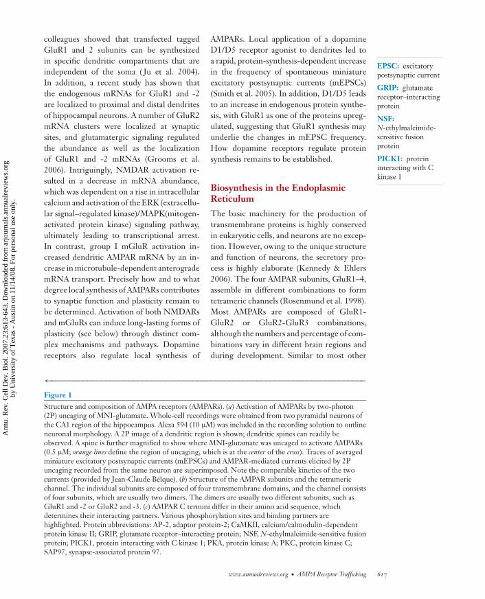
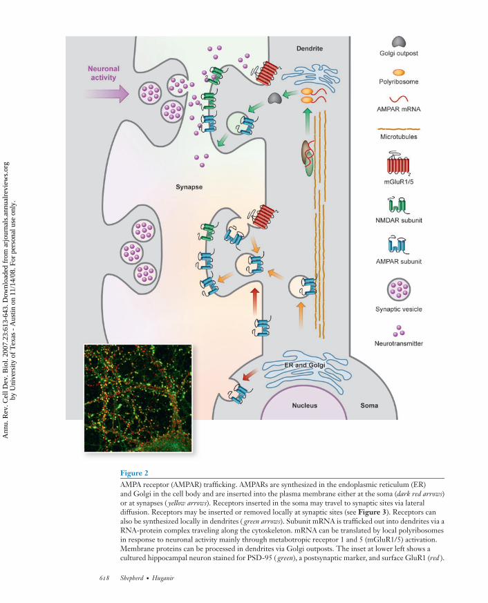
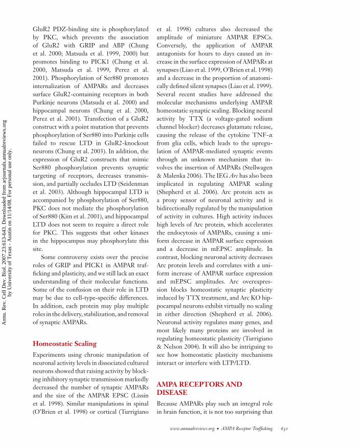
Figure 2AMPA receptor (AMPAR) trafficking. AMPARs are synthesized in the endoplasmic reticulum (ER)and Golgi in the cell body and are inserted into the plasma membrane either at the soma (dark red arrows)or at synapses ( yellow arrows). Receptors inserted in the soma may travel to synaptic sites via lateraldiffusion. Receptors may be inserted or removed locally at synaptic sites (see Figure 3). Receptors canalso be synthesized locally in dendrites ( green arrows). Subunit mRNA is trafficked out into dendrites via aRNA-protein complex traveling along the cytoskeleton. mRNA can be translated by local polyribosomesin response to neuronal activity mainly through metabotropic receptor 1 and 5 (mGluR1/5) activation.Membrane proteins can be processed in dendrites via Golgi outposts. The inset at lower left shows acultured hippocampal neuron stained for PSD-95 ( green), a postsynaptic marker, and surface GluR1 (red ).
618 Shepherd · Huganir
Ann
u. R
ev. C
ell D
ev. B
iol.
2007
.23:
613-
643.
Dow
nloa
ded
from
arj
ourn
als.
annu
alre
view
s.or
gby
Uni
vers
ity o
f T
exas
- A
ustin
on
11/1
4/08
. For
per
sona
l use
onl
y.
ANRV324-CB23-23 ARI 24 August 2007 20:16
multimeric membrane proteins, AMPARs arefirst assembled in the ER. Once in the ER,the N-terminal signal sequence is cleaved,and a high-mannose glycosylation attachesto the first extracellular domain at specificasparagine residues (Rogers et al. 1991). Theprecise mechanisms that govern subunit as-sembly, especially the different combinations,are not well understood but depend on lumi-nal interactions between the N-terminal do-mains (NTDs) of the subunits (Kuusinen et al.1999). Tetramers form from dimers, whichrequire the transmembrane segments and theextracellular S2 loop for assembly (Ayalon &Stern-Bach 2001). A recent study showed thatinteractions between the LBDs of the AM-PARs subunits are critical for the dimer-to-tetramer transition (Greger et al. 2006). TheNTD, which is not required for homomericassembly, seems to play an important role inthe preferential assembly of heteromers (Ay-alon et al. 2005). GluR1/2 heteromers exit theER rapidly, whereas GluR2/3 heteromers areretained longer in the ER. The GluR2 subunithas an arginine-based ER retention motif thatconsists of a single arginine residue (R607)in the transmembrane domain (Greger et al.2002). As discussed above, this arginine isgenerated by RNA editing of the originalsequence coding for glutamine. This residueis also critical for channel properties andconfers calcium impermeability and a charac-teristic linear rectifying property to GluR2-containing AMPARs (Burnashev et al. 1992).Transgenic mice with impaired Q/R editingexhibit epileptic seizures and die within twoweeks after birth (Brusa et al. 1995). The Q/Rediting results in a stable pool of GluR2 sub-units in the ER that exits more efficiently ina heteromeric complex. Knockout mice thatlack GluR2 do form GluR1/3 heteromers,as well as GluR1 and 3 homomers, but thesechannels are poorly translocated to synapses(Sans et al. 2003). It is unclear how the Q/Rsite regulates ER retention, but a retentionprotein that binds to the edited site may beresponsible. Recent studies have suggestedthat other editing sites also regulate secretory
PSD: postsynapticdensity
trafficking of AMPARs. A developmentallyregulated editing site, R/G at position 743in the S2 domain, governs whether GluR2homomers will form. Following editing, theformation of homomers is precluded (Gregeret al. 2006). In cell lines, the flip and flopforms of AMPARs seem to traffic differen-tially to the surface. Homomeric AMPARsof the flop form accumulate in the ER,whereas those of the flip form traffic effi-ciently to the cell surface (Coleman et al.2006). As a caveat, many of these studiesmake use of transfected recombinant proteinsin cell lines or in primary neurons, andtherefore it is not clear if native AMPARsare similarly regulated. It would be inter-esting to know if RNA editing and ERretention/export are modulated by neuronalactivity or other signaling pathways that couldchange the composition of active surfaceAMPARs.
AMPARs associate with the ER chaper-ones BiP and calnexin (Rubio & Wenthold1999), and GluR2 colocalizes extensivelywith BiP in the ER (Greger et al. 2002).These or other unidentified chaperonesmay control ER retention. Researchers haveidentified many proteins that interact withthe C termini of the AMPARs (see Figure 1)(Song & Huganir 2002), and some of theseproteins may also regulate ER retention orexit (Figure 3). The GluR2 C terminus hasa PDZ consensus motif (SVKI) that interactswith several PDZ domain–containing pro-teins, including the protein interacting withC kinase 1 (PICK1) (Xia et al. 1999), whichappears to control AMPAR endocytosisand/or recycling but may also be necessaryfor the exit of GluR2 from the ER (Gregeret al. 2002). Another PDZ interaction, thatbetween the GluR1 C terminus (ATGL site)and SAP97 (synapse-associated protein 97)(Leonard et al. 1998, Rumbaugh et al. 2003),seems to occur in the ER (Sans et al. 2001).The SAP97-interacting region of GluR1 isnecessary for correct synaptic targeting, but itis unclear if this is due to ER retention of theprotein (Hayashi et al. 2000). Intriguingly,
www.annualreviews.org • AMPA Receptor Trafficking 619
Ann
u. R
ev. C
ell D
ev. B
iol.
2007
.23:
613-
643.
Dow
nloa
ded
from
arj
ourn
als.
annu
alre
view
s.or
gby
Uni
vers
ity o
f T
exas
- A
ustin
on
11/1
4/08
. For
per
sona
l use
onl
y.
ANRV324-CB23-23 ARI 24 August 2007 20:16
TARPs:transmembraneAMPAR regulatoryproteins
LTP: long-termpotentiation
LTD: long-termdepression
glutamate binding and ion permeationthrough the pore are also required for normalexpression of AMPARs; ligand mutants thatcannot bind glutamate, and pore mutantsthat block ion permeation, exhibit decreasedsurface/synaptic expression and are retainedin the ER (Grunwald & Kaplan 2003). Thissuggests that there may be a surveillancemechanism in the ER that allows onlycorrectly folded and active channels to exit.
Stargazin, a calcium channel γ-subunithomolog, seems to control AMPAR traffick-ing at multiple points during the secretoryprocess (Nicoll et al. 2006). Stargazin wasoriginally identified as the mutant gene in theStargazer mouse, which exhibits profoundcerebellar ataxia and epilepsy (Osten & Stern-Bach 2006). Stargazin and its closely relatedγ-3, γ-4, and γ-8 paralogs [collectivelycalled TARPs (transmembrane AMPARregulatory proteins)] interact directly withall the AMPAR subunits to promote theirtransport to the cell surface (Chen et al. 2000,Tomita et al. 2003) and to modulate channelfunction (Priel et al. 2005, Tomita et al. 2005).Stargazer cerebellar granule cells exhibit astriking lack of surface AMPARs, and alarge portion of the intracellularly retainedreceptors exhibit immature glycosylation(Tomita et al. 2003). FRET studies suggestthat stargazin and the AMPARs may first
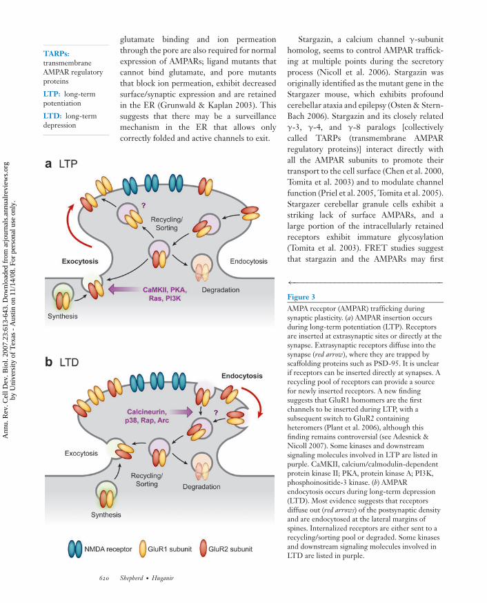
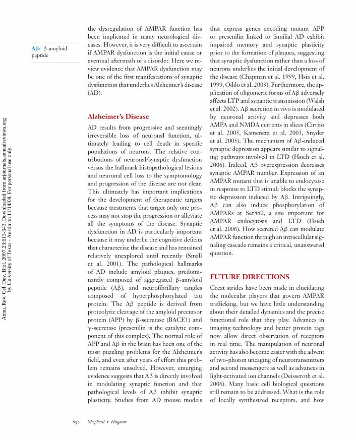
←−−−−−−−−−−−−−−−−−−−−−−−−−−−−−−−−−Figure 3AMPA receptor (AMPAR) trafficking duringsynaptic plasticity. (a) AMPAR insertion occursduring long-term potentiation (LTP). Receptorsare inserted at extrasynaptic sites or directly at thesynapse. Extrasynaptic receptors diffuse into thesynapse (red arrow), where they are trapped byscaffolding proteins such as PSD-95. It is unclearif receptors can be inserted directly at synapses. Arecycling pool of receptors can provide a sourcefor newly inserted receptors. A new findingsuggests that GluR1 homomers are the firstchannels to be inserted during LTP, with asubsequent switch to GluR2 containingheteromers (Plant et al. 2006), although thisfinding remains controversial (see Adesnick &Nicoll 2007). Some kinases and downstreamsignaling molecules involved in LTP are listed inpurple. CaMKII, calcium/calmodulin-dependentprotein kinase II; PKA, protein kinase A; PI3K,phosphoinositide-3 kinase. (b) AMPARendocytosis occurs during long-term depression(LTD). Most evidence suggests that receptorsdiffuse out (red arrows) of the postsynaptic densityand are endocytosed at the lateral margins ofspines. Internalized receptors are either sent to arecycling/sorting pool or degraded. Some kinasesand downstream signaling molecules involved inLTD are listed in purple.
620 Shepherd · Huganir
Ann
u. R
ev. C
ell D
ev. B
iol.
2007
.23:
613-
643.
Dow
nloa
ded
from
arj
ourn
als.
annu
alre
view
s.or
gby
Uni
vers
ity o
f T
exas
- A
ustin
on
11/1
4/08
. For
per
sona
l use
onl
y.
ANRV324-CB23-23 ARI 24 August 2007 20:16
interact in the ER (Bedoukian et al. 2006).This suggests that stargazin may play a rolein trafficking AMPARs from the ER to thecis-Golgi. In addition, stargazer granule cellsexhibit an upregulated ER unfolded proteinresponse (Vandenberghe et al. 2005), suggest-ing a role for stargazin in AMPAR folding orassembly. Mice lacking γ-8, the predominanthippocampal TARP, also exhibit dramaticintracellular retention of AMPARs in the ERand Golgi (Rouach et al. 2005), suggesting aconserved function. Stargazin is also able toalleviate the block in the ER exit of the flopisoforms in cell lines (Coleman et al. 2006),suggesting that stargazin may act as an escortchaperone. However, it is unclear if TARPSare absolutely required for correct AMPARfolding as an auxiliary subunit of the channelor whether they only enhance channel assem-bly. Further work is required to fully under-stand the role of TARPs in AMPAR foldingand assembly during the biosynthetic process.
Biosynthesis in the Golgi
Neurons have both somatic and dendriticGolgi compartments, suggesting that proteinstrafficking through the secretory pathway canexit in the soma or at specialized dendriticsites (Horton & Ehlers 2004). Indeed, theseGolgi outposts may allow posttranslationalmodification of proteins that are locallytranslated in dendrites. All AMPAR subunitspossess N-glycosylation sites but becomefully glycosylated only in the Golgi. AMPARshave mostly complex oligosaccharide forms;they are relatively insensitive to treatmentwith Endo H (Rogers et al. 1991). nPIST,a protein enriched in the Golgi, dendritictubulovesicles, and the postsynaptic density(PSD), interacts with the C-tail of stargazinand may help AMPARs exit the Golgi (Cuadraet al. 2004). Lipid modification of proteinsalso occurs in the Golgi. Recent studieshave suggested that these modifications,especially palmitoylation, play importantroles in synaptic function (El-Husseini et al.2002, Huang & El-Husseini 2005). Palmi-
toylation is a reversible process that occursby a covalent attachment of palmitate viathioester bonds to cytosolic cysteine residues.Protein palmitoylation is regulated by thebalance of palmitoyl acyl transferase (PAT)and palmitoyl thioesterase activities. Palmi-toylation of the scaffolding protein PSD-95regulates AMPAR accumulation at synapses,and AMPAR internalization requires de-palmitoylation of PSD-95 (El-Husseini et al.2002). In addition, other AMPAR-interactingproteins important for membrane traffickingof the AMPARs, such as the glutamatereceptor–interacting proteins GRIP1b andGRIP2b (pABP-L), are also palmitoylated(DeSouza et al. 2002, Yamazaki et al. 2001).A recent study showed that all the AMPARsubunits are palmitoylated at two sites andthat these modifications are important forcorrect AMPAR trafficking (Hayashi et al.2005). Palmitoylation occurs at a C-terminalcysteine that lies just after the final trans-membrane domain and also at a cysteine inthe TMD 2 region, three amino acids awayfrom the Q/R editing site. Palmitoylationof this second site is increased by the Golgiapparatus–specific PAT GODZ [Golgi-specific DHHC (Asp-His-His-Cys) zincfinger protein], which promotes the accumu-lation of the receptor in the Golgi (Hayashiet al. 2005, Uemura et al. 2002). Palmitoy-lation of the C-terminal domain inhibitsAMPAR interaction with the 4.1N protein,which stabilizes AMPAR surface expression(Shen et al. 2000). Mutation of the palmitoy-lation sites increases GluR1 association with4.1N and inhibits the regulated endocytosisof AMPARs (Hayashi et al. 2005) (see below).
AMPA RECEPTORTRAFFICKING
Neurons pose many unique problems for thetrafficking of membrane proteins because oftheir highly polarized and elaborate structure.Membrane proteins must travel extremelylong distances, and transmembrane proteinsmay be inserted at plasma membrane domains
www.annualreviews.org • AMPA Receptor Trafficking 621
Ann
u. R
ev. C
ell D
ev. B
iol.
2007
.23:
613-
643.
Dow
nloa
ded
from
arj
ourn
als.
annu
alre
view
s.or
gby
Uni
vers
ity o
f T
exas
- A
ustin
on
11/1
4/08
. For
per
sona
l use
onl
y.
ANRV324-CB23-23 ARI 24 August 2007 20:16
far from their final location. Indeed, sites ofsynaptic contact contain their own milieuof proteins, and in some cases individualsynapses contain specific receptor subtypesand scaffolding proteins that are differentfrom their neighboring synapses only micronsaway.
Vesicular/Cytoskeletal Trafficking
Early in neuronal development, packets of re-ceptors and scaffolding proteins travel alongdendrites (Gerrow et al. 2006, Washbourneet al. 2002). The precise cues that governwhere these receptors ultimately stop andform synapses are unknown. The traffick-ing of these packets is microtubule depen-dent, and transport is an active process in-volving motor proteins such as dynein andkinesin (Hirokawa & Takemura 2005). Themultiple PDZ domain–containing proteinGRIP1/ABP interacts directly with the heavychain of conventional kinesin (KIF5) (Setouet al. 2002) and binds to the C-terminalPDZ motif of GluR2 and GluR3 (Donget al. 1997). A complex of GluR2, GRIP1,and kinesin can be immunoprecipitated frombrain lysates, and the expression of dominant-negative versions of kinesin decreases synap-tic abundance of AMPARs (Setou et al. 2002).The kinesin KIF1 interacts with Liprin-α,which also interacts with the GluR2/GRIP1complex (Wyszynski et al. 2002). KIF1 andAMPARs can be coimmunoprecipitated withKIF1 in brain lysates (Shin et al. 2003), and theexpression of Liprin mutants that cannot bindGRIP1 blocks synaptic targeting of AMPARs(Wyszynski et al. 2002). These results indi-cate that the GRIP1/ABP protein serves as anadaptor to link AMPARs to kinesins and pro-mote dendritic transport.
Although dendrites contain microtubulesalong which most cargo is transported, den-drites are also enriched in actin, especially inspines, which are small protrusions along den-drites that form small microcompartmentsimportant for synaptic function. Myosins, themain actin-dependent motor proteins, have
recently been implicated in AMPAR trans-port. Myosin Vb has been associated withGluR1 (Lise et al. 2006), and the expressionof the myosin Vb tail domain in develop-ing hippocampal neurons enhances the accu-mulation of GluR1 in the soma but reducesthe expression of GluR1 at the surface (Liseet al. 2006). Myosin VI has also been impli-cated in AMPAR trafficking (Osterweil et al.2005). Myosin VI–deficient neurons exhibitdeficits in activity-dependent AMPAR inter-nalization as well as a decrease in the numberof synapses and dendritic spines (Osterweilet al. 2005). Myosin VI is found in an AMPARcomplex that includes the endocytosis adap-tor protein AP-2 and the scaffolding proteinSAP97, suggesting that Myosin VI may be se-lectively involved in clathrin-dependent en-docytosis of AMPARs (Osterweil et al. 2005,Wu et al. 2002). Another actin adaptor, pro-tein 4.1N, also associates with the AMPARsand appears to stabilize the surface expressionof GluR1 (Shen et al. 2000). RIL (reversion-induced LIM protein), which has a PDZ do-main, may also be involved in actin-dependenttrafficking of GluR1 (Schulz et al. 2004). Live-imaging experiments of fluorescently taggedAMPARs have shown that GluR1 is consti-tutively and rapidly transported throughoutthe neuron. In contrast, GluR2 is less mobileand mostly retained in relatively immobilemembrane-associated clusters, some of whichare synapses. Interestingly, these receptor dy-namics are independent of neuronal activity(Perestenko & Henley 2003).
Exocytosis
Precise synaptic targeting and insertion of re-ceptors are extremely complicated, given thatan average neuron contains approximately10,000 synapses. Despite intense study, itis still unclear whether AMPARs are firstinserted into the extrasynaptic plasma mem-brane or directly into synapses (see Figures 2and 3). One possibility is that AMPARs firstare inserted into the plasma membrane in thesoma at extrasynaptic sites and then travel
622 Shepherd · Huganir
Ann
u. R
ev. C
ell D
ev. B
iol.
2007
.23:
613-
643.
Dow
nloa
ded
from
arj
ourn
als.
annu
alre
view
s.or
gby
Uni
vers
ity o
f T
exas
- A
ustin
on
11/1
4/08
. For
per
sona
l use
onl
y.
ANRV324-CB23-23 ARI 24 August 2007 20:16
out into dendrites via lateral diffusion in theplasma membrane until they finally reach thesynapse and become anchored in the PSD. Arecent study using an innovative method tomeasure receptor insertion of AMPARs hassuggested that most receptors are insertedin the somatic plasma membrane (Adesniket al. 2005). In this study, a membrane-impermeable photoreactive AMPAR antago-nist derived from ANQX was used to photoin-activate surface receptors, and the subsequentexocytosis of AMPARs was then mea-sured electrophysiologically. The recovery ofsynaptic receptors measured with this methodwas surprisingly slow, taking hours rather thanminutes. In contrast, exocytosis of AMPARsin the soma was much faster. However, thesedata are inconsistent with many studies,including data from the same laboratory (Luet al. 2001), that find more rapid insertionof receptors into the plasma membrane atdendrites and synapses. Another possibilityis that AMPARs are trafficked intracellularlyinto dendrites via the cytoskeleton-associatedmotors and then directly inserted at synapticsites. A recent study of the role of the exocystcomplex in AMPAR delivery has suggestedthat Exo70 mediates AMPAR insertiondirectly within the PSD rather than at ex-trasynaptic membranes (Gerges et al. 2006).A third possibility is that AMPARs are syn-thesized in dendritic compartments and theninserted directly into synapses. Studies usingcleavable extracellular-tagged transfectedreceptors suggest that AMPARs are insertedalong dendrites (Passafaro et al. 2001) and thatthis occurs in a subunit-dependent manner.Other studies using different types of epitopetags, for example, a bungarotoxin-bindingsite or bi-arsenical dies, have observed similardendritic insertion ( Ju et al. 2004, Sekine-Aizawa & Huganir 2004). AMPAR insertionis blocked by the introduction of intracellulartetanus toxin, implying that AMPARs areinserted via SNARE-dependent exocytosis(Lu et al. 2001). Most likely a combinationof all these processes occurs, depending onthe subunit composition of the receptors
and the context of the neuron’s activitystate.
Many studies have shown that plasmamembrane insertion of AMPARs is depen-dent on the subunit composition of the recep-tor. Surface insertion of GluR1 and -4 (or thelong-tailed AMPARs) occurs slowly in basalconditions and is stimulated by neuronal ac-tivity and NMDAR activation (Hayashi et al.2000). In contrast, GluR2 insertion in manyneurons is rapid and occurs constitutively un-der basal conditions, without the need forsynaptic activity (Passafaro et al. 2001, Shiet al. 2001). Endogenous receptors consistmostly of either GluR1/2 or GluR2/3 het-eromers, and the GluR1 trafficking signalsdominate over GluR2 in controlling inser-tion. When GluR1/2 heteromeric channelsare expressed, the activity-dependent traffick-ing of GluR1 dominates, whereas GluR2/3heteromeric channels behave like GluR2 ho-momeric channels and constitutively trafficinto the synapse. These subunit-specific rulesfor trafficking have led to a simple model inwhich GluR2-GluR3 receptors continuouslycycle in and out of synapses, preserving the to-tal number of synaptic AMPARs (the consti-tutive pathway), whereas GluR1-GluR2 (andGluR4) receptors are added into synapses inan activity-dependent manner during synapticplasticity (the regulated pathway) (Malinowet al. 2000). The constitutive pathway maymaintain synaptic strength despite proteinturnover, and the regulated pathway may acttransiently upon the induction of synapticplasticity.
This differential trafficking of the AMPARsubunits is dependent on their C-terminaltails (see Figure 1 for details). Expression ofthe C terminus of GluR2 decreases AMPAEPSCs, whereas expression of the GluR1 Cterminus has no effect on the basal synap-tic transmission but blocks activity-dependentincreases in AMPA responses. The differen-tial behavior of the tails seems to be governedby their interacting proteins. The GluR2 Cterminus binds to N-ethylmaleimide-sensitivefusion protein (NSF) (Nishimune et al. 1998,
www.annualreviews.org • AMPA Receptor Trafficking 623
Ann
u. R
ev. C
ell D
ev. B
iol.
2007
.23:
613-
643.
Dow
nloa
ded
from
arj
ourn
als.
annu
alre
view
s.or
gby
Uni
vers
ity o
f T
exas
- A
ustin
on
11/1
4/08
. For
per
sona
l use
onl
y.
ANRV324-CB23-23 ARI 24 August 2007 20:16
Osten et al. 1998, Song et al. 1998), andthis site seems to regulate the rapid exocy-tosis of GluR2 at synaptic sites (Beretta et al.2005). The mechanism by which NSF reg-ulates AMPAR trafficking may relate to itsclassical role in controlling membrane fu-sion (Rothman 1994). However, NSF mayalso regulate the interaction of GluR2 withanother interacting protein, PICK1 (Hanleyet al. 2002). PICK1 may bind to and stabilizeintracellular pools of GluR2 that may providea ready source of receptors for quick mem-brane insertion (Gardner et al. 2005, Liu &Cull-Candy 2005, Steinberg et al. 2004). NSFbinding may dissociate the GluR2-PICK1complex (Hanley et al. 2002), thus allowingmembrane insertion. However, the precisemolecular role of NSF in regulating AMPARinsertion remains elusive.
Synaptic Targeting and MembraneDiffusion
AMPARs are concentrated at synapses, wherethey are precisely localized to efficientlymediate the response to glutamate releasedfrom presynaptic terminals. Whether ornot AMPARs are inserted into the plasmamembrane at extrasynaptic regions or morelocally at synapses, there must be moleculesthat retain the receptors at synapses tomaintain the high density of receptors at thesynapse. The molecular mechanisms under-lying the synaptic retention of AMPARs arenot clear, but AMPAR-interacting proteinsseem to be critical in this process (Song &Huganir 2002). Indeed, PSD-95 and othermembers of the PSD-95 protein family arecritical determinants for synaptic targeting ofAMPARs. Overexpression of PSD-95 en-hances AMPAR-mediated synaptic currents(Beique & Andrade 2003, El-Husseini et al.2000). Moreover, knocking out or knockingdown PSD-95 and its family membersdecreases the synaptic levels of AMPARs(Beique et al. 2006, Elias et al. 2006). Al-though PSD-95 does not directly interactwith AMPARs, it does bind to stargazin
and other members of the TARP family(Chen et al. 2000, Tomita et al. 2005), whichprovide the link to AMPARs. TARPs bind toall four AMPAR subunits and also interactthrough their C-terminal domains with thePDZ domains of PSD-95. This interactionbetween the TARPs and PSD-95 appears tobe crucial for AMPAR targeting to synapses(Chen et al. 2000, Tomita et al. 2005).Another family of AMPAR-interacting pro-teins that has been reported to be involvedin the synaptic retention is the neuronalpentraxins, NARP, NP1, and NPR (Song &Huganir 2002). These multimeric proteinsbind to all AMPAR subunits and promoteclustering of the receptor (O’Brien et al.1999, 2002; Xu et al. 2003). Overexpressionof NARP in neurons increases the numberof synaptic AMPARs, and the expression ofdominant-negative forms of NARP decreasesAMPAR clusters (O’Brien et al. 1999, 2002).
How these AMPAR-interacting proteinspromote the retention of receptors at synapsesis not clear, but it is likely that they reduce thelateral membrane diffusion and endocytosis ofthe receptors. PSD-95 and stargazin presum-ably stabilize receptors by linking the recep-tors with the PSD. In contrast, the multimericpentraxins likely link the receptors with ex-tracellular proteins stabilizing the receptorsat synapses. To test the mobility of recep-tors at synapses, the real-time lateral diffu-sion of surface AMPARs in the plasma mem-brane has recently been investigated directlyby optical monitoring of the movement ofsingle receptors, which relies on the use ofsmall latex particles or quantum dots cou-pled via antibodies to the AMPAR extracel-lular domain (Borgdorff & Choquet 2002,Tardin et al. 2003). These studies showed that,whereas extrasynaptic AMPARs are highlymobile, synaptic AMPARs are relatively im-mobile under basal conditions. Neuronal ac-tivity significantly increases the movementof AMPARs, especially in extrasynaptic re-gions. Extrasynaptic receptors continuouslymove with high (10−1–10−2 μm2 s−1) and low(less than 10−4 μm2 s−1) diffusion rates and
624 Shepherd · Huganir
Ann
u. R
ev. C
ell D
ev. B
iol.
2007
.23:
613-
643.
Dow
nloa
ded
from
arj
ourn
als.
annu
alre
view
s.or
gby
Uni
vers
ity o
f T
exas
- A
ustin
on
11/1
4/08
. For
per
sona
l use
onl
y.
ANRV324-CB23-23 ARI 24 August 2007 20:16
transiently interact with scaffolding proteinclusters at synaptic sites. Receptor movementsbetween scaffold clusters were Brownian innature, moving in random steps. This indi-cates that scaffold proteins may act as molec-ular determinants of receptor exchange be-tween extrasynaptic and synaptic membranecompartments. As discussed above, PSD-95and stargazin are ideally suited for this role;together they interact with AMPARs but alsobind to many other PSD proteins via multiplePDZ interactions in a scaffold-like manner.Recently the interaction between stargazinand PSD-95 has been shown to be crucial forAMPAR diffusion in and out of synapses (Batset al. 2007). Disruption of this interactionled to an increase in AMPAR diffusion andprevented AMPAR accumulation at synapticsites (Bats et al. 2007). This result indicatesthat the stargazin–PSD-95 complex limitsAMPAR lateral diffusion at synapses andis critical for the retention of receptors atsynapses.
Endocytosis
Clathrin-mediated endocytosis is a generalmechanism of membrane protein regulation,and the core endocytic protein machinery ishighly conserved in most species and cell types(Mousavi et al. 2004). Some of these proteins,such as dynamin and endophilin, are essen-tial for endosome formation, whereas others,such as AP-2, act as clathrin adaptor moleculesthat link specific cargo with the clathrin lat-tice. However, many of these proteins alsoserve specific roles in specialized endocyticpathways such as synaptic vesicle recyclingin the presynaptic nerve terminal. The clas-sic dynamin mutation, shibire, causes paraly-sis because of a severe defect in synaptic vesi-cle recycling at the neuromuscular junction(Poodry et al. 1973).
Postsynaptic endocytosis of receptors isthought to be mediated by a similar reper-toire of proteins, although perhaps via spe-cific protein isoforms. Dynamin 2 and 3 aremostly postsynaptic and are localized to the
PSD via their interaction with the postsynap-tic scaffolding proteins Shank and Homer,respectively (Gray et al. 2003, Okamotoet al. 2001). Distinct isoforms of endophilins(2 and 3) are localized to postsynaptic mem-branes, whereas endophilin 1 is localizedpredominantly presynaptically (Chowdhuryet al. 2006). Endocytosis of AMPARs issimilar to the stimulated endocytosis of Gprotein–coupled receptors in that both pro-cesses occur via clathrin-coated pits and re-quire dynamin. Numerous methods of block-ing clathrin-dependent endocytosis, such asthe expression of a dominant-negative formof dynamin, high concentrations of sucrose, orpeptide-mediated disruption of the dynamin-amphiphysin complex, all block AMPAR en-docytosis (Carroll et al. 1999a, Man et al.2000, Wang & Linden 2000). After internal-ization, AMPARs are sorted either (a) withinearly endosomes to a specialized recycling en-dosome compartment that allows quick rein-sertion to the surface or (b) to late endosomesand lysosomes that allow degradation (Ehlers2000, Lee et al. 2004).
Specific endocytic zones, segregated fromthe PSD, can be found in the lateral marginsof excitatory synapses (Blanpied et al. 2002).These sites appear to be sites of glutamate re-ceptor internalization (Racz et al. 2004). Thisis particularly evident in electron micrographsof dendritic spines, which show the presenceof clathrin-coated pits and vesicles (Petraliaet al. 2003, Spacek & Harris 1997). Thesestudies also suggest that clathrin-mediated en-docytosis varies during the development ofneurons. Immature neurons are more abun-dant in dendritically localized clathrin pits,which are hot spots of rapid and repeatedclathrin coat assembly and disassembly. Incontrast, clathrin coats in mature dendritesare more stable but fewer in number.
Specific postsynaptic proteins are selec-tively involved in the endocytosis of AMPARs.The immediate early gene (IEG) CPG2 me-diates both constitutive and activity-regulatedglutamate receptor internalization (Cottrellet al. 2004) and localizes to the endocytic zone
www.annualreviews.org • AMPA Receptor Trafficking 625
Ann
u. R
ev. C
ell D
ev. B
iol.
2007
.23:
613-
643.
Dow
nloa
ded
from
arj
ourn
als.
annu
alre
view
s.or
gby
Uni
vers
ity o
f T
exas
- A
ustin
on
11/1
4/08
. For
per
sona
l use
onl
y.
ANRV324-CB23-23 ARI 24 August 2007 20:16
of excitatory synapses. CPG2 knockdowndisrupted constitutive AMPAR and NMDARinternalization as well as activity-inducedAMPAR internalization. Another IEG, Arc, isinduced by neuronal activity associated withcognition and long-term forms of synapticplasticity. Arc mRNA is exquisitely regulated:The transcribed message is targeted to thedendrites of neurons as they engage ininformation processing and storage, and itis locally translated at activated synapses(Steward & Worley 2001). Arc regulatesAMPAR trafficking by interacting with theintegral endocytic proteins dynamin andendophilin (Chowdhury et al. 2006, RialVerde et al. 2006). High levels of Arc acceler-ate AMPAR endocytosis and decrease surfaceand synaptic AMPAR levels. These effects arespecific to AMPARs, suggesting that Arc actsas an adaptor protein that localizes AMPARsto the endocytic machinery. These recentstudies elucidate some of the specific proteinmachinery involved in AMPAR endocytosis,but the signals that regulate them still remainrelatively unknown. The elucidation ofthese signals will be critical for understand-ing AMPAR trafficking because activity-dependent endocytosis of AMPARs leads toseveral forms of synaptic plasticity (see below).
Recycling
Recent studies suggest that recycling endo-somes contain a pool of AMPARs that is asource for the rapid insertion of receptors.Activation of NMDA receptors can regulatethe kinetics of recycling and significantly af-fect the relative amount of receptors that aremaintained intracellularly versus on the sur-face (Park et al. 2004). This is particularlyevident after long-term potentiation (LTP)-inducing stimuli, for which an increase in thegeneral recycling of endocytic cargo occursand recycling endosomes physically translo-cate into spines (Park et al. 2004, 2006). Thisenhancement of recycling also provides addi-tional lipid membrane, which is critical for thestructural growth and expansion of dendritic
spines during LTP (Park et al. 2006). The cou-pling of AMPAR insertion and membrane ad-dition may be an explanation for the tightlycorrelated scaling of spine size with AMPAR-mediated synaptic currents (Matsuzaki et al.2001). The specific molecular players thatregulate recycling are beginning to be eluci-dated, although precisely how NMDA activityregulates recycling remains unclear. One pro-tein, NEEP21 (neuron-enriched endosomalprotein of 21 kDa), is localized to early and re-cycling endosomes (Steiner et al. 2002) and in-teracts with general endocytic/recycling pro-teins such as syntaxin 13 (Prekeris et al. 1998).Downregulation of NEEP21 leads to im-paired recycling of internalized transferrin re-ceptor (Steiner et al. 2002) and neurotensinreceptor 2 (Debaigt et al. 2004), suggesting ageneral role in receptor recycling. However,suppression of NEEP21 also significantly re-tards GluR1 and GluR2 recycling followingNMDA-induced internalization (Steiner et al.2002), which may occur through the interac-tion of NEEP21 with GRIP and syntaxin 13(Steiner et al. 2005). These results suggest thatthe AMPAR-interacting proteins GRIP andPICK1 may play some role in the regulationof receptor recycling to modulate the level ofsynaptic receptors.
Degradation
Little is known about how AMPARs are de-graded at synapses, but recent studies sug-gest a role for the ubiquitin/proteasome sys-tem (UPS). The UPS consists of a numberof proteins that coordinately regulate manycellular functions, including protein degrada-tion and endocytosis. Ubiquitination can sig-nal endocytosis to occur through specializedmachinery that regulates clathrin endocytosis,and it has been implicated in the trafficking ofreceptor tyrosine kinases (Mukhopadhyay &Riezman 2007). Generally, monoubiquitina-tion signals endocytosis, whereas polyubquiti-nation ultimately leads to degradation viathe proteasome. However, monoubiquitina-tion also seems to play a role as a sorting
626 Shepherd · Huganir
Ann
u. R
ev. C
ell D
ev. B
iol.
2007
.23:
613-
643.
Dow
nloa
ded
from
arj
ourn
als.
annu
alre
view
s.or
gby
Uni
vers
ity o
f T
exas
- A
ustin
on
11/1
4/08
. For
per
sona
l use
onl
y.
ANRV324-CB23-23 ARI 24 August 2007 20:16
signal that targets its substrates to multi-vesicular bodies, which is the first step lead-ing to lysosomes, where degradation can alsooccur.
Initial studies in Caenorhabditis elegansfound that direct ubiquitination of GLR-1glutamate receptors (the AMPAR homologs)at synapses induced the removal of recep-tors from the postsynaptic membrane, a pro-cess that required the clathrin adaptor AP180(Burbea et al. 2002). Loss-of-function mu-tants in the multisubunit APC ubiquitin lig-ase complex exhibit increased levels of GLR-1( Juo & Kaplan 2004). A similar phenotypeis observed at the neuromuscular synapse ofDrosophila APC mutants, which also have in-creased postsynaptic glutamate receptor clus-tering and defects in synaptic transmission(van Roessel et al. 2004). GLR-1 does notseem to be a direct substrate of the APC, butthe mutant phenotype can be suppressed bythe introduction of a loss-of-function alleleof the AP180, suggesting that APC activityis linked to the endocytic pathway ( Juo &Kaplan 2004). Recently, the neuronal BTB-Kelch protein KEL-8 was identified as an-other player in the regulation of GLR-1(Schaefer & Rongo 2006). KEL-8 mutantsalso exhibit increased GLR-1 clustering, andthe phenotype is rescued by a loss of functionof AP180. KEL-8 biochemically purifies withthe cullin protein CUL-3, which is part of thelarge SCF ubiquitin ligase complex, suggest-ing that GLR-1 degradation occurs throughthis pathway (Schaefer & Rongo 2006).
The role of the UPS in the traffickingof AMPARs in mammalian neurons is lessclear. Direct ubiquitination of AMPARs hasnot been observed, although the ubiquitina-tion of AMPAR-interacting proteins occursand seems to be important for AMPAR traf-ficking. Direct ubiquitination of PSD-95 bythe ubiquitin ligase Mdm2 (Colledge et al.2003) causes a loss of surface AMPARs. How-ever, PSD-95 ubiquitination is not detectablein all conditions (Bingol & Schuman 2004,Ehlers 2003), possibly owing to subtle differ-ences in experimental conditions. Other stud-
ies showed that the overexpression of ubiqui-tin mutated at lysine 48 (K48R), which oc-cludes chain formation but allows monoubiq-uitination, prevents AMPA-induced receptorinternalization (Patrick et al. 2003). The pre-cise role of the UPS in AMPAR traffickingthus remains an important area of study.
AMPA RECEPTORS INSYNAPTIC PLASTICITY
Changes in synaptic strength are thought tounderlie memory storage in the brain (Martinet al. 2000). LTP and long-term depression(LTD) are the two most-studied and pre-vailing cellular models of synaptic plasticity(Malenka & Bear 2004). Multiple mechanismsserve different forms of LTP and LTD, withmechanisms differing across brain regions.However, in many cases changes in AMPARlevels have been implicated in the expressionand maintenance of these forms of plasticity(see Figure 3). A full review of the LTP/LTDliterature is beyond the scope of this article(for further reading, see Bredt & Nicoll 2003,Collingridge et al. 2004, Song & Huganir2002). If left unchecked, LTP and LTD cansaturate synaptic strength. Homeostatic plas-ticity may compensate for these forms ofsynaptic plasticity by scaling neuronal out-put without changing the relative strengthof individual synapses, and this may occurthrough global changes in synaptic AMPARs(Turrigiano & Nelson 2004) (see Figure 4).In the sections below, we concentrate on thecellular and signaling processes that regulateAMPARs in the context of synaptic plasticity.
Long-Term Potentiation
Protein phosphorylation plays an importantrole in the regulation of neuronal function,as it does in almost all cell types (Greengard2001). Protein kinases play integral roles insynaptic plasticity and have helped to eluci-date many important signaling pathways in-volved in LTP and LTD (Thomas & Huganir2004). Emerging evidence utilizing knock-in
www.annualreviews.org • AMPA Receptor Trafficking 627
Ann
u. R
ev. C
ell D
ev. B
iol.
2007
.23:
613-
643.
Dow
nloa
ded
from
arj
ourn
als.
annu
alre
view
s.or
gby
Uni
vers
ity o
f T
exas
- A
ustin
on
11/1
4/08
. For
per
sona
l use
onl
y.
ANRV324-CB23-23 ARI 24 August 2007 20:16
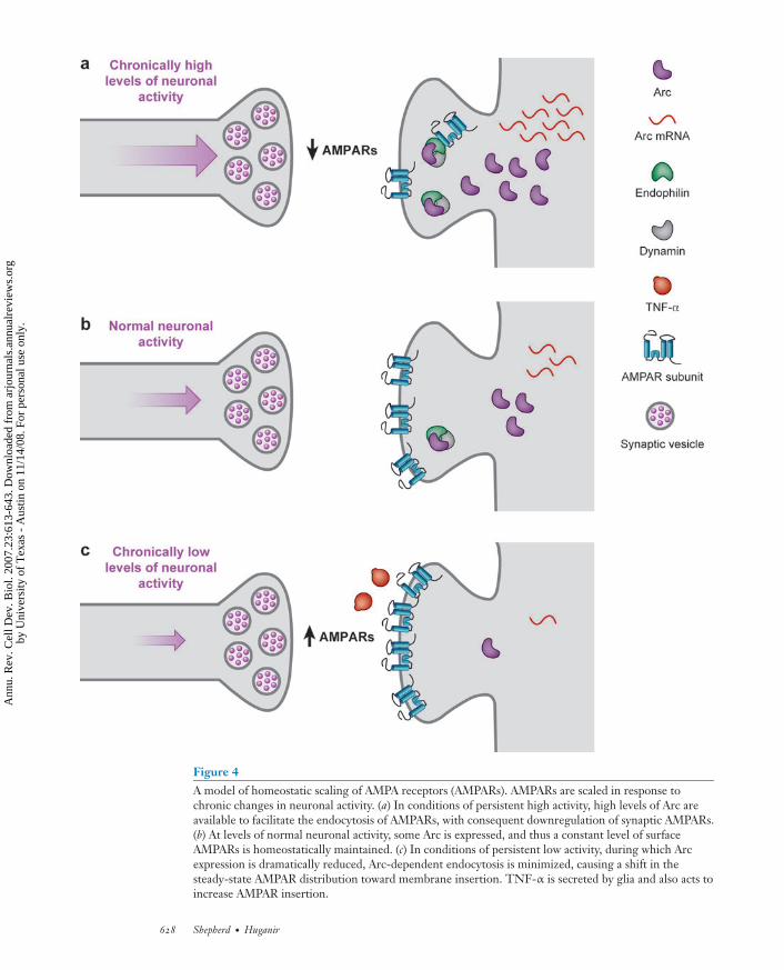
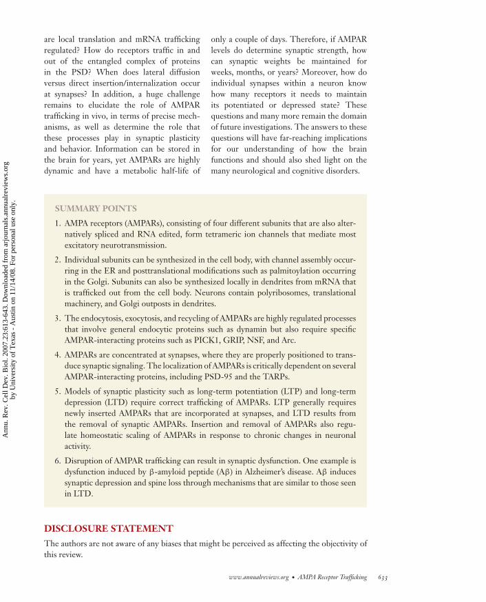
Figure 4A model of homeostatic scaling of AMPA receptors (AMPARs). AMPARs are scaled in response tochronic changes in neuronal activity. (a) In conditions of persistent high activity, high levels of Arc areavailable to facilitate the endocytosis of AMPARs, with consequent downregulation of synaptic AMPARs.(b) At levels of normal neuronal activity, some Arc is expressed, and thus a constant level of surfaceAMPARs is homeostatically maintained. (c) In conditions of persistent low activity, during which Arcexpression is dramatically reduced, Arc-dependent endocytosis is minimized, causing a shift in thesteady-state AMPAR distribution toward membrane insertion. TNF-α is secreted by glia and also acts toincrease AMPAR insertion.
628 Shepherd · Huganir
Ann
u. R
ev. C
ell D
ev. B
iol.
2007
.23:
613-
643.
Dow
nloa
ded
from
arj
ourn
als.
annu
alre
view
s.or
gby
Uni
vers
ity o
f T
exas
- A
ustin
on
11/1
4/08
. For
per
sona
l use
onl
y.
ANRV324-CB23-23 ARI 24 August 2007 20:16
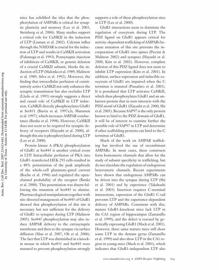
mice has solidified the idea that the phos-phorylation of AMPARs is critical for synap-tic plasticity and memory (Lee et al. 2003,Steinberg et al. 2006). Many studies supporta critical role for CaMKII in the inductionof LTP (Lisman et al. 2002). Calcium influxthrough the NMDAR is crucial for the induc-tion of LTP and results in CaMKII activation(Fukunaga et al. 1993). Postsynaptic injectionof inhibitors of CaMKII, or genetic deletionof a crucial CaMKII subunit, blocks the in-duction of LTP (Malenka et al. 1989, Malinowet al. 1989, Silva et al. 1992). Moreover, thefinding that intracellular perfusion of consti-tutively active CaMKII not only enhances thesynaptic transmission but also occludes LTP(Lledo et al. 1995) strongly suggests a directand causal role of CaMKII in LTP induc-tion. CaMKII directly phosphorylates GluR1at Ser831 (Barria et al. 1997a,b; Mammenet al. 1997), which increases AMPAR conduc-tance (Benke et al. 1998). However, CaMKIIdoes not seem to be required for synaptic de-livery of receptors (Hayashi et al. 2000), al-though this site is phosphorylated during LTP(Lee et al. 2000).
Protein kinase A (PKA) phosphorylationof GluR1 at Ser845 is another critical eventin LTP. Intracellular perfusion of PKA intoGluR1-transfected HEK 293 cells resulted ina 40% potentiation of the peak amplitudeof the whole-cell glutamate-gated current(Roche et al. 1996) and regulated the open-channel probability of the receptor (Bankeet al. 2000). This potentiation was absent fol-lowing the mutation of Ser845 to alanine.Pharmacological manipulations together withsite-directed mutagenesis of Ser845 of GluR1showed that phosphorylation of this site isnecessary but not sufficient for the deliveryof GluR1 to synapses during LTP (Malinow2003). Ser845 phosphorylation may also in-duce AMPAR delivery to the extrasynapticmembrane and then to the synapse via surfacediffusion (Man et al. 2007, Oh et al. 2006).The fact that LTP was diminished in a knock-in mouse in which Ser831 and Ser845 weremutated to prevent phosphorylation strongly
supports a role of these phosphorylation sitesin LTP (Lee et al. 2000).
GluR1 interactions seem to dominate theregulation of exocytosis during LTP. ThePDZ ligand on GluR1 appears critical foractivity-dependent trafficking of AMPARs be-cause mutation of this site prevents the in-corporation of GluR1 into spines (Piccini &Malinow 2002) and synapses (Hayashi et al.2000, Kim et al. 2001). However, completedeletion of this PDZ ligand does not seem toinhibit LTP expression (Kim et al. 2001). Inaddition, surface expression and inducible ex-ocytosis of GluR1 are impaired when the Cterminus is mutated (Passafaro et al. 2001).It is postulated that LTP activates CaMKII,which then phosphorylates GluR1 and an un-known protein that in turn interacts with thePDZ motif of GluR1 (Hayashi et al. 2000, Shiet al. 2001). Because SAP97 is the only proteinknown to bind to the PDZ domain of GluR1,it will be of interest to examine further thepossible role of SAP97 in LTP and determineif other scaffolding proteins can bind to the Cterminus of GluR1.
Much of the work on AMPAR traffick-ing has involved the use of recombinantAMPARs. In most cases, these constructsform homomeric channels that allow for thestudy of subunit specificity in trafficking, butdo not elucidate the regulation of endogenousheteromeric channels. Recent experimentshave shown that endogenous AMPARs canbe driven into the synapse during LTP (Shiet al. 2001) and by experience (Takahashiet al. 2003). Insertion requires C-terminalinteractions; expression of the GluR1 C-tailprevents LTP and the experience-dependentdelivery of AMPARs. Consistent with this,mature GluR1-knockout mice lack LTP inthe CA1 region of hippocampus (Zamanilloet al. 1999), and the defect is rescued by ge-netically expressing GluR1 (Mack et al. 2001).However, these same mature mice still showsome LTP in the dentate gyrus (Zamanilloet al. 1999) and also show LTP in the CA1 re-gion in young mice (Mack et al. 2001), whichindicates that GluR1-independent LTP also
www.annualreviews.org • AMPA Receptor Trafficking 629
Ann
u. R
ev. C
ell D
ev. B
iol.
2007
.23:
613-
643.
Dow
nloa
ded
from
arj
ourn
als.
annu
alre
view
s.or
gby
Uni
vers
ity o
f T
exas
- A
ustin
on
11/1
4/08
. For
per
sona
l use
onl
y.
ANRV324-CB23-23 ARI 24 August 2007 20:16
exists. GluR4, which shares similarities withGluR1 in its C-terminal tail, may substitutefor GluR1 in younger animals (Zhu et al.2000).
Long-Term Depression
Many studies show that LTD results fromthe endocytosis of surface AMPARs (Beattieet al. 2000, Lissin et al. 1999). However,AMPAR internalization can occur in responseto many stimuli (Ehlers 2000, Lin et al. 2000,Man et al. 2000), which has made the precisemolecular pathways that underlie LTD diffi-cult to isolate. Because most of these studieswere performed in cultured cells, it is hard toknow which pathways occur in vivo and whichsignaling molecules are the most important.
The first experimental support for therole of AMPA endocytosis in LTD came fromimmunocytochemical data, which showedthat NMDAR-dependent chemical LTD inhippocampal cultures caused a decrease inthe number of synapses containing surfaceAMPARs (Carroll et al. 1999b). Furthermore,hippocampal LTD induced in vivo caused adecrease in the number of AMPARs in synap-toneurosomes (Heynen et al. 2000). CA1pyramidal neurons or cerebellar Purkinje cellsloaded with a peptide that disrupts dynaminfunction blocked LTD (Luscher et al. 1999,Wang & Linden 2000). The inhibition of en-docytosis also blocked the actions of insulin,which can cause a depression of synapticcurrents that occludes LTD (Lin et al. 2000,Man et al. 2000). Indeed, the activationof NMDARs (Beattie et al. 2000, Carrollet al. 1999a, Ehlers 2000), mGluR receptors(Snyder et al. 2001, Xiao et al. 2001), or insulinreceptors (Lin et al. 2000, Man et al. 2000)can cause a loss of synaptic/surface AMPARs.NMDA-induced AMPAR endocytosis re-sembles LTD: It requires calcium influxand activation of the calcium-dependentphosphatase calcineurin (Beattie et al. 2000,Ehlers 2000, Zhou et al. 2001).
Regulation of the phosphorylation ofAMPAR subunits is also important for LTD
expression. During hippocampal LTD, thePKA site on GluR1, Ser845, is dephosphory-lated, whereas LTD induction in previouslypotentiated synapses leads to dephospho-rylation of the CaMKII site, Ser831 (Leeet al. 2000). Mice that have these two sitesmutated exhibit major deficits in LTD andAMPAR internalization induced by NMDARactivation (Lee et al. 2003). The mechanismby which the phosphorylation state of GluR1affects AMPAR internalization is unknownbut may involve differential regulation ofAMPAR binding partners.
The interactions of GluR2 with severalproteins are also involved in LTD. Theclathrin adaptor protein AP-2 interacts witha site that overlaps with the NSF-binding siteon GluR2, and this interaction is critical forNMDA-induced internalization of AMPARs(Lee et al. 2002). Moreover, the applica-tion of a peptide that specifically blocked theGluR2-AP2 interaction blocked the inductionof LTD (Lee et al. 2002). Recent studies haveindicated that the GluR2-interacting proteinsGRIP, ABP/GRIP2, and PICK1 (all of whichbind via the extreme C-terminal PDZ do-main) also play a critical role in AMPAR en-docytosis and LTD. A mutant form of GluR2that does not bind to GRIP/ABP in hip-pocampal neurons targeted appropriately tothe surface, but its accumulation at synapseswas significantly reduced when comparedwith wild-type GluR2 (Osten et al. 2000, Shiet al. 2001). These data suggest that AMPARsare stabilized at the synapse by the bindingof GluR2-containing receptors to GRIP/ABP,perhaps by limiting their endocytosis or by in-creasing recycling of GluR2.
Studies of LTD in cerebellar Purkinje cellsprovide further complexity to the signalingpathways triggering AMPAR endocytosis.Cerebellar LTD requires the activation ofprotein kinase C (PKC) (Linden & Connor1991), which is required to stimulate AMPARinternalization (Xia et al. 2000). Activationof PKC with a phorbol ester is sufficientto induce LTD and AMPAR internalization(Chung et al. 2000). Ser880 within the
630 Shepherd · Huganir
Ann
u. R
ev. C
ell D
ev. B
iol.
2007
.23:
613-
643.
Dow
nloa
ded
from
arj
ourn
als.
annu
alre
view
s.or
gby
Uni
vers
ity o
f T
exas
- A
ustin
on
11/1
4/08
. For
per
sona
l use
onl
y.
ANRV324-CB23-23 ARI 24 August 2007 20:16
GluR2 PDZ-binding site is phosphorylatedby PKC, which prevents the associationof GluR2 with GRIP and ABP (Chunget al. 2000; Matsuda et al. 1999, 2000) butpromotes binding to PICK1 (Chung et al.2000, Matsuda et al. 1999, Perez et al.2001). Phosphorylation of Ser880 promotesinternalization of AMPARs and decreasessurface GluR2-containing receptors in bothPurkinje neurons (Matsuda et al. 2000) andhippocampal neurons (Chung et al. 2000,Perez et al. 2001). Transfection of a GluR2construct with a point mutation that preventsphosphorylation of Ser880 into Purkinje cellsfailed to rescue LTD in GluR2-knockoutneurons (Chung et al. 2003). In addition, theexpression of GluR2 constructs that mimicSer880 phosphorylation prevents synaptictargeting of receptors, decreases transmis-sion, and partially occludes LTD (Seidenmanet al. 2003). Although hippocampal LTD isaccompanied by phosphorylation of Ser880,PKC does not mediate the phosphorylationof Ser880 (Kim et al. 2001), and hippocampalLTD does not seem to require a direct rolefor PKC. This suggests that other kinasesin the hippocampus may phosphorylate thissite.
Some controversy exists over the preciseroles of GRIP and PICK1 in AMPAR traf-ficking and plasticity, and we still lack an exactunderstanding of their molecular functions.Some of the confusion on their role in LTDmay be due to cell-type-specific differences.In addition, each protein may play multipleroles in the delivery, stabilization, and removalof synaptic AMPARs.
Homeostatic Scaling
Experiments using chronic manipulation ofneuronal activity levels in dissociated culturedneurons showed that raising activity by block-ing inhibitory synaptic transmission markedlydecreased the number of synaptic AMPARsand the size of the AMPAR EPSC (Lissinet al. 1998). Similar manipulations in spinal(O’Brien et al. 1998) or cortical (Turrigiano
et al. 1998) cultures also decreased theamplitude of miniature AMPAR EPSCs.Conversely, the application of AMPARantagonists for hours to days caused an in-crease in the surface expression of AMPARs atsynapses (Liao et al. 1999, O’Brien et al. 1998)and a decrease in the proportion of anatomi-cally defined silent synapses (Liao et al. 1999).Several recent studies have addressed themolecular mechanisms underlying AMPARhomeostatic synaptic scaling. Blocking neuralactivity by TTX (a voltage-gated sodiumchannel blocker) decreases glutamate release,causing the release of the cytokine TNF-αfrom glia cells, which leads to the upregu-lation of AMPAR-mediated synaptic eventsthrough an unknown mechanism that in-volves the insertion of AMPARs (Stellwagen& Malenka 2006). The IEG Arc has also beenimplicated in regulating AMPAR scaling(Shepherd et al. 2006). Arc protein acts asa proxy sensor of neuronal activity and isbidirectionally regulated by the manipulationof activity in cultures. High activity induceshigh levels of Arc protein, which acceleratesthe endocytosis of AMPARs, causing a uni-form decrease in AMPAR surface expressionand a decrease in mEPSC amplitude. Incontrast, blocking neuronal activity decreasesArc protein levels and correlates with a uni-form increase of AMPAR surface expressionand mEPSC amplitudes. Arc overexpres-sion blocks homeostatic synaptic plasticityinduced by TTX treatment, and Arc KO hip-pocampal neurons exhibit virtually no scalingin either direction (Shepherd et al. 2006).Neuronal activity regulates many genes, andmost likely many proteins are involved inregulating homeostatic plasticity (Turrigiano& Nelson 2004). It will also be intriguing tosee how homeostatic plasticity mechanismsinteract or interfere with LTP/LTD.
AMPA RECEPTORS ANDDISEASE
Because AMPARs play such an integral rolein brain function, it is not too surprising that
www.annualreviews.org • AMPA Receptor Trafficking 631
Ann
u. R
ev. C
ell D
ev. B
iol.
2007
.23:
613-
643.
Dow
nloa
ded
from
arj
ourn
als.
annu
alre
view
s.or
gby
Uni
vers
ity o
f T
exas
- A
ustin
on
11/1
4/08
. For
per
sona
l use
onl
y.
ANRV324-CB23-23 ARI 24 August 2007 20:16
Aβ: β-amyloidpeptide
the dysregulation of AMPAR function hasbeen implicated in many neurological dis-eases. However, it is very difficult to ascertainif AMPAR dysfunction is the initial cause oreventual aftermath of a disorder. Here we re-view evidence that AMPAR dysfunction maybe one of the first manifestations of synapticdysfunction that underlies Alzheimer’s disease(AD).
Alzheimer’s Disease
AD results from progressive and seeminglyirreversible loss of neuronal function, ul-timately leading to cell death in specificpopulations of neurons. The relative con-tributions of neuronal/synaptic dysfunctionversus the hallmark histopathological lesionsand neuronal cell loss to the symptomologyand progression of the disease are not clear.This ultimately has important implicationsfor the development of therapeutic targetsbecause treatments that target only one pro-cess may not stop the progression or alleviateall the symptoms of the disease. Synapticdysfunction in AD is particularly importantbecause it may underlie the cognitive deficitsthat characterize the disease and has remainedrelatively unexplored until recently (Smallet al. 2001). The pathological hallmarksof AD include amyloid plaques, predomi-nantly composed of aggregated β-amyloidpeptide (Aβ), and neurofibrillary tanglescomposed of hyperphosphorylated tauprotein. The Aβ peptide is derived fromproteolytic cleavage of the amyloid precursorprotein (APP) by β-secretase (BACE1) andγ-secretase (presenilin is the catalytic com-ponent of this complex). The normal role ofAPP and Aβ in the brain has been one of themost puzzling problems for the Alzheimer’sfield, and even after years of effort this prob-lem remains unsolved. However, emergingevidence suggests that Aβ is directly involvedin modulating synaptic function and thatpathological levels of Aβ inhibit synapticplasticity. Studies from AD mouse models
that express genes encoding mutant APPor presenilin linked to familial AD exhibitimpaired memory and synaptic plasticityprior to the formation of plaques, suggestingthat synaptic dysfunction rather than a loss ofneurons underlies the initial development ofthe disease (Chapman et al. 1999, Hsia et al.1999, Oddo et al. 2003). Furthermore, the ap-plication of oligomeric forms of Aβ adverselyaffects LTP and synaptic transmission (Walshet al. 2002). Aβ secretion in vivo is modulatedby neuronal activity and depresses bothAMPA and NMDA currents in slices (Cirritoet al. 2005, Kamenetz et al. 2003, Snyderet al. 2005). The mechanism of Aβ-inducedsynaptic depression appears similar to signal-ing pathways involved in LTD (Hsieh et al.2006). Indeed, Aβ overexpression decreasessynaptic AMPAR number. Expression of anAMPAR mutant that is unable to endocytosein response to LTD stimuli blocks the synap-tic depression induced by Aβ. Intriguingly,Aβ can also induce phosphorylation ofAMPARs at Ser880, a site important forAMPAR endocytosis and LTD (Hsiehet al. 2006). How secreted Aβ can modulateAMPAR function through an intracellular sig-naling cascade remains a critical, unansweredquestion.
FUTURE DIRECTIONS
Great strides have been made in elucidatingthe molecular players that govern AMPARtrafficking, but we have little understandingabout their detailed dynamics and the precisefunctional role that they play. Advances inimaging technology and better protein tagsnow allow direct observation of receptorsin real time. The manipulation of neuronalactivity has also become easier with the adventof two-photon uncaging of neurotransmittersand second messengers as well as advances inlight-activated ion channels (Deisseroth et al.2006). Many basic cell biological questionsstill remain to be addressed. What is the roleof locally synthesized receptors, and how
632 Shepherd · Huganir
Ann
u. R
ev. C
ell D
ev. B
iol.
2007
.23:
613-
643.
Dow
nloa
ded
from
arj
ourn
als.
annu
alre
view
s.or
gby
Uni
vers
ity o
f T
exas
- A
ustin
on
11/1
4/08
. For
per
sona
l use
onl
y.
ANRV324-CB23-23 ARI 24 August 2007 20:16
are local translation and mRNA traffickingregulated? How do receptors traffic in andout of the entangled complex of proteinsin the PSD? When does lateral diffusionversus direct insertion/internalization occurat synapses? In addition, a huge challengeremains to elucidate the role of AMPARtrafficking in vivo, in terms of precise mech-anisms, as well as determine the role thatthese processes play in synaptic plasticityand behavior. Information can be stored inthe brain for years, yet AMPARs are highlydynamic and have a metabolic half-life of
only a couple of days. Therefore, if AMPARlevels do determine synaptic strength, howcan synaptic weights be maintained forweeks, months, or years? Moreover, how doindividual synapses within a neuron knowhow many receptors it needs to maintainits potentiated or depressed state? Thesequestions and many more remain the domainof future investigations. The answers to thesequestions will have far-reaching implicationsfor our understanding of how the brainfunctions and should also shed light on themany neurological and cognitive disorders.
SUMMARY POINTS
1. AMPA receptors (AMPARs), consisting of four different subunits that are also alter-natively spliced and RNA edited, form tetrameric ion channels that mediate mostexcitatory neurotransmission.
2. Individual subunits can be synthesized in the cell body, with channel assembly occur-ring in the ER and posttranslational modifications such as palmitoylation occurringin the Golgi. Subunits can also be synthesized locally in dendrites from mRNA thatis trafficked out from the cell body. Neurons contain polyribosomes, translationalmachinery, and Golgi outposts in dendrites.
3. The endocytosis, exocytosis, and recycling of AMPARs are highly regulated processesthat involve general endocytic proteins such as dynamin but also require specificAMPAR-interacting proteins such as PICK1, GRIP, NSF, and Arc.
4. AMPARs are concentrated at synapses, where they are properly positioned to trans-duce synaptic signaling. The localization of AMPARs is critically dependent on severalAMPAR-interacting proteins, including PSD-95 and the TARPs.
5. Models of synaptic plasticity such as long-term potentiation (LTP) and long-termdepression (LTD) require correct trafficking of AMPARs. LTP generally requiresnewly inserted AMPARs that are incorporated at synapses, and LTD results fromthe removal of synaptic AMPARs. Insertion and removal of AMPARs also regu-late homeostatic scaling of AMPARs in response to chronic changes in neuronalactivity.
6. Disruption of AMPAR trafficking can result in synaptic dysfunction. One example isdysfunction induced by β-amyloid peptide (Aβ) in Alzheimer’s disease. Aβ inducessynaptic depression and spine loss through mechanisms that are similar to those seenin LTD.
DISCLOSURE STATEMENT
The authors are not aware of any biases that might be perceived as affecting the objectivity ofthis review.
www.annualreviews.org • AMPA Receptor Trafficking 633
Ann
u. R
ev. C
ell D
ev. B
iol.
2007
.23:
613-
643.
Dow
nloa
ded
from
arj
ourn
als.
annu
alre
view
s.or
gby
Uni
vers
ity o
f T
exas
- A
ustin
on
11/1
4/08
. For
per
sona
l use
onl
y.
ANRV324-CB23-23 ARI 24 August 2007 20:16
ACKNOWLEDGMENT
J.D.S. was a graduate student in the Cellular and Molecular Medicine Graduate Program inthe Department of Neuroscience at the Johns Hopkins School of Medicine when this reviewwas written. We thank Jean-Claude Beique for the data presented in Figure 1a.
LITERATURE CITED
Adesnik H, Nicoll RA. 2007. Conservation of glutamate receptor 2-containing AMPA receptorsduring long-term potentiation. J. Neurosci. 27:4598–602
Adesnik H, Nicoll RA, England PM. 2005. Photoinactivation of native AMPA receptors revealstheir real-time trafficking. Neuron 48:977–85
Ayalon G, Segev E, Elgavish S, Stern-Bach Y. 2005. Two regions in the N-terminal domain ofionotropic glutamate receptor 3 form the subunit oligomerization interfaces that controlsubtype-specific receptor assembly. J. Biol. Chem. 280:15053–60
Ayalon G, Stern-Bach Y. 2001. Functional assembly of AMPA and kainate receptors is mediatedby several discrete protein-protein interactions. Neuron 31:103–13
Banke TG, Bowie D, Lee H, Huganir RL, Schousboe A, Traynelis SF. 2000. Control of GluR1AMPA receptor function by cAMP-dependent protein kinase. J. Neurosci. 20:89–102
Barria A, Derkach V, Soderling T. 1997a. Identification of the Ca2+/calmodulin-dependentprotein kinase II regulatory phosphorylation site in the alpha-amino-3-hydroxyl-5-methyl-4-isoxazole-propionate-type glutamate receptor. J. Biol. Chem. 272:32727–30
Barria A, Muller D, Derkach V, Griffith LC, Soderling TR. 1997b. Regulatory phosphorylationof AMPA-type glutamate receptors by CaM-KII during long-term potentiation. Science276:2042–45
Bass BL. 2002. RNA editing by adenosine deaminases that act on RNA. Annu. Rev. Biochem.71:817–46
Bats C, Groc L, Choquet D. 2007. The interaction between Stargazin and PSD-95 regulatesAMPA receptor surface trafficking. Neuron 53:719–34
Beattie EC, Carroll RC, Yu X, Morishita W, Yasuda H, et al. 2000. Regulation of AMPAreceptor endocytosis by a signaling mechanism shared with LTD. Nat. Neurosci. 3:1291–300
Bedoukian MA, Weeks AM, Partin KM. 2006. Different domains of the AMPA receptor di-rect stargazin-mediated trafficking and stargazin-mediated modulation of kinetics. J. Biol.Chem. 281:23908–21
Beique JC, Andrade R. 2003. PSD-95 regulates synaptic transmission and plasticity in ratcerebral cortex. J. Physiol. 546:859–67
Beique JC, Lin DT, Kang MG, Aizawa H, Takamiya K, Huganir RL. 2006. Synapse-specificregulation of AMPA receptor function by PSD-95. Proc. Natl. Acad. Sci. USA 103:19535–40
Benke TA, Luthi A, Isaac JT, Collingridge GL. 1998. Modulation of AMPA receptor unitaryconductance by synaptic activity. Nature 393:793–97
Beretta F, Sala C, Saglietti L, Hirling H, Sheng M, Passafaro M. 2005. NSF interaction isimportant for direct insertion of GluR2 at synaptic sites. Mol. Cell Neurosci. 28:650–60
Bergles DE, Roberts JD, Somogyi P, Jahr CE. 2000. Glutamatergic synapses on oligodendro-cyte precursor cells in the hippocampus. Nature 405:187–91
Bingol B, Schuman EM. 2004. A proteasome-sensitive connection between PSD-95 and GluR1endocytosis. Neuropharmacology 47:755–63
634 Shepherd · Huganir
Ann
u. R
ev. C
ell D
ev. B
iol.
2007
.23:
613-
643.
Dow
nloa
ded
from
arj
ourn
als.
annu
alre
view
s.or
gby
Uni
vers
ity o
f T
exas
- A
ustin
on
11/1
4/08
. For
per
sona
l use
onl
y.
ANRV324-CB23-23 ARI 24 August 2007 20:16
Blanpied TA, Scott DB, Ehlers MD. 2002. Dynamics and regulation of clathrin coats at spe-cialized endocytic zones of dendrites and spines. Neuron 36:435–49
Borgdorff AJ, Choquet D. 2002. Regulation of AMPA receptor lateral movements. Nature417:649–53
Borges K, Dingledine R. 2001. Functional organization of the GluR1 glutamate receptorpromoter. J. Biol. Chem. 276:25929–38
Bredt DS, Nicoll RA. 2003. AMPA receptor trafficking at excitatory synapses. Neuron 40:361–79
Brusa R, Zimmermann F, Koh DS, Feldmeyer D, Gass P, et al. 1995. Early-onset epilepsyand postnatal lethality associated with an editing-deficient GluR-B allele in mice. Science270:1677–80
Burbea M, Dreier L, Dittman JS, Grunwald ME, Kaplan JM. 2002. Ubiquitin and AP180regulate the abundance of GLR-1 glutamate receptors at postsynaptic elements in C.elegans. Neuron 35:107–20
Burnashev N, Monyer H, Seeburg PH, Sakmann B. 1992. Divalent ion permeability of AMPAreceptor channels is dominated by the edited form of a single subunit. Neuron 8:189–98
Carroll RC, Beattie EC, Xia H, Luscher C, Altschuler Y, et al. 1999a. Dynamin-dependentendocytosis of ionotropic glutamate receptors. Proc. Natl. Acad. Sci. USA 96:14112–17
Carroll RC, Lissin DV, von Zastrow M, Nicoll RA, Malenka RC. 1999b. Rapid redistributionof glutamate receptors contributes to long-term depression in hippocampal cultures. Nat.Neurosci. 2:454–60
Chapman PF, White GL, Jones MW, Cooper-Blacketer D, Marshall VJ, et al. 1999. Impairedsynaptic plasticity and learning in aged amyloid precursor protein transgenic mice. Nat.Neurosci. 2:271–76
Chen L, Chetkovich DM, Petralia RS, Sweeney NT, Kawasaki Y, et al. 2000. Stargazin regu-lates synaptic targeting of AMPA receptors by two distinct mechanisms. Nature 408:936–43
Chowdhury S, Shepherd JD, Okuno H, Lyford G, Petralia RS, et al. 2006. Arc/Arg3.1 interactswith the endocytic machinery to regulate AMPA receptor trafficking. Neuron 52:445–59
Chung HJ, Steinberg JP, Huganir RL, Linden DJ. 2003. Requirement of AMPA receptorGluR2 phosphorylation for cerebellar long-term depression. Science 300:1751–55
Chung HJ, Xia J, Scannevin RH, Zhang X, Huganir RL. 2000. Phosphorylation of theAMPA receptor subunit GluR2 differentially regulates its interaction with PDZ domain-containing proteins. J. Neurosci. 20:7258–67
Cirrito JR, Yamada KA, Finn MB, Sloviter RS, Bales KR, et al. 2005. Synaptic activity regulatesinterstitial fluid amyloid-beta levels in vivo. Neuron 48:913–22
Coleman SK, Moykkynen T, Cai C, von Ossowski L, Kuismanen E, et al. 2006. Isoform-specificearly trafficking of AMPA receptor flip and flop variants. J. Neurosci. 26:11220–29
Colledge M, Snyder EM, Crozier RA, Soderling JA, Jin Y, et al. 2003. Ubiquitination regulatesPSD-95 degradation and AMPA receptor surface expression. Neuron 40:595–607
Collingridge GL, Isaac JT, Wang YT. 2004. Receptor trafficking and synaptic plasticity. Nat.Rev. Neurosci. 5:952–62
Cottrell JR, Borok E, Horvath TL, Nedivi E. 2004. CPG2: a brain- and synapse-specificprotein that regulates the endocytosis of glutamate receptors. Neuron 44:677–90
Cuadra AE, Kuo SH, Kawasaki Y, Bredt DS, Chetkovich DM. 2004. AMPA receptor synaptictargeting regulated by stargazin interactions with the Golgi-resident PDZ protein nPIST.J. Neurosci. 24:7491–502
Debaigt C, Hirling H, Steiner P, Vincent JP, Mazella J. 2004. Crucial role of neuron-enrichedendosomal protein of 21 kDa in sorting between degradation and recycling of internalizedG-protein-coupled receptors. J. Biol. Chem. 279:35687–91
www.annualreviews.org • AMPA Receptor Trafficking 635
Ann
u. R
ev. C
ell D
ev. B
iol.
2007
.23:
613-
643.
Dow
nloa
ded
from
arj
ourn
als.
annu
alre
view
s.or
gby
Uni
vers
ity o
f T
exas
- A
ustin
on
11/1
4/08
. For
per
sona
l use
onl
y.
ANRV324-CB23-23 ARI 24 August 2007 20:16
Deisseroth K, Feng G, Majewska AK, Miesenbock G, Ting A, Schnitzer MJ. 2006. Next-generation optical technologies for illuminating genetically targeted brain circuits. J.Neurosci. 26:10380–86
DeSouza S, Fu J, States BA, Ziff EB. 2002. Differential palmitoylation directs the AMPAreceptor-binding protein ABP to spines or to intracellular clusters. J. Neurosci. 22:3493–503
Dong H, O’Brien RJ, Fung ET, Lanahan AA, Worley PF, Huganir RL. 1997. GRIP: a synapticPDZ domain-containing protein that interacts with AMPA receptors. Nature 386:279–84
Ehlers MD. 2000. Reinsertion or degradation of AMPA receptors determined by activity-dependent endocytic sorting. Neuron 28:511–25
Ehlers MD. 2003. Activity level controls postsynaptic composition and signaling via theubiquitin-proteasome system. Nat. Neurosci. 6:231–42
El-Husseini AE, Schnell E, Chetkovich DM, Nicoll RA, Bredt DS. 2000. PSD-95 involvementin maturation of excitatory synapses. Science 290:1364–68
El-Husseini AE, Schnell E, Dakoji S, Sweeney N, Zhou Q, et al. 2002. Synaptic strengthregulated by palmitate cycling on PSD-95. Cell 108:849–63
Elias GM, Funke L, Stein V, Grant SG, Bredt DS, Nicoll RA. 2006. Synapse-specific and de-velopmentally regulated targeting of AMPA receptors by a family of MAGUK scaffoldingproteins. Neuron 52:307–20
Fukunaga K, Stoppini L, Miyamoto E, Muller D. 1993. Long-term potentiation is associatedwith an increased activity of Ca2+/calmodulin-dependent protein kinase II. J. Biol. Chem.268:7863–67
Gardner SM, Takamiya K, Xia J, Suh JG, Johnson R, et al. 2005. Calcium-permeable AMPAreceptor plasticity is mediated by subunit-specific interactions with PICK1 and NSF.Neuron 45:903–15
Gerges NZ, Backos DS, Rupasinghe CN, Spaller MR, Esteban JA. 2006. Dual role of theexocyst in AMPA receptor targeting and insertion into the postsynaptic membrane. EMBOJ. 25:1623–34
Gerrow K, Romorini S, Nabi SM, Colicos MA, Sala C, El-Husseini A. 2006. A preformedcomplex of postsynaptic proteins is involved in excitatory synapse development. Neuron49:547–62
Gray NW, Fourgeaud L, Huang B, Chen J, Cao H, et al. 2003. Dynamin 3 is a component ofthe postsynapse, where it interacts with mGluR5 and Homer. Curr. Biol. 13:510–15
Greengard P. 2001. The neurobiology of slow synaptic transmission. Science 294:1024–30Greger IH, Akamine P, Khatri L, Ziff EB. 2006. Developmentally regulated, combinatorial
RNA processing modulates AMPA receptor biogenesis. Neuron 51:85–97Greger IH, Khatri L, Ziff EB. 2002. RNA editing at arg607 controls AMPA receptor exit from
the endoplasmic reticulum. Neuron 34:759–72Grooms SY, Noh KM, Regis R, Bassell GJ, Bryan MK, et al. 2006. Activity bidirectionally
regulates AMPA receptor mRNA abundance in dendrites of hippocampal neurons. J.Neurosci. 26:8339–51
Grunwald ME, Kaplan JM. 2003. Mutations in the ligand-binding and pore domains controlexit of glutamate receptors from the endoplasmic reticulum in C. elegans. Neuropharma-cology 45:768–76
Hanley JG, Khatri L, Hanson PI, Ziff EB. 2002. NSF ATPase and alpha-/beta-SNAPs disas-semble the AMPA receptor-PICK1 complex. Neuron 34:53–67
Hayashi T, Rumbaugh G, Huganir RL. 2005. Differential regulation of AMPA receptor subunittrafficking by palmitoylation of two distinct sites. Neuron 47:709–23
636 Shepherd · Huganir
Ann
u. R
ev. C
ell D
ev. B
iol.
2007
.23:
613-
643.
Dow
nloa
ded
from
arj
ourn
als.
annu
alre
view
s.or
gby
Uni
vers
ity o
f T
exas
- A
ustin
on
11/1
4/08
. For
per
sona
l use
onl
y.

ANRV324-CB23-23 ARI 24 August 2007 20:16
Hayashi Y, Shi SH, Esteban JA, Piccini A, Poncer JC, Malinow R. 2000. Driving AMPAreceptors into synapses by LTP and CaMKII: requirement for GluR1 and PDZ domaininteraction. Science 287:2262–67
Heynen AJ, Quinlan EM, Bae DC, Bear MF. 2000. Bidirectional, activity-dependent regulationof glutamate receptors in the adult hippocampus in vivo. Neuron 28:527–36
Hirokawa N, Takemura R. 2005. Molecular motors and mechanisms of directional transportin neurons. Nat. Rev. Neurosci. 6:201–14
Hollmann M, Heinemann S. 1994. Cloned glutamate receptors. Annu. Rev. Neurosci. 17:31–108Horton AC, Ehlers MD. 2004. Secretory trafficking in neuronal dendrites. Nat. Cell Biol.
6:585–91Hsia AY, Masliah E, McConlogue L, Yu GQ, Tatsuno G, et al. 1999. Plaque-independent
disruption of neural circuits in Alzheimer’s disease mouse models. Proc. Natl. Acad. Sci.USA 96:3228–33
Hsieh H, Boehm J, Sato C, Iwatsubo T, Tomita T, et al. 2006. AMPAR removal underliesAβ-induced synaptic depression and dendritic spine loss. Neuron 52:831–43
Huang K, El-Husseini A. 2005. Modulation of neuronal protein trafficking and function bypalmitoylation. Curr. Opin. Neurobiol. 15:527–35
Ju W, Morishita W, Tsui J, Gaietta G, Deerinck TJ, et al. 2004. Activity-dependent regulationof dendritic synthesis and trafficking of AMPA receptors. Nat. Neurosci. 7:244–53
Juo P, Kaplan JM. 2004. The anaphase-promoting complex regulates the abundance of GLR-1glutamate receptors in the ventral nerve cord of C. elegans. Curr. Biol. 14:2057–62
Kacharmina JE, Job C, Crino P, Eberwine J. 2000. Stimulation of glutamate receptor proteinsynthesis and membrane insertion within isolated neuronal dendrites. Proc. Natl. Acad. Sci.USA 97:11545–50
Kamenetz F, Tomita T, Hsieh H, Seabrook G, Borchelt D, et al. 2003. APP processing andsynaptic function. Neuron 37:925–37
Kennedy MJ, Ehlers MD. 2006. Organelles and trafficking machinery for postsynaptic plas-ticity. Annu. Rev. Neurosci. 29:325–62
Kim CH, Chung HJ, Lee HK, Huganir RL. 2001. Interaction of the AMPA receptor subunitGluR2/3 with PDZ domains regulates hippocampal long-term depression. Proc. Natl.Acad. Sci. USA 98:11725–30
Kuusinen A, Abele R, Madden DR, Keinanen K. 1999. Oligomerization and ligand-bindingproperties of the ectodomain of the alpha-amino-3-hydroxy-5-methyl-4-isoxazole propi-onic acid receptor subunit GluRD. J. Biol. Chem. 274:28937–43
Lee HK, Barbarosie M, Kameyama K, Bear MF, Huganir RL. 2000. Regulation of dis-tinct AMPA receptor phosphorylation sites during bidirectional synaptic plasticity. Nature405:955–59
Lee HK, Takamiya K, Han JS, Man H, Kim CH, et al. 2003. Phosphorylation of the AMPAreceptor GluR1 subunit is required for synaptic plasticity and retention of spatial memory.Cell 112:631–43
Lee SH, Liu L, Wang YT, Sheng M. 2002. Clathrin adaptor AP2 and NSF interact withoverlapping sites of GluR2 and play distinct roles in AMPA receptor trafficking and hip-pocampal LTD. Neuron 36:661–74
Lee SH, Simonetta A, Sheng M. 2004. Subunit rules governing the sorting of internalizedAMPA receptors in hippocampal neurons. Neuron 43:221–36
Leonard AS, Davare MA, Horne MC, Garner CC, Hell JW. 1998. SAP97 is associated withthe alpha-amino-3-hydroxy-5-methylisoxazole-4-propionic acid receptor GluR1 subunit.J. Biol. Chem. 273:19518–24
www.annualreviews.org • AMPA Receptor Trafficking 637
Ann
u. R
ev. C
ell D
ev. B
iol.
2007
.23:
613-
643.
Dow
nloa
ded
from
arj
ourn
als.
annu
alre
view
s.or
gby
Uni
vers
ity o
f T
exas
- A
ustin
on
11/1
4/08
. For
per
sona
l use
onl
y.
ANRV324-CB23-23 ARI 24 August 2007 20:16
Liao D, Zhang X, O’Brien R, Ehlers MD, Huganir RL. 1999. Regulation of morphologi-cal postsynaptic silent synapses in developing hippocampal neurons. Nat. Neurosci. 2:37–43
Lin JW, Ju W, Foster K, Lee SH, Ahmadian G, et al. 2000. Distinct molecular mechanisms anddivergent endocytotic pathways of AMPA receptor internalization. Nat. Neurosci. 3:1282–90
Linden DJ, Connor JA. 1991. Participation of postsynaptic PKC in cerebellar long-term de-pression in culture. Science 254:1656–59
Lise MF, Wong TP, Trinh A, Hines RM, Liu L, et al. 2006. Involvement of myosin Vb inglutamate receptor trafficking. J. Biol. Chem. 281:3669–78
Lisman J, Schulman H, Cline H. 2002. The molecular basis of CaMKII function in synapticand behavioural memory. Nat. Rev. Neurosci. 3:175–90
Lissin DV, Gomperts SN, Carroll RC, Christine CW, Kalman D, et al. 1998. Activity differen-tially regulates the surface expression of synaptic AMPA and NMDA glutamate receptors.Proc. Natl. Acad. Sci. USA 95:7097–102
Lissin DV, Malenka RC, Von Zastrow M. 1999. An immunocytochemical assay for activity-dependent redistribution of glutamate receptors from the postsynaptic plasma membrane.Ann. N.Y. Acad. Sci. 868:550–53
Liu SJ, Cull-Candy SG. 2005. Subunit interaction with PICK and GRIP controls Ca2+ per-meability of AMPARs at cerebellar synapses. Nat. Neurosci. 8:768–75
Lledo PM, Hjelmstad GO, Mukherji S, Soderling TR, Malenka RC, Nicoll RA. 1995.Calcium/calmodulin-dependent kinase II and long-term potentiation enhance synaptictransmission by the same mechanism. Proc. Natl. Acad. Sci. USA 92:11175–79
Lomeli H, Mosbacher J, Melcher T, Hoger T, Geiger JR, et al. 1994. Control of kineticproperties of AMPA receptor channels by nuclear RNA editing. Science 266:1709–13
Lu W, Man H, Ju W, Trimble WS, MacDonald JF, Wang YT. 2001. Activation of synap-tic NMDA receptors induces membrane insertion of new AMPA receptors and LTP incultured hippocampal neurons. Neuron 29:243–54
Luscher C, Xia H, Beattie EC, Carroll RC, von Zastrow M, et al. 1999. Role of AMPA receptorcycling in synaptic transmission and plasticity. Neuron 24:649–58
Mack V, Burnashev N, Kaiser KM, Rozov A, Jensen V, et al. 2001. Conditional restoration ofhippocampal synaptic potentiation in Glur-A-deficient mice. Science 292:2501–4
Malenka RC, Bear MF. 2004. LTP and LTD: an embarrassment of riches. Neuron 44:5–21Malenka RC, Kauer JA, Perkel DJ, Mauk MD, Kelly PT, et al. 1989. An essential role for
postsynaptic calmodulin and protein kinase activity in long-term potentiation. Nature340:554–57
Malinow R. 2003. AMPA receptor trafficking and long-term potentiation. Philos. Trans. R. Soc.London Ser. B 358:707–14
Malinow R, Mainen ZF, Hayashi Y. 2000. LTP mechanisms: from silence to four-lane traffic.Curr. Opin. Neurobiol. 10:352–57
Malinow R, Schulman H, Tsien RW. 1989. Inhibition of postsynaptic PKC or CaMKII blocksinduction but not expression of LTP. Science 245:862–66
Mammen AL, Kameyama K, Roche KW, Huganir RL. 1997. Phosphorylation of theα-amino-3-hydroxy-5-methylisoxazole-4-propionic acid receptor GluR1 subunit bycalcium/calmodulin-dependent kinase II. J. Biol. Chem. 272:32528–33
Man HY, Lin JW, Ju WH, Ahmadian G, Liu L, et al. 2000. Regulation of AMPA receptor-mediated synaptic transmission by clathrin-dependent receptor internalization. Neuron25:649–62
638 Shepherd · Huganir
Ann
u. R
ev. C
ell D
ev. B
iol.
2007
.23:
613-
643.
Dow
nloa
ded
from
arj
ourn
als.
annu
alre
view
s.or
gby
Uni
vers
ity o
f T
exas
- A
ustin
on
11/1
4/08
. For
per
sona
l use
onl
y.
ANRV324-CB23-23 ARI 24 August 2007 20:16
Man HY, Sekine-Aizawa Y, Huganir RL. 2007. Regulation of α-amino-3-hydroxy-5-methyl-4-isoxazolepropionic acid receptor trafficking through PKA phosphorylation of the Glureceptor 1 subunit. Proc. Natl. Acad. Sci. USA 104:3579–84
Martin SJ, Grimwood PD, Morris RG. 2000. Synaptic plasticity and memory: an evaluationof the hypothesis. Annu. Rev. Neurosci. 23:649–711
Matsuda S, Launey T, Mikawa S, Hirai H. 2000. Disruption of AMPA receptor GluR2 clus-ters following long-term depression induction in cerebellar Purkinje neurons. EMBO J.19:2765–74
Matsuda S, Mikawa S, Hirai H. 1999. Phosphorylation of serine-880 in GluR2 by protein kinaseC prevents its C terminus from binding with glutamate receptor-interacting protein. J.Neurochem. 73:1765–68
Matsuzaki M, Ellis-Davies GC, Nemoto T, Miyashita Y, Iino M, Kasai H. 2001. Dendriticspine geometry is critical for AMPA receptor expression in hippocampal CA1 pyramidalneurons. Nat. Neurosci. 4:1086–92
Monyer H, Seeburg PH, Wisden W. 1991. Glutamate-operated channels: developmentallyearly and mature forms arise by alternative splicing. Neuron 6:799–810
Mousavi SA, Malerod L, Berg T, Kjeken R. 2004. Clathrin-dependent endocytosis. Biochem.J. 377:1–16
Mu Y, Otsuka T, Horton AC, Scott DB, Ehlers MD. 2003. Activity-dependent mRNA splicingcontrols ER export and synaptic delivery of NMDA receptors. Neuron 40:581–94
Mukhopadhyay D, Riezman H. 2007. Proteasome-independent functions of ubiquitin in en-docytosis and signaling. Science 315:201–5
Myers SJ, Peters J, Huang Y, Comer MB, Barthel F, Dingledine R. 1998. Transcriptional reg-ulation of the GluR2 gene: neural-specific expression, multiple promoters, and regulatoryelements. J. Neurosci. 18:6723–39
Nicoll RA, Tomita S, Bredt DS. 2006. Auxiliary subunits assist AMPA-type glutamate receptors.Science 311:1253–56
Nishimune A, Isaac JT, Molnar E, Noel J, Nash SR, et al. 1998. NSF binding to GluR2regulates synaptic transmission. Neuron 21:87–97
O’Brien RJ, Kamboj S, Ehlers MD, Rosen KR, Fischbach GD, Huganir RL. 1998. Activity-dependent modulation of synaptic AMPA receptor accumulation. Neuron 21:1067–78
O’Brien RJ, Xu D, Petralia RS, Steward O, Huganir RL, Worley P. 1999. Synaptic clusteringof AMPA receptors by the extracellular immediate gene product NARP. Neuron 23:309–23
Oddo S, Caccamo A, Shepherd JD, Murphy MP, Golde TE, et al. 2003. Triple-transgenicmodel of Alzheimer’s disease with plaques and tangles: intracellular Aβ and synaptic dys-function. Neuron 39:409–21
Oh MC, Derkach VA, Guire ES, Soderling TR. 2006. Extrasynaptic membrane traffickingregulated by GluR1 serine 845 phosphorylation primes AMPA receptors for long-termpotentiation. J. Biol. Chem. 281:752–58
Okamoto PM, Gamby C, Wells D, Fallon J, Vallee RB. 2001. Dynamin isoform-specific inter-action with the shank/ProSAP scaffolding proteins of the postsynaptic density and actincytoskeleton. J. Biol. Chem. 276:48458–65
Osten P, Khatri L, Perez JL, Kohr G, Giese G, et al. 2000. Mutagenesis reveals a role forABP/GRIP binding to GluR2 in synaptic surface accumulation of the AMPA receptor.Neuron 27:313–25
Osten P, Srivastava S, Inman GJ, Vilim FS, Khatri L, et al. 1998. The AMPA receptor GluR2C terminus can mediate a reversible, ATP-dependent interaction with NSF and alpha-and beta-SNAPs. Neuron 21:99–110
www.annualreviews.org • AMPA Receptor Trafficking 639
Ann
u. R
ev. C
ell D
ev. B
iol.
2007
.23:
613-
643.
Dow
nloa
ded
from
arj
ourn
als.
annu
alre
view
s.or
gby
Uni
vers
ity o
f T
exas
- A
ustin
on
11/1
4/08
. For
per
sona
l use
onl
y.
ANRV324-CB23-23 ARI 24 August 2007 20:16
Osten P, Stern-Bach Y. 2006. Learning from stargazin: the mouse, the phenotype and theunexpected. Curr. Opin. Neurobiol. 16:275–80
Osterweil E, Wells DG, Mooseker MS. 2005. A role for myosin VI in postsynaptic structureand glutamate receptor endocytosis. J. Cell Biol. 168:329–38
Park M, Penick EC, Edwards JG, Kauer JA, Ehlers MD. 2004. Recycling endosomes supplyAMPA receptors for LTP. Science 305:1972–75
Park M, Salgado JM, Ostroff L, Helton TD, Robinson CG, et al. 2006. Plasticity-inducedgrowth of dendritic spines by exocytic trafficking from recycling endosomes. Neuron52:817–30
Passafaro M, Piech V, Sheng M. 2001. Subunit-specific temporal and spatial patterns of AMPAreceptor exocytosis in hippocampal neurons. Nat. Neurosci. 4:917–26
Patrick GN, Bingol B, Weld HA, Schuman EM. 2003. Ubiquitin-mediated proteasome activityis required for agonist-induced endocytosis of GluRs. Curr. Biol. 13:2073–81
Perestenko PV, Henley JM. 2003. Characterization of the intracellular transport of GluR1and GluR2 α-amino-3-hydroxy-5-methyl-4-isoxazole propionic acid receptor subunits inhippocampal neurons. J. Biol. Chem. 278:43525–32
Perez JL, Khatri L, Chang C, Srivastava S, Osten P, Ziff EB. 2001. PICK1 targets activatedprotein kinase Ca to AMPA receptor clusters in spines of hippocampal neurons and reducessurface levels of the AMPA-type glutamate receptor subunit 2. J. Neurosci. 21:5417–28
Petralia RS, Wang YX, Wenthold RJ. 2003. Internalization at glutamatergic synapses duringdevelopment. Eur. J. Neurosci. 18:3207–17
Piccini A, Malinow R. 2002. Critical postsynaptic density 95/disc large/zonula occludens-1interactions by glutamate receptor 1 (GluR1) and GluR2 required at different subcellularsites. J. Neurosci. 22:5387–92
Plant K, Pelkey KA, Bortolotto ZA, Morita D, Terashima A, et al. 2006. Transient incorporationof native GluR2-lacking AMPA receptors during hippocampal long-term potentiation.Nat. Neurosci. 9:602–4
Poodry CA, Hall L, Suzuki DT. 1973. Developmental properties of Shibire: a pleiotropicmutation affecting larval and adult locomotion and development. Dev. Biol. 32:373–86
Prekeris R, Klumperman J, Chen YA, Scheller RH. 1998. Syntaxin 13 mediates cycling ofplasma membrane proteins via tubulovesicular recycling endosomes. J. Cell Biol. 143:957–71
Priel A, Kolleker A, Ayalon G, Gillor M, Osten P, Stern-Bach Y. 2005. Stargazin reducesdesensitization and slows deactivation of the AMPA-type glutamate receptors. J. Neurosci.25:2682–86
Racz B, Blanpied TA, Ehlers MD, Weinberg RJ. 2004. Lateral organization of endocyticmachinery in dendritic spines. Nat. Neurosci. 7:917–18
Rial Verde EM, Lee-Osbourne J, Worley PF, Malinow R, Cline HT. 2006. Increased expres-sion of the immediate-early gene arc/arg3.1 reduces AMPA receptor-mediated synaptictransmission. Neuron 52:461–74
Roche KW, O’Brien RJ, Mammen AL, Bernhardt J, Huganir RL. 1996. Characterization ofmultiple phosphorylation sites on the AMPA receptor GluR1 subunit. Neuron 16:1179–88
Rogers SW, Hughes TE, Hollmann M, Gasic GP, Deneris ES, Heinemann S. 1991. Thecharacterization and localization of the glutamate receptor subunit GluR1 in the rat brain.J. Neurosci. 11:2713–24
Rosenmund C, Stern-Bach Y, Stevens CF. 1998. The tetrameric structure of a glutamatereceptor channel. Science 280:1596–99
Rothman JE. 1994. Mechanisms of intracellular protein transport. Nature 372:55–63
640 Shepherd · Huganir
Ann
u. R
ev. C
ell D
ev. B
iol.
2007
.23:
613-
643.
Dow
nloa
ded
from
arj
ourn
als.
annu
alre
view
s.or
gby
Uni
vers
ity o
f T
exas
- A
ustin
on
11/1
4/08
. For
per
sona
l use
onl
y.
ANRV324-CB23-23 ARI 24 August 2007 20:16
Rouach N, Byrd K, Petralia RS, Elias GM, Adesnik H, et al. 2005. TARP γ-8 controlshippocampal AMPA receptor number, distribution and synaptic plasticity. Nat. Neurosci.8:1525–33
Rubio ME, Wenthold RJ. 1999. Calnexin and the immunoglobulin binding protein (BiP)coimmunoprecipitate with AMPA receptors. J. Neurochem. 73:942–48
Rumbaugh G, Sia GM, Garner CC, Huganir RL. 2003. Synapse-associated protein-97 isoform-specific regulation of surface AMPA receptors and synaptic function in cultured neurons.J. Neurosci. 23:4567–76
Sans N, Racca C, Petralia RS, Wang YX, McCallum J, Wenthold RJ. 2001. Synapse-associatedprotein 97 selectively associates with a subset of AMPA receptors early in their biosyntheticpathway. J. Neurosci. 21:7506–16
Sans N, Vissel B, Petralia RS, Wang YX, Chang K, et al. 2003. Aberrant formation of glutamatereceptor complexes in hippocampal neurons of mice lacking the GluR2 AMPA receptorsubunit. J. Neurosci. 23:9367–73
Schaefer H, Rongo C. 2006. KEL-8 is a substrate receptor for CUL3-dependent ubiquitinligase that regulates synaptic glutamate receptor turnover. Mol. Biol. Cell 17:1250–60
Schulz TW, Nakagawa T, Licznerski P, Pawlak V, Kolleker A, et al. 2004. Actin/α-actinin-dependent transport of AMPA receptors in dendritic spines: role of the PDZ-LIM proteinRIL. J. Neurosci. 24:8584–94
Seidenman KJ, Steinberg JP, Huganir R, Malinow R. 2003. Glutamate receptor subunit 2serine 880 phosphorylation modulates synaptic transmission and mediates plasticity inCA1 pyramidal cells. J. Neurosci. 23:9220–28
Sekine-Aizawa Y, Huganir RL. 2004. Imaging of receptor trafficking by using α-bungarotoxin-binding-site-tagged receptors. Proc. Natl. Acad. Sci. USA 101:17114–19
Setou M, Seog DH, Tanaka Y, Kanai Y, Takei Y, et al. 2002. Glutamate-receptor-interactingprotein GRIP1 directly steers kinesin to dendrites. Nature 417:83–87
Shen L, Liang F, Walensky LD, Huganir RL. 2000. Regulation of AMPA receptor GluR1subunit surface expression by a 4.1N-linked actin cytoskeletal association. J. Neurosci.20:7932–40
Shepherd JD, Rumbaugh G, Wu J, Chowdhury S, Plath N, et al. 2006. Arc/Arg3.1 mediateshomeostatic synaptic scaling of AMPA receptors. Neuron 52:475–84
Shi S, Hayashi Y, Esteban JA, Malinow R. 2001. Subunit-specific rules governing AMPAreceptor trafficking to synapses in hippocampal pyramidal neurons. Cell 105:331–43
Shin H, Wyszynski M, Huh KH, Valtschanoff JG, Lee JR, et al. 2003. Association of the kinesinmotor KIF1A with the multimodular protein liprin-α. J. Biol. Chem. 278:11393–401
Silva AJ, Stevens CF, Tonegawa S, Wang Y. 1992. Deficient hippocampal long-term potenti-ation in α-calcium-calmodulin kinase II mutant mice. Science 257:201–6
Small DH, Mok SS, Bornstein JC. 2001. Alzheimer’s disease and Aβ toxicity: from top tobottom. Nat. Rev. Neurosci. 2:595–98
Smith WB, Starck SR, Roberts RW, Schuman EM. 2005. Dopaminergic stimulation of lo-cal protein synthesis enhances surface expression of GluR1 and synaptic transmission inhippocampal neurons. Neuron 45:765–79
Snyder EM, Nong Y, Almeida CG, Paul S, Moran T, et al. 2005. Regulation of NMDAreceptor trafficking by amyloid-β. Nat. Neurosci. 8:1051–58
Snyder EM, Philpot BD, Huber KM, Dong X, Fallon JR, Bear MF. 2001. Internalization ofionotropic glutamate receptors in response to mGluR activation. Nat. Neurosci. 4:1079–85
Sommer B, Keinanen K, Verdoorn TA, Wisden W, Burnashev N, et al. 1990. Flip andflop: a cell-specific functional switch in glutamate-operated channels of the CNS.Science 249:1580–85
www.annualreviews.org • AMPA Receptor Trafficking 641
Ann
u. R
ev. C
ell D
ev. B
iol.
2007
.23:
613-
643.
Dow
nloa
ded
from
arj
ourn
als.
annu
alre
view
s.or
gby
Uni
vers
ity o
f T
exas
- A
ustin
on
11/1
4/08
. For
per
sona
l use
onl
y.
ANRV324-CB23-23 ARI 24 August 2007 20:16
Song I, Huganir RL. 2002. Regulation of AMPA receptors during synaptic plasticity. TrendsNeurosci. 25:578–88
Song I, Kamboj S, Xia J, Dong H, Liao D, Huganir RL. 1998. Interaction of the N-ethylmaleimide-sensitive factor with AMPA receptors. Neuron 21:393–400
Spacek J, Harris KM. 1997. Three-dimensional organization of smooth endoplasmic reticulumin hippocampal CA1 dendrites and dendritic spines of the immature and mature rat. J.Neurosci. 17:190–203
Steinberg JP, Huganir RL, Linden DJ. 2004. N-Ethylmaleimide-sensitive factor is required forthe synaptic incorporation and removal of AMPA receptors during cerebellar long-termdepression. Proc. Natl. Acad. Sci. USA 101:18212–16
Steinberg JP, Takamiya K, Shen Y, Xia J, Rubio ME, et al. 2006. Targeted in vivo mutations ofthe AMPA receptor subunit GluR2 and its interacting protein PICK1 eliminate cerebellarlong-term depression. Neuron 49:845–60
Steiner P, Alberi S, Kulangara K, Yersin A, Sarria JC, et al. 2005. Interactions between NEEP21,GRIP1 and GluR2 regulate sorting and recycling of the glutamate receptor subunit GluR2.EMBO J. 24:2873–84
Steiner P, Sarria JC, Glauser L, Magnin S, Catsicas S, Hirling H. 2002. Modulation of receptorcycling by neuron-enriched endosomal protein of 21 kD. J. Cell Biol. 157:1197–209
Stellwagen D, Malenka RC. 2006. Synaptic scaling mediated by glial TNF-α. Nature 440:1054–59
Steward O, Worley PF. 2001. Selective targeting of newly synthesized Arc mRNA to activesynapses requires NMDA receptor activation. Neuron 30:227–40
Sutton MA, Schuman EM. 2006. Dendritic protein synthesis, synaptic plasticity, and memory.Cell 127:49–58
Takahashi T, Svoboda K, Malinow R. 2003. Experience strengthening transmission by drivingAMPA receptors into synapses. Science 299:1585–88
Tardin C, Cognet L, Bats C, Lounis B, Choquet D. 2003. Direct imaging of lateral movementsof AMPA receptors inside synapses. EMBO J. 22:4656–65
Thomas GM, Huganir RL. 2004. MAPK cascade signalling and synaptic plasticity. Nat. Rev.Neurosci. 5:173–83
Tomita S, Adesnik H, Sekiguchi M, Zhang W, Wada K, et al. 2005. Stargazin modulates AMPAreceptor gating and trafficking by distinct domains. Nature 435:1052–58
Tomita S, Chen L, Kawasaki Y, Petralia RS, Wenthold RJ, et al. 2003. Functional studies anddistribution define a family of transmembrane AMPA receptor regulatory proteins. J. CellBiol. 161:805–16
Turrigiano GG, Leslie KR, Desai NS, Rutherford LC, Nelson SB. 1998. Activity-dependentscaling of quantal amplitude in neocortical neurons. Nature 391:892–96
Turrigiano GG, Nelson SB. 2004. Homeostatic plasticity in the developing nervous system.Nat. Rev. Neurosci. 5:97–107
Uemura T, Mori H, Mishina M. 2002. Isolation and characterization of Golgi apparatus-specific GODZ with the DHHC zinc finger domain. Biochem. Biophys. Res. Commun.296:492–96
van Roessel P, Elliott DA, Robinson IM, Prokop A, Brand AH. 2004. Independent regulationof synaptic size and activity by the anaphase-promoting complex. Cell 119:707–18
Vandenberghe W, Nicoll RA, Bredt DS. 2005. Interaction with the unfolded protein responsereveals a role for stargazin in biosynthetic AMPA receptor transport. J. Neurosci. 25:1095–102
642 Shepherd · Huganir
Ann
u. R
ev. C
ell D
ev. B
iol.
2007
.23:
613-
643.
Dow
nloa
ded
from
arj
ourn
als.
annu
alre
view
s.or
gby
Uni
vers
ity o
f T
exas
- A
ustin
on
11/1
4/08
. For
per
sona
l use
onl
y.
ANRV324-CB23-23 ARI 24 August 2007 20:16
Walsh DM, Klyubin I, Fadeeva JV, Cullen WK, Anwyl R, et al. 2002. Naturally secretedoligomers of amyloid β protein potently inhibit hippocampal long-term potentiation invivo. Nature 416:535–39
Wang YT, Linden DJ. 2000. Expression of cerebellar long-term depression requires postsy-naptic clathrin-mediated endocytosis. Neuron 25:635–47
Washbourne P, Bennett JE, McAllister AK. 2002. Rapid recruitment of NMDA receptortransport packets to nascent synapses. Nat. Neurosci. 5:751–59
Wu H, Nash JE, Zamorano P, Garner CC. 2002. Interaction of SAP97 with minus-end-directed actin motor myosin VI. Implications for AMPA receptor trafficking. J. Biol. Chem.277:30928–34
Wyszynski M, Kim E, Dunah AW, Passafaro M, Valtschanoff JG, et al. 2002. Interactionbetween GRIP and liprin-α/SYD2 is required for AMPA receptor targeting. Neuron 34:39–52
Xia J, Chung HJ, Wihler C, Huganir RL, Linden DJ. 2000. Cerebellar long-term depres-sion requires PKC-regulated interactions between GluR2/3 and PDZ domain-containingproteins. Neuron 28:499–510
Xia J, Zhang X, Staudinger J, Huganir RL. 1999. Clustering of AMPA receptors by the synapticPDZ domain-containing protein PICK1. Neuron 22:179–87
Xiao MY, Zhou Q, Nicoll RA. 2001. Metabotropic glutamate receptor activation causes a rapidredistribution of AMPA receptors. Neuropharmacology 41:664–71
Xu D, Hopf C, Reddy R, Cho RW, Guo L, et al. 2003. Narp and NP1 form heterocomplexesthat function in developmental and activity-dependent synaptic plasticity. Neuron 39:513–28
Yamazaki M, Fukaya M, Abe M, Ikeno K, Kakizaki T, et al. 2001. Differential palmitoylationof two mouse glutamate receptor interacting protein 1 forms with different N-terminalsequences. Neurosci. Lett. 304:81–84
Zamanillo D, Sprengel R, Hvalby O, Jensen V, Burnashev N, et al. 1999. Importance ofAMPA receptors for hippocampal synaptic plasticity but not for spatial learning. Science284:1805–11
Zhou Q, Xiao M, Nicoll RA. 2001. Contribution of cytoskeleton to the internalization ofAMPA receptors. Proc. Natl. Acad. Sci. USA 98:1261–66
Zhu JJ, Esteban JA, Hayashi Y, Malinow R. 2000. Postnatal synaptic potentiation: delivery ofGluR4-containing AMPA receptors by spontaneous activity. Nat. Neurosci. 3:1098–106
www.annualreviews.org • AMPA Receptor Trafficking 643
Ann
u. R
ev. C
ell D
ev. B
iol.
2007
.23:
613-
643.
Dow
nloa
ded
from
arj
ourn
als.
annu
alre
view
s.or
gby
Uni
vers
ity o
f T
exas
- A
ustin
on
11/1
4/08
. For
per
sona
l use
onl
y.