Embed Size (px)
Citation preview
The Cell CycleIntroduction to Biology

The Key Roles of Cell Division
• The ability to reproduce is one of the key features that separates life from non-life.
• All cells have the ability to reproduce, by making exact copies of themselves.
• In unicellular organisms, division of one cell reproduces the entire organism
• In multicellular organisms, cell division is needed for:o Development of an embryo from a sperm/eggo Growtho Repair
LE 12-2
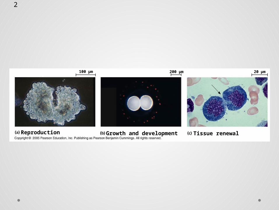
Reproduction
100 µm
Tissue renewalGrowth and development
20 µm200 µm
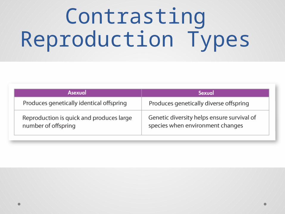
Asexual Reproduction• Asexual reproduction is reproduction that
involves a single parent producing an offspring.o The offspring produced are, in most cases,
genetically identical to the single cell that produced them.
o Asexual reproduction is a simple, efficient, and effective way for an organism to produce a large number of offspring.
o Prokaryotic organisms (like bacteria) reproduce asexually, as do some eukaryotes (like sponges)
Sexual Reproduction• In sexual reproduction, offspring are
produced by the fusion of two sex cells – one from each of two parents. These fuse into a single cell before the offspring can grow.o The offspring produced inherit some genetic
information from both parents.o Most animals and plants, and many single-celled
organisms, reproduce sexually.
Contrasting Reproduction Types
Cell Division• Cells duplicate their genetic material before
they divide, ensuring that each daughter cell receives an exact copy of the genetic material, DNA.
• A dividing cell duplicates its DNA, allocates the two copies to opposite ends of the cell, and only then splits into daughter cells.
Cellular Organization of the Genetic
Material• A cell’s endowment of DNA (its genetic
information) is called its genome.• DNA molecules in a cell are packaged into
chromosomes.
• The genetic information that is passed on from one generation of cells to the next is carried by chromosomes.
• Every cell must copy its genetic information before cell division begins.
• Each daughter cell gets its own copy of that genetic information.
• Cells of every organism have a specific number of chromosomes.
Chromosomes
Prokaryotic Chromosomes
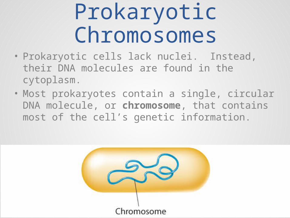
• Prokaryotic cells lack nuclei. Instead, their DNA molecules are found in the cytoplasm.
• Most prokaryotes contain a single, circular DNA molecule, or chromosome, that contains most of the cell’s genetic information.
Eukaryotic Chromosomes
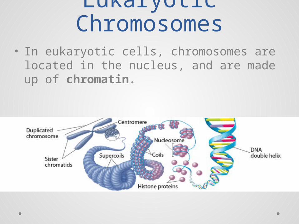
• In eukaryotic cells, chromosomes are located in the nucleus, and are made up of chromatin.
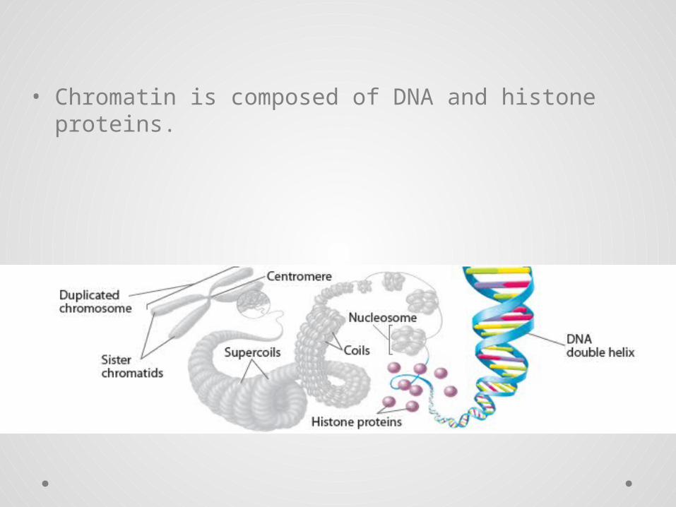
• Chromatin is composed of DNA and histone proteins.
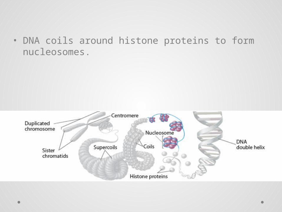
• DNA coils around histone proteins to form nucleosomes.
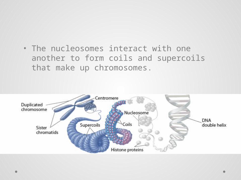
• The nucleosomes interact with one another to form coils and supercoils that make up chromosomes.
Chromosomes During Cell Division
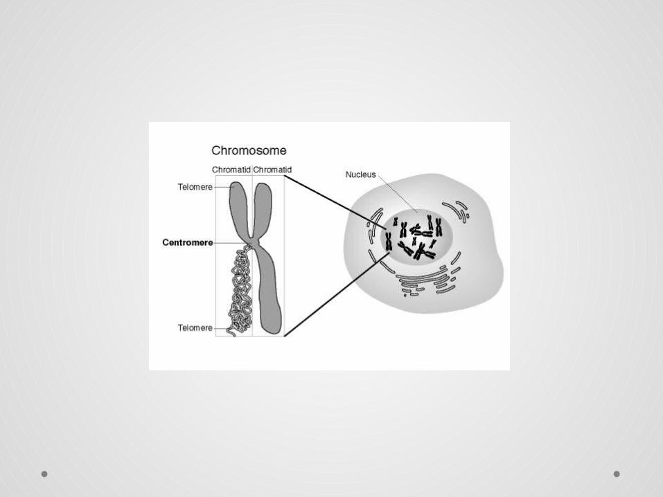
• In preparation for cell division, DNA is replicated and the chromosomes condense
• Each duplicated chromosome has two sister chromatids, which separate during cell division
• The centromere is the narrow “waist” of the duplicated chromosome, where the two chromatids are most closely attached
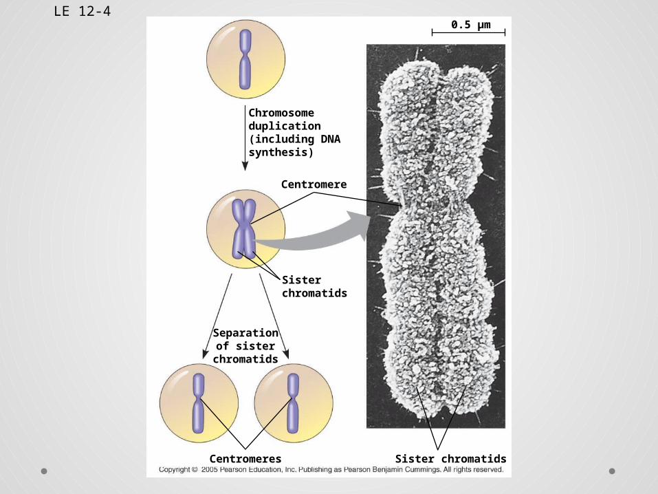
LE 12-4
Chromosomeduplication(including DNAsynthesis)
0.5 µm
Centromere
Sisterchromatids
Separationof sister
chromatids
Centromeres Sister chromatids
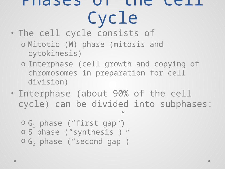
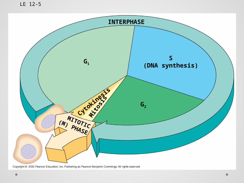
Phases of the Cell Cycle
• The cell cycle consists ofo Mitotic (M) phase (mitosis and cytokinesis)o Interphase (cell growth and copying of
chromosomes in preparation for cell division)
• Interphase (about 90% of the cell cycle) can be divided into subphases:o G1 phase (“first gap”)o S phase (“synthesis”)o G2 phase (“second gap”)
LE 12-5
G1
G2
S(DNA synthesis)
INTERPHASE
Cytokin
esis
MITOTIC(M) PHASE
Mito
sis
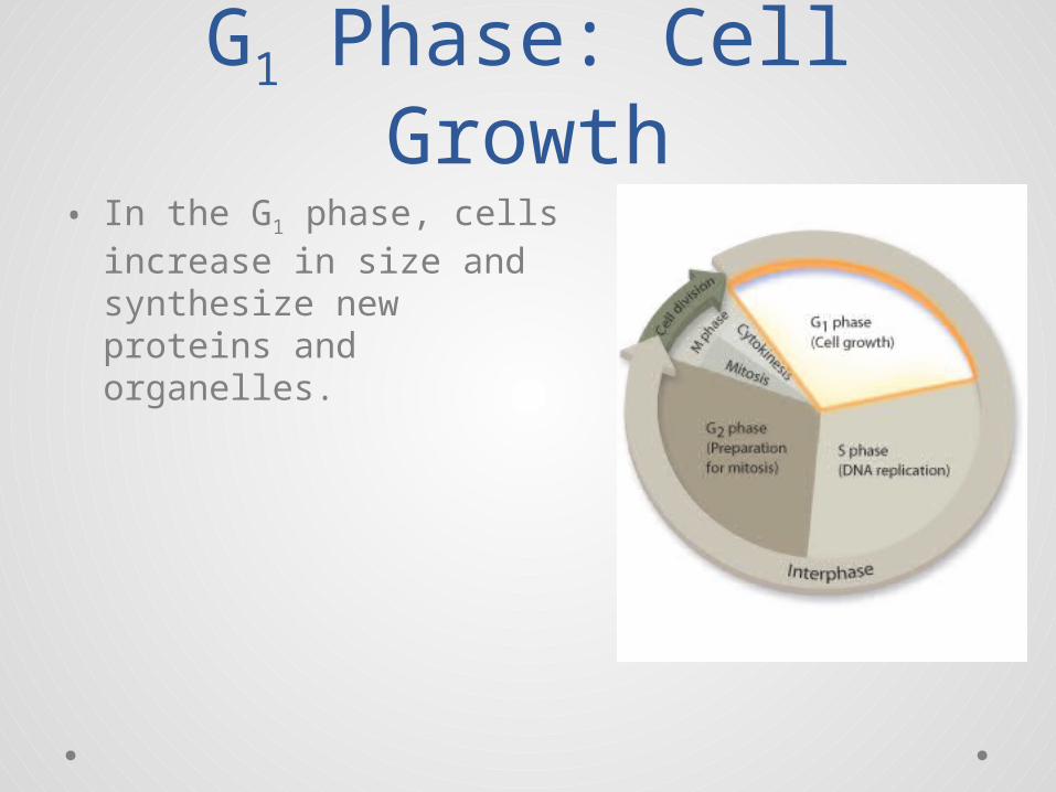
G1 Phase: Cell Growth• In the G1 phase, cells
increase in size and synthesize new proteins and organelles.
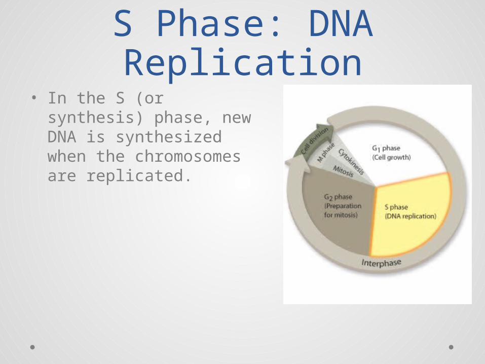
S Phase: DNA Replication
• In the S (or synthesis) phase, new DNA is synthesized when the chromosomes are replicated.
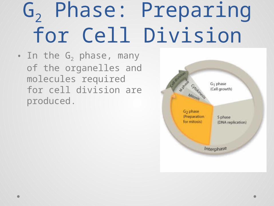
G2 Phase: Preparing for Cell Division
• In the G2 phase, many of the organelles and molecules required for cell division are produced.
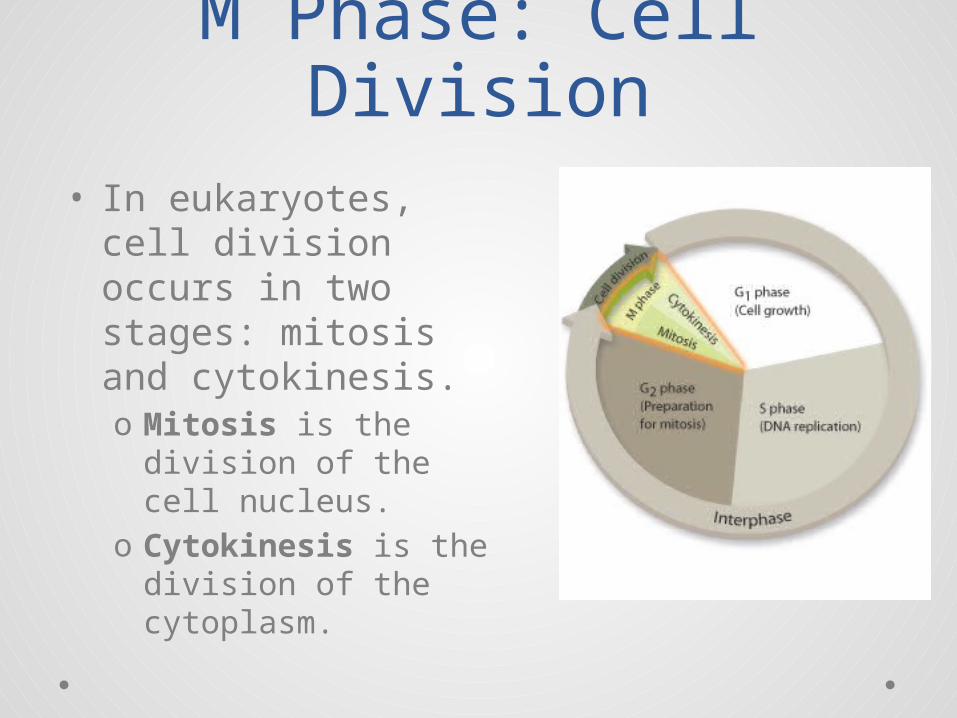
M Phase: Cell Division• In eukaryotes, cell
division occurs in two stages: mitosis and cytokinesis.o Mitosis is the
division of the cell nucleus.
o Cytokinesis is the division of the cytoplasm.
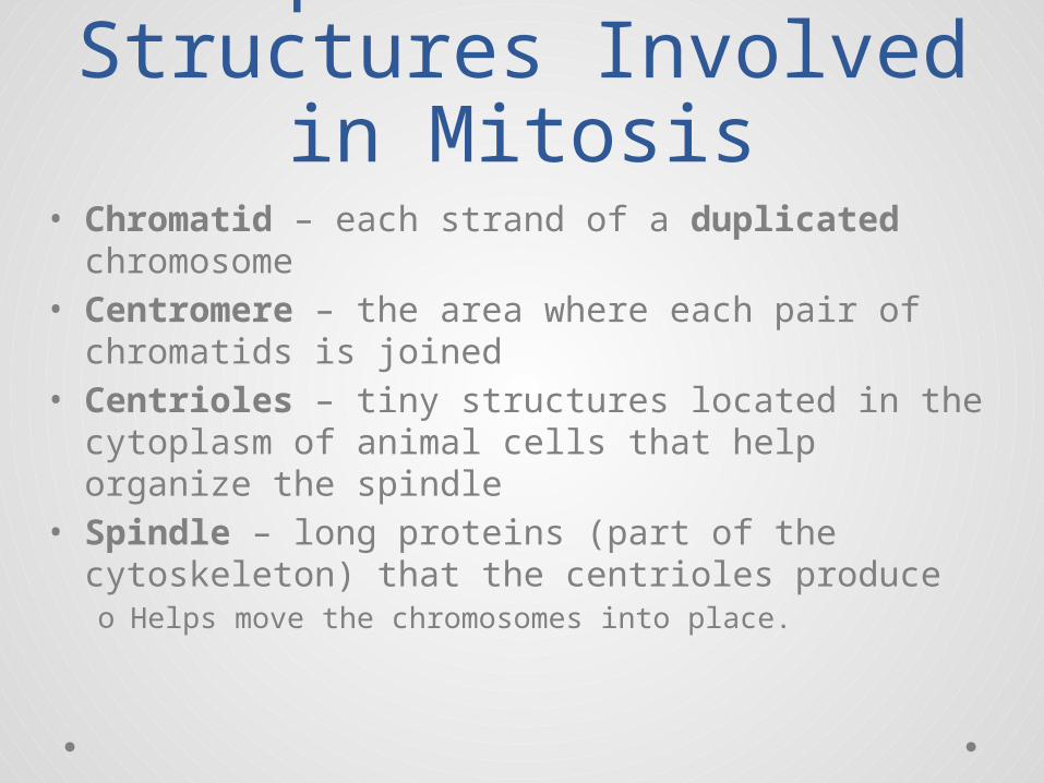
Important Cell Structures Involved in
Mitosis• Chromatid – each strand of a duplicated
chromosome• Centromere – the area where each pair of
chromatids is joined• Centrioles – tiny structures located in the
cytoplasm of animal cells that help organize the spindle
• Spindle – long proteins (part of the cytoskeleton) that the centrioles produceo Helps move the chromosomes into place.
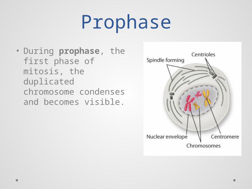
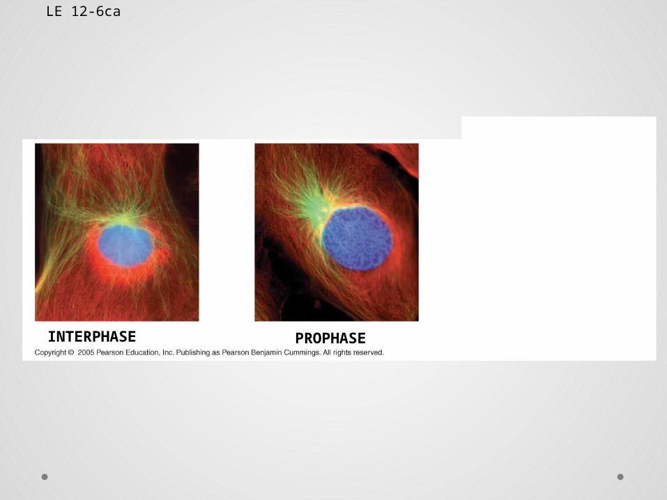
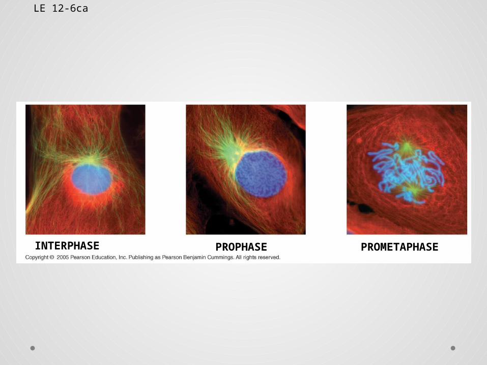
Prophase• During prophase, the
first phase of mitosis, the duplicated chromosome condenses and becomes visible.
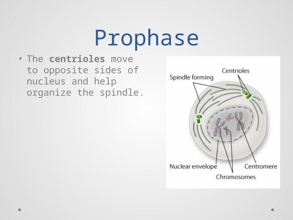
Prophase• The centrioles move
to opposite sides of nucleus and help organize the spindle.
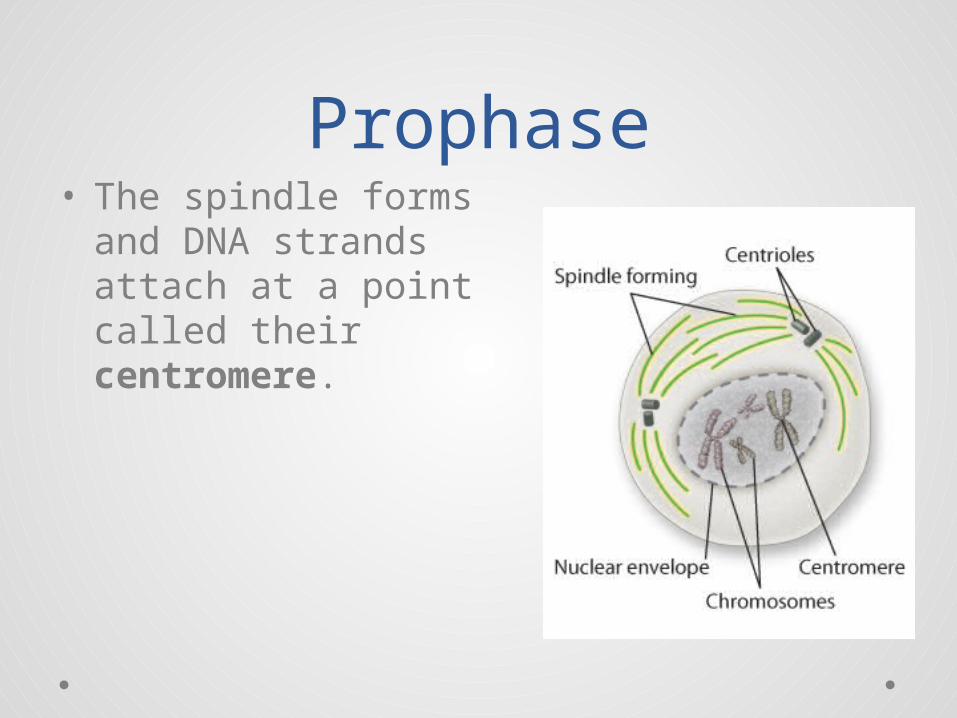
Prophase• The spindle forms
and DNA strands attach at a point called their centromere.
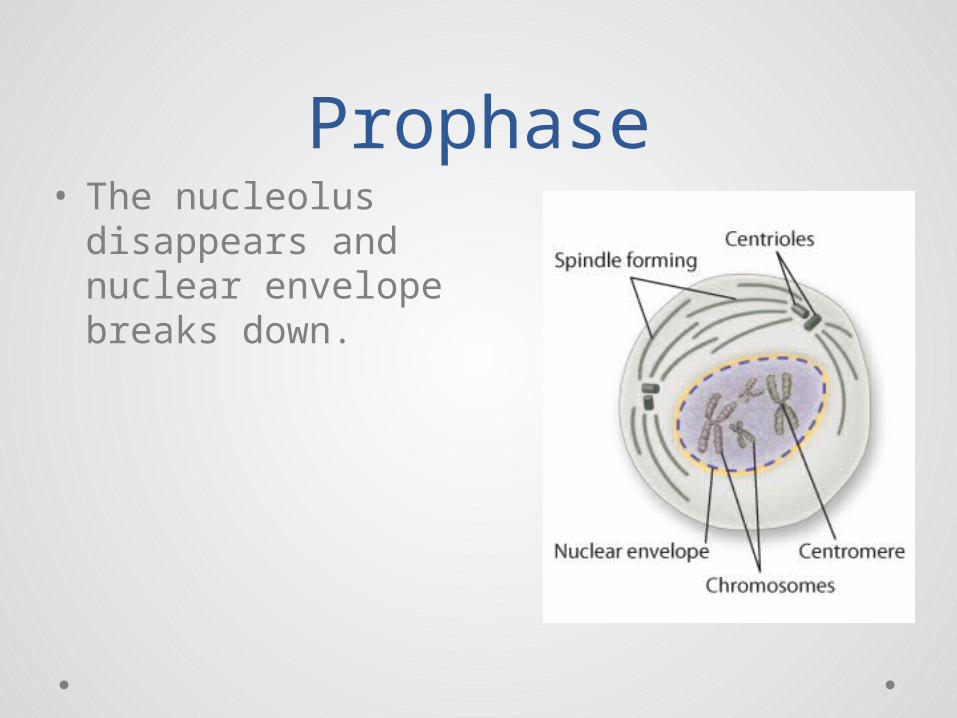
Prophase• The nucleolus
disappears and nuclear envelope breaks down.
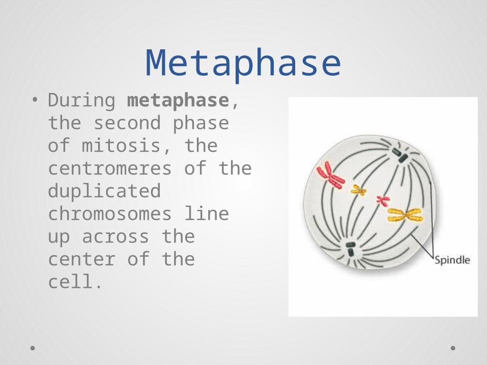
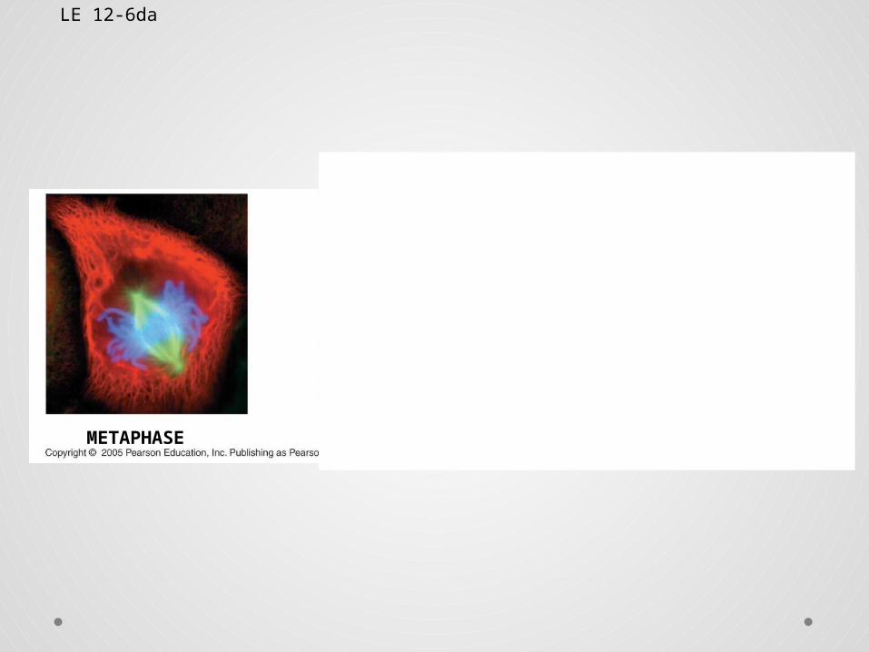
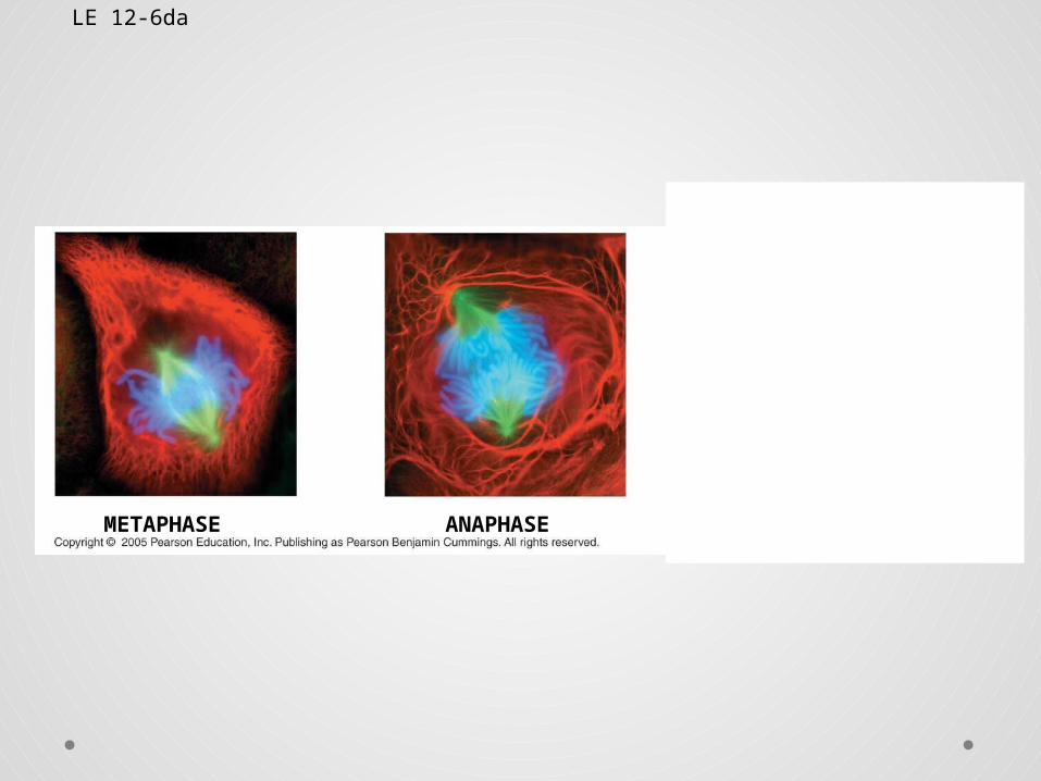
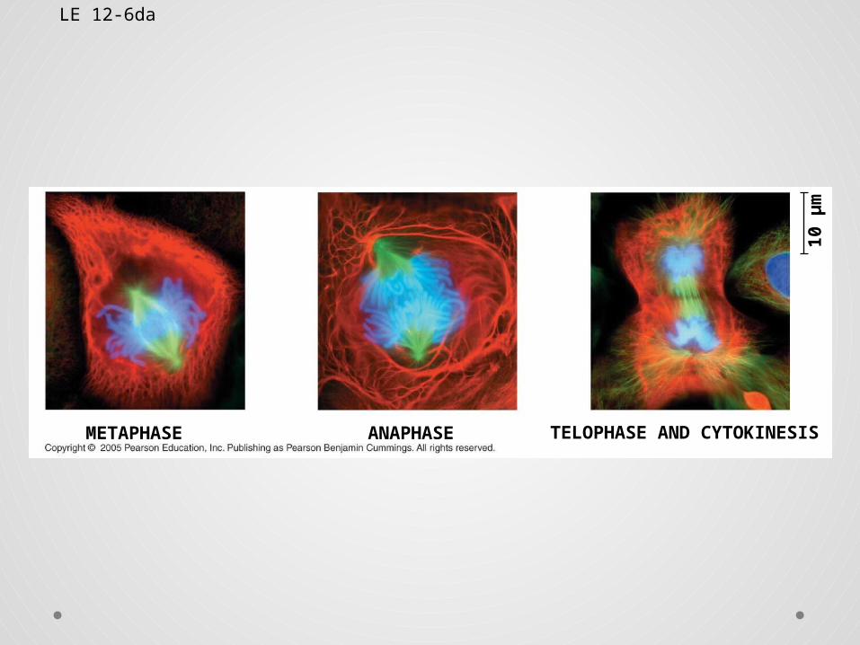
Metaphase• During metaphase,
the second phase of mitosis, the centromeres of the duplicated chromosomes line up across the center of the cell.
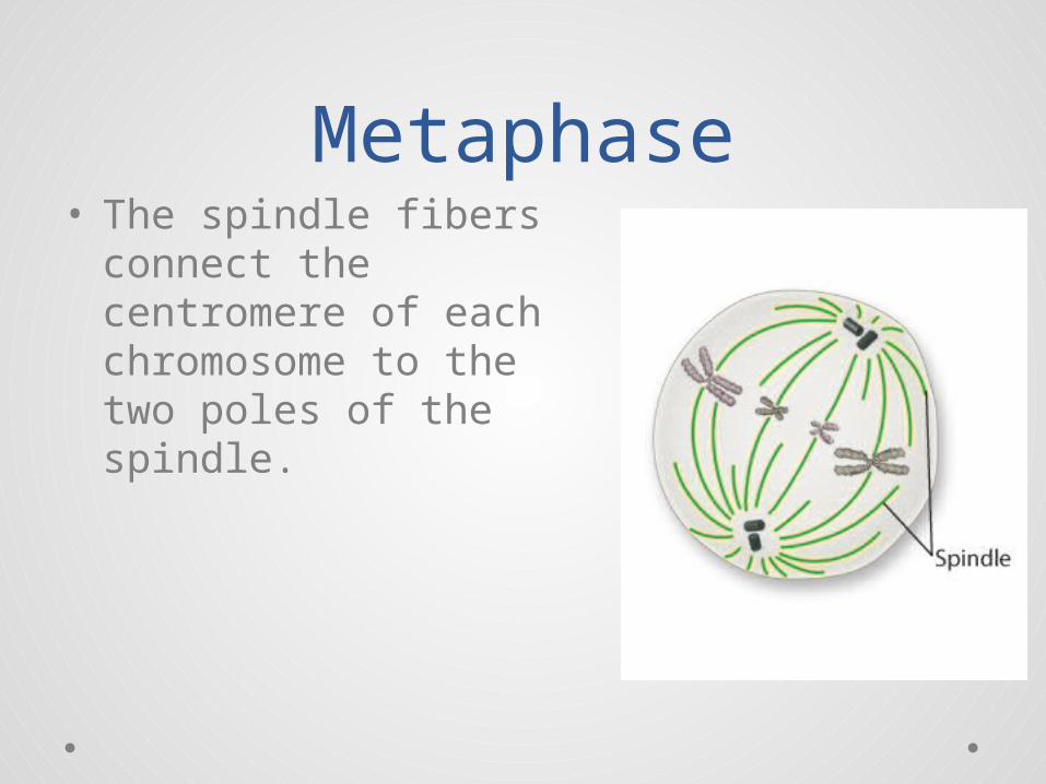
Metaphase• The spindle fibers
connect the centromere of each chromosome to the two poles of the spindle.
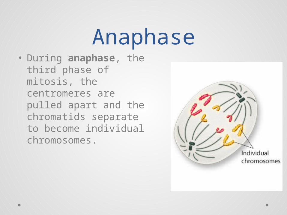
Anaphase• During anaphase, the
third phase of mitosis, the centromeres are pulled apart and the chromatids separate to become individual chromosomes.
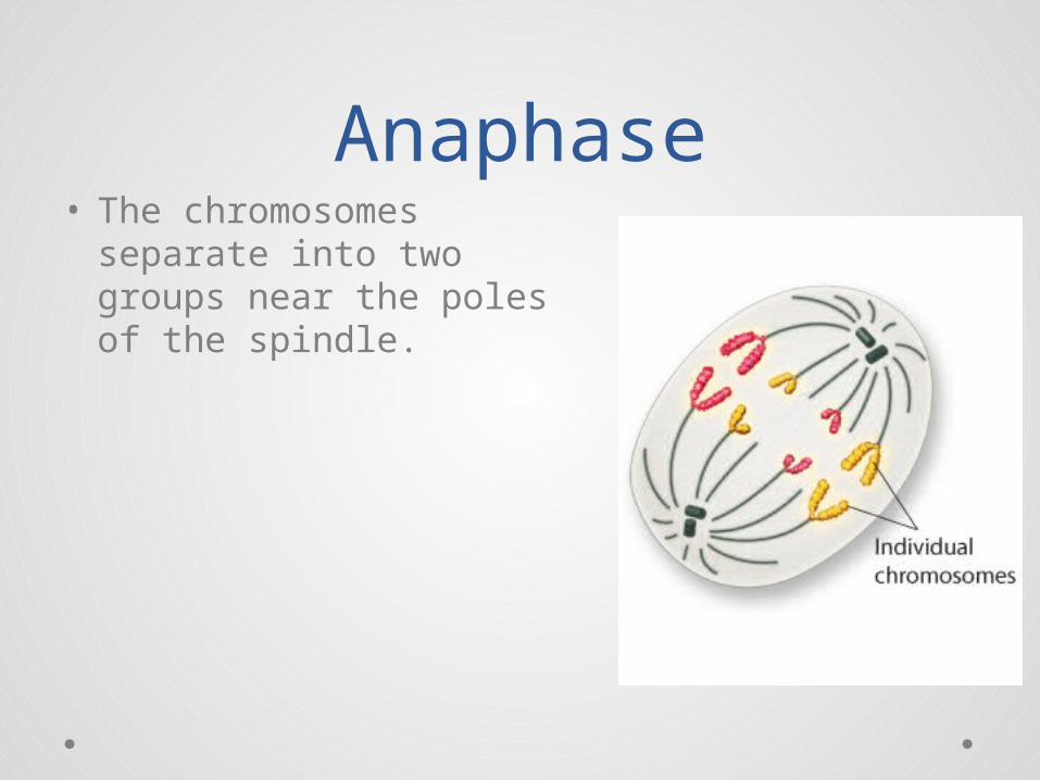
Anaphase• The chromosomes
separate into two groups near the poles of the spindle.
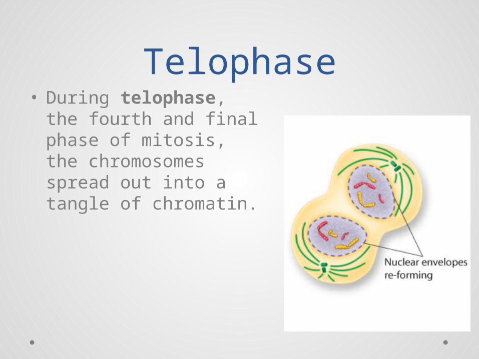
Telophase• During telophase, the
fourth and final phase of mitosis, the chromosomes spread out into a tangle of chromatin.
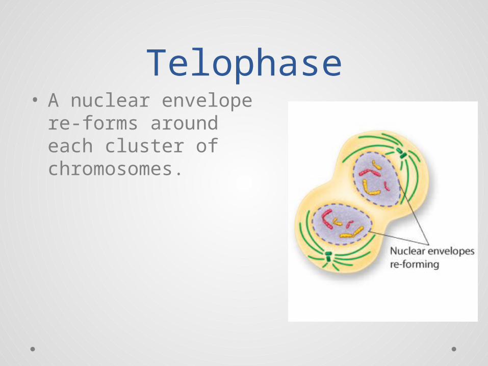
Telophase• A nuclear envelope re-
forms around each cluster of chromosomes.
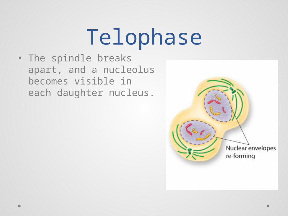
Telophase• The spindle breaks apart,
and a nucleolus becomes visible in each daughter nucleus.
• Cytokinesis is the division of the cytoplasm.
• The process of cytokinesis is different in animal and plant cells.
Cytokinesis
Cytokinesis in Animal Cells
• The cell membrane is drawn in until the cytoplasm is pinched into two equal parts.
• Each part contains its own nucleus and organelles.
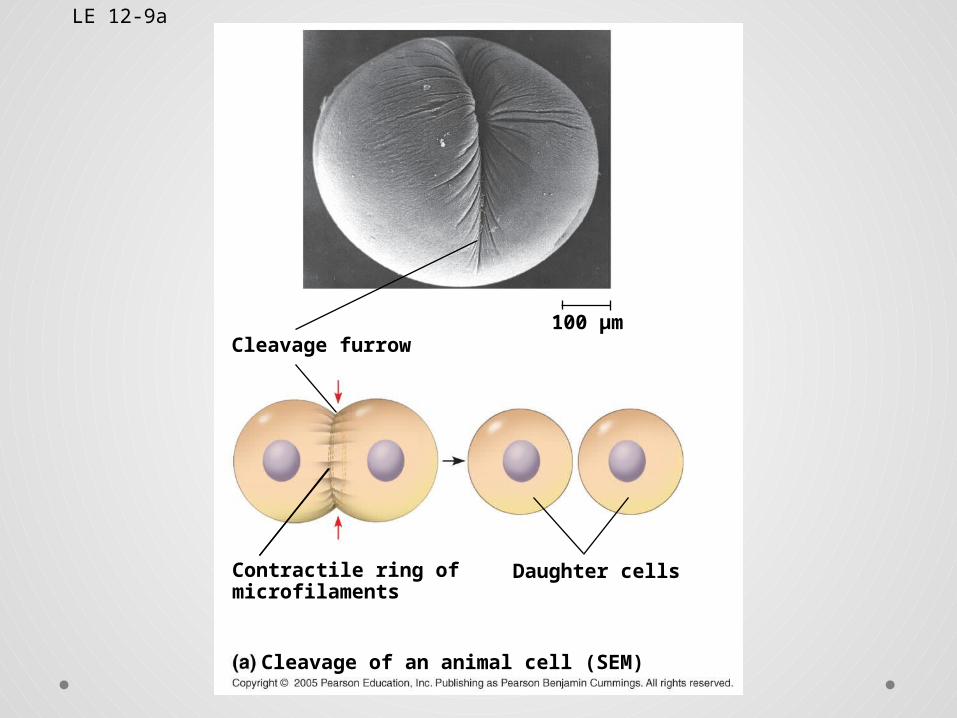
LE 12-9a
Cleavage furrow100 µm
Contractile ring ofmicrofilaments
Daughter cells
Cleavage of an animal cell (SEM)
Cytokinesis in Animal Cells
• In plants, the cell membrane is not flexible enough to draw inward because of the rigid cell wall.
• Instead, a cell plate forms between the divided nuclei that develops into cell membranes.
• A cell wall then forms in between the two new membranes.
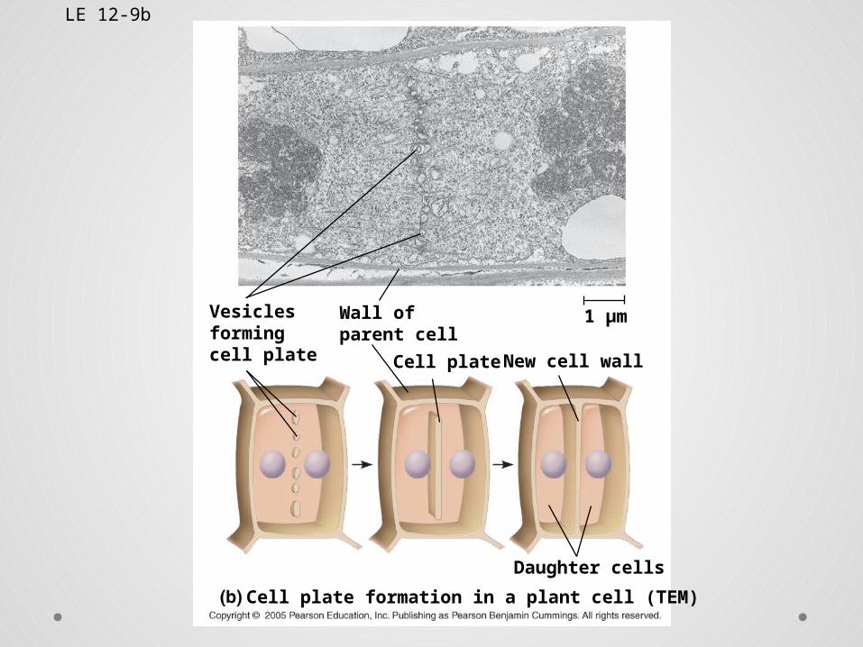
LE 12-9b
1 µm
Daughter cells
Cell plate formation in a plant cell (TEM)
New cell wallCell plate
Wall ofparent cell
Vesiclesformingcell plate
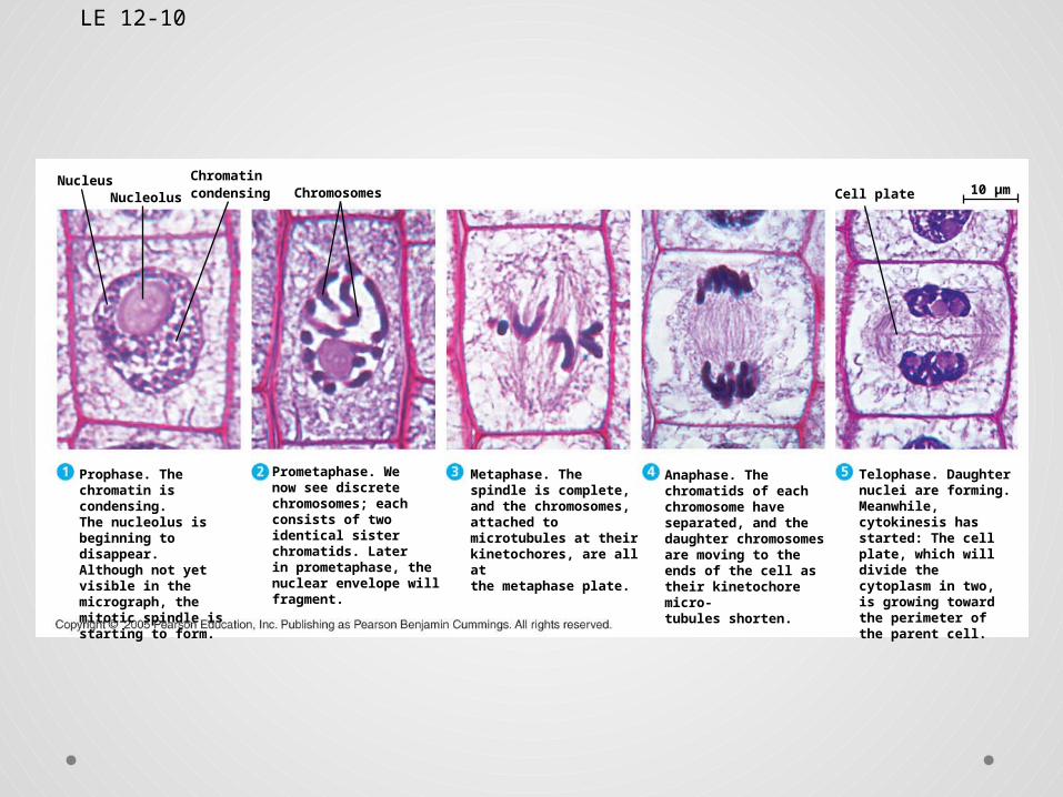
LE 12-10
NucleusCell plateChromosomesNucleolus
Chromatincondensing 10 µm
Prophase. The chromatin is condensing.The nucleolus is beginning to disappear.Although not yet visible in the micrograph, the mitotic spindle is starting to form.
Prometaphase. Wenow see discrete chromosomes; each consists of two identical sister chromatids. Laterin prometaphase, the nuclear envelope will fragment.
Metaphase. The spindle is complete, and the chromosomes, attached to microtubules at their kinetochores, are all at the metaphase plate.
Anaphase. The chromatids of each chromosome have separated, and the daughter chromosomes are moving to the ends of the cell as their kinetochore micro- tubules shorten.
Telophase. Daughter nuclei are forming. Meanwhile, cytokinesis has started: The cell plate, which will divide the cytoplasm in two, is growing toward the perimeter of the parent cell.
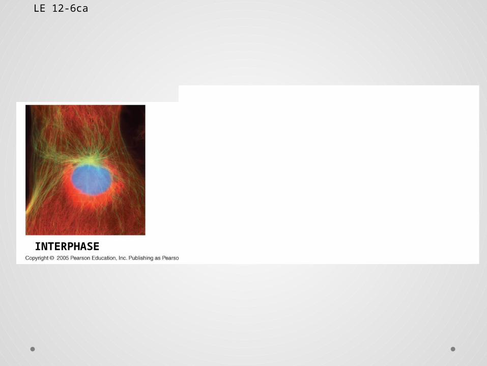
LE 12-6ca
INTERPHASE PROPHASE PROMETAPHASE
LE 12-6ca
INTERPHASE PROPHASE PROMETAPHASE
LE 12-6ca
INTERPHASE PROPHASE PROMETAPHASE
LE 12-6da
METAPHASE ANAPHASE TELOPHASE AND CYTOKINESIS
10
µm
LE 12-6da
METAPHASE ANAPHASE TELOPHASE AND CYTOKINESIS
10
µm
LE 12-6da
METAPHASE ANAPHASE TELOPHASE AND CYTOKINESIS
10
µm
Virtual Onion Root Tip Mitosis Lab
Click here to start
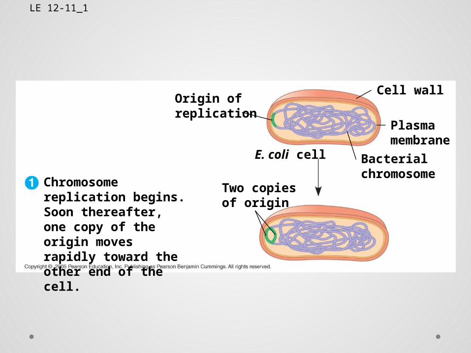
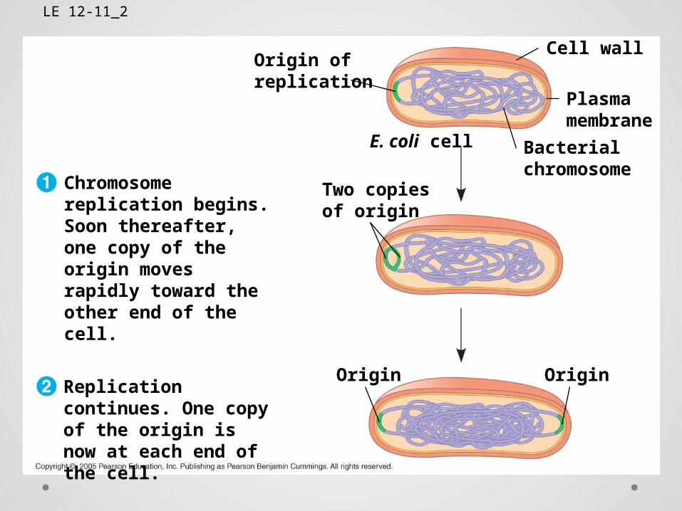
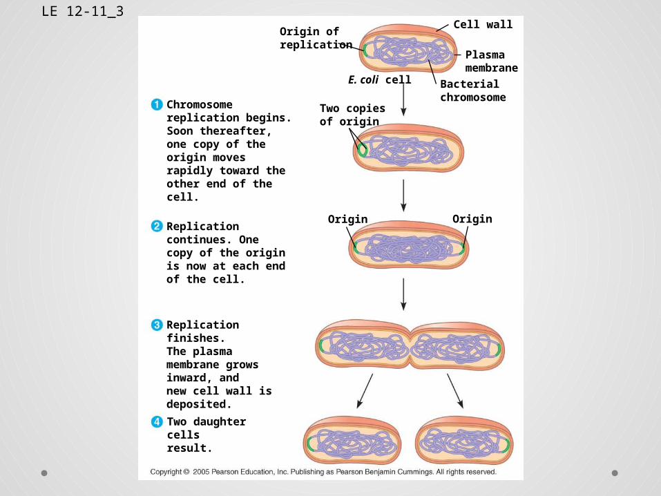
Binary Fission• Prokaryotes (bacteria and archaea) reproduce by a
type of cell division called binary fission• In binary fission, the chromosome replicates
(beginning at the origin of replication), and the two daughter chromosomes actively move apart
LE 12-11_1
Origin ofreplication
Cell wall
Plasmamembrane
Bacterialchromosome
E. coli cell
Two copiesof origin
Chromosome replication begins. Soon thereafter, one copy of the origin moves rapidly toward the other end of the cell.
LE 12-11_2
Origin ofreplication
Cell wall
Plasmamembrane
Bacterialchromosome
E. coli cell
Two copiesof origin
Chromosome replication begins. Soon thereafter, one copy of the origin moves rapidly toward the other end of the cell.
Replication continues. One copy of the origin is now at each end of the cell.
Origin Origin
LE 12-11_3Origin ofreplication
Cell wall
Plasmamembrane
Bacterialchromosome
E. coli cell
Two copiesof origin
Chromosome replication begins. Soon thereafter, one copy of the origin moves rapidly toward the other end of the cell.
Replication continues. One copy of the origin is now at each end of the cell.
Origin Origin
Replication finishes. The plasma membrane grows inward, and new cell wall is deposited.
Two daughtercells result.
The Evolution of Mitosis
• Since prokaryotes evolved before eukaryotes, mitosis probably evolved from binary fission
• Certain protists exhibit types of cell division that seem intermediate between binary fission and mitosis
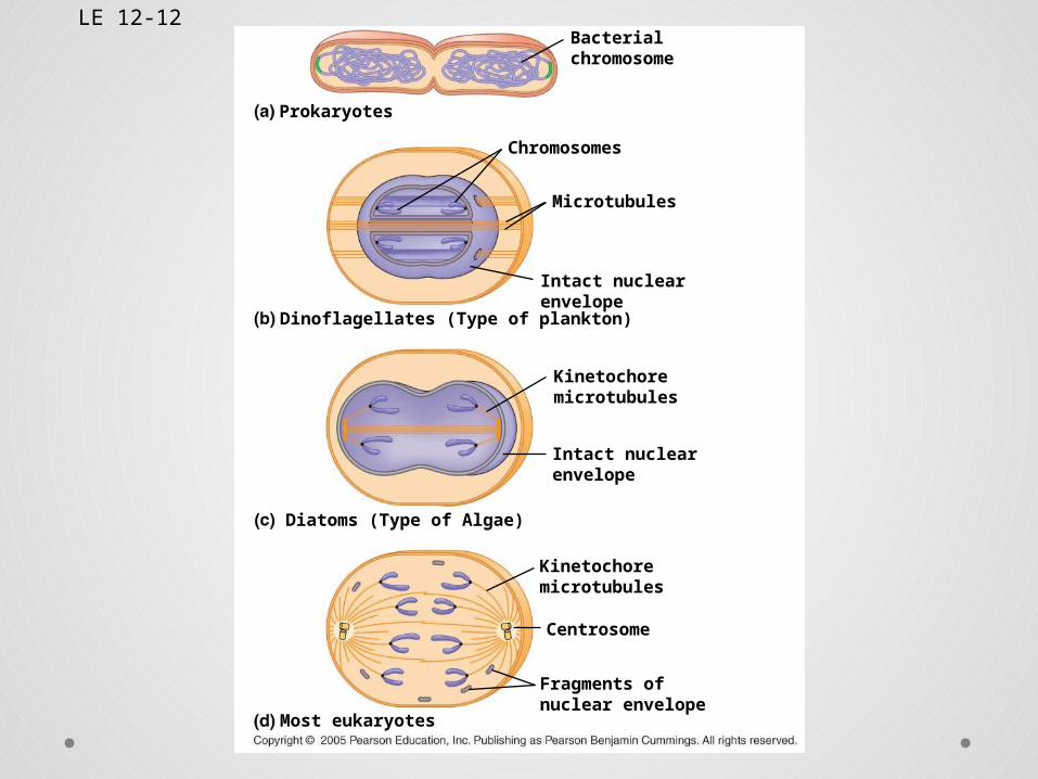
LE 12-12Bacterialchromosome
Chromosomes
Microtubules
Prokaryotes
Dinoflagellates (Type of plankton)
Intact nuclearenvelope
Kinetochoremicrotubules
Kinetochoremicrotubules
Intact nuclearenvelope
Diatoms (Type of Algae)
Centrosome
Most eukaryotes
Fragments ofnuclear envelope
The Cell Cycle Control System
• The sequential events of the cell cycle are directed by a distinct cell cycle control system, which is similar to a clock
• The clock has specific checkpoints where the cell cycle stops until a go-ahead signal is received
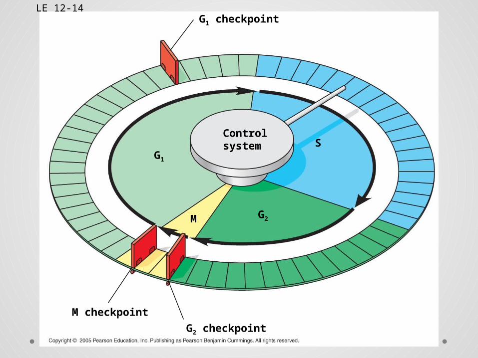
LE 12-14G1 checkpoint
G1
S
M
M checkpoint
G2 checkpoint
G2
Controlsystem
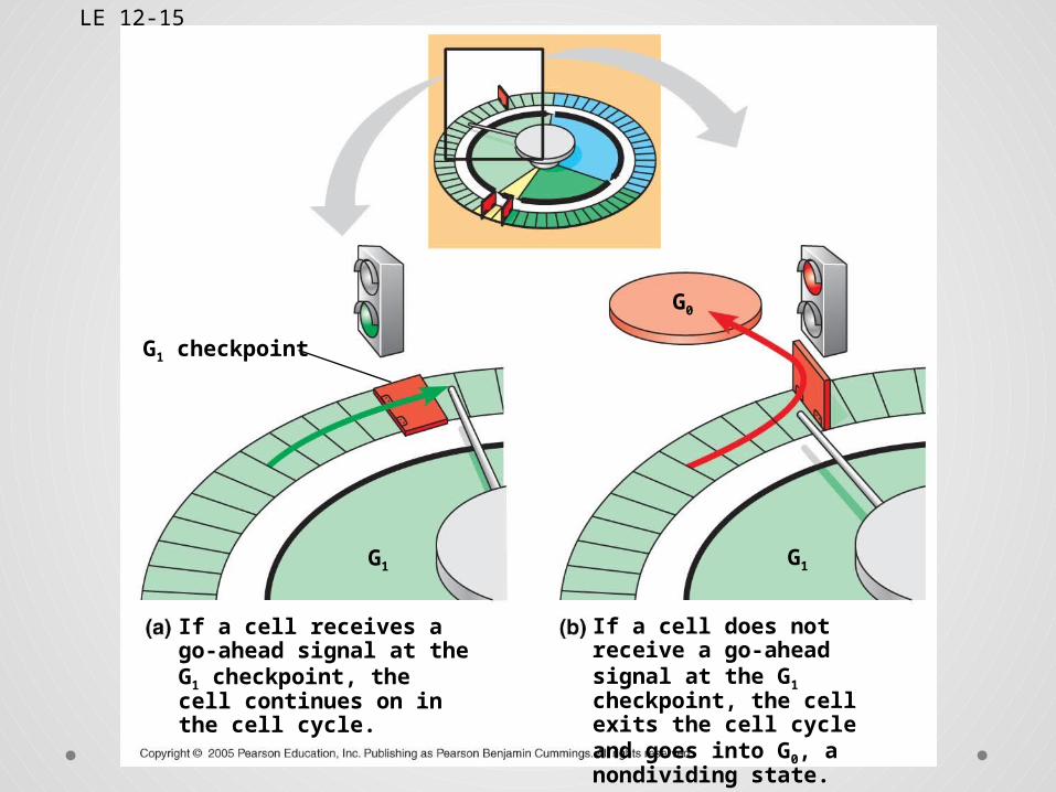
• For many cells, the G1 checkpoint seems to be the most important one
• If a cell receives a go-ahead signal at the G1 checkpoint, it will usually complete the S, G2, and M phases and divide
• If the cell does not receive the go-ahead signal, it will exit the cycle, switching into a nondividing state called the G0 phase
LE 12-15
G1
G1 checkpoint
G1
G0
If a cell receives a go-ahead signal at the G1 checkpoint, the cell continues on in the cell cycle.
If a cell does not receive a go-ahead signal at the G1 checkpoint, the cell exits the cell cycle and goes into G0, a nondividing state.
• An example of external signals is density-dependent inhibition, in which crowded cells stop dividing
• Most animal cells also exhibit anchorage dependence, in which they must be attached to a substratum (connective tissue) in order to divide
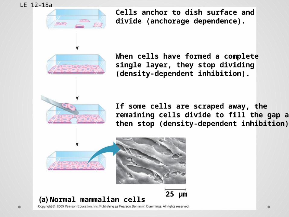
LE 12-18aCells anchor to dish surface anddivide (anchorage dependence).
When cells have formed a completesingle layer, they stop dividing(density-dependent inhibition).
If some cells are scraped away, theremaining cells divide to fill the gap andthen stop (density-dependent inhibition).
25 µmNormal mammalian cells
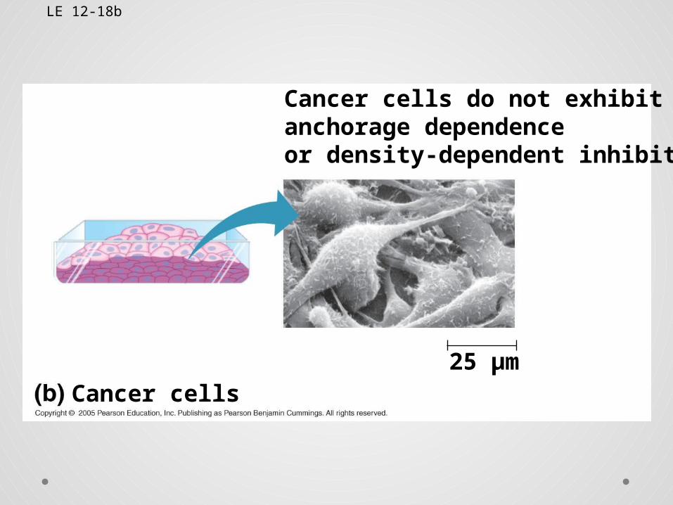
LE 12-18b
Cancer cells do not exhibitanchorage dependenceor density-dependent inhibition.
Cancer cells25 µm
Loss of Cell Cycle Controls in Cancer
Cells• Cancer cells do not respond normally to the
body’s control mechanisms• Cancer cells form tumors, masses of abnormal
cells within otherwise normal tissue• If abnormal cells remain at the original site,
the lump is called a benign tumor• Malignant tumors invade surrounding tissues
and can metastasize, exporting cancer cells to other parts of the body, where they may form secondary tumors
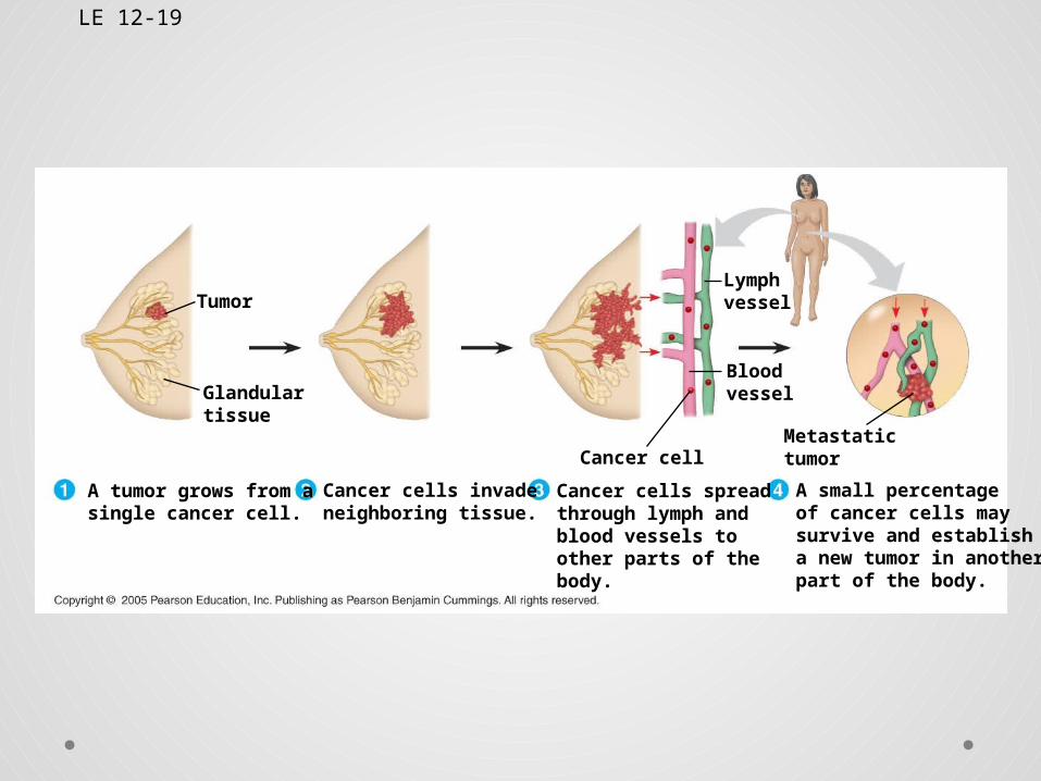
LE 12-19
Cancer cell
Bloodvessel
LymphvesselTumor
Glandulartissue
Metastatictumor
A tumor grows from asingle cancer cell.
Cancer cells invadeneighboring tissue.
Cancer cells spreadthrough lymph andblood vessels toother parts of thebody.
A small percentageof cancer cells maysurvive and establisha new tumor in anotherpart of the body.