Embed Size (px)
Citation preview

Development 110, 371-378 (1990)Printed in Great Britain © The Company of Biologists Limited 1990
371
The cellular retinoic-acid-binding protein is expressed in tissues associated
with retinoic-acid-induced malformations
MARIE-JOSEE VAESSEN1, J. H. CAREL MEIJERS2*, DIRK BOOTSMA1, and
AD GEURTS VAN KESSEL3
'Department of Cell Biology and Genetics, Erasmus University, PO Box 1738, 3000 DR Rotterdam, The Netherlands2Department of Pediatric Surgery, Erasmus University Medical School and Sophia Children's Hospital, Rotterdam, The Netherlands3Department of Anthropogenetics, University of Nijmegen, The Netherlands
'Present address: European Molecular Biology Laboratory, Heidelberg, FRG
Summary
Retinoic acid (RA) is thought to play a role in embryonicpattern formation in vertebrates. A naturally occurringgradient of endogenous RA has been demonstrated inthe developing chick limb bud, while local application ofRA leads to the formation of additional digits. Inmammals, a well-defined spectrum of birth defects hasbeen reported as a result of fetal exposure to excess RA.In analogy to the chick limb bud, it may be speculatedthat these malformations are the result of disturbance ofmorphogenetic RA concentration gradients.
A candidate gene involved in the regulation ofendogenous RA concentrations is the gene encodingcellular RA binding protein (CRABP). We have isolateda partial cDNA clone corresponding to the chickenhomolog of CRABP, and performed in situ hybridization
experiments on sections of embryos at various stages ofdevelopment. CRABP expression was detected in theCNS, the craniofacial mesenchyme, ganglia of theperipheral nervous system, the limb bud, and thevisceral arch area. Our results indicate that thespatiotemporally specified expression pattern displayedby the CRABP gene exhibits a striking correspondenceto the tissues that are affected by exposure of avian ormammalian embryos to RA. We hypothesize thatCRABP plays an important role in normal embryogen-esis and that embryonic tissues showing high CRABPexpression are susceptible to the adverse effects of excessRA.
Key words: retinoic acid, morphogenesis, CRABP.
Introduction
All-trans retinoic acid (RA), a biologically activemetabolite of retinol (vitamin A), plays an importantrole in cellular differentiation and pattern formationduring embryonic development of vertebrate animals.In chicken embryos, RA was found to be present inphysiological concentrations similar to those known tobe sufficient for the induction of EC cell differentiationin vitro (Thaller and Eichele, 1987). In the same study,Thaller and Eichele also demonstrated that RA ispresent in a higher concentration in the posterior part ofthe developing limb bud as compared to the anteriorpart. Local application of RA to the anterior margin ofthe limb bud results in dose-dependent changes in theskeletal pattern (Tickle etal. 1982; Eichele, 1989; Tickleet al. 1989). Local application of RA to a chickblastoderm grown in vitro was shown to interfere withaxis formation (Mitrani and Shimoni, 1989). In Xeno-pus laevis, RA was found to act on differentiation of thecentral nervous system (CNS), causing transformationof anterior neural tissue to a posterior neural specifi-
cation (Durston et al. 1989). Thus, the conclusion iswarranted that RA acts as a morphogen in variousdevelopmental processes.
RA has long been known to be teratogenic in humans(Kochar, 1967), and has been reported to be the causepi severe birth defects when administered to pregnantwomen (Lammer et al. 1985). RA-induced malforma-tions include defects of the CNS (mainly hydrocepha-lus), cleft lip and cleft palate, and congenital heartdefects. In rodents, the same spectrum of birth defectswas observed, while high doses of RA have also beenreported to result in limb deformations (Kochar, 1973).Also in chicken embryos, the tissues affected by RAtreatment seem to be similar to those reported formammals (Jelinek and Kistler, 1981). Excess RAobviously disturbs differentiation of the CNS inXenopus laevis (Durston et al. 1989). From theseobservations it seems likely that RA-induced malforma-tions represent a phenomenon general to all ver-tebrates.
If RA gradients influence morphogenetic processesduring embryonic development, it is important to know

372 M. -J. Vaessen and others
which mechanisms regulate RA concentrations. Inaddition to regulation of the synthesis and degradationof RA, fine-tuning of RA levels may be achieved at thecellular level. In this respect, a possible role could bereserved for the cellular RA-binding protein CRABP.This 15.5xlO3 MT protein, which displays a highlyspecific binding affinity for RA (Ong and Chytil, 1975;Jetten and Jetten, 1979), shows a structural similarity tothe P2 family of proteins (Eriksson et al. 1981; Sundelinet al. 1985). Members of this family, which also includescellular retinol binding protein (CRBP), are small,cytoplasmic proteins that have been implicated in thetransport of specific small hydrophobic molecules.
CRABP exhibits a spatiotemporally restricted ex-pression pattern in the mouse embryo (Vaessen et al.1989a). Using in situ hybridization techniques, wedetected a high level of CRABP transcripts in asubpopulation of cells in the CNS, as well as in thecraniofacial mesenchyme. These results were laterconfirmed by others (Perez-Castro et al. 1989). It wasalso shown that CRABP is differentially expressed inthe developing limbs of the mouse (Perez-Castro et al.1989; Dolle" etal. 1989). Maden etal. (1989) reported onthe immunocytochemical localization of CRABP in thechicken embryo. Apart from particular cells of theneural tube, CRABP was also found in various neuralcrest derivatives, including dorsal root ganglia andenteric ganglia. Earlier, these authors showed that agradient of CRABP is present at the tip of the chicklimb bud, with its maximum concentration in theanterior part (Maden et al. 1988).
Apart from the limb bud, all major CRABPexpression sites reported are of neurectodermal origin,and notably include many neural crest derivatives.Because of this observation, and also because neuralcrest cells have been mentioned as candidate targets forRA-induced malformations (Poswillo, 1975; Lammer etal. 1985), it would be of interest to know whether otherneural crest cell derivatives also show high levels ofCRABP expression. In order to study CRABP ex-pression during embryogenesis, we performed in situhybridization experiments on chicken embryos atvarious stages of development. Use of the chickenembryo for expression studies facilitated the acquisitionof well-standardized material of early developmentalstages. Furthermore, neural crest cell migration hasbeen most thoroughly studied in avian embryos, andseveral markers for early neural crest cells have becomeavailable (reviewed by Anderson, 1989). We chose touse the monoclonal antibody HNK-1 (Abo and Balch,1981), which recognizes a sulfated glucuronic acidpresent on several cell adhesion molecules (Kruse et al.1984, 1986; Pesheva et al. 1987; Rathjen et al. 1987;Hoffman and Edelman, 1988). In the chicken embryoHNK-1 binds to most premigratory neural crest cellsand neuronal neural crest cell derivatives (Vincent et al.1983; Vincent and Thiery, 1984; Tucker et al. 1984). Inaddition, HNK-1 is an early marker for neuraldifferentiation induced by RA in murine embryonalcarcinoma cells (McBurney et al. 1988).
Our results show that the CRABP gene exhibits a
spatiotemporally specified expression pattern, whichoffers a striking correlation to parts of the embryo thatcorrespond to the tissues commonly affected by RA-induced malformations. The .observed correlation ishighly suggestive for a role of CRABP in RA-mediatedmorphogenetic processes, and may help to understandthe underlying molecular mechanisms.
Materials and methods
Isolation of a chicken CRABP cDNA cloneA chicken embryo cDNA library, consisting of oligo(dT)-primed cDNAs inserted into the EcoKl site of bacteriophagelambda gtll (Clontech), was screened with a mouse CRABPcDNA probe. This probe, a 170bp Taql-Taql fragmentisolated from clone MoT-CAll and containing part of theCRABP-coding region (Vaessen et al. 1989a), was labeledwith ^P using random priming (Feinberg and Vogelstein,1983). The filters were hybridized at 56°C, in a buffercontaining 6xSSC and 9% dextran sulphate, and washed atthe same temperature, twice in 3xSSC, 0.1% NaDodSO4,and twice in lxSSC, 0.1% NaDodSO4.
Screening of approximately lxlO6 bacteriophage plaquesresulted in the isolation of a cDNA clone, designated C4.DNA isolated from clone C4 was digested with EcoBJ, andligated to EcoRl plasmid vector pTZ18R (Pharmacia).Transformation of E. coli DH5aF' yielded the subcloneChCRABP C4.5, which contains a cDNA insert of approxi-mately 300 bp.
Sequence analysisFor determination of the nucleotide sequence, the ChCRABPC4.5 cDNA insert was subcloned into bacteriophageM13 mpl9. The cDNA insert was cloned in both orientations,allowing both strands to be read. After isolation of single-stranded DNA, the nucleotide sequence was determined viathe dideoxy chain termination method (Sanger et al. 1977)using Sequenase (United States Biochemical) according toinstructions by the manufacturer.
Computer-assisted analysis of the DNA sequence wascarried out with a Microgenie software package (Beckman).
Chicken embryosFertilized eggs from the White Leghorn (Gallus domesticus)were incubated at 38 °C in a forced-draft incubator at arelative humidity of 80%. Staging of the embryos wasperformed according to Hamburger and Hamilton (1951).
RNA isolation and blot hybridizationTotal RNA was isolated from whole chicken embryos usingthe LiCl/Urea method described by Auffray and Rougeon(1980). For RNA blot analysis, RNA was electrophoresed on1 % agarose gels in the presence of formaldehyde. Prior toelectrophoresis, ethidium bromide was added to the RNAsamples in order to allow visualisation of the ribosomal RNAsin the gel. In this way, it was ascertained that the amounts ofRNA in the different lanes were approximately the same.
Following electrophoresis, the RNA was transferred tonitrocellulose filters (Maniatis etal. 1982). DNA probes werelabeled as described above. The RNA blots were hybridizedat 42°C in a buffer containing 6xSSC, 9 % dextran sulphate,and 50% formamide, and washed at 56°C, twice in 3xSSC,0.1% NaDodSO4, and twice in lxSSC, 0.1% NaDodSO4.

CRABP and RA-induced malformations 373
Preparation of embryo sectionsChicken embryos were fixed for 24 h in 4 % paraformaldehydein phosphate-buffered saline at 4°C. After fixation, theembryos were embedded in paraffin. Sections were cut at5/an, placed on chromium(III)potassium sulphate coatedslides, and air dried.
Prior to in situ hybridization or immunocytochemicaltreatment, the sections were deparaffinized and hydrated.
In situ hybridization techniquesWe used the cDNA insert of ChCRABP C4.5, labeled with35S via nick translation, and treated with DNAsel to obtainfragments of approximately 50-100 bp as a probe for in situhybridization to the chicken embryo sections.
Section pretreatment and in situ hybridization procedureswere based on standard methods (Akam, 1983; Hafen et al.1983). For autoradiography, slides were dipped in KodakNBT-2 emulsion, and developed after 5-10 days of exposure.Counterstaining with haematoxylin was carried out asrequired.
ImmunocytochemistryThe HNK-1 hybridoma cell line was purchased from theAmerican Tissue Culture Collection (ATCC TIB 200). HNK-1 immunoperoxidase staining was performed using undilutedsupernatant. Rabbit anti-mouse peroxidase-conjugated im-munoglobulins (Dako, Denmark) were used in a dilution of1:100. In order to reduce background staining, 2% chickserum was added to the conjugate. Peroxidase was visualizedby 0.1% 3,3'diaminobenzidine.4HCl (Serva) and 0.02%hydrogen peroxide. All rinsing and diluting was done inphosphate-buffered saline with 0.1 % Tween 20.
EcoRI _G_ S E N F D E L L K A L G V N A MGAATTCTGGCAGCGAGAATTTCWCtMGCTCCTCAAAGCGCTGGGTGTCAACGCCATG
+ • + •
CAGCAGCGAGAATTTCGACGAGCTCCTCAAGGCGTTGGGTGTGAACGCCATGS S E N F D E L L K A L G V N A M
L R K V A V A A A S K P H V E I R Q D GCTCAGGAAGGTGGC<K7rGGCGGCCGCCTCCAAACCCCA«rrGGAGATCCGCCAGGACGGG
L H K V V A A A S K P H R Q D G
D Q F Y I K T S T T V R T T E I N F K IGACCAGTTCTACATCAAAACTT«:ACCACTGTCCGCACCACGGAAATCAACTTCAAAATC
GATCAGTTCTACATCAAGACATCCACTACrcrGCGCACCACGGAGATCAACTTCAAGGTCD Q F Y I K T S T T V R T T E I N F K V
G E _ S _ F E E E T V D G R K C R S L _A_ T WGGGGAGAGCTTCGAGGAGGAGACGGTGGATOGCCGAAAATGCAGGAGTTTGGCCACCTGG
GGAGAGGCKTTCGAGGAGGAGACAGTGGACGGACGCAAATGCAGGAGTTTACCCACGTGGG E G F E E E T V D G R K C R S L P T W
E N E N K I _Y_ C _K_ Q T L _I_ E G D G P K TGAGAATGAAAACAAGATCTATTGCAAACAAACTCTTATTGAGGGAGATGGTCCTAAAACA
GAGAATGAGAACAACWTTCACTGCACACACUC^CTTCTrcAGGGGGATGGCCCTAAAACTE N E N K I H C T Q T L L E G D G P K T
Y W T EcoRITACTGGACTCGAATTC
TACTGGACCCY W T
Fig. 1. Sequence of the partial cDNA clone encodingchicken CRABP. The cDNA sequence and deduced aminoacid sequence of ChCRABP C4.5 are shown in bold face,with underneath the corresponding sequence of mouseCRABP cDNA clone MoT-CAll. Non-homologousnucleotides are indicated by asterisks, and non-homologousamino acids are underlined. The £coRI sites that flank theC4.5 cDNA insert are indicated.
Results
Isolation of a chicken CRABP cDNA cloneWe obtained a chicken CRABP probe by screening achicken embryo cDNA library with a Taql-Taqlfragment containing nucleotides 147-315 of mouseCRABP cDNA clone MoT-CAll. This resulted in theisolation of bacteriophage clone C4, from which anEcoRI subclone in plasmid vector pTZ18R was derived.This subclone, which was named ChCRABP C4.5,contains a 310 bp cDNA insert, which corresponds tonucleotides 129-438 of mouse CRABP clone MoT-CAll. The sequence of ChCRABP C4.5 and itscomparison with the mouse CRABP sequence is shownin Fig. 1. The chicken cDNA clone contains part of theCRABP-coding sequence. Homology with mouseCRABP is 87 % at the nucleotide level and 94 % at theamino acid level.
The 32P-labeled ChCRABP C4.5 insert DNA washybridized to nitrocellulose blots containing total RNAisolated from chicken embryos at various stages ofdevelopment. This resulted in detection of one singletranscript of approximately 1 kb (see Fig. 2), which is inagreement with the size of the mouse CRABP mRNA(Vaessen et al. 1989a). Thus, ChCRABP C4.5 specifi-cally hybridizes to chicken CRABP mRNA and can beused to study CRABP expression patterns in the chickembryo.
Localization of CRABP transcripts in chickenembryosWe performed in situ hybridization experiments withthe radiolabeled cDNA insert of ChCRABP C4.5 onsections obtained from chicken embryos at variousstages of development. The CRABP gene exhibits astrongly restricted expression pattern in day 2, day 3and day 4 embryos, which tends to become more diffusefrom day 5 onwards. High CRABP expression wasfound in the CNS, in the craniofacial mesenchyme, inthe visceral arches, and in the ganglia of the peripheralnervous system. CRABP transcripts were also detectedin the limb bud, where expression was predominantlyfound in mesenchymal cells located in the anterior part.
In order to establish a possible relationship betweenexpression sites of CRABP and the occurrence ofmigratory neural crest cells, we performed immuno-staining experiments with the neural crest cell markerHNK-1 on sections serial to those used for the in situhybridizations.
The following results were obtained:
(A) CRABP expression in stage 12 embryosFigs 3 and 4 show serial sections of a stage 12 embryo(day 2) that were subjected to in situ hybridization witha CRABP probe (Fig. 3) and immunostaining withHNK-1 (Fig. 4). Occasional CRABP-positive cells wereobserved in the outer cell layer of the mesencephalon,

374 M.-J. Vaessen and others
a b c d
28S>
18S>
Fig. 2. Hybridization of ChCRABP C4.5 to RNA samplesisolated from (a) day 2; (b) day 3; (c) day 4; (d) day 5;and (e) day 6 chicken embryos. The position of the 28Sand 18S ribosomal RNAs is indicated. The 18S ribosomalRNAs, stained with ethidium bromide, are shownunderneath the corresponding lanes.
rhombencephalon and spinal cord, but not in theprosencephalon. In the cells of the auditory pit, which isdeep and wide open at this stage, CRABP transcriptswere also detected. These cells exhibit strong ex-pression of the HNK-1 epitope, while the cells of theneural tube are negative. CRABP expression was alsoobserved in the craniofacial mesenchyme anterior tothe telencephalon and diencephalon. These mesenchy-mal cells are also positive for HNK-1.
(B) CRABP expression in stage 22 embryosIn stage 22 embryos (day 3), cells that show a stronghybridization to the CRABP probe were detected in theouter cell layers of mesencephalon, rhombencephalonand spinal cord. CRABP-positive cells are lying apartor in small clusters, but are more abundant than in stage12 embryos (see Fig. 5). The distribution of CRABPtranscripts coincides with HNK-1 expression, as illus-trated by Fig. 6. As can be seen in Fig. 7, CRABPexpression is also observed in the neuroepithelial cells
of the auditory vesicle, which is HNK-1 positive. Thecraniofacial mesenchyme, located anterior to telen-cephalon and diencephalon, and posteriorly confinedby the eyes, exhibits a high level of CRABP transcripts(see Fig. 5). These cells no longer express the HNK-1epitope at this stage.
In addition to the CNS and the craniofacial mesen-chyme, CRABP-positive cells were detected in thevisceral arches, as illustrated by Figs 7 and 8. TheCRABP-positive region extends from the craniofacialmesenchyme of the head into the first or mandibulararch, where an even higher level of CRABP transcriptswas observed. High CRABP expression continues intothe second, third and fourth arch. The hybridizationsignal appears to be strongest in the mesenchymal cellsbordering the overlying epithelium of pharynx, visceralclefts, and pericardial cavity, while the epithelium itselfis CRABP negative. In the visceral arch area, onlyoccasional HNK-1 positive cells were observed.
CRABP expression was also detected in cranial anddorsal root ganglia, which are positive for HNK-1.
(C) CRABP expression in stage 24 embryosIn stage 24 embryos (day 4), a high level of CRABPexpression was observed in the dorsolateral part ofmesencephalon and metencephalon, extending to lat-eral and ventral, and continuing into myelencephalonand spinal cord. CRABP-positive cells were alsoobserved in the ventral part of telencephalon anddiencephalon, posteriorly confined by the olfactory pit,which is CRABP negative. The strong CRABP-specifichybridization signal has extended to nearly the wholeouter neural cell layer, as shown in Fig. 9. In embryosof this stage, HNK-1 expression was observed all alongthe neural tissue.
In the craniofacial mesenchyme, CRABP expressionis still detectable, but reduced as compared to stage 22embryos. Expression in the visceral arch area issimilarly diminished, with the strongest hybridizationsignal appearing at the ventral side of the arches. At thisstage, neither the craniofacial mesenchyme nor thevisceral arches showed any immunoreactivity withHNK-1. An elevated level of CRABP transcripts wasstill detected in the spinal cord, and in the dorsal rootganglia and cranial ganglia, which showed a continu-ously strong expression of the HNK-1 epitope. Fig. 10shows a transverse section through the spinal cord, witha characteristic distribution of CRABP transcripts onthe dorsolateral sides and in two cell groups locatedventrally, near the notochord.
Discussion
We have isolated a cDNA clone encoding part ofchicken CRABP and used it for in situ hybridizationstudies of CRABP expression during chicken embryo-genesis. After determination of the nucleotide se-quence of our partial cDNA clone ChCRABP C4.5, wecompared the deduced amino acid sequence withmouse CRABP (Vaessen et al. 1989a). Only 6 out of 100
I•
•^s?K^-.
r ? ^>:$$*
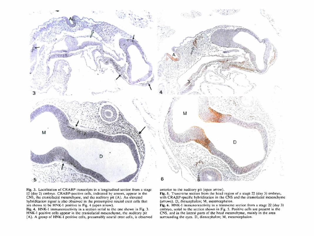
Fig. 3. Localisation of CRABP transcripts in a longitudinal section from a stage12 (day 2) embryo. CRABP-positive cells, indicated by arrows, appear in theCNS, the craniofacial mesenchyme, and the auditory pit (A). An elevatedhybridization signal is also observed in the presumpiive neural crest cells thatore shown to be HNK-1 positive in Fig. 4 (open arrow).Fig. 4. HNK-l immu no reactivity in a section serial to the one shown in Fig. 3.HNK-1 positive cells appear in the craniofacial mesenchyme, the auditory pit(A). A group of HNK-1 positive cells, presumably neural crest cells, is observed
anterior to the auditory pit (open arrow).Fig, 5. Transverse section from the head region of a stage 22 (day 3) embryo,with CRABP-specific hybridization in the CNS and ihe craniofacial mesenchyme(arrows). D, diencephalon; M. mesenoephalon.Fig. 6. HNK-1 immunoreactivity in a transverse section from a stage 22 (day 3)embryo, serial to the section shown in Fig. 5. Positive cells are present in theCNS, and in the lateral parts of the head mesenchyme, mainly in the areasurrounding ihe eyes. D, diencephalon; M, mesencephalon.

***3F&«:
rV M V
Fig. 7. CRABP-specific hybridization in a longitudinal section from a stage 22(day 3) embryo, showing CRABP expression in the visceral arch area, and inthe auditory vesicie (A). I, I I , 111, firsi, second, and third visceral arch.Fig. S. Transverse section from a stage 72 (day 3) embryo, demonstratingCRABP expression in the mantle layer of the myelencephalon (My), in the firstviscera] arch, and in the fifth cranial ganglion (arrow). P, pharynx.
5v -: .
• > •
'_ t
; -
*r . •
m
Fig. 9. CRABP-specific hybridization in a transversal section from a stage 24 (day4) embryo, with positive ceils in the mantle layer of the myelencephalon (My).Fig. 10. Transversal section from a stage 24 (day 4) embryo, showing CRABPexpression in the neural tube and in a dorsal root ganglion (open arrow). TheCRABP-positive cells located on the ventral side of the neural tube areindicated by solid arrows. /V=notochord.

CRABP and RA-induced malformations 375
amino acids were different, with most substitutionsconcerning amino acids with similar physicochemicalproperties. The high degree of conservation observedfor CRABP in cow (Sundelin et al. 1985), mouse andchicken suggests that the biological function of theprotein does not allow important structural variation.The protein sequence of ChCRABP C4.5 is inagreement with the partial sequence of chickenCRABP type I as reported by Kitamoto et al. (1988).These authors state that two types of CRABPcontaining 6 amino acid replacements in the NH2terminal region are present in the chick embryo: amajor one, type I, and a minor one, type II. It would beof interest to know whether CRABP I and II areencoded by different loci, or are the result of e.g.differential mRNA splicing. However, Southern blot-ting experiments performed on mouse and chickenDNA as well as on DNA isolated from mouse-hamsterand human-hamster somatic cell hybrids did not, sofar,give any indication for the existence of more than oneCRABP gene (Vaessen et al. 1989a,b).
Our in situ data demonstrate that CRABP expressionin the CNS is strictly limited to a subpopulation ofneural cells in day 2, day 3 and day 4 embryos. CRABPexpression begins in a few single cells, graduallyexpanding to the whole outer cell layer of mesencepha-lon, rhombencephalon and spinal cord. In agreementwith the results reported earlier for the mouse embryo,we also detected a high level of CRABP transcripts incells of the craniofacial mesenchyme. In addition, wehere report that CRABP is highly expressed in thevisceral arches. As a matter of fact, CRABP expressionin the visceral arch area also occurs in the mouseembryo (Vaessen, unpublished results). Thus, CRABPexpression patterns in chicken and in mouse embryosshow a strong similarity.
The major CRABP expression sites reported hereexhibit a striking correspondence to tissues commonlyaffected by exposure of mammalian embryos to RA.Clinical reports include malformations of the CNS -mainly hydrocephalus; of structures derived from thecraniofacial and mandibular arch mesenchyme -microtia/anotia, micrognathia and cleft palate; con-genital heart defects - predominantly conotmncal orbranchial-arch mesenchymal tissue defects, occasion-ally combined with thymic defects (Lammer etal. 1985).Although the adverse effects of RA on the chickenembryo are less well documented, the target tissuesseem to be the same (Jelinek and Kistler, 1981). Severalreports implicate Vitamin A in the inhibition of cranialneural crest cell development in the chick embryo(Hassell et al. Wll; Keith, 1977). In addition, recentresults obtained in our own group demonstrate that RAtreatment of chicken embryos may give rise tocongenital heart defects and craniofacial malformationsas well as to limb deformations (M. Broekhuyzen, pers.comm.). Apart from leading to digit duplications, localapplication of RA to the chick wing bud may also resultin upper beak defects (Tickle et al. 1982; Tamarin et al.1984; Wedden and Tickle, 1986). Interestingly, aftertreatment with RA we also obtained an embryo with a
cleft lower beak (Vaessen, unpublished results),suggesting that in the chicken embryo the first arch - thelower beak primordium - is susceptible to the teratoge-nic effects of RA, just as in the mammalian embryo.The fact that Wedden and Tickle did not observe effectsof RA on the lower beak must probably be ascribed totheir using local application of RA in the chick wing budas a means to generate malformations. In conclusion,the resemblance of CRABP expression patterns be-tween chicken and mouse embryos, and the fact that inboth species the CRABP-positive tissues appear to besusceptible to the adverse effects of RA, leads to theassumption that similar RA-sensitive morphogeneticprocessess take place in both species, and that CRABPexpression has an important function in theseprocesses.
The tissues that are frequently affected by RA-induced malformations share a common embryonicorigin in that they have all received contributions fromthe cephalic neural crest. It has been suggested thatexcess RA has an adverse influence on cephalic neuralcrest cells, possibly by interfering with normal neuralcrest cell migration (Thorogood et al. 1982; Pratt et al.1987). This theory is supported by the observation thatRA interferes with the cell - substratum adhesion ofneural crest cells in vitro (Smith-Thomas et al. 1987). Ifendogenous RA concentrations also determine themigratory behaviour of neural crest cells in vivo,differential expression of CRABP could play a role inthe regulation of this proces. Maden et al. (1989)described the occurrence of single CRABP-positivecells in a line from the dorsal neural tube to the lateraledge of the dorsal aorta and suggest that these cells maybe neural crest cells in the progress of migration. Thefact that these CRABP-expressing cells have escapedour attention can be explained by immunocytochem-istry being better suited for detection of single positivecells than in situ hybridization, due to the higherbackground levels obtained with the latter technique.However, we observed two additional CRABP ex-pression sites that may be related to neural crest cellmigration: the craniofacial mesenchyme and the vis-ceral arch area.
For a more detailed investigation of a possiblerelationship between CRABP expression sites andneural crest cell migration, we employed the mono-clonal antibody HNK-1 (Abo and Balch, 1981), whichrecognizes an epitope present on several cell adhesionmolecules. Canning and Stern (1988) showed thatHNK-1 identifies tissues involved in mesoderm forma-tion in the chick embryo. Prior to mesoderm induction,HNK-1 binds to the inducing tissue (hypoblast) andreveals a mosaic pattern in the responding tissue(epiblast). After primitive streak formation, the epi-blast displays an anteroposterior gradient of HNK-1expression. At the end of gastrulation, the primitivestreak region loses its HNK-1 reactivity. HNK-1expression is next seen in cells of the forming notochordand in cranial neural crest cells (Canning and Stern,1988; Stern and Canning, 1990). In later stages ofchicken embryogenesis, HNK-1 recognizes most pre-

376 M.-J. Vaessen and others
migratory neural crest cells and neuronal neural crestcell derivatives (Vincent et al. 1983; Vincent andThiery, 1984; Tucker et al. 1984). However, the absenceof HNK-1 immunoreactivity does not rule out theposibility that migratory neural crest cells are presentsince disappearance of the HNK-1 epitope from cranialneural crest cells at certain stages of migration has beenreported (Vincent and Thiery, 1984; Bronner-Fraser,1987).
In view of the existence of multiple HNK-1 antigensduring development and adult life, it might seemsurprising that migrating neural crest cells in the avianembryo can be visualized specifically with HNK-1 orrelated antibodies. Although reactivity is not restrictedto neural crest cells, use of HNK-1 as a marker forneural crest cells is possible because the staining ofother antigenic lineages does not overlap topographi-cally or temporally with the distibution of crest cells.For one thing, neural crest cells do not become HNK-1positive until they leave the neuroepithelium and startto migrate. Additional proof for the presence of theHNK-1 epitope on migrating neural crest cells is the factthat injection of HNK-1 antibodies lateral to themesencephalic neural tube perturbs cranial neural crestcell migration (Bronner-Fraser, 1987).
In our investigation of a possible relationshipbetween CRABP expression and neural crest cells wefound that the cranial and dorsal root ganglia arepositive for both CRABP and HNK-1. In the CNS, apartial coincidence of CRABP and HNK-1 expressionis observed, with a remarkable colocalization at stage22. In stage 24 embryos, CRABP continues to bedifferentially expressed, while HNK-1 reactivity occursall along the neural tissue. However, the observedexpression of the HNK-1 epitope in the CNS isprobably unrelated to neural crest cell migration.
The cells of the craniofacial mesenchyme are initially(stage 12) positive for HNK-1 and also expressCRABP. Later during development (stage 22) theystart to differentiate into cartilage, muscle and bone,and lose the HNK-1 epitope. In contrast, CRABPexpression in the craniofacial mesenchyme continuesuntil stage 24. The visceral arches are filled with cells ofneural crest origin (Le Lievre and Le Douarin, 1975)which contribute to the development of the heart andarch arteries (Bockman et al. 1987; Philips et al. 1987).While showing a high level of CRABP expression,these neural crest derived cells do not express the HNK-1 epitope anymore. After administration of RA tochicken embryos Jelinek and Kistler (1981) showed thattreatment on day 3 frequently gave rise to heart defects.Administration on day 4 resulted in a high incidence ofheart defects and craniofacial malformations, as well aslimb deformations. A tentative suggestion is evokedthat the period of sensitivity to RA treatment of aparticular morphogenetic system coincides withCRABP expression and loss of HNK-1 reactivity.
The role of CRABP in RA-mediated morphogeneticprocesses is still poorly understood. It has beensuggested that CRABP has a function as a transportprotein, mediating transfer of RA to the nucleus, where
RA is thought to exert its biological activity (Takase etal. 1986; Shubeita et al. 1987). On the other hand, thehuman myelocytic leukemia cell line HL60, which isdeficient in CRABP (Breitman et al. 1982; Douer andKoeffler, 1982), is still able to differentiate in responseto RA, indicating that binding to CRABP is notobligatory for transport of RA to its nuclear receptorsites.
A different model, proposed by Hirschel-Scholz etal.suggests that CRABP has a buffer function, protectingthe cell from the deleterious effects of unbound RA(Hirschel-Scholz et al. 1989). Maden et al. reason alongthe same lines, proposing a function for CRABP in thesequestering of RA in the cytoplasm (Maden et al.1989). The existence of reciprocating concentrationgradients for RA and CRABP in the developing chicklimb bud (Thaller and Eichele, 1987; Maden etal. 1988)is consistent with these models. While the concen-tration of RA itself is highest in the posterior part,CRABP is present in a higher concentration in theanterior part. Thus, CRABP could be effective inreducing the concentration of free RA in the anteriorpart.
Recently, a nuclear receptor for RA (RARar) wasidentified, which was shown to be related to thesteroid/thyroid hormone receptor family (Petkovich etal. 1987; Giguere et al. 1987). In addition, three moreRA receptors were identified, designated RAR/3,RARy, and RAR6, which were related to RARar butwere obviously encoded by different genes (Brand et al.1988; Krust et al. 1989; Ragsdale et al. 1989).
It is assumed that the different RA receptors act astranscription factors, mediating expression of specificsets of genes. This suggests that RA-induced malforma-tions occur as the result of aberrant gene expression.Evidently, genes known from in vitro experiments to besusceptible to RA induction, such as the gene encodingGrowth Hormone (Bedo et al. 1989), and the homeo-box-containing genes (Colberg-Poley et al. 1985a,6;Breier et al. 1986; Deschamps et al. 1987; Mavilio et al.1988), are candidate target genes involved in RA-induced malformations.
We propose that a high level of CRABP expression incertain tissues reflects a particular sensitivity to RA.CRABP would be instrumental in protecting cells fromthe developmentally important action of RA byprohibiting aberrant activation of RA responsive genesequences during critical stages. This would explainwhy fetal exposure to excess RA predominantly affectsthose tissues that exhibit a high level of CRABPexpression during certain stages of embryogenesis.
The authors thank R. Beekhuizen for technical assistance.This work was supported by the Netherlands Cancer Society(Koningin Wilhelmina Fonds).
'The nucleotide sequence data reported here will appear inthe EMBL, GenBank and DDBJ Nucleotide SequenceDatabases under the accession number X53701 CHICKENCRABP.'
CRABP and RA-induced malformations 377
References
ABO, T. AND BALCH, C. M. (1981). A differentiation antigen ofhuman NK and K cells identified by a monoclonal antibody(HNK-1). /. Immunol. 127, 1024-1029.
Akam, M. E. (1983). The location of Uhrabithorax transcripts inDrosophila tissue sections. EMBO J. 2, 2075-2084.
ANDERSON, D. J. (1989). The neural crest cell lineage problem:Neuropoiesis? Neuron 3, 1-12.
AUFFRAY, C. AND ROUGEON, F. (1980). Purification of mouseimmunoglobulin heavy chain messenger RNAs from totalmyeloma tumor RNA Eur. J. Biochem. 107, 303-314.
BEDO, G., SANTISTEBAN, P. AND ARANDA, A. (1989). Retinoic acidregulates growth hormone gene expression. Nature 339,231-234.
BOCKMAN, D. E., REDMOND, M. E., WALDO, K., DAVIS, H. ANDKIRBY, M. L. (1987). Effect of neural crest ablation ondevelopment of the heart and arch arteries in the chick. Am. J.Anat. 180, 332-341.
BRAND, N., PETKOVICH, M., KRUST, A., CHAMBON, P., DE TH£, H.,MARCHIO, A., TIOLLAIS, P. AND DEJEAN, A. (1988).Identification of a second human retinoic acid receptor. Nature332, 850-853.
BREIER, G., BUCAN, M., FRANCKE, U., COLBERG-POLEY, A. M. ANDGRUSS, P. (1986). Sequential expression of murine homeoboxgenes during F9 EC cell differentiation. EMBO J. 5, 2209-2215.
BREITMAN, T. R., COLLINS, S. J. AND KEENE, B. R. (1982).Terminal differentiation of Human promyelocytic leukemic cellsin primary culture in response to retinoic acid. Blood 57,1000-1004.
BRONNER-FRASER, M. (1987). Perturbation of cranial neural crestmigration by the HNK-1 antibody. Devi Biol. 123, 321-331.
CANNING, D. R. AND STERN, C. D. (1988). Changes in theexpression of the carbohydrate epitope HNK-1 associated withmesoderm induction in the chick embryo. Development 104,643-655.
COLBERG-POLEY, A . M. , VOSS, S. D . , CHOWDHURY, K. AND GRUSS,P. (1985a). Structural analysis of murine genes containinghomeo box sequences and their expression in embryonalcarcinoma cells. Nature 314, 713-718.
COLBERG-POLEY, A. M., Voss, S. D., CHOWDHURY, K., STEWART,C. L., WAGNER, E. F. AND GRUSS, P. (1985b). Clusteredhomeoboxes are differentially expressed during raunnedevelopment. Cell 43, 39-45.
DESCHAMPS, J., DE LAAF, R., JOOSEN, L., MEJJUNK, F. ANDDESTREE, O. (1987). Abundant expression of homeobox genes inmouse embryonal carcinoma cells correlates with chemicallyinduced differentiation. Proc. natn. Acad. Sci. U.S.A. 84,1304-1308.
DOLLE, P., RUBERTE, E., KASTNER, P., PETKOVICH, M., STONER, C.M., GUDAS, L. J. AND CHAMBON, P. (1989). Differentialexpression of genes encoding a, f} and gamma retinoic acidreceptors and CRABP in the developing limbs of the mouse.Nature 342, 702-705.
DOUER, D. AND KOEFFLER, H. P. (1982). Retinoic acid enhancescolony-stimulating factor-induced clonal growth of normalhuman myeloid progenitor cells in vitro. Expl Cell Res. 138,193-198.
DURSTON, A . J . , TlMMERMANS, J. P . M . , H A G E , W. J . , HENDRIKS,H. F. J., DE VRIES, N. J., HEIDEVELD, M. AND NIEUWXOOP, P.D. (1989). Retinoic acid causes an anterioposteriortransformation in the developing central nervous system. Nature340, 140-144.
EICHELE, G. (1989). Retinoids and vertebrate limb patternformation. TIG 5, 246-251.
ERIKSSON, U., SUNDEUN, J., RASK, L. AND PETERSON, P. A.(1981). The NH2-terminal amino acid sequence of cellularretinoic acid-binding protein from rat testis. FEBS Lett. 135,70-72.
FHNBERG, A. P. AND VOGELSTEIN, B. (1983). A technique forradiolabeling DNA restriction endonuclease fragments to highspecific activity. Anal. Biochem. 132, 6-13.
GIGUERE, V., ONG, E. S., SEGUI, P. AND EVANS, R. M. (1987).Identification of a receptor for the morphogen retinoic acid.Nature 330, 624-629.
HAFEN, E., LEVINE, M., GARBER, R. L. AND GEHRTNG, W. J.(1983). An improved in situ hybridization method for thedetection of cellular RNAs in Drosophila tissue sections and itsapplication for localizing transcripts of the homeoticAntennapedia gene complex. EMBO J. 2, 617-623.
HAMBURGER, V. AND HAMILTON, H. L. (1951). A series of normalstages in the development of the chick embryo. J. Morph. 88,49-92.
HASSELL, J. R., GREENBERG, J. H. AND JOHNSTON, M. C. (1977).Inhibition of cranial neural crest cell development by Vitamin Ain the cultured chick embryo. J. Embryol. exp. Morph. 39,267-271.
HlRSCHEL-SCHOLZ, S., SlEGENTHALER, G . AND SAURAT, J . - H .(1989). Ligand-specific and non-specific in vivo modulation ofhuman epidermal cellular retinoic acid binding protein(CRABP). Eur. J. din. Invest. 19, 220-227.
HOFFMAN, S. AND EDELMAN, G. (1988). A proteoglycan withHNK-1 antigenic determinants is a neuron associated ligand forcytotactin. Proc. natn. Acad. Sci. U.S.A. 84, 2523-2527.
JELINEK, R. AND KISTLER, A. (1981). Effect of Retinoic Acid uponthe chick embryonic morphogenetic systems. I. Theembryotoxicity dose range. Teratology 23, 191-195.
JETTEN, A. M. AND JETTEN, M. E. R. (1979). Possible role ofretinoic acid binding protein in retinoid stimulation ofembryonal carcinoma cell differentiation. Nature 278, 180-182.
KEITH, J. (1977). Effects of excess vitamin A on the cranial neuralcrest in the chick embryo. Annals of the Royal College ofSurgeons of England 59, 479-483
KITAMOTO, T., MOMOI, T. AND MOMOI, M. (1988). The presence ofa novel cellular retinoic acid-binding protein in chick embryos:purification and partial characterization. Biochem. biophys. Res.Commun. 157, 1302-1308.
KOCHAR, D. M. (1967). Teratogenic activity of retinoic acid. ActaPathol. Microbiol. Scand. 70, 398-404.
KOCHAR, D. M. (1973). Limb development in mouse embryos. I.Analysis of teratogenic effects of retinoic acid. Teratology 7,289-298.
KRUSE, J., KEILHAUER, G., FAISSNER, A., TIMPL, R. ANDSCHACHNER, M. (1986). The Jl glycoprotein - a novel nervoussystem cell adhesion molecule of the L2/HNK-1 family. Nature316, 146-148.
KRUSE, J., MAILHAMMER, R., WERNECKE, H., FAISSNER, A.,SOMMER, I., GORIDIS, C. AND SCHACHNER, M. (1984). Neural celladhesion molecules and myelin-associated glycoprotein share acarbohydrate moiety recognized by monoclonal antibodies L2and HNK-1. Nature 311, 153-155.
KRUST, A., KASTNER PH PETKOVICH, M., ZELENT, A. ANDCHAMBON, P. (1989). A third human retinoic acid receptor,hRAR-gamma. Proc. natn. Acad. Sci. U.S.A. 86, 5310-5314.
LAMMER, E. J., CHEN, D. T., HOAR, R. M., AGNISH, N. D.,BENKE, P. J., BRAUN, J. T., CURRY, C. J., FERNHOFF, P. M.,GRIX, A. W., LOTT, I. T., RICHAID, J. M. AND SUN, S. C.(1985). Retinoic acid embryopathy. N. Engl. J. Med. 313,837-841.
LE LIEVRE, C. S. AND LE DOUARIN, N. M. (1975). Mesenchymalderivatives of the neural crest: Analysis of chimeric quail andchick embryos. J. Embryol. exp. Morph. 34, 125-154.
MADEN, M., ONG, D. E., SUMMERBELL, D. AND CHYTIL, F. (1988).Spatial distribution of cellular protein binding to retinoic acid inthe chick limb bud. Nature 335, 733-735.
MADEN, M., ONG, D. E., SUMMERBELL, D. AND CHYTIL, F. (1989).The role of retinoid-binding proteins in the generation ofpattern in the developing limb, the regenerating limb and thenervous system. Development 107 Supplement, 109-119.
MADEN, M., ONG, D. E., SUMMERBELL, D., CHYTIL, F. AND HIRST,E. A. (1989). Cellular retinoic acid-binding protein and the roleof retinoic acid in the development of the chick embryo. DeviBiol. 135, 124-132.
MANIATIS, T., FRTTSCH, E. F. AND SAMBROOK, J. (1982). Extraction,purification, and analysis of mRNA from eukaryotic cells. InMolecular Cloning: a Laboratory Manual. Cold Spring HarborLaboratory, Cold Spring Harbour, N.Y., pp. 202-203.

378 M. -J. Vaessen and others
MAVIUO, F . , StMEONE, A . , BONCINELLI, E . AND ANDREWS, P.(1988). Activation of four homeobox gene clusters in humanembryonal carcinoma cells induced to differentiate by retinoicacid. Differentiation 37, 73-79.
MCBURNEY, M. W., REUHL, K. R., AXLY, A. I., NASIPURI, S.,BELL, J. C. AND CRAIG, J. (1988). Differentiation andmaturation of embryonal carcinoma-derived neurons in cellculture. J. Neurosci. 8, 1063-1073.
MITRANI, E. AND SHIMONI, Y. (1989). Retinoic acid inhibits growthin agarose of early chick embryonic cells and may be involved inthe regulation of axis formation. Development 107, 275-280.
ONG, D. E. AND CHYTIL, F. (1975). Retinoic acid binding proteinin rat tissue. /. biol. Chem. 250, 6113-6117.
PEREZ-CASTRO, A. V., TOTH-ROGIER, L. E., WEI, L. N. ANDNGUYEN-HUU, M. C. (1989). Spatial and temporal pattern ofexpression of the cellular retinoic acid-binding protein and thecellular retinol-binding protein during mouse embryogenesis.Proc. natn. Acad. Sa. U.S.A. 86, 8813-8817.
PESHEVA, P., HORWITZ, A. F. AND SCHACHNER, M. (1987).Integrin, the cell surface receptor for fibronectin and laminin,expresses the L2/HNK-1 and L3 carbohydrate structures sharedby adhesion molecules. Neurosci. Lett. 83, 303-306.
PETKOVICH, M., BRAND, N., KRUST, A. AND CHAMBON, P. (1987).A human retinoic acid receptor which belongs to the family ofnuclear receptors. Nature 330, 444—450.
PHILIPS, M. T., KIRBY, M. L. AND FORBES, G. (1987). Analysis ofcranial neural crest distribution in the developing heart usingquail-chick chimeras. Circ. Res. 60, 27-30.
POSWILLO, D. (1975). Causal mechanisms of craniofacial deformity.Brit. med. Bull. 31, 101-106.
PRATT, R. M., GOULDING, E. H. AND ABBOTT, B. D. (1987).Retinoic acid inhibits migration of cranial neural crest cells inthe cultured mouse embryo. J. Craniofacial Genet, devl Biol. 7,205-217.
RAGSDALE, C. W. JR, PETKOVICH, M., GATES, P. B., CHAMBON, P.AND BROCKES, J. P. (1989). Identification of a novel retinoic acidreceptor in regenerative tissues of the newt. Nature 341,654-657.
RATHJEN, F. G., WOLFF, J. M., FRANK, R., BONHOEFFER, F. ANDRUTISHAUSER, U. (1987). Membrane glycoproteins involved inneurite fasciculation. J. Cell Biol. 104, 343-353.
SANGER, F., NICKLEN, S. AND COULSON, A. R. (1977). DNAsequencing using chain-terminating inhibitors. Proc. natn. Acad.Sci. U.S.A. 74, 5463-5467.
SHUBEITA, H. E., SAMBROOK, J. F. AND MCCORMICK, A. M. (1987).Molecular cloning and analysis of functional cDNA and genomicclones encoding bovine cellular retinoic acid-binding protein.Proc. natn. Acad. Sci. U.S.A. 84, 5645-5649.
SMITH-THOMAS, L., LOTT, I. AND BRONNER-FRASER, M. (1987).Effects of isotretinoin on the behaviour of neural crest cells invitro. Devi Biol. 123, 276-281.
STERN, C. D. AND CANNING, D. R. (1990). Origin of cells givingrise to mesoderm and endoderm in chick embryo. Nature 343,273-275.
STRICKLAND, S. AND MAHDAVI, V. (1978). The induction ofdifferentiation of teratocarcinoma stem cells by retinoic acid.Cell 15, 393-403.
SUNDELTN, J., DAS, S. R., ERIKSSON, U., RASK, L. AND PETERSON,P. A. (1985). The primary structure of bovine cellular retinoicacid-binding protein. /. biol. Chem. 260, 6494-6499.
TAKASE, S., ONG, D. E. AND CHYTIL, F. (1986). Transfer ofretinoic acid from its complex with cellular retinoic acid-bindingprotein to the nucleus. Archs Biochem. biophys. 247, 328-334.
TAMARIN, A., CRAWLEY, A., LEE, J. AND TICKLE, C. (1984).Analysis of upper beak defects in chicken embryos following(treatment?) with retinoic acid. J. Embryol. exp. Morph. 84,113-6117.
THALLER, C. AND EICHELE, G. (1987). Identification and spatialdistribution of retinoids in the developing chick limb bud.Nature 327, 625-628.
THOROGOOD, P., SMITH, L., NICOL, A., MCGINTY, R. AND GARROD,D. (1982). Effects of vitamin A on the behaviour of migratoryneural crest cells in vitro. J. Cell Sci. 57, 331-350.
TICKLE, C , ALBERTS, B., WOLPERT, L. AND LEE, J. (1982). Localapplication of retinoic acid to the limb bond mimics the actionof the polarizing region. Nature 296, 564-566.
TICKLE, C , CRAWLEY, A. AND FARRAR, J. (1989). Retinoic acidapplication to chick wing buds leads to a dose-dependentreorganization of the apical ectodermal ridge that is mediated bythe mesenchyme. Development 106, 691-705.
TUCKER, G. C , AOYAMA, H., LIPINSKY, M., TURSZ, T. ANDTHIERY, J. P. (1984). Identical reactivity of monoclonalantibodies HNK-1 and NC-1: conservation in vertebrates oncells derived from the neural primordium and of someleukocytes. Cell. Differ. 14, 223-230.
VAESSEN, M.-J., KOOTWUK, E., MUMMERY, C , HILKENS, J.,BOOTSMA, D. AND GEURTS VAN KESSEL, A. (1989a). Preferentialexpression of cellular retinoic acid binding protein in asubpopulation of neural cells in the developing mouse embryo.Differentiation 40, 99-105.
VAESSEN, M. J., RXIKS, L., DEKKER, E. J., KOOTWUK, E.,BOOTSMA, D., WESTERVELD, A. AND GEURTS VAN KESSEL, A.(1989b). Localisation of the human cellular retinoic acid-bindingprotein (CRABP) on chromosome 15. Human Gene MappingConference 10, Cytogenet. Cell Genet. 51, 44.
VINCENT, M., DUBAND, J. L. AND THIERY, J. P. (1983). A cellsurface determinant expressed early on migrating neural crestcells. Dev. Brain Res. 9, 235-238.
VINCENT, M. AND THIERY, J. P. (1984). A cell surface marker forneural crest and placodal cells: further evolution in peripheraland central nervous system. Devi Biol. 103, 468-481.
WEDDEN, S. AND TICKLE, C. (1986). Quantitative analysis of theeffect of retinoids on facial morphogenesis. /. Craniofac. Genet.Devi Biol. 1 2 3 , 27 6-281.
{Accepted 5 July 1990)