Embed Size (px)
Citation preview

Gene Therapy and Molecular Biology Vol 1, page 173
173
Gene Ther Mol Biol Vol 1, 173-214. March, 1998.
The challenge of liposomes in gene therapy
Francis Martin1 and Teni Boulikas2
1. SEQUUS Pharmaceuticals, Inc., 960 Hamilton Court, Menlo Park, California 94025
2. Institute of Molecular Medical Sciences, 460 Page Mill Road, Palo Alto, California 94306 and Regulon Inc., 249Matadero Avenue, Palo Alto, CA 94306
__________________________________________________________________________________Correspondence: Francis Martin, Vice President and Chief Scientist, Tel: (650) 323-9011, Fax: 617-3080, E-mail: [email protected]
SummaryRecently , l iposomes have gained a special interest as gene delivery systems: over 30 humanclinical trials for gene del ivery using cationic l iposomes have been approved; al l these del iverymethods use intratumoral, subcutaneous and other local delivery but not systemic delivery due tothe toxicity of cat ionic l ipids . Stealth l iposomes (coated with polyethyleneglycol to camouflagethe l iposome and evade detect ion by the immune system) have a remarkable longevity in bodyfluids, have negligible toxicity with respect to their l ipid components, reduce the toxicity of theencapsulated drug, and can deliver efficiently their doxorubicin payload (DOXIL) or cis-platin totumor les ions . The mechanism of stealth l iposome accumulation in tumors involves theirextravasation through gaps in the endothelium of tumor vessels. DOXIL can sustain a much higherconcentration of Doxorubicin in tumor tissue compared to free drug administration at comparabledoses. Liposomes tagged with folate-PEG or with antibodies can target specific t issues . Wepropose that “stealth” l iposomes, could f ind future applications to systemically deliver plasmidDNA with therapeutic genes (p53 , H S V - tk , angiostatin) to primary tumors and their metastasesleading to complete cancer eradication.
Abbreviations:AUC, area-under-the-plasma concentration vstime curveCHOL, cholesterolCL, cardiolipinDDAB, dimethyldioctadecylammoniumbromideDOGS, dioctadecylamidoglycylspermineDOPE, dioleyl phosphatidylethanolamine
DOSPA , (2,3-dioleyloxy-N-[20({2,5-bis[(3-aminopropyl)amino]-1-oxypentyl}amino)ethyl]-N,N-dimethyl-2,3-bis(9-octadecenyloxy)-1-propanaminium trifluoroacetateDOTMA or lipofectin, N-[1-(2,3-dioleyloxy)propyl]-N, N, N trimethylammonium chlorideDOX, doxorubicinDOXIL. “stealth” liposomes loaded withdoxorubicinDSPC, distearoyl phosphatidyl choline
DXR, doxorubicinEPC, egg phosphatidyl cholineEPG, egg phosphatidyl glycerolHSPE, hydrogenated soy phosphatidylethanolamineMPS, mononuclear phagocyte system,PC, phosphatidyl cholinePEG, polyethyleneglycolSM, sphingomyelin
I. IntroductionLiposome-mediated drug delivery has clear advantages
compared to administration of free drugs: (i ) Because of theslow releasing of drugs, encapsulated into the lumen oftheir lipid bilayer, into the blood stream of animalsincluding humans. Soon after Alec Bangham and hiscolleagues first described liposomes in the mid 1960s asclosed vesicular structures able to envelop water solublemolecules, pharmacologists recognized their potentialvalue in drug delivery (Bangham and Horne, 1964); therationale was simple: use liposomes as a safe vehicle fordelivering drugs more specifically to sites of disease whilelimiting exposure of normal tissues. (i i ) Because of theminimization of allergic and other untoward reactions
caused by drugs and proteins after their encapsulation inliposomes (Gregoriadis and Neerunjun, 1975). Cytotoxicanti-tumor agents were of particular interest as these ingeneral have a narrow therapeutic window, i.e., dose-limiting side effects limit their therapeutic utility. (i i i )Because the liposome uptake by cells warrants entrance ofchemicals and other molecules into otherwise inaccessiblecells (Segal et al, 1974). (iv ) Because of the tendency of“stealth” liposomes to preferentially accumulate in tumortissues (Gabizon and Papahadjopoulos, 1988; Gabizon etal, 1994); during tumor growth and vascularization of thecell mass blood vessels are formed from epithelial cellswhich protrude inside the tumor cell mass at sites digestedwith collagenases; the new blood vessels need a maturationtime to attain the vein/artery-type of wall; during this time

Martin and Boulikas: The challenge of liposomes in gene therapy
174
the vessel wall can be penetrated by liposomes. (v )Because of the possibility of adding various substances ontheir surface to target particular cell types. Liposomestagged with folate-PEG (Lee and Low, 1995) or withantibodies (Straubinger et al, 1988; Ahmad et al, 1992,1993) are promising vehicles for drug and gene delivery.
Liposomes can be prepared by various methodsincluding (i) reverse phase evaporation, (ii) dehydration-rehydration, (iii) detergent dialysis, and (iv) thin filmhydration followed by sonication or repeated extrusionsthrough membranes of 400 down to 50-nm diameter pores.
Liposomes have found wide applications. As an exoticexample, liposomes are also being used for the delivery ofwater-soluble antibiotics and of the chemotherapeuticstrimethoprim and sulfamethoxazole to the larvae of aquaticanimals, such as to nauplii of Artemia fransiscana, whichare uptaken and concentrated into larvae tissues; theselarvae are the main food source of marine fish larvaedelivering the drugs to treat infectious diseases in fishcultures (Touraki et al, 1995, 1996).
Liposomes can be divided into two major classes:nearly neutral or “true” liposomes and cationic liposomesor complexes of cationic lipids with plasmid DNA.Cationic liposomes, because of their instant interactionwith the strongly anionic DNA, have been used widely ingene delivery. The field of drug and gene delivery usingliposomes has been reviewed by Lasic and Martin, 1995;Lasic and Papahadjopoulos, 1995; Ledley, 1995; Boulikas,1996a, 1998a; Martin, 1997.
There is no limit on the size of DNA to be delivered tocells with liposomes compared with the upper limit of 7.5kb that can be accommodated into viral/retroviral vectorsbecause of packaging limitations.
Several key steps can be conceptualized for effectivegene transfer to somatic cells using liposomes: (i ) choiceof type of lipid, size of liposome, and type of complexwith plasmid DNA which will determine the time for itsclearance from body fluids, biodistribution in tissues, andefficacy of delivery; (i i ) interaction of the gene-lipidcomplex with components in the serum or body fluids(plasma proteins, macrophages, immune response cells);(i i i ) targeting to the cell type, organ, tumor, and bindingto the cell surface; (iv ) mode of entrance to the cell(poration through the cell membrane, receptor-mediatedendocytosis); (v ) release from cytoplasmic compartments(endosomes, lysosomes) and release of the plasmid DNAfrom its lipid complex. The remaining of the steps(nuclear import, maintenance of the plasmid as anextrachromosomal element or integration into thechromosomes of the cell, transcription, splicing andprocessing of the transcript to mature mRNA, export tothe cytoplasm, translation onto polyribosomes into
protein, posttranslational modification of the protein, andin some cases, addition of a signal peptide for export of theprotein outside of the cell) would depend on the type ofDNA control elements added to the therapeutic gene andnot on the liposome. All steps can be experimentallymanipulated and improvements in each one of them canenormously enhance the level of expression and therapeuticindex of a gene therapy approach.
In this article we shall elaborate on the use of stealthliposomes for the delivery of the antineoplastic drugAdriamycin (also called Doxorubicin) to tumors. We willthen review the use of cationic liposomes in gene delivery,the ongoing Clinical trials using cationic lipids, andspeculate on possible future applications of stealthliposome technology on systemic delivery of plasmidDNA with therapeutic genes for the treatment of primarytumors and their metastases.
II. Drug delivery with conventionalliposomes
A. Doxorubicin as an antineoplasticdrug
Doxorubicin (DOX, DXR) is one of the most widelyused anticancer drugs with the broadest spectrum ofantitumor effects. For example, DOX combined with 5-fluorouracil has been used for the treatment of D3 stage ofprostate cancer causing 50% reduction in PSA in 11 out of18 patients. However, this treatment, like other cytotoxicchemotherapies (e.g. cyclophosphamide plus granulocyte-macrophage colony stimulating factor supplementation),either alone or in combination with endocrine therapy,have shown only marginal survival benefits; hormonerefractory prostate cancer is resistant to cytotoxic agentslikely via a mechanism involving overexpression of theMDR1 gene by prostate cancer cells in the advanced stageof the disease (reviewed by Hsieh and Simons, 1993).
DOX has also been linked to monoclonal antibodies orproteins in order to reduce its toxicity. The chemistryincludes ester bond formation and C-N linkages between14-bromodaunorubicin and proteins or poly-L-amino acids,and the use of enzyme-sensitive or acid sensitive spacerarms (for references see Nagy et al, 1996).
For drug targeting to specific cell types, the 2-pyrrolino-doxorubicin, a derivative 500-1000 times morepotent than doxorubicin, was coupled to agonistic andantagonistic analogs of luteinizing hormone-releasinghormone (LH-RH); this coupling preserved the bindingcapacity for rat pituitary LH-RH receptors; the highlycytotoxic 2-pyrrolino-DOX/LH-RH analogs couldconstitute anticancer drugs for various tumors expressingLH-RH receptors (Nagy et al, 1996).
Gene Therapy and Molecular Biology Vol 1, page 175
175
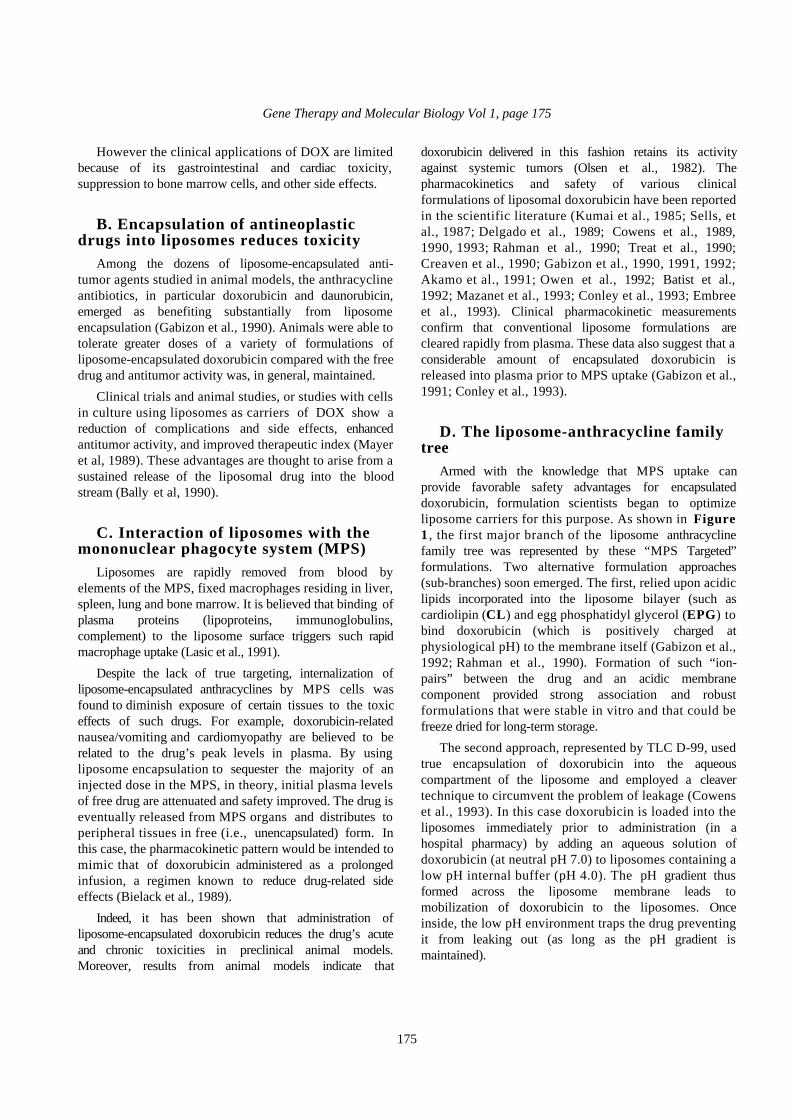
However the clinical applications of DOX are limitedbecause of its gastrointestinal and cardiac toxicity,suppression to bone marrow cells, and other side effects.
B. Encapsulation of antineoplasticdrugs into liposomes reduces toxicity
Among the dozens of liposome-encapsulated anti-tumor agents studied in animal models, the anthracyclineantibiotics, in particular doxorubicin and daunorubicin,emerged as benefiting substantially from liposomeencapsulation (Gabizon et al., 1990). Animals were able totolerate greater doses of a variety of formulations ofliposome-encapsulated doxorubicin compared with the freedrug and antitumor activity was, in general, maintained.
Clinical trials and animal studies, or studies with cellsin culture using liposomes as carriers of DOX show areduction of complications and side effects, enhancedantitumor activity, and improved therapeutic index (Mayeret al, 1989). These advantages are thought to arise from asustained release of the liposomal drug into the bloodstream (Bally et al, 1990).
C. Interaction of liposomes with themononuclear phagocyte system (MPS)
Liposomes are rapidly removed from blood byelements of the MPS, fixed macrophages residing in liver,spleen, lung and bone marrow. It is believed that binding ofplasma proteins (lipoproteins, immunoglobulins,complement) to the liposome surface triggers such rapidmacrophage uptake (Lasic et al., 1991).
Despite the lack of true targeting, internalization ofliposome-encapsulated anthracyclines by MPS cells wasfound to diminish exposure of certain tissues to the toxiceffects of such drugs. For example, doxorubicin-relatednausea/vomiting and cardiomyopathy are believed to berelated to the drug’s peak levels in plasma. By usingliposome encapsulation to sequester the majority of aninjected dose in the MPS, in theory, initial plasma levelsof free drug are attenuated and safety improved. The drug iseventually released from MPS organs and distributes toperipheral tissues in free (i.e., unencapsulated) form. Inthis case, the pharmacokinetic pattern would be intended tomimic that of doxorubicin administered as a prolongedinfusion, a regimen known to reduce drug-related sideeffects (Bielack et al., 1989).
Indeed, it has been shown that administration ofliposome-encapsulated doxorubicin reduces the drug’s acuteand chronic toxicities in preclinical animal models.Moreover, results from animal models indicate that
doxorubicin delivered in this fashion retains its activityagainst systemic tumors (Olsen et al., 1982). Thepharmacokinetics and safety of various clinicalformulations of liposomal doxorubicin have been reportedin the scientific literature (Kumai et al., 1985; Sells, etal., 1987; Delgado et al., 1989; Cowens et al., 1989,1990, 1993; Rahman et al., 1990; Treat et al., 1990;Creaven et al., 1990; Gabizon et al., 1990, 1991, 1992;Akamo et al., 1991; Owen et al., 1992; Batist et al.,1992; Mazanet et al., 1993; Conley et al., 1993; Embreeet al., 1993). Clinical pharmacokinetic measurementsconfirm that conventional liposome formulations arecleared rapidly from plasma. These data also suggest that aconsiderable amount of encapsulated doxorubicin isreleased into plasma prior to MPS uptake (Gabizon et al.,1991; Conley et al., 1993).
D. The liposome-anthracycline familytree
Armed with the knowledge that MPS uptake canprovide favorable safety advantages for encapsulateddoxorubicin, formulation scientists began to optimizeliposome carriers for this purpose. As shown in Figure1 , the first major branch of the liposome anthracyclinefamily tree was represented by these “MPS Targeted”formulations. Two alternative formulation approaches(sub-branches) soon emerged. The first, relied upon acidiclipids incorporated into the liposome bilayer (such ascardiolipin (CL) and egg phosphatidyl glycerol (EPG) tobind doxorubicin (which is positively charged atphysiological pH) to the membrane itself (Gabizon et al.,1992; Rahman et al., 1990). Formation of such “ion-pairs” between the drug and an acidic membranecomponent provided strong association and robustformulations that were stable in vitro and that could befreeze dried for long-term storage.
The second approach, represented by TLC D-99, usedtrue encapsulation of doxorubicin into the aqueouscompartment of the liposome and employed a cleavertechnique to circumvent the problem of leakage (Cowenset al., 1993). In this case doxorubicin is loaded into theliposomes immediately prior to administration (in ahospital pharmacy) by adding an aqueous solution ofdoxorubicin (at neutral pH 7.0) to liposomes containing alow pH internal buffer (pH 4.0). The pH gradient thusformed across the liposome membrane leads tomobilization of doxorubicin to the liposomes. Onceinside, the low pH environment traps the drug preventingit from leaking out (as long as the pH gradient ismaintained).

Martin and Boulikas: The challenge of liposomes in gene therapy
176
Figure 1: Family Tree illustrating the relationship between formulation strategy and the development of liposomalanthracycline products.
The ion pair formulations have been tested clinicallybut have not progressed beyond phase 1-2 studies. TLC-D99 is in advanced phase 3 trails in metastatic breastcancer.
Recognizing that MPS uptake represented the mainobstacle to targeting, another branch of liposomesdeveloped were liposomes that resist binding/interactionwith plasma proteins (opsonization) with a view towardprolonging liposome blood residence times and targetingpotential. Early work suggested that a modest degree of“MPS avoiding” activity could be obtained byformulations composed of high phase transition lipids andcholesterol. Size was also a critical parameter, the smallerthe liposome the longer it circulated: 300-nm in diameterliposomes are cleared from the blood approximately threetimes faster than small 100-nm liposomes (Huang et al,1992).
This “pure lipid” subbranch arrived at two formulationsof small diameter (∼50nm), one composed ofDSPC/cholesterol (Presant et al, 1990) and the other ofsphingomyelin/cholesterol (Webb et al, 1995) both ofwhich showed relatively slow MPS clearance.DaunoXome, a DSPC/cholesterol formulation ofdaunorubicin, is the only product to emerge from this purelipid approach. DaunoXome is approved in the US and
Europe for the treatment of AIDS-related Kaposi’s sarcoma(see below).
III. “Stealth” liposomesA. Polyethylene glycol (PEG)-coated
liposomes circulate for long periods inbody fluids
Coating the surface of liposomes with inert materialsdesigned to camouflage the liposome from the body’s hostdefense systems was shown to increase remarkably theplasma longevity of liposomes. The biological paradigmfor this “surface modified” subbranch was the erythrocyte,a cell which is coated with a dense layer of carbohydrategroups, and which manages to evade immune systemdetection and to circulate for several months (before beingremoved by the same type of cell responsible for removingliposomes).
The first breakthrough came in 1987 when a glycolipid(the brain tissue-derived ganglioside GM1) was identifiedwhich, when incorporated within the lipid matrix, allowedliposomes to circulate for many hours in the blood stream(Allen and Chonn, 1987). A second glycolipid,phosphatidylinositol, was also found to impart longplasma residence times to liposomes and, since it wasextracted from soy beans, not brain tissue, was believed to

Gene Therapy and Molecular Biology Vol 1, page 177
177
be a more pharmaceutically acceptable excipient (Gabizonet al, 1989).
A major advance in the surface-modified subbranch wasthe development of polymer-coated liposomes(Allen et al,1991). Polyethylene glycol (PEG) modification had beenused for many years to prolong the half-lives of biologicalproteins (such as enzymes and growth factors) and toreduce their immunogenicity (e.g. Beauchamp et al, 1983).It was reported in the early 1990s that PEG-coatedliposomes circulated for remarkably long times afterintravenous administration. Half-lives in the order of 24 hwere seen in mice and rats and over 30 hours in dogs. Theterm “stealth” was applied to these liposomes because oftheir ability of evade interception by the immune system(in much the same way as the stealth bomber was able toevade radar) (Gabizon and Papahadjopoulos, 1988;Klibanov et al, 1990; Papahadjopoulos et al, 1991; Senioret al, 1991; Huang et al, 1994). The increasedhydrophilicity of the liposomes after their coating with theamphipathic PEG5000 leads to a reduction in nonspecificuptake by the reticuloendothelial system.
Whereas the half-life of antimyosin immunoliposomeswas 40 min, their coating with PEG increased their half-life to 1000 min after intravenous injection to rabbits(Torchilin et al, 1992).
B. Mechanism of loading ofdoxorubicin into “stealth” liposomes
DOXIL, the PEG-coated liposomes packed withdoxorubicin, (also called CAELYX in Europe) is the firstproduct to emerge from the surface-modified liposome
subbranch. It, too, is approved in the US and Europe fortreatment of Kaposi’s sarcoma.
The mechanism of doxorubicin loading into liposomesis explained in Figure 2 . Liposomes composed ofHSPC:CHOL (1:1) and 5% PEG-DSPE of a diameter of85 nm are prepared in high ammonium sulfate; these arethen brought into a solution of high concentration ofDoxorubicin in ammonium chloride at a higher pH.Loading is mediated via exchange of ammonia moleculeswith uncharged doxorubicin; ammonia molecules passfrom the inside of the liposomes to the outside, whereasdoxorubicin enters liposomes. Loading is driven by thegradient of a chemical potential across the membrane ofthe liposome, the efflux of ammonia to a larger externalvolume, the precipitation of doxorubicin inside theliposome, and the pH gradient.
The reactions in the outside volume betweenliposomes involve removal of a proton from thedoxorubicin-ammonium chloride complex and its bindingto ammonia converting it into ammonium ions; theneutral doxorubicin-ammonia complex crosses theliposome bilayer. The reactions inside the liposomeinvolve protonation of the doxorubicin-ammonia complexfrom a hydrogen ion removed from the ammonium ion andits precipitation as doxorubicin sulfate (Lasic, 1995).
High resolution cryo-electron microscopy has shown aprecipitate of Doxorubicin molecules with sulfate ionsinside liposomes into fibrilar colloidal complexes whichalign into bundles (Figure 3 ). The structure has beenconfirmed by small angle X-ray scattering where theperiodicity of 2.7 nm observed was thought to representthe thickness of the gel fibers (Lasic et al, 1992).
Figure 2 . The mechanism of loading of DOX into liposomes.

Martin and Boulikas: The challenge of liposomes in gene therapy
178
Figure 3 . The coffee bean appearance of the precipitated DOX sulfate within liposomes. Courtesy of Dan Lasic.
Figure 4 . Cut-away view of a DOXIL particle.
Figure 4 shows schematically a cut-away view of aDOXIL particle.
DOX encapsulated into PEG-coated liposomes iscleared 450 times slower than free DOX (Gabizon et al,1994). For example, uptake of DOX encapsulated intoliposomes coated with PEG-folate was found to be uptakenby KB cells in culture, which express high levels of the
folate receptor, at a rate 45-fold higher than liposomalDOX although at a rate only 1.6 times higher than that offree DOX (Lee and Low, 1995). The enhanced release ofDOX from folate-PEG-liposomes internalized into theacidic endosomal organelles known as caveolae seemed tobe responsible for the increased cytotoxicity of DOX totumor cells (Lee and Low, 1995).

Gene Therapy and Molecular Biology Vol 1, page 179
179
C. Preclinical antitumor activity ofDOXIL
The efficacy of DOXIL has been evaluated in a varietyof different tumor models, including several humanxenograft models (Papahadjopoulos et al, 1991; Huang etal, 1992; Vaage et al, 1992, 1993, 1994; Williams et al,1993; Siegal et al, 1995; Amantea et al, 1997). In everymodel examined DOXIL was more effective than the samedoses of doxorubicin (i ) at inhibiting or halting tumor
growth, (i i ) at effecting cures and/or (i i i ) at prolongingsurvival times of tumor-bearing animals. Most often, allthree endpoints were improved by DOXIL, and in no casewas DOXIL less effective than doxorubicin. DOXIL wasmore active in both solid and dispersed tumors, and wasmore effective than doxorubicin in preventing spontaneousmetastases from intramammary implants of two differentmammary tumors in mice. These findings are alsosupported by studies done with DOXIL in several murinetumor models and human xenograft models (Figure 5).
Figure 5 : Growth kinetics of human lung cancer xenografts (A) and human prostate cancer (PC-1) xenografts (B ) implantedin SCID mice. Groups of 10 animals were engrafted in the flank with 2 x 10⁶ TL-1 cells at day 0 and treated weekly (via tail veininjection) for 10 weeks starting one week post engraftment with saline (circles), doxorubicin (also called Adriamycin) (3 mg/kg,diamonds) or DOXIL (3 mg/kg, squares). Adapted from Siegal et al, 1995.

Gene Therapy and Molecular Biology Vol 1, page 180
180
Figure 6 . Electronmicrographs showing colloidalgold (arrows) in the intracellularvesicles of a typical mononuclearphagocyte. The particles are oftenseen within the endosomes (lowerinsert) and secondary withinlysosomes (upper insert) ofmacrophages at the border of theliver-implanted tumor.
In general, the efficacy of doxorubicin in these modelswas limited by its toxicity at high doses. Typically,DOXIL could be used at a higher dose, offering anincreased therapeutic advantage. Pharmacokinetic and tissuedistribution studies suggest that the greater persistence,particularly in tumor tissue, achieved with DOXILcompared to conventional doxorubicin also contributes atherapeutic advantage. The efficacy of DOXIL compared tothat of conventional liposomal (non-Stealth) doxorubicinindicated that DOXIL was significantly more effective thanconventional liposomal doxorubicin, demonstrating theimpact of the long-circulating Stealth liposome. Based onthe results of these nonclinical studies, DOXIL appears tobe an effective agent for the treatment of both solid anddispersed tumors.
Tissue distribution of sterically-stabilized liposomeswas studied by Huang et al, (1992). Following tail veininjection the microscopic localization of liposomes labeledwith encapsulated colloidal gold was found predominantlyin Kupffer cells (macrophages) of the liver (not inhepatocytes) (Figure 6) and within macrophages of thebone marrow. Electron microscopy showed the presence ofgold in endosomes and lysosomes of fixed sinusoidallining macrophages in the liver.
D. Combinations of DOXIL with otheranticancer drugs are effective anticancerregiments in preclinical studies
Humanized monoclonal antibodies directed againstreceptors overexpressed on malignant cells such as HER2or EGFR have been used in the treatment of malignanciesbut are not active on their own (Chrysogelos et al, 1994;Baselga et al, 1996). Figure 7 shows that thecombination of such anticancer regiments with DOXILresults in a synergistic antitumor activity: a combinationof the DOXIL with the C225 EGFR antibody had aspectacular effect in inhibiting growth of human breastcancer cells in nude mice.
IV. Clinical trials using DOXILA. Pharmacokinetics of DOXIL in
human patientsPopulation pharmacokinetic analysis has been
conducted on a group of 83 patients receiving DOXIL atdoses ranging from 10 to 60 mg/m2 (17 females, 66males) (Gabizon et al, 1994; Northfelt et al, 1996). Atdoses ranging from 10 to 40 mg/m2, DOXILpharmacokinetics were linear. At dosages above 40 mg/m2,

Gene Therapy and Molecular Biology Vol 1, page 181
181
DOXIL displayed nonlinear pharmacokinetics as evidencedby a disproportionate increase in the area-under-the-plasmaconcentration versus time curve (AUC) with increasingdose amounts. In general, drugs that display nonlinearpharmacokinetics have a potential to accumulate to toxiclevels in the plasma if not monitored regularly (e.g.,phenytoin). In the case of DOXIL, this is not a concernsince the drug is administered a minimum of every threeweeks, after which time no drug is detectable in the plasmaof patients. Table 1 lists the statistics of selectedpharmacokinetic parameters for all 83 patients. There wasno evidence of accumulation at dose intervals of ≥ 3weeks.
Utilizing the fitted pharmacokinetic parameter resultsfrom this analysis, simulated plasma concentration versus
time profiles of DOXIL were generated at doses of 10 – 60mg/m2 (Figure 8 ). The nonlinearity of DOXILpharmacokinetics at higher doses is most evident at dosesgreater than 40 mg/m2.
No correlations were observed betweenpharmacokinetic parameters and age, weight, body surfacearea, tumor type, sex, and renal (as determined by serumcreatinine) and hepatic function (as determined by totalbilirubin levels).
DOXIL has also been used as single-agent therapy foradvanced breast cancer among elderly patients in a phaseclinical trial (Ranson et al, 1997) and as primary therapyfor refractory ovarian cancer also in a phase II study
Figure 7 . Growth kinetics ofhuman xenograft of A431 breast tumorimplanted subcutaneously in nudemice. Groups of animals were treatedvia tail vein injection as indicated inthe figure. C225 is a monoclonalantibody against epidermal growthfactor receptor EGFR)
Table 1: DOXIL pharmacokinetic parameter estimates n = 83
Sta t i s t i c Vss (L/m 2) CL i (L/h/m 2) Km (mg/L) AUC50 ( m g / L ˙h)
Mean 3.40 0.108 2.01 3260
CV% 18.2 54.2 66.3 54.8
Median 3.42 0.0950 1.85 3018
Minimum 2.20 0.0269 0.428 535
Maximum 5.67 0.393 8.84 9520
Vss – volume of distribution at steady-state, CLi – intrinsic clearance, Km – Michaelis Menton Constant, AUC50 – area-under-the-curve normalized for a 50 mg/m2 dose of DOXIL

Gene Therapy and Molecular Biology Vol 1, page 182
182
(Muggia et al, 1997). While generally manageable in bothtype of trials, epithelial cell toxicity manifesting itself aspalmar-plantar erythrodysesthesia (PPE, hand-footsyndrome) may limit the amount of DOXIL patients areable to tolerate.
B. Amount of non-liposomalDoxorubicin in plasma
Several lines of evidence support the conclusion thatthe majority of the doxorubicin (between 93% and 99%)in plasma is encapsulated within the liposome after i.v.administration of DOXIL. The most convincing come
from work by Gabizon et al who conducted a pilotpharmacokinetic of DOXIL (Druckmann et al, 1989). Inthis study the fraction of the liposome-encapsulated andfree, non-liposomal drug in circulation after DOXILadministration was quantitated directly using a Dowexcolumn separation method that is able to accurately andreproducibly quantitate _ 7% free drug in the plasma(Speth et al, 1988). Using this method, essentially all thedoxorubicin measured in plasma was liposome-associated(Figure 9 ). These findings suggest that at least90 to 95% of the doxorubicin measured in plasma, andpossibly more, is liposome-encapsulated.
Figure 8: Simulated plasma clearance kinetics of doxorubicin after a single 30 minute infusion of DOXIL at doses rangingfrom 10 to 60 mg/m2.
Figure 9: Clearance over a one week period of total vs. encapsulated doxorubicin after a single 50 mg/m2 dose of DOXIL incancer patients. Data points represent mean values ± standard deviation for 14 patients in the DOXIL group and 4 patients in thedoxorubicin group (adapted from Druckmann et al, 1989). The method described in Speth et al, 1988 was used to separate theencapsulated from released drug fractions.

Gene Therapy and Molecular Biology Vol 1, page 183
183
The amount of doxorubicin that remains liposome-associated while circulating in plasma is an importantpoint that deserves further emphasis from a safetyperspective. Acute adverse reactions associated withdoxorubicin administration including nausea and vomitingand chronic cardiotoxicity are believed to be directly relatedto peak concentrations of the drug in plasma. As pointedout above, while in the circulation, DOXIL liposomesremain intact, retaining virtually all of the doxorubicin inencapsulated form (Speth et al, 1987a). Although totalplasma levels of doxorubicin may be relatively high forseveral days after DOXIL administration, the majority ofthe dose is sequestered within the liposome during thisperiod and thus is not bioavailable to distribute (as freedrug molecules) to tissues, including the GI tract andmyocardium. With respect to level of available drug inplasma, DOXIL resembles more that of a 96 hourcontinuous infusion of doxorubicin than the usual 30minute infusion. Prolonged infusion of doxorubicin isknown to reduce cardiotoxicity and GI irritation.
C. Comparison of pharmacokineticparameters: DOXIL vs. Doxorubicin
According to literature reports, an i.v. bolus injectionof doxorubicin in humans produces high plasma
concentrations of doxorubicin that decline quickly due torapid and extensive distribution into tissues (Greene et al,1982). Apparent volumes of distribution range from 1400to 3000 L, reflective of the drug’s extensive tissuedistribution. The doxorubicin plasma concentration-timecurve in humans is biphasic, with a distribution half-lifeof 5 to 10 minutes and terminal phase elimination half-lifeof 30 hours (Benjamin et al, 1984; Speth et al, 1987a, b).A triphasic curve has also been described with a terminalplasma half-life of approximately 30 hours (Benjamin etal, 1977).
Clearance of doxorubicin after doxorubicin
administration ranges from 24 to 73 L/hour (Greene et al,1982). No accumulation in plasma occurs after repeatedinjections (Benjamin et al, 1984; Speth et al, 1987a, b).
The pharmacokinetics of DOXIL are significantlydifferent from those reported for doxorubicin.Administration of DOXIL results in a significantly higherdoxorubicin area-under-the-plasma concentration vs timecurve (AUC), lower rate of clearance (approximately 0.1L/hour) and smaller volume of distribution (5 to 7 L)relative to administration of doxorubicin (Figure 10 ).The first phase of the biexponential plasma concentration-time curve after DOXIL administration is relatively short(approximately 5 hours), and the second phase, whichrepresents the majority of the AUC, is prolonged (half-life50 to 55 hours).

Martin and Boulikas: The challenge of liposomes in gene therapy
184
Doxorubicin Cmax after DOXIL administration is 15- to40-fold higher than after the same dose of doxorubicin, andthe ratio quickly increases as doxorubicin is rapidly clearedfrom circulation. Importantly, the vast majority of thetotal plasma doxorubicin remains liposome-encapsulatedafter DOXIL treatment. Because of the high percentage ofliposome encapsulation in DOXIL, the amount of free(i.e., “bioavailable”) drug in the plasma appears to besignificantly lower than that measured after administrationof an equal dose of doxorubicin.
This conclusion is supported by the same type ofcalculations presented above, which derive the apparentconcentration of free doxorubicin based on the reportedrelationship between doxorubicinol and doxorubicinconcentrations in plasma. For example, five minutes after
the end of the infusion, the mean doxorubicinol levelfollowing a 20 mg/m2 dose of DOXIL was approximately22 ng/mL. Using the doxorubicinol:doxorubicinconcentration ratio reported in the literature, as describedabove, predicted free doxorubicin concentration at this timepoint would be 54 ng/mL in DOXIL-treated patients (thetotal plasma concentration measured at this time point was8863 ng/mL). Comparatively, patients in the Northfelt etal study (1996) who received a dose of doxorubicin 20mg/m2, had initial plasma concentrations of doxorubicin ofapproximately 500 ng/mL.
Studies on human cancer xenografts in nude micedemonstrate an increased accumulation of Doxorubicin atthe lesion after DOXIL treatment compared toadministration of free Adriamycin (Figures 11, 12).
Figure 11 . Increased accumulation ofDoxorubicin in prostate cancer xenografts(A) and pancreatic carcinoma xenografts(B ) after DOXIL treatment compared toadministration of free Adriamycin.
Gene Therapy and Molecular Biology Vol 1, page 185
185
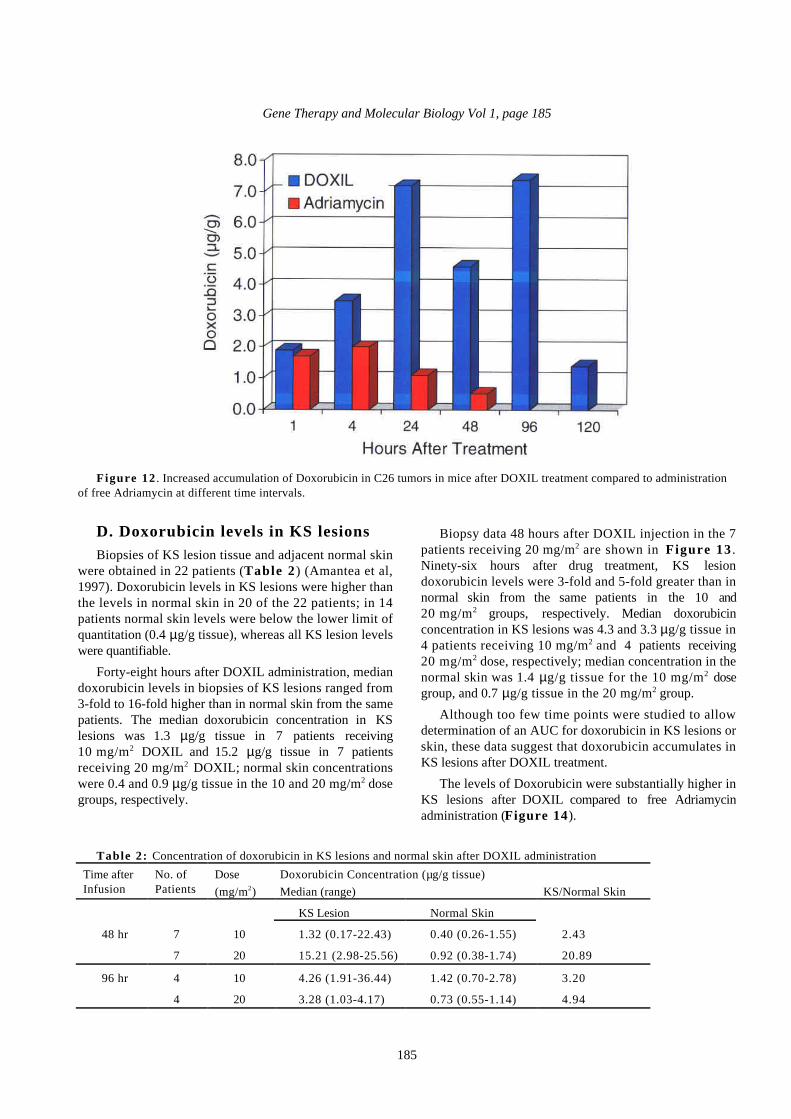
Figure 12 . Increased accumulation of Doxorubicin in C26 tumors in mice after DOXIL treatment compared to administrationof free Adriamycin at different time intervals.
D. Doxorubicin levels in KS lesionsBiopsies of KS lesion tissue and adjacent normal skin
were obtained in 22 patients (Table 2 ) (Amantea et al,1997). Doxorubicin levels in KS lesions were higher thanthe levels in normal skin in 20 of the 22 patients; in 14patients normal skin levels were below the lower limit ofquantitation (0.4 µg/g tissue), whereas all KS lesion levelswere quantifiable.
Forty-eight hours after DOXIL administration, mediandoxorubicin levels in biopsies of KS lesions ranged from3-fold to 16-fold higher than in normal skin from the samepatients. The median doxorubicin concentration in KSlesions was 1.3 µg/g tissue in 7 patients receiving10 mg/m2 DOXIL and 15.2 µg/g tissue in 7 patientsreceiving 20 mg/m2 DOXIL; normal skin concentrationswere 0.4 and 0.9 µg/g tissue in the 10 and 20 mg/m2 dosegroups, respectively.
Biopsy data 48 hours after DOXIL injection in the 7patients receiving 20 mg/m2 are shown in Figure 13 .Ninety-six hours after drug treatment, KS lesiondoxorubicin levels were 3-fold and 5-fold greater than innormal skin from the same patients in the 10 and20 mg/m2 groups, respectively. Median doxorubicinconcentration in KS lesions was 4.3 and 3.3 µg/g tissue in4 patients receiving 10 mg/m2 and 4 patients receiving20 mg/m2 dose, respectively; median concentration in thenormal skin was 1.4 µg/g tissue for the 10 mg/m2
dosegroup, and 0.7 µg/g tissue in the 20 mg/m2 group.
Although too few time points were studied to allowdetermination of an AUC for doxorubicin in KS lesions orskin, these data suggest that doxorubicin accumulates inKS lesions after DOXIL treatment.
The levels of Doxorubicin were substantially higher inKS lesions after DOXIL compared to free Adriamycinadministration (Figure 14).
Table 2: Concentration of doxorubicin in KS lesions and normal skin after DOXIL administration
Time afterInfusion
No. ofPatients
Dose
(mg/m2)
Doxorubicin Concentration (µg/g tissue)
Median (range) KS/Normal Skin
KS Lesion Normal Skin
48 hr 7 10 1.32 (0.17-22.43) 0.40 (0.26-1.55) 2.43
7 20 15.21 (2.98-25.56) 0.92 (0.38-1.74) 20.89
96 hr 4 10 4.26 (1.91-36.44) 1.42 (0.70-2.78) 3.20
4 20 3.28 (1.03-4.17) 0.73 (0.55-1.14) 4.94

Gene Therapy and Molecular Biology Vol 1, page 186
186
Figure 13: Doxorubicin concentration in KS lesion tissue and adjacent normal skin tissue. Seven KS patients were given a20 mg/m2 dose of DOXIL and, 96 hours later, biopsies were taken of a representative cutaneous KS lesion and normal skin near thelesion. The tissue was homogenized, extracted with solvents and total doxorubicin measured by HPLC.
Figure 14 . DOXIL compared to Adriamycin sustains a higher concentration of Doxorubicin in Kaposi’s sarcoma lesions inhuman patients. From Northfelt et al (1996) J Clin Pharmacol 36, 55-63.
E. Combinations of DOXIL with otheranticancer drugs in clinical trials
The dose-limiting toxicity of Navelbine (vinorelbinetartrate) is granulocytopenia. In combination withdoxorubicin, Navelbine produced a 57% overall objectiveresponse rate as first-line therapy of advanced breast cancer,
however, the incidence of grade 4 granulocytopenia was83%, with 8% requiring hospitalization due to febrileneutropenia and one septic death (Hochster, 1995).Substitution of doxorubicin with DOXIL in thiscombination is being explored as a means of maintainingthe favorable tumor response of the combination whilereducing the incidence of hematological toxicity.

Gene Therapy and Molecular Biology Vol 1, page 187
187
Navelbine would not be expected to contribute to the skintoxicity seen with DOXIL.
The excitement generated by Gianni et al (1995) whoreported a greater than 90% objective response rate inmetastatic breast for a taxol/doxorubicin combination istempered by the rather unfavorable side effects profile ofthis regimen. Severe, febrile neutropenia was commonand peripheral neuropathy occurred in one third of thepatients. Perhaps more troubling was the development ofreversible congestive heart failure (CHF) in 18% ofwomen after a median of 480 mg/m2 doxorubicin, resultswhich raise the specter of taxol-related enhancement ofdoxorubicin cardiotoxicity.
These encouraging preclinical findings are supported byresults of a pilot clinical study done by Berry et al (1996).These authors have reported the results of endomyocardialbiopsies performed on a series of AIDS-KS patients whoreceived cumulative doses of DOXIL ranging from 469 to860 mg/m2. These findings support the rationale forcombining taxol (paclitaxel) with DOXIL. Both drugshave demonstrated activity in breast and ovarian cancer.With respect to toxicities, the incidence of severeneutropenia and peripheral neuropathy are lower forDOXIL than taxol. Preclinical and early clinical biopsyresults strongly suggest that DOXIL produces less damageto the myocardium relative to comparable cumulativedoses of doxorubicin. Thus the cardio-protective effect ofDOXIL may translate into a reduced risk of cardiotoxicityrelative to the highly active taxol-doxorubicincombination. Moreover, taxol causes relatively little skintoxicity. Based on these considerations, several phase 1dose-finding trials of DOXIL and taxol have been launched.
V. Extravasation of “stealth”liposomes into tumors
A. Mechanism of enhanced DOXILaccumulation in tumors
An understanding of the mechanisms by whichliposome-encapsulated doxorubicin accumulates withinsolid tumors after DOXIL administration, and how thisdeposition pattern and subsequent slow release of drugimprove the antitumor activity of DOXIL relative totreatment with the free drug, is now emerging (refer toFigure 15).
B. Plasma stability and long plasmaresidence times are critical requirements
DOXIL liposomes are intend to carry their payload ofdoxorubicin directly to tumors. So, any premature releaseof the drug, while the liposomes are still in route (i.e., inthe circulation), would detract from the total amount ofencapsulated doxorubicin able to reach the desired target.This requirement highlights the importance of engineeringplasma stability into DOXIL liposomes. As mentionedearlier, conventional liposome formulations of doxorubicinhave been shown to release a significant proportion oftheir payload into the bloodstream soon after injection(Gabizon et al, 1991; Conley et al, 1993). Drug releaseappears to follow protein adsorption/intercalation into theliposome which disrupts the barrier properties of themembrane. Moreover, the liposomes, together with anyremaining drug, are removed by cells of the MPS withinseveral minutes to a few hours after injection. As aconsequence of this rapid clearance, doxorubicin deliveredin conventional liposomes has little opportunity to reachtumors in encapsulated form.
By virtue of the PEG groups grafted to their surface,DOXIL liposomes are stable in plasma and release verylittle drug while in the circulation (see discussion above).Moreover, the PEG coating provides slow clearance; after asingle injection, DOXIL can be detected in the circulationfor 2-3 weeks. Slow clearance kinetics provide anopportunity for these liposomes to reach sites of diseasesuch a tumors. Measurements made in tumor-bearinganimals and cancer patients indicate that uptake ofpegylated liposomes by tumors is also slow process. Inpreclinical tumor models, the peak uptake of DOXIL isreached 24-48 hours after injection (Vaage et al, 1993;Working et al, 1994).
In cancer patients given 111Indium encapsulated inpegylated liposomes of the same composition and size asDOXIL, peak uptake in tumors is seen 48-72 hours afterinjection (Figure 16 , Stewart Simon, personalcommunication, May 20, 1997). Slow uptake in tumorshighlights the importance of long circulation times; ifliposomes are to have an opportunity to reach and entertumors in significant numbers, they must circulate forperiods of days after injection.
Figures 17 and 18 also show localization of 111In-labeled Stealth liposomes into a T4 squamous cellcarcinoma of the tongue and a squamous cell carcinoma ofthe lung, respectively.
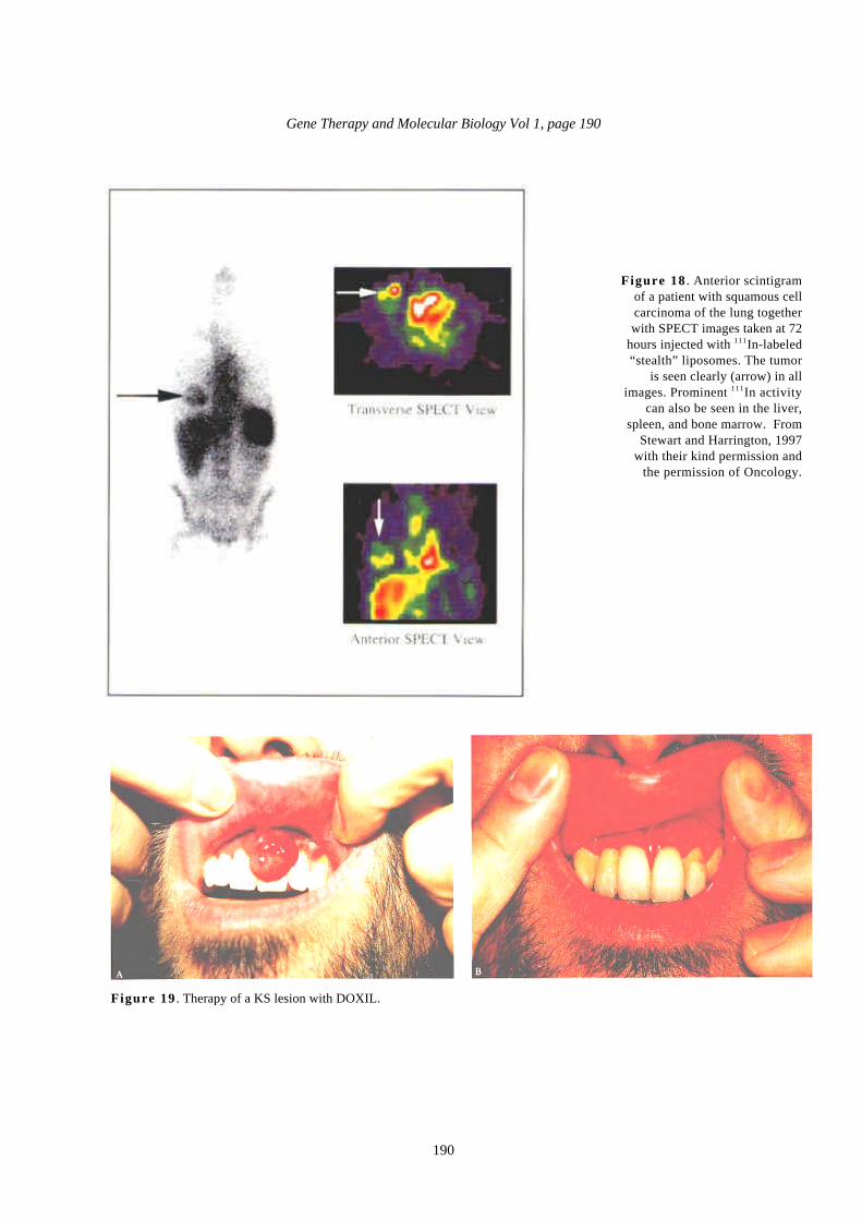
Figure 19 shows complete eradication of a KS lesionafter six cycles of treatment with DOXIL.

Martin and Boulikas: The challenge of liposomes in gene therapy
188
Figure 15: Proposed mechanismfor DOXIL accumulation in tumors. ¬Liposomes containing doxorubicincirculate for 2-3 weeks after injection.During this period virtually all of thedrug remains encapsulated. Theliposomes pass many times throughthe blood vessels feeding growingtumors. Intact liposomes extravasatethrough defects/gaps present in newlysprouting vessels and enter the tissue
compartment; lodging in the tumorinterstitium near the vessel. ® Drugmolecules are released from theextravasated liposomes. Liposomeleakage is believed to be theconsequence of conditions present inthe interstitial fluid surrounding tumorswhich lead to physical/chemicalbreakdown of the liposome membrane(low pH, oxidizing agents, enzymes,uptake by macrophages). ¯ Free drug
molecules penetrate deeply into thetumor and enter tumor cells. °Doxorubicin molecules bind to nucleicacids and kill tumor cells. Note thatsuch a mechanism does not require aclose physical encounter between aliposome and target cell, since free drugmolecules are able to diffuse throughbarriers that may intercept liposomes.

Gene Therapy and Molecular Biology Vol 1, page 189
189
Figure 16: Gamma scintigraphic image of a lung cancer patient 48 and 96 hours after administration of DOXIL liposomescontaining 111Indium. Note that both images are posterior views. Uptake of the radioactive liposomes is seen in certain normaltissues including spleen, liver, bone marrow. The activity visible in the central chest (substernal) and upper abdomen representliposomes that are still circulating in the heart and major vessels at these time points. The liposomes are taken up by a large tumorin the left upper lung. The density of radioactivity is as high or higher in the tumor than in any normal organ.
Figure 17 . Plain anterior view scintigrams of a patient with T4 squamous cell carcinoma of the tongue injected with 111In-labeled “stealth” liposomes. The image at 4 h postinjection shows the blood pool, early uptake by the liver reticuloendothelialsystem, and EDTA-chelated 111In in the bladder. The tumor is seen clearly (white arrow) 72 hours after injection and is still visibleat 10 days. From Stewart and Harrington, 1997 with their kind permission and the permission of Oncology.
Gene Therapy and Molecular Biology Vol 1, page 190
190
Figure 18 . Anterior scintigramof a patient with squamous cellcarcinoma of the lung togetherwith SPECT images taken at 72
hours injected with 111In-labeled“stealth” liposomes. The tumor
is seen clearly (arrow) in allimages. Prominent 111In activity
can also be seen in the liver,spleen, and bone marrow. From
Stewart and Harrington, 1997with their kind permission and
the permission of Oncology.
Figure 19 . Therapy of a KS lesion with DOXIL.

Gene Therapy and Molecular Biology Vol 1, page 191
191
Figure 20 . Histological preparation of KS-like lesion nodule. Early lesion and adjacent normal skin in transgenic mice byliposome-encapsulated colloidal gold. A-C: Sections of KS-like lesion nodule were from a 16-month old F2 mouse that had alocalized 5-mm spherical erythematous lesion on its back. The sections reveal that the gold particles are localized predominantlyin the lesion region. Arrows in C show labeling of spindle cells. D: Section of an early lesion invisible to the naked eye in a 8-month-old C4 mouse showing that the gold marker is scattered extravasated erythrocytes in the collagenous dermis. E: Normalskin adjacent to the tumor shown in A. From Huang et al, 1993.
Martin and Boulikas: The challenge of liposomes in gene therapy
192
C. Liposomes extravasate throughgaps in the endothelium of tumor vessels
Stealth liposomes of the same size and lipidcomposition as DOXIL, but containing entrapped colloidalgold designed to serve as a marker to follow liposomedistribution by microscopic techniques, have been shownto enter solid colon tumors implanted in mice (Huang etal, 1992) and KS-like lesions in HIV-transgenic mice(Huang et al, 1993) (Figure 20) . In these mouse models,movement of liposomes from the vascular lumen into thetumor interstitium was visualized by light and electronmicroscopy. Transcytosis of liposomes from the lumen ofblood vessels, through endothelial cells, and into theextravascular compartment of KS lesions was seen, as wasintracellular uptake of liposomes by some spindle cellswithin lesions. However, these processes appear to berestricted to a minority of the particles entering the tumor(Huang et al, 1993). The vast majority of the liposomeswere seen to enter through gaps in the endothelial cellwall.
This finding is consistent with results reported byYuan, et al who used pegylated liposomes ranging in sizefrom 100-600 nm to probe the cut off size of the gapspresent in a human adenocarcinoma xenograft implanted innude mice (Yuan et al, 1995). This tumor was permeableto liposomes up to 400 nm in diameter, suggesting the cutoff size in this tumor is between 400-600 nm. Given theirsmall size (85 nm) and long circulation times, DOXILliposomes would be expected to extravasate in tumors thatexhibit gaps of such dimensions. Gaps/defects are knownto be present in solid tumors (Seymour, 1992; Jain, 1989)and KS lesions (Francis et al, 1986; Vogel et al, 1988).Indeed, fluorescent pegylated liposomes of <100 nm indiameter have been visualized by video microscopyextravasating in real time into the interstitium ofimplanted tumors using window chamber models (Yuan etal, 1994; Huang et al, 1995; Dewhirst and Needam, 1995).
D. Release of drug followingextravasation
Encapsulated doxorubicin is released from the DOXILliposomes after extravasation in tumors (Dewhirst andNeedam, 1995). Several possible factors may contribute toliposome breakdown and drug release in tumors: (i )conditions present in the interstitial fluid surroundingtumors may cause breakdown of the liposomes, such aslow pH, (Stubbs et al, 1992) and lipases released from deador dying tumor cells (Sakayama et al, 1994); (i i )inflammatory cells (which are often found in tumors(Dvorak et al, 1981) may release factors that lead toliposome destabilization such as enzymes or superoxide
and other oxidizing agents (Cobbs et al, 1995); or (i i i )phagocytic cells residing in tumors (Pupa et al, 1996)which are known to engulf liposomes (Huang et al, 1995),may digest the lipid matrix intracellularly and releasedoxorubicin (or its active metabolites) back into theinterstitial fluid (Gabizon et al, 1991). A combination ofthese possibilities may well be responsible for theobserved release of doxorubicin after extravasation ofDOXIL liposomes in tumors (Gabizon et al, 1995).
The rate of release of doxorubicin within a tumor hasyet to be measured directly. In order to do so, it would benecessary to separate encapsulated drug (i.e., drugmolecules that have not been released from intactliposomes) from free drug in a solid tissue. Although sucha separation is possible in biological fluids (such asplasma; Druckmann et al, 1989) it is technically difficultto conduct in solid tissues such as tumors; the conditionsneeded for quantitative extraction of doxorubicin lead toliposome disruption. Despite the difficulty of directlymeasuring release kinetics, indirect methods suggest thatthe release of doxorubicin from DOXIL liposomes occursover a period of days to perhaps weeks followingadministration. In a recent study using a human pancreaticxenograft model in nude mice, Vaage et al showed thattumor levels of doxorubicin peak at 24-48 hours afterDOXIL, and fall slowly over a period of a week (Vaage etal, 1997). These results suggest that the liposomesentering the tumor release their drug locally at quite a slowrate.
The improved antitumor activity of DOXIL relative toa comparable dose of free doxorubicin can be partiallyattributed to these slow in situ release kinetics. Considerthe distribution kinetics after a dose of free doxorubicin.Drug molecules enter the tumor (and other tissues)quickly, reaching maximal exposure (i.e., peakconcentrations) within minutes (Working et al, 1994).During the subsequent 24 hours, tumor doxorubicinconcentration drops precipitously to undetectable levels.During this brief “pulse” of doxorubicin, those cells notexposed to a cytotoxic concentration for a sufficientamount of time, or which are not at a sensitive point inthe cell cycle, can escape therapy and continue toproliferate. A typical course of doxorubicin is given on athree week cycle. This length of time between injections isneeded to allow for recovery from the hematologic toxicityassociated with doxorubicin therapy. Following such aschedule, it is quite likely that tumor cells are exposed tocytotoxic levels of drug for only a few hours during the 3week interval between injections. In the case of DOXILwhich is also given in a 2-4 week cycle, not only doesmore drug reach the tumor, but, by virtue of the slow insitu release kinetics provided by the liposomes, tumor

Gene Therapy and Molecular Biology Vol 1, page 193
193
cells are exposed to drug over a period of several days toperhaps a week or more after a single dose. Such a releasepattern may contribute to DOXIL’s antitumor response.
E. Tumor cell penetration andcytotoxicity
Given its amphipathic nature, a doxorubicin moleculethat is released from a liposome can quickly diffusethrough surrounding fluids and connective tissue, entertumor cells, bind to nucleic acids and inhibit DNAsynthesis. Indeed, it is quite likely that drug moleculesreleased from DOXIL can penetrate many cell layers intothe tumor, well beyond the point that the liposome itselfhas reached. Early findings suggest that penetration of“free” drug in this fashion may be essential for DOXIL’santitumor activity.
As mentioned above, microscopic observations indicatethat liposomes extravasate in tumors at particular sites;primarily through vessels forming at the advancing edge ofangiogenesis (Yuan et al, 1994). The deposition ofextravasated liposomes in these areas is perivascular andfocal, occurring primarily at the roots of capillary sproutswhere weak spots (possibly defects or gaps) in theendothelium are believed to occur. Given the geometry ofthe system, liposomes that enter through such gaps maynot be able to penetrate deeply into the tumor interstitium.Liposome penetration may be limited by a range ofphysical obstacles including tight cell-cell junctions (oftenfound in highly differentiated epithelial cell tumors), denseconnective tissue stroma, small extracellular volume andhigh interstitial fluid viscosity (that may be caused byfibrin cross-linking) (Nagy et al, 1995). Ideally all tumorcells, regardless of their proximity to blood vessels or theliposome depots that may from near them, would beexposed to a cytotoxic dose of drug. So, the observationthat drug molecules released from focal, perivasculardeposits of liposomes are able to penetrate deeply into thetumor mass may be a critical requirement for expression ofDOXIL’s antitumor activity.
VI. Encapsulation of other drugs intostealth liposomes
A. cis-Platinum (SPI-77)Cisplatin (Platinol) is active alone and or combination
chemotherapy regimens against a wide rage of epithelialmalignancies including testicular, ovarian, head and neck,lung, bladder, and cervical cancers (Loehler and Einhorn,1984). Cisplatin chemotherapy is often limited by sideeffects that prohibit continued treatment. In addition,some tumors are initially resistant or acquire cisplatinresistance with continued exposure. The major dose-limiting cisplatin-induced toxicity in humans is renal
toxicity, although significant nausea and vomiting,ototoxicity, peripheral neuropathy and myelotoxicity arealso induced by cisplatin administration. Attempts toameliorate cisplatin-induced toxicity and/or resistance havefocused on the development of platinum derivatives thatare less toxic and/or more active than the parent compound(Schilder et al, 1994; Kelland and McKeage, 1994).Alternative approaches include altering the pharmacologyof the drug by altering the treatment schedule, hydratingpatients prior to and during therapy, or administering renalprotectant therapy. Encapsulating the drug withinliposomes has shown improved therapeutic capacity(Steerenberg et al, 1987; Potkul et al, 1991).
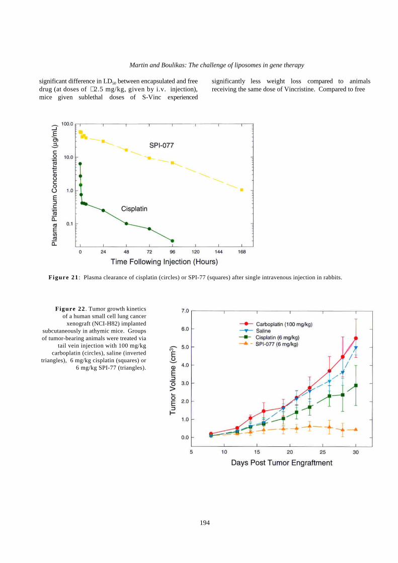
SPI-77 is a formulation of cisplatin encapsulated invirtually the same type of liposome as DOXIL. SPI-77exhibits plasma pharmacokinetics characteristic ofsterically stabilized (Stealth) liposomes, with longcirculation, high Cmax and area-under-the-plasmaconcentration vs time curve (AUC), and low clearance andvolume of distribution compared to non-liposomalcisplatin (Figure 21). In vitro leakage studies suggestthat plasma levels of platinum primarily or solelyrepresent liposomal cisplatin, i.e., drug that is inliposomes and free or bound to proteins.
The therapeutic activity of SPI-77 has been evaluatedand compared to non-liposomal cisplatin in various tumormodels, including the C26 colon carcinoma in Balb/c miceand a xenograft of the NCI-H82 small cell lung tumor inathymic mice (Figure 22). SPI-77 showed meaningfulanti-tumor activity in these tumor models. Cisplatin wasonly effective in the NCI-H82 xenograft model;carboplatin (Paraplatin) was ineffective in both. SPI-77only occasionally produced complete tumor responses, butdid cause a persistent inhibition of tumor growth duringand after treatment. In many animals, tumors grew slowlyto intermediate size and then were apparently arrested, withlittle additional growth evident. Although cisplatintreatment resulted in better inhibition of tumor growth inboth trials in the NCI-H82 xenograft models, SPI-77 wasmore effective in producing a prolonged response totreatment, with persistent inhibition of tumor growth.
B. Stealth VincristineVincristine is used clinically both as a single agent and
in combination regimens, for the treatment ofhematological malignancies, head and neck cancer,Kaposi’s sarcoma and lung cancer. Early work withconventional liposomal Vincristine showed noimprovement in safety or therapeutic activity relative tothe free drug (Layton and Trouet, 1980).
Stealth liposome-encapsulated Vincristine (S-Vinc)prolonged the drug’s distribution phase plasma half-life inrats from 0.22 to 10.5 hours. While there was no
Martin and Boulikas: The challenge of liposomes in gene therapy
194
significant difference in LD50 between encapsulated and freedrug (at doses of ≅2.5 mg/kg, given by i.v. injection),mice given sublethal doses of S-Vinc experienced
significantly less weight loss compared to animalsreceiving the same dose of Vincristine. Compared to free
Figure 21 : Plasma clearance of cisplatin (circles) or SPI-77 (squares) after single intravenous injection in rabbits.
Figure 22 . Tumor growth kineticsof a human small cell lung cancer
xenograft (NCI-H82) implantedsubcutaneously in athymic mice. Groupsof tumor-bearing animals were treated via
tail vein injection with 100 mg/kgcarboplatin (circles), saline (inverted
triangles), 6 mg/kg cisplatin (squares) or6 mg/kg SPI-77 (triangles).

Gene Therapy and Molecular Biology Vol 1, page 195
195
Figure 23 . Tumor volume inBLAB/c mice given multipletail vein injections of saline,1.3 mg/kg Vincristine(Oncovin) or 1.3 mg/kg Stealthliposomal Vincristine (S-VINC). Treatment was given ondays 10,17 and 24 afterimplantation of the murine C26colon carcinoma. (NMT = nomeasurable tumor)
drug, S-Vinc was more active against intraperitoneally andsubcutaneously implanted tumors. In a subcutaneously-implanted murine colon tumor model, multiple doses offree drug did little to retard tumor growth, but S-Vincslowed tumor growth and improved long-term survival inseveral dosing regimens (Figure 23 ; Allen et al, 1995).Stealth liposomes extravasate preferentially to tumors byleaking through new vessels during the process ofangiogenesis of the tumor (Papahadjopoulos et al, 1991;Huang et al, 1992, 1993; Gabizon et al, 1994).
Steric hindrance by coating the liposome surface withPEG can inhibit recognition of targeting ligands, such asantibodies, by cell membrane proteins on the targeted cell(Mori et al, 1991; Torchilin et al, 1992). This obstacle canbe in part overcome by conjugating a water-soluble drug atthe end of the PEG polymer. For example, 66-nm indiameter liposomes can be efficiently targeted to tumorcells that express folate receptors (KB cells) viaconjugation of the folate to a PEG spacer of 25 nm inlength; shorter PEG spacers were not efficient in mediatingbinding of the liposomes to KB cells (Lee and Low,1995). Antibodies attached to long PEG spacers can givestealth liposomes that are effective in target binding andexhibit prolonged circulation times (Papahadjopoulos et al,1991; Blume et al, 1993).
VII. Cell targeting with liposomesA. IgG-coated liposomes can target
specific cell typesInjected liposomes are localized mainly in the fixed
macrophages of the liver and spleen tissue; indeed the
reticuloendothelial system of the body rapidly removesliposomes from the blood. Gangliosides andsphingomyelin, when included into the lipids of theliposome, act synergistically to diminish the rate of uptakeof liposomes by macrophages of the host defense system;this results in extended circulation times of these largeunilamellar liposomes (Allen and Chonn, 1987).
Attempts to generate cell-targeting have focusedprimarily on the addition of monoclonal antibodies to thesurface of the liposome. Liposomes tagged on their surfacewith IgG immunoglobulins directed against a variety ofcell membrane proteins and desialylated fetuin which bindsto the parenchymal cells of the liver can deliver bleomycinand mediate selective cellular uptake of the entrapped drug(Gregoriadis and Neerunjun, 1975). Apparently thehydrophobic IgG regions penetrate the lipid bilayerswhereas the immunologically active portions are facing theexterior of the liposomes and are available for interactionwith cells.
Immunoliposomes tagged with monoclonal antibodiesagainst c-ErbB2 (other names Neu or HER2), product ofthe protooncogene c-erbB2, a growth factor receptor-tyrosine kinase, were bound preferentially to breast cancercells in culture which overexpress this receptor; loadingthese immunoliposomes with doxorubicin made themmore toxic to cell lines overexpressing the c-erbB2oncogene; furthermore, when this immunoliposome bulletwas injected into SCID mice bearing human breast tumorxenografts it was able to deliver the cytotoxic doxorubicinto the tumor cells (Park et al, 1995).
More recently, production of cell-targeting ligands hasbeen achieved by cell-binding peptides specific for different

Martin and Boulikas: The challenge of liposomes in gene therapy
196
cell types in culture; these peptides are selected throughseveral rounds of binding to a particular cell type fromrandom peptide-presenting phage libraries (Devlin et al,1990; Cwirla et al, 1990; Barry et al, 1996).
Antibodies have been attached to neutral liposomes(Straubinger et al, 1988; Ahmad et al, 1992, 1993). Adisadvantage of using antibodies that are bulky inliposome formulations is the increase in the volume of theliposome: small liposomes extravasate at the site of atumor more readily than large liposomes and largerliposomes are captured more frequently by macrophages inanimal and human studies; thus, keeping the size of theliposome small offers a clear advantage for its use as adelivery system.
Often antibodies are loaded to preassembled liposomesin order to avoid exposure of the antibody to organicsolvents; in other cases antibodies are reacted withpreassembled liposomes containing lipids with activatedhead groups (Heath et al, 1983; Matthay et al, 1989).Antibodies have also been conjugated to N-glutaryl-phosphatidylethanolamine in aqueous dispersions and havebeen reassembled with the drug bullet and bilayer lipids bydetergent dialysis into targeting liposomes (Maruyama etal, 1990; Lundberg et al, 1993); however, theencapsulation efficiency with hydrophilic drugs is verylow.
B. Liposomes tagged with folatereceptor and the caveolae vesicle
1. Caveolae
The purpose of this approach is to bypass thelysosomal compartment that could modify and degradeforeign DNA during DNA delivery.
Caveolae, also known as plasmalemmal vesicles, arecell membrane organelles appearing under transmissionelectron microscopy as 50-100 nm invaginations of theplasma membrane (Bundgaard et al, 1979; Montesano etal, 1982). Caveolae are abundant in endothelial cells andare rich in glycosyl-phosphatidylinositol (GPI); caveolaeconcentrate specific proteins that bind to GPI lipids andmediate a unique transcytosis or potocytosis mechanismwhere the engulfed material is not presented to lysosomesbut to Golgi or is emptied to the cytoplasm. Proteinsinteracting with the GPI lipid components include SRCtyrosine kinases, an anchorage mediated by theirpalmitoylation (Robbins et al, 1995), the folate receptorsα, β, and γ , and G protein-coupled receptors, and may thusconstitute integral components of the signal transductionfrom the cell exterior to cytoplasm and the nucleus acrossthe cell membrane (reviewed by Anderson, 1993a,b;Lisanti et al, 1994).
Cholera toxin trafficking, observed by fluorescenceconfocal microscopy, might occur via caveolae directed tothe Golgi compartment (Bastiaens et al, 1996); cationicamphiphilic drugs (also cationic liposomes?) inhibit theinternalization of cholera toxin to the Golgi (Sofer andFuterman, 1995). These studies support a model forinternalization of cationic or amphiphilic liposomes viacaveolae.
2. The GPI anchor
The mechanism of GPI anchoring involves covalentattachment of the glycosyl-phosphatidylinositol moiety tothe C-terminus of the protein through an ethanolaminelinkage. The GPI anchor precursor is synthesized in theendoplasmic reticulum and linked to protein post-translationally. This occurs soon after the proteinsynthesis; the GPI anchor is added in the lumen of theendoplasmic reticulum (Takahashi et al, 1996) and mightinvolve either a protease linked to a transferase or a singletranspeptidase which breaks a peptide bond at the C-terminus and forms the amide bond to the ethanolamine(Ferguson and Williams, 1988). Synthesis of the GPIanchor involves several steps; the first reaction is transferof the N-acetyl-glucosamine (GlcNAc) from UDP-GlnNActo phosphatidylinositol (PI) ; deacetylation of thismolecule is then followed by the sequential addition ofthree mannosyl residues (Man); the last step involvestransfer EtN-P to the third mannose from phosphatidyl-ethanolamine. Most of the genes involved in GPIsynthesis have been cloned (see Takahashi et al, 1996).The core backbone of the GPI anchor is conserved fromyeast to mammals and has the structure: ethanolamine-P-6Mana1,2Mana1,6Mana1,4GlcNa1,6myoinositol1-P-lipid(Ferguson and Williams, 1988; Takahashi et al, 1996).
The phosphatidylinositol glycan of complementationclass B (PIG-B) is a ER transmembrane protein involvedin transferring the third mannose; about 60 aa are to thecytoplasmic site and the large C-terminal portion of 470aa, that contains the active site, lies within the lumen ofthe ER (Takahashi et al, 1996). A somatic mutation in theX-linked PIG-A gene involved in the first step in GPIsynthesis results in defective GPI anchor and is asomatically acquired genetic disease known as paroxysmalnocturnal hemoglobinuria; the defect arises from afflictedclonal hematopoietic cells (Takeda et al, 1993).
The nascent proteins that are to be GPI anchored have asignal peptide sequence at their C-terminus; this C-terminal peptide is cleaved and the new C-terminus islinked to the ethanolamine of the GPI anchor (seeTakahashi et al, 1996 for more references). Once attachedto the GPI anchor, proteins are transported to the plasmamembrane by vesicular transport via the Golgi apparatus;protein molecules with GPI anchors are more mobile in
Gene Therapy and Molecular Biology Vol 1, page 197
197
the lipid bilayer than proteins with a transmembranedomain and such proteins are thought to be localized atspecialized regions of the plasma membrane. Theimportance of the GPI anchor is obvious in the case of theneural acetylcholinesterase, also attached to the membranevia a GPI anchor: rapid destruction of acetylcholine in theregion of the synapse triggers neurotransmission. Otherprotein molecules attached to membranes via a GPI anchorinclude alkaline phosphatase, 5' nucleotidase, alkalinephosphodiesterase, the lymphoid antigens Thy-1 and RT-6and others (reviewed by Ferguson and Williams, 1988).
The GPI anchor can be broken by proteases as in thecase of folate receptor (Lacey et al, 1989) or by activationof phospholipase C (PLC) in response to triggering at thecell surface; one of the cleavage products ofphosphoinositides by PLC is diacylglycerol whichstimulates protein kinase C and another cleavage product isinositol phosphate that triggers the release of Ca++ fromintracellular stores. This has led to the idea that breakdownof GPI anchors might be a component of receptor mediatedtriggering pathway.
A number of membrane proteins are known to beassociated with caveolae. The protein-tyrosine kinasep59hck is first myristoylated and then palmitoylated atanother site, cysteine-3; palmitoylation targets p59hck tocaveolae vesicles (Robbins et al, 1995). The FYN tyrosinekinase is also anchored to caveolae membrane via itspalmitoylation at cysteine-3 (Shenoy-Scaria et al, 1994).Among the SRC family of tyrosine kinases,myristoylation is a prerequisite for their anchorage to thecell membrane and mutations at the N-terminal glycinewhere myristoylation takes place results in the exclusiveretain of the kinase in the cytoplasm (Resh, 1994).Different types of interactions have been evoked to explainanchorage of the SRC family of tyrosine kinases to thecell membrane including insertion of the myristate moietyinto the lipid bilayer, electrostatic protein-lipidinteractions, and interactions between the anchor part ofthe SRC proteins with protein domains already embeddedin the cell membrane (Resh and Ling, 1990; Sigal et al,1994). This suggests that caveolae participate in thetransduction of signals across the plasma membrane(Anderson, 1993a,b; Lisanti et al, 1994).
3. Folate as an essential cofactor inpurine/pyrimidine biosynthesis
Folic acid, broadly distributed in plant leafs, isessential for mammals (vitamin) supporting cell growth;its reduced form, tetrahydrofolate, serves as an intermediatecarrier of hydroxymethyl (-CH2OH), formyl (-CHO), or
methyl (-CH3) groups in a large number of enzymatic
reactions in particular those involved in the intermediarymetabolism of purines, pyrimidines, and amino acids;
tetrahydrofolate (THF) is composed of the two condensedring compound 2-amino-4-hydroxy-6-methyltetrahydropteridine linked to p-aminobenzoic acid which is esterified
with the amino group of glutamic acid. N5-methyl-THF isformed by removal of the -CH2OH group from serine and
its reduction to -CH3; the methyl group is then donated to
homocysteine to form methionine. N10-formyl-THF is acofactor of the enzyme phosphoribosylaminoimidazole-
carboxamide formyltransferase; N5, N10-methylene-THFis a cofactor of the enzyme phosphoribosylglycinamideformyltransferase; both derivatives of THF donate a formylgroup to two different intermediates during biosynthesis ofinosinic acid, the precursor of adenylic and guanylic acids(AMP and GMP) during the building of the purine ring on
D-ribose-5-phosphate. N5, N10-methylene-THF is also acofactor of the enzyme thymidylate synthetase whichcatalyzes methylation of the 5 position of deoxyuridylicacid (dUMP) to deoxythymidylic acid (dTMP); theantifolate drugs aminopterin and amethopterin, used asantineoplastic drugs, are competitive inhibitors ofdihydrofolate reductase (DHFR) that converts DHF intoTHF (Lehninger, 1975).
4. Folate receptor (FR) is overexpressed intumor cells
The FR molecule is maximally expressed on thesurface of cells cultured in low folate medium and mediatesthe high affinity accumulation of 5-methyltetrahydrofolatein the cytoplasm of these cells. Because of their increasedmetabolic rates tumor cells have increased needs for folateand overexpress folate receptor (Matsue et al, 1992;Weitman et al, 1992; Mayor et al, 1994). A specialinterest for the FR emerged from the finding that itsdensity on the cell membrane is considerably (more than20-fold) higher in tumor than in normal cells especiallyovarian adenocarcinoma and cervical carcinoma cell lines;FR expression, albeit at lower levels, was detected innormal bone marrow, spleen, thymus and ovarian anduterine carcinoma tissue explants (Weitman et al, 1992).FR is also expressed in subsets of breast, lung, and coloncancer, in neuroendocrine carcinomas and rare gliomas(Garin-Chesa et al, 1993). Folate receptor (also calledfolate-binding protein) was identified by cDNA cloning asthe ovarian cancer-associated antigen recognized by themonoclonal antibody MOv18; this monoclonal antibodywas used for immunodiagnosis of ovarian cancers. FR wasnot amplified in 16 out of 16 carcinoma cell linesexamined and thus the overexpression of this gene inovarian cancer involves other mechanisms (Campbell et al,1991). The overexpression of FR in cancer cells has raisedthe possibility of targeting tumor cells with folate attachedto different ligands such as PEG-liposome-encapsulateddoxorubicin (Lee and Low, 1995, see below).
Martin and Boulikas: The challenge of liposomes in gene therapy
198
The folate receptor (FR) is a membrane protein linkedto glycosyl-phosphatidylinositol. The anchor to GPI of theprotein molecule is a C-terminal 19 aa residue segment:WAAWPFLLSLALMLLWLLS (Lacey et al, 1989; Coneyet al, 1991). Thus FR is not a transmembrane proteinsince it lacks a cytoplasmic tail. The protein is releasedfrom the membrane by cleavage of its anchor withphosphatidylinositol phospholipase C, apparently enrichedin plasma and responsible for a soluble form of the FR inplasma as well as milk. The cDNA cloning also revealed asignal peptide at the N-terminus responsible for targetingto the lumen of the endoplasmic reticulum:MAQRMTTQ LLLLLVWVAVV GEAQT, with thehydrophobic core of the sequence underlined (Lacey et al,1989). It is noted here that the FR possesses a putativeweak nuclear localization signal AKHHKEKPGPEDK;thus, a possible cleavage of the membrane anchor of themolecule inside the cytoplasm, if occurring at all underphysiological or pathological conditions, could give asoluble form of the folate receptor similar to that found inhuman and cow milk, able to enter the nucleus (Boulikas,1996b).
The part of the cell membrane with a high density ofRFs (clusters of about 750 protein molecules; Rothberg etal, 1990) is potocytosed forming a special type of vesicleknown as caveolae; according to a model proposed byRothberg and coworkers (1990) caveolae, enclosing the RFwith the folate bound to it, remain attached to themembrane at the cytoplasmic side of the cell; the pHinside the caveolae vesicle drops by one unit as a result ofa proton pump on the vesicle increasing the concentrationof H+ inside the vesicle and causing the dissociation of thefolate from its receptor; folate then moves across thecaveolae membrane via the transporter using the energygenerated by the H+ gradient; finally, folate is modified bya chain of glutamic acid residues, a modificationentrapping it into the cytoplasm, and the caveolae unsealsand presents the receptor to the exterior of the cell foranother cycle.
About 600,000 RF molecules per cell have beenestimated for MA104 monkey kidney epithelial cells inculture; these are grouped into about 800 clusters per celleach containing 750 RF molecules (Rothberg et al, 1990).
5. Folate-PEG-liposomes in tumor therapy
The folate receptor has been predicted from cDNAmolecular cloning and sequencing to be anchored in themembrane via a glycosyl-phosphatidylinositol (GPI)linkage (Lacey et al, 1989). GPI in membranes has aspecial function (Low and Saltiel, 1988) and moleculesinternalized into cells via GPI-enriched caveolae do not
pass to the lysosomal compartment as do clathrin-coatedpits (Rothberg et al, 1990).
One additional advantage of the folate-PEG-liposome isthat the conjugation of folate-PEG-distearoylphosphatidylethanolamine (DSPE) is performed prior to liposomeassembly and is thus compatible with the differentmethods of liposome preparation (Lee and Low, 1995).
VIII. Cationic liposomes in genedelivery
A. Principle of cationic liposome-mediated gene transfer
Cationic liposomes have gained wide recognition asdelivery vehicles for plasmid DNA in somatic cell genetransfer often circumventing the shortcomings of the viraland retroviral systems (Lasic and Papahadjopoulos, 1995;Ledley, 1995; Aliño et al, 1996; Cao et al, 1995); oneadvantage using cationic liposomes is that there is nolimit on the size of DNA to be delivered to cells comparedwith the upper limit of 7.5 kb that can be accommodatedinto viral/retroviral vectors. The elimination oftherapeutically important cells from the body by theimmune system due to expression of viral proteins after exvivo delivery of genes with recombinant adenovirus seemsto be an additional drawback of viral methods (Dai et al,1995).
Two approaches have been used for the liposomaldelivery of genes: (i ) encapsulation of plasmids (Kaneda etal, 1989) and oligonucleotides (Thierry and Dritschilo,1992) into true liposomes and (i i ) formation of a complexbetween liposomes composed of cationic lipids andplasmid DNA (Aliño et al, 1996; Cao et al, 1995). Use ofpH-sensitive liposomes (Wang and Huang, 1987) orliposomes with folate ligands exposed on their surface (Leeand Low, 1995) have been used to circumvent thecumbersome uptake of such complexes into endosomes(lysosomes) by phagocytosis resulting in DNAdegradation.
Important parameters affecting cationic liposome-mediated transfection efficiency are (i ) the type of lipid,(i i ) the ratio of lipid to DNA, (i i i ) the presence of DNAcondensing agents such as spermine, polylysine, histones,(iv ) whether cells in culture or somatic cells in animals invivo are being targeted, (v ) presence of fusogenic peptidesin the complex (Wagner et al, 1992), and (vi ) the type ofcontrol elements that drive the reporter or therapeuticallyimportant gene. The physicochemical properties of suchcomplexes and their interaction with the cell surface arenot well understood (Lasic and Papahadjopoulos, 1995).
The calcium phosphate coprecipitation method andhigh molecular weight polycations (dextran) are still
Gene Therapy and Molecular Biology Vol 1, page 199
199
extensively used for the introduction of plasmid DNA intocells; however, these methods display a high variability intransfection, are toxic to cells, and result in theintroduction of many copies of DNA into a single cellwhereas the majority of cells may not be transfected at all.Furthermore, multiple copies of foreign DNA may becomeintegrated into the host's genome; the mechanismsinvolved have not been fully elucidated.
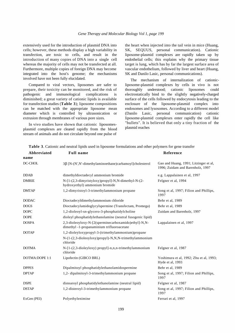
Compared to viral vectors, liposomes are safer toprepare, their toxicity can be monitored, and the risk ofpathogenic and immunological complications isdiminished; a great variety of cationic lipids is availablefor transfection studies (Table 3); liposome compositionscan be matched with the appropriate liposome meandiameter which is controlled by ultrasonication orextrusion through membranes of various pore sizes.
In vivo studies have shown that cationic liposomes-plasmid complexes are cleared rapidly from the bloodstream of animals and do not circulate beyond one pulse of
the heart when injected into the tail vein in mice (Huang,SK, SEQUUS, personal communication). Cationicliposome-plasmid complexes are rapidly taken up byendothelial cells; this explains why the primary tissuetarget is lung, which has by far the largest surface area ofvascular endothelium, followed by liver and heart (Huang,SK and Danilo Lasic, personal communications).
The mechanism of internalization of cationic-liposome-plasmid complexes by cells in vivo is notthoroughly understood; cationic liposomes couldelectrostatically bind to the slightly negatively-chargedsurface of the cells followed by endocytosis leading to theenclosure of the liposome-plasmid complex intoendosomes and lysosomes. According to a different model(Danilo Lasic, personal communication) cationicliposome-plasmid complexes enter rapidly the cell like"bullets". It is believed that only a tiny fraction of theplasmid reaches
Table 3. Cationic and neutral lipids used in liposome formulations and other polymers for gene transfer
Abbreviatedname
Full name Reference
DC-CHOL 3β [N-(N',N'-dimethylaminoethane)carbamoyl]cholesterol Gao and Huang, 1991; Litzinger et al,1996; Zuidam and Barenholz, 1997
DDAB dimethyldioctadecyl ammonium bromide e.g. Lappalainen et al, 1997
DMRIE N-[1-(2,3-dimyristyloxy)propyl]-N,N-dimethyl-N-(2-hydroxyethyl) ammonium bromide
Felgner et al, 1994
DMTAP 1,2-dimyristoyl-3-trimethylammonium propane Song et al, 1997; Filion and Phillips,1997
DODAC Dioctadecyldimethylammonium chloride Behr et al, 1989
DOGS Dioctadecylamidoglycylspermine (Transfectam, Promega) Behr et al, 1989
DOPC 1,2-dioleoyl-sn-glycero-3-phosphatidylcholine Zuidam and Barenholz, 1997
DOPE dioleyl phosphatidylethanolamine (neutral fusogenic lipid)
DOSPA 2,3-dioleoyloxy-N-[2(sperminecarboxamido)ethyl]-N,N-dimethyl -1-propanaminium trifluoroacetate
Lappalainen et al, 1997
DOTAP 1,2-dioleyloxypropyl-3-(trimethylammonium)propane
N-(1-(2,3-dioleoyloxy)propyl)-N,N,N-trimethylammoniumchloride
DOTMA N-[1-(2,3-dioleyloxy) propyl]-n,n,n-trimethylammoniumchloride
Felgner et al, 1987
DOTMA:DOPE 1:1 Lipofectin (GIBCO BRL) Yoshimura et al, 1992; Zhu et al, 1993;Hyde et al, 1993
DPPES Dipalmitoyl phosphatidylethanolamidospermine Behr et al, 1989
DPTAP 1,2- dipalmitoyl-3-trimethylammonium propane Song et al, 1997; Filion and Phillips,1997
DSPE distearoyl phosphatidylethanolamine (neutral lipid) Felgner et al, 1987
DSTAP 1,2-disteroyl-3-trimethylammonium propane Song et al, 1997; Filion and Phillips,1997
ExGen (PEI) Polyethylenimine Ferrari et al, 1997
Gene Therapy and Molecular Biology Vol 1, page 200
200
intact the cytoplasmic compartment and even a smallerfraction is imported into nuclei (Boulikas, 1998b).
There is no upper limit in plasmid DNA size to becomplexed with cationic liposomes as opposed toadenoviruses, AAV, and retroviruses that can accommodatea maximum of 7.5 kb of foreign DNA because ofpackaging limitations. This factor is of utmost importancewhen large genomic regions need to be transferred in orderto obtain correct developmental expression aftertransduction of fetuses or newborn animals.
Positively-charged liposomes containing distearoylphosphatidylethanolamine, a lipid which promotes fusionof liposomes with membranes, has been used for thetransfer of plasmid DNA into cells (Felgner et al, 1987;Felgner and Ringold, 1989). Liposomes containing headgroups able to form discrete complexes with DNA inducewrapping of DNA around unilamellar 80- to 100-nm indiameter vesicles in a way reminiscent of the wrapping of167 bp of DNA around the 55-nm core histone octamerforming nucleosomes (see Behr et al, 1989).
The positively-charged groups of the lipids interactwith DNA causing both a condensation of the plasmid bydiminishing the negative electrostatic repulsions on DNAas well as electrostatic binding of DNA. The liposome-plasmid DNA complex is then presented to cells in cultureor is injected into animals intravenously, intraperitoneally,subcutaneously, intratracheally, or via other routes.
B. Studies on cationic liposome-mediated gene transfer
A number of studies in gene therapy have used cationicliposomes as means of delivering DNA. Examples includetransfer of prostaglandin G/H synthase to protect lungs inrabbits against endotoxin-induced inflammation andpulmonary hypertension (Conary et al 1994); of α1-
antitrypsin cDNA to protect lungs (Canonico et al, 1994)or α1-antitrypsin cDNA encapsulated into negatively-
charged liposomes to protect connective tissue from thelytic action of the leukocyte neutrophil elastase (Aliño etal, 1996); of the human CFTR (cystic fibrosistransmembrane conductance regulator) gene in lungs inCFTR-deficient transgenic mice (Hyde et al, 1993; Altonet al, 1993) or in normal mice (Yoshimura et al, 1992) bytracheal instillation for the transduction of airwayepithelial cells for cystic fibrosis; of the MHC class IHLA-B7 heavy chain gene for the treatment of cancer (Lewet al, 1995); of tyrosine hydroxylase (TH) gene in order toalleviate degeneration of dopaminergic nigrostriatalneurons in rat models of Parkinson's disease (Jiao et al,1993; Cao et al, 1995); of the wild-type p53 gene to treatnude mice inoculated with breast carcinoma cells (Lesoon-
Wood et al, 1995); and of the IL-2 gene for prostate cancertherapy (Vieweg et al, 1995).
Cationic liposomes have also been used for arterialgene transfer (Nabel et al, 1990, 1993, Takeshita et al,1994).
Stable cationic lipid/DNA complexes were formed bysolubilizing DOSPA:DOPE in 1% octylglucoside, 10 mMTris pH 7.4 followed by the addition of DNA andexhaustive dialysis of the complex against 10 mM Tris,pH 7.4, 5% dextrose; the lipid-DNA complex had a storagelife of up to 3 months after formation with respect to itsability to transfect tissue culture cells and was competentof transfecting cells in the presence of 15% fetal bovineserum (FBS) whereas convenient cationic liposome-DNAcomplexes are unable to transfect cells in the presence ofFBS; a precipitate of the stable cationic lipid-DNA
complex formed after 14 days on shelf storage at 40Ccould be pelleted retaining all the transfection efficiencyand displaying lower toxicity because of the removal of thefree uncomplexed lipid in the supernatant (Hofland et al,1996).
DOTAP
DOTAP, a monocationic lipid, has been used totransfer efficiently the lacZ reporter gene and CFTR cDNAinto mice without any inflammatory response(McLachlanet al, 1995).
Lipofectin
Lipofectin (a 1:1 mixture of the cationic DOTMA withthe neutral DOPE) has been shown to deliver reportergenes to the rodent airways after direct intratrachealinjection (Yoshimura et al, 1992) or after intravenousadministration (Zhu et al, 1993). Lipofectin has been usedto alleviate the symptoms of cystic fibrosis in transgenicCF mice after transfer of the CFTR gene (Hyde et al,1993). Lipofectin has been used for the transfer of theCCK gene to suppress audiogenic epileptic seizures, aswell as for the transfer of reporter genes directly injectedinto mouse brain (Ono et al, 1990; Roessler and Davidson,1994).
DOGS (Transfectam)
DOGS appears to be more efficient than Lipofectin andDOTAP for the transfer of the luciferase gene to polyp andtracheal lung epithelial cells in culture; this may be due tothe presence of a secondary amine with a pKa=5.4 in theDOGS molecule which might be able to buffer the acidicendosomes and protect the plasmid DNA from degradation;
Gene Therapy and Molecular Biology Vol 1, page 201
201
DOGS, however, was inefficient for gene transfer tosubmucosal gland cells which are active in producingsticky mucus in CF patients and should be the target cellsto be corrected by CFTR gene transfer (Ferrari et al, 1997).
Compaction of plasmid DNA with dipalmitoylphosphatidylethanolamidospermine (DPPES) anddioctadecylamidoglycylspermine (DOGS), collectivelyknown as lipospermines, gave lipid-coated plasmid DNArather than liposome-plasmid complexes; liposperminesinteract strongly with DNA eventually promoting coatingof supercoiled DNA plasmids and promote binding of thecomplex to the cell membrane (Behr et al, 1989). Whendispersed in water (either by sonication or by dilution of anethanol solution) they form unilamellar vesicles of 80-100nm, unstable in ionic media, and interacting cooperativelywith plasmids because of the strong affinity of the
spermine group for DNA (105-10
7 M
-1). Small unilamellar
vesicles formed between DOGS and egg yolk lecithin wereunable to mediate transfection and thus DOGS-mediatedtransfection is not a liposome-mediated process but aprocess mediated by cationic lipid-coated plasmid (Behr etal, 1989).
Although the in vitro conditions for transfection seemto favor a ratio of positive charges of lipids to negativephosphate charges on DNA of about 1:8, the in vivooptimal conditions are 1 lipid molecule/30 phosphatesusing DDAB:DOPE and DDAD:Chol (Boulikas et al, inpreparation). Previous studies by Behr and coworkers(1989; reviewed by Behr, 1994) and Schwartz et al (1995)using DOGS have also shown that the in vivo optimalconditions require a lower lipid:DNA charge ratio; theexplanation might be that anionic proteins in the bloodserum or in the extracellular matrix could interact withcationic lipid particles inhibiting their uptake by the cellsin tissues.
The in vivo efficacy for transferring episomallyreplicating plasmids containing the human papovavirusBKV origin of replication/early regulatory region and thelarge T antigen gene, as well as the luciferase reporter geneunder control of RSV promoter were investigated byThierry and coworkers (1995); dioctadecylamidoglysyl-spermidine:DOPE liposomes hydrated in the presence ofplasmid DNA, injected into the vein of mice, sustainedexpression in the liver, lung, spleen, heart, and othertissues for up to three months postinjection. These vectorswere found to replicate extrachromosomally in the lungtissue; use of nonepisomal vectors under the sameconditions gave only transient expression of the luciferasegene which disappeared after about 4-5 days.
Cationic liposomes have been used for targeting braincells after direct intracranial injection of the plasmid-liposome complex. DOGS:DOPE liposomes havesuccessfully transferred the luciferase reporter gene under
control of CMV promoter to striatal parenchyma andparaventricular brain cells in neonatal mice; however, theexpression of the transgene, although significant at earlytimes postinjection diminished over time (Schwartz et al,1995).
C. Advantages and drawbacks usingcationic lipids for gene delivery
Cationic lipids may show low efficiency oftransfection despite the relatively large amounts of DNAused.
Cationic lipid-DNA complexes with a net positivecharge may interact with circulating serum proteins oranionic components of the extracellular matrix in thevarious tissues; this interaction reduces their bio-availability (Schwartz et al, 1995). In addition positively-charged complexes activate complement and complement-dependent phagocytosis by macrophages in thereticuloendothelial system and are, thus, cleared rapidlyfrom body fluids (Plank et al, 1996). Thus, although theoptimal lipid-DNA ratio may be to an excess of positivecharges for the transfection of cells in culture (mediated bythe ability of cationic lipids to interact with the relativelynegatively-charged external surface of the cell membraneand higher poration through the membrane) the optimalratio in vivo is nearly that which gives a neutral complex(Boulikas et al, in preparation, see Boulikas 1998a).
D. Entry of liposome-DNA into cellsNeutral liposomes are internalized via the endocytic
uptake mechanism. However, cationic liposome-plasmidcomplexes (Capaccioli et al, 1993; Boutorine and Kostina,1993) as well as Sendai virus-derived liposomes(Compagnon et al, 1992; Morishita et al, 1993) seem topermit a direct passage of the oligonucleotide load throughthe cell membrane (see also Bongartz et al, 1994).
E. Enhancement of cationic liposomedelivery
A number of methods have been invented to augmentthe efficiency of internalization or release from endosomesof DNA-liposome complexes, or to enhance nuclearimport of the plasmid after its release in the cytoplasm.The external cell surface has a net negative charge and thusliposome-DNA particles with a net positive charge can beelectrostatically anchored to the external cell membrane.
Liposomes are internalized into endosomes whichdegrade the plasmid; use of folate attached to ligands on thesurface of the liposome (Gottschalk et al, 1993) changesthe route of internalization and the particles are taken upby the caveolae vesicles rather than endosomes which lack
Martin and Boulikas: The challenge of liposomes in gene therapy
202
nucleases and release more readily their content to theinterior of the cell compared with endosomes.
Use of pH-sensitive liposomes, such as DOPE:cholesterol: oleic acid liposomes (Wang and Huang, 1987;Huang et al, 1987) seem to induce their break-down at thelower pH of the endosomes releasing the DOPE to theendosome; DOPE then induces endosome membranebreakdown and release of its content to the cytoplasm.
Use of nuclear proteins in complex with plasmid DNAencapsulated into true (as opposed to cationic) liposomeshas been found to increase transfection efficiency; DNAwas rapidly transported into the nuclei and its expressionreached a maximum within 6-8h after transfection (Kanedaet al, 1989; Kato et al, 1991). According to this procedureSendai virus was used to fuse DNA-loaded gangliosideliposomes with protein-containing membrane vesiclespurified from red blood cells; cointroduction of HMG-1protein showed rapid uptake of plasmids by nuclei whereaswith BSA in the place of HMG the grains of the in situhybridization were located in the cytoplasm after 6 hreaching the nucleus only after about 24h (Kaneda et al,1989).
F. Problems and advantages forliposomal delivery of genes
During in vivo delivery foreign DNA can be attackedby macrophages, lymphocytes, or other components of theimmune system and the vast majority is cleared fromblood, intracellular, or other body fluids before it is giventhe chance to reach the membrane of the cell target; thehalf-life of naked plasmids injected intravenously intoanimals is about 5 min (reviewed by Boulikas, 1998a). Onthe other hand “stealth” liposomes persist in the bodyfluids for days as seen in the whole body scintigraphs ofempty radioactively-labeled “stealth “ liposomes (Figures17-19); the encapsulated plasmid DNA would alsocirculate for long times. However, tumor cells , where the“stealth” liposomes are localized are very often ofepithelial origin (brain, colon, breast, head & neck,prostate tumors) and not engaged in the uptake ofliposomes or other colloidal particles. Thus, stealthliposomes remain in the extracellular space and slowlyreleasing their material after lysis over days. One strategyto circumvent this bottleneck would be to include cationiclipids in the lipid bilayer and devise methods for PEGcoating to fall off; the cationic lipids are then expected tomediate rapid poration through the cell membrane oruptake, as cationic lipids are known to penetrate rapidlymost types of cells.
The elimination of therapeutically important cells fromthe body by the immune system due to expression of viralproteins after ex vivo delivery of genes with recombinantadenovirus (Dai et al, 1995) would not apply to liposomaldelivery of genes.
IX. Clinical trials using liposome-mediated gene transfer
All RAC-approved protocols for gene transfer tohuman patients with liposomes use cationic lipids (Table4 ). Because of the toxicity of cationic lipids none of theseprotocols employs systemic intravenous injection of thecationic liposome-plasmid complex; instead the type ofadministration involves (i ) direct intratumoral injection forimmunotherapy of melanoma, lymphoma, renal carcinomaand a great variety of metastatic malignancies; (i i )subcutaneous injection for glioblastoma antisense-IGFtherapy, or for immunotherapy of melanoma, colon withhepatic metastases, renal, breast, and small cell lungcancer; (i i i ) intranasal delivery for CFTR and alpha-1-antitrypsin deficiency; (iv ) intradermal injection for theimmunotherapy of ovarian cancer and advanced ormetastatic prostate cancer using IL-2 cDNA; and (v )intraperitoneal and intrapleural delivery of the adenoviralE1A gene to control the regulation of the HER-2/neuoncogene in ovarian and breast cancer (Table 4).
X. ProspectsTo-date, only very few conventional (radiation/
chemotherapy) regimens on cancer patients lead to tumoreradication. Most treatments, after cancer is detected at anadvanced stage, prolong the life of the patient by fewmonths but reduce its quality because of the adverse effectsof antineoplastic treatments. Gene therapy offers stronghopes to millions of desperate people, both patients andrelatives.
Although "stealth" liposomes extravasate preferentiallyin solid tumors, they remain in the extracellular space andare not readily internalized by tumor cells; liposomesloaded with doxorubicin concentrate the drug in the solidtumor and release their content over a period of severaldays. Use of substance P, a peptide known to increasevascular permeability, increased liposome extravasation intissues that posses receptors for substance P (trachea,esophagus, and urinary bladder); extravasated liposomesremained in the extracellular space beyond the postcapillaryvenular endothelium at early times. The colloidal goldparticles encapsulated into liposomes were localizedintracellularly in both endothelial cells and macrophages at24 h postinjection (Rosenecker et al, 1996).

Gene Therapy and Molecular Biology Vol 1, page 203
203
Table 4. RAC-approved human gene therapy protocols using liposomes(For clinical trials using viruses see Appendix 1 in Boulikas, 1998, pages 159-172 of this volume).
Protoco lnumber & humandisease
Gene and route ofadministrat ion
Inves t iga tors /A f f i l i a t i o n
Tit le of protocol & date ofRAC/NIH approval
187. 9202-013 (Closed)Gene Therapy /Phase I/Cancer /Melanoma/Adenocarcinoma/Immunotherapy
In Vivo /Autologous Tumor Cells/Cationic Liposome Complex /DC-Chol /HLA-B7 /Beta-2 MicroglobulincDNA /Intratumoral /DirectInjection /Catheter Delivery toPulmonary Nodules
Nabel, Gary J.; University ofMichigan, Ann Arbor,Michigan
Immunotherapy of Malignancy by InVivo Gene Transfer into Tumors. RACApproval: 2-10-92 /NIH Approval: 4-17-92 Closed: 11-19-92 (Replaced byProtocol #9306-045)
188. 9306-045 (Open)Gene Therapy /Phase I/CancerImmunotherapy
In Vivo /Autologous Tumor Cells/Cationic Liposome Complex /HLA-B7 /Beta-2 Microglobulin cDNA /Intratumoral /Direct Injection/Catheter Delivery to PulmonaryNodules
Nabel, Gary J.; University ofMichigan Medical Center, AnnArbor, Michigan
Immunotherapy for Cancer by DirectGene Transfer into Tumors. RACApproval: 6-7-93 /NIH Approval: 9-3-93
189. 9306-052 (Open)Gene Therapy /Phase I/Cancer / Glioblastoma/Antisense
In Vitro /Autologous Tumor Cells/Lethally Irradiated / CationicLiposome Complex /Lipofectin (GibcoBRL) /Insulin-like Growth FactorAntisense / Subcutaneous Injection
Ilan, Joseph; Case WesternReserve University School ofMedicine and UniversityHospitals of Cleveland,Cleveland, Ohio
Gene Therapy for Human Brain TumorsUsing Episome-Based Antisense cDNATranscription of Insulin-Like GrowthFactor I. RAC Approval: 6-8-93 /NIHApproval: 12-2-93
190. 9309-053 (Open)Gene Therapy /Phase I/Cancer / Small CellLung Cancer /Immunotherapy
In Vitro /Autologous Tumor Cells/Lethally Irradiated / CationicLiposome Complex /Lipofectin (GibcoBRL) /Cytokine /Interleukin-2 cDNA/Neomycin Phosphotransferase cDNA/Subcutaneous Injection
Cassileth, Peter; Podack,Eckhard R.; Sridhar, Kasi;University of Miami; andSavaraj, Niramol; MiamiVeterans AdministrationHospital, Miami, Florida
Phase I Study of Transfected CancerCells Expressing the Interleukin-2 GeneProduct in Limited Stage Small Cell LungCancer. RAC Approval: 9-9-93 /NIHApproval: 12-2-93
191. 9312-063 (Open)Gene Therapy /Phase I/Cancer /Melanoma/Immunotherapy
In Vitro /Allogeneic Tumor Cells/Lethally Irradiated /CationicLiposome Complex /Lipofectin (GibcoBRL) /B7 (CD80) cDNA /NeomycinPhosphotransferase cDNA/Subcutaneous Injection
Sznol, Mario; NationalInstitutes of Health, Frederick,Maryland
A Phase I Trial of B7-TransfectedLethally Irradiated Allogeneic MelanomaCell Lines to Induce Cell MediatedImmunity Against Tumor-AssociatedAntigens Presented by HLA-A2 or HLA-A1 in Patients with Stage IV Melanoma.RAC Approval: 12-3-93 /NIH Approval:4-19-94
192. 9312-064 (Closed)Gene Therapy /Phase I/Cancer /Colon /HepaticMetastases/Immunotherapy
In Vivo /Autologous Tumor Cells/Cationic Liposome Complex/DMRIE-DOPE Vical VCL-1005/HLA-B7 /Beta-2 MicroglobulincDNA /Intratumoral /HepaticInjection
Rubin, Joseph; Mayo Clinic,Rochester, Minnesota
Phase I Study of Immunotherapy ofAdvanced Colorectal Carcinoma byDirect Gene Transfer into HepaticMetastases.
Sponsor: Vical, Incorporated RACApproval: 12-3-93 /NIH Approval: 4-19-94 Closed: 3-16-95
193. 9312-066 (Open)Gene Therapy /Phase I/Monogenic Disease/Cystic Fibrosis
In Vivo /Nasal Epithelial Cells/Cationic Liposome Complex/DMRIE-DOPE /Cystic FibrosisTransmembrane ConductanceRegulator cDNA /Intranasal
Sorscher, Eric J. and Logan,James L.; University ofAlabama, Birmingham,Alabama
Gene Therapy for Cystic Fibrosis UsingCationic Liposome Mediated GeneTransfer: A Phase I Trial of Safety andEfficacy in the Nasal Airway. RACApproval: 12-3-93 /NIH Approval: 1-4-95
194. 9403-070 (Open)Gene Therapy /Phase I/Monogenic Disease/Alpha-1-AntitrypsinDeficiency
In Vivo /Nasal Epithelial Cells/Respiratory Epithelial Cells /CationicLiposome Complex /DC-Chol-DOPE/Alpha-1 Antitrypsin cDNA/Intranasal /Respiratory TractAdministration (Bronchoscope)
Brigham, Kenneth; ClinicalResearch Center at VanderbiltUniversity Medical Center,Nashville, Tennessee
Expression of an ExogenouslyAdministered Human Alpha-1-AntitrypsinGene in the Respiratory Tract of Humans.
Sponsor: Gene Medicine, Inc. RACApproval: 3-3-94 /NIH Approval: 10-25-94
195. 9403-071 (Closed)Gene Therapy /Phase I/Cancer /Renal Cell/Immunotherapy
In Vivo /Autologous Tumor Cells/Cationic Liposome Complex/DMRIE-DOPE Vical VCL-1005/HLA-B7 /Beta-2 MicroglobulincDNA /Intratumoral /DirectInjection
Vogelzang, Nicholas; theUniversity of Chicago,Chicago, Illinois
Phase I Study of Immunotherapy forMetastatic Renal Cell Carcinoma byDirect Gene Transfer into MetastaticLesions.
Sponsor: Vical, Incorporated RACApproval: 3-4-94 /NIH Approval: 4-19-94Closed: 4-5-95
196. 9403-072 (Closed)Gene Therapy /Phase I/Cancer /Melanoma/Immunotherapy
In Vivo /Autologous Tumor Cells/Cationic Liposome Complex/DMRIE-DOPE Vical VCL-1005/HLA-B7 /Beta-2 MicroglobulincDNA /Intratumoral /DirectInjection
Hersh, Evan; ArizonaCancer Center, Tucson,Arizona; and Akporiaye;Harris; Stopeck; Unger; andWarneke; University ofArizona, Tucson, Arizona
Phase I Study of Immunotherapy ofMalignant Melanoma by Direct GeneTransfer.
Sponsor: Vical, Incorporated RACApproval: 3-4-94 /NIH Approval: 4-19-94
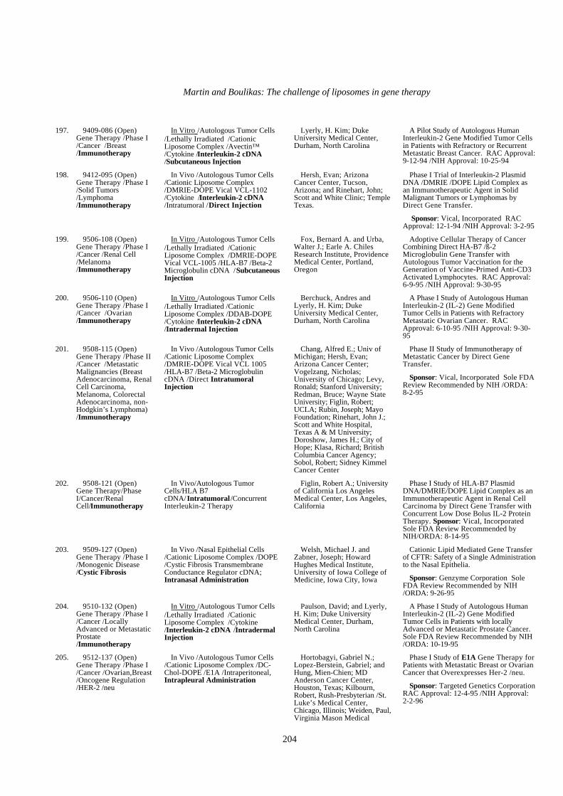
Martin and Boulikas: The challenge of liposomes in gene therapy
204
197. 9409-086 (Open)Gene Therapy /Phase I/Cancer /Breast/Immunotherapy
In Vitro /Autologous Tumor Cells/Lethally Irradiated /CationicLiposome Complex /Avectin™/Cytokine /Interleukin-2 cDNA/Subcutaneous Injection
Lyerly, H. Kim; DukeUniversity Medical Center,Durham, North Carolina
A Pilot Study of Autologous HumanInterleukin-2 Gene Modified Tumor Cellsin Patients with Refractory or RecurrentMetastatic Breast Cancer. RAC Approval:9-12-94 /NIH Approval: 10-25-94
198. 9412-095 (Open)Gene Therapy /Phase I/Solid Tumors/Lymphoma/Immunotherapy
In Vivo /Autologous Tumor Cells/Cationic Liposome Complex/DMRIE-DOPE Vical VCL-1102/Cytokine /Interleukin-2 cDNA/Intratumoral /Direct Injection
Hersh, Evan; ArizonaCancer Center, Tucson,Arizona; and Rinehart, John;Scott and White Clinic; TempleTexas.
Phase I Trial of Interleukin-2 PlasmidDNA /DMRIE /DOPE Lipid Complex asan Immunotherapeutic Agent in SolidMalignant Tumors or Lymphomas byDirect Gene Transfer.
Sponsor: Vical, Incorporated RACApproval: 12-1-94 /NIH Approval: 3-2-95
199. 9506-108 (Open)Gene Therapy /Phase I/Cancer /Renal Cell/Melanoma/Immunotherapy
In Vitro /Autologous Tumor Cells/Lethally Irradiated /CationicLiposome Complex /DMRIE-DOPEVical VCL-1005 /HLA-B7 /Beta-2Microglobulin cDNA /SubcutaneousInjection
Fox, Bernard A. and Urba,Walter J.; Earle A. ChilesResearch Institute, ProvidenceMedical Center, Portland,Oregon
Adoptive Cellular Therapy of CancerCombining Direct HA-B7 /ß-2Microglobulin Gene Transfer withAutologous Tumor Vaccination for theGeneration of Vaccine-Primed Anti-CD3Activated Lymphocytes. RAC Approval:6-9-95 /NIH Approval: 9-30-95
200. 9506-110 (Open)Gene Therapy /Phase I/Cancer /Ovarian/Immunotherapy
In Vitro /Autologous Tumor Cells/Lethally Irradiated /CationicLiposome Complex /DDAB-DOPE/Cytokine /Interleukin-2 cDNA/Intradermal Injection
Berchuck, Andres andLyerly, H. Kim; DukeUniversity Medical Center,Durham, North Carolina
A Phase I Study of Autologous HumanInterleukin-2 (IL-2) Gene ModifiedTumor Cells in Patients with RefractoryMetastatic Ovarian Cancer. RACApproval: 6-10-95 /NIH Approval: 9-30-95
201. 9508-115 (Open)Gene Therapy /Phase II/Cancer /MetastaticMalignancies (BreastAdenocarcinoma, RenalCell Carcinoma,Melanoma, ColorectalAdenocarcinoma, non-Hodgkin’s Lymphoma)/Immunotherapy
In Vivo /Autologous Tumor Cells/Cationic Liposome Complex/DMRIE-DOPE Vical VCL 1005/HLA-B7 /Beta-2 MicroglobulincDNA /Direct IntratumoralInjection
Chang, Alfred E.; Univ ofMichigan; Hersh, Evan;Arizona Cancer Center;Vogelzang, Nicholas;University of Chicago; Levy,Ronald; Stanford University;Redman, Bruce; Wayne StateUniversity; Figlin, Robert;UCLA; Rubin, Joseph; MayoFoundation; Rinehart, John J.;Scott and White Hospital,Texas A & M University;Doroshow, James H.; City ofHope; Klasa, Richard; BritishColumbia Cancer Agency;Sobol, Robert; Sidney KimmelCancer Center
Phase II Study of Immunotherapy ofMetastatic Cancer by Direct GeneTransfer.
Sponsor: Vical, Incorporated Sole FDAReview Recommended by NIH /ORDA:8-2-95
202. 9508-121 (Open)Gene Therapy/PhaseI/Cancer/RenalCell/Immunotherapy
In Vivo/Autologous TumorCells/HLA B7cDNA/Intratumoral /ConcurrentInterleukin-2 Therapy
Figlin, Robert A.; Universityof California Los AngelesMedical Center, Los Angeles,California
Phase I Study of HLA-B7 PlasmidDNA/DMRIE/DOPE Lipid Complex as anImmunotherapeutic Agent in Renal CellCarcinoma by Direct Gene Transfer withConcurrent Low Dose Bolus IL-2 ProteinTherapy. Sponsor: Vical, Incorporated Sole FDA Review Recommended byNIH/ORDA: 8-14-95
203. 9509-127 (Open)Gene Therapy /Phase I/Monogenic Disease/Cystic Fibrosis
In Vivo /Nasal Epithelial Cells/Cationic Liposome Complex /DOPE/Cystic Fibrosis TransmembraneConductance Regulator cDNA;Intranasal Administration
Welsh, Michael J. andZabner, Joseph; HowardHughes Medical Institute,University of Iowa College ofMedicine, Iowa City, Iowa
Cationic Lipid Mediated Gene Transferof CFTR: Safety of a Single Administrationto the Nasal Epithelia.
Sponsor: Genzyme Corporation SoleFDA Review Recommended by NIH/ORDA: 9-26-95
204. 9510-132 (Open)Gene Therapy /Phase I/Cancer /LocallyAdvanced or MetastaticProstate/Immunotherapy
In Vitro /Autologous Tumor Cells/Lethally Irradiated /CationicLiposome Complex /Cytokine/Interleukin-2 cDNA /IntradermalInjection
Paulson, David; and Lyerly,H. Kim; Duke UniversityMedical Center, Durham,North Carolina
A Phase I Study of Autologous HumanInterleukin-2 (IL-2) Gene ModifiedTumor Cells in Patients with locallyAdvanced or Metastatic Prostate Cancer.Sole FDA Review Recommended by NIH/ORDA: 10-19-95
205. 9512-137 (Open)Gene Therapy /Phase I/Cancer /Ovarian,Breast/Oncogene Regulation/HER-2 /neu
In Vivo /Autologous Tumor Cells/Cationic Liposome Complex /DC-Chol-DOPE /E1A /Intraperitoneal,Intrapleural Administration
Hortobagyi, Gabriel N.;Lopez-Berstein, Gabriel; andHung, Mien-Chien; MDAnderson Cancer Center,Houston, Texas; Kilbourn,Robert, Rush-Presbyterian /St.Luke’s Medical Center,Chicago, Illinois; Weiden, Paul,Virginia Mason Medical
Phase I Study of E1A Gene Therapy forPatients with Metastatic Breast or OvarianCancer that Overexpresses Her-2 /neu.
Sponsor: Targeted Genetics CorporationRAC Approval: 12-4-95 /NIH Approval:2-2-96

Gene Therapy and Molecular Biology Vol 1, page 205
205
Center, Seattle, Washington
206. 9512-142 (Open)Gene Therapy /Phase I/Gene Therapy /Cancer/Head and NeckSquamous CellCarcinoma/Immunotherapy
In Vivo /Autologous Tumor Cells/Cationic Liposome Complex/DMRIE-DOPE Vical VCL 1005/HLA-B7 /Beta-2 MicroglobulincDNA /Direct IntratumoralInjection
Gluckman, Jack L.;University of CincinnatiMedical Center, Cincinnati,Ohio
Allovectin-7 in the Treatment ofSquamous Cell Carcinoma of the Headand Neck. Sole FDA ReviewRecommended by NIH /ORDA: 12-15-95
207. 9608-156 (Open)Gene Therapy /Phase I/Cancer /Breast/Immunotherapy
InVitro /Allogeneic Tumor Cells/Lethally Irradiated /CationicLiposome Complex /B7(CD80) cDNA/Subcutaneous Injection
Urba, Walter J., ProvidencePortland Medical Center,Portland, Oregon
Phase I Trial Using a CD80-ModifiedAllogeneic Breast Cancer Line toVaccinate HLA-A2-Positive Women withBreast Cancer . Sole FDA ReviewRecommended by NIH /ORDA: 8-6-96
208. 9609-161 (Open)Gene Therapy /Phase I/Cancer /Small CellLung Cancer/Immunotherapy
In Vitro /Autologous Tumor Cells/Lethally Irradiated /CationicLiposome Complex/Lipofectin(GibcoBRL) /B7-1(CD80)cDNA /Subcutaneous Injection
Antonia, Scott J., H. LeeMoffitt Cancer Center, Tampa,Florida
Treatment of Small Cell Lung CancerPatients In Partial Remission Or At RelapseWith B7-1 Gene-Modified AutologousTumor Cells As A Vaccine With SystemicInterferon Gamma. Sole FDA ReviewRecommended by NIH /ORDA: 10-10-96
209. 9610-162 (Open)Gene Therapy /Phase I/Cancer /Solid Tumors/Oncogene Regulation/HER-2 /neu
In Vivo /Autologous Tumor Cells/Cationic Liposome Complex /DC-Chol-DOPE /E1A /IntratumoralInjection
LaFollette, Suzanne, Rush/Presbyterian /St. Luke’sMedical Center, Chicago,Illinois; Murray, James L.,M.D. Anderson Cancer Center,Houston, Texas; Yoo, George,Wayne State University,Detroit, Michigan
A Phase I Multicenter Study ofIntratumoral E1A Gene Therapy forPatients with Unresectable or MetastaticSolid Tumors that Overexpress HER-2/neu.
Sponsor: Targeted Genetics CorporationSole FDA Review Recommended by NIH/ORDA: 10-29-96
210. 9611-168 (Open)Gene Therapy /Phase II/Cancer /Melanoma/Immunotherapy
In Vivo /Autologous Tumor Cells/Cationic Liposome Complex/DMRIE-DOPE Vical VCL 1005/HLA-B7 /Beta-2 MicroglobulincDNA /Direct IntratumoralInjection
Hersh, Evan M., ArizonaCancer Center, Tucson,Arizona; Klasa, Richard,British Columbia CancerAgency, Vancouver, B.C.,Canada; Gonzales, Rene,University of Colorado CancerCenter, Denver, Colorado;Silver, Gary, NorthernCalifornia Melanoma Clinic,San Francisco,California;Thompson, JohnA.,U. of Washington MedicalCenter, Seattle, Washington
Phase II Study of Immunotherapy ofMetastatic Melanoma by Direct GeneTransfer.
Sponsor: Vical, Incorporated NIH/ORDA Receipt Date: 11-26-96. Sole FDAReview Recommended by NIH /ORDA:1-6-97
211. 9611-169 (Open)Gene Therapy /Phase I/II /Cancer /SolidTumors/Immunotherapy
In Vivo /Autologous Tumor Cells/Cationic Liposome Complex/DMRIE-DOPE Vical VCL 1102/Cytokine /Interleukin-2 cDNA/Direct Intratumoral Injection
Hersh, Evan, M., ArizonaCancer Center, Tucson,Arizona; Rinehart, John, Scottand White Clinic, Temple,Texas; Rubin, Joseph, MayoClinic, Rochester, Minnesota;Sondak, Vernon K., Universityof Michigan Medical Center,Ann Arbor, Michigan;Gonzales, Rene, University ofColorado Cancer Center,Denver, Colorado; Sobol,Robert E., Sharp HealthCare,San Diego, California; andForscher, Charles A., Cedars-Sinai Comprehensive CancerCenter, Los Angeles, California
Phase I /II Trial of Interleukin-2 DNA/DMRIE /DOPE Lipid Complex as anImmunotherapeutic Agent in Cancer byDirect Gene Transfer.
Sponsor: Vical, Incorporated NIH/ORDA Receipt Date: 11-26-96. Sole FDAReview Recommended by NIH /ORDA:1-17-97
212. 9612-170 (Open)Gene Therapy /Phase I/Monogenic Disease/Cystic Fibrosis
In Vivo /Lung and Nasal EpithelialCells /Cationic Liposome Complex/DOPE /CFTR cDNA /AerosolAdministration
Sorscher, Eric, University ofAlabama, Birmingham,Medical Center
Safety and Efficiency of Gene Transferof Aerosol Administration of a SingleDose of a Cationic Lipid /DNAFormulation fo the Lungs and Nose ofPatients with Cystic Fibrosis.
Sponsor: Genzyme Corporation NIH/ORDA Receipt Date: 12-17-96. Sole FDAReview Recommended by NIH /ORDA:1-6-97
213. 9703-184 (Open)Gene Therapy /Phase I/Cancer /Prostate Cancer/Immunotherapy
In Vivo /Autologous Tumor Cells/Cationic Liposome Complex/DMRIE-DOPE Vical VCL-1102/Cytokine /Interleukin-2 cDNA/Intratumoral Injection
Belldegrun, Arie, Universityof California, Los Angeles,School of Medicine, LosAngeles, California
A Phase I Study Evaluating the Safetyand Efficacy of Interleukin-2 GeneTherapy Delivered by Lipid MediatedGene Transfer (Leuvectin) in ProstateCancer Patients.
Sponsor: Vical, Inc. NIH /ORDAReceipt Date: 3-24-97. Sole FDA Review
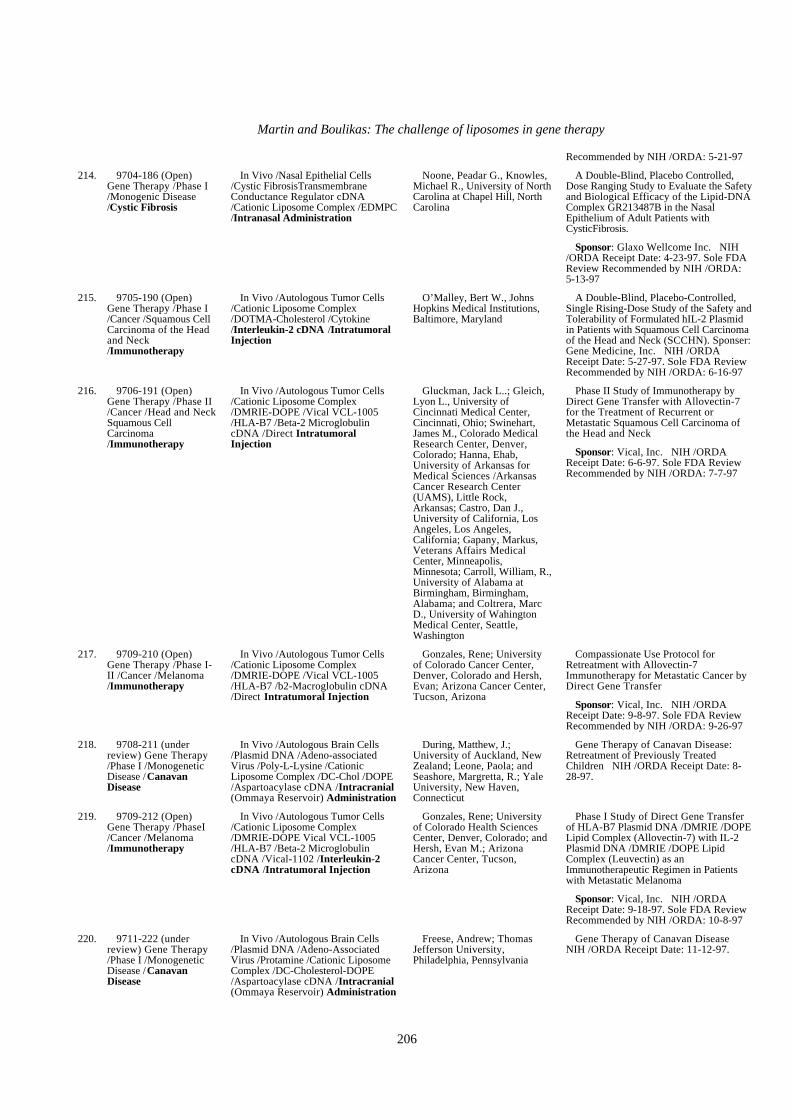
Martin and Boulikas: The challenge of liposomes in gene therapy
206
Recommended by NIH /ORDA: 5-21-97
214. 9704-186 (Open)Gene Therapy /Phase I/Monogenic Disease/Cystic Fibrosis
In Vivo /Nasal Epithelial Cells/Cystic FibrosisTransmembraneConductance Regulator cDNA/Cationic Liposome Complex /EDMPC/Intranasal Administration
Noone, Peadar G., Knowles,Michael R., University of NorthCarolina at Chapel Hill, NorthCarolina
A Double-Blind, Placebo Controlled,Dose Ranging Study to Evaluate the Safetyand Biological Efficacy of the Lipid-DNAComplex GR213487B in the NasalEpithelium of Adult Patients withCysticFibrosis.
Sponsor: Glaxo Wellcome Inc. NIH/ORDA Receipt Date: 4-23-97. Sole FDAReview Recommended by NIH /ORDA:5-13-97
215. 9705-190 (Open)Gene Therapy /Phase I/Cancer /Squamous CellCarcinoma of the Headand Neck/Immunotherapy
In Vivo /Autologous Tumor Cells/Cationic Liposome Complex/DOTMA-Cholesterol /Cytokine/Interleukin-2 cDNA /IntratumoralInjection
O’Malley, Bert W., JohnsHopkins Medical Institutions,Baltimore, Maryland
A Double-Blind, Placebo-Controlled,Single Rising-Dose Study of the Safety andTolerability of Formulated hIL-2 Plasmidin Patients with Squamous Cell Carcinomaof the Head and Neck (SCCHN). Sponser:Gene Medicine, Inc. NIH /ORDAReceipt Date: 5-27-97. Sole FDA ReviewRecommended by NIH /ORDA: 6-16-97
216. 9706-191 (Open)Gene Therapy /Phase II/Cancer /Head and NeckSquamous CellCarcinoma/Immunotherapy
In Vivo /Autologous Tumor Cells/Cationic Liposome Complex/DMRIE-DOPE /Vical VCL-1005/HLA-B7 /Beta-2 MicroglobulincDNA /Direct IntratumoralInjection
Gluckman, Jack L..; Gleich,Lyon L., University ofCincinnati Medical Center,Cincinnati, Ohio; Swinehart,James M., Colorado MedicalResearch Center, Denver,Colorado; Hanna, Ehab,University of Arkansas forMedical Sciences /ArkansasCancer Research Center(UAMS), Little Rock,Arkansas; Castro, Dan J.,University of California, LosAngeles, Los Angeles,California; Gapany, Markus,Veterans Affairs MedicalCenter, Minneapolis,Minnesota; Carroll, William, R.,University of Alabama atBirmingham, Birmingham,Alabama; and Coltrera, MarcD., University of WahingtonMedical Center, Seattle,Washington
Phase II Study of Immunotherapy byDirect Gene Transfer with Allovectin-7for the Treatment of Recurrent orMetastatic Squamous Cell Carcinoma ofthe Head and Neck
Sponsor: Vical, Inc. NIH /ORDAReceipt Date: 6-6-97. Sole FDA ReviewRecommended by NIH /ORDA: 7-7-97
217. 9709-210 (Open)Gene Therapy /Phase I-II /Cancer /Melanoma/Immunotherapy
In Vivo /Autologous Tumor Cells/Cationic Liposome Complex/DMRIE-DOPE /Vical VCL-1005/HLA-B7 /b2-Macroglobulin cDNA/Direct Intratumoral Injection
Gonzales, Rene; Universityof Colorado Cancer Center,Denver, Colorado and Hersh,Evan; Arizona Cancer Center,Tucson, Arizona
Compassionate Use Protocol forRetreatment with Allovectin-7Immunotherapy for Metastatic Cancer byDirect Gene Transfer
Sponsor: Vical, Inc. NIH /ORDAReceipt Date: 9-8-97. Sole FDA ReviewRecommended by NIH /ORDA: 9-26-97
218. 9708-211 (underreview) Gene Therapy/Phase I /MonogeneticDisease /CanavanDisease
In Vivo /Autologous Brain Cells/Plasmid DNA /Adeno-associatedVirus /Poly-L-Lysine /CationicLiposome Complex /DC-Chol /DOPE/Aspartoacylase cDNA /Intracranial(Ommaya Reservoir) Administration
During, Matthew, J.;University of Auckland, NewZealand; Leone, Paola; andSeashore, Margretta, R.; YaleUniversity, New Haven,Connecticut
Gene Therapy of Canavan Disease:Retreatment of Previously TreatedChildren NIH /ORDA Receipt Date: 8-28-97.
219. 9709-212 (Open)Gene Therapy /PhaseI/Cancer /Melanoma/Immunotherapy
In Vivo /Autologous Tumor Cells/Cationic Liposome Complex/DMRIE-DOPE Vical VCL-1005/HLA-B7 /Beta-2 MicroglobulincDNA /Vical-1102 /Interleukin-2cDNA /Intratumoral Injection
Gonzales, Rene; Universityof Colorado Health SciencesCenter, Denver, Colorado; andHersh, Evan M.; ArizonaCancer Center, Tucson,Arizona
Phase I Study of Direct Gene Transferof HLA-B7 Plasmid DNA /DMRIE /DOPELipid Complex (Allovectin-7) with IL-2Plasmid DNA /DMRIE /DOPE LipidComplex (Leuvectin) as anImmunotherapeutic Regimen in Patientswith Metastatic Melanoma
Sponsor: Vical, Inc. NIH /ORDAReceipt Date: 9-18-97. Sole FDA ReviewRecommended by NIH /ORDA: 10-8-97
220. 9711-222 (underreview) Gene Therapy/Phase I /MonogeneticDisease /CanavanDisease
In Vivo /Autologous Brain Cells/Plasmid DNA /Adeno-AssociatedVirus /Protamine /Cationic LiposomeComplex /DC-Cholesterol-DOPE/Aspartoacylase cDNA /Intracranial(Ommaya Reservoir) Administration
Freese, Andrew; ThomasJefferson University,Philadelphia, Pennsylvania
Gene Therapy of Canavan DiseaseNIH /ORDA Receipt Date: 11-12-97.

Gene Therapy and Molecular Biology Vol 1, page 207
207
A breakthrough would be the encapsulation ofplasmids with therapeutic genes (e.g. antitumor genes suchas p53, HSV-tk, angiostatin) into Stealth liposomes withmechanisms inducing PEG fall off after their accumulationinto tumors; when a certain amount of cationic liposomesis included into the lipid composition of the Stealthliposome, the PEG-free liposome is expected to be takenrapidly by tumor cells. Fusogenic peptides or otherstrategies could be combined to release the liposomes fromendosomes. These regiments could be combined withnontoxic levels of antineoplastic drugs encapsulated intoStealth liposomes that might act synergistically with thetumor suppressor or tumor killer genes to eradicate cancer.The major advantage, no doubt, would be the systemicdelivery of genes with this approach, able to hit, like noother gene therapy regiment currently available, theprimary tumor and its metastases.
AcknowledgementsWe are indebted to Stewart Simon for providing
Figures for this article and to Dan Lasic, DemetriosPapahadjopoulos, Peter Working, Ken Huang, and JoeVallner for stimulating discussions.
ReferencesAhmad I and Allen TM (1 9 9 2 ) Antibody-mediated specific
binding and cytotoxicity of liposome-entrappeddoxorubicin to lung cancer cells in vitro. Cancer Res .52, 4817-4820.
Ahmad I, Longenecker M, Samuel J and Allen TM (1 9 9 3 )Antibody-targeted delivery of doxorubicin entrapped insterically stabilized liposomes can eradicate lung cancerin mice. Cancer Res . 53, 1484-1488.
Akamo, Y., Mizuno, I., Ichino, T., Yamamoto, T. et al.(1991) Endoscopic injection of liposomal Adriamycintargeting lymph node metastasis of gastric cancer. Jpn. J.Cancer Chemother. 18, 1822-1824.
Aliño SF, Bobadilla M, Crespo J, and Lejarreta M (1 9 9 6 )Human α1-antitrypsin gene transfer to in vivo mousehepatocytes. Hum Gene Ther 7, 531-536.
Allen, T.M. and Chonn, A. (1 9 8 7 ) Large unilamellarliposomes with low uptake into the reticuloendothelialsystem. FEBS Lett 223, 42-46.
Allen, T.M., Hansen, C., Martin, F., et al. (1 9 9 1 ) Liposomescontaining synthetic lipid derivatives of polyethyleneglycol show prolonged circulation half-lives in vivo.B i o c h i m . B i o p h y s . A c t a 1066:29-36.
Allen, T.M., Newman, M.S., Woodle, M.C., et al. (1 9 9 5 )Pharmacokinetics and anti-tumor activity of vincristineencapsulated in sterically stabilized liposomes. Int . J .Cancer 62, 199-204.
Alton EWFW, Middleton PG, Caplen NJ, Smith SN, Steel DM,Munkonge FM, Jeffery PK, Geddes DM, Hart SL,Williamson R, Fasold KI, Miller AD, Dickinson P,Stevenson BJ, McLachlan G, Dorin JR, Porteous DJ(1 9 9 3 ) Non-invasive liposome-mediated gene deliverycan correct the ion transport defect in cystic fibrosismutant mice. Nature Genet 5, 135-142.
Amantea, M.A., Forrest, A., Northfelt, D.W. and Mamelok, R.(1 9 9 7 ) Population pharmacokinetics andpharmacodynamics of pegylated liposomal doxorubicin inpatients with AIDS-related Kaposi’s sarcoma. C l i n .Pharmacol. Therapeutics , 61:301-311.
Anderson RGW (1 9 9 3 a ) Caveolae: Where incoming andoutgoing messengers meet. Proc Nat l Acad Sci USA90, 10909-10913.
Anderson RGW (1 9 9 3 b ) Plasmalemmal caveolae and GPI-anchored membrane proteins. Curr Opin Cel l Bio l 5,647-652.
Bally MB, Nayar R, Masin D, Hope MJ, Cullis PR and MayerLD (1 9 9 0 ) Liposomes with entrapped doxorubicinexhibit extended blood residence times. B i o c h i m .Biophys . Ac ta . 1023, 133-139.
Bangham, A.D. and Horne, R.W. (1 9 6 4 ) Negative staining ofphospholipids and their structural modification bysurface-active agents as observed in the electronmicroscope. J . M o l B i o l . 8:660-668.
Barry MA, Dower WJ, Johnston SA (1 9 9 6 ) Toward cell-targeting gene therapy vectors: Selection of cell-bindingpeptides from random peptide-presenting phage libraries.Nature Med 2, 299-305.
Baselga, J., Tripathy, D., Mendelsohn, J, et al. (1 9 9 6 )Phase II study of weekly intravenous recombinanthumanized anti-p185HER2 monoclonal antibody in patientswith HER2/new-overexpressing metastatic breast cancer.J . Cl in . Onco l 14, 737-744.
Bastiaens PI, Majoul IV, Verveer PJ, Soling HD, Jovin TM(1 9 9 6 ) Imaging the intracellular trafficking and state ofthe AB5 quaternary structure of cholera toxin. EMBO J15, 4246-4253.
Batist, G., Ahlgren, P., Panasci, J., et al. (1 9 9 2 ) Phase IIstudy of liposomal doxorubicin (TLC-D99) in metastaticbreast cancer (Abstract). Proc. Am. S o c . C l i n .Oncol . 11:82.
Beauchamp CO, Gonias SL, Menapace DP, Pizzo SV ( 1 9 8 3 )A new procedure for the synthesis of polyethylene glycol-protein adducts; effects on function, receptor recognition,and clearance of superoxide dismutase, lactoferrin, andalpha 2-macroglobulin. Anal Biochem 131, 25-33.
Behr J-P (1 9 9 4 ) Gene transfer with synthetic cationicamphiphiles; prospects for gene therapy.Bioconjugate Chem 5, 382-389.
Behr J-P, Demeneix B, Loeffler J-P, and Perez-Mutul J (1 9 8 9 )Efficient gene transfer into mammalian primary endocrinecells with lipopolyamine-coated DNA. Proc Natl AcadSci USA 86, 6982-6986.
Martin and Boulikas: The challenge of liposomes in gene therapy
208
Benjamin, R.S., Wiernik, P.H. and Bachur, N.R. (1 9 7 4 )Adriamycin chemotherapy--efficacy, safety, andpharmacologic basis of an intermittent singe high-dosageschedule. Cancer 33:19-27.
Berry, G., Billingham, M., Alderman, E, et al. (1 9 9 6 )Reduced cardiotoxicity of DOXIL (pegylated liposomaldoxorubicin) in AIDS Kaposi’s sarcoma patients comparedto a matched control groups of cancer patients givendoxorubicin. Proc . Am. Soc . Cl in . Oncol . 15, 843.
Bielack, S.S., Erttmann, R., Winkler, K. and Landbeck, G.(1 9 8 9 ) Doxorubicin: effect of different schedules ontoxicity and anti-tumor efficacy. Eur. J . Cancer Cl in .Oncol . 25:873-882.
Blume G, Cevc G, Crommelin MDJA, Bakker-WoudenbergIAJM, Kluft C and Storm G (1 9 9 3 ) Specific targetingwith poly(ethylene glycol)-modified liposomes:Coupling of homing devices to the ends of the polymericchains combines effective target binding with longcirculation times. Bioch im Biophys Acta 1149, 180-184.
Bongartz J-P, Aubertin A-M, Milhaud PG, and Lebleu B(1 9 9 4 ) Improved biological activity of antisenseoligonucleotides conjugated to a fusogenic peptide.Nucleic Acids Res 22, 4681-4688.
Boulikas T (1 9 9 4 ) A compilation and classification of DNAbinding sites for protein transcription factors fromvertebrates. C r i t . R e v . E u k . G e n e E x p r e s s i o n 4 ,117-321.
Boulikas T (1 9 9 6 a ) Liposome DNA delivery and uptake bycells. Oncol Rep . 3, 989-995.
Boulikas T. (1 9 9 6 b ) Nuclear import of protein kinases andcyclins. J . Ce l l . B iochem. 60, 61-82.
Boulikas T (1 9 9 8 a ) Status of gene therapy in 1997:molecular mechanisms, disease targets, and clinicalapplications. Gene Ther Mol Biol 1, 1-172.
Boulikas T (1 9 9 8 b ) Nucleocytoplasmic trafficking:implications for the nuclear import of plasmid DNA duringgene therapy. Gene Ther Mol Biol 1, 713-740.
Boutorine AS and Kostina EV (1 9 9 3 ) Reversible covalentattachment of cholesterol to oligodeoxyribonucleotidesfor studies of the mechanisms of their penetration intoeucaryotic cells. B i o c h i m i e 75, 35-41.
Bundgaard M, Frøkjaer-Jensen J and Crone C (1 9 7 9 )Endothelial plasmalemmal vesicles as elements in asystem of branching invaginations from the cell surface.Proc. Natl . Acad. Sci . USA 76, 6439-6442.
Campbell IG, Jones TA, Foulkes WD and Trowsdale J (1 9 9 1 )Folate-binding protein is a marker for ovarian cancer.Cancer Res 51, 5329-5338.
Canonico AE, Conary JT, Meyrick BO, Brigham KL (1 9 9 4 )Aerosol and intravenous transfection of human α1-antitrypsin gene to lungs of rabbits. Am J Respir Ce l lM o l B i o l 10, 24-29.
Cao L, Zheng Z-C, Zhao Y-C, Jiang Z-H, Liu Z-G, Chen S-D,Zhou C-F, and Liu X-Y (1 9 9 5 ) Gene therapy of Parkinsondisease model rat by direct injection of plasmid DNA-lipofectin complex. Hum Gene Ther 6, 1497-1501.
Capaccioli S, Di Pasquale G, Mini E, Mazzei T, Quattrone A(1 9 9 3 ) Cationic lipids improve antisenseoligonucleotide uptake and prevent degradation in culturedcells and in human serum. Biochem B i o p h y s ResCommun 197, 818-825.
Chrysogelos, S.A. and Dickson, R.B. (1 9 9 4 ) EGF receptorexpression, regulation and function in breast cancer.Breast Cancer Res . Treat. 29, 29-40.
Cobbs, C.S., Brenman, J.E., Aldape, K.D., et al. (1 9 9 5 )Expression of nitric oxide synthase in human centralnervous system tumors. Cancer Res . 15, 727-730.
Compagnon B, Milhaud P, Bienvenue A, Philippot JR(1 9 9 2 ) Targeting of poly(rI)-poly(rC) by fusogenic (Fprotein) immunoliposomes. E x p C e l l R e s 200, 333-338.
Conary JT, Parker RE, Christman BW, Raulks RD, King GA,Meyrick BO, Brigham KL (1 9 9 4 ) Protection of rabbitlungs from endotoxin injury by in vivo hyperexpressionof the prostaglandin G/H synthase gene. J C l i n I n v e s t93, 1834-1840.
Coney LR, Tomassetti A, Carayannopoulos L, Frasca V,Kamen BA, Colnaghi MI and Zurawski VR (1 9 9 1 )Cloning of a tumor-associated antigen: MOv18 andMOv19 antibodies recognize a folate-binding protein.Cancer Res . 51, 6125-6132.
Conley, B., Egorin, M., Whitacre, M., Carter, D., Zuhowski,E. and Echo, D. (1 9 9 3 ) Phase I and pharmacokinetic trialof liposome-encapsulated doxorubicin. Cancer Chem.Pharm. 33, 107-112.
Cowens, J.W., Creaven, P.J, Greco, D.E., et al. (1 9 9 3 )Initial clinical (phase I) trial of TLC-D99 (doxorubicinencapsulated liposomes). Cancer Res . 53, 2796-2802.
Cowens, J.W., Creaven. P.J., Brenner, D.E., et al. (1 9 9 0 a )Phase I study of doxorubicin encapsulated in liposomes(Abstract). Proc . Am. Soc . Cl in . Oncol . 9, 7.
Cowens, J.W., Greco, W., Ginsberg, R. (1 9 9 0 b )Pharmacokinetics of doxorubicin encapsulated inliposomes in patients with advanced cancer (Abstract).Proc . Am. Soc . Cl in . Onco l . 8, 87.
Cowens, J.W., Kanter, P., Brenner, D.E., et al. (1 9 8 9 ) PhaseI study of doxorubicin encapsulated in liposomes(Abstract). Proc . Am. Soc . Cl in . Oncol . 8, 69.
Creaven, P.J., Cowens, J.W., Ginsberg, R., Ostro, M. andBrowman, G. (1 9 9 0 ) Clinical studies with liposomaldoxorubicin. J . L iposome Res . 1, 481-490.
Cwirla SE, Peters EA, Barrett RW, and Dower WJ (1 9 9 0 )Peptides on phage: a vast library of peptides foridentifying ligands. Proc Natl Acad Sc i USA 87,6378-6382.

Gene Therapy and Molecular Biology Vol 1, page 209
209
Dai Y, Schwarz EM, Gu D, Zhang W-W, Sarvetnick N, andVerma IM (1 9 9 5 ) Cellular and humoral immune responsesto adenoviral vectors containing factor IX gene:Tolerization of factor IX and vector antigens allows forlong-term expression. Proc Nat l Acad Sc i USA 92,1401-1405.
Delgado, G., Potkul, R.K., Treat, J.A., et al. (1 9 8 9 ) A PhaseI/II study of intraperitoneally administered doxorubicinentrapped in cardiolipin liposomes in patients withovarian cancer. A m . J . O b s t e t . G y n e c o l . 160:812-819.
Devlin JJ, Panganiban LC, Devlin PE (1 9 9 0 ) Randompeptide libraries: A source of specific binding proteinmolecules. Sc ience 249, 404-406.
Dewhirst, M.W. and Needam, D. (1 9 9 5 ) Extravasation ofStealth liposomes into tumors: direct measurement ofaccumulation and vascular permeability using a windowchamber. In: D. Lasic and F. Martin eds. Steal thLiposomes . Boca Raton: CRC Press; pp 127-137.
Druckmann, S., Gabizon, A. and Barenholz, Y. (1 9 8 9 )Separation of liposome-associated doxorubicin from non-liposome-associated doxorubicin in human plasma.B i o c h i m . B i o p h y s . Acta . 980, 381-384.
Dvorak, H.F., Dickerson, G.R., Dvorak, A.M., et al. (1 9 8 1 )Human breast carcinoma: fibrin deposits, anddesmoplasia, inflammatory cell type and distribution,microvasculature and infarction. J . Nat l . Cancer Ins t .67, 335-345.
Embree, L., Gelmon, K., Lohr, A., Mayer, L., Coldman, A.,Cullis, P., Palaitis, W., Piliewicz, F., Hudon, N., Heggie,J. and Goldie, J. (1 9 9 3 ) Chromatographic analysis andpharmacokinetics of liposome-encapsulated doxorubicinin non-small lung cancer patients. J . Pharm. Sc i . 82,627-634.
Felgner JH, Kumar R, Sridhar CN, Wheeler CJ, Tsai YJ, BorderR, Ramsey P, Martin M, Felgner PL (1 9 9 4 ) Enhancedgene delivery and mechanism studies with a novel seriesof cationic lipid formulations. J Bio l Chem 269, 2550-2561.
Felgner PL and Ringold GM (1 9 9 1 ) Cationic liposome-mediated transfection. Nature 337, 387-388.
Felgner PL, Gadek TR, Holm M, Roman R, Chan HW, WenzM, Northrop JP, Ringold GM, and Danielsen M (1 9 8 7 )Lipofection: a highly efficient, lipid-mediated DNA-transfection procedure. Proc Nat l Acad Sc i USA 84,7413-7417.
Ferguson MAJ and Williams AF (1 9 8 8 ) Cell-surfaceanchoring of proteins via glycosyl-phosphatidylinositolstructures. Ann. Rev . B iochem . 57, 285-320.
Ferrari S, Moro E, Pettenazzo A, Behr J-P, Zacchello F, andScarpa M (1 9 9 7 ) ExGen 500 is an efficient vector forgene edelivery to lung epithelial cells in vitro and invivo. Gene Therapy 4, 1100-1106.
Filion MC and Phillips NC (1 9 9 7 ) Toxicity andimmunomodulatory activity of liposomal vectors
formulated with cationic lipids toward immune effectorcells. Bioch im Biophys Acta 1329, 345-356.
Francis, N.D., Parkin, J.M., Weber, J., Boylston, A.W.(1 9 8 6 ) Kaposi’s sarcoma in acquired immune deficiencysyndrome (AIDS). J . Cl in . Patho l . 39, 469-474.
Gabizon A, Papahadjopoulos D (1 9 8 8 ) Liposomeformulations with prolonged circulation time in blood andenhanced uptake by tumors. Proc Nat l Acad Sci USA85, 6949-6953.
Gabizon, A., Amselem, S., Goren, D., et al. (1 9 9 0 )Preclinical and clinical experience with a doxorubicin-liposome preparation. J . Liposome Res . 1, 491-502.
Gabizon, A., Catane, R., Uziely, B., Kaufman, B., Safra, T.,Cohen, R., Martin, F., Huang, A. and Barenholz, Y.(1 9 9 4 ) Prolonged circulation time and enhancedaccumulation in malignant exudates of doxorubicinencapsulated in polyethylene-glycol coated liposomes(DOXIL®). Cancer Res . 54, 987-992.
Gabizon, A., Chisin, R., Amselem, S., Druckmann, S.,Cohen, R., Goren, D., Fromer, I., Peretz, T., Sulkes, A.,Barenholz, Y. (1 9 9 1 ) Pharmacokinetic and imagingstudies in patients receiving a formulation of liposome-associated Adriamycin. Br. J. Cancer . 64, 1125-1132.
Gabizon, A., Peretz, T., Sulkes, A., et al. (1 9 9 2 ) Systemicadministration of doxorubicin-containing liposomes incancer patients: a Phase I study. Eur. J . Cancer Cl in .Oncol . 25, 795-1803.
Gabizon, A., Shiota, R., Papahadjopoulos, D. (1 9 8 9 )Pharmacokinetics and tissue localization of doxorubicinencapsulated in stable liposomes with long circulationtimes. J. Natl . Cancer Inst . 81, 1484-1488.
Gao X, Huang L (1 9 9 1 ) A novel cationic liposome reagentfor efficient transfection of mammalian cells. BiochemBiophys Res Commun 179, 280-285.
Garin-Chesa P, Campbell I, Saigo PE, Lewis JL, Old LJ andRettig WJ (1 9 9 3 ) Trophoblast and ovarian cancerantigen LK26: Sensitivity and specificity inimmunopathology and molecular identification as afolate-binding protein. Amer J Path. 142, 557-567.
Gianni, L., Munzone E., Capri, G., et al (1 9 9 5 ) Paclitaxel by3-hour infusion in combination with bolus doxorubicin inwomen with untreated metastatic breast cancer: highantitumor efficacy and cardiac in a dose-finding andsequence-finding study. J . Cl in . Onco l . 13, 2688-99.
Gottschalk S, Christiano RJ, Smith LC, and Woo SLC (1 9 9 3 )Folate receptor mediated DNA delivery into tumor cells:Potosomal disruption results in enhanced geneexpression. Gene Ther 1, 185-191.
Greene, R.F., Collins, J.M., Jenkins, J.F., et al. (1 9 8 2 )Plasma pharmacokinetics of Adriamycin andadriamycinol: implications for the design of in vitroexperiments and treatment protocols. Cancer Res . 43,3417-3421.
Martin and Boulikas: The challenge of liposomes in gene therapy
210
Gregoriadis G and Neerunjun ED (1 9 7 5 ) Homing ofliposomes to target cells. Biochem B i o p h y s ResComm 65, 537-544.
Heath TD, Montgomery JA, Piper JR and Papahadjopoulos D(1 9 8 3 ) Antibody-targeted liposomes: Increase in specifictoxicity of methotrexate-γ-aspartate. Proc Natl AcadSci USA 80, 1377-1381.
Hochster, H.S. (1 9 9 5 ) Combined doxorubicin/vinorelbine(Navelbine) therapy in the treatment of advanced breastcancer. Semin . Oncol . 22(2 Suppl 5), 55-59.
Hochster, H.S. (1995) Combined doxorubicin/vinorelbine(Navelbine) therapy in the treatment of advanced breastcancer. Semin. Oncol. 22(2 Suppl 5):55-59.
Hofland HEJ, Shephard L, Sullivan SM (1 9 9 6 ) Formation ofstable cationic lipid/DNA complexes for gene transfer.Proc Natl Acad Sci USA 93, 7305-7309.
Hofland HEJ, Shephard L, Sullivan SM (1 9 9 6 ) Formation ofstable cationic lipid/DNA complexes for gene transfer.Proc Natl Acad Sci USA 93, 7305-7309.
Hsieh W-S and Simons JW (1 9 9 3 ) Tumor review. Sustemictherapy of prostate cancer. New concepts from prostatecancer tumor biology. Cancer Treatm Rev 19, 229-260.
Huang L, Connor J, Wang CY (1 9 8 7 ) pH-sensitiveimmunoliposomes. Methods Enzymol 149, 88-99
Huang SK, Lee K-D, Hong K, Friend DS, and PapahadjopoulosD (1 9 9 2 ) Microscopic localization of stericallystabilized liposomes in colon carcinoma-bearing mice.Cancer Res 52, 5135-5143.
Huang SK, Martin FJ, Jay G, Vogel J, Papahadjopoulos D, andFriend DS (1 9 9 3 ) Extravasastion and transcytosis ofliposomes in Kaposi's sarcoma-like dermal lesions oftransgenic mice bearing the HIV Tat gene. Am J Pathol143, 10-14.
Huang SK, Stauffer PR, Hong K, Guo JWH, Phillips TL, HuangA and Papahadjopoulos D (1 9 9 4 ) Liposomes andhyperthermia in mice: Increased tumor uptake andtherapeutic efficacy of doxorubicin in sterically stabilizedliposomes. Cancer Res . 54, 2186-2191.
Huang, S.K., Lee, K.D., Hong, K., Friend, D.S. andPapahadjopoulos, D. (1 9 9 2 ) Microscopic localization ofsterically stabilized liposomes in colon carcinoma-bearing mice. Cancer Res . 52, 5135-5143.
Huang, S.K., Mayhew, E., Gilani, S., Lasic, D.D., Martin,F.J. and Papahadjopoulos, D. (1 9 9 2 ) Pharmacokineticsand therapeutics of sterically stabilized liposomes in micebearing C-26 colon carcinoma. Cancer Res . 52, 6774-6781.
Hunag, S.K., Martin, F.J., Friend, D.S. and Papahadjopoulos,D. (1 9 9 5 ) Mechanism of Stealth liposome accumulationin some pathological tissues. In: D. Lasic and F. Martineds. Steal th Liposome s. Boca Raton: CRC Press;pp119-126.
Hyde SC, Gill DR, Higgins CF, Trezise AEO, MacVinish LJ,Cuthbert AW, Ratcliff R, Evans MJ, Colledge WH (1 9 9 3 )Correction of the ion transport defect in cystic fibrosistransgenic mice by gene therapy. Nature 362, 250-255.
Jain, R. (1 9 8 9 ) Delivery of novel therapeutic agents intumors: physiological barriers and strategies. J . N a t l .Cancer Inst . 81, 570-576.
Jiao S, Gurevich V, and Wolff JA (1 9 9 3 ) Long-termcorrection of rat model of Parkinson's disease by genetherapy. Nature 362, 450-453.
Kaneda Y, Iwai K, and Uchida T (1 9 8 9 ) Increased expressionof DNA cointroduced with nuclear protein in adult rat liver.Sc ience 243, 375-378.
Kato K, Nakanishi M, Kaneda Y, Uchida T, Okada Y (1 9 9 1 )Expression of hepatitis B virus surface antigen in adult ratliver. J Biol Chem 266, 3361-3364.
Kelland RL, McKeage MJ. (1 9 9 4 ) New platinum agents. Acomparison in ovarian cancer. Drugs Aging 5, 85-95.
Klibanov AL, Maruyama K, Torchilin VP and Huang L (1 9 9 0 )Amphipathic polyethyleneglycols effectively prolongthe circulation time of liposomes. FEBS Lett 268, 235-237.
Kumai, K., Takahashi, T., Tsubouchi, T., et al. (1 9 8 5 )Selective hepatic arterial infusion of liposomescontaining antitumor agents. Jpn. J . CancerChemother . 12, 1946-1948.
Lacey SW, Sanders JM, Rothberg KG, Anderson RGW andKamen BA (1 9 8 9 ) Complementary DNA for the folatebinding protein correctly predicts anchoring to themembrane by glycosyl-phosphatidylinositol. J. C l i n .Inves t . 84, 715-720.
Lappalainen K, Miettinen R, Kellokoski J, Jaaskelainen I,Syrjanen S (1 9 9 7 ) Intracellular distribution ofoligonucleotides delivered by cationic liposomes: lightand electron microscopic study. HistochemCytochem 45, 265-274.
Lasic D and Martin F (1 9 9 5 ) Editors in "Steal thl i p o s o m e s "CRC Press Boca Raton Florida, pp 1-289.
Lasic D, and Papahadjopoulos D (1 9 9 5 ) Liposomes revisited.Sc ience 267, 1275-1276.
Lasic, D.D (1 9 9 5 ) Pharmacokinetics and atitumor activity ofanthracyclines precipitates in sterically stabilized(stealth) liposomes. In: D. Lasic and F. Martin eds.Steal th Liposome s. Boca Raton: CRC Press; pp139-147.
Lasic, D.D., Martin, F.J., Gabizon, A, et al. (1 9 9 1 )Sterically stabilized liposomes: a hypothesis on themolecular origin ot the extended circulation times.B i o c h i m . B i o p h y s . A c t a 1070:187-192.
Lasic, DD, Frederick P, Stuart MC, Barenholz Y, and McIntoshTJ (1 9 9 2 ) Gelation of liposome interior: a novel methodfor drug encapsulation. FEBS Lett 312, 255-258.
Layton, D. and Trouet, A., (1 9 8 0 ) A comparison of thetherapeutic effects of free and liposomally encapsulated

Gene Therapy and Molecular Biology Vol 1, page 211
211
vincristine in leukemia mice. Eur. J. Cancer 16, 949-950.
Ledley FD (1 9 9 5 ) Nonviral gene therapy: The promise ofgenes as pharmaceutical products. Hum Gene Ther 6 ,1129-1144.
Lee RJ, Low PS (1 9 9 5 ) Folate-mediated tumor cell targetingof liposome-entrapped doxorubicin in vitro. B i o c h i mBiophys Acta 1233, 134-144.
Lehninger AL (1 9 7 5 ) Biochemistry, second edition. WorthPublishers Inc, New York
Lesoon-Wood LA, Kim WH, Kleinman HK, Weintraub BD, andMixson AJ (1 9 9 5 ) Systemic gene therapy with p53reduces growth and metastases of a malignant humanbreast cancer in nude mice. Hum Gene Ther 6, 395-405.
Lew D, Parker SE, Latimer T, Abai AM, Kuwahara-Rundell A,Doh SG, Yang Z-Y, Laface D, Gromkowski SH, Nabel GJ,Manthorpe M, and Norman J (1 9 9 5 ) Cancer gene therapyusing plasmid DNA: pharmacokinetic study of DNAfollowing injection in mice. Hum Gene Ther 6, 553-564.
Lisanti MP, Scherer PE, Tang Z and Sargiacomo M (1 9 9 4 )Caveolae, caveolin and caveolin-rich membrane domains:A signalling hypothesis. T r e n d s C e l l B i o l . 4, 231-235.
Litzinger DC, Brown JM, Wala I, Kaufman SA, Van GY,Farrell CL, Collins D (1 9 9 6 ) Fate of cationic liposomesand their complex with oligonucleotide in vivo.Bioch im Biophys Acta 1281, 139-149.
Loeher PJ, Einhorn LH. (1 9 8 4 ) Drugs five years later:cisplatin. Ann. Intern. Med . 100, 704-13.
Low MG and Saltiel AR (1 9 8 7 ) Structural and functional rolesof glycosyl-phosphatidylinositol in membranes.Sc ience 239, 268-275.
Lundberg B, Hong K, Papahadjopoulos D (1 9 9 3 )Conjugation of apolipoprotein B with liposomes andtargeting to cells in culture. B i o c h i m B i o p h y s A c t a1149, 305-312.
Martin FC (1 9 9 7 ) Future prospects for Stealth liposomes incancer therapy. O n c o l o g y 11, (Suppl 11) 63-68.
Martin FJ (1 9 9 7 ) Pegylated liposomal doxorubicin:scientific rationale and preclinical pharmacology.O n c o l o g y 11, 11-20.
Maruyama K, Kennel SJ and Huang L (1 9 9 0 ) Lipidcomposition is important for highly efficient targetbinding and retention of immunoliposomes. Proc.Natl . Acad. Sci . USA 87, 5744-5748.
Matsue H, Rothberg KG, Takashima A, Kamen BA, AndersonRGW and Lacey SW (1 9 9 2 ) Folate receptor allows cellsto grow in low concentrations of 5-methyltetrahydrofolate. Proc . Nat l . Acad . Sc i . USA89, 6006-6009.
Matthay KK, Abai AM, Cobb S, Hong K, Papahadjopoulos Dand Straubinger RM (1 9 8 9 ) Role of ligand in antibody-
directed endocytosis of liposomes by human T-leukemiacells. Cancer Res . 49, 4879-4886.
Mayer LD, Tai LCL, Ko DSC, Masin D, Ginsberg RS, CullisPR and Bally MB (1 9 8 9 ) Influence of vesicle size, lipidcomposition, and drug-to-lipid ratio on the biologicalactivity of liposomal doxorubicin in mice. Cancer Res .49, 5922-5930.
Mayor S, Rothberg KG and Maxfield FR (1 9 9 4 ) Sequestrationof GPI-anchored proteins in caveolae triggered by cross-linking. Sc ience 264, 1948-1951.
Mazanet, R., Seidenberg, S., Bartel, S., Elias, A., Saletan, S.and Faber, D. (1 9 9 3 ) A phase I study of liposomeencapsulated doxorubicin (TLC-99) with G-CSF.(Abstract). Proc. Amer. Soc . C l in . Onco l . 12:154.
McLachlan G et al, (1 9 9 5 ) Evaluation in vitro and in vivo ofcationic liposome-expression construct complexes forcystic fibrosis gene therapy. Gene Therapy 2, 614-622.
Montesano R, Roth J, Robert A and Orci L (1 9 8 2 ) Non-coated membrane invaginations are involved in bindingand internalization of cholera and tetanus toxins. Nature296, 651-653.
Mori A, Klibanov AL, Torchilin VP and Huang L (1 9 9 1 )Influence of the steric barrier activity of amphipathicpoly(ethyleneglycol) and ganglioside GM1 on thecirculation time of liposomes and on the target binding ofimmunoliposomes in vivo. FEBS Lett . 284, 263-266.
Morishita R, Gibbons GH, Ellison KE, Nakajima M, Zhang L,Kaneda Y, Ogihara T, Dzau VJ (1 9 9 3 ) Single intraluminaldelivery of antisense cdc2 kinase and proliferating-cellnuclear antigen oligonucleotides results in chronicinhibition of neointimal hyperplasia. Proc Nat l AcadSci USA 90, 8474-8478.
Muggia, F.M., Hainsworth J.D., Jeffers, S., et. al. (1 9 9 7 )Phase 2 study of DOXIL in refractory ovarian cancer:antitumor activity and toxicity modification by liposomalencapsulation. J . Cl in . Onco l 15, 987-993.
Nabel EG, Plautz G, and Nabel GJ (1 9 9 0 ) Site-specific geneexpression in vivo by direct gene transfer into arterialwall. Sc ience 249, 1285-1288.
Nabel EG, Yang Z, Liptay S, San H, Gordon D, HaudenschildCC, et al. (1 9 9 3 ) Recombinbant platelet-derived growthfactor B gene expression in porcine arteries inducesintimal hyperplasia in vivo. J C l i n I n v e s t 91, 1822-1829.
Nagy A, Schally AV, Armatis P, Szepeshzi K, Halmos G,Kovacs M, Zarandi M, Groot K, Miyazaki M, Jungwirth A,Horvath J (1 9 9 6 ) Cytotoxic analogs of luteinizinghormone-releasing hormone containing doxorubicin or 2-pyrrolinodoxorubicin, a derivative 500-1000 times morepotent. Proc Natl Acad Sci USA 93, 7269-7273.
Nagy, J.A., Morgan, E.S., Herzberg, K.T., et al. (1 9 9 5 )Pathogenesis of ascites tumor growth: angiogenesis,vascular remodeling, and stroma formation in theperitoneal cavity. Cancer Res 55, 376-385.
Martin and Boulikas: The challenge of liposomes in gene therapy
212
Northfelt, D.M., Martin, F.J., Working, P., et al. (1 9 9 6 )Doxorubicin encapsulated in liposomes containingsurface-bound polyethylene glycol: pharmacokinetics,tumor localization and safety in patients with AIDS-related Kaposi’s sarcoma. J Clin Pharm . 36:55-63.
Olson, F., Mayhew, E., Maslow, D., Rustum, Y. and Szoka, F.(1 9 8 2) Characterization, toxicity and therapeutic efficacyof Adriamycin encapsulated in liposomes. Eur. J . CancerClin. Oncol . 18, 167.
Owen, R.R., Sells, R.A., Gilmore, I.T., et al. (1 9 9 2 ) A PhaseI clinical evaluation of liposome-entrapped doxorubicin(Lip-dox) in patients with primary and metastatic hepaticmalignancy. Anti-cancer Drugs 3, 101-107.
Papahadjopoulos D, Allen TM, Gabizon A, Mayhew E,Matthay K, Huang SK, Lee K-D, Woodle MC, Lasic DD,Redemann C and Martin FJ (1 9 9 1 ) Sterically stabilizedliposomes: Improvements in pharmacokinetics andantitumor therapeutic efficacy. Proc Natl Acad S c iUSA 88, 11460-11464.
Park JW, Hong K, Carter P, Asgari H, Guo LY, Keller GA,Wirth C, Shalaby R, Kotts C, Wood WI, Papahadjopoulos
D, and Benz CC (1 9 9 5 ) Development of anti-p185HER2
immunoliposomes for cancer therapy. Proc Nat l AcadSci USA 92, 1327-1331.
Plank C, Mechtler K, Szoka FC, and Wagner E (1 9 9 6 )Activation of the complement system by synthetic DNAcomplexes: a potential barrier for intravenous genedelivery. Hum Gene Ther 7, 1437-1446.
Potkul RK, Gondal J, Bitterman P, Dretchen KL, Rahman A.(1 9 9 1 ) Toxicities in rats with free versus liposomalencapsulated cisplatin. Am. J . Obste t . Gyneco l .164, 652-658.
Presant, C.A., Blayney, D., Proffitt, R.T., et al. (1 9 9 0 )Preliminary report: imaging of Kaposi’s sarcoma andlymphoma in AIDS with indium-111-labeled liposomes.Lancet 335, 130801310.
Pupa, S.M., Bufalino. R., Invernizzi, A.M. (1 9 9 6 )Macrophage infiltrate and prognosis in c-erbB-2-overexpressing breast carcinomas. J C l i n O n c o l . 14,85-94.
Rahman, A., Treat, J., Roh, J.K., Potkul, L.A., Alvord, W.G.,Forst, D. and Woolley, P.V. (1 9 9 0 ) A Phase I clinicaltrial and pharmacokinetic evaluation of liposome-encapsulated doxorubicin. J . C l i n . Oncol . 8, 1093-1100.
Ranson, M.R., Carmichael, J., O’Byrne, K., et. al. (1 9 9 7 )Treatment of advanced breast cancer with Stealthliposomal doxorubicin (CAELYX): results of a multicenterphase 2 trial. J . Cl in . Onco l , in press.
Resh MD (1 9 9 4 ) Myristoylation and palmitoylation of srcfamily members: The facts of the matter. C e l l 76, 411-413.
Resh MD and Ling H-P (1 9 9 0 ) Identification of a 32 Kplasma membrane protein that binds to the myristylated
amino-terminal sequence of p60v-src. Nature 346, 84-86.
Robbins SM, Quintrell NA, and Bishop JM (1 9 9 5 )Myristoylation and differential palmitoylation of theHCK protein-tyrosine kinases govern their attachment tomembranes and association with caveolae. Mol C e l lB i o l 15, 3507-3515.
Roessler BJ, Davidson BL (1 9 9 4 ) Direct plasmid mediatedtransfection of adult murine brain cells in vivo usingcationic liposomes. Neurosc i Lett 167, 5-10.Ono T,Fujino Y, Tsuchiya T, Usuda M (1 9 9 0 ) Plasmid DNAsdirectly injected into mouse brain with lipofectin can beincorporated and expressed by brain cells. NeurosciLett 117, 259-263.
Rosenecker J, Zhang W, Hong K, Lausier J, Geppetti P,Yoshihara S, Papahdjopoulos D, Nadel JA (1 9 9 6 )Increased liposome extravasation in selected tissues:Effect of substance P. Proc Nat l Acad Sc i USA 93,7236-7241.
Rothberg KG, Ying Y, Kolhouse JF, Kamen BA, and AndersonRGW (1 9 9 0 ) The glycophospholipid-linked folatereceptor internalizes folate without entering the clathrin-coated pit endocytic pathway. J C e l l B i o l 110, 637-649.
Sakayama K., Masuno, H., Miyazaki, T., et al (1 9 9 4 )Exostance of lipoprotein lipase in human sarcomas andcarcinomas. Jpn. J. Cancer Res . 85, 515-521.
Schilder RJ, LaCreta FP, Perez RP, et al. (1 9 9 4 ) Phase I andpharmacokinetic study of ormaplatin (tetraplatin, NSC363812) administered on a day 1 and 8 schedule. CancerRes . 54, 709-17.
Schwartz B, Benoist C, Abdallah B, Scherman D, Behr J-P,and Demeneix BA (1 9 9 5 ) Lipospermine-based genetransfer into the newborn mouse brain is optimized by alow lipospermine/DNA charge ratio. Hum Gene Ther 6,1515-1524.
Segal AW, Wills EJ, Richmond JE, Slavin G, Black CD,Gregoriadis G ( 1 9 7 4 ) Morphological observations onthe cellular and subcellular destination of intravenouslyadministered liposomes. Br J Exp Path 55, 320-327.
Sells, R.A., Gilmore, I.T., Owen, R.R., et al. (1 9 8 7 )Reduction in doxorubicin toxicity following liposomaldelivery. Cancer Treat. Rev . 14, 383-387.
Sells, R.A., Owen, R.R., New, R.R.C., et al. (1 9 8 7 )Reduction in toxicity of doxorubicin by liposomalentrapment. Lancet 11, 624-625.
Senior J, Delgado C, Fisher D, Tilcock C and Gregoriadis G(1 9 9 1 ) Influence of surface hydrophilicity of liposomeson their interaction with plasma protein and clearancefrom the circulation: Studies with poly(ethylene glycol)-coated vesicles. B i o c h i m B i o p h y s A c t a 1062, 77-82.

Gene Therapy and Molecular Biology Vol 1, page 213
213
Seymour, L.W. (1 9 9 2 ) Passive tumor targeting of solublemacromolecules and drug conjugates. Crit . Rev. Ther .Drug Carrier Systems 9, 135-187.
Shenoy-Scaria AM, Dietzen DJ, Kwong J, Link DC, and
Lublin DM (1 9 9 4 ) Cysteine3 of src family proteintyrosine kinases determines palmitoylation andlocalization in caveolae. J Ce l l B io l 126, 353-363.
Siegal T. Horowitz A. and Gabizon A. (1 9 9 5 ) Doxorubicinencapsulated in sterically stabilized liposomes for thetreatment of a brain tumor model: biodistribution andtherapeutic efficacy. J Neurosurg 83, 1029-1037.
Sigal CT, Zhou W, Buser CA, McLaughlin S, and Resh MD(1 9 9 4 ) Amino-terminal basic residues of Src mediatemembrane binding through electrostatic interaction withacidic phospholipids. P r o c N a t l A c a d S c i U S A 91,12253-12257.
Sofer A and Futerman AH (1 9 9 5 ) Cationic amphiphilic drugsinhibit the internalization of cholera toxin to the Golgiapparatus and the subsequent elevation of cyclic AMP. JBio l Chem 270, 12117-12122.
Song YK, Liu F, Chu S, Liu D (1 9 9 7 ) Characterization ofcationic liposome-mediated gene transfer in vivo byintravenous administration. Hum Gene Ther 8, 1585-1594.
Speth, P.A.J., Linssen, P.C.M., Boezeman, J.B.M., et al.(1 9 8 7 ) Cellular and plasma Adriamycin concentrations inlong-term infusion therapy of leukemia patients. CancerChemother . Pharmacol . 20, 305-310.
Speth, P.A.J., Linssen, P.C.M., Holdrinet, R.S.G. andHaanen, C. (1 9 8 7 ) Plasma and cellular Adriamycinconcentrations in patients with myeloma treated withninety-six-hour continuous infusion. C l i n .Pharmacol. Therap . 41, 661-665.
Speth, P.A.J., van Hoesel, Q.G.C.M. and Haanen, C. (1 9 8 8 )Clinical pharmacokinetics of doxorubicin. C l i n .Pharmacokin . 15, 15-31.
Steerenberg PA, Storm G, de Groot G, Bergers JJ, Claessen A,de Jong WH. (1 9 8 7 ) Liposomes as a drug carrier systemfor cis-diamminedichloroplatinum(II). I. Bindingcapacity, stability and tumor growth inhibition in vitro.Int. J. Pharmaceutics 40, 51-62.
Stewart S and Harrington KJ (1 9 9 7 ) The biodistribution andpharmacokinetics of Stealth liposomes in patients withsolid tumors. O n c o l o g y 11, (Suppl 11) 33-37.
Straubinger RM, Lopez NG, Debs RJ, Hong K andPapahadjopoulos D (1 9 8 8 ) Liposome-based therapy ofhuman ovarian cancer: parameters determining potency ofnegatively charged and antibody-targeted liposomes.Cancer Res . 48, 5237-5245.
Stubbs, M., Bhujwalla, Z.M., Tozer, G.M., et al (1992) Anassessment of 31 P MRC as a method of measuring pH in rattumors. NMR Biomed 5:351-359.
Takahashi M, Inoue N, Ohishi K, Maeda Y, Nakamura N, EndoY, Fujita T, Takeda J, Kinoshita T (1 9 9 6 ) PIG-B, amembrane protein of the endoplasmic reticulum with a
large lumenal domain, is involved in transferring the thirdmannose of the GPI anchor. EMBO J 15, 4254-4261.
Takeda J, Miyata T, Kawagoe K, Iida Y, Endo Y, Fujita T,Takahashi M, Kitani T, Kinoshita T (1 9 9 3 ) Deficiency ofthe GPI anchor caused by a somatic mutation of the PIG-Agene in paroxysmal nocturnal hemoglobinuria. C e l l 73,703-711.
Takeshita S, Losordo DW, Kearney M, Rossow ST, and IsnerJM (1 9 9 4 ) Time course of recombinant protein secretionafter liposome-mediated gene transfer in a rabbit arterialorgan culture model. Lab Invest 71, 387-391.
Thierry AR and Dritschilo A (1 9 9 2 ) Intracellular availabilityof unmodified, phosphorothioated and liposomallyencapsulated oligodeoxynucleotides for antisenseactivity. Nucleic Acids Res 20, 5691-5698.
Thierry AR, Lunardi-Iskandar Y, Bryant JL, Rabinovitch P,Gallo RC, and Mahan LC (1 9 9 5 ) Systemic gene therapy:biodistribution and long-term expression of a transgenein mice. Proc Natl Acad Sci USA 92, 9742-9746.
Torchilin VP, Klibanov AL, Huang L, O’Donnell S, NossiffND and Khaw BA (1 9 9 2 ) Targeted accumulation ofpolyethylene glycol-coated immunoliposomes ininfarcted rabbit myocardium. FASEB J. 6, 2716-2719.
Touraki M, Mourelatos S, Karamanlidou G, Kalaitzopoulou S,and Kastritsis C (1 9 9 6 ) Bioencapsulation ofchemotherapeutics in Artemia as a means of preventionand treatment of infectious diseases of marine fish fry.Aquacult Engin 155, 133-147.
Touraki M, Rigas P, and Kastritsis C (1 9 9 5 ) Liposome-mediated delivery of water soluble antibiotics to the larvaeof aquatic animals. Aquaculture 136, 1-10.
Treat, J., Greenspan, A., Forst, D., Sanchez, J.A., Ferrans,V.J., Potkul, L.A., Woolley, P.V. and Rahman, A.(1 9 9 0 ) Antitumor activity of liposome-encapsulateddoxorubicin in advanced breast cancer: a Phase II study. J .Natl . Cancer Inst . 82, 1706-1710.
Vaage J. Mayhew E, Lasic D, Martin F. (1 9 9 2 ) Therapy ofprimary and metastatic mouse mammary carcinoma withdoxorubicin encapsulated in long circulating liposomes.Int J Cancer 51, 942-948.
Vaage, J., Barbera-Guillem, E., Abra, R., Huang, A. andWorking, P. (1 9 9 4 ) Tissue distribution and therapeuticeffect of Intravenous free or encapsulated liposomaldoxorubicin on human prostate carcinoma xenografts.Cancer 73, 1478-1484.
Vaage, J., Donovan, D., Mayhew, E., Abra, R., and Huang, A.(1 9 9 3 ) Therapy of human ovarian carcinoma xenograftsusing doxorubicin encapsulated in sterically stabilizedliposomes. Cancer 72, 3671-3675.
Vaage, J., Donovan, D., Uster, P. and Working, P.K. (1 9 9 7 ).Tumor uptake of doxorubicin in polyethylene glycol-coated liposomes and therapeutic effect againstxenografted pancreatic carcinoma. Bri t . J . Cancer 75,482-486.
Martin and Boulikas: The challenge of liposomes in gene therapy
214
Vieweg J, Boczkowski D, Roberson KM, Edwards DW, PhilipM, Philip R, Rudoll T, Smith C, Robertson C, and GilboaE (1 9 9 5 ) Efficient gene transfer with adeno-associatedvirus-based plasmids complexed to cationic liposomes forgene therapy of human prostate cancer. Cancer Res 55,2366-2372.
Vogel, J., Hinrichs, S.H., Reynolds, R.K., Luciw, P.A. andJay, G. (1 9 8 8 ) The HIV tat gene induces dermal lesionsresembling Kaposi’s sarcoma in transgenic mice. Nature335, 606-611.
Wagner E, Plank C, Zatloukal K, Cotten M, and Birnstiel ML(1 9 9 2 ) Influenza virus hemagglutinin HA-2 N-terminalfusogenic peptides augment gene transfer by transferrin-polylysine-DNA complexes: Toward a synthetic virus-likegene-transfer vehicle. P r o c N a t l A c a d S c i U S A 89,7934-7938.
Wang C-Y and Huang L (1 9 8 7 ) pH-sensitiveimmunoliposomes mediate target-cell-specific deliveryand controlled expression of a foreign gene in mouse.Proc Natl Acad Sci USA 84, 7851-7855.
Webb, M.S., Harasym, T.O., Masin, D. et al. (1 9 9 5 )Sphingomyelin-cholesterol liposomes significantlyenhance the pharmacokinetic and therapeutic properties ofvincristine in murine and human tumor models. B r . J .Cancer 72, 896-904.
Weitman SD, Lark RH, Coney LR, Fort DW, Frasca V,Zurawski VR and Kamen BA (1 9 9 2 ) Distribution of thefolate receptor GP38 in normal and malignant cell linesand tissues. Cancer Res . 52, 3396-3401.
Williams, S., Alosco, T., Mayhew, E., Lasic, D., Martin, F.and Bankert, R. (1 9 9 3 ) Arrest of human lung tumorxenograft growth in severe combined immunodeficientmice using doxorubicin encapsulated in stericallystabilized liposomes. Cancer Res . 53, 3964-3967.
Working, P.K., Newman, M.S., Huang, S.K., et al (1 9 9 4 )Pharmacokinetics, biodistribution and therapeuticefficacy of doxorubicin encapsulated in Stealth liposomes(DOXIL). J Liposome Res 4, 667-687.
Yoshimura K, Rosenfeld MA, Nakamura H, Scherer EM,Pavirani A, Lecocq J-P, Crystal RG (1 9 9 2 ) Expression ofthe human cystic fibrosis transmembrane conductanceregulator gene in the mouse lung after in vivointratracheal plasmid-mediated gene transfer. NucleicAcids Res 30, 3233-3240.
Yuan, F., Dellian, M., Fukumura, D., et al. (1 9 9 5 ) Vascularpermeability in human xenograft: molecular sizedependence and cut off size. Cancer Res . 55, 3752-3756.
Yuan, F., Leunig, M., Huang, S.K., et al. (1 9 9 4 )Microvascular permeability and interstitial penetration ofsterically stabilized (Stealth) liposomes in human tumorxenografts. Cancer Res . 54, 3352-3356.
Zhu N, Liggitt D, Liu Y, and Debs R (1 9 9 3 ) Systemic geneexpression after intravenous DNA delivery into adultmice. Sc ience 261, 209-211.
Zuidam NJ, Barenholz Y (1 9 9 7 ) Electrostatic parameters ofcationic liposomes commonly used for gene delivery asdetermined by 4-heptadecyl-7-hydroxycoumarin.Bioch im Biophys Acta 1329, 211-222.