Embed Size (px)
Citation preview
Journal of Nanoscience and Nanoengineering
Vol. 1, No. 4, 2015, pp. 198-205
http://www.aiscience.org/journal/jnn
*
Coresponding author:
E-mail address: [email protected] (C. K. H. Saputro), [email protected] (C. K. H. Saputro)
The Characterization and Effectiveness Penetrasion of Caffeine Trapped and Coated Chitosan Nanoparticles as Anti-Cellulite
Pipih Suptijah1, Joshita Djajadisastra2, Candra Kirana Hartuti Saputro1, *, Taufik Hidayat1
1Departement Aquatic Produc Technology, Faculty Fiosheries and Marine Science Bogor Agricultural University, West Java, Indonesia
2Faculty Pharmacy, University of Indonesia, Depok, Indonesia
Abstract
Chitosan used as drug carrier because of is natural polycationic and easily modified in chemical and physical properties. In this
research chitosan was chemically modified by coating and entrapping anti-cellulite active substance of caffeine and physically
modified by minimizing chitosan particle size into nanoparticles size. The purpose of this research were to characterize the
chitosan nanoparticles from its morphology, particle size, function of group, the value of adsorption efficiency, and
effectiveness of chitosan nanoparticles against In Vitro penetration of caffeine as anti-cellulite using Franz diffusion cell. The
morphology characterization test of caffeine trapped in chitosan nanoparticles and caffeine coated by chitosan nanoparticles
resulting a smooth surface, slight convex shape, and agglomerated particles; average size of particles are 232.74 nm and
226.62 nm respectively; the function group showed a shift of wave number of amide III groups (-CN) and hydroxyl groups (-
OH); the caffeine adsorption efficiency are 51.35% and 64.63% respectively. The result of effective penetration were 1.089.65
± 10.7 µg/cm2 and 2.170.03 ± 6.85 µg/cm
2 respectively.
Keywords
Anti-Cellulite, Caffeine, Chitosan, Chitosan Nanoparticles
Received: August 29, 2015 / Accepted: September 8, 2015 / Published online: September 25, 2015
@ 2015 The Authors. Published by American Institute of Science. This Open Access article is under the CC BY-NC license.
http://creativecommons.org/licenses/by-nc/4.0/
1. Introduction
Chitosan is a polymer which can be obtained from the
deacetylation of chitin; non-toxic, easily degradable
biologically, has a polycationic nature in acidic conditions
due to the protonation of an amino group, and form a gel.
Chitosan structure similar to cellulose that has properties
similar matrix in drug delivery systems orally and topically
(Sutriyo et al. 2005). Chitosan is widely used as a conductive
matrix polycationic drug because it is a natural,
biodegradable, biocompatible, mucoadhesiveness, and easy
to modify the chemical and physical properties (Lee et al.
2006).
Chitosan chemical modification results in improved stability
of the chitosan through the activity of existing functional
groups, improvement of chitosan pore size using porogen
compound, and can increase the adsorption capacity of
chitosan chitosan when combined with other polymers. One
chemical modification can be done through the formation of
crosslinked structures produce chitosan chitosan gel (Wang et
al. 2004). Physical modification leads to form nanoparticles.
According to Mohanraj and Chen (2006) nanoparticles
having a size range of 1-1000 nm. Manufacture nanoparticles
of chitosan is affected by several factors, including the
composition of the materials and methods used. The
composition of materials used in the manufacture
nanoparticles of chitosan is chitosan, STPP and surfactant
Journal of Nanoscience and Nanoengineering Vol. 1, No. 4, 2015, pp. 198-205 199
(Tween 80). Many methods of making nanoparticles of
chitosan nanoparticles and developed to produce a uniform
morphology (Wahyono 2010).
Chitosan nanoparticles act as a carrier (carrier) by dissolving,
trapping, encapsulating, or attach the drug in the matrix and
deliver drugs orally and topically (Tiyaboonchai 2003). In
this research, a method of making nanoparticles of chitosan
by means of trapping and menyalutkan active substance to
deliver them topically. One of the topical treatment of the
skin increased demand due to an issue of aesthetics skin anti-
cellulite preparations, namely adult women. According to the
WHO survey (2012) of approximately 90% of adult women
in the world have cellulite disorders. Cellulite (Gynoid
limphodystrophy) is a form of condition-grated grater
Uneven skin that looks like orange peel, occur in women and
usually appears on certain body parts, namely the thigh,
abdomen, and bokong.Selulit occur due to damage to the
blood vessels and lymph causing changes in the structure of
the layer of fat and collagen matrix that surrounds (Rona et
al. 2006).
Cellulite skin care is done by interfering with the function
of the stratum corneum barrier system using chitosan
nanoparticles carrying active substances such as caffeine.
Caffeine has no effect on topical lipolysis via
phosphodiesterase enzyme inhibition and increases the
amount of cyclic monophosphate (Rossi and Vergranini
2000). Carrier material in the form of chitosan
nanoparticles combined with caffeine in the preparation of
anti-cellulite can affect the penetration of caffeine on the
skin. If the barrier function of the skin may be bothered by
the active ingredients are absorbed by chitosan
nanoparticles topically, then inhibiting fat synthesis may
also be included in the formulation of anti-cellulite (Murray
et al 2003).
2. Material and Methods
Materials used in the manufacture of nanokitosan antiselulit
is chitosan (degree of deacetylation min 70%) (CV. Bio
Chitosan, Indonesia), acetic acid 1% (Merk, Germany),
tween 80 (BRATACO, Indonesia), sodium tripolyphosphate
(Aditya Birla, Thailand), caffeine anhydrous (BRATACO,
Indonesia), aquademineralisata (BRATACO, Indonesia),
potassium dihydrogen phosphate (Merck, Germany), sodium
hydroxide (BRATACO, Indonesia), and the strain Sprague
Dawley female rats aged 2-3 months, weighing ± 150 grams
(Bogor Agricultural University, Indonesia).
Equipment used in the manufacture of nanokitosan is
ultrasonikasi (As One 110 volt, Coda JBIC Loan), a
homogenizer (Ultra Turrax T8, Wika), analytical balance
(EB-330 type, Shimadzhu), and tools laboratory glassware.
The tools used for the analysis is the Franz diffusion cell
receptor compartment with a volume of 14.0 mL (Glass
Workshop ITB, Bandung), UV-VIS spectrophotometer (Type
1600, Shimadzhu), Electron Microscopy Microscope (JSM-
5310 LV, JEOL Ltd.), Particle Size Analyzer (TM Delsa
Nano, Cordouan), Fourier trasform Infrared (Type MB3000,
ABB Group), digital cameras (SEL 1855, Sony), and surgical
instruments.
This study covers the stages of manufacture nanoparticles of
chitosan, chitosan nanoparticles characteristics testing,
calibration of caffeine, the entrapment efficiency of
nanoparticles of chitosan to caffeine, and penetration testing
by Franz diffusion cells in vitro. This research was conducted
with the two treatments, namely caffeine coated
nanoparticles of chitosan and chitosan nanoparticles trapped
caffeine. Chitosan is made with a concentration of 2.5%. A
total of 2.5 grams of chitosan dissolved using a homogenizer
for 30 minutes in 100 mL of 1% acetic acid in order to obtain
chitosan concentration of 2.5% (w / v). Then the solubility of
chitosan added 50 mL of STPP as much as 0.84 mg / mL
soluble chitosan solution akuademineralisata with STPP were
then divided into control and two treatments. Control with
caffeine unallocated caffeine, chitosan nanoparticles trapped
first treatment, and a second treatment with caffeine coated
chitosan nanoparticles.
Control treatment with a solution of chitosan-STPP added
20 mL Tween 80 as much as 0.1 mg / mL dissolved in
akuademineralisata. The first treatment with caffeine added
40 mL of 0.8 mg / mL dissolved in akuademineralisata first,
and then added 20 mL of Tween 80 as much as 0.1 mg / mL
dissolved in akuademineralisata. The second treatment
solution of chitosan-STPP first, then added 40 mL of
caffeine as much as 0.8 mg/mL dissolved in
akuademineralisata.
Furthermore, the two treatments in ultrasonikasi with a
frequency of 20 kHz for 1 hour. Nanoparticles of chitosan-
caffeine solution that has been broken then divided into two
parts. The first part in the form of a liquid solution and the
second part is dried by spray dryer (spray dryer) at a
temperature of 173°C so that the results obtained in powder
form.Tests were conducted to nanoparticles of chitosan in this
study is to determine the morphology of nanoparticles of
chitosan and testing Microscopy Electron Microscopy (SEM),
the analysis of particle measurement of nanoparticles of
chitosan produced by testing Particle Size Analyzer (PSA), the
analysis of functional groups nanoparticles of chitosan
generated by testing Fourier Transform Infrared (FTIR),
testing the efficiency of the adsorption of caffeine on chitosan
nanoparticles with UV-VIS spectrophotometer, and testing the
effectiveness of chitosan nanoparticles on the penetration of
caffeine in vitro using Franz diffusion cell vertical type.
200 Pipih Suptijah et al.: The Characterization and Effectiveness Penetrasion of Caffeine Trapped and
Coated Chitosan Nanoparticles as Anti-Cellulite
3. Results and Discussion
3.1. Characteristics of Chitosan Nanoparticles
Characteristics of caffeine stuck and coated chitosan
nanoparticles were conducted in this research include
morphological analysis, particle measurement analysis,
analysis of functional groups, and the efficiency of the
adsorption of caffeine on chitosan nanoparticles.
3.2. Morphology
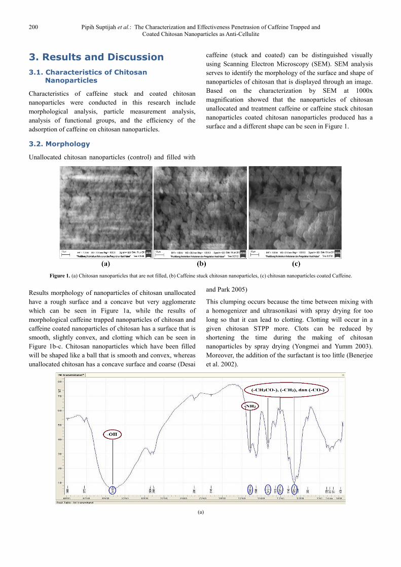
Unallocated chitosan nanoparticles (control) and filled with
caffeine (stuck and coated) can be distinguished visually
using Scanning Electron Microscopy (SEM). SEM analysis
serves to identify the morphology of the surface and shape of
nanoparticles of chitosan that is displayed through an image.
Based on the characterization by SEM at 1000x
magnification showed that the nanoparticles of chitosan
unallocated and treatment caffeine or caffeine stuck chitosan
nanoparticles coated chitosan nanoparticles produced has a
surface and a different shape can be seen in Figure 1.
Figure 1. (a) Chitosan nanoparticles that are not filled, (b) Caffeine stuck chitosan nanoparticles, (c) chitosan nanoparticles coated Caffeine.
Results morphology of nanoparticles of chitosan unallocated
have a rough surface and a concave but very agglomerate
which can be seen in Figure 1a, while the results of
morphological caffeine trapped nanoparticles of chitosan and
caffeine coated nanoparticles of chitosan has a surface that is
smooth, slightly convex, and clotting which can be seen in
Figure 1b-c. Chitosan nanoparticles which have been filled
will be shaped like a ball that is smooth and convex, whereas
unallocated chitosan has a concave surface and coarse (Desai
and Park 2005)
This clumping occurs because the time between mixing with
a homogenizer and ultrasonikasi with spray drying for too
long so that it can lead to clotting. Clotting will occur in a
given chitosan STPP more. Clots can be reduced by
shortening the time during the making of chitosan
nanoparticles by spray drying (Yongmei and Yumm 2003).
Moreover, the addition of the surfactant is too little (Benerjee
et al. 2002).
(a)
Journal of Nanoscience and Nanoengineering Vol. 1, No. 4, 2015, pp. 198-205 201
(b)
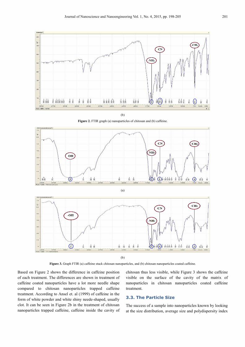
Figure 2. FTIR graph (a) nanoparticles of chitosan and (b) caffeine.
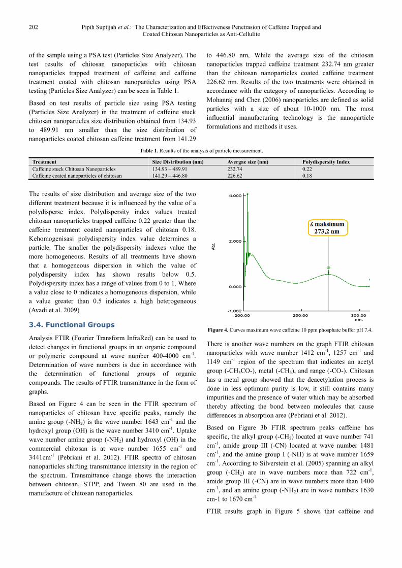
(a)
(b)
Figure 3. Graph FTIR (a) caffeine stuck chitosan nanoparticles, and (b) chitosan nanoparticles coated caffeine.
Based on Figure 2 shows the difference in caffeine position
of each treatment. The differences are shown in treatment of
caffeine coated nanoparticles have a lot more needle shape
compared to chitosan nanoparticles trapped caffeine
treatment. According to Ansel et. al (1999) of caffeine in the
form of white powder and white shiny neede-shaped, usually
clot. It can be seen in Figure 2b in the treatment of chitosan
nanoparticles trapped caffeine, caffeine inside the cavity of
chitosan thus less visible, while Figure 3 shows the caffeine
visible on the surface of the cavity of the matrix of
nanoparticles in chitosan nanoparticles coated caffeine
treatment.
3.3. The Particle Size
The success of a sample into nanoparticles known by looking
at the size distribution, average size and polydispersity index
202 Pipih Suptijah et al.: The Characterization and Effectiveness Penetrasion of Caffeine Trapped and
Coated Chitosan Nanoparticles as Anti-Cellulite
of the sample using a PSA test (Particles Size Analyzer). The
test results of chitosan nanoparticles with chitosan
nanoparticles trapped treatment of caffeine and caffeine
treatment coated with chitosan nanoparticles using PSA
testing (Particles Size Analyzer) can be seen in Table 1.
Based on test results of particle size using PSA testing
(Particles Size Analyzer) in the treatment of caffeine stuck
chitosan nanoparticles size distribution obtained from 134.93
to 489.91 nm smaller than the size distribution of
nanoparticles coated chitosan caffeine treatment from 141.29
to 446.80 nm, While the average size of the chitosan
nanoparticles trapped caffeine treatment 232.74 nm greater
than the chitosan nanoparticles coated caffeine treatment
226.62 nm. Results of the two treatments were obtained in
accordance with the category of nanoparticles. According to
Mohanraj and Chen (2006) nanoparticles are defined as solid
particles with a size of about 10-1000 nm. The most
influential manufacturing technology is the nanoparticle
formulations and methods it uses.
Table 1. Results of the analysis of particle measurement.
Treatment Size Distribution (nm) Avergae size (nm) Polydispersity Index
Caffeine stuck Chitosan Nanoparticles 134.93 – 489.91 232.74 0.22
Caffeine coated nanoparticles of chitosan 141.29 – 446.80 226.62 0.18
The results of size distribution and average size of the two
different treatment because it is influenced by the value of a
polydisperse index. Polydispersity index values treated
chitosan nanoparticles trapped caffeine 0.22 greater than the
caffeine treatment coated nanoparticles of chitosan 0.18.
Kehomogenisasi polydispersity index value determines a
particle. The smaller the polydispersity indexes value the
more homogeneous. Results of all treatments have shown
that a homogeneous dispersion in which the value of
polydispersity index has shown results below 0.5.
Polydispersity index has a range of values from 0 to 1. Where
a value close to 0 indicates a homogeneous dispersion, while
a value greater than 0.5 indicates a high heterogeneous
(Avadi et al. 2009)
3.4. Functional Groups
Analysis FTIR (Fourier Transform InfraRed) can be used to
detect changes in functional groups in an organic compound
or polymeric compound at wave number 400-4000 cm-1
.
Determination of wave numbers is due in accordance with
the determination of functional groups of organic
compounds. The results of FTIR transmittance in the form of
graphs.
Based on Figure 4 can be seen in the FTIR spectrum of
nanoparticles of chitosan have specific peaks, namely the
amine group (-NH2) is the wave number 1643 cm-1
and the
hydroxyl group (OH) is the wave number 3410 cm-1
. Uptake
wave number amine group (-NH2) and hydroxyl (OH) in the
commercial chitosan is at wave number 1655 cm-1
and
3441cm-1
(Pebriani et al. 2012). FTIR spectra of chitosan
nanoparticles shifting transmittance intensity in the region of
the spectrum. Transmittance change shows the interaction
between chitosan, STPP, and Tween 80 are used in the
manufacture of chitosan nanoparticles.
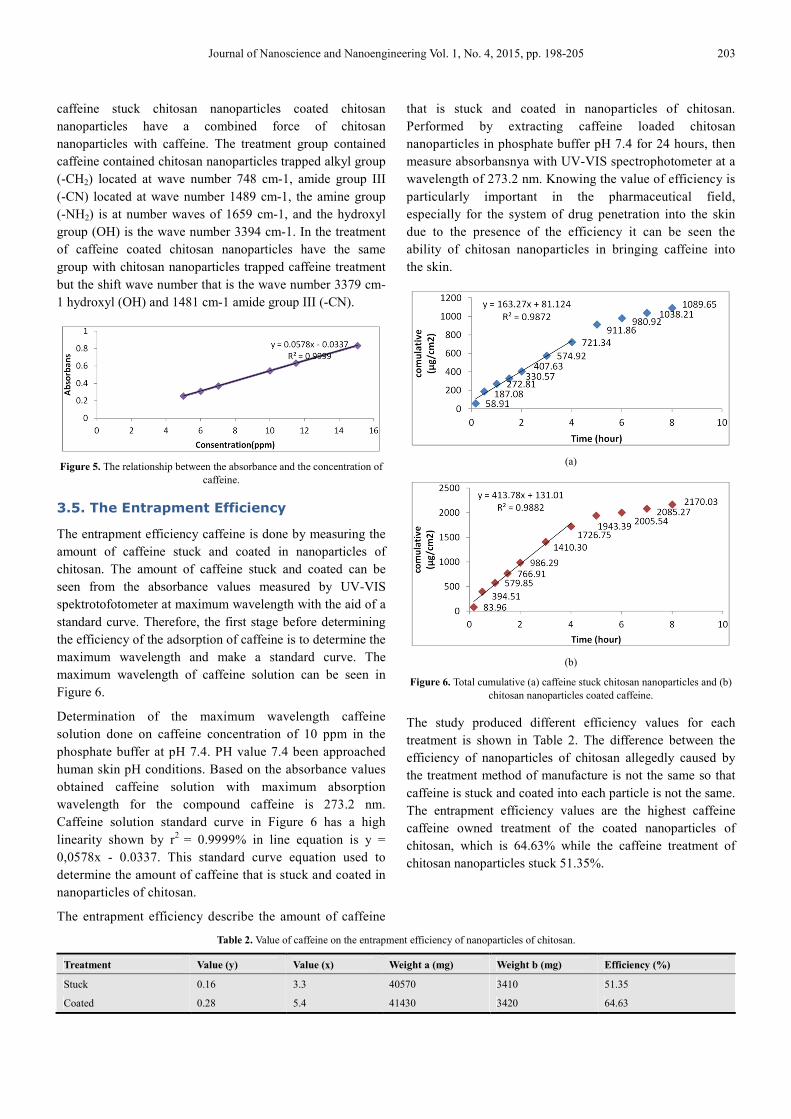
Figure 4. Curves maximum wave caffeine 10 ppm phosphate buffer pH 7.4.
There is another wave numbers on the graph FTIR chitosan
nanoparticles with wave number 1412 cm-1
, 1257 cm-1
and
1149 cm-1
region of the spectrum that indicates an acetyl
group (-CH3CO-), metal (-CH3), and range (-CO-). Chitosan
has a metal group showed that the deacetylation process is
done in less optimum purity is low, it still contains many
impurities and the presence of water which may be absorbed
thereby affecting the bond between molecules that cause
differences in absorption area (Pebriani et al. 2012).
Based on Figure 3b FTIR spectrum peaks caffeine has
specific, the alkyl group (-CH2) located at wave number 741
cm-1
, amide group III (-CN) located at wave number 1481
cm-1
, and the amine group I (-NH) is at wave number 1659
cm-1
. According to Silverstein et al. (2005) spanning an alkyl
group (-CH2) are in wave numbers more than 722 cm-1
,
amide group III (-CN) are in wave numbers more than 1400
cm-1
, and an amine group (-NH2) are in wave numbers 1630
cm-1 to 1670 cm-1.
FTIR results graph in Figure 5 shows that caffeine and
Journal of Nanoscience and Nanoengineering Vol. 1, No. 4, 2015, pp. 198-205 203
caffeine stuck chitosan nanoparticles coated chitosan
nanoparticles have a combined force of chitosan
nanoparticles with caffeine. The treatment group contained
caffeine contained chitosan nanoparticles trapped alkyl group
(-CH2) located at wave number 748 cm-1, amide group III
(-CN) located at wave number 1489 cm-1, the amine group
(-NH2) is at number waves of 1659 cm-1, and the hydroxyl
group (OH) is the wave number 3394 cm-1. In the treatment
of caffeine coated chitosan nanoparticles have the same
group with chitosan nanoparticles trapped caffeine treatment
but the shift wave number that is the wave number 3379 cm-
1 hydroxyl (OH) and 1481 cm-1 amide group III (-CN).
Figure 5. The relationship between the absorbance and the concentration of
caffeine.
3.5. The Entrapment Efficiency
The entrapment efficiency caffeine is done by measuring the
amount of caffeine stuck and coated in nanoparticles of
chitosan. The amount of caffeine stuck and coated can be
seen from the absorbance values measured by UV-VIS
spektrotofotometer at maximum wavelength with the aid of a
standard curve. Therefore, the first stage before determining
the efficiency of the adsorption of caffeine is to determine the
maximum wavelength and make a standard curve. The
maximum wavelength of caffeine solution can be seen in
Figure 6.
Determination of the maximum wavelength caffeine
solution done on caffeine concentration of 10 ppm in the
phosphate buffer at pH 7.4. PH value 7.4 been approached
human skin pH conditions. Based on the absorbance values
obtained caffeine solution with maximum absorption
wavelength for the compound caffeine is 273.2 nm.
Caffeine solution standard curve in Figure 6 has a high
linearity shown by r2
= 0.9999% in line equation is y =
0,0578x - 0.0337. This standard curve equation used to
determine the amount of caffeine that is stuck and coated in
nanoparticles of chitosan.
The entrapment efficiency describe the amount of caffeine
that is stuck and coated in nanoparticles of chitosan.
Performed by extracting caffeine loaded chitosan
nanoparticles in phosphate buffer pH 7.4 for 24 hours, then
measure absorbansnya with UV-VIS spectrophotometer at a
wavelength of 273.2 nm. Knowing the value of efficiency is
particularly important in the pharmaceutical field,
especially for the system of drug penetration into the skin
due to the presence of the efficiency it can be seen the
ability of chitosan nanoparticles in bringing caffeine into
the skin.
(a)
(b)
Figure 6. Total cumulative (a) caffeine stuck chitosan nanoparticles and (b)
chitosan nanoparticles coated caffeine.
The study produced different efficiency values for each
treatment is shown in Table 2. The difference between the
efficiency of nanoparticles of chitosan allegedly caused by
the treatment method of manufacture is not the same so that
caffeine is stuck and coated into each particle is not the same.
The entrapment efficiency values are the highest caffeine
caffeine owned treatment of the coated nanoparticles of
chitosan, which is 64.63% while the caffeine treatment of
chitosan nanoparticles stuck 51.35%.
Table 2. Value of caffeine on the entrapment efficiency of nanoparticles of chitosan.
Treatment Value (y) Value (x) Weight a (mg) Weight b (mg) Efficiency (%)
Stuck 0.16 3.3 40570 3410 51.35
Coated 0.28 5.4 41430 3420 64.63
204 Pipih Suptijah et al.: The Characterization and Effectiveness Penetrasion of Caffeine Trapped and
Coated Chitosan Nanoparticles as Anti-Cellulite
The entrapment efficiency factor is not one aspect of which is
reviewed to determine the feasibility of chitosan
nanoparticles as systems of drug penetration into the skin
(Silva 2006). The higher the value, the better the expected
efficiency of the formulation because the amount of caffeine
that is entrapped in the chitosan nanoparticles more. The high
value of the efficiency of treatment of caffeine coated
nanoparticles of chitosan may be caused by participating
terekstraksinya whole caffeine coated in nanoparticles of
chitosan either on the surface or in the cavity of the matrix of
nanoparticles then caffeine would be more easily extracted
out, while the treatment of caffeine trapped nanoparticles of
chitosan inside the cavity chitosan will require a long time to
extracted out (Wahyono 2010).
3.6. Penetration Effectiveness Stuck
Caffeine and Chitosan Nanoparticles
Coated
In vitro penetration test is performed using Franz diffusion
cells. Penetration testing was conducted to determine the
amount of caffeine that can penetrate the membrane during
a certain time interval of chitosan nanoparticles. The
membranes used are skin abdominal strain Sprague Dawley
female rats aged 2-3 months, weighing ± 150 grams. Rat
skin membrane is used because it has a permeability which
is almost equal to the permeability of human skin
(Rawlings 2006).
Franz diffusion cell penetration test performed for 8 hours
with intervals of 10, 30, 60, 90, 120, 180, 240, 300, 360,
and 480 minutes. Each time interval of 0.5 mL of fluid
samples taken from the receptor compartment and diluted to
5 mL with phosphate buffer pH 7.4 (Franz 2005). Each
fluid samples taken from the receptor compartment must
always be replaced with the same amount of volume of
liquid that is picked to maintain the receptor fluid volume
remains kostan. 5 mL sample dilution is measured by uv-vis
spectrophotometer at a wavelength of 273.2 nm to
determine the absorbance.
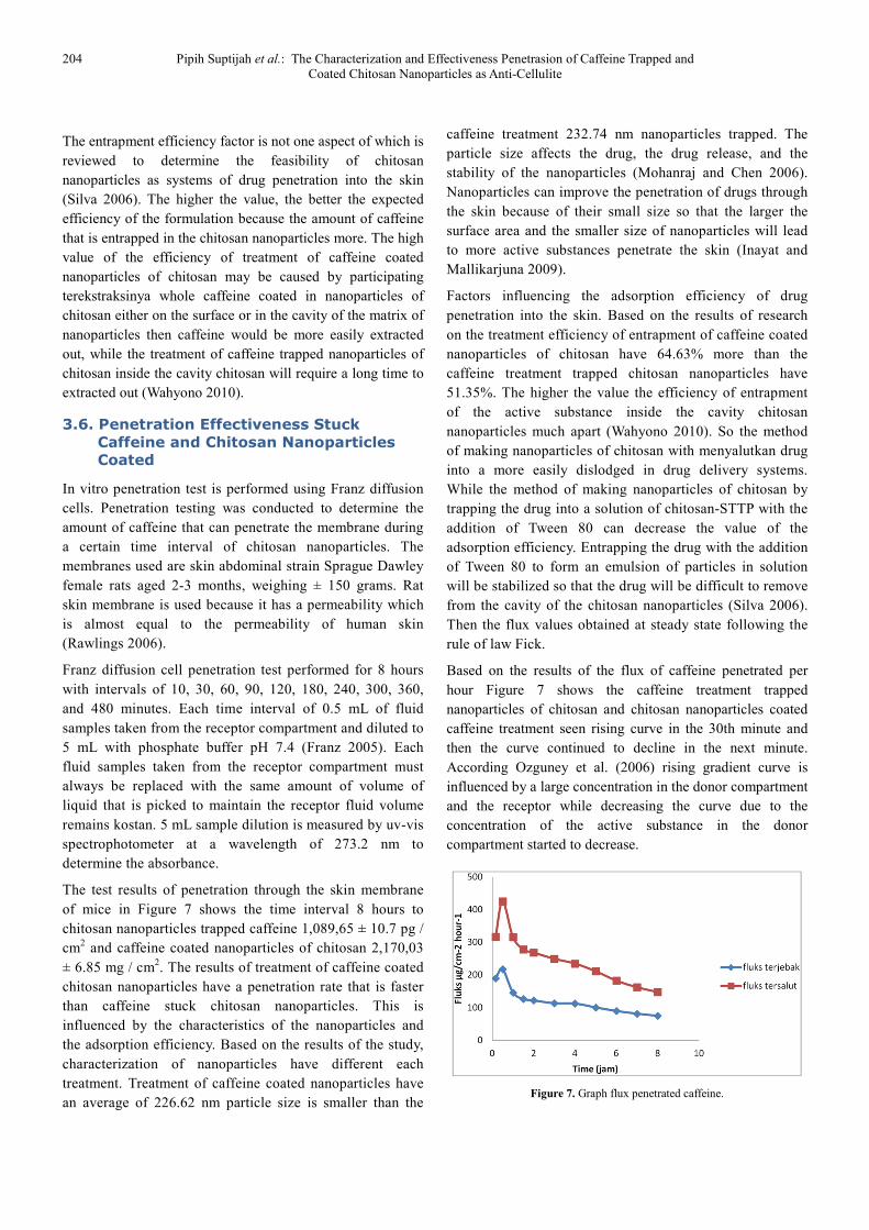
The test results of penetration through the skin membrane
of mice in Figure 7 shows the time interval 8 hours to
chitosan nanoparticles trapped caffeine 1,089,65 ± 10.7 pg /
cm2 and caffeine coated nanoparticles of chitosan 2,170,03
± 6.85 mg / cm2. The results of treatment of caffeine coated
chitosan nanoparticles have a penetration rate that is faster
than caffeine stuck chitosan nanoparticles. This is
influenced by the characteristics of the nanoparticles and
the adsorption efficiency. Based on the results of the study,
characterization of nanoparticles have different each
treatment. Treatment of caffeine coated nanoparticles have
an average of 226.62 nm particle size is smaller than the
caffeine treatment 232.74 nm nanoparticles trapped. The
particle size affects the drug, the drug release, and the
stability of the nanoparticles (Mohanraj and Chen 2006).
Nanoparticles can improve the penetration of drugs through
the skin because of their small size so that the larger the
surface area and the smaller size of nanoparticles will lead
to more active substances penetrate the skin (Inayat and
Mallikarjuna 2009).
Factors influencing the adsorption efficiency of drug
penetration into the skin. Based on the results of research
on the treatment efficiency of entrapment of caffeine coated
nanoparticles of chitosan have 64.63% more than the
caffeine treatment trapped chitosan nanoparticles have
51.35%. The higher the value the efficiency of entrapment
of the active substance inside the cavity chitosan
nanoparticles much apart (Wahyono 2010). So the method
of making nanoparticles of chitosan with menyalutkan drug
into a more easily dislodged in drug delivery systems.
While the method of making nanoparticles of chitosan by
trapping the drug into a solution of chitosan-STTP with the
addition of Tween 80 can decrease the value of the
adsorption efficiency. Entrapping the drug with the addition
of Tween 80 to form an emulsion of particles in solution
will be stabilized so that the drug will be difficult to remove
from the cavity of the chitosan nanoparticles (Silva 2006).
Then the flux values obtained at steady state following the
rule of law Fick.
Based on the results of the flux of caffeine penetrated per
hour Figure 7 shows the caffeine treatment trapped
nanoparticles of chitosan and chitosan nanoparticles coated
caffeine treatment seen rising curve in the 30th minute and
then the curve continued to decline in the next minute.
According Ozguney et al. (2006) rising gradient curve is
influenced by a large concentration in the donor compartment
and the receptor while decreasing the curve due to the
concentration of the active substance in the donor
compartment started to decrease.
Figure 7. Graph flux penetrated caffeine.
Journal of Nanoscience and Nanoengineering Vol. 1, No. 4, 2015, pp. 198-205 205
4. Conclusion
Caffeine chitosan nanoparticles were made using two
different treatments namely caffeine stuck and coated
chitosan nanoparticles. Characteristics of nanoparticles of
chitosan caffeine generated in this study is based on the
morphology of the caffeine treatment trapped nanoparticles
of chitosan and chitosan nanoparticles coated caffeine has a
smooth surface and slightly convex but slightly lumpy, the
average size of a row 232.74 nm and 226.62 nm. Analysis of
functional groups that showed a shift wavenumber
wavenumber amide group III (-CN) and wave number of the
hydroxyl group (OH), as well as the efficiency of entrapment
of the highest caffeine caffeine owned treatment of the coated
nanoparticles of chitosan 64.63% while the caffeine
treatment trapped nanoparticles 51.35% chitosan. Caffeine
chitosan nanoparticle characterization results demonstrate the
effectiveness of the penetration value in the treatment of
caffeine coated chitosan nanoparticles have a penetration rate
that is faster 2170.03 ± 6.85 mg / cm2 compared to chitosan
nanoparticles trapped caffeine treatment 1089.65 ± 10.7 mg /
cm2.
Refferences
[1] Ansel H, LV Allen, NG Popovich 1999. Pharmaceutical Dosage Forms and Drug Systems 7th edition. Maryland: Lippincot Williams and Willkins.
[2] Avadi MR, Sadeghi AM, M Mohammadpour. 2009. Preparation and characterization of insulin nanoparticles using chitosan and arabic gum with ionic gelation metod. Journal Nanomeicine: 6. 58-63.
[3] Benerjee T, Mitra S, Singh AK, Sharma RK, Maitra A. 2002. Preparation, characterization, biodistribution of ultrafine chitosan nanoparticles. International Journal of Pharmaceutics. 243. 93-105.
[4] Desai KGH, Park HJ. 2005. Preparation and characterization of drug-loaded chitosan-tripolyphosphate microspheres by spray drying. Journal Drug Development Research. 64: 114-128.
[5] Franz H. 2005. Phosphatdylcholine treatment to induce lipolysis. Journal of Cosmetic Dermatology. 4: 308-313.
[6] Inayat BP, Mallikarjuna S. 2009. Chemical penetration enhancer for transdermal drug delivery systems. Tropical Journal of Pharmaceutical Research. 8: 173-179.
[7] Lee DW, Shirley SA, Lockey RF, Mohapatra SS. 2006.
Thiolated chitosan nanoparticles enhace anti-inflammatory effects of intranasally delivered theophylline. Journal Medical Central. 7: 1-10.
[8] Mohanraj UJ, Chen Y. 2006. Nanoparticles – A Review. Tropical Journal of Pharmaceutical Research. 5 (1): 561-573.
[9] Murray RK, Ganner DK, Mayes PA, Rodwell VW. 2003. Harper’s Biochemistry. Melville: EGC.
[10] Ozguney IS, HY Karasulu, G Katarci, S Sozer, T Guneri, G Ertan. 2006. Transdermal delivery of diclofenac sodium though rat skin from various formulations. Journal Pharmaceutical Sciences Technology. 7(4): 88-103.
[11] Pebriani RH, Rilda Y, Zulhajri. 2012. Modifikasi Komposisi Kitosan Pada Proses Sintesis Komposit TiO2 – Kitosan. Jurnal Kimia Universitas Padjajaran. 1: 12-34.
[12] Rawling AV. 2006. Cellulite and its treatment. Journal Cosmetic Science. 28: 175-190.
[13] Rona C, Carrera M, Berardesca E. 2006. Testing anticellulite products. International Journal of Cosmetic Sciences. 28:169-173.
[14] Rossi ABR, Vergnanini AL. 2000. Cellulite. Journal of Pharmaceutical Research. 14: 121-262.
[15] Silva CM. 2006. Microencapsulation of hemoglobin in chitosan-coasted alginate microspheres prepared by emulsification internal gelation. Pharmaceutical Sciences Journal. 7 (4): 78-89.
[16] Silverstein RM, Webster FX, Kiemle DJ. 2005. Spectrometric identification of organic compounds. English: Willey Amazon.
[17] Sutriyo, Joshita D, Indah R. 2005. Perbandingan pelepasan propanol hidroklorida dari matriks kitosan, etil selulosa, dan hidroksipropil metal selulosa. Majalah Ilmu Kefarmasian. 2: 145-153.
[18] Tiyaboonchai W. 2003. Chitosan nanoparticles: a promosing system for drug delivery. Naresuan University Journal. 11 (3): 51-66.
[19] Wahyono D. 2010. Ciri nanopartikel kitosan dan pengaruhnya pada ukuran partikel dan efisiensi penyaluran ketoprofen [tesis]. Bogor (ID): Institut Pertanian Bogor.
[20] Wang X, Du Y, Liu H. 2004. Preparation, characterization, and antimicrobial activity of chitosan-Zn Complex. Journal of Medical Sciences. 56: 21-26.
[21] [WHO] World Health Organization. 2012. Cellulite [internet]. [diunduh 2012 April 8]. Tersedia pada http://www.who.int /mediacentre/ factsheets/ fs117/en/ index.html.
[22] Yongmei X, Yumm D. 2003. Effect of moleculer structure of chitosan on protein delivery properties of chitosan nanoparticles. International Journal of Pharmaceutics. 250: 215-226.