Embed Size (px)
Citation preview

_______________________________________
Characterization of actin variants in
Dictyostelium discoideum
_______________________________________
Dissertation der Fakultät für Biologie der Ludwig-Maximilians-Universität München zur
Erlangung des akademischen Grades
„Doktor der Naturwissenschaften“
(Dr. rer. nat.)
vorgelegt von
Linda Sanftenberg
München, 2014
Eidesstattliche Erklärung
Ich versichere hiermit an Eides statt, dass die vorgelegte Dissertation von mir selbständig und ohne
unerlaubte Hilfe angefertigt worden ist.
München, den 10.09.14
Linda Sanftenberg
Ort der Durchführung
Der experimentelle Teil der Dissertation wurde von Oktober 2011 bis Juni 2014 im Labor von Prof. Dr.
Michael Schleicher am Institut für Anatomie und Zellbiologie der Ludwig-Maximilians-Universität
München ausgeführt.
1.Gutachter: Prof. Dr. Michael Schleicher
2.Gutachter: Prof. Dr. Angelika Boettger
Eingereicht am: 10.09.14
Tag der mündlichen Prüfung: 17.11.14

Presentations _____________________________________________________________________________________
Poster Presentations
Sanftenberg L, Gallinger J, Gallinger C, Joseph JM, Rieger D, Henze F,
Müller-Taubenberger A, Schleicher M,
Actin variants in Dictyostelium discoideum,
ASCB 53th international meeting, 2013, New Orleans, LA, USA
Sanftenberg L, Gallinger J, Gallinger C, Joseph JM, Rieger D, Henze F,
Müller-Taubenberger A, Schleicher M,
Actin variants in Dictyostelium amoebae,
Annual Meeting of the German Society for Cell Biology, 2014, Regensburg, Germany
Sanftenberg L, Gallinger J, Gallinger C, Joseph JM, Rieger D, Henze F,
Müller-Taubenberger A, Schleicher M,
Characterization of actin variants in D. discoideum,
Annual International Dictyostelium Conference, 2014, Potsdam, Germany

Table of contents _____________________________________________________________________________________
Table of contents
Summary 1
Zusammenfassung 3
1 Introduction 5 1.1. D. discoideum as a model organism 5 1.2. Structure and domains of actin 6 1.3. The actin cytoskeleton 7 1.4. The actinome of D. discoideum and its regulation 11 1.5. The actin variants Act3, Act18 and Act31 in D. discoideum 13 1.6. Aims of the thesis 16
2 Materials and Methods 17 2.1 Materials 17
2.1.1 Instruments 17 2.1.2 Computer programs 18 2.1.3 Laboratory consumables 19 2.1.4 Reagents 19 2.1.5 Antibodies 20 2.1.6 Vectors 20 2.1.7 Bacterial strains 20 2.1.8 Yeast strains 21 2.1.9 D. discoideum strains 21
2.2 Methods 22 2.2.1 Molecular biological methods 22 2.2.2 Biochemical methods 22
2.2.2.1.1 SDS-polyacrylamide gel electrophoresis and western blotting 22 2.2.2.1.2 Commassie and silver staining 22 2.2.2.1.3 Actin preparation from rabbit skeletal muscle 23 2.2.2.1.4 Labeling of actin with pyrene 24 2.2.2.1.5 Flag-tagged protein expression using the baculoviral
amplification system 24 2.2.2.1.6 In vitro actin polymerization assay 25 2.2.2.1.7 Low shear viscometry 25 2.2.2.1.8 Gel filtration using the AEKTA 100 system 26 2.2.2.1.9 Transmission electron microscopy 26 2.2.2.1.10 Immunoprecipitation 26
2.2.3 Cell biological methods 27 2.2.3.1.1 Cell culture and transformation of D. discoideum 27 2.2.3.1.2 Live-cell microscopy of D. discoideum 27 2.2.3.1.3 Immunofluorescence microscopy 28 2.2.3.1.4 Induction of nuclear and cytoplasmic rods 28 2.2.3.1.5 Growth and germination 28 2.2.3.1.6 Phagocytosis measurements 29 2.2.3.1.7 Analysis of phototaxis and development 29
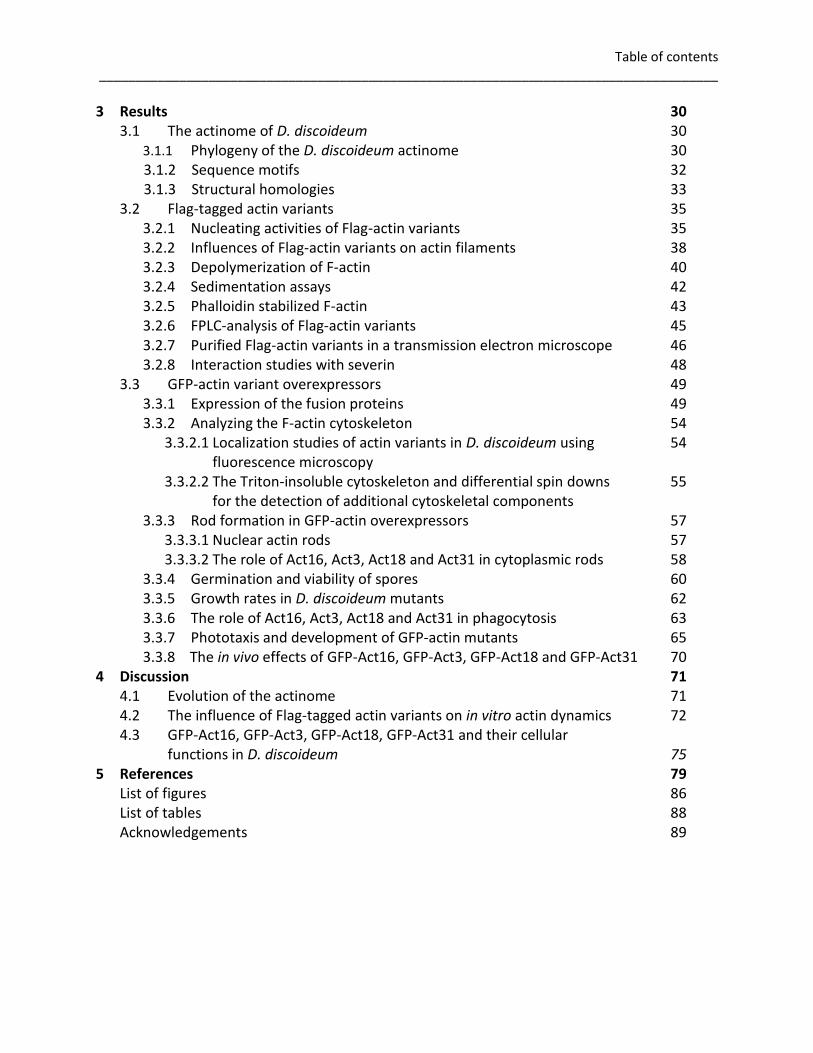
Table of contents _____________________________________________________________________________________
3 Results 30 3.1 The actinome of D. discoideum 30
3.1.1 Phylogeny of the D. discoideum actinome 30
3.1.2 Sequence motifs 32 3.1.3 Structural homologies 33
3.2 Flag-tagged actin variants 35 3.2.1 Nucleating activities of Flag-actin variants 35 3.2.2 Influences of Flag-actin variants on actin filaments 38 3.2.3 Depolymerization of F-actin 40 3.2.4 Sedimentation assays 42 3.2.5 Phalloidin stabilized F-actin 43 3.2.6 FPLC-analysis of Flag-actin variants 45 3.2.7 Purified Flag-actin variants in a transmission electron microscope 46 3.2.8 Interaction studies with severin 48
3.3 GFP-actin variant overexpressors 49 3.3.1 Expression of the fusion proteins 49 3.3.2 Analyzing the F-actin cytoskeleton 54
3.3.2.1 Localization studies of actin variants in D. discoideum using 54 fluorescence microscopy
3.3.2.2 The Triton-insoluble cytoskeleton and differential spin downs 55 for the detection of additional cytoskeletal components
3.3.3 Rod formation in GFP-actin overexpressors 57 3.3.3.1 Nuclear actin rods 57 3.3.3.2 The role of Act16, Act3, Act18 and Act31 in cytoplasmic rods 58
3.3.4 Germination and viability of spores 60 3.3.5 Growth rates in D. discoideum mutants 62 3.3.6 The role of Act16, Act3, Act18 and Act31 in phagocytosis 63 3.3.7 Phototaxis and development of GFP-actin mutants 65 3.3.8 The in vivo effects of GFP-Act16, GFP-Act3, GFP-Act18 and GFP-Act31 70
4 Discussion 71 4.1 Evolution of the actinome 71 4.2 The influence of Flag-tagged actin variants on in vitro actin dynamics 72 4.3 GFP-Act16, GFP-Act3, GFP-Act18, GFP-Act31 and their cellular
functions in D. discoideum 75 5 References 79
List of figures 86 List of tables 88 Acknowledgements 89
Summary _____________________________________________________________________________________
1
Summary
The actin cytoskeleton in eukaryotic cells is crucial for a wide range of cellular functions
including cell shape changes, cell motility, cell division or intracellular transport. As
Dictyostelium discoideum harbors a relatively large actinome composed of 33 actin genes and
eight genes that code for actin related proteins, this social amoebae serves as an excellent
model organism to study the actin system. The main objective of this study was to get an
overview of the huge variety of actin variants in D. discoideum. Conventional actin is encoded
by 17 distinct genes (Act8 group), whereas the protein sequences of the other 16 actin variants
can be almost identical with conventional actin or differ rather drastically. To cover a broad
range of relative similarities we studied Act3 (97% identity), Act18 (88% identity) and Act31
(37% identity). As a reference we used Act16, a member of the Act8 group.
Although the amino acid sequences of the compared actin variants show different levels of
similarity, the alignment of the sequence motifs and the computed ribbon models suggest that
structure and fundamental functions of the compared proteins are strongly conserved.
We used Sf9 cells to express the actin variants Flag-Act16, Flag-Act3, Flag-Act18 and Flag-Act31.
The conducted in vitro experiments showed that Flag-Act16 and Flag-Act3 promote the
polymerization of short actin filaments, whereas Flag-Act18 and Flag-Act31 do not affect actin
polymerization. Cosedimentation assays indicated that Flag-Act16, Flag-Act3 and Flag-Act31 are
associated with polymerized actin, whereas Flag-Act18 cannot be detected together with actin
filaments. Flag-Act16 and Flag-Act31 are not able to form filaments without conventional actin,
not even in the presence of stabilizing phalloidin. Patches of assembled Flag-Act3 and Flag-
Act18 monomers occurred when we added phalloidin. Gel filtration assays propose a
tetrameric structure of Flag-Act16. Furthermore, denatured aggregates of Flag-Act3 are
detectable, whereas an ordered oligomerization of Flag-Act18 and Flag-Act31 can be excluded.
Images taken at the electron microscope suggest that none of the tested Flag-tagged actin
variants is able to form stable actin filaments without conventional actin. Additionally, the
formation of the actin network is not disturbed by the Flag-tagged variants. Protein interaction
studies showed that the actin-binding protein severin binds Flag-Act16, Flag-Act3, Flag-Act18
and Flag-Act31 in a Ca2+-dependent manner.
Summary _____________________________________________________________________________________
2
In vivo studies were performed using GFP-Act16, GFP-Act3, GFP-Act18 and GFP-Act31
overexpressing cells of D. discoideum. Immunofluorescence studies and biochemical
approaches showed that GFP-Act16 and GFP-Act3 are colocalizing with endogenous actin in the
cell cortex, and partially in the cytoplasm. In contrast, GFP-Act18 and GFP-Act31 seem to be
soluble proteins without interactions with the actin cytoskeleton. A most peculiar behavior is
the stress-induced appearance of GFP-Act16 and GFP-Act3 in nuclear actin rods. GFP-Act18 and
GFP-Act31 expressing cells are able to form nuclear rods, but these actin variants are not part
of the rods. The tested GFP-actin variant overexpressors do not form typical needle-shaped
cytoplasmic rods, but GFP-Act16 and GFP-Act3 are part of crescent-shaped aggregates within
the cytoplasm.
As the viability of GFP-actin variant overexpressing spores is reduced in all mutants, we assume
a disturbed activation of the actin cytoskeleton due to the excess of GFP-actin variant
monomers, which could sequester actin binding proteins that are not available anymore
for constitutively synthesized actins during germination. The size and shape of the tested
spores is not affected as well as the overexpression of the GFP-actin variants has no influence
on cytokinesis. GFP-Act16 and GFP-Act3 are accumulated in the phagocytic cup, whereas GFP-
Act18 and GFP-Act31 are not enriched in this actin driven structure. During the 24 hours
developmental cycle, smaller fruiting bodies of GFP-Act18 overexpressing cells and their
increased density per area are conspicuous. In parallel, the expression of the aggregation
marker glycoprotein A (CsA) is reduced in GFP-Act18 cells, which could restrict the formation of
stable intercellular contacts.
Taken together, these data suggest that Act16 and Act3 are part of the F-actin network,
whereas less related isoforms like Act18 and Act31 could even exhibit cytoskeleton-
independent subcellular functions.
Zusammenfassung _____________________________________________________________________________________
3
Zusammenfassung
Das Aktinzytoskelett in Nicht-Muskelzellen ist äußerst wichtig für eine Vielzahl zellulärer
Funktionen wie die Dynamik der Zellform, der Zellmigration, der Zellteilung oder auch des
intrazellulären Transports. Da Dictyostelium discoideum ein relativ großes Aktinom besitzt,
bestehend aus 33 Aktingenen und acht Genen, die für aktinverwandte Proteine codieren, bietet
sich diese soziale Amoebe als ausgezeichneter Modellorganismus an, um ausgewählte
Komponenten des Aktinsystems zu untersuchen. Ziel dieser Arbeit war es, sich einen Überblick
über die enorme Vielfalt der Aktinvarianten in D. discoideum zu verschaffen. Konventionelles
Aktin wird über 17 verschiedene Gene (Akt8 Gruppe) codiert, wohingegen die 16 übrigen
Aktinvarianten mehr oder weniger große Unterschiede zu konventionellem Aktin aufweisen.
Die wichtigsten Aktine dieser Arbeit waren: Akt16, ein Mitglied der Akt8 Gruppe
konventionellen Aktins (100% Referenz), Akt3 (zu 97% identisch), Akt18 (zu 88% identisch) und
Akt31 (zu 37% identisch).
Obwohl die Aminosäuresequenzen der verglichenen Aktinvarianten verschiedene Stufen an
Ähnlichkeit zueinander aufweisen, verdeutlichen der Abgleich der Sequenzmotive und die
berechneten 3D Modelle, dass die Strukturen und grundlegenden Funktionen der verglichenen
Proteine stark konserviert sind.
Wir nutzten Sf9 Zellen, um die Aktinvarianten Flag-Akt16, Flag-Akt3, Flag-Akt18 und Flag-Akt31
zu exprimieren. In vitro Versuche zeigten, dass Flag-Akt16 und Flag-Akt3 die Polymerisation
kurzer Aktinfilamente fördern, wohingegen Flag-Akt18 und Flag-Akt31 sich nicht auf die
Aktinpolymerisation auswirken. Versuche zur Cosedimentation deuteten darauf hin, dass Flag-
Akt16, Flag-Akt3 und Flag-Akt31 mit polymerisiertem Aktin verbunden sind, dies gilt nicht für
Flag-Akt18. Flag-Akt16 und Flag-Akt31 können keine Aktinfilamente ohne konventionelles Aktin
ausbilden, auch nicht unter Verwendung von stabilisierendem Phalloidin. Jedoch konnten unter
Einfluss von Phalloidin Stücke aneinandergefügter Flag-Akt3 und Flag-Akt18 Monomere
nachgewiesen werden. Gelfiltrationsmessungen deuten auf eine tetramere Struktur von Flag-
Act16 hin. Des Weiteren sind denaturierte Aggregate von Flag-Akt3 nachweisbar, wohingegen
eine geordnete Oligomerisierung von Flag-Akt18 und Flag-Akt31 ausgeschlossen werden kann.
Elektronenmikroskopische Aufnahmen lassen vermuten, dass keine der untersuchten Flag-
Aktinvarianten stabile Filamente ohne konventionelles Aktin hervorbringt. Die Ausbildung des
Aktinnetzwerks wird von den Flag-Aktinvarianten nicht gestört. Proteininteraktionsstudien
zeigten, dass das aktinbindende Protein Severin Ca2+-abhängig an Flag-Akt16, Flag-Akt3, Flag-
Akt18 und Flag-Akt31 bindet.
Zusammenfassung _____________________________________________________________________________________
4
In vivo Experimente wurden unter Einsatz von GFP-Akt16, GFP-Akt3, GFP-Akt18 und GFP-Akt31
überexprimierenden Zellen von D. discoideum durchgeführt. Studien zur Immunfluoreszenz und
biochemische Ansätze zeigten, dass GFP-Akt16 und GFP-Akt3 gemeinsam mit endogenem Aktin
im Zellcortex und teilweise im Zytoplasma lokalisieren. Hingegen scheinen GFP-Akt18 und GFP-
Akt31 lösliche Proteine des Zytoplasmas zu sein. Besonders auffällig ist das stressinduzierte
Auftreten von GFP-Akt16 und GFP-Akt3 in intranukleären, stäbchenförmigen Proteinaggregaten
(„nuclear rods“). GFP-Akt18 und GFP-Akt31 Zellen bilden zwar nuclear rods aus, beide
Aktinvarianten sind aber nicht Teil dieser Strukturen. Die untersuchten GFP-Aktin
Überexpressoren bilden nicht die typischen nadelförmigen cytoplasmatischen Aktinbündel,
jedoch sind GFP-Akt16 und GFP-Akt3 Teil halbmondförmiger Aggregate innerhalb des
Zytoplasmas.
Da die Keimfähigkeit aller GFP-Aktin überexprimierenden Sporen reduziert ist, gehen wir von
einer gestörten Aktivierung des Aktinzytoskeletts aus, welche durch den Überschuss an GFP-
Aktin Monomeren entsteht. Aktinbindeproteine könnten dadurch abfangen werden, die folglich
nicht mehr den konstitutiv synthetisierten Aktinen während der Keimung zur Verfügung stehen.
Größe und Form der untersuchten Sporen waren nicht verändert. Die Zytokinese selbst ist bei
keiner GFP-Aktin Mutante verändert. GFP-Akt16 und GFP-Akt3 reichern sich in Phagozytosen
an, wohingegen GFP-Akt18 und GFP-Akt31 hier nicht konzentriert vorliegen. Während des 24
Stunden dauernden Entwicklungszyklus fallen kleinere Fruchtkörper der GFP-Akt18
Überexpressoren und deren erhöhte Dichte auf. Zugleich ist die Expression des Markerproteins
für Zellaggregation Glycoprotein A verringert, so dass die Ausprägung stabiler Kontakte
zwischen den einzelnen Zellen begrenzt ist.
Zusammenfassend weisen diese Daten darauf hin, dass Akt16 und Akt3 Teile des F-
Aktinnetzwerks sind, wohingegen weniger nah verwandte Isoformen wie Akt18 und Akt31
möglicherweise sogar Funktionen unabhängig vom Zytoskelett besitzen.

Introduction _____________________________________________________________________________________
5
1. Introduction
1.1 Dictyostelium discoideum as a model organism
The eukaryotic soil amoeba Dictyostelium discoideum (D. discoideum) is an excellent model
organism to study cellular processes. Dictyostelia belong to the phylum mycetazoa and are
described as social organisms (Raper, 1935). Cellular dynamics as growth, germination, cell
adhesion, phagocytosis, development and phototaxis can be analyzed easily, as well as the
required signaling processes. Moreover, the high motility of D. discoideum facilitates insights
into the actin cytoskeleton and its regulatory machinery. The extraordinary life cycle classifies
the amoeba as a linker between unicellular and multicellular organisms due to its transition
from autonomous single cells to higher organized organisms. Usually the professional
phagocyte lives on forest soil and feeds on bacteria, but upon starvation it undergoes a specific
developmental program, which leads to the aggregation of about 105 single cells induced and
mediated by a cyclic adenosine monophosphate (cAMP) gradient. The movement of an
organism in response to a chemical stimulus is called chemotaxis. Later on, these aggregates
can form slug-shaped bodies, which migrate towards light sources.
The slug rises from the underlying substratum during culmination and forms a fruiting body
consisting of a basal disk, stalk and a spore head, completing the life cycle. The spores of the
fruiting body are useful to endure harsh environmental periods as starvation, heat or frost.
Under favorable conditions germination into amoebae is induced. The entire developmental
cycle can be completed within 24 hours under laboratory conditions (Chisholm & Firtel, 2004).
Figure 1: Life cycle of D. discoideum (Chisholm
& Firtel, 2004). Single, vegetative cells start the
developmental cycle to form a mature fruiting
body, by developing a multicellular organism
induced via cAMP. Directional streaming of the
cells into multicellular aggregates is
characteristic for this part of morphogenesis.
The resulting multicellular organism is called
mound, respectively tipped mound in the next
phase of the cycle. The arising finger forms the
adjacent slug, which culminates into a fruiting
body. The head of the fruiting body incorporates
the elliptical spores.
Introduction _____________________________________________________________________________________
6
With a total size of 34Mb the genome of D. discoideum contains about 12.000 genes and has
therefore the extent of the Drosophila genome. The availability of the completely sequenced
genome was an enormous help towards our understanding of actins and actin-related proteins
(ARPs; Eichinger et al., 2005). As a founder of a large protein family, any sequenced genome can
be used to study actin. We selected the recently unraveled D. discoideum genome since it
contains 33 genes that code for bona fide actin. In contrast, the genome of the budding yeast
contains only one single gene that codes for actin, mouse harbors 35, and the plant Arabidopsis
thaliana contains 10 actin genes. As a conclusion the number of actin genes does not tell us
very much about the complexity of an organism. Elimination of redundant genes during
evolution can only be avoided if they represent a selective advantage and it is still unclear why
evolution allows this seemingly luxurious feature (Schleicher & Jockusch, 2008). The haploid
genome of D. discoideum is easily susceptible to manipulation via recombinational methods
and facilitates analyzing the functions of single protein isoforms. We used the wild type strain
AX2 in this work that can grow in the simplified axenic media AX and HL5 (Schwalb & Roth,
1970). Consequently, laboratory culture is easy and inexpensive, highly accessible for
biochemical, molecular and cell biological studies. To sum up, D. discoideum is a prime
organism to analyze the activities of single molecules in their cellular environment.
1.2 Structure and domains of actin
Despite of their different protein sequences, actin turned out to be a structural homolog of
proteins like hexokinases, the Hsp70 familiy, other sugar kinases and prokaryotic cell cycle
proteins such as MreB, FtsA and StbA (Doolittle & York, 2002). It is assumed that convergent
evolution with high pressure towards structure and function is the reason for analogies (Csete
& Doyle, 2002). Actin has a molecular mass of 42kDa and exhibits a characteristic structure of
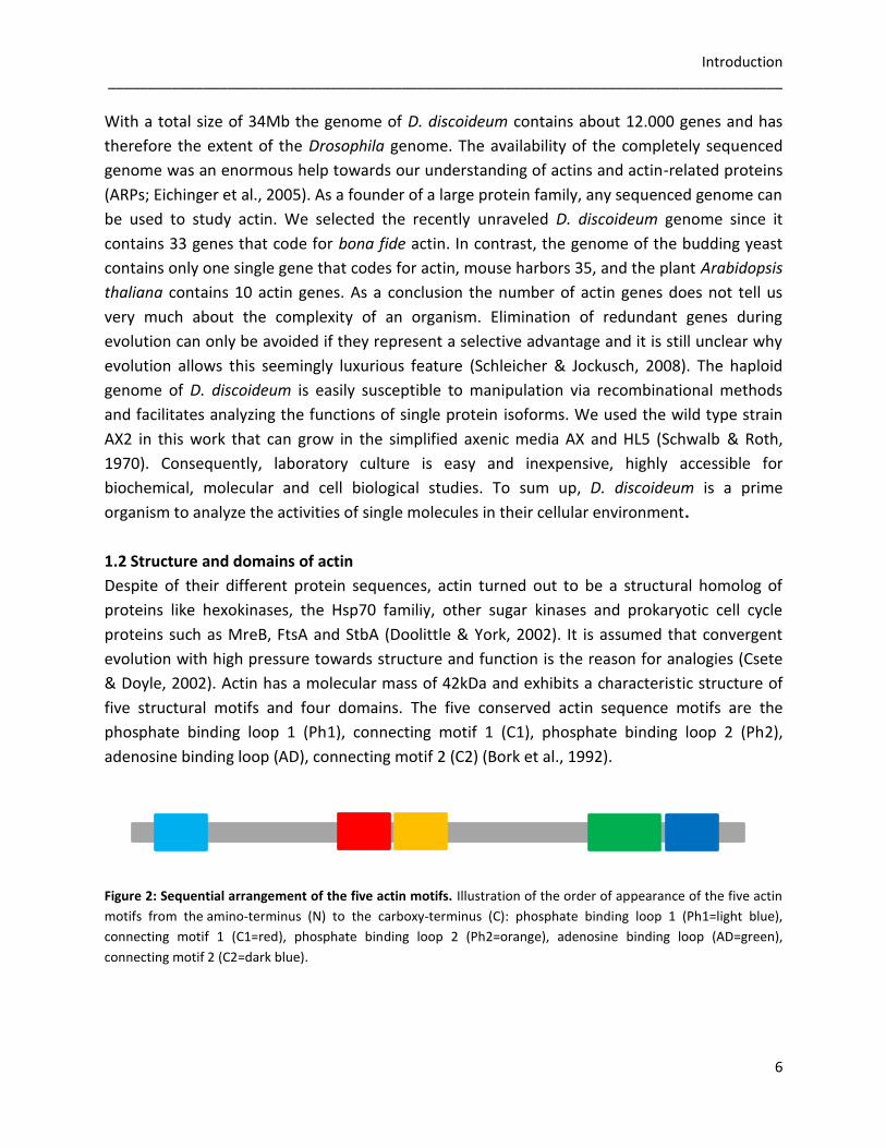
five structural motifs and four domains. The five conserved actin sequence motifs are the
phosphate binding loop 1 (Ph1), connecting motif 1 (C1), phosphate binding loop 2 (Ph2),
adenosine binding loop (AD), connecting motif 2 (C2) (Bork et al., 1992).
Figure 2: Sequential arrangement of the five actin motifs. Illustration of the order of appearance of the five actin
motifs from the amino-terminus (N) to the carboxy-terminus (C): phosphate binding loop 1 (Ph1=light blue),
connecting motif 1 (C1=red), phosphate binding loop 2 (Ph2=orange), adenosine binding loop (AD=green),
connecting motif 2 (C2=dark blue).

Introduction _____________________________________________________________________________________
7
Domain I and domain II are two structurally similar globular domains connected by a flexible
hinge that forms the core of actin. Most important for the actin dynamic is the interface
between the two domains, which forms an adenosine triphosphate (ATP) binding pocket.
Domain I as well as domain II are composed of two subdomains in each case 1 (Ia), 2 (Ib), 3 (IIa)
and 4 (IIb). The barbed end of an actin filament is defined by the subdomains 1 and 3, opposed
by the pointed end composed by subdomain 2 and 4 (Carlier, 1990). The structural similarity
suggests that duplication of one ancestral domain could have formed the two domains of G-
actin (Bork et al., 1992).
Figure 3: Typical G-actin structure in the ADP-state. The computed ribbon model of Act8 (P07830) of
D. discoideum in the ADP-state (Joseph et al., 2008) shows that the ATP-binding pocket is exposed between the
domains I and II. The four subdomains are marked (1-4) as well as the five structural motifs (Ph1= light blue,
C1=red, Ph2=orange, Ad=green and C2=dark blue).
1.3 The actin cytoskeleton
The cytoskeleton in eukaryotic cells plays an important role in cell shape, structure, migration,
cytokinesis, intracellular transport and provides one of the machineries for actively moving
organelles within the cytoplasm. A complex and dynamic network of protein filaments is
therefore spread throughout the cell consisting of microfilaments (diameter~7nm),
intermediate filaments (diameter~10nm) and microtubules (diameter~24nm). Actin belongs
to the microfilament system, is highly conserved and an abundant protein making up to 5-10%
of the total cell protein (Pollard & Earnshaw, 2004).
Actin is present in two states, as monomeric globular actin (G-actin) and as polymeric,
filamentous actin (F-actin). Two helical, interlaced F-actin strands build a right handed helix and
form an actin filament. The addition of physiological salts like Mg2+ or K+ to a G-actin pool above
critical concentration induces polymerization in vivo.
Introduction _____________________________________________________________________________________
8
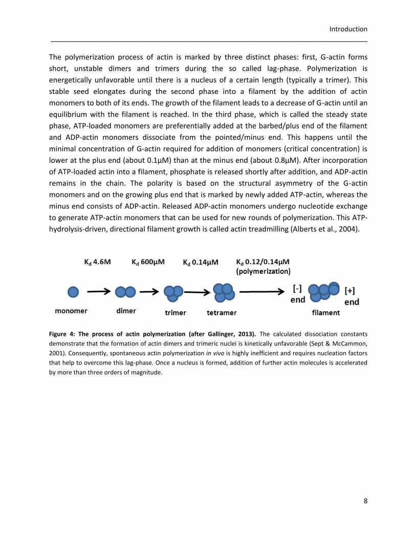
The polymerization process of actin is marked by three distinct phases: first, G-actin forms
short, unstable dimers and trimers during the so called lag-phase. Polymerization is
energetically unfavorable until there is a nucleus of a certain length (typically a trimer). This
stable seed elongates during the second phase into a filament by the addition of actin
monomers to both of its ends. The growth of the filament leads to a decrease of G-actin until an
equilibrium with the filament is reached. In the third phase, which is called the steady state
phase, ATP-loaded monomers are preferentially added at the barbed/plus end of the filament
and ADP-actin monomers dissociate from the pointed/minus end. This happens until the
minimal concentration of G-actin required for addition of monomers (critical concentration) is
lower at the plus end (about 0.1µM) than at the minus end (about 0.8µM). After incorporation
of ATP-loaded actin into a filament, phosphate is released shortly after addition, and ADP-actin
remains in the chain. The polarity is based on the structural asymmetry of the G-actin
monomers and on the growing plus end that is marked by newly added ATP-actin, whereas the
minus end consists of ADP-actin. Released ADP-actin monomers undergo nucleotide exchange
to generate ATP-actin monomers that can be used for new rounds of polymerization. This ATP-
hydrolysis-driven, directional filament growth is called actin treadmilling (Alberts et al., 2004).
Figure 4: The process of actin polymerization (after Gallinger, 2013). The calculated dissociation constants
demonstrate that the formation of actin dimers and trimeric nuclei is kinetically unfavorable (Sept & McCammon,
2001). Consequently, spontaneous actin polymerization in vivo is highly inefficient and requires nucleation factors
that help to overcome this lag-phase. Once a nucleus is formed, addition of further actin molecules is accelerated
by more than three orders of magnitude.
Introduction _____________________________________________________________________________________
9
Actin-binding proteins are able to fulfill a large variety of tasks including the control of actin
assembly and disassembly, as well as regulating filament branching and bundling to help
arranging actin filaments into higher ordered structures. While actin monomer binding proteins
control the amount and availability of monomers for polymerization, proteins that bind F-actin
are involved in barbed and pointed end capping, filament severing and filament crosslinking.
Among others, the ATP/G-actin binding proteins ARP2/3, WASP and formin induce the
nucleation of F-actin, whereas profilin and thymosin control the pool of G-actin monomers via
sequestering (Schüler et al., 2006). Profilin promotes the ADP/ATP exchange of the actin
monomers, and thus ensures the delivery of ATP-actin for incorporation into growing filaments
by actin polymerizing machineries (Dickinson et al. 2002). ß-thymosin binds G-actin
stoichiometrically and prevents G-actin even above its critical concentration from assembling to
F-actin (Lodish et al., 2000). Gelsolin, cofilin or severin bind ADP/G-actin within the polymerized
filament to disassemble the actin network. A severing protein cuts the filament and remains
bound at the plus end of the resulting fragments, where it prevents the addition or exchange of
actin subunits, an activity called capping. The minus ends of fragments remain uncapped and
are rapidly shortened. Thus severing promotes the turnover of actin filaments by creating new
minus ends and causes disintegration of the actin network. All of the mentioned severing
proteins are regulated in a Ca2+-dependent manner (Lodish et al., 2000). Capping at the plus
end to block the addition and loss of actin subunits is also observed under the influence of
Cap32/34 in D. discoideum. This heterodimeric protein consists of two subunits (32kDa and
34kDa) that can be inhibited by phosphatidyl bisphosphate, an important component in signal
transduction during chemotaxis (Haus et al., 1991). The resulting lowered viscosity is a
prerequisite to allow the flow of cellular contents during movement of the cell. Besides
nucleation, ARP2/3 is also operating as a minus end capping protein, like tropomodulin. ARP2/3
inhibits both monomer addition and dissociation at the pointed ends of actin filaments and
increases the critical concentration for polymerization at the pointed end. The high affinity of
the ARP2/3 complex for pointed ends and its abundance in amoebae suggests that in vivo all
pointed ends of actin filaments are capped by the ARP2/3 complex (Mullins et al., 1998).
Furthermore ARP2/3 is important to allow dendritic branching that is found at the leading edge
of motile cells (Pollard & Borisy, 2003). Besides the mentioned interactors, numerous other
actin binding proteins are of great importance to the cell in the integration of structure and
signaling between the cytoskeletal elements and the maintenance of cell integrity.

Introduction _____________________________________________________________________________________
10
Figure 5: Actin binding proteins. Different actin binding proteins regulate the actin polymerization kinetics and
stability of the microfilament network. Nucleation is mediated e.g. via ARP2/3, WASP or formins (N). These
proteins are specific for binding ATP/G-actin monomers, like profilin (E) moderating nucleotide exchange. Severing
and capping of ADP-Pi/G-actin is procured by gelsolin, fragmin or vilin (S), if lower viscosity is required in the
cytoplasm. Thymosins (C) can bind ATP/G-actin subunits and ADP/G-actin monomers to induce
capping/sequestration. For disassembly of the F-actin network the actin-depolymerization factor ADF/cofilin (D)
binds ADP/G-actin and therefore communicates severing and depolymerization (after Winder & Ayscough, 2005).
If actin dynamics in human cells are disturbed, many diseases including muscular, neurological,
immunological, vascular diseases and even cancer can occur (Cleuren & Boonstra, 2012). Also
nemaline myopathies are caused by dysfunctions of the actin protein. The mutation of
Val163Leu in ACTA1 (actin gene expressed in the human skeletal muscle) leads often to the
intranuclear rod myopathy (IRM). This genetic defect results in the accumulation of rod-shaped
protein aggregates in the nuclei and cytoplasm of human muscle cells (Domazetovska et al.,
2007; Kaimaktchiev et al., 2006; Sparrow et al. 2003; Vandebrouck et al., 2010). Mutations in
actin, nebulin, cofilin, troponin and tropomyosin could be ascertained in the affected patients,
whereas the exact composition, formation and biophysics of these rod-shaped structures are
yet unknown. One possible explanation for the presented disease pattern could be cell death,
induced via harmed chromatins within the cells due to the stiff rod-shaped aggregates in the
nucleus (Sparrow et al., 2003).
Introduction _____________________________________________________________________________________
11
1.3 The actinome of D. discoideum and its regulation
To fulfill all the required functions and dynamics, the regulation of the actin cytoskeleton is not
only controlled by actin binding proteins. Also posttranslational modifications on different actin
isoforms play important roles in the modification of the actin network. Acetylation, acylation,
serine/threonine/tyrosine-phosphorylation and ubiquitinylation of actin are possible. One can
assume that a developmentally regulated expression of actin genes requires a similarly
regulated expression of enzymes that catalyze posttranslational modifications (Schleicher &
Jockusch, 2008). The discovery of ARPs made this picture even more complex. Due to its huge
variety of actin genes, the D. discoideum genome provides a very good basis to study the
actinome for potential cellular targets and conserved sequence motifs (Joseph et al., 2008). The
actinome is comprised of 41 actins and ARPs. Seven potential pseudogenes are part of the
D. discoideum genome, as well as eight ARPs. ARPs vary in presence and copy in different
organisms and show altered degrees of similarity with actin (Muller et al., 2005). A few of them
have preserved the actin structural fold, and are assumed to have originated from a common
ancestor parallel to the actin isoforms. First identified in Saccharomyces cerevisiae
(S. cerevisiae), the ARPs are named on decreasing order according to their relative identity with
the conventional actin sequences, where ARP1 is the most similar and ARP10 the least similar
(Poch & Winsor, 1997).
To get an overview of the actinome presented in D. discoideum, the genetic organization was
analyzed using multiple sequence alignments and profile-hidden Markov models from the
‘Pfam’ protein family database. Altogether 33 actins and 8 ARPs have been identified (Joseph et
al., 2008). 95% of the cellular actin consists of the Act8 group of conventional actin, which
contains 17 distinct genes coding for identical amino acid sequences. The range of differences
in the amino acid sequences in the other 17 actin genes varies from a single substituted residue
(0.3%) for example in Act10, up to 295 (78%) non-identical amino acids in Act33 (Joseph et al.,
2008).
Introduction _____________________________________________________________________________________
12
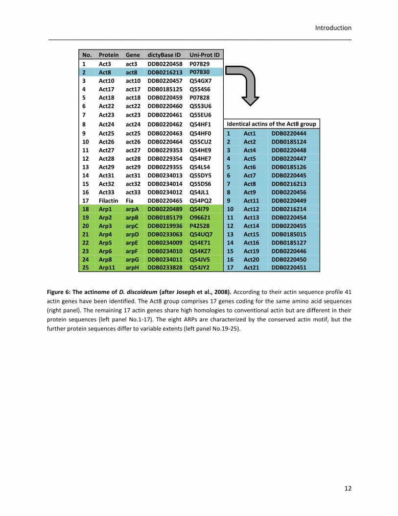
Figure 6: The actinome of D. discoideum (after Joseph et al., 2008). According to their actin sequence profile 41
actin genes have been identified. The Act8 group comprises 17 genes coding for the same amino acid sequences
(right panel). The remaining 17 actin genes share high homologies to conventional actin but are different in their
protein sequences (left panel No.1-17). The eight ARPs are characterized by the conserved actin motif, but the
further protein sequences differ to variable extents (left panel No.19-25).
No. Protein Gene dictyBase ID Uni-Prot ID 1 Act3 act3 DDB0220458 P07829 2 Act8 act8 DDB0216213 P07830
3 Act10 act10 DDB0220457 Q54GX7 4 Act17 act17 DDB0185125 Q554S6 5 Act18 act18 DDB0220459 P07828 6 Act22 act22 DDB0220460 Q553U6 7 Act23 act23 DDB0220461 Q55EU6 8 Act24 act24 DDB0220462 Q54HF1 Identical actins of the Act8 group
9 Act25 act25 DDB0220463 Q54HF0 1 Act1 DDB0220444
10 Act26 act26 DDB0220464 Q55CU2 2 Act2 DDB0185124
11 Act27 act27 DDB0229353 Q54HE9 3 Act4 DDB0220448
12 Act28 act28 DDB0229354 Q54HE7 4 Act5 DDB0220447
13 Act29 act29 DDB0229355 Q54L54 5 Act6 DDB0185126
14 Act31 act31 DDB0234013 Q55DY5 6 Act7 DDB0220445
15 Act32 act32 DDB0234014 Q55DS6 7 Act8 DDB0216213
16 Act33 act33 DDB0234012 Q54JL1 8 Act9 DDB0220456
17 Filactin Fia DDB0220465 Q54PQ2 9 Act11 DDB0220449
18 Arp1 arpA DDB0220489 Q54I79 10 Act12 DDB0216214
19 Arp2 arpB DDB0185179 O96621 11 Act13 DDB0220454
20 Arp3 arpC DDB0219936 P42528 12 Act14 DDB0220455
21 Arp4 arpD DDB0233063 Q54UQ7 13 Act15 DDB0185015
22 Arp5 arpE DDB0234009 Q54E71 14 Act16 DDB0185127
23 Arp6 arpF DDB0234010 Q54KZ7 15 Act19 DDB0220446
24 Arp8 arpG DDB0234011 Q54JV5 16 Act20 DDB0220450 25 Arp11 arpH DDB0233828 Q54JY2 17 Act21 DDB0220451
Introduction _____________________________________________________________________________________
13
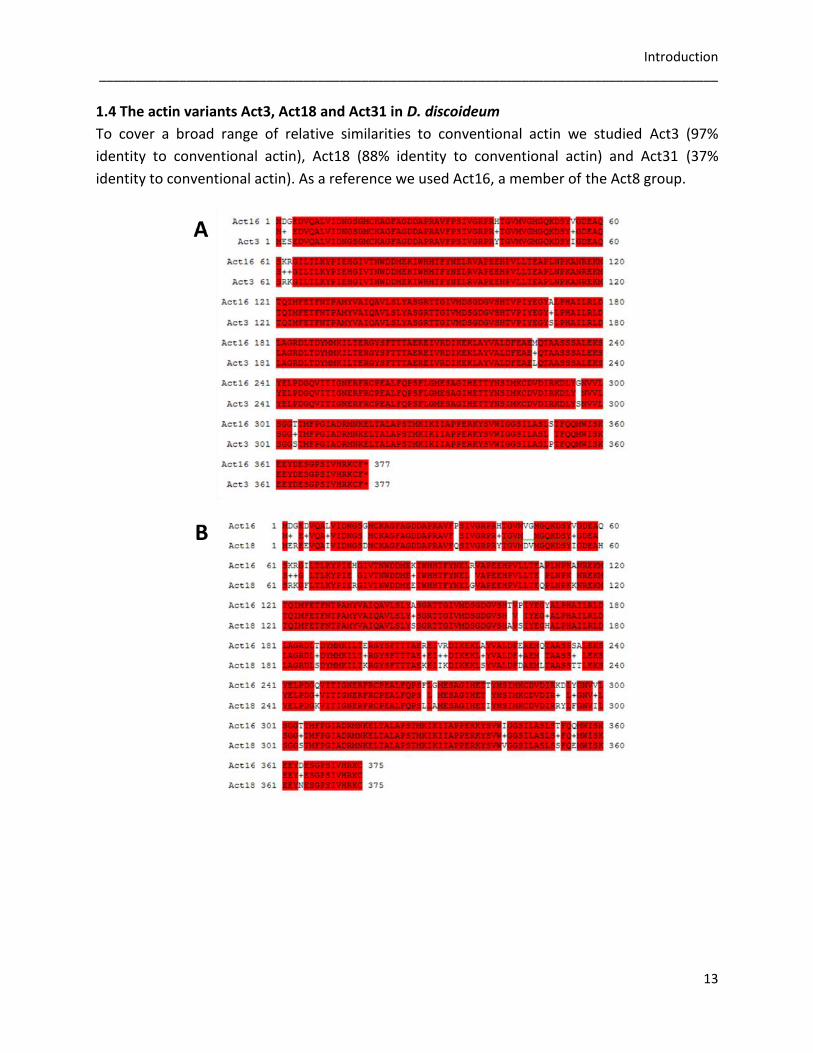
1.4 The actin variants Act3, Act18 and Act31 in D. discoideum
To cover a broad range of relative similarities to conventional actin we studied Act3 (97%
identity to conventional actin), Act18 (88% identity to conventional actin) and Act31 (37%
identity to conventional actin). As a reference we used Act16, a member of the Act8 group.
Introduction _____________________________________________________________________________________
14
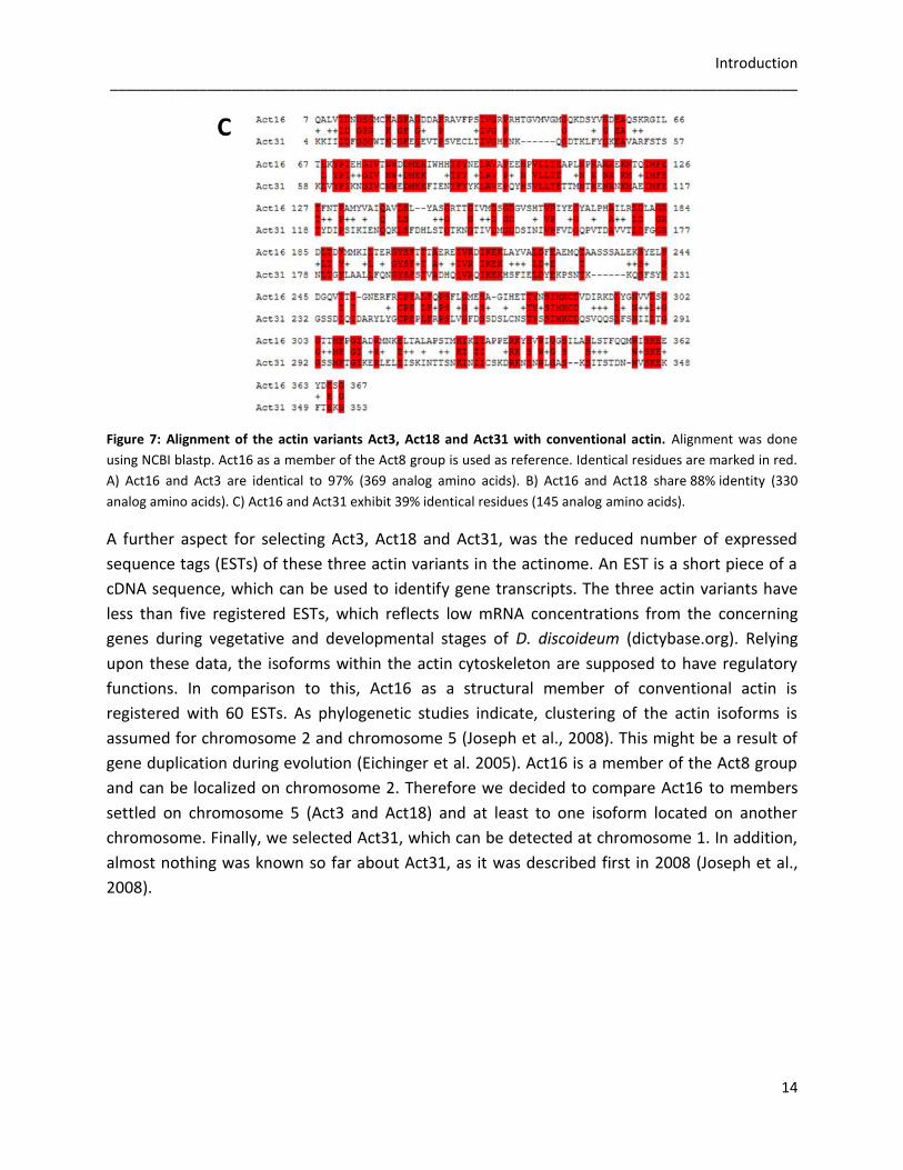
Figure 7: Alignment of the actin variants Act3, Act18 and Act31 with conventional actin. Alignment was done
using NCBI blastp. Act16 as a member of the Act8 group is used as reference. Identical residues are marked in red.
A) Act16 and Act3 are identical to 97% (369 analog amino acids). B) Act16 and Act18 share 88% identity (330
analog amino acids). C) Act16 and Act31 exhibit 39% identical residues (145 analog amino acids).
A further aspect for selecting Act3, Act18 and Act31, was the reduced number of expressed
sequence tags (ESTs) of these three actin variants in the actinome. An EST is a short piece of a
cDNA sequence, which can be used to identify gene transcripts. The three actin variants have
less than five registered ESTs, which reflects low mRNA concentrations from the concerning
genes during vegetative and developmental stages of D. discoideum (dictybase.org). Relying
upon these data, the isoforms within the actin cytoskeleton are supposed to have regulatory
functions. In comparison to this, Act16 as a structural member of conventional actin is
registered with 60 ESTs. As phylogenetic studies indicate, clustering of the actin isoforms is
assumed for chromosome 2 and chromosome 5 (Joseph et al., 2008). This might be a result of
gene duplication during evolution (Eichinger et al. 2005). Act16 is a member of the Act8 group
and can be localized on chromosome 2. Therefore we decided to compare Act16 to members
settled on chromosome 5 (Act3 and Act18) and at least to one isoform located on another
chromosome. Finally, we selected Act31, which can be detected at chromosome 1. In addition,
almost nothing was known so far about Act31, as it was described first in 2008 (Joseph et al.,
2008).
Introduction _____________________________________________________________________________________
15
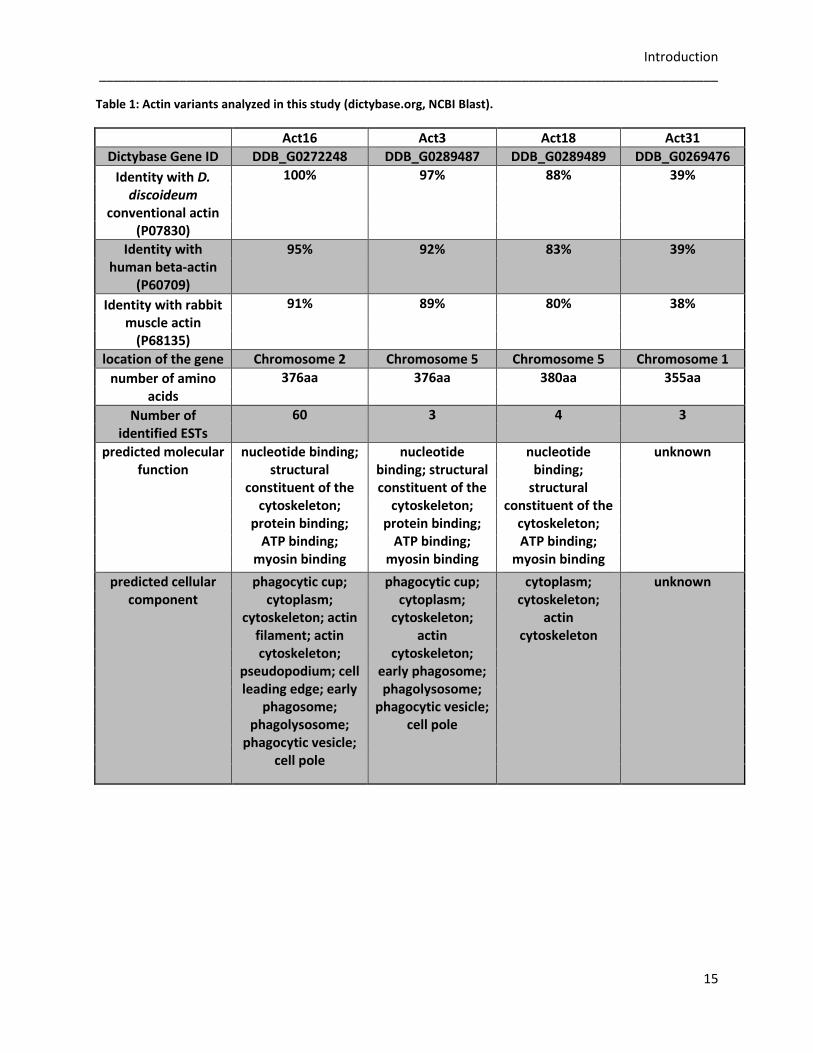
Table 1: Actin variants analyzed in this study (dictybase.org, NCBI Blast).
Act16 Act3 Act18 Act31
Dictybase Gene ID DDB_G0272248 DDB_G0289487 DDB_G0289489 DDB_G0269476
Identity with D. discoideum
conventional actin (P07830)
100% 97% 88% 39%
Identity with human beta-actin
(P60709)
95% 92% 83% 39%
Identity with rabbit muscle actin
(P68135)
91% 89% 80% 38%
location of the gene Chromosome 2 Chromosome 5 Chromosome 5 Chromosome 1
number of amino acids
376aa 376aa 380aa 355aa
Number of identified ESTs
60 3 4 3
predicted molecular function
nucleotide binding; structural
constituent of the cytoskeleton;
protein binding; ATP binding;
myosin binding
nucleotide binding; structural constituent of the
cytoskeleton; protein binding;
ATP binding; myosin binding
nucleotide binding;
structural constituent of the
cytoskeleton; ATP binding;
myosin binding
unknown
predicted cellular component
phagocytic cup; cytoplasm;
cytoskeleton; actin filament; actin cytoskeleton;
pseudopodium; cell leading edge; early
phagosome; phagolysosome;
phagocytic vesicle; cell pole
phagocytic cup; cytoplasm;
cytoskeleton; actin
cytoskeleton; early phagosome; phagolysosome;
phagocytic vesicle; cell pole
cytoplasm; cytoskeleton;
actin cytoskeleton
unknown
Introduction _____________________________________________________________________________________
16
1.6 Aims of the thesis
Given that D. discoideum comprises 41 actins and ARPs, several approaches should be
performed to get an overview over this huge actinome. It is still unclear why such a multitude
of genes is kept active within the genome of D. discoideum, where other organisms vary their
proteomes simply via splicing or posttranslational modifications. Accordingly, we assumed that
the actin variants which are not part of the Act8 group are not necessary for the cytoskeleton
itself, but are rather regulators of its dynamical features or subunits in larger protein
complexes. Therefore, we aimed at the functions of the actin variants Act3, Act18 and Act31 in
the microfilament system in vitro and in vivo. Act16 as a member of the Act8 group served as a
control.
First, the in vitro characterization of the selected actin variants should be performed. For this
purpose the baculoviral amplification system (Sf9 cells) should be used to express N-terminal
Flag-tagged actin variants. To analyze the role of Flag-Act16, Flag-Act3, Flag-Act18 and Flag-
Act31 in actin dynamics, actin polymerization assays have to be performed using fluorometric
approaches, low shear viscometry, spin down assays and the transmission electron microscopy.
Additionally, oligomerization of the Flag-tagged actin variants should be checked via FPLC-
gelfiltration and immunoprecipitation studies could reveal potential binding partners of the
actin variants.
The in vivo assays should be performed using green fluorescent protein-actin variant (GFP-actin
variant) overexpressing cells of D. discoideum. To determine if the actin variants are part of the
microfilament system, immunofluorescence and spin down assays were to be performed.
Furthermore, phenotypical characterization should be carried out with the analysis of growth,
germination, phagocytosis, development and phototaxis. Additionally, we were interested in
the role of GFP-Act16, GFP-Act3, GFP-Act18 and GFP-Act31 concerning the formation of actin
bundles, like nuclear and cytoplasmic rods. In all in vivo assays the influence of the N-terminal
GFP-tag had to be compared to the results to GFP-overexpressing wild type cells from
D. discoideum (AX2-GFP cells).
Materials and Methods _____________________________________________________________________________________
17
2. Materials and Methods
2.1 Materials
2.1.1 Instruments
AEKTA purifier 100
BioDocAnalyze
Dounce homogenizer
fluorescence spectrometer LS55
Gene pulse electroporator Xcell
Gelsystem MiniPROTEAN
PCR-Thermocycler Tpersonal
pH-meter pH720
plasma cleaner
Power supplies
Protein Transfer Transblot Semi-Dry
Protein Transfer TF 77XP
Shaker Orbital Incubator SI500
Shakers for Dictyostelium cultures
Thermomixer
Tabletop Film Processor Curix 60
Vortex Genie 2
Waterbath
GE Healthcare
Biometra
Braun/Wheaton
Perkin Elmer
BioRad
BioRad
Biometra
Inolab WTW series
Diener, Ebhausen
Biorad, Biometra, Consort
BioRad
Serva
Memmert
Kühner
Eppendorf
Agfa
Bender & Hobein
GFL, Kühner
Microscopes
Binocular microscope Stereo Discovery.V8
LSM 510 confocal microscope
Carl Zeiss
Carl Zeiss
JEM-1200 EX II Electron microscope JEOL
Objectives
Achromat S 0.63x FWD 115mm
40x LD A-Plan 0.50 Ph2
63x Neofluar 1.4 oil immersion objective
100x Neofluar 1.3 oil immersion objective
Carl Zeiss
Carl Zeiss
Carl Zeiss
Carl Zeiss

Materials and Methods _____________________________________________________________________________________
18
Centrifuges
GS-6KR
J2-21M/E
J6-HC
Microcentrifuge 5415 D, 5417 R
Optima LE-80K
Optima TL ultracentrifuge
Beckman
Beckman
Beckman
Eppendorf
Beckman
Beckman
Rotors
JA-10, JA-15, JA-20
Ti35, Ti45, Ti70
TLA 100.3
Beckman
Beckman
Beckman
2.1.2 Computer programs
Adobe Creative Suite 2
ApE plasmid editor v1.10.4
AxioVision
BioDoc Analyze
BioEdit 7.0.9.0
ClustalX2
FL Winlab
LSM 5, 4.2 SP1
Microsoft Office
Multalin 5.4.1
NCBI
OpenAstexViewer
Pfam 22.0
Swissmodel Expasy
Treeview 1.6.6
Unicorn 5.20
Uniprot
Weblogo2.0
ZEN
Adobe Systems
M. Wayne
Carl Zeiss
Biometra
Tom Hall
European Molecular Biology Laboratory
Perkin Elmer
Carl Zeiss
Mircosoft Corporation
Florence Corpet
National Center for Biotech Information
Carl Zeiss
Trust Sanger Institute
Swiss Institute of Bioinformatics
Java
GE Healthcare
European Bioinformatics Institute
University of California, Berkley
Carl Zeiss
Materials and Methods _____________________________________________________________________________________
19
2.1.3 Laboratory consumables
1.5ml centrifuge tubes
Amersham Hyperfilm ECL
Cell culture plates, 24 wells
Cell culture dishes, Ø 100mm x 20mm
100Carbon Support Films-grids Cu 200
Dialysis tubings Type 8, 20, 27
Gel-blotting paper 3MM Chr
GFP-Nano-Trap
High Pure Plasmid Isolation Kit
High Pure PCR Product Purification Kit
High precision cuvettes 10mm
Nitrocellulose transfer membrane Protran
Parafilm
PCR tubes Thermo Tube 0.2ml
Phusion High-Fidelity DNA-Polymerase
Petri dishes Ø 92mmx 16mm
Pipettes 10ml, 25ml
Pipette tips
Plasmid DNA Purification Maxi Kit
600 mesh Cu2+-grids
Restriction Enzymes
Sterile filter, Filtropur S 0.2
Tubes 15ml, 50ml
Ultracentrifuge tubes 1.5ml
Eppendorf
GE Healthcare
Starlab Int.
Greiner bio-one
QUANTIFOIL
Biomol
Whatman
Chromotek
Roche
Roche
Hellma
Whatman
American National Can
Peqlab
New England Biolabs
Sarstedt
Sarstedt
Biozym, Gilson, Starlab
Machery Nagel
Plano
New England Biolabs
Sarstedt
Sarstedt
Beckman
2.1.4 Reagents
Standard laboratory chemicals were mainly purchased from Biomol, Biorad, Fluka, Invitrogen,
Merck, Peqlab, Roche, Roth, Serva or Sigma-Aldrich and had the degree of purity ‘p.a.’ unless
otherwise mentioned. Media and buffers used in this study were prepared with de-ionised
water (Millipore), sterilized either by autoclaving or passing through a micro-filter (pore size
0.2µm).

Materials and Methods _____________________________________________________________________________________
20
2.1.5 Antibodies
Primary antibodies
Actin, D. discoideum (Act-1)
Actin, D. discoideum (224-236-1)
Contact site A glycoprotein (33-294-17)
Filactin (4S-59-4)
Flag-tag (6F7)
GFP (K3-184-2)
Severin (102-200-1)
Simpson et al. (1984)
Westphal et al. (1997)
Bertholdt et al. (1985)
Israel (2002)
Chromotek
Noegel et al. (2004)
Andre et al. (1989)
Secondary antibodies
Goat-anti-mouse IgG Cy3-conjugated
Anti-mouse IgG horseradish peroxidase-
linked (ECL)
Invitrogen
GE Healthcare
2.1.6 Vectors
pDEX-(g418)-GFP-act16 (Mo-1)
pDEX-(blasticidin)-GFP-act3 (#614_2)
pDEX-(blasticidin)-GFP-act18 (#681_43)
pDEX-(blasticidin)-GFP-act31 (#668_1)
pFastBac1
Annette Müller-Taubenberger
Annette Müller-Taubenberger
Annette Müller-Taubenberger
Annette Müller-Taubenberger
Invitrogen
Constructs generated in this study
pFastBac1 Act16
pFastBac1 Act3
pFastBac1 Act18
pFastBac1 Act31
SalI/KpnI
SalI/KpnI
SalI/KpnI
SalI/KpnI
2.1.7 Bacterial strains
E.coliDH5α Invitrogen
E.coliDH10Bac
E.coliDH10Bac FLAG-Act16
E.coliDH10Bac FLAG-Act3
E.coliDH10Bac FLAG-Act18
E.coliDH10Bac FLAG-Act31
Invitrogen
Present study
Present study
Present study
Present study
K. aerogenes Williams & Newell, 1976
Materials and Methods _____________________________________________________________________________________
21
2.1.8 Yeast strains
S. cerevisiae YSC-II Sigma -Aldrich
2.1.9 D. discoideum strains
Strain name
AX2
AX2-GFP
Cherry-cofilin
GFP-Act16
GFP-Act3
GFP-Act18
GFP-Act31
Resistance
/
G10
G10
G10
B10
B10
B10
Source
laboratory wild type
Meino Rohlfs
Hellen Ishikawa-Ankerhold
Annette Müller-Taubenberger
Annette Müller-Taubenberger
Annette Müller-Taubenberger
Annette Müller-Taubenberger
Materials and Methods _____________________________________________________________________________________
22
2.2 Methods
2.2.1 Molecular methods
To generate various GFP- and Flag-tagged protein constructs standard molecular biological
methods were used. Polymerase chain reactions (PCRs) were performed with Phusion
High-Fidelity DNA-Polymerase (New England Biolabs) according to the manufacturer’s manual.
Extraction and purification of DNA from Tris-borate-EDTA agarose gels were performed using
the High Pure PCR Product Purification Kit (Roche). PCR products were cloned into the
appropriate plasmids using standard restriction enzyme mediated cloning techniques. Plasmid
DNA was obtained from E.coli by using standard alkaline lysis miniprep or by using the silica-
based mini- and maxiprep kits (Roche, Macherey Nagel). Chemically competent E. coli cells
were prepared according to the CaCl2 method (Dagert & Ehrlich, 1979). The correctness of the
DNA sequences inserted into the respective expression vectors was controlled by sequencing
using specific primers (Eurofins MWG Operon, Ebersberg).
2.2.2 Biochemical methods
2.2.2.1 SDS-polyacrylamide gel electrophoresis and western blotting
Standard discontinuous SDS-page was used to separate protein mixtures (Laemmli, 1970).
Afterwards transfer buffer (25mM Tris; pH8.5, 190mM glycine, 20% methanol, 0.02% SDS)
transmitted these proteins onto a nitrocellulose membrane via semi-dry western blotting.
Nonfat milk powder in NCP buffer (10mM Tris; pH7.3, 150mM NaCl, 0.05% Tween20) blocked
the membranes, before incubation steps with the appropriate primary and secondary
antibodies followed. The Enhanced Chemiluminescence System (ECL) was used for developing
the membranes finally.
2.2.2.2 Coomassie Blue and silver staining
To detect proteins after electrophoretic separation on polyacrylamide gels, staining with
Coomassie Brilliant Blue R250 was performed. If sensitivity in the low nanogram range was
needed, we conducted silver staining. This is compatible with downstream processing such as
mass spectrometry analysis after protein digestion. The sequential phases of silver staining
were protein fixation (40% ethanol, 10% acetate; 30 minutes) then sensitization (30% ethanol,
sodium thiosulfate, 0.83M sodium acetate, 0.125 % freshly added glutaraldehyde; 30 minutes).
After washing three times with ddH2O (10 minutes each), the gels were stained in silver
impregnation (0.25% silver nitrate, 0.015% freshly added formaldehyde; 30 minutes). Gels were
rinsed with ddH2O and exposed to developing solution (23.5mM sodium carbonate, 0.015%
freshly added formaldehyde) until the desired grade of staining intensity was reached. The
reaction was stopped by addition of an aqueous EDTA solution.
Materials and Methods _____________________________________________________________________________________
23
2.2.2.3 Actin preparation from rabbit skeletal muscle
In the present thesis rabbit muscle actin was used as a control in many assays. Routine actin
purification methods were used to obtain actin essentially as described (Spudich & Watt, 1971).
The upper and back thigh muscles of a rabbit were detached, chilled and ground twice. A high
salt extraction buffer (0.5M KCl, 0.1M K2HPO4) was applied for 10-15 minutes on a stirrer to
remove myosin. Next, the mixture was centrifuged (4.000g, 4°C, 10 minutes) and reextracted.
Until the pellet swelled, stirring in cold distilled water for 10 minutes and centrifugational steps
were repeated again and again. Afterwards the pellet was incubated with cold acetone for
about 30 minutes, filtered and dried overnight. For subsequent actin preparations the acetone
powder was stored at -20°C.
Regularly, 10g acetone powder were extracted with 200ml G-actin buffer (2mM Tris; pH 8.0,
0.2mM ATP, 0.5mM DTT, 0.2mM CaCl2, 0.01% NaN3) at 4°C for 30 minutes, followed by
filtration through nylon sets and reextraction for 15 minutes. Past centrifugation of the filtrate
(30.000g, 30 minutes, 4°C) actin polymerization was induced by adding 50mM KCl, 2mM MgCl2
and 1mM ATP dissolved in 100mM NaOH, and the mixture was incubated at 4°C for two hours,
respectively overnight. Up to 0.8M solid KCl was slowly added to remove tropomyosin. To
accumulate F-actin, centrifugation at 150.000g for three hours at 4°C was performed and the
supernatant was discarded. Homogenization was executed by a douncer. To achieve
depolymerization, F-actin was dialyzed against G-buffer for two-three days with a total of about
six buffer changes. Again the remaining F-actin fraction was separated by centrifugation at
150.000g and 4°C for three hours, then 65% from the top supernatant were further purified
using a Sephacryl S300 gel filtration column (2.5x45cm; Pharmacia). The actin concentrations of
the different fractions were determined by measuring the optical density at 290nm (1mg/ml
pure actin: OD290nm=0.65). Adjacent falling ball viscometry verified the quality. Freshly prepared
rabbit muscle actin can be stored on ice for up to three weeks for most applications and was
dialyzed against freshly prepared G-buffer before usage.

Materials and Methods _____________________________________________________________________________________
24
2.2.2.4 Labeling of actin with pyrene
Actin was labeled for in vitro polymerization assays with N-(1-pyrenyl) iodoacetamide (pyrene;
Kouyama & Mihashi, 1981). After the first ultracentrifugation step during actin preparation, the
F-actin pellet was dialyzed against P-buffer (1mM NaHCO3; pH7.6, 0.1mM CaCl2, 0.2mM ATP,
0.1mM ß-mercaptoethanol) for two-three days with a total of about six buffer changes. 65% of
the top supernatant was used, after centrifugation at 150.000g for three hours at 4°C. In
parallel, 100mM KCl and 1mM MgCl2 were added to promote actin polymerization, while a
three-five fold molar excess of pyrene dissolved in DMSO was dropwise stirred into the actin
solution. Given that pyrene is light sensitive, the following working steps had to be performed
under protection of light. The solution was mixed gently overnight by turning end-over-end in a
50ml tube at room temperature before F-actin was pelleted again at 150.000g. G-buffer was
supplemented to the pellet of F-actin for homogenization and dialysis to effect
depolymerization. A further centrifugational step at 150.000g for three hours at 4°C was
undertaken, before the pyrenylated G-actin was further purified via gel filtration as described in
2.2.3.3. By using the respective extinction coefficients (OD290nm=2.6100µM actin; OD344nm=
100µM pyrene; micromolar concentration pyrene/micromolar concentration actin= % of
pyrene-labeled actin) the percentage of labeled actin could be calculated when the
concentrations of actin and pyrene were measured at the photometer. 60-90% of the actin
fraction should be successfully labeled. Before usage the aliquots were dialyzed against G-
buffer. Storage at -80°C.
2.2.2.5 Flag-tagged protein expression using the baculoviral amplification system
To ensure correct expression, folding and oligomerization of the selected actin variants, the
eukaryotic Bac-to-Bac baculoviral expression system was utilized to express recombinant
protein constructs. First of all, the appropriate nucleotide sequence with an N-terminal Flag-tag
was cloned into the pFastBac1 vector. Thereupon, DH10Bac cells were transformed with 300ng
of the newly cloned pFastBac1 construct. DH10Bac cells contained the baculoviral shuttle
vector (bacmid) with a mini-attTn7 target site and a helper plasmid that encoded certain
transposition proteins. The transposition of the mini-Tn7 element on the pFastBac1 donor
plasmid to the mini-attTn7 target site on the bacmid was supported by the special transposition
proteins. If the recombination was successful, the lacZ gene on the bacmid was disrupted,
which allowed a blue/white screening in the presence of X-Gal and IPTG. After isolation of the
bacmid via miniprep, immortalized Sf9 insect cells could be transfected and cultivated at 28°C.
For this purpose 200µl Sf-900 II SFM medium (Invitrogen), 10µl cellfectin (Invitrogen) and 30-
100µg bacmid DNA were mixed and incubated for 45 minutes at room temperature. Afterwards
the mix was added to 2ml of Sf9 cells (density 5x10⁵cells/ml) which were placed in wells of a 6-
well-plate and incubated at 28°C for five hours.
Materials and Methods _____________________________________________________________________________________
25
The transfection mixture was removed, before SFM medium supplemented with 5% fetal
bovine serum and gentamycin (0.1mg/ml) was added. The virus (P0 virus) was harvested after
72 hours of incubation at 28°C, sterile-filtered and stored at 4°C. The virus was amplified by
generating P1 and P2 generations. For protein expression >97%, viable Sf9 cells were infected
at a density of 2x10⁶cells/ml with P2 virus at a ratio of about 1/10 (10ml P2 virus+ 90ml cells),
and incubated as a shaking culture at 110rpm at 28°C for 48-72 hours. Cells were harvested by
centrifugation (2000g, 15 minutes, 4°C) and pellets of a 100ml culture were resuspended in 5ml
lysis buffer (15mM Hepes, pH 7.4; 30mM NaCl, 1mM EGTA, 1mM DTT, 0.1mM magnesium-ATP,
one Complete Protease Inhibitor cocktail tablet (Roche) per 20ml buffer, 2% glycerol, 1% Triton-
X100). The lysates were centrifuged at 30.000g for 10 minutes at 4°C and the supernatant was
incubated at 4°C under gentle end-over-end mixing for 90 minutes with 100µl Anti-Flag M2
Affinity Gel (Sigma) for lysates of a 100ml culture. The matrix was washed in washing buffer
(15mM Hepes, pH7.4; 30mM NaCl, 1mM DTT, 0.1mM magnesium-ATP, one Complete Protease
Inhibitor cocktail tablet (Roche) per 20ml buffer, 2% glycerol), with 10-30 column volumes in a
Poly-Prep Chromatography column (Biorad). For elution, the column outlet was closed and
200µl elution buffer (wash buffer containing 100µg Flag-peptides; Sigma) were used to elute
the protein from 100µl Anti-Flag M2 Affinity Gel for 60 minutes. The eluate was collected and
the concentration was determined by SDS-page analysis. The purified protein was subsequently
used in further experiments or stored at -80°C.
2.2.2.6 In vitro actin polymerization assay
A Perkin Elmer fluorometer was used for fluorometric actin polymerization assays by the
application of pyrenylated actin. 20x increase of fluorescence could be obtained if actin
polymerizes and the environment of pyrene-actin changes. A convenient signal occurred
already with about 10% pyrenylated actin. The polymerization-induced fluorescence was
captured at 386nm using an excitation wavelength of 365nm and 8nm slit widths.
Polymerization was induced by application of polymerization buffer (10mM imidazole, pH7.2;
3mM MgCl2, 1mM Na-ATP, 0.2mM CaCl2). G-actin, Flag-tagged actin variants and different
buffering conditions (e.g. elution buffer or 100mM KCl) were mixed and preincubated for about
one-two minutes prior to addition of polymerization buffer and recording. Filament-disrupting
measurements were performed with prepolymerized actin samples.
2.2.2.7 Low shear viscometry
Low shear viscometry assays were carried out in a falling ball viscometer (MacLean-Fletcher &
Pollard, 1980). Polymerization of the incubated actin proteins was started by the addition of
polymerization buffer. The mixture was briefly vortexed, immediately filled into the capillary
and allowed to polymerize for 15 minutes. The viscosity of the solution was measured by
recording the time a mini steel ball took to pass a certain distance.
Materials and Methods _____________________________________________________________________________________
26
2.2.2.8 Gel filtration using the AEKTA 100 system
The gel filtration column Superose 6 10/300GL (GE Healthcare) provides the appropriate range
of pore sizes for the selected Flag-actin constructs. The column was equilibrated with IEDANBP
buffer (10mM imidazole, 1mM EGTA, 1mM DTT, 0.02% NaN3, 50mM NaCl, 1mM benzamidine,
1mM PMSF; pH 7.3). The flow rate was set between 0.1-0.5ml/min. Before starting size analysis
the column was calibrated using protein molecular weight standards in a range between 12.4–
450kDa (Serva). The void volume (V0) was identified with Dextran blue 2000 (GE Healthcare).
Usually 0.2mg purified protein was injected, 0.5ml fractions were collected and analyzed by
SDS-page.
2.2.2.9 Transmission electron microscopy
First the grids were cleaned using a plasma cleaner. About 10µl of the protein samples
(50µg/ml, respectively 25µg/ml) were pipetted onto a parafilm, the grids were placed on the
sample drops with their carbon coated side and incubated for one-two minutes. Afterwards the
grids were washed in a drop of H2O two times. By using a filter paper superfluous fluid was
removed and the grids were placed in a 2% uranyl acetate solution, incubated for two minutes
and dried. Images were taken on a CM20 electron microscope (JEOL) at 70kV and 10.000x
magnifications.
2.2.2.10 Immunoprecipitation
The GFP-Nano-Trap system (Chromotek) was used to identify proteins interacting with the
cloned GFP-actin variants (Zolghadr et al., 2012). 5x10⁷cells of the relevant D. discoideum
strains were harvested and opened in lysis buffer (25mM HEPES; pH7.4, 50mM NaCl, 1mM
EDTA, 1mM EGTA, 1mM DTT, 5mM benzamidine, 1µM PMSF, one Complete Protease Inhibitor
cocktail tablet (Roche) per 20ml buffer, 5% Glycerol, 1% Triton X-100). The lysate was
centrifuged for 15-30 minutes at 10.000g and 4°C. Afterwards the supernatant was incubated
with 15-20µl packed GFP-Trap agarose beads equilibrated in lysis buffer. The following
incubation via end-over-end mixing at 4°C took 60-90 minutes. As described in the
manufacturer’s protocol the beads were washed, and the GFP-tagged proteins with their
potential interaction partners were eluted by boiling in SDS-sample buffer. Proteins were
separated by SDS-page and silver stained (O-Connel & Stults, 1997). Bands of interest were cut
out from the gel and analyzed via MALDI-TOF mass spectrometry (ZfP, LMU Munich).

Materials and Methods _____________________________________________________________________________________
27
2.2.3 Cell biological methods
2.2.3.1 Cell culture and transformation of D. discoideum
The wild type strain of D. discoideum AX2, or mutant cells derived from it, were cultured
axenically in either AX medium (14.3g peptone, 7.15g yeast extract, 50mM glucose, 3.5mM
Na2HPO4, 3.5mM KH2PO4 in 1l H2O) or HL5 medium (5g yeast extract, 10g proteose peptone,
50mM glucose, 8.5mM KH2PO4, made up to 1l with H2O; pH7.5). Cells can be brought up in
culture dishes, in shaking cultures at 150rpm or on lawns of non-pathogenic Klebsiella
aerogenes. To cultivate the mutants the media were supplemented with the appropriate
antibiotics. For long time storage, spores of mature fruiting bodies were brought up on
Soerensen phosphate agar plates (10g Bacto-agar, dissolved in 1l Soerensen buffer), and were
resuspended in Soerensen phosphate buffer (14.6mM KH2PO4, 2mM Na2HPO4; pH6.0).
Afterwards the spores were shock-frozen in liquid nitrogen and stored at -80°C.
For transformation, electroporation was used with the appropriate plasmids. 2x10⁷cells were
extensively washed in cold Soerensen buffer and electroporation buffer (50mM sucrose, 10mM
KH2PO4; pH6.1) and resuspended in 1ml cold electroporation buffer. In the presence of 25µg
DNA, the cells were electroporated in a 4mm electroporation cuvette using a Gene Pulser XCell
(Biorad), applying the standard settings (square wave, V=1.0kV, 1ms pulse length, two pulses,
five seconds pulse interval). After gentle shaking (50rpm, room temperature, 15 minutes) in a
cell culture dish 2µM CaCl2 and 2µM MgCl2 were added. The cells were incubated in HL5
medium after additional 15 minutes, and the cells were allowed to recover for about 24 hours
before the respective antibiotic (either 10µg/ml geniticin or blasticidin) was applied to select
the transformants.
Single clones were amplified via spreader dilution with non-pathogenic K. aerogenes on SM
agar plates (9g agar, 10g peptone, 50mM glucose, 1g yeast extract, 4mM MgSO4, 16mM
KH2PO4, 5.7mM K2HPO4 in 1l with H2O; pH6.5). The overexpression of fluorescently labeled
proteins was checked via live-cell microscopy.
2.2.3.2 Live-cell microscopy of D. discoideum amoebae
After cells were transferred onto coverslips and allowed to settle down, they were washed
twice with Soerensen buffer. Confocal images were taken by the usage of an inverted laser
scanning microscope (LSM) 510 confocal microscope (Zeiss) equipped with a 40x LDA-Plan 0.50
Ph2, a 63x Neofluar 1.4 or a 100x Neofluar 1.3 oil immersion objective. For excitation, the
488nm argon ion laser line and the 543nm helium neon laser lines were used. Emission was
collected using a 510-525nm band-pass and a 585-615nm band pass filter.
For recording of phagocytosis at the LSM 510 confocal microscope (Zeiss), cells were incubated
with a suspension of heat-killed S. cerevisiae cells (Sigma-Aldrich) labeled with tetramethyl
rhodamine isothiocyanate (TRITC; Sigma) in Soerensen buffer for 20 minutes, and recorded at
intervals of 10 seconds.
Materials and Methods _____________________________________________________________________________________
28
2.2.3.3 Immunofluorescence microscopy
Indirect immunofluorescence studies were used to investigate subcellular localization of
proteins. In advance, cover slips were washed with 3.6% HCl and rinsed with H2O. 5x10⁶cells/ml
were harvested, washed twice in Soerensen buffer and allowed to settle on the coverslips for
15 minutes. Subsequently, the medium was removed and the cells were fixed with methanol
(-20°C for 10 minutes). Afterwards, the coverslips were washed several times with phosphate
buffered saline (PBS; 137mM NaCl, 2.7mM KCl, 8.1mM Na2HPO4, 1.5mM KH2PO4; pH7.4)
supplemented with 100mM glycine. After washing the samples with phosphate buffered
glucose (PBS + 0.5% BSA, 0.045% fish gelatin), they were incubated with the primary antibody
overnight at 4°C. The next day, preparations were washed with PBG and incubated for 60
minutes with the secondary adequate antibody, which was fluorescence-dye labeled (e.g. goat-
anti-mouse IgG Cy3-conjugated). TOPRO (Invitrogen) was used to stain the DNA. At last the
coverslips were quickly rinsed with H2O, embedded in gelvatol mounting medium (0.14M NaCl,
0.01M KH2PO4 /Na2HPO4 pH7.2, 5.7M polyvinyl alcohol, 0.5mM glycerol, 0.9mM DABCO) and
stored in the dark at 4°C. Confocal microscopy data were acquired on an inverted LSM 510
confocal microscope (Zeiss) with a 63x or 100x oil immersion objectives with a numerical
aperture of 1.4 and 1.3, respectively. Excitation of fluorophores was achieved with the 488nm
argon ion laser line, the 543nm and 633nm helium laser lines, and emission was collected using
510-525nm band-pass, 585-615nm band-pass or 650nm long-pass filters.
2.2.3.4 Induction of nuclear and cytoplasmic rods
D. discoideum cells were exposed to 5% dimethylsulfoxide (DMSO) in HL5 medium to induce
nuclear protein rod formation, respectively 10mM sodium azide (NaN3) dissolved in HL5
medium, to provoke the formation of cytoplasmic protein rods.
Both applications were carried out at 28°C for one hour. Cells with induced nuclear rods were
treated consecutively as described in immunofluorescence microscopy (2.2.2.4), whereas
cytoplasmic rods were imaged under live-cell conditions (2.2.2.3).
2.2.3.5 Growth and germination
To determine any differences in growth of AX2 mutants, D. discoideum cells were brought up to
densities below 5x10⁶cells/ml. After washing the cells in Soerensen buffer, they were adjusted
to a density of 5x10⁴cells/ml. 30ml of cell suspension were shaken at 150rpm, 21°C in 100ml
Erlenmeyer flasks without any supplementary antibiotics. Densities of cell cultures were
determined two times daily.
100 spores were plated together with the non-pathogenic feeding bacterium K. aerogenes on
SM agar plates. The viability of spores was investigated by counting the number of plaques
after 96 hours.
Materials and Methods _____________________________________________________________________________________
29
2.2.3.6 Phagocytosis measurements
Phagocytosis of yeast by D. discoideum cells was measured through fluorescence spectroscopy.
Cells were grown to 6x10⁶cells/ml, harvested, washed, resuspended in Soerensen buffer and
adjusted to a density of 2x10⁶cells/ml. 12ml of this solution were shaken at 150rpm and 21°C in
a 25ml Erlenmeyer flask for one hour. TRITC-labeled yeast cells (120µl of 109cells/ml) were
added. Samples of 1ml were withdrawn every 10 minutes and added to 100µl of trypan blue
solution (20mg/ml dissolved in 20mM sodium citrate containing 150mM NaCl), which quenched
the fluorescence of non-internalized yeast cells. After three minutes of agitated incubation,
cells were spun and the supernatant was removed carefully. After resuspension in Soerensen
buffer, fluorescence was measured in a fluorometer using 544nm light for excitation.
To check the ability of phagocyting bacteria, single clones from D. discoideum cells were picked
from the edges of colonies growing on plated non-pathogenic K. aerogenes and transferred to
the middle of new non-pathogenic K. aerogenes lawns on SM agar plates. The diameter of the
developing plaque was measured daily.
2.2.3.7 Analysis of phototaxis and development
Using a sterile inoculation loop, cells were transferred onto water agar plates from the edges of
colonies growing on non-pathogenic K. aerogenes lawns to test the phototaxis efficiency of
different cell lines. The plates were stored in a darkly colored box with a 2mm wide opening for
the entry of light. After incubation at 21°C for 48 hours, the tracks of the migrating slugs could
be visualized by transferring them onto a nitrocellulose membrane and final amido black
staining.
The 24 hours cell cycle was induced in washed 5x10⁶cells/ml. Wild type and mutant cells were
transferred onto nutrient free phosphate agar plates to document developmental stages by
time-lapse photography. Pictures were taken every 10 minutes for 24 hours.
Results _____________________________________________________________________________________
30
3. Results
3.1 The actinome of D. discoideum
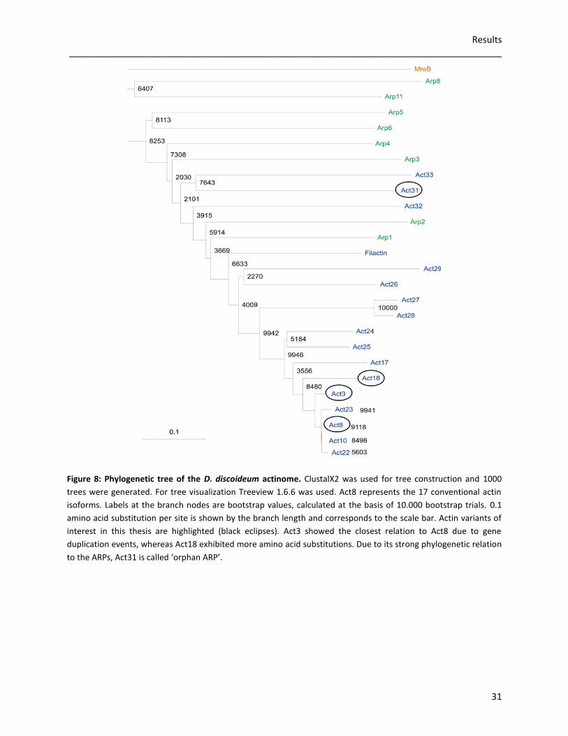
3.1.1 Phylogeny of the D. discoideum actinome
The bacterial actin murein cluster e proteins (MreB) have been known for a long time to be cell
shape determinants and are present in gram-positive and gram-negative bacterial cells (Wachi
et al., 1987). They also contain five conserved sequence motifs to determine the three
dimensional fold similar to that of actin, including the nucleotide binding pocket (Carballido-
Lopez, 2006). Furthermore, MreB filaments are generated by actin-like polymerization and
show similar dynamics. Although just 15% identity exists between MreB and conventional actin,
they are assumed to be structural and functional homologues of actin (Bork et al., 1992). As
MreB and actins are putative descendants from a common ATP-binding ancestor, MreB (E.coli;
E0J6V5) was used as outgroup for the phylogenetic tree of the D. discoideum actinome. All 17
conventional actin genes are represented by Act8. The ARPs are displayed in green, the actin
variants are shown in dark blue. Phylogenetic analyses illustrated that Act3 is closely related to
conventional actin (11 different amino acids), whereas Act18 contains more mutations (45
different amino acids). Act31 has evolved independently (224 different amino acids) and is
more closely related to the protein sequences of the ARPs (Eichinger et al., 2005).
Results _____________________________________________________________________________________
31
Figure 8: Phylogenetic tree of the D. discoideum actinome. ClustalX2 was used for tree construction and 1000
trees were generated. For tree visualization Treeview 1.6.6 was used. Act8 represents the 17 conventional actin
isoforms. Labels at the branch nodes are bootstrap values, calculated at the basis of 10.000 bootstrap trials. 0.1
amino acid substitution per site is shown by the branch length and corresponds to the scale bar. Actin variants of
interest in this thesis are highlighted (black eclipses). Act3 showed the closest relation to Act8 due to gene
duplication events, whereas Act18 exhibited more amino acid substitutions. Due to its strong phylogenetic relation
to the ARPs, Act31 is called ‘orphan ARP’.
Results _____________________________________________________________________________________
32
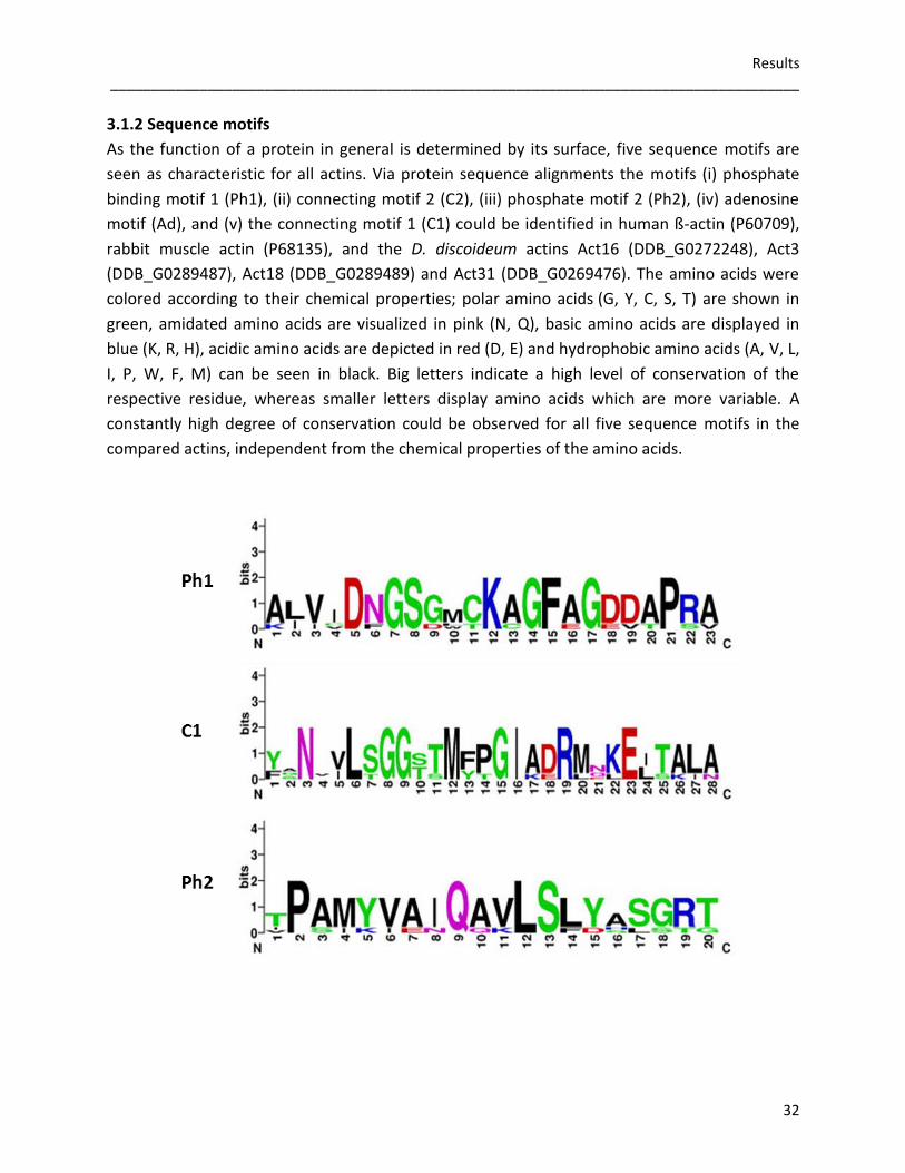
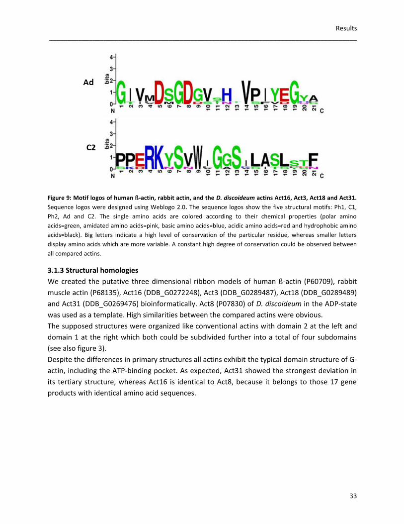
3.1.2 Sequence motifs
As the function of a protein in general is determined by its surface, five sequence motifs are
seen as characteristic for all actins. Via protein sequence alignments the motifs (i) phosphate
binding motif 1 (Ph1), (ii) connecting motif 2 (C2), (iii) phosphate motif 2 (Ph2), (iv) adenosine
motif (Ad), and (v) the connecting motif 1 (C1) could be identified in human ß-actin (P60709),
rabbit muscle actin (P68135), and the D. discoideum actins Act16 (DDB_G0272248), Act3
(DDB_G0289487), Act18 (DDB_G0289489) and Act31 (DDB_G0269476). The amino acids were
colored according to their chemical properties; polar amino acids (G, Y, C, S, T) are shown in
green, amidated amino acids are visualized in pink (N, Q), basic amino acids are displayed in
blue (K, R, H), acidic amino acids are depicted in red (D, E) and hydrophobic amino acids (A, V, L,
I, P, W, F, M) can be seen in black. Big letters indicate a high level of conservation of the
respective residue, whereas smaller letters display amino acids which are more variable. A
constantly high degree of conservation could be observed for all five sequence motifs in the
compared actins, independent from the chemical properties of the amino acids.
Results _____________________________________________________________________________________
33
Figure 9: Motif logos of human ß-actin, rabbit actin, and the D. discoideum actins Act16, Act3, Act18 and Act31.
Sequence logos were designed using Weblogo 2.0. The sequence logos show the five structural motifs: Ph1, C1,
Ph2, Ad and C2. The single amino acids are colored according to their chemical properties (polar amino
acids=green, amidated amino acids=pink, basic amino acids=blue, acidic amino acids=red and hydrophobic amino
acids=black). Big letters indicate a high level of conservation of the particular residue, whereas smaller letters
display amino acids which are more variable. A constant high degree of conservation could be observed between
all compared actins.
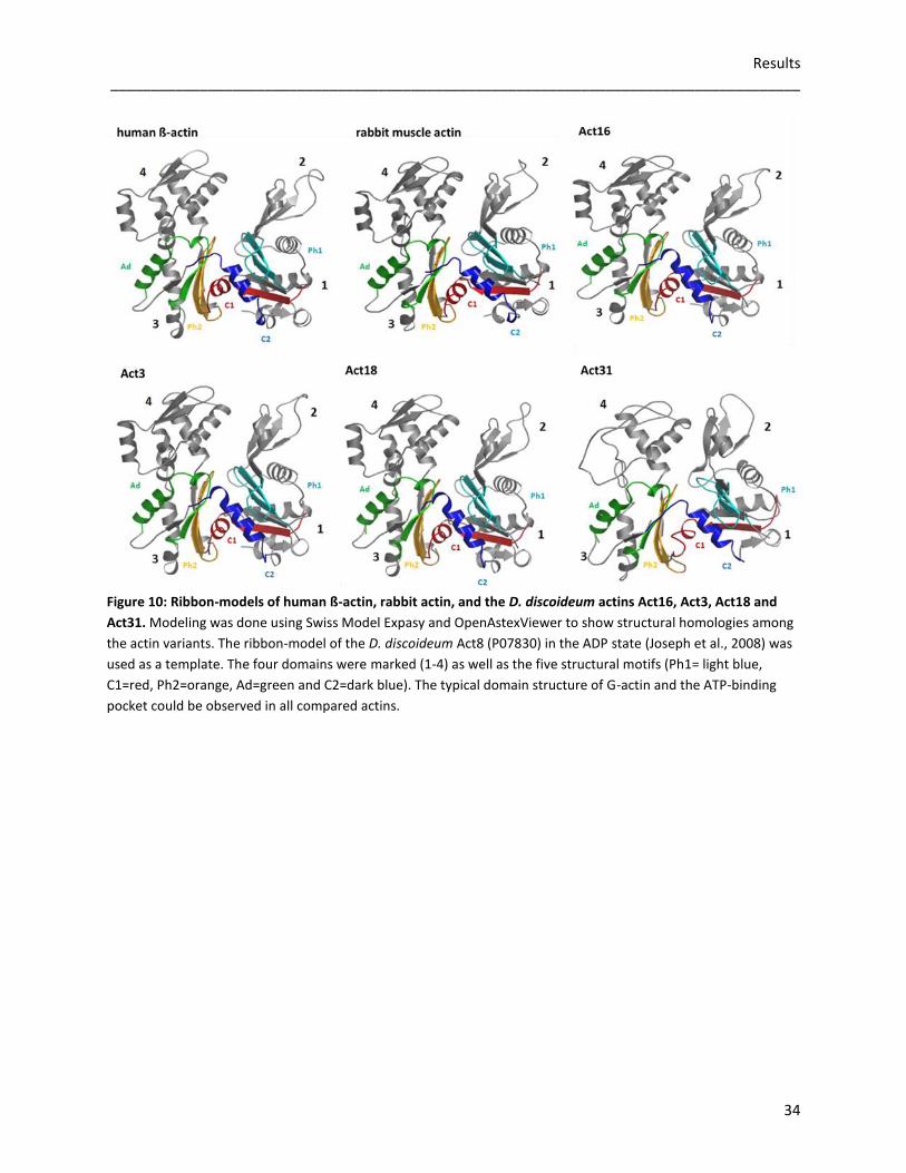
3.1.3 Structural homologies
We created the putative three dimensional ribbon models of human ß-actin (P60709), rabbit
muscle actin (P68135), Act16 (DDB_G0272248), Act3 (DDB_G0289487), Act18 (DDB_G0289489)
and Act31 (DDB_G0269476) bioinformatically. Act8 (P07830) of D. discoideum in the ADP-state
was used as a template. High similarities between the compared actins were obvious.
The supposed structures were organized like conventional actins with domain 2 at the left and
domain 1 at the right which both could be subdivided further into a total of four subdomains
(see also figure 3).
Despite the differences in primary structures all actins exhibit the typical domain structure of G-
actin, including the ATP-binding pocket. As expected, Act31 showed the strongest deviation in
its tertiary structure, whereas Act16 is identical to Act8, because it belongs to those 17 gene
products with identical amino acid sequences.
Results _____________________________________________________________________________________
34
Figure 10: Ribbon-models of human ß-actin, rabbit actin, and the D. discoideum actins Act16, Act3, Act18 and
Act31. Modeling was done using Swiss Model Expasy and OpenAstexViewer to show structural homologies among
the actin variants. The ribbon-model of the D. discoideum Act8 (P07830) in the ADP state (Joseph et al., 2008) was
used as a template. The four domains were marked (1-4) as well as the five structural motifs (Ph1= light blue,
C1=red, Ph2=orange, Ad=green and C2=dark blue). The typical domain structure of G-actin and the ATP-binding
pocket could be observed in all compared actins.
Results _____________________________________________________________________________________
35
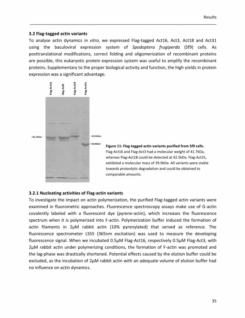
3.2 Flag-tagged actin variants
To analyse actin dynamics in vitro, we expressed Flag-tagged Act16, Act3, Act18 and Act31
using the baculoviral expression system of Spodoptera frugiperda (Sf9) cells. As
posttranslational modifications, correct folding and oligomerization of recombinant proteins
are possible, this eukaryotic protein expression system was useful to amplify the recombinant
proteins. Supplementary to the proper biological activity and function, the high yields in protein
expression was a significant advantage.
3.2.1 Nucleating activities of Flag-actin variants
To investigate the impact on actin polymerization, the purified Flag-tagged actin variants were
examined in fluorometric approaches. Fluorescence spectroscopy assays make use of G-actin
covalently labeled with a fluorescent dye (pyrene-actin), which increases the fluorescence
spectrum when it is polymerized into F-actin. Polymerization buffer induced the formation of
actin filaments in 2µM rabbit actin (10% pyrenylated) that served as reference. The
fluorescence spectrometer LS55 (365nm excitation) was used to measure the developing
fluorescence signal. When we incubated 0.5µM Flag-Act16, respectively 0.5µM Flag-Act3, with
2µM rabbit actin under polymerizing conditions, the formation of F-actin was promoted and
the lag-phase was drastically shortened. Potential effects caused by the elution buffer could be
excluded, as the incubation of 2µM rabbit actin with an adequate volume of elution buffer had
no influence on actin dynamics.
Figure 11: Flag-tagged actin variants purified from Sf9 cells.
Flag-Act16 and Flag-Act3 had a molecular weight of 41.7kDa,
whereas Flag-Act18 could be detected at 42.5kDa. Flag-Act31,
exhibited a molecular mass of 39.9kDa. All variants were stable
towards proteolytic degradation and could be obtained to
comparable amounts.
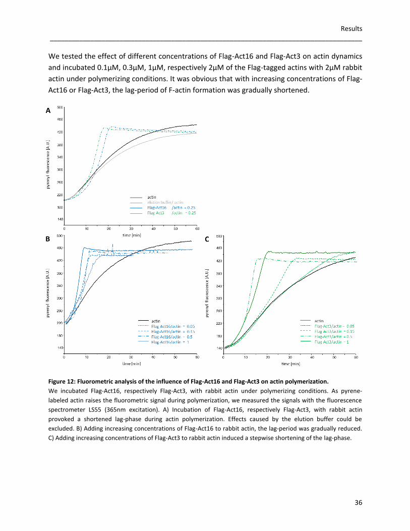
Results _____________________________________________________________________________________
36
We tested the effect of different concentrations of Flag-Act16 and Flag-Act3 on actin dynamics
and incubated 0.1µM, 0.3µM, 1µM, respectively 2µM of the Flag-tagged actins with 2µM rabbit
actin under polymerizing conditions. It was obvious that with increasing concentrations of Flag-
Act16 or Flag-Act3, the lag-period of F-actin formation was gradually shortened.
Figure 12: Fluorometric analysis of the influence of Flag-Act16 and Flag-Act3 on actin polymerization.
We incubated Flag-Act16, respectively Flag-Act3, with rabbit actin under polymerizing conditions. As pyrene-
labeled actin raises the fluorometric signal during polymerization, we measured the signals with the fluorescence
spectrometer LS55 (365nm excitation). A) Incubation of Flag-Act16, respectively Flag-Act3, with rabbit actin
provoked a shortened lag-phase during actin polymerization. Effects caused by the elution buffer could be
excluded. B) Adding increasing concentrations of Flag-Act16 to rabbit actin, the lag-period was gradually reduced.
C) Adding increasing concentrations of Flag-Act3 to rabbit actin induced a stepwise shortening of the lag-phase.
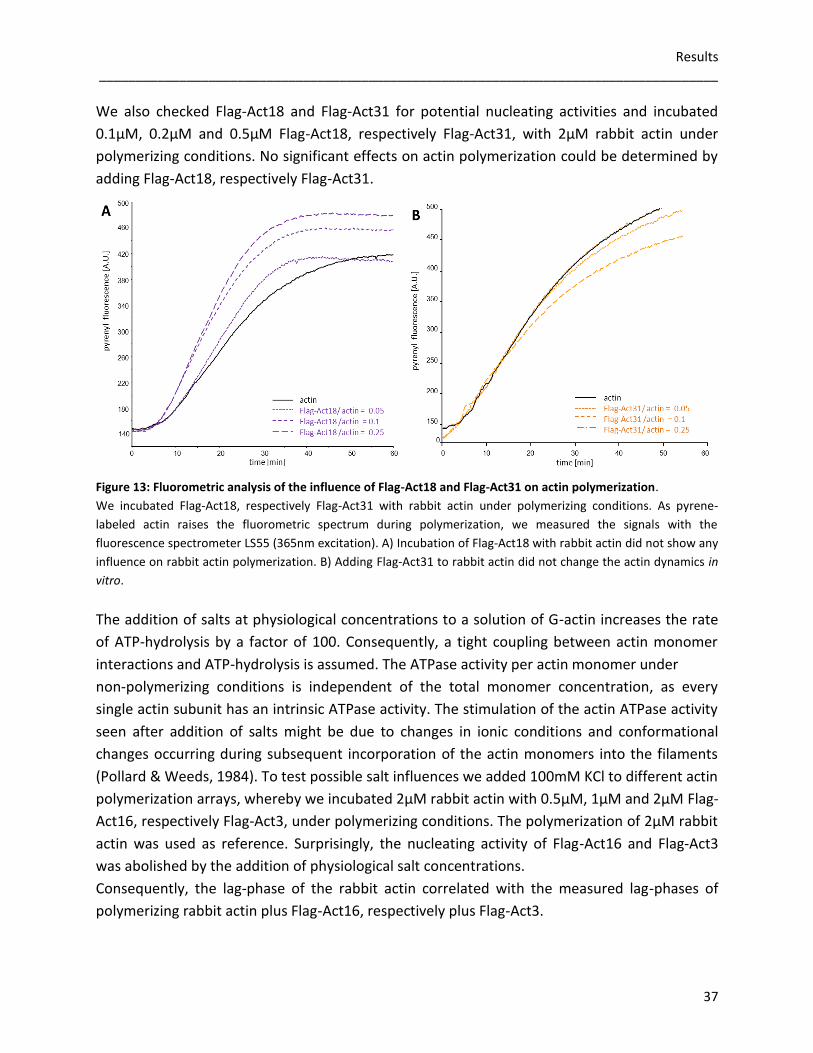
Results _____________________________________________________________________________________
37
We also checked Flag-Act18 and Flag-Act31 for potential nucleating activities and incubated
0.1µM, 0.2µM and 0.5µM Flag-Act18, respectively Flag-Act31, with 2µM rabbit actin under
polymerizing conditions. No significant effects on actin polymerization could be determined by
adding Flag-Act18, respectively Flag-Act31.
Figure 13: Fluorometric analysis of the influence of Flag-Act18 and Flag-Act31 on actin polymerization.
We incubated Flag-Act18, respectively Flag-Act31 with rabbit actin under polymerizing conditions. As pyrene-
labeled actin raises the fluorometric spectrum during polymerization, we measured the signals with the
fluorescence spectrometer LS55 (365nm excitation). A) Incubation of Flag-Act18 with rabbit actin did not show any
influence on rabbit actin polymerization. B) Adding Flag-Act31 to rabbit actin did not change the actin dynamics in
vitro.
The addition of salts at physiological concentrations to a solution of G-actin increases the rate
of ATP-hydrolysis by a factor of 100. Consequently, a tight coupling between actin monomer
interactions and ATP-hydrolysis is assumed. The ATPase activity per actin monomer under
non-polymerizing conditions is independent of the total monomer concentration, as every
single actin subunit has an intrinsic ATPase activity. The stimulation of the actin ATPase activity
seen after addition of salts might be due to changes in ionic conditions and conformational
changes occurring during subsequent incorporation of the actin monomers into the filaments
(Pollard & Weeds, 1984). To test possible salt influences we added 100mM KCl to different actin
polymerization arrays, whereby we incubated 2µM rabbit actin with 0.5µM, 1µM and 2µM Flag-
Act16, respectively Flag-Act3, under polymerizing conditions. The polymerization of 2µM rabbit
actin was used as reference. Surprisingly, the nucleating activity of Flag-Act16 and Flag-Act3
was abolished by the addition of physiological salt concentrations.
Consequently, the lag-phase of the rabbit actin correlated with the measured lag-phases of
polymerizing rabbit actin plus Flag-Act16, respectively plus Flag-Act3.
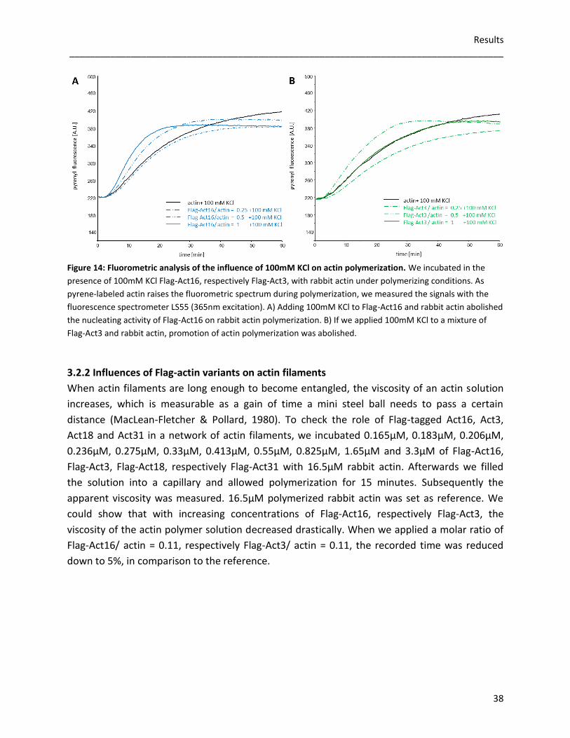
Results _____________________________________________________________________________________
38
Figure 14: Fluorometric analysis of the influence of 100mM KCl on actin polymerization. We incubated in the
presence of 100mM KCl Flag-Act16, respectively Flag-Act3, with rabbit actin under polymerizing conditions. As
pyrene-labeled actin raises the fluorometric spectrum during polymerization, we measured the signals with the
fluorescence spectrometer LS55 (365nm excitation). A) Adding 100mM KCl to Flag-Act16 and rabbit actin abolished
the nucleating activity of Flag-Act16 on rabbit actin polymerization. B) If we applied 100mM KCl to a mixture of
Flag-Act3 and rabbit actin, promotion of actin polymerization was abolished.
3.2.2 Influences of Flag-actin variants on actin filaments
When actin filaments are long enough to become entangled, the viscosity of an actin solution
increases, which is measurable as a gain of time a mini steel ball needs to pass a certain
distance (MacLean-Fletcher & Pollard, 1980). To check the role of Flag-tagged Act16, Act3,
Act18 and Act31 in a network of actin filaments, we incubated 0.165µM, 0.183µM, 0.206µM,
0.236µM, 0.275µM, 0.33µM, 0.413µM, 0.55µM, 0.825µM, 1.65µM and 3.3µM of Flag-Act16,
Flag-Act3, Flag-Act18, respectively Flag-Act31 with 16.5µM rabbit actin. Afterwards we filled
the solution into a capillary and allowed polymerization for 15 minutes. Subsequently the
apparent viscosity was measured. 16.5µM polymerized rabbit actin was set as reference. We
could show that with increasing concentrations of Flag-Act16, respectively Flag-Act3, the
viscosity of the actin polymer solution decreased drastically. When we applied a molar ratio of
Flag-Act16/ actin = 0.11, respectively Flag-Act3/ actin = 0.11, the recorded time was reduced
down to 5%, in comparison to the reference.

Results _____________________________________________________________________________________
39
Figure 15: Viscometric analysis of the effects of Flag-Act16 and Flag-Act3. We incubated 16.5µM rabbit actin with
different concentrations of Flag-Act16, respectively Flag-Act3, and induced polymerization within a capillary. Three
independent experiments were conducted. A) With increasing amounts of Flag-Act16 in the polymerizing actin
solution, the viscosity declined strongly. At a molar ratio of 0.11, the recorded time was reduced to 5% in
comparison to the reference. B) Performing the low shear viscosity assay with increasing ratios of Flag-Act3, the
viscosity decreased gradually. Adding a molar ratio of 0.11 of Flag-Act3 minimizes as well the measured time down
to 5%, in comparison to the reference.
The influence of Flag-Act18 and Flag-Act31 on polymerized rabbit actin was much less dramatic,
with Flag-Act18 even neglectable. This suggests that the dramatically reduced viscosity with
Act16 and Act3 is a rather specific activity.
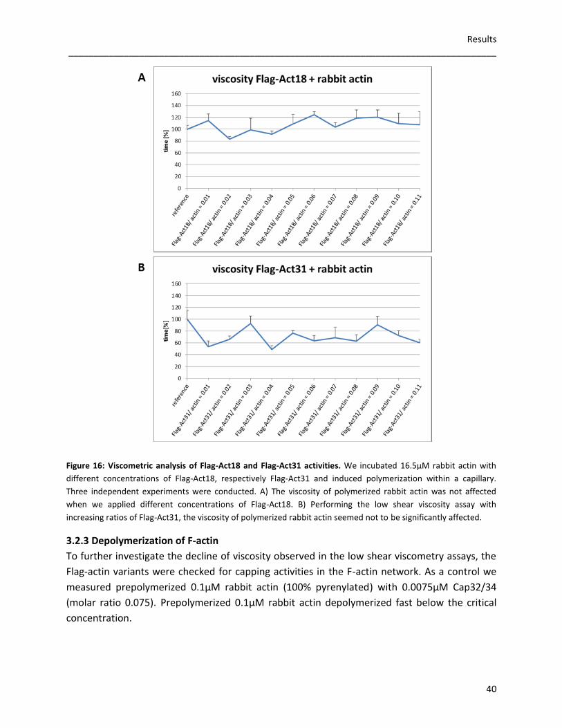
Results _____________________________________________________________________________________
40
Figure 16: Viscometric analysis of Flag-Act18 and Flag-Act31 activities. We incubated 16.5µM rabbit actin with
different concentrations of Flag-Act18, respectively Flag-Act31 and induced polymerization within a capillary.
Three independent experiments were conducted. A) The viscosity of polymerized rabbit actin was not affected
when we applied different concentrations of Flag-Act18. B) Performing the low shear viscosity assay with
increasing ratios of Flag-Act31, the viscosity of polymerized rabbit actin seemed not to be significantly affected.
3.2.3 Depolymerization of F-actin
To further investigate the decline of viscosity observed in the low shear viscometry assays, the
Flag-actin variants were checked for capping activities in the F-actin network. As a control we
measured prepolymerized 0.1µM rabbit actin (100% pyrenylated) with 0.0075µM Cap32/34
(molar ratio 0.075). Prepolymerized 0.1µM rabbit actin depolymerized fast below the critical
concentration.
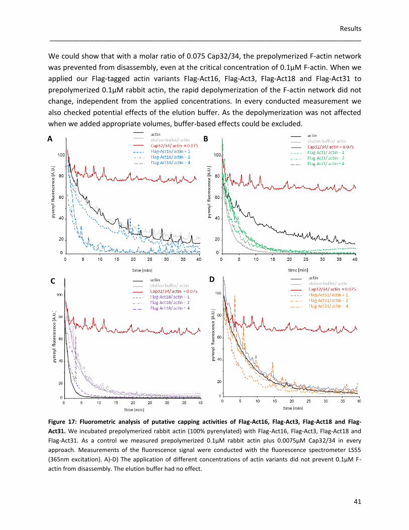
Results _____________________________________________________________________________________
41
We could show that with a molar ratio of 0.075 Cap32/34, the prepolymerized F-actin network
was prevented from disassembly, even at the critical concentration of 0.1µM F-actin. When we
applied our Flag-tagged actin variants Flag-Act16, Flag-Act3, Flag-Act18 and Flag-Act31 to
prepolymerized 0.1µM rabbit actin, the rapid depolymerization of the F-actin network did not
change, independent from the applied concentrations. In every conducted measurement we
also checked potential effects of the elution buffer. As the depolymerization was not affected
when we added appropriate volumes, buffer-based effects could be excluded.
Figure 17: Fluorometric analysis of putative capping activities of Flag-Act16, Flag-Act3, Flag-Act18 and Flag-
Act31. We incubated prepolymerized rabbit actin (100% pyrenylated) with Flag-Act16, Flag-Act3, Flag-Act18 and
Flag-Act31. As a control we measured prepolymerized 0.1µM rabbit actin plus 0.0075µM Cap32/34 in every
approach. Measurements of the fluorescence signal were conducted with the fluorescence spectrometer LS55
(365nm excitation). A)-D) The application of different concentrations of actin variants did not prevent 0.1µM F-
actin from disassembly. The elution buffer had no effect.

Results _____________________________________________________________________________________
42
3.2.4 Sedimentation assays
Besides fluorescence spectroscopy and low shear viscometry, sedimentation assays are also
helpful tools to analyze actin polymerization. The basis of the sedimentation assay is the ability
to pellet F-actin, but not G-actin. Therefore, Flag-actin variants which were associated with
rabbit actin could be detected in the pellet. 2µM rabbit actin (42.0kDa) was used as reference.
After a short 10.000g centrifugational step to remove short oligomers and byproducts, we
incubated 2µM of Flag-Act16, Flag-Act3, Flag-Act18 and Flag-Act31 with 5µM rabbit actin under
polymerizing conditions and centrifuged them at 100.000g. For exact detection of proteins,
silver staining of the SDS-gel was conducted. Concomitantly as in previous experiments, about
60% of Flag-Act16 and Flag-Act3 (both 42.7kDa) seemed to be associated with rabbit F-actin,
whereas the remaining 40% were detectable in the supernatant. In contrast, Flag-Act18
(43.5kDa) could not be pelleted at all and was found exclusively in the supernatant after
ultracentrifugation. Surprisingly, about 40% of Flag-Act31 (40.9kDa) were found in the pellet
after ultracentrifugation. The remaining 60% seemed to be in the supernatant.
Figure 18: Sedimentation assay to
study if Flag-Act16, Flag-Act3, Flag-
Act18 and Flag-Act31 bind to rabbit
F-actin. We incubated 2µM of the
respective Flag-actin variant with
5µM rabbit actin under polymerizing
conditions and pelleted the mixture
at 100.000g. The analysis was done
by silver staining the SDS-gel. Rabbit
actin alone was used as reference.
A) About 80% of the applied rabbit
actin (42.0kDa) could be detected in
the pellet. B) According to the signal
in the pellet, about 60% of Flag-Act16
(42.7kDa) seemed to be associated
with rabbit actin. C) Comparable to
Flag-Act16, about 60% of Flag-Act3
(42.7kDa) are present in the 100.000g
pellet. D) FlagAct18 (43.5kDa) was
apparently not copolymerizing with
rabbit actin. E) Concomitantly with
previous experiments, about 40% of
Flag-Act31 (40.9kDa) occurred in the
100.000g pellet.
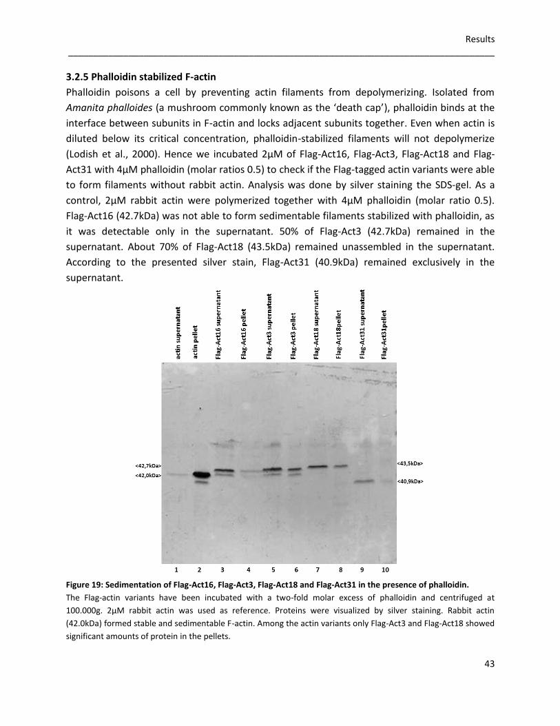
Results _____________________________________________________________________________________
43
3.2.5 Phalloidin stabilized F-actin
Phalloidin poisons a cell by preventing actin filaments from depolymerizing. Isolated from
Amanita phalloides (a mushroom commonly known as the ‘death cap’), phalloidin binds at the
interface between subunits in F-actin and locks adjacent subunits together. Even when actin is
diluted below its critical concentration, phalloidin-stabilized filaments will not depolymerize
(Lodish et al., 2000). Hence we incubated 2µM of Flag-Act16, Flag-Act3, Flag-Act18 and Flag-
Act31 with 4µM phalloidin (molar ratios 0.5) to check if the Flag-tagged actin variants were able
to form filaments without rabbit actin. Analysis was done by silver staining the SDS-gel. As a
control, 2µM rabbit actin were polymerized together with 4µM phalloidin (molar ratio 0.5).
Flag-Act16 (42.7kDa) was not able to form sedimentable filaments stabilized with phalloidin, as
it was detectable only in the supernatant. 50% of Flag-Act3 (42.7kDa) remained in the
supernatant. About 70% of Flag-Act18 (43.5kDa) remained unassembled in the supernatant.
According to the presented silver stain, Flag-Act31 (40.9kDa) remained exclusively in the
supernatant.
Figure 19: Sedimentation of Flag-Act16, Flag-Act3, Flag-Act18 and Flag-Act31 in the presence of phalloidin.
The Flag-actin variants have been incubated with a two-fold molar excess of phalloidin and centrifuged at
100.000g. 2µM rabbit actin was used as reference. Proteins were visualized by silver staining. Rabbit actin
(42.0kDa) formed stable and sedimentable F-actin. Among the actin variants only Flag-Act3 and Flag-Act18 showed
significant amounts of protein in the pellets.
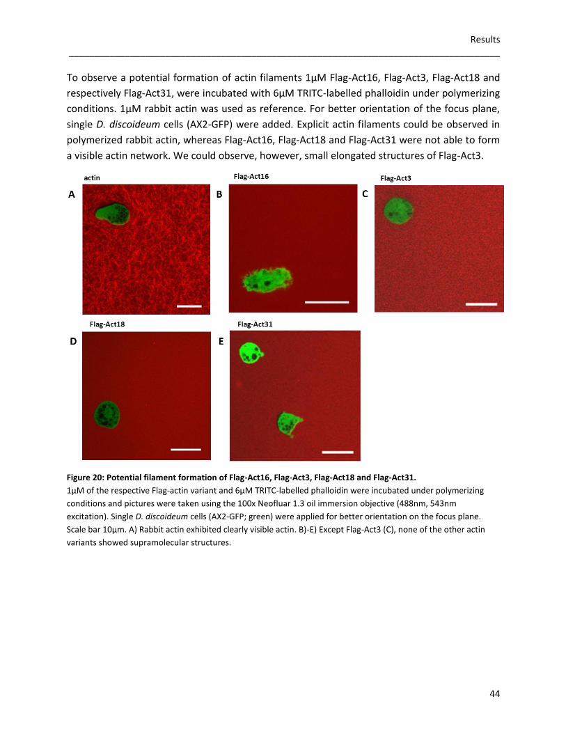
Results _____________________________________________________________________________________
44
To observe a potential formation of actin filaments 1µM Flag-Act16, Flag-Act3, Flag-Act18 and
respectively Flag-Act31, were incubated with 6µM TRITC-labelled phalloidin under polymerizing
conditions. 1µM rabbit actin was used as reference. For better orientation of the focus plane,
single D. discoideum cells (AX2-GFP) were added. Explicit actin filaments could be observed in
polymerized rabbit actin, whereas Flag-Act16, Flag-Act18 and Flag-Act31 were not able to form
a visible actin network. We could observe, however, small elongated structures of Flag-Act3.
Figure 20: Potential filament formation of Flag-Act16, Flag-Act3, Flag-Act18 and Flag-Act31.
1µM of the respective Flag-actin variant and 6µM TRITC-labelled phalloidin were incubated under polymerizing
conditions and pictures were taken using the 100x Neofluar 1.3 oil immersion objective (488nm, 543nm
excitation). Single D. discoideum cells (AX2-GFP; green) were applied for better orientation on the focus plane.
Scale bar 10µm. A) Rabbit actin exhibited clearly visible actin. B)-E) Except Flag-Act3 (C), none of the other actin
variants showed supramolecular structures.
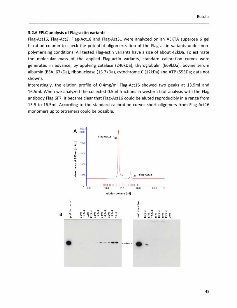
Results _____________________________________________________________________________________
45
3.2.6 FPLC analysis of Flag-actin variants
Flag-Act16, Flag-Act3, Flag-Act18 and Flag-Act31 were analyzed on an AEKTA superose 6 gel
filtration column to check the potential oligomerization of the Flag-actin variants under non-
polymerizing conditions. All tested Flag-actin variants have a size of about 42kDa. To estimate
the molecular mass of the applied Flag-actin variants, standard calibration curves were
generated in advance, by applying catalase (240kDa), thyroglobulin (669kDa), bovine serum
albumin (BSA; 67kDa), ribonuclease (13.7kDa), cytochrome C (12kDa) and ATP (553Da; data not
shown).
Interestingly, the elution profile of 0.4mg/ml Flag-Act16 showed two peaks at 13.5ml and
16.5ml. When we analyzed the collected 0.5ml fractions in western blot analysis with the Flag
antibody Flag 6F7, it became clear that Flag-Act16 could be eluted reproducibly in a range from
13.5 to 16.5ml. According to the standard calibration curves short oligomers from Flag-Act16
monomers up to tetramers could be possible.
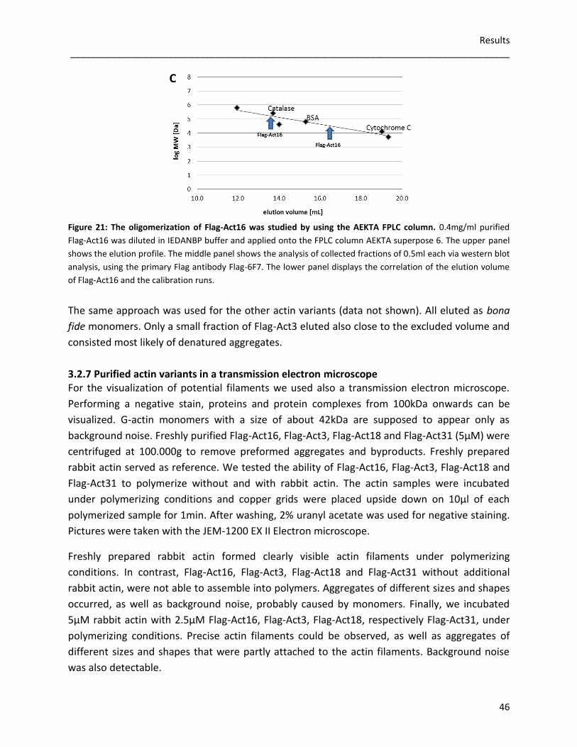
Results _____________________________________________________________________________________
46
Figure 21: The oligomerization of Flag-Act16 was studied by using the AEKTA FPLC column. 0.4mg/ml purified
Flag-Act16 was diluted in IEDANBP buffer and applied onto the FPLC column AEKTA superpose 6. The upper panel
shows the elution profile. The middle panel shows the analysis of collected fractions of 0.5ml each via western blot
analysis, using the primary Flag antibody Flag-6F7. The lower panel displays the correlation of the elution volume
of Flag-Act16 and the calibration runs.
The same approach was used for the other actin variants (data not shown). All eluted as bona
fide monomers. Only a small fraction of Flag-Act3 eluted also close to the excluded volume and
consisted most likely of denatured aggregates.
3.2.7 Purified actin variants in a transmission electron microscope For the visualization of potential filaments we used also a transmission electron microscope.
Performing a negative stain, proteins and protein complexes from 100kDa onwards can be
visualized. G-actin monomers with a size of about 42kDa are supposed to appear only as
background noise. Freshly purified Flag-Act16, Flag-Act3, Flag-Act18 and Flag-Act31 (5µM) were
centrifuged at 100.000g to remove preformed aggregates and byproducts. Freshly prepared
rabbit actin served as reference. We tested the ability of Flag-Act16, Flag-Act3, Flag-Act18 and
Flag-Act31 to polymerize without and with rabbit actin. The actin samples were incubated
under polymerizing conditions and copper grids were placed upside down on 10µl of each
polymerized sample for 1min. After washing, 2% uranyl acetate was used for negative staining.
Pictures were taken with the JEM-1200 EX II Electron microscope.
Freshly prepared rabbit actin formed clearly visible actin filaments under polymerizing
conditions. In contrast, Flag-Act16, Flag-Act3, Flag-Act18 and Flag-Act31 without additional
rabbit actin, were not able to assemble into polymers. Aggregates of different sizes and shapes
occurred, as well as background noise, probably caused by monomers. Finally, we incubated
5µM rabbit actin with 2.5µM Flag-Act16, Flag-Act3, Flag-Act18, respectively Flag-Act31, under
polymerizing conditions. Precise actin filaments could be observed, as well as aggregates of
different sizes and shapes that were partly attached to the actin filaments. Background noise
was also detectable.

Results _____________________________________________________________________________________
47
Figure 22: Flag-Act16, Flag-Act3, Flag-
Act18 and Flag-Act31 imaged using a
transmission electron microscope.
Rabbit actin alone (A), Flag-tagged actin
variants without rabbit actin (B-E), and
Flag-tagged actin variants in the presence
of rabbit actin (F-I), were incubated under
polymerizing conditions. After washing and
negative staining with 2% uranyl acetate,
the copper grids were examined and
imaged using the 10.000x magnifications at
the JEOL 1200EX II transmission electron
microscope (70kV). Scale bar 200nm.
A)-E): Only rabbit actin alone showed the
well-known filaments, all actin variants
formed at most supramolecular aggregates.
F)-I): In the presence of rabbit actin,
filaments were present in all cases which
suggest that the actin variants did not
inhibit filament formation.

Results _____________________________________________________________________________________
48
3.2.8 Interaction studies with severin
To identify potential interaction partners of Act16, Act3, Act18 and Act31, GFP-Traps
(Chromotek) were performed with GFP-Act16, GFP-Act3, GFP-Act18 and GFP-Act31 cells from
D. discoideum. The GFP-fusion proteins bind to beads coated with the GFP-antibody and can be
pulled down together with their putative interaction partners. In mass spectrometry analysis,
the actin binding protein severin was reproducibly detected to interact with GFP-Act31. As the
severin that was available in the laboratory was His-tagged, beaded agarose derivatized with
the nitrilotriacetic acid (NTA) chelation moiety and loaded with divalent nickel ions (Ni2+) could
be used for detection of severin. When we performed a western blot analysis by using the
monoclonal Flag antibody 6F7, we could show that all tested Flag-tagged actin interacted in a
Ca2+-dependent manner with severin.
Figure 23: Interaction studies of Flag-Act16, Flag-Act3, Flag-Act18 and Flag-Act31 with severin.
We mixed Flag-Act16, Flag-Act3, Flag-Act18 and Flag-Act31 with severin (molar ratios 1) and added 1mM Ca2+
,
respectively 1mM EGTA. These approaches were incubated with Ni2+
-NTA beaded agarose for 90 minutes at 4°C
and subsequently centrifuged. Western blots were performed by using the monoclonal Flag antibody 6F7.
A)-D): All Flag-tagged actin variants interacted Ca2+
-dependent with severin, as they could be pulled down with
Ni2+
-NTA beaded agarose. Adding EGTA, no interaction of the Flag-tag actin variants and severin occurred as the
Flag-actin variants remained in the supernatant.
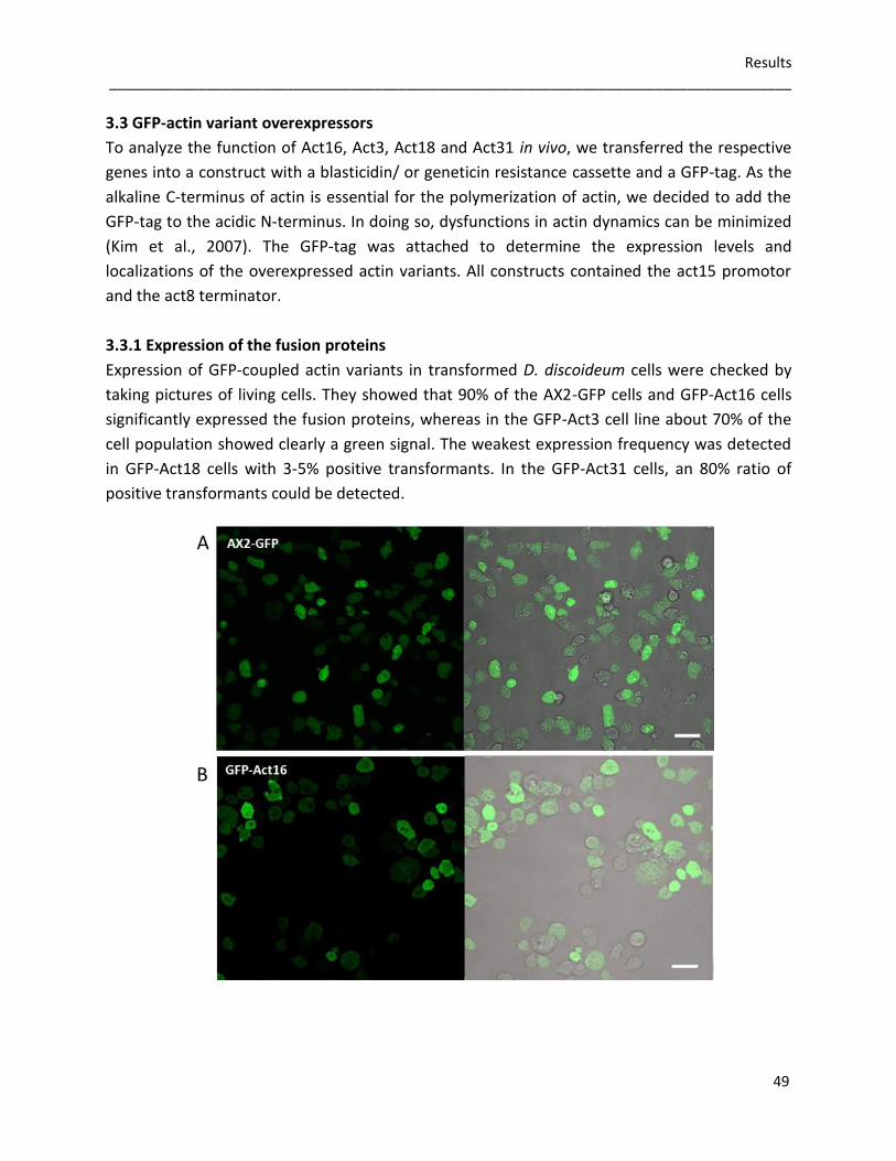
Results _____________________________________________________________________________________
49
3.3 GFP-actin variant overexpressors
To analyze the function of Act16, Act3, Act18 and Act31 in vivo, we transferred the respective
genes into a construct with a blasticidin/ or geneticin resistance cassette and a GFP-tag. As the
alkaline C-terminus of actin is essential for the polymerization of actin, we decided to add the
GFP-tag to the acidic N-terminus. In doing so, dysfunctions in actin dynamics can be minimized
(Kim et al., 2007). The GFP-tag was attached to determine the expression levels and
localizations of the overexpressed actin variants. All constructs contained the act15 promotor
and the act8 terminator.
3.3.1 Expression of the fusion proteins
Expression of GFP-coupled actin variants in transformed D. discoideum cells were checked by
taking pictures of living cells. They showed that 90% of the AX2-GFP cells and GFP-Act16 cells
significantly expressed the fusion proteins, whereas in the GFP-Act3 cell line about 70% of the
cell population showed clearly a green signal. The weakest expression frequency was detected
in GFP-Act18 cells with 3-5% positive transformants. In the GFP-Act31 cells, an 80% ratio of
positive transformants could be detected.

Results _____________________________________________________________________________________
50
Figure 24: Different expression of GFP-actin variants in transformed D. discoideum cells.
The ratio of GFP-positive to unlabeled cells within the transformed cell lines was documented using the 40x LDA-
Plan 0.50 Ph2 (488nm excitation) of the LSM 510 confocal microscope. The left panel shows the fluorescence
picture. The right panel visualizes a merge of the bright-field illumination and the GFP-signal. Scale bar 10µm.
A) The GFP control was detectable in 90% of the AX2-GFP cells. B) GFP-Act16 was expressed in 90% of the
transformed cells. C) GFP-Act3 was detectable in about 70% of the transformed cells. D) GFP-Act18 was detectable
in 3-5% of the cells. E) GFP-Act31 occurred in 90% of the transformed cells.
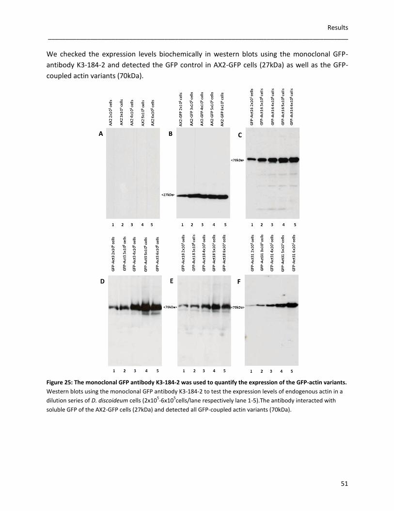
Results _____________________________________________________________________________________
51
We checked the expression levels biochemically in western blots using the monoclonal GFP-
antibody K3-184-2 and detected the GFP control in AX2-GFP cells (27kDa) as well as the GFP-
coupled actin variants (70kDa).
Figure 25: The monoclonal GFP antibody K3-184-2 was used to quantify the expression of the GFP-actin variants.
Western blots using the monoclonal GFP antibody K3-184-2 to test the expression levels of endogenous actin in a
dilution series of D. discoideum cells (2x10⁵-6x10
5cells/lane respectively lane 1-5).The antibody interacted with
soluble GFP of the AX2-GFP cells (27kDa) and detected all GFP-coupled actin variants (70kDa).
-
-
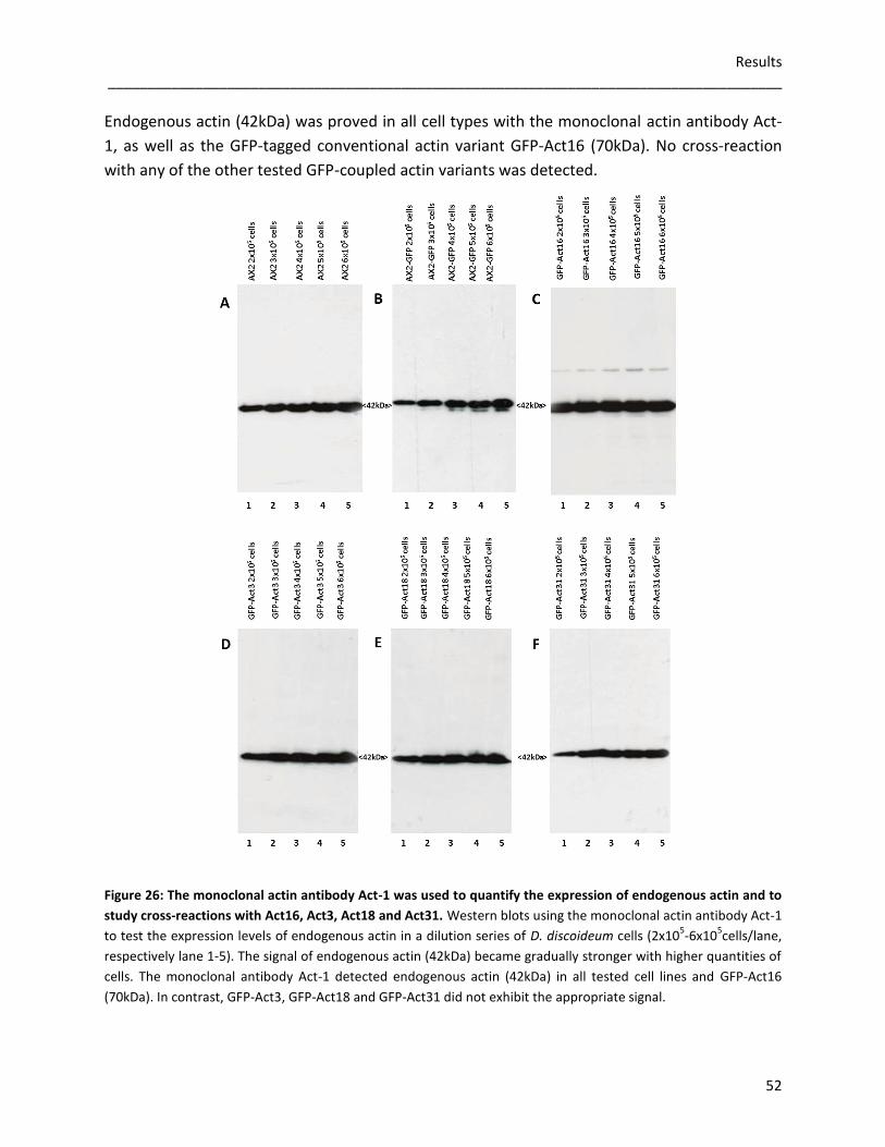
Results _____________________________________________________________________________________
52
Endogenous actin (42kDa) was proved in all cell types with the monoclonal actin antibody Act-
1, as well as the GFP-tagged conventional actin variant GFP-Act16 (70kDa). No cross-reaction
with any of the other tested GFP-coupled actin variants was detected.
Figure 26: The monoclonal actin antibody Act-1 was used to quantify the expression of endogenous actin and to
study cross-reactions with Act16, Act3, Act18 and Act31. Western blots using the monoclonal actin antibody Act-1
to test the expression levels of endogenous actin in a dilution series of D. discoideum cells (2x10⁵-6x10
5cells/lane,
respectively lane 1-5). The signal of endogenous actin (42kDa) became gradually stronger with higher quantities of
cells. The monoclonal antibody Act-1 detected endogenous actin (42kDa) in all tested cell lines and GFP-Act16
(70kDa). In contrast, GFP-Act3, GFP-Act18 and GFP-Act31 did not exhibit the appropriate signal.
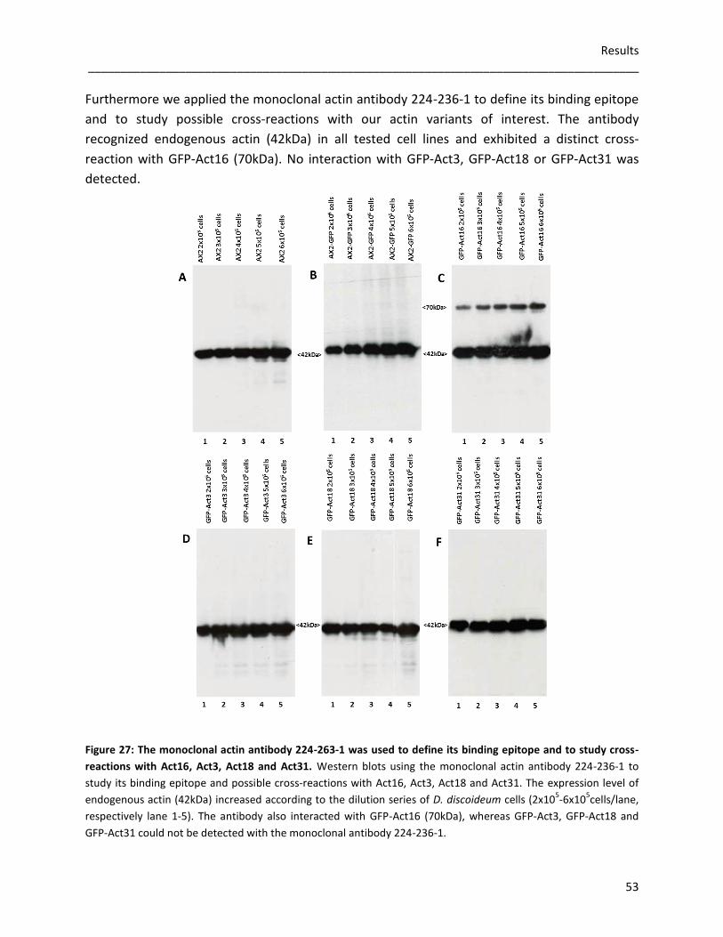
Results _____________________________________________________________________________________
53
Furthermore we applied the monoclonal actin antibody 224-236-1 to define its binding epitope
and to study possible cross-reactions with our actin variants of interest. The antibody
recognized endogenous actin (42kDa) in all tested cell lines and exhibited a distinct cross-
reaction with GFP-Act16 (70kDa). No interaction with GFP-Act3, GFP-Act18 or GFP-Act31 was
detected.
Figure 27: The monoclonal actin antibody 224-263-1 was used to define its binding epitope and to study cross-
reactions with Act16, Act3, Act18 and Act31. Western blots using the monoclonal actin antibody 224-236-1 to
study its binding epitope and possible cross-reactions with Act16, Act3, Act18 and Act31. The expression level of
endogenous actin (42kDa) increased according to the dilution series of D. discoideum cells (2x10⁵-6x10
5cells/lane,
respectively lane 1-5). The antibody also interacted with GFP-Act16 (70kDa), whereas GFP-Act3, GFP-Act18 and
GFP-Act31 could not be detected with the monoclonal antibody 224-236-1.

Results _____________________________________________________________________________________
54
The application of the monoclonal filactin antibody 4S-59-4 in western blot analysis showed
that the tested cell lines AX2, AX2-GFP, GFP-Act16, GFP-Act3, GFP-Act18 and GFP-Act31
expressed filactin (105kDa; data not shown). Expected interactions with endogenous actin
(42kDa) could be observed in all tested cells, whereas no interaction with Act16, Act3, Act18
and Act31 occurred (Gallinger, 2013).
3.3.2 Analyzing the F-actin cytoskeleton
3.3.2.1 Localization studies of actin variants in D. discoideum using fluorescence microscopy
To study the localization of GFP-Act16, GFP-Act3, GFP-Act18 and GFP-Act31 within the
transformed D. discoideum cells, fixed cells were examined under the LSM 510 confocal
microscope. GFP-Act16 and GFP-Act3 colocalized predominantly with endogenous actin at the
cell cortex and were also detectable in the cytoplasm. GFP-Act18 and GFP-Act31 could not be
visualized in the cell cortex, but were distributed throughout the cytoplasm. The GFP of the
AX2-GFP cells was not associated with actin and was, as expected, spread homogenously in the
cytoplasm of the dedicated cells.
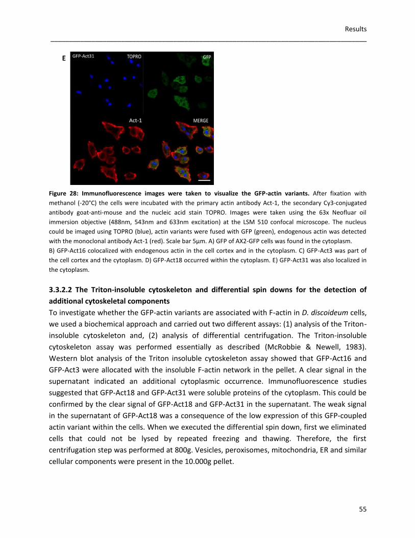
Results _____________________________________________________________________________________
55
Figure 28: Immunofluorescence images were taken to visualize the GFP-actin variants. After fixation with
methanol (-20°C) the cells were incubated with the primary actin antibody Act-1, the secondary Cy3-conjugated
antibody goat-anti-mouse and the nucleic acid stain TOPRO. Images were taken using the 63x Neofluar oil
immersion objective (488nm, 543nm and 633nm excitation) at the LSM 510 confocal microscope. The nucleus
could be imaged using TOPRO (blue), actin variants were fused with GFP (green), endogenous actin was detected
with the monoclonal antibody Act-1 (red). Scale bar 5µm. A) GFP of AX2-GFP cells was found in the cytoplasm.
B) GFP-Act16 colocalized with endogenous actin in the cell cortex and in the cytoplasm. C) GFP-Act3 was part of
the cell cortex and the cytoplasm. D) GFP-Act18 occurred within the cytoplasm. E) GFP-Act31 was also localized in
the cytoplasm.
3.3.2.2 The Triton-insoluble cytoskeleton and differential spin downs for the detection of
additional cytoskeletal components
To investigate whether the GFP-actin variants are associated with F-actin in D. discoideum cells,
we used a biochemical approach and carried out two different assays: (1) analysis of the Triton-
insoluble cytoskeleton and, (2) analysis of differential centrifugation. The Triton-insoluble
cytoskeleton assay was performed essentially as described (McRobbie & Newell, 1983).
Western blot analysis of the Triton insoluble cytoskeleton assay showed that GFP-Act16 and
GFP-Act3 were allocated with the insoluble F-actin network in the pellet. A clear signal in the
supernatant indicated an additional cytoplasmic occurrence. Immunofluorescence studies
suggested that GFP-Act18 and GFP-Act31 were soluble proteins of the cytoplasm. This could be
confirmed by the clear signal of GFP-Act18 and GFP-Act31 in the supernatant. The weak signal
in the supernatant of GFP-Act18 was a consequence of the low expression of this GFP-coupled
actin variant within the cells. When we executed the differential spin down, first we eliminated
cells that could not be lysed by repeated freezing and thawing. Therefore, the first
centrifugation step was performed at 800g. Vesicles, peroxisomes, mitochondria, ER and similar
cellular components were present in the 10.000g pellet.
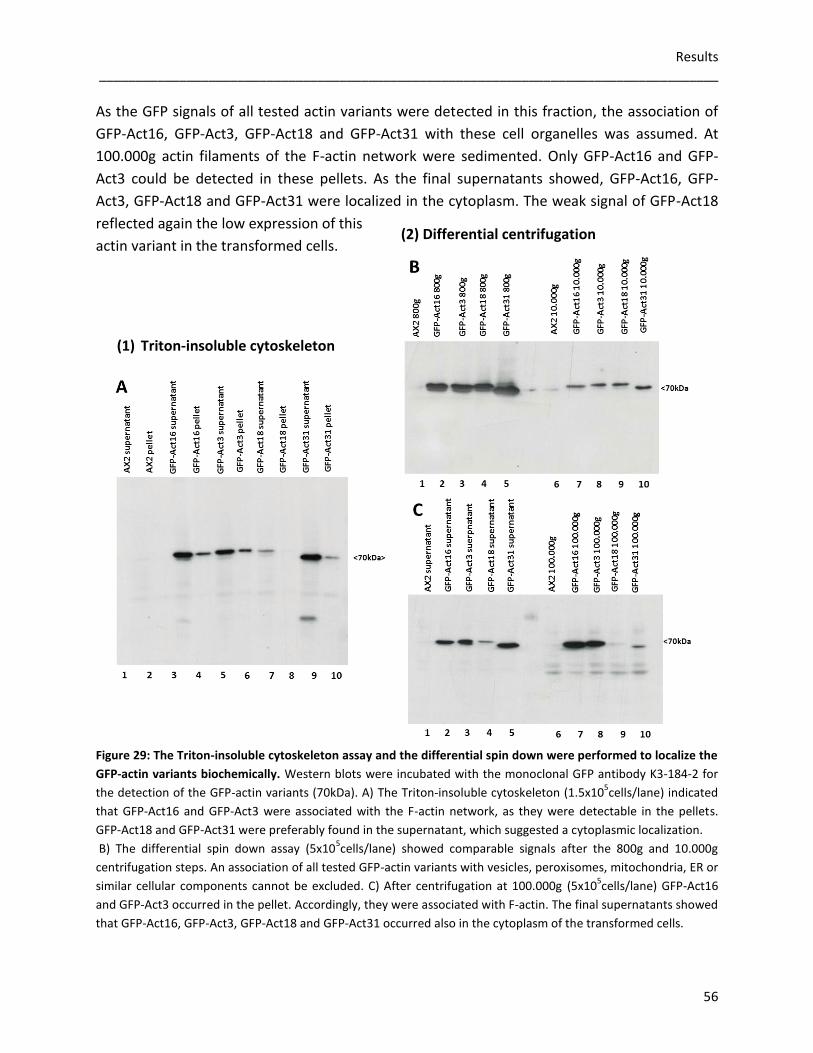
Results _____________________________________________________________________________________
56
As the GFP signals of all tested actin variants were detected in this fraction, the association of
GFP-Act16, GFP-Act3, GFP-Act18 and GFP-Act31 with these cell organelles was assumed. At
100.000g actin filaments of the F-actin network were sedimented. Only GFP-Act16 and GFP-
Act3 could be detected in these pellets. As the final supernatants showed, GFP-Act16, GFP-
Act3, GFP-Act18 and GFP-Act31 were localized in the cytoplasm. The weak signal of GFP-Act18
reflected again the low expression of this
actin variant in the transformed cells.
Figure 29: The Triton-insoluble cytoskeleton assay and the differential spin down were performed to localize the
GFP-actin variants biochemically. Western blots were incubated with the monoclonal GFP antibody K3-184-2 for
the detection of the GFP-actin variants (70kDa). A) The Triton-insoluble cytoskeleton (1.5x10⁵cells/lane) indicated
that GFP-Act16 and GFP-Act3 were associated with the F-actin network, as they were detectable in the pellets.
GFP-Act18 and GFP-Act31 were preferably found in the supernatant, which suggested a cytoplasmic localization.
B) The differential spin down assay (5x10⁵cells/lane) showed comparable signals after the 800g and 10.000g
centrifugation steps. An association of all tested GFP-actin variants with vesicles, peroxisomes, mitochondria, ER or
similar cellular components cannot be excluded. C) After centrifugation at 100.000g (5x10⁵cells/lane) GFP-Act16
and GFP-Act3 occurred in the pellet. Accordingly, they were associated with F-actin. The final supernatants showed
that GFP-Act16, GFP-Act3, GFP-Act18 and GFP-Act31 occurred also in the cytoplasm of the transformed cells.
(1) Triton-insoluble cytoskeleton
(2) Differential centrifugation
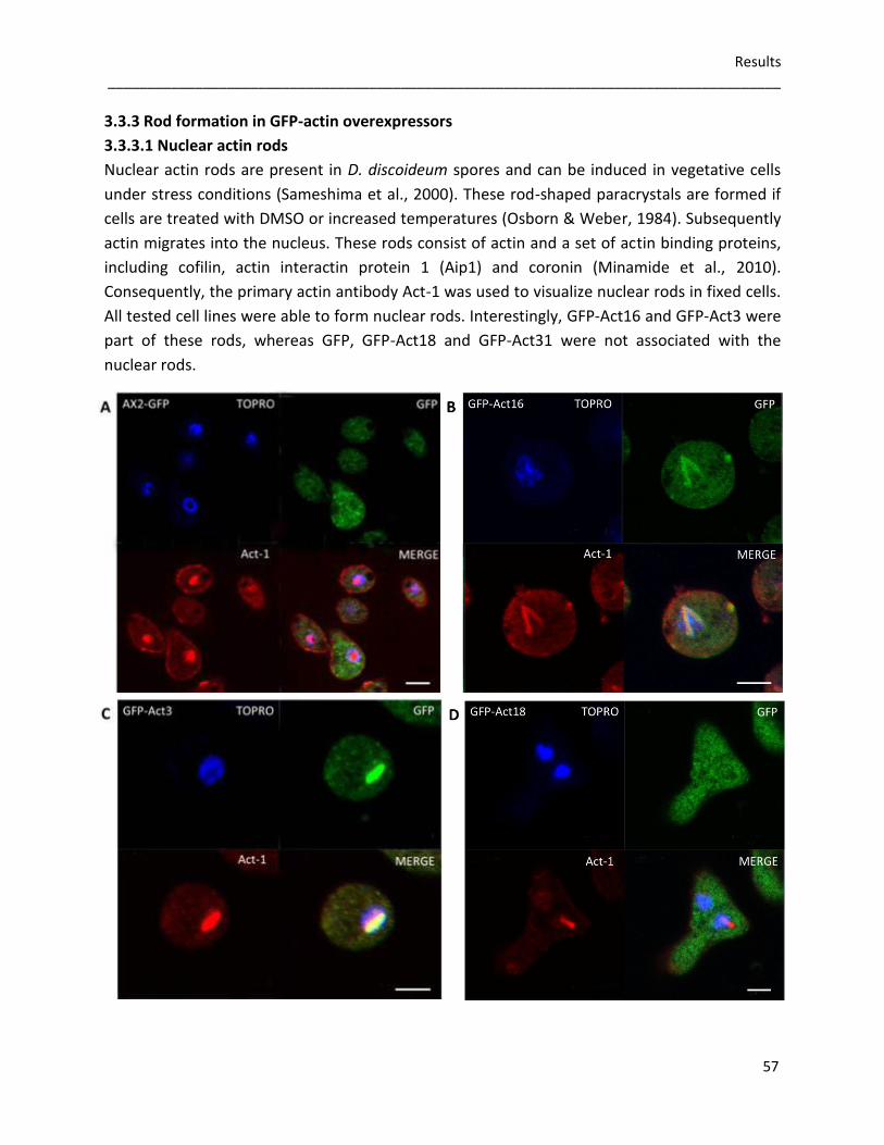
Results _____________________________________________________________________________________
57
3.3.3 Rod formation in GFP-actin overexpressors
3.3.3.1 Nuclear actin rods
Nuclear actin rods are present in D. discoideum spores and can be induced in vegetative cells
under stress conditions (Sameshima et al., 2000). These rod-shaped paracrystals are formed if
cells are treated with DMSO or increased temperatures (Osborn & Weber, 1984). Subsequently
actin migrates into the nucleus. These rods consist of actin and a set of actin binding proteins,
including cofilin, actin interactin protein 1 (Aip1) and coronin (Minamide et al., 2010).
Consequently, the primary actin antibody Act-1 was used to visualize nuclear rods in fixed cells.
All tested cell lines were able to form nuclear rods. Interestingly, GFP-Act16 and GFP-Act3 were
part of these rods, whereas GFP, GFP-Act18 and GFP-Act31 were not associated with the
nuclear rods.
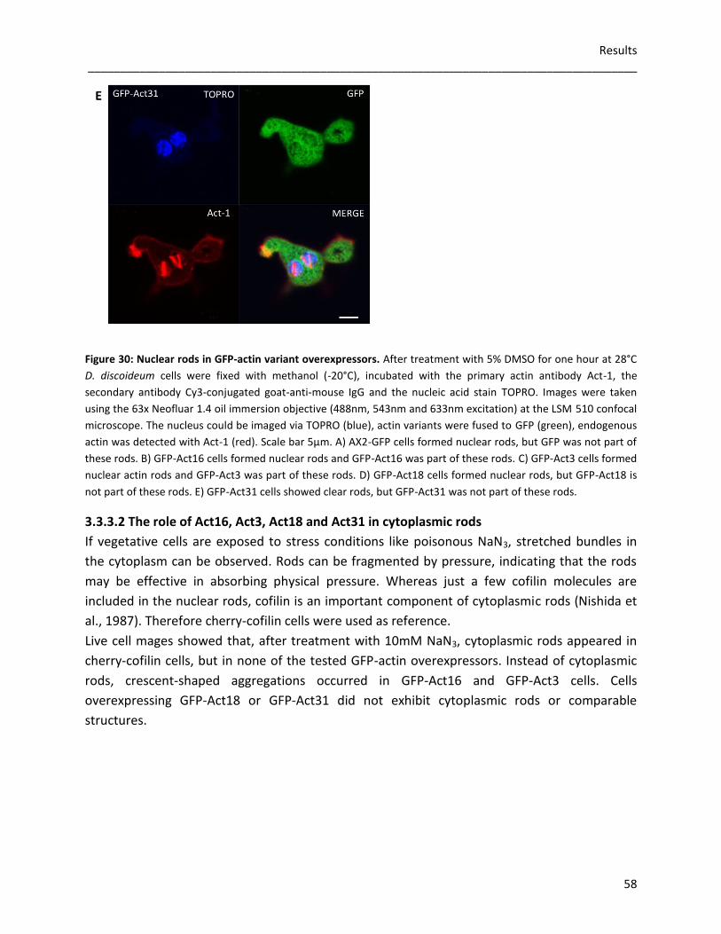
Results _____________________________________________________________________________________
58
Figure 30: Nuclear rods in GFP-actin variant overexpressors. After treatment with 5% DMSO for one hour at 28°C
D. discoideum cells were fixed with methanol (-20°C), incubated with the primary actin antibody Act-1, the
secondary antibody Cy3-conjugated goat-anti-mouse IgG and the nucleic acid stain TOPRO. Images were taken
using the 63x Neofluar 1.4 oil immersion objective (488nm, 543nm and 633nm excitation) at the LSM 510 confocal
microscope. The nucleus could be imaged via TOPRO (blue), actin variants were fused to GFP (green), endogenous
actin was detected with Act-1 (red). Scale bar 5µm. A) AX2-GFP cells formed nuclear rods, but GFP was not part of
these rods. B) GFP-Act16 cells formed nuclear rods and GFP-Act16 was part of these rods. C) GFP-Act3 cells formed
nuclear actin rods and GFP-Act3 was part of these rods. D) GFP-Act18 cells formed nuclear rods, but GFP-Act18 is
not part of these rods. E) GFP-Act31 cells showed clear rods, but GFP-Act31 was not part of these rods.
3.3.3.2 The role of Act16, Act3, Act18 and Act31 in cytoplasmic rods
If vegetative cells are exposed to stress conditions like poisonous NaN3, stretched bundles in
the cytoplasm can be observed. Rods can be fragmented by pressure, indicating that the rods
may be effective in absorbing physical pressure. Whereas just a few cofilin molecules are
included in the nuclear rods, cofilin is an important component of cytoplasmic rods (Nishida et
al., 1987). Therefore cherry-cofilin cells were used as reference.
Live cell mages showed that, after treatment with 10mM NaN3, cytoplasmic rods appeared in
cherry-cofilin cells, but in none of the tested GFP-actin overexpressors. Instead of cytoplasmic
rods, crescent-shaped aggregations occurred in GFP-Act16 and GFP-Act3 cells. Cells
overexpressing GFP-Act18 or GFP-Act31 did not exhibit cytoplasmic rods or comparable
structures.
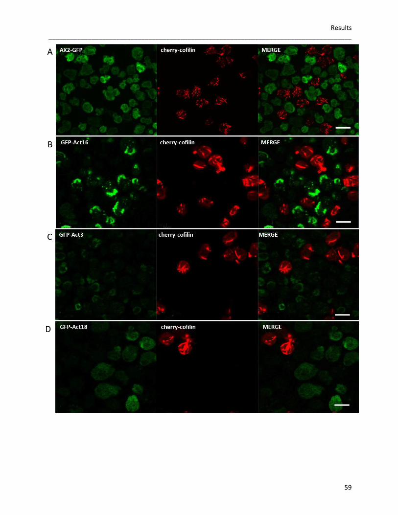
Results _____________________________________________________________________________________
59
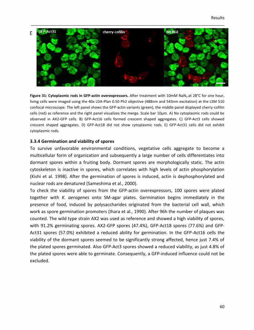
Results _____________________________________________________________________________________
60
Figure 31: Cytoplasmic rods in GFP-actin overexpressors. After treatment with 10mM NaN3 at 28°C for one hour,
living cells were imaged using the 40x LDA-Plan 0.50 Ph2 objective (488nm and 543nm excitation) at the LSM 510
confocal microscope. The left panel shows the GFP-actin variants (green), the middle panel displayed cherry-cofilin
cells (red) as reference and the right panel visualizes the merge. Scale bar 10µm. A) No cytoplasmic rods could be
observed in AX2-GFP cells. B) GFP-Act16 cells formed crescent shaped aggregates. C) GFP-Act3 cells showed
crescent shaped aggregates. D) GFP-Act18 did not show cytoplasmic rods. E) GFP-Act31 cells did not exhibit
cytoplasmic rods.
3.3.4 Germination and viability of spores
To survive unfavorable environmental conditions, vegetative cells aggregate to become a
multicellular form of organization and subsequently a large number of cells differentiates into
dormant spores within a fruiting body. Dormant spores are morphologically static. The actin
cytoskeleton is inactive in spores, which correlates with high levels of actin phosphorylation
(Kishi et al. 1998). After the germination of spores is induced, actin is dephosphorylated and
nuclear rods are denatured (Sameshima et al., 2000).
To check the viability of spores from the GFP-actin overexpressors, 100 spores were plated
together with K. aerogenes onto SM-agar plates. Germination begins immediately in the
presence of food, induced by polysaccharides originated from the bacterial cell wall, which
work as spore germination promoters (Ihara et al., 1990). After 96h the number of plaques was
counted. The wild type strain AX2 was used as reference and showed a high viability of spores,
with 91.2% germinating spores. AX2-GFP spores (47.4%), GFP-Act18 spores (77.6%) and GFP-
Act31 spores (57.0%) exhibited a reduced ability for germination. In the GFP-Act16 cells the
viability of the dormant spores seemed to be significantly strong affected, hence just 7.4% of
the plated spores germinated. Also GFP-Act3 spores showed a reduced viability, as just 4.8% of
the plated spores were able to germinate. Consequently, a GFP-induced influence could not be
excluded.
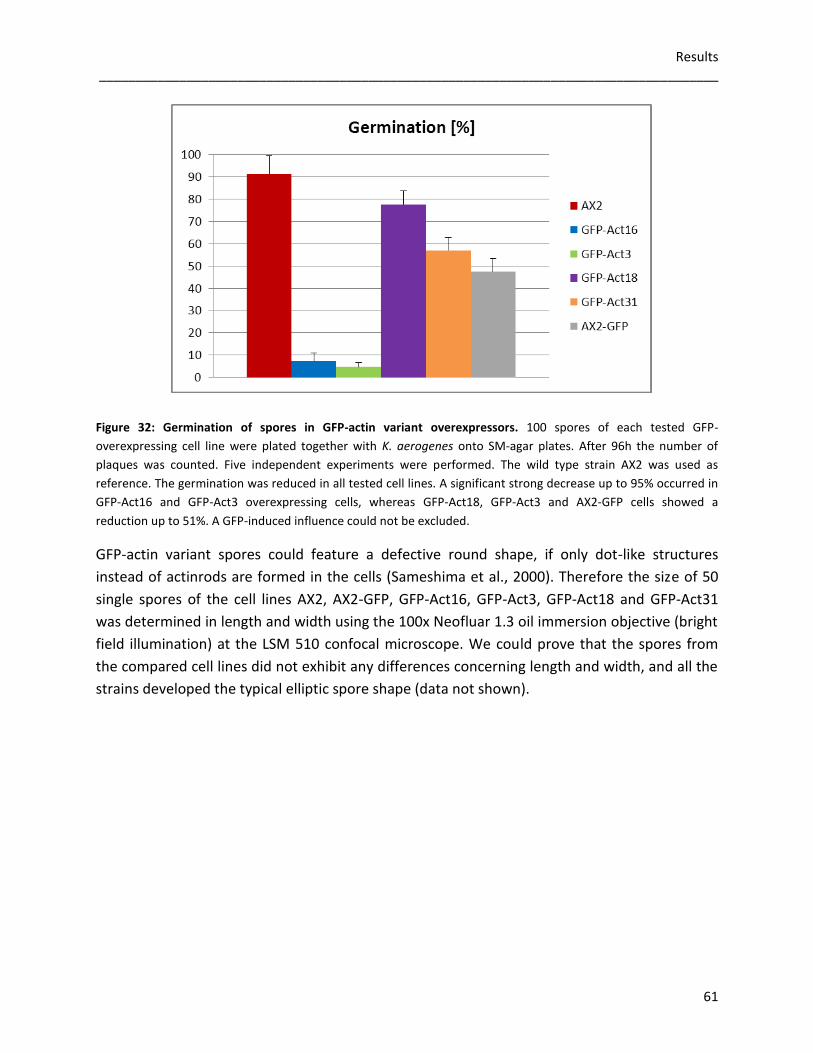
Results _____________________________________________________________________________________
61
Figure 32: Germination of spores in GFP-actin variant overexpressors. 100 spores of each tested GFP-
overexpressing cell line were plated together with K. aerogenes onto SM-agar plates. After 96h the number of
plaques was counted. Five independent experiments were performed. The wild type strain AX2 was used as
reference. The germination was reduced in all tested cell lines. A significant strong decrease up to 95% occurred in
GFP-Act16 and GFP-Act3 overexpressing cells, whereas GFP-Act18, GFP-Act3 and AX2-GFP cells showed a
reduction up to 51%. A GFP-induced influence could not be excluded.
GFP-actin variant spores could feature a defective round shape, if only dot-like structures
instead of actinrods are formed in the cells (Sameshima et al., 2000). Therefore the size of 50
single spores of the cell lines AX2, AX2-GFP, GFP-Act16, GFP-Act3, GFP-Act18 and GFP-Act31
was determined in length and width using the 100x Neofluar 1.3 oil immersion objective (bright
field illumination) at the LSM 510 confocal microscope. We could prove that the spores from
the compared cell lines did not exhibit any differences concerning length and width, and all the
strains developed the typical elliptic spore shape (data not shown).
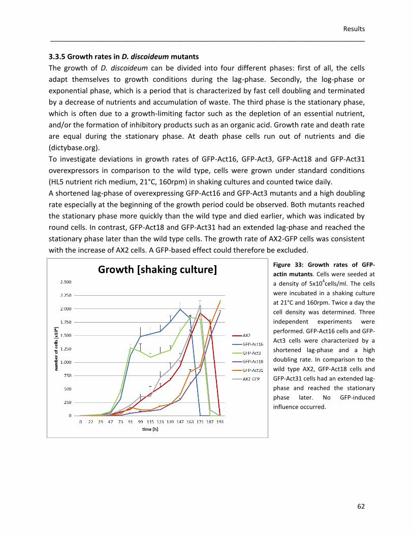
Results _____________________________________________________________________________________
62
3.3.5 Growth rates in D. discoideum mutants
The growth of D. discoideum can be divided into four different phases: first of all, the cells
adapt themselves to growth conditions during the lag-phase. Secondly, the log-phase or
exponential phase, which is a period that is characterized by fast cell doubling and terminated
by a decrease of nutrients and accumulation of waste. The third phase is the stationary phase,
which is often due to a growth-limiting factor such as the depletion of an essential nutrient,
and/or the formation of inhibitory products such as an organic acid. Growth rate and death rate
are equal during the stationary phase. At death phase cells run out of nutrients and die
(dictybase.org).
To investigate deviations in growth rates of GFP-Act16, GFP-Act3, GFP-Act18 and GFP-Act31
overexpressors in comparison to the wild type, cells were grown under standard conditions
(HL5 nutrient rich medium, 21°C, 160rpm) in shaking cultures and counted twice daily.
A shortened lag-phase of overexpressing GFP-Act16 and GFP-Act3 mutants and a high doubling
rate especially at the beginning of the growth period could be observed. Both mutants reached
the stationary phase more quickly than the wild type and died earlier, which was indicated by
round cells. In contrast, GFP-Act18 and GFP-Act31 had an extended lag-phase and reached the
stationary phase later than the wild type cells. The growth rate of AX2-GFP cells was consistent
with the increase of AX2 cells. A GFP-based effect could therefore be excluded.
Figure 33: Growth rates of GFP-
actin mutants. Cells were seeded at
a density of 5x10⁴cells/ml. The cells
were incubated in a shaking culture
at 21°C and 160rpm. Twice a day the
cell density was determined. Three
independent experiments were
performed. GFP-Act16 cells and GFP-
Act3 cells were characterized by a
shortened lag-phase and a high
doubling rate. In comparison to the
wild type AX2, GFP-Act18 cells and
GFP-Act31 cells had an extended lag-
phase and reached the stationary
phase later. No GFP-induced
influence occurred.
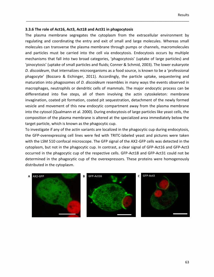
Results _____________________________________________________________________________________
63
3.3.6 The role of Act16, Act3, Act18 and Act31 in phagocytosis
The plasma membrane segregates the cytoplasm from the extracellular environment by
regulating and coordinating the entry and exit of small and large molecules. Whereas small
molecules can transverse the plasma membrane through pumps or channels, macromolecules
and particles must be carried into the cell via endocytosis. Endocytosis occurs by multiple
mechanisms that fall into two broad categories, ‘phagocytosis’ (uptake of large particles) and
‘pinocytosis’ (uptake of small particles and fluids; Conner & Schmid, 2003). The lower eukaryote
D. discoideum, that internalizes microorganisms as a food source, is known to be a ‘professional
phagocyte’ (Bozzaro & Eichinger, 2011). Accordingly, the particle uptake, sequestering and
maturation into phagosomes of D. discoideum resembles in many ways the events observed in
macrophages, neutrophils or dendritic cells of mammals. The major endocytic process can be
differentiated into five steps, all of them involving the actin cytoskeleton: membrane
invagination, coated pit formation, coated pit sequestration, detachment of the newly formed
vesicle and movement of this new endocytic compartment away from the plasma membrane
into the cytosol (Qualmann et al. 2000). During endocytosis of large particles like yeast cells, the
composition of the plasma membrane is altered at the specialized area immediately below the
target particle, which is known as the phagocytic cup.
To investigate if any of the actin variants are localized in the phagocytic cup during endocytosis,
the GFP-overexpressing cell lines were fed with TRITC-labeled yeast and pictures were taken
with the LSM 510 confocal microscope. The GFP signal of the AX2-GFP cells was detected in the
cytoplasm, but not in the phagocytic cup. In contrast, a clear signal of GFP-Act16 and GFP-Act3
occurred in the phagocytic cup of the respective cells. GFP-Act18 and GFP-Act31 could not be
determined in the phagocytic cup of the overexpressors. These proteins were homogenously
distributed in the cytoplasm.

Results _____________________________________________________________________________________
64
Figure 34: The localization of GFP-actin variants during phagocytosis. 2x10⁶cells/ml of every tested cell line were
starved one hour in nutrient deficient medium and challenged with 1/10 of TRITC-labeled yeast cells. Images were
taken by using a 100x Neofluar 1.3 oil immersion objective (488nm and 543nm excitation) at the LSM 510 confocal
microscope. Scale bar 2µm A) In AX2-GFP cells, GFP was spread within the cytoplasm, but not enriched in the
phagocytic cup. B) GFP-Act16 accumulated in the phagocytic cup during the uptake of yeast cells. C) Also GFP-Act3
was concentrated in the phagocytic cup during phagocytosis. D) In GFP-Act18 cells, the GFP-signal occurred in the
cytoplasm, but not enriched in the phagocytic cup. E) GFP-Act31 occurred in the cytoplasm, but not accumulated in
the phagocytic cup.
Given that GFP-Act16 and GFP-Act3 were enriched in the phagocytic cup during phagocytosis,
whereas an accumulation of GFP-Act18 and GFP-Act31 could not be detected in the phagocytic
cup, the uptake of TRITC-labeled yeast was measured at the fluorescence spectrometer LS55.
Surprisingly, all mutants showed a disturbed uptake of yeast and had only about 50% of the
phagocytosis capacity compared to the wild type. As the uptake of the AX2-GFP cell line was
equally low, an influence of GFP could not be excluded.
Figure 35: Phagocytosis of TRITC-labeled yeast. 2x10⁶cells/ml were starved one hour in nutrient deficient medium
and fed with 1/10 of fluorescent yeast cells. Fluorescence from internalized yeasts was measured at the
fluorescence spectrometer LS55 (544nm excitation) every ten minutes. Three independent experiments were
performed. In all mutants the uptake of yeast was reduced up to 50%. GFP-induced effects are possible, as the
uptake was also reduced to 50% in AX2-GFP cells.
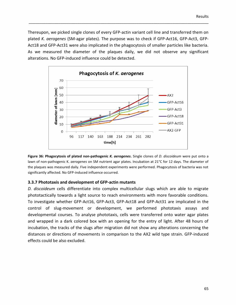
Results _____________________________________________________________________________________
65
Thereupon, we picked single clones of every GFP-actin variant cell line and transferred them on
plated K. aerogenes (SM-agar plates). The purpose was to check if GFP-Act16, GFP-Act3, GFP-
Act18 and GFP-Act31 were also implicated in the phagocytosis of smaller particles like bacteria.
As we measured the diameter of the plaques daily, we did not observe any significant
alterations. No GFP-induced influence could be detected.
Figure 36: Phagocytosis of plated non-pathogenic K. aerogenes. Single clones of D. discoideum were put onto a
lawn of non-pathogenic K. aerogenes on SM nutrient agar plates. Incubation at 21°C for 12 days. The diameter of
the plaques was measured daily. Five independent experiments were performed. Phagocytosis of bacteria was not
significantly affected. No GFP-induced influence occurred.
3.3.7 Phototaxis and development of GFP-actin mutants
D. discoideum cells differentiate into complex multicellular slugs which are able to migrate
phototactically towards a light source to reach environments with more favorable conditions.
To investigate whether GFP-Act16, GFP-Act3, GFP-Act18 and GFP-Act31 are implicated in the
control of slug-movement or development, we performed phototaxis assays and
developmental courses. To analyse phototaxis, cells were transferred onto water agar plates
and wrapped in a dark colored box with an opening for the entry of light. After 48 hours of
incubation, the tracks of the slugs after migration did not show any alterations concerning the
distances or directions of movements in comparison to the AX2 wild type strain. GFP-induced
effects could be also excluded.

Results _____________________________________________________________________________________
66
Figure 37: Phototaxis of GFP-actin variant overexpressing cells. D .discoideum cells were transferred onto water-
agar plates and incubated in a dark colored box with a 2mm wide opening for the light source (*). After 48 hours
the tracks of the migrated slugs could be visualized via amido black staining. Five independent experiments were
performed. Scale bar 1cm. A) No alterations in distance and direction occurred in the GFP-actin overexpressors in
comparison to the AX2 wild type strain. No GFP-induced effects could be observed. B) Measured distances did not
show any significant differences between the tested cell lines.
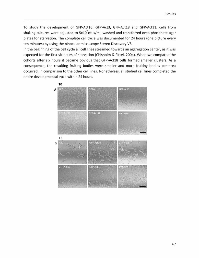
Results _____________________________________________________________________________________
67
To study the development of GFP-Act16, GFP-Act3, GFP-Act18 and GFP-Act31, cells from
shaking cultures were adjusted to 5x10⁶cells/ml, washed and transferred onto phosphate-agar
plates for starvation. The complete cell cycle was documented for 24 hours (one picture every
ten minutes) by using the binocular microscope Stereo Discovery.V8.
In the beginning of the cell cycle all cell lines streamed towards an aggregation center, as it was
expected for the first six hours of starvation (Chisholm & Firtel, 2004). When we compared the
cohorts after six hours it became obvious that GFP-Act18 cells formed smaller clusters. As a
consequence, the resulting fruiting bodies were smaller and more fruiting bodies per area
occurred, in comparison to the other cell lines. Nonetheless, all studied cell lines completed the
entire developmental cycle within 24 hours.

Results _____________________________________________________________________________________
68
Figure 38: Studying the 24 hours life cycle of GFP-Act16, GFP-Act3, GFP-Act18 and GFP-Act31 overexpressing
cells. 5x10⁶cells/ml of D. discoideum cells from shaking cultures were transferred onto phosphate-agar plates for
starvation. Cell cycles were documented by using the Achromat S 0.63x FWS 115mm objective (bright field) at the
Stereo Discovery.V8 binocular microscope. Pictures were taken every 10 minutes for 24 hours (T0=start, T6=6
hours, T12=12 hours, T18=18 hours, T24=24 hours). Three independent experiments were performed. Scale bar
1cm. A) Identical densities of vegetative cells were transferred onto the phosphate agar plates. B) The cells
streamed towards an aggregation center. GFP-Act18 overexpressing cells formed smaller cohorts. C) AX2, GFP-
Act16, GFP-Act3, GFP-Act31 and AX2-GFP cells developed migrating slugs, whereas GFP-Act18 cells did not exhibit
typical slugs, but aggregates of cells. D) AX2, GFP-Act16, GFP-Act3, GFP-Act31 and AX2-GFP slugs culminated into
fruiting bodies. GFP-Act18 cells developed slugs and fruiting bodies more slowly. E) AX2, GFP-Act16, GFP-Act3,
GFP-Act31 and AX2-GFP showed fruiting bodies of comparable sizes and densities. GFP-Act18 cells formed smaller
fruiting bodies with a higher density per area.
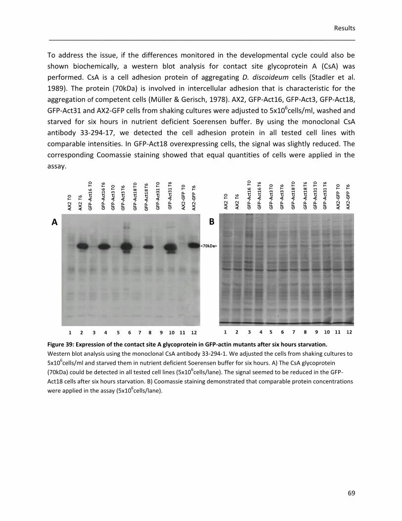
Results _____________________________________________________________________________________
69
To address the issue, if the differences monitored in the developmental cycle could also be
shown biochemically, a western blot analysis for contact site glycoprotein A (CsA) was
performed. CsA is a cell adhesion protein of aggregating D. discoideum cells (Stadler et al.
1989). The protein (70kDa) is involved in intercellular adhesion that is characteristic for the
aggregation of competent cells (Müller & Gerisch, 1978). AX2, GFP-Act16, GFP-Act3, GFP-Act18,
GFP-Act31 and AX2-GFP cells from shaking cultures were adjusted to 5x10⁶cells/ml, washed and
starved for six hours in nutrient deficient Soerensen buffer. By using the monoclonal CsA
antibody 33-294-17, we detected the cell adhesion protein in all tested cell lines with
comparable intensities. In GFP-Act18 overexpressing cells, the signal was slightly reduced. The
corresponding Coomassie staining showed that equal quantities of cells were applied in the
assay.
Figure 39: Expression of the contact site A glycoprotein in GFP-actin mutants after six hours starvation.
Western blot analysis using the monoclonal CsA antibody 33-294-1. We adjusted the cells from shaking cultures to
5x10⁶cells/ml and starved them in nutrient deficient Soerensen buffer for six hours. A) The CsA glycoprotein
(70kDa) could be detected in all tested cell lines (5x10⁶cells/lane). The signal seemed to be reduced in the GFP-
Act18 cells after six hours starvation. B) Coomassie staining demonstrated that comparable protein concentrations
were applied in the assay (5x10⁶cells/lane).
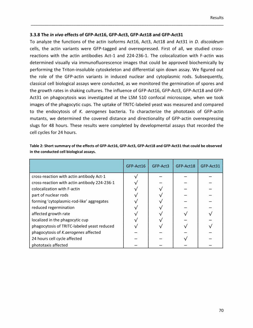
Results _____________________________________________________________________________________
70
3.3.8 The in vivo effects of GFP-Act16, GFP-Act3, GFP-Act18 and GFP-Act31
To analyze the functions of the actin isoforms Act16, Act3, Act18 and Act31 in D. discoideum
cells, the actin variants were GFP-tagged and overexpressed. First of all, we studied cross-
reactions with the actin antibodies Act-1 and 224-236-1. The colocalization with F-actin was
determined visually via immunofluorescence images that could be approved biochemically by
performing the Triton-insoluble cytoskeleton and differential spin down assay. We figured out
the role of the GFP-actin variants in induced nuclear and cytoplasmic rods. Subsequently,
classical cell biological assays were conducted, as we monitored the germination of spores and
the growth rates in shaking cultures. The influence of GFP-Act16, GFP-Act3, GFP-Act18 and GFP-
Act31 on phagocytosis was investigated at the LSM 510 confocal microscope, when we took
images of the phagocytic cups. The uptake of TRITC-labeled yeast was measured and compared
to the endocytosis of K. aerogenes bacteria. To characterize the phototaxis of GFP-actin
mutants, we determined the covered distance and directionality of GFP-actin overexpressing
slugs for 48 hours. These results were completed by developmental assays that recorded the
cell cycles for 24 hours.
Table 2: Short summary of the effects of GFP-Act16, GFP-Act3, GFP-Act18 and GFP-Act31 that could be observed
in the conducted cell biological assays.
GFP-Act16 GFP-Act3 GFP-Act18 GFP-Act31
cross-reaction with actin antibody Act-1 √ − − −
cross-reaction with actin antibody 224-236-1 √ − − −
colocalization with F-actin √ √ − −
part of nuclear rods √ √ − −
forming ‘cytoplasmic-rod-like’ aggregates √ √ − −
reduced regermination √ √ − −
affected growth rate √ √ √ √
localized in the phagocytic cup √ √ − −
phagocytosis of TRITC-labeled yeast reduced √ √ √ √
phagocytosis of K.aerogenes affected − − − −
24 hours cell cycle affected − − √ −
phototaxis affected − − − −
Discussion _____________________________________________________________________________________
71
4. Discussion
The major goal of this work was to analyze the functions of selected actin variants in actin
dynamics in the model organism D. discoideum in vitro and in vivo. Although the compared
actin variants show different levels of similarity, the basic structure of the proteins is strongly
conserved. Analysis of the studied proteins revealed that Flag-Act16 and Flag-Act3 might be
part of the actin cytoskeleton, due to their ability to promote actin polymerization. Together
with rabbit actin both actin variants are able to enhance the formation of short actin filaments.
In contrast, effects of Flag-Act18 and Flag-Act31 on actin polymerization could not be observed.
In vivo characterization suggested that GFP-Act16 and GFP-Act3 are associated with F-actin. The
overexpression might support the organization of the F-actin network during growth and could
disturb the activation of the actin cytoskeleton in the process of regermination. The association
of the GFP-actin variants with cellular organelles might impair the phagocytic capacity of
D. discoideum cells. Furthermore, GFP-Act18 mutants exhibit a reduced expression of CsA,
which seems to affect the cellular development.
4.1 Evolution of the actinome
Sequence analysis showed that the actinome of D. discoideum can be divided into three
sections: actins, actin variants and ARPs (Joseph et al., 2008). Therefore it was a prerequisite for
further in vitro and in vivo studies to perform thorough bioinformatic comparisons. The
phylogenetic analysis involving all members of the D. discoideum actinome revealed the
evolutionary relation of the different actins to each other in correlation to the bacterial
ancestor MreB. The conventional actins, also named the Act8 group, are assumed to be the tip
of a series of duplication events on chromosome 2, as 20% of all proteins in D. discoideum have
arisen by a relatively recent duplication (Eichinger et al., 2005). When members of a protein
family are clustered on one chromosome, the physical distance between family members often
correlates strongly with their evolutionary divergence. Another example of putative tandem
duplication events in D. discoideum is to be found on chromosome 5, where either single genes
or blocks containing several consecutive genes are repeated. Prime examples for this event are
Act3 (coordinates 2884652 to 2885782, Watson strand) and Act18 (coordinates 2886806 to
2887948, Watson strand), which share 88% identity on the protein level (dictybase.org; NCBI
blastp). In contrast, Act31 is localized on chromosome 1 (coordinates 2844541 to 2845608,
Watson strand) and does not belong to an actin gene cluster (Joseph et al., 2008). Like the
ARPs, Act31 did not branch off from any D. discoideum actin and was not related to any other
actin in this tree. It must have taken its separate track way before the evolution of
D. discoideum. Therefore, Act31 is also called ‘orphan ARP’ (Joseph et al., 2008).
Discussion _____________________________________________________________________________________
72
When we performed protein sequence alignments of the phosphate binding motif 1 (Ph1), the
connecting motif 2 (C2), the phosphate motif 2 (Ph2), the adenosine motif (Ad), and the
connecting motif 1 (C1) of human ß-actin (P60709), rabbit muscle actin (P68135), Act16
(DDB_G0272248), Act3 (DDB_G0289487), Act18 (DDB_G0289489) and Act31 (DDB_G0269476),
it became obvious that the sequence motifs were highly conserved within the analyzed actin
variants. Although the amino acid sequences of the compared actins variants show different
levels of similarity, the proclaimed ribbon models suggested that the general three-dimensional
forms of the biopolymers human ß-actin (P60709), rabbit muscle actin (P68135), Act16
(DDB_G027224), Act3 (DDB_G0289487), Act18 (DDB_G0289489) and Act31 (DDB_G0269476)
are almost identical with Act8 (P07830) in the ADP-state. This suggests that the actins were
apparently under huge pressure in terms of structure and function to keep this perfect
molecular design.
The presence of the Act8 group in D. discoideum is still a mystery. There are 17 genes (Act8
group) which have distinct DNA coding sequences, they have complete promoters and
terminators, they are all expressed, some of them are even developmentally regulated, but
they code for identical proteins, the conventional actin (Joseph et al., 2008). One knows since
years that on the protein level more than 95% of actin in D. discoideum represent conventional
actin and the remaining 5% were just a careful calculation about the limits of detectability
(Vandekerckhove & Weber, 1980). Why keeps D. discoideum an obviously luxurious actin
cytoskeleton that is guaranteed up to 17 fold throughout evolution? It is certainly very difficult,
if not presently impossibly, to analyze this via gene disruptions. Perhaps one should shed light
on the presence of distinct mRNAs and putative RNA complexes at different times during
development. In view of the overwhelming amount of conventional actin, the actin variants
cover apparently only a tiny niche in the actinome of a D. discoideum cell.
4.2 The influence of Flag-tagged actin variants on in vitro actin dynamics
We used the baculoviral expression system of the Sf9 cells to express the Flag-tagged actin
variants Flag-Act16, Flag-Act3, Flag-Act18 and Flag-Act31. The indigenous chaperones of insect
cells are necessary for correct folding and oligomerization of recombinant proteins. Many
chaperones are heat shock proteins (for example HSP40, HSP60 and HSP70) and are needed to
prevent aggregation and misfolding during the folding of newly synthesized chains, to prevent
nonproductive interactions with other cell components, to direct the assembly of larger
proteins and multi-protein complexes, and to protect previously folded proteins from unfolding
due to stress exposition (Fink, 1999). As the Sf9 expression system is eukaryotic,
posttranslational modifications are possible and engineered polypeptides with many domains
can be fold by sequential and cotranslational folding of their domains, whereas in prokaryotic
cells proteins usually are folded posttranslationally, which could lead to an intramolecular
misfolding (Netzer & Hartl, 1997).
Discussion _____________________________________________________________________________________
73
In addition, differences between the in vivo and in vitro nature of the interactions of
chaperones with actin have been reported (Frydman & Hartl, 1996).
Flag-Act16 and Flag-Act3 promoted the polymerization of rabbit actin as the lag-phase during
the polymerization assay measured at the fluorometer was significantly shortened. This effect
was intensified with increasing concentrations of Flag-Act16 and Flag-Act3. In contrast, Flag-
Act18 and Flag-Act31 did not affect the polymerization of rabbit actin.
One can assume that the used Flag-Act16 and Flag-Act3 samples contained already short
oligomers that could not be removed by the previous clear spin. These short aggregates could
act as nucleators to overcome the energetically unfavorable lag-phase and induce the actin
polymerization. The application of 100mM KCl to the actin polymerization assays abolished the
observed effects. Higher salt concentrations in vitro seemed to lower the affinity of Flag-Act16
and Flag-Act3 for rabbit actin. The viscosity of an actin solution was gradually reduced when we
incubated rabbit actin with increasing concentrations of Flag-Act16 and Flag-Act3 under
polymerizing conditions. In contrast, the incubation of rabbit actin with different
concentrations of Flag-Act18 and Flag-Act31 did not affect the viscosity of the polymerized
samples. The disassembly of F-actin at the critical concentration of 0.1µM was not altered
under the application of different concentrations of Flag-Act16, Flag-Act3, Flag-Act18 and Flag-
Act31. A role as potential capping proteins could be therefore excluded for all tested actin
variants. To sum up, the incubation of rabbit actin with increasing shares of Flag-Act16 and
Flag-Act3 promoted the polymerization of actin on one hand and lowered the viscosity of the
polymerized samples on the other hand. One possible explanation could be that Flag-Act16 and
Flag-Act3 induce the formation of short actin filaments. The elongation of actin filaments would
lead to an amplification of the fluorescence signal, whereas the viscosity of the actin solution
would decrease, as the actin filaments are shorter under the influence of Flag-Act16 and Flag-
Act3, than actin filaments of conventional actin with or without Flag-Act18 and Flag-Act31.
Cosedimentation studies showed that about 60% of Flag-Act16 and Flag-Act3 were associated
with polymerized rabbit actin, whereas Flag-Act18 was not part of the sedimented actin
fraction. About 40% of Flag-Act31 could be detected together with rabbit F-actin in the pellet.
Without rabbit actin, Flag-Act16 and Flag-Act31 were not able to form stable actin filaments,
not even under the application of stabilizing phalloidin. 40 % of Flag-Act3 and 30% of Flag-Act18
assembled under polymerizing conditions when we added phalloidin. These observations could
be partly confirmed, when we checked the formation of TRITC-phalloidin stabilized Flag-actin
filaments microscopically. Here, Flag-Act3 was the only Flag-actin variant which exhibited stable
patches. When we checked the oligomerization of the Flag-tagged actin variants, Flag-Act16
was measurable in a range of 40-160kDa, meaning that a polymerization of Flag-Act16 up to a
tetramer is indicated. Denatured aggregates of Flag-Act3 were detectable, whereas an ordered
oligomerization of Flag-Act18 and Flag-Act31 could be excluded.
Discussion _____________________________________________________________________________________
74
Images taken at the transmission electron microscope visualized that no polymerization of Flag-
Act16, Flag-Act3, Flag-Act18 and Flag-Act31 was possible without the supplementation of
freshly prepared rabbit actin. Aggregates of different sizes and shapes occurred, but the precise
number of assembled G-actin monomers could not be identified. Incubation of freshly prepared
rabbit actin with Flag-Act16, Flag-Act3, Flag-Act18 and Flag-Act31 under polymerizing
conditions proved that the formation of filamentous rabbit actin was not disturbed by the Flag-
actin variants.
Severin is a conserved, Ca2+-dependent, well studied actin binding protein and therefore a good
marker for the activity and integrity of actin itself. It consists of three domains which all have
different actin binding activities: domain 1 binds to G-actin and caps actin filaments at the
barbed end, domain 2 binds to the side of actin filaments, domain 3 binds to G-actin as well and
thus can sever filaments in combination with domain 1, the three domains share very similar
three dimensional structures (Andre et al., 1988; Eichinger & Schleicher, 1992; Schnuchel et al.,
1995). The D. discoideum protein is a homolog of human gelsolin (Yin et al. 1990). Severin could
be confirmed as a binding partner for all studied Flag-actin variants in immunoprecipitation
analysis and mass spectrometry. The binding of severin to actin requires micromolar
concentrations of Ca2+ which also induces its severing activity. Severin binds to other
conformational species of actin like high speed supernatant actin (HSS-actin) or fluorescein
isothiocyanate labeled actin as well (Giffard et al., 1984). Therefore, one can conclude that the
expression of actin variants in Sf9 cells leads to at least partially folded recombinant proteins.
This is not a trivial assumption because it is after decades of intensive experimental approaches
still impossible to purify a fully recombinant actin.
Consequently, Flag-Act16 did not show all the characteristics of conventional actin, although
Act16 is part of the Act8 group and exhibits the identical amino acid sequence as the other 17
actins. For the detection and purification of proteins in a wide variety of settings, affinity tags
have become essential tools for the production of recombinant proteins. The Flag-tag structure
has been optimized for compatibility with other proteins it is attached to, in that it is more
hydrophilic than other common epitope tags and therefore less likely to denature or inactivate
proteins to which it is appended (Joel et al., 2004). To determine potential influences of the
Flag-tag concerning actin dynamics in vitro, we tried to remove the Flag-peptide (1012 Da).
Freshly purified Flag-Act16 was incubated with a specific recombinant enterokinase (Novagen)
that should bind to the amino acid sequence of the Flag-tag for subsequent removal. After
numerous trials under different conditions we came to the conclusion that the removal of the
N-terminal Flag-tag was not successful. The inability of a protease to cleave a fusion protein
may have been caused by sterical hindrance. For example, the cleavage site may have been too
close to the ordered structure in the target protein (Joel et al., 2004).

Discussion _____________________________________________________________________________________
75
Results obtained using tagged proteins should be carefully controlled and interpreted with
caution, taking into consideration changes to the proteins natural state upon addition of a
fusion tag. Additionally, some proteins need to be activated by certain conditions or factors,
therefore in vitro assays are just significant to a limited extend. Furthermore, the interactions of
the actin variants in the cytoskeleton of D. discoideum are extremely specific and the use of
rabbit muscle actin for the in vitro actin polymerization assays could be unfavorable. Hence, the
in vitro assays should be repeated with purified conventional actin from D. discoideum.
4.3 GFP-Act16, GFP-Act3, GFP-Act18, GFP-Act31 and their cellular functions in D. discoideum
Generating the GFP-overexpressors was quite a challenge and expression levels of the GFP-
tagged actin variants varied drastically. As Act16 belongs to the Act8 group, GFP-Act16 could be
detected with both monoclonal actin antibodies Act-1 and 224-236-1. The respective epitopes
are very short and specific for conventional actin (Simpson et al., 1984; Westphal et al., 1997).
Consequently, also endogenous actin of the cell lysates could be detected easily. In contrast,
GFP-Act3, GFP-Act18 and GFP-Act31 could not be recognized by using Act-1 and 224-236-1,
although e.g. Act3 had only 11 different amino acids as compared to the conventional actin
Act16. To draw a conclusion, the binding epitopes in the actin variants GFP-Act3, GFP-Act18 and
GFP-Act31 might have been too divergent or possibly, the different amino acids in the actin
variants caused an antigenic change at a topologically different site within the protein.
With regard to the actin cytoskeleton of D. discoideum, we analyzed the localization of GFP-
Act16, GFP-Act3, GFP-Act18 and GFP-Act31 by conducting microscopical and biochemical
approaches. GFP-Act16 and GFP-Act3 colocalized in immunofluorescence studies with
endogenous actin in the cell cortex, and to some extent in the cytoplasm. Keeping in mind that
Act18 and Act31 differ rather drastically from actins of the Act8 group, the predominant
localization of GFP-Act18 and GFP-Act31 within the cytoplasm was after all not surprising.
These observations were confirmed biochemically by performing the Triton insoluble
cytoskeleton assay and the differential spin downs. Also in these approaches Act18 and Act31
behaved like soluble proteins without any interactions with conventional actin in and/or on
microfilaments.
Interestingly, GFP-Act16 and GFP-Act3 were part of the DMSO induced nuclear actin rods,
which is another clear indication that these actin variants are part of the actin cytoskeleton.
Also in this case one cannot unequivocally show whether these actin variants are intrinsic parts
of the filaments, because binding along filaments would be sufficient to detect these proteins in
nuclear rods. In principle, the designation ‘actin rods’ is somewhat misleading because other
cytoskeletal proteins were identified in the rods as well, such as cofilin, Aip1 and coronin
(Vandebrouck et al., 2010).
Discussion _____________________________________________________________________________________
76
Many of these proteins are rather large (50kDa and more) and cannot simply pass the nuclear
membrane through the pores without a specific transporting system. The GFP-tagged actin
variants Act16 and Act3 have a size of about 70kDa and also need to be imported or exported
by a transporter system (Gerace & Burke, 1988).
There exist also cytoplasmic rods and cofilin can be taken as a marker for both nuclear rods,
and cytoplasmic rods as well. Cytoplasmic rods can be induced via inhibition of ATP synthesis by
NaN3 (Minamide et al., 2010). The tested GFP-actin variant overexpressors were not able to
form typical needle-shaped cytoplasmic rods, but GFP-Act16 and GFP-Act3 seemed to be part
of crescent-shaped aggregates within the cytoplasm.
During unfavorable environmental conditions, cells and spores of D. discoideum and their
intracellular particles remain in an immobile state. After the germination of spores is induced,
the actin synthesis is activated until actin constitutes 2-3% of the germinating spore (MacLeod
et al., 1980). But spore cells retain only two of the actin mRNA sequences initially present in the
pseudoplasmodium and it is still unclear whether these actin mRNA sequences encode identical
actin proteins (Tsang et al., 1982). The viability of GFP-Act16 and GFP-Act3 spores was
significantly reduced, whereas the regermination of GFP-Act18 and GFP-Act31 spores was just
slightly decreased. The size and shape of the tested spores was not affected in our experiments.
Expression of GFP-actin can affect the morphology and function of D. discoideum cells severely,
possibly as a result of the large increase in the number of actin monomers that occurs during
overexpression of GFP-actin fusion proteins (Aizawa et al., 1997). As a result the excess of GFP-
actin variant monomers could sequester actin binding proteins that are not available anymore
for the actin proteins that were synthesized constitutively during germination, which could
result in a disturbed activation of the actin cytoskeleton. Furthermore, there remains the
inevitable problem that whenever we label in vivo, we will always interfere with the biology of
the labeled structures. As a central structure, the actin cytoskeleton interacts with numerous
molecules that regulate its dynamics. Any marker will interfere with those biological processes
by occupying the limited binding sites along the actin cytoskeleton. If those binding sites are
saturated with markers, they will not bind the factors that regulate actin structure (Du & Ren,
2011).
In shaking culture experiments GFP-Act16 and GFP-Act3 overexpressors reproducibly showed a
reduced lag-phase. In contrast, GFP-Act18 and GFP-Act31 mutants exhibited a delayed lag-
phase in comparison to the AX2 or AX2-GFP cells. It is known that at reduced densities
D. discoideum cells grow slower, as the cells have to adapt to their new environment, before
the synthesis of actin increases and cell doubling starts (dictybase.org). As GFP-Act16 and GFP-
Act3 seems to be part of the F-actin fraction in the cell cortex it could be involved in the F-actin
organization during growth, which seems to promote the entry into the log-phase.
Discussion _____________________________________________________________________________________
77
GFP-Act18 and GFP-Act31 are assumed to be part of the cytoplasm with no association to the F-
actin network. A large increase in the number of GFP-Act18 and GFP-Act31 monomers might
disturb the organization and regulation of the F-actin network. The cytokinesis of the GFP-actin
variant overexpressors was not affected.
During phagocytosis of TRITC-labeled yeast cells GFP-Act16 and GFP-Act3 accumulated in the
phagocytic cup, whereas GFP-Act18 and GFP-Act31 were spread homogenously in the
cytoplasm. As the phagocytic cup is a surface protuberance, the F-actin associated proteins
GFP-Act16 and GFP-Act3 are concentrated in this structure, whereas the cytoplasmic proteins
GFP-Act18 and GFP-Act31 are not enriched in the phagocytic cup. When we measured the
uptake of TRITC-labeled yeast at the fluorometer, a reduced uptake down to 50% in comparison
to the wild type was detected in all cell lines. For phagocytosis of large particles, the membrane
of a phagocytic cup has to be remodeled extensively by the actin cytoskeleton and its
stimulated polymerization (Lee et al., 2007; Griffin et al., 1975). Intracellular compartments like
recycling endosomes or the ER are necessary for the contribution of extra membrane to
completely surround the particles (Gagnon et al., 2002). As we proved in differential spin
downs, the GFP-Act16, GFP-Act3, GFP-Act18 and GFP-Act31 are associated with cell organelles
like vesicles, mitochondria and ER. The recruitment of extra membrane could be disturbed
consequently, which influences the phagocytic capacity. Furthermore, the phagocytosis is also
reduced in AX2-GFP cells, so that the interference of the GFP-tag with general biological
processes has to be considered again.
The need for insertion of endomembrane to completely surround the particles also explains
why large particles are more susceptible to inhibition than their smaller counterparts (Lee et al.,
2007). For the uptake of smaller particles, the actin driven formation of membrane protrusions
do not ‘zipper up’ along a ligand-coated particle, but instead they collapse to fuse with the
plasma membrane (Connor & Schmid, 2003). Consequently, the phagocytosis of plated
K. aerogenes was not influenced by the overexpression of the GFP-actin variants.
The overexpression of the GFP-actin variants had no significant effect on the covered distance
or the directionality in the performed phototaxis assay. Imaging the cell cycle of the GFP-actin
variants for 24 hours proved that GFP-Act18 overexpressing cells produced smaller fruiting
bodies and a higher density of fruiting bodies per area, in comparison to the other tested cell
lines. In parallel, the expression of the early aggregation marker CsA was delayed in the GFP-
Act18 cells after six hours of starvation. It can be assumed that if the formation of stable
intercellular contacts is restricted, due to a reduced expression of CsA, fewer cells aggregate to
form a single fruiting body. As the 24 hours cell cycle assay was performed with the same
number of vegetative cells for every cell line, a higher density of fruiting bodies per area
occurred.

Discussion _____________________________________________________________________________________
78
As the in vivo assays showed, GFP-actin overexpressors are useful to characterize possible
functions of different actin variants in the model organism D. discoideum. Nonetheless, it has to
be considered that besides the discussed fundamental limitations due to the GFP-labeling of
actin, the overexpression of a specific protein often leads to inconsistencies concerning the
observed phenotype. Overexpression can influence different organelles of the cell at the same
time, though it is not uncommon for an overexpressing strain to display lethality or to show no
phenotype at all. Consequently, to analyze the roles of Act16, Act3, Act18 and Act31 in vivo
precisely, it might be worthwhile to create knock-out mutants for further characterization.

References _____________________________________________________________________________________
79
5. References
Aizawa H, Sameshima M, Yahara I (1997): A green fluorescent protein-actin fusion protein
dominantly inhibits cytokinesis, cell spreading and locomotion in Dictyostelium. Cell Structure
and Function 22: 335-345.
Andre E, Lottspeich F, Schleicher M, Noegel A (1988): Severin, gelsolin and villin share
homologues sequence in regions presumed to contain F-actin severing domains. The Journal of
Biological Chemistry 263: 722-727.
Andre E, Brink M, Gerisch G, Isenberg G, Noegel A, Schleicher M, Segall JE, Wallraff E (1989): A
Dictyostelium mutant deficient in severin, an F-actin fragmenting protein, shows normal
motility and chemotaxis. The Journal of Cell Biology 108: 985-995.
Alberts B, Johnson A, Lewis J, Raff M, Roberts K, Walter P (2004): Molecular biology of the cell.
Wiley-VCH Verlag GmbH & Co KgaA; ISBN 3-527-30492-4.
Bertholdt G, Stadler J, Bozzaro S, Fichtner B, Gerisch G (1985). Carbohydrate and other epitopes
of the contact site A glycoprotein of Dictyostelium discoideum as characterized by monoclonal
antibodies. Cell Differentiation 16: 187-202.
Bork P, Sander C, Valencia A (1992): An ATPase domain common to prokaryotic cell cycle
proteins, sugar kinases, actin and HSP70 heat shock proteins. Proceedings of the National
Academy of Sciences USA 89: 7290-7294.
Bozzaro S & Eichinger L (2011): The professional phagocyte Dictyostelium discoideum as a
model host for bacterial pathogens. Current Drug Targets 12: 942–954.
Carballido-Lopez R (2006): The bacterial actin-like cytoskeleton. Microbiology and Molecular
Biology Reviews 70: 888-909.
Carlier MF (1990): Actin polymerization and ATP hydrolysis. Advances in Biophysics 26: 51-73.
Cleuren YT & Boonstra J (2012): Actin: structure, functions and disease. Consuelas VA, Minas DJ
(eds), Chapter 2: 61-96. Nova Publishers.
Chisholm RL & Firtel RA (2004): Insights into morphogenesis from a simple developmental
system. Nature Reviews Molecular Cell Biology 5: 531-41.
Conner SD & Schmid SL (2003): Regulated portals of entry into the cell. Nature 422: 37-44.
Csete ME & Doyle JC (2002): Reverse engineering of biological complexity. Science 295: 1664-
1669.
References _____________________________________________________________________________________
80
Dickinson RB, Southwick FS, Purich DL (2002): A direct-transfer polymerization model explains
how the multiple profilin-binding sites in the actoclampin motor promote rapid actin-based
motility. Archives of Biochemistry and Biophysics 406: 296-301.
Domazetovska A, Ilkovski B, Cooper ST, Ghoddusi M, Haderman EC, Minamide LS, Gunning PW,
Bamburg JR, North KN (2007): Mechanisms underlying intranuclear rod formation. Brain 130:
3275-3284.
Doolittle RF & York AL (2002): Bacterial actins? An evolutionary perspective. Bioessays 24: 293-
296.
Du F & Ren H (2010): Development and application of probes for labeling the actin cytoskeleton
in living plant cells. Protoplasma 248: 239-250.
Eichinger L & Schleicher M (1992): Characterization of actin- and lipid-binding domains in
severin, a Ca2+-dependent F-actin fragmenting protein. Biochemistry 31: 4779-4787.
Eichinger L, Pachebat JA, Glöckner G, Rajandream MA, Sucgang R, Berriman M, Song J, Olsen R,
Szafranski K, Xu Q, Tunggal B, Kummerfeld S, Madera M, Konfortov BA, Rivero F, Bankier AT,
Lehmann R, Hamlin N, Davies R, Gaudet P, Fey P, Pilcher K, Chen G, Saunders D, Sodergren E,
Davis P, Kerhornou A, Nie X, Hall N, Anjard C, Hemphill L, Bason N, Farbrother P, Desany B, Just
E, Morio T, Rost R, Churcher C, Cooper J, Haydock S, van Driessche N, Cronin A, Goodhead I,
Muzny D, Mourier T, Pain A, Lu M, Harper D, Lindsay R, Hauser H, James K, Quiles M, Madan
Babu M, Saito T, Buchrieser C, Wardroper A, Felder M, Thangavelu M, Johnson D, Knights A,
Loulseged H, Mungall K, Oliver K, Price C, Quail MA, Urushihara H, Hernandez J, Rabbinowitsch
E, Steffen D, Sanders M, Ma J, Kohara Y, Sharp S, Simmonds M, Spiegler S, Tivey A, Sugano S,
White B, Walker D, Woodward J, Winckler T, Tanaka Y, Shaulsky G, Schleicher M, Weinstock G,
Rosenthal A, Cox EC, Chisholm RL, Gibbs R, Loomis WF, Platzer M, Kay RR, Williams J, Dear PH,
Noegel AA, Barrell B, Kuspa A. (2005): The genome of the social amoebae Dictyostelium
discoideum. Nature 435: 43-57.
Fink A (1999): Chaperone-mediated protein folding. Physiological reviews 79: 425-442.
Frydman J & Hartl FU (1996): Principles of chaperone-assisted protein folding: differences
between in vitro and in vivo mechanisms. Science 272: 1497-1502.
Gagnon E, Duclos S, Rondeau C, Chevet E, Cameron PH, Steele-Mortimer O, Paiement J,
Bergeron JJM, Desjardins M (2002): Endoplasmic reticulum-mediated phagocytosis is a
mechanism of entry into macrophages. Cell 110: 119-131.
References _____________________________________________________________________________________
81
Gallinger J (2013): WH2 domains and actin variants as multifunctional organizers of the actin
cytoskeleton. PhD Thesis, Ludwig-Maximilians-Universität München.
Gerace L & Burke B (1988): Functional organization of the nuclear envelope. Annual Review of
Cell and Developmental Biology 4: 335-74.
Gerisch G (1962): Cell functions and cell function changes in the development of Dictyostelium
discoideum. Inhibitors of aggregation, their effects on cellular contact formation and
morphogenetic movements. Experimental Cell Research 26: 462-484.
Giffard RG, Weeds AG, Spudich JA (1984): Ca2+-dependent binding of severin to actin: a one-to-
one complex is formed. The Journal of Cell Biology 98: 1796-1803.
Griffin FM, Griffin JA, Leider JE, Silverstein SC (1975): Studies on the mechanism of
phagocytosis. Requirements for circumferential attachment of particle-bound ligands to specific
receptors on the macrophage plasma membrane. The Journal of Experimental Medicine 142:
1263-1282.
Haus U, Hartmann H, Trommler P, Noegel A, Schleicher M (1991): F-actin capping by Cap32/34
requires heterodimeric conformation and can be inhibited by PIP2. Biochemical and Biophysical
Research Communications 181: 833-839.
Ihara M, Tanaka Y, Hashimoto Y, Yanagisawa K (1990): Partial purification and some properties
of spore germination promotor(s) for Dictyostelium discoideum excreted by Klebsiella
aerogenes. Agricultural and Biological Chemistry 54: 2619-2627.
Israel L (2002): Domänenanalyse des Cyclase-assoziierten Proteins (CAP) und Charakterisierung
von Filactin, einem neuartigen actinähnlichen Protein aus Dictyostelium discoideum. PhD
Thesis, Ludwig-Maximilians-Universität München.
Joel PB, Fagnant PM, Trybus KM (2004): Expression of a nonpolymerizable actin mutant in Sf9
cells. Biochemistry 43: 11554-11559.
Joseph JM, Fey P, Ramalingnam N, Liu XI, Rohlfs M (2008): The actinome of Dictyostelium
discoideum in comparison to actins and actin-related proteins from other organisms. PlosOne 3:
e2654.
Kaimaktchiev V, Goebel H, Laing N, Narus M, Weeks D, Nixon R (2006): Intracellular nemaline
rod myopathy. Muscle Nerve 34: 369-372.

References _____________________________________________________________________________________
82
Kim DJ, Kim SH, Kim SM, Bae JI, Ahnn JJ, Song WK (2007): F-actin binding region of SPIN90 C-
terminus is essential for actin polymerization and lamellipodia formation. Cell Communication
and Adhesion 14: 33-43.
Kishi Y, Clements C, Mahadeo DC, Cotter DA, Sameshima M (1998): High levels of actin tyrosine
phosphorylation: correlation with the dormant state of Dictyostelium spores. Journal of Cell
Science 111: 2923-2932.
Kouyama T & Mihashi K (1981): Fluorimetry study of N-(1-pyrenyl)iodoacetamide-labelled F-
actin. Local structural change of actin protomer both on polymerization and on binding of
heavy meromyosin. European Journal of Biochemistry 114: 33-38.
Laemmli UK (1970): Cleavage of structural proteins during the assembly of the head of
bacteriophage T4. Nature 227: 680-685.
Lee WL, Mason D, Schreiber AD, Grinstein S (2007): Quantitative analysis of membrane
remodeling at the phagocytic cup. Molecular Biology of the Cell 18: 2883-2892.
Lodish H, Berk A, Zipursky SL, Matsudaira P, Baltimore D, Darnell J (2000): Molecular cell biology
4th edition New York: W. H. Freeman; 2000. ISBN-10: 0-7167-3136-3.
MacLean-Fletcher SD & Pollard TS (1980): Viscometric analysis of the gelation of Acanthamoeba
extracts and purification of two gelation factors. Journal of Cell Biology 85: 414-428.
MacLeod C, Firtel RA, Papkoff J (1980): Regulation of actin gene expression during spore
germination in Dictyostelium discoideum. Developmental Biology 76: 263-274.
McRobbie SJ & Newell PC (1983): Changes in actin associated with the cytoskeleton following
chemotactic stimulation of Dictyostelium discoideum. Biochemical and Biophysical Research
Communications 115: 351-359.
Minamide LS, Maiti S, Boyle JA, Davis RC, Coppinger JA, Bao Y, Huang TY, Yates J, Bockoch GM,
Bamburg JR (2010): Isolation and characterization of cytoplasmic cofilin-actin rods. The Journal
of Biological Chemistry 285: 5450-5460.
Müller K & Gerisch G (1978): A specific glycoprotein as the target site of adhesion blocking fab
in aggregating Dictyostelium cells. Nature 274: 445-449.
Muller J, Oma Y, Vallar L, Friedrich E, Poch O, Winsor B (2005): Sequence and comparative
genomic analysis of actin-related proteins. Molecular Biology of the Cell 16: 5736-5748.
References _____________________________________________________________________________________
83
Mullins RD, Heuser JA, Pollard TD (1998): The interaction of ARP2/3 complex with actin:
nucleation, high affinity pointed end capping, and formation of branching networks of
filaments. Proceedings of the National Academy of Sciences USA 95: 6181-6186.
Netzer WJ & Hartl FU (1997): Recombination of protein domains facilitated by co-translational
folding in eukaryotes. Nature 388: 343-349.
Nishida E, Iida K, Yonezawa N, Koyasu S, Yahara I, Sakai H (1987): Cofilin is a component of
intranuclear and cytoplasmic actin rods induced in cultures cells. Proceedings of the National
Academy of Sciences USA 84: 5262-5266.
Noegel AA, Blau-Wasser R, Sultana H, Müller R, Israel L, Schleicher M, Patel H, Weijer CJ (2004):
The cyclase-associated protein CAP as regulator of cell polarity and cAMP signaling in
Dictyostelium. Molecular Biology of the Cell 15: 934-945.
Osborn M & Weber K (1984): Actin paracrystal induction by forskolin and by db-cAMP in CHO
cells. Experimental Cell Research 150: 408-418.
O-Connel KL & Stults JT (1997): Identification of mouse liver proteins in two-dimensional
electrophoresis gels by matrix-assisted laser desorption/ ionization mass spectrometry of in situ
enzymatic digests. Electrophoresis 18: 349-359.
Poch O & Winsor B: Who's who among the Saccharomyces cerevisiae actin-related proteins? A
classification and nomenclature proposal for a large family. Yeast 13: 1053-1058.
Pollard TD & Weeds AG (1984): The rate constant for ATP hydrolysis by polymerized actin.
Federation of European Biochemical Societies Letters 170: 94-98.
Pollard TD & Borisy GG (2003): Cellular motility driven by assembly and disassembly of actin
filaments. Cell 112: 453-465.
Pollard TD & Earnshaw WD (2004): Cell Biology, First Edition, Saunders, ISBN 1-4160-2388-7.
Qualmann B, Kessels MM, Kelly RB (2000): Molecular links between endocytosis and the actin
cytoskeleton. Journal of Cell Biology 150: 111-116.
Raper KB (1935): Dictyostelium discoideum, a new species of slime mold from decaying forest
leaves. Journal of Agricultural Research 50: 135-148.
Sameshima M, Kishi Y, Osumi M, Mahadeo D, Cotter DA (2000): Novel actin cytoskeleton: actin
tubules. Cell Structure and Function 25: 291-295.
References _____________________________________________________________________________________
84
Sept D & McCammon JA (2001): Thermodynamics and kinetics of actin filament nucleation.
Biophysical journal 81: 667-674.
Schleicher M & Jockusch B (2008): Actin: its cumbersome pilgrimage through cellular
compartments. Histochemistry and Cell Biology 129: 695-704.
Schnuchel A, Wiltscheck R, Eichinger L, Schleicher M, Holak TA (1995): Structure of severin
domain 2 in solution. Journal of Molecular Biology 247: 21-27.
Schwalb M & Roth R (1979): Axenic growth and development of the cellular slime mould
Dictyostelium discoideum. Journal of General Microbiology 60: 283-286.
Schüler H, Karlsson R, Schutt CE, Lindberg U (2006): The connection between actin ATPase and
polymerization. Advances in Molecular and Cell Biology 37: 49-61.
Simpson PA, Spudich JA, Parham P (1984): Monoclonal antibodies prepared against
Dictyostelium actin: characterization and interactions with actin. The Journal of Cell Biology 99:
287-295.
Sparrow JC, Nowak KJ, Durling HJ, Beggs AH, Wallgreen-Pettersson C, Romero N, Nonaka I,
Laing NG (2003): Muscle diseases caused by mutations in the skeletal muscle alpha-actin gene
(ACTA1). Neuromuscular Disorders 13: 519-531.
Spudich JA & Watt S (1971): The regulation of rabbit skeletal muscle contraction. Biochemical
studies of the interaction of the tropomyosin-troponin complex with actin and the proteolytic
fragments of myosin. The Journal of Biological Chemistry 246: 4866-4871.
Stadler J, Keenan TW, Bauer G, Gerisch G (1989): The contact site A glycoprotein of Dictyostelium discoideum carries a phospholipid anchor of a novel type. The European Molecular Biology Organization Journal 8: 371–377.
Tsang AS, Mahbubani H, Williams JG (1982): Cell-type specific actin mRNA populations in Dictyostelium discoideum. Cell 31: 375-382.
Vandebrouck A, Domazetovska A, Mokbel N, Cooper ST, Ilkovski B, North KN (2010): In vitro
analysis of rod composition and actin dynamics in inherited myopathies. Journal of
Neuropathology and Experimental Neurology 69: 429-441.
Vandekerckhove J & Weber K (1980): Vegetative Dictyostelium cells containing 17 actin genes
express a single major actin. Nature 284: 475-477.
References _____________________________________________________________________________________
85
Wachi M, Doi M, Tamaki S, Park S, Nakajima-Ilijima S, Matsuhashi M (1987): Mutant isolation
and molecular cloning of mre genes, which determine cell shape, sensitivity to mecillinam, and
the amount of penicillin-binding proteins in Escherichia coli cell. Journal of Bacteriology 169:
4935-4940.
Westphal M, Jungbluth A, Heidecker M, Mühlbauer B, Heizer C, Schwartz JM, Marriott G,
Gerisch G (1997): Microfilament dynamics during cell movement and chemotaxis monitored
using a GFP-actin fusion protein. Current Biology 7: 176-183.
Williams KL & Newell PC: A genetic study of aggregation in the cellular slime mould Dictyostelium discoideum using complementation analysis. Genetics 82: 287-307.
Winder SJ & Ayscough KR (2005): Actin-binding proteins. Journal of Cell Science 118: 651-654.
Witke W, Schleicher M, Noegel A (1992): Redundancy in the microfilament system: abnormal
development of Dictyostelium cells lacking two F-actin cross-linking proteins. Cell 68: 53-62.
Yin HL, Janmey PA, Schleicher M (1990): Severin is a gelsolin prototype. Federation of European
Biochemical Societies Letters 264: 78-80.
Zolghadr K, Rothbauer U, Leonhardt H (2008): The fluorescent two-hybrid (F2H) assay for direct
analysis of protein-protein interactions in living cells. Methods of Molecular Biology 812: 275-
282.
List of figures _____________________________________________________________________________________
86
List of figures
Figure 1: Life cycle of D. discoideum (Chisholm & Firtel, 2004).
Figure 2: Sequential arrangement of the five actin motifs.
Figure 3: Typical G-actin structure in the ADP-state.
Figure 4: The process of actin polymerization (after Gallinger, 2013).
Figure 5: Actin binding proteins.
Figure 6: The actinome of D. discoideum (after Joseph et al., 2008).
Figure 7: Alignment of the actin variants Act3, Act18 and Act31 with conventional actin.
Figure 8: Phylogenetic tree of the D. discoideum actinome.
Figure 9: Motif logos of human ß-actin, rabbit actin, and the D. discoideum actins Act16, Act3,
Act18 and Act31.
Figure 10: Ribbon-models of human ß-actin, rabbit actin, and the D. discoideum actins Act16,
Act3, Act18 and Act31.
Figure 11: Flag-tagged actin variants purified from Sf9 cells.
Figure 12: Fluorometric analysis of the influence of Flag-Act16 and Flag-Act3 on actin
polymerization.
Figure 13: Fluorometric analysis of the influence of Flag-Act18 and Flag-Act31 on actin
polymerization.
Figure 14: Fluorometric analysis of the influence of 100mM KCl on actin polymerization.
Figure 15: Viscometric analysis of the effects of Flag-Act16 and Flag-Act3.
Figure 16: Viscometric analysis of Flag-Act18 and Flag-Act31 activities.
Figure 17: Fluorometric analysis of putative capping activities of Flag-Act16, Flag-Act3, Flag-
Act18 and Flag-Act31.
Figure 18: Sedimentation assay to study if Flag-Act16, Flag-Act3, Flag-Act18 and Flag-Act31 bind
to rabbit F-actin.
Figure 19: Sedimentation of Flag-Act16, Flag-Act3, Flag-Act18 and Flag-Act31 in the presence of
phalloidin.
List of figures _____________________________________________________________________________________
87
Figure 20: Potential filament formation of Flag-Act16, Flag-Act3, Flag-Act18 and Flag-Act31.
Figure 21: The oligomerization of Flag-Act16 was studied by using the AEKTA FPLC column.
Figure 22: Flag-Act16, Flag-Act3, Flag-Act18 and Flag-Act31 imaged using a transmission
electron microscope.
Figure 23: Interaction studies of Flag-Act16, Flag-Act3, Flag-Act18 and Flag-Act31 with severin.
Figure 24: Different expression of GFP-actin variants in transformed D. discoideum cells.
Figure 25: The monoclonal GFP antibody K3-184-2 was used to quantify the expression of the
GFP-actin variants.
Figure 26: The monoclonal actin antibody Act-1 was used to quantify the expression of
endogenous actin and to study cross-reactions with Act16, Act3, Act18 and Act31.
Figure 27: The monoclonal actin antibody 224-263-1 was used to define its binding epitope and
to study cross-reactions with Act16, Act3, Act18 and Act31.
Figure 28: Immunofluorescence images were taken to visualize the GFP-actin variants.
Figure 29: The Triton-insoluble cytoskeleton assay and the differential spin down were
performed to localize the GFP-actin variants biochemically.
Figure 30: Nuclear rods in GFP-actin variant overexpressors.
Figure 31: Cytoplasmic rods in GFP-actin overexpressors.
Figure 32: Germination of spores in GFP-actin variant overexpressors.
Figure 33: Growth rates of GFP-actin mutants.
Figure 34: The localization of GFP-actin variants during phagocytosis.
Figure 35: Phagocytosis of TRITC-labeled yeast.
Figure 36: Phagocytosis of plated non-pathogenic K. aerogenes.
Figure 37: Phototaxis of GFP-actin variant overexpressing cells.
Figure 38: Studying the 24 hours life cycle of GFP-Act16, GFP-Act3, GFP-Act18 and GFP-Act31
overexpressing cells.
Figure 39: Expression of the contact site A glycoprotein in GFP-actin mutants after six hours
starvation.

List of tables _____________________________________________________________________________________
88
List of tables
Table 1: Actin variants analyzed in this study.
Table 2: Short summary of the effects of GFP-Act16, GFP-Act3, GFP-Act18 and GFP-Act31 that
could be observed in our conducted cell biological assays.

Acknowledgments _____________________________________________________________________________________
89
Acknowledgements
First of all, I want to thank Prof. Michael Schleicher for giving me the opportunity to do the
experimental part of this work in his laboratory, for the stimulating discussions and the
enormous support during my PhD time.
Many thanks go to Prof. Dr. Wolfgang Baumeister and Oana Mihalache for sharing precious
reagents and expert knowledge. I thank Prof. Dr. Kiebler and Dr. Bastian Popper for supporting
me with valuable materials, operational experience, scientific discussions and inspiration.
I want to thank all lab members for creating such a great working atmosphere where I found all
sorts of help, support, motivation and friends. It was a great pleasure to work with you!
Many thanks go to PD Dr. Annette Müller-Taubenberger for sharing advice, valuable reagents,
cell lines and vectors. I sincerely thank Daniela Rieger for her professional and personal advice.
I thank Dr. Hellen Ishikawa Ankerhold for great teamwork, scientific discussions and methodical
support. I am grateful for technical and moral help from Gudrun Trommler, Thi-Heu Ho,
Stefanie Lindholz and Maria Beer.
Very special thanks go to my colleagues and bench mates Dr. Julia & Christoph Gallinger,
Heike Roth, Dr. Matthias Samereier and Süleyman Kösem for sharing the same problems with
me and for having so much fun together inside and outside of the lab.
I want to thank Christoph for comments on this thesis, his personal and professional coaching
and unforgettable moments during my PhD time.
In would like to thank Dr. Peter Batsios, Dr. Dennis Zimmermann, Dr. Marija Vukajlovic and
Angela Oberhofer for stimulating scientific discussions and sharing leisure time together.
Finally, very special thanks go to my friends and family, who always supported me in my plans.
Thanks for all your love, trust and patience throughout all the years of my studies.


![CYTOSKELETON NEWS - fnkprddata.blob.core.windows.net · Dynamic remodeling of the actin cytoskeleton [i.e., rapid cycling between filamentous actin (F-actin) and monomer actin (G-actin)]](https://img.pdfslide.net/doc/110x75/609edd2b88630103265d18ee/cytoskeleton-news-dynamic-remodeling-of-the-actin-cytoskeleton-ie-rapid-cycling.jpg)

![Review Actin-targeting natural products: structures ... · actin-binding proteins actively break or ‘sever’ actin filaments [e.g. actin-depolymerizing factor (ADF) and cofilin]](https://img.pdfslide.net/doc/110x75/5f0f85bd7e708231d44494d0/review-actin-targeting-natural-products-structures-actin-binding-proteins-actively.jpg)