Embed Size (px)
Citation preview

RESEARCH ARTICLE
The Chemopotential Effect of Annonamuricata Leaves against Azoxymethane-Induced Colonic Aberrant Crypt Foci in Ratsand the Apoptotic Effect of AcetogeninAnnomuricin E in HT-29 Cells: A Bioassay-Guided ApproachSoheil Zorofchian Moghadamtousi1, Elham Rouhollahi2, Hamed Karimian2,Mehran Fadaeinasab3, Mohammad Firoozinia1, Mahmood Ameen Abdulla2, Habsah AbdulKadir1*
1 Biomolecular Research Group, Biochemistry Program, Institute of Biological Sciences, Faculty of Science,University of Malaya, Kuala Lumpur, Malaysia, 2 Department of Biomedical Science, Faculty of Medicine,University of Malaya, Kuala Lumpur, Malaysia, 3 Department of chemistry, Faculty of Science, University ofMalaya, Kuala Lumpur, Malaysia
AbstractAnnonamuricata has been used in folk medicine for the treatment of cancer and tumors. This
study evaluated the chemopreventive properties of an ethyl acetate extract of A.muricataleaves (EEAML) on azoxymethane-induced colonic aberrant crypt foci (ACF) in rats. More-
over, the cytotoxic compound of EEAML (Annomuricin E) was isolated, and its apoptosis-
inducing effect was investigated against HT-29 colon cancer cell line using a bioassay-guided
approach. This experiment was performed on five groups of rats: negative control, cancer
control, EEAML (250 mg/kg), EEAML (500 mg/kg) and positive control (5-fluorouracil). Methy-
lene blue staining of colorectal specimens showed that application of EEAML at both doses
significantly reduced the colonic ACF formation compared with the cancer control group.
Immunohistochemistry analysis showed the down-regulation of PCNA and Bcl-2 proteins and
the up-regulation of Bax protein after administration of EEAML compared with the cancer con-
trol group. In addition, an increase in the levels of enzymatic antioxidants and a decrease in
the malondialdehyde level of the colon tissue homogenates were observed, suggesting the
suppression of lipid peroxidation. Annomuricin E inhibited the growth of HT-29 cells with an
IC50 value of 1.62 ± 0.24 μg/ml after 48 h. The cytotoxic effect of annomuricin E was further
substantiated by G1 cell cycle arrest and early apoptosis induction in HT-29 cells. Annomuri-
cin E triggered mitochondria-initiated events, including the dissipation of the mitochondrial
membrane potential and the leakage of cytochrome c from the mitochondria. Prior to these
events, annomuricin E activated caspase 3/7 and caspase 9. Upstream, annomuricin E in-
duced a time-dependent upregulation of Bax and downregulation of Bcl-2 at the mRNA and
PLOSONE | DOI:10.1371/journal.pone.0122288 April 10, 2015 1 / 28
OPEN ACCESS
Citation: Zorofchian Moghadamtousi S, RouhollahiE, Karimian H, Fadaeinasab M, Firoozinia M, AmeenAbdulla M, et al. (2015) The Chemopotential Effect ofAnnona muricata Leaves against Azoxymethane-Induced Colonic Aberrant Crypt Foci in Rats and theApoptotic Effect of Acetogenin Annomuricin E in HT-29 Cells: A Bioassay-Guided Approach. PLoS ONE10(4): e0122288. doi:10.1371/journal.pone.0122288
Academic Editor: Mahitosh Mandal, Indian Instituteof Technology, INDIA
Received: November 1, 2014
Accepted: February 10, 2015
Published: April 10, 2015
Copyright: © 2015 Zorofchian Moghadamtousi et al.This is an open access article distributed under theterms of the Creative Commons Attribution License,which permits unrestricted use, distribution, andreproduction in any medium, provided the originalauthor and source are credited.
Data Availability Statement: All relevant data arewithin the paper and its Supporting Information files.
Funding: This research was supported by theUniversity of Malaya High Impact ResearchChancellery (UM.C/625/1/HIR/175), University ofMalaya Research Grant (RP001-2012C), andPostgraduate Research Fund (PG118-2013A). Thefunders had no role in study design, data collectionand analysis, decision to publish, or preparation ofthe manuscript.

protein levels. In conclusion, these findings substantiate the usage of A.muricata leaves inethnomedicine against cancer and highlight annomuricin E as one of the contributing com-
pounds in the anticancer activity ofA.muricata leaves.
IntroductionThe complex and multistep process of carcinogenesis generally involves three main stages: ini-tiation, promotion and progression [1]. Perturbations in the genetic level as a result of exposureto carcinogenic agents, including chemical, physical or viral agents, can trigger the initiationphase [2]. Morphological changes and the expansion of altered cells are paramount characteri-zations of the promotion stage. In the progression stage, genotypic and phenotypic conversionsare accompanied with malignancy and metastasis [3].
Colorectal cancer evolves through the deregulation and aberrant growth of epithelial cells inthe appendix, colon or rectum [4]. Early detection is pivotal to reduce the number of colorectalcancer victims [5]. The promotion stage in this type of cancer is characterized by aberrant cryptfoci (ACF), which are the earliest identifiable precancerous lesions in colon carcinogenetic mod-els in both animals and humans [6]. Therefore, monitoring for ACF is widely employed to in-spect the effects of various anticarcinogens against colorectal cancer [7]. The carcinogenazoxymethane (AOM, C2H6N2O), an oxide of azomethane, has been widely utilized to start theinitiation phase of colorectal cancer, thus stimulating AOM-induced ACF in experimental mod-els. This carcinogenic agent is particularly effective for the induction of colorectal cancer [8].
The evasion of apoptosis is an important property of human cancers, which effectivelycause tumor formation and cancer progression [9]. The resistance of cancer cells to apoptosisin response to pertinent stimuli is a critical rationale behind treatment failure [10,11]. There-fore, the majority of strategies used in cancer treatment, including chemotherapy and radiationtherapy, are generally based on inducing apoptosis in cancer cells [12]. The induction of apo-ptosis in cancer cells is primarily triggered through two apoptosis pathways: the intrinsic (mi-tochondrial) pathway and the extrinsic (receptor) pathway, which both eventually lead to theexecutioner phase via caspase activation [13]. Caspases, including initiators and executioners,are a family of enzymes that act as death effector proteins in different types of cell death [14].
The long history of employing natural products in ethnomedicine with low-prices and limit-ed side effects, in contrast to expensive synthetic drugs with severe adverse side effects, was themain reason for the development of new pharmaceutical drugs from natural sources [15,16].In addition, a marked similarity between numerous plant ingredients and the compositions ofthe human body has evolved acceptable immunity to the majority of plant-derived products.Over the past few decades, natural compounds with apoptosis-inducing effects have attractednoteworthy interest in the area of anticancer pharmaceutical agents [15,16]. There is a growingtrend towards natural products with high hopes for new anticancer drugs with similar effect tocamptothecin (Camptotheca acuminata) and paclitaxel (Taxus brevifolia) [17]. Numerousplants were subjected to detailed scientific scrutiny and plenty of them, including Allium sati-vum [18], Andrographis paniculata [19], Glycine max [20], Gynura procumbens [21], Panaxginseng [22], Zingiber officinale [23], reported to possess noteworthy anticancer and antitumoractivity. Therefore, screening for new plant-derived anticancer agents may lead to cost-effectivechemotherapeutic drugs with diminished side effects while maintaining therapeutic efficacy.
Annona muricata L. (A.muricata), commonly named “graviola” or “soursop”, is a smalltropical tree from the Annonaceae family, also known the custard apple family [24,25]. This
The Chemopotential Effect of Annona muricata
PLOSONE | DOI:10.1371/journal.pone.0122288 April 10, 2015 2 / 28
Competing Interests: The authors have declaredthat no competing interests exist.
popular fruit tree, known as “the cancer killer”, has an extensive traditional history in the treat-ment of cancer and tumors in South America and tropical Africa, especially Nigeria [26–28].Different studies on A.muricata leaves have demonstrated noteworthy cytotoxic effects againstvarious cancer cell lines [28–30]. In our previous cytotoxicity screening, the ethyl acetate ex-tract of A.muricata leaves (EEAML) was found to induce apoptosis in A549, HT-29 and HCT-116 cancer cells [28,30]. Moreover, the safety of EEAML for animal studies was proven by theacute toxicity study in rats, which showed no sign of toxicity, even at a high dose of 2 g/kg [25].The present study was designed to evaluate the chemopreventive properties of EEAML on thedevelopment and growth of AOM-induced colorectal cancer in rats by analyzing the incidenceof ACF. Moreover, EEAML was subjected to a bioassay-guided approach to isolate the cytotox-ic compound annomuricin E from A.muricata and examine its apoptosis-inducing effects.
Materials and Methods
General Experimental ProceduresColumn chromatography (CC) was run on a silica gel 60 column (40–63 μm particle size,Merck, Darmstadt, Germany). Thin layer chromatography (TLC) was performed on an alumi-num supported silica gel 60 F254 column (Merck). Preparative TLC (PTLC) was run on glasscoated with silica gel 60 F254 (Merck). 1H NMR and 13C NMR spectra were analyzed in CDCl3on a JEOL JNM-FX500 spectrometer (Tokyo, Japan). The ultraviolet absorption spectra wereobtained on a Shimadzu UV-160A spectrophotometer (Kyoto, Japan) using methanol(CH3OH) as a solvent. The separation was performed on a HPLC machine (Gilson, Inc., Mid-dleton, WI, USA) with a photodiode array (PDA) detector and an ODS C18 column (Phenom-enex, Torrance, CA, USA). The mass spectra were measured with an Agilent 6530 massspectrometer (Santa Clara, CA, USA). The infrared spectra were obtained on a Perkin ElmerSpectrum 400-FTIR spectrometer (Waltham, MA, USA) with CHCl3 as a solvent.
Plant Material and ExtractionFresh leaves of the A.muricata plant were collected from Ipoh, Malaysia, in March 2013. Weobtained prior permission from all landowners and no endangered or protected species weresampled. Botanical identification was performed by Dr. Yong Kien Thai, an ethnobotanistfrom the Department of Biological Sciences at the University of Malaya. A voucher specimen(No. KLU47978) has been deposited in the herbarium of the University of Malaya. The driedpowdered leaves (3 kg) of A.muricata were macerated with ethyl acetate (3 × 2,500 ml) threetimes at room temperature. The extracting solvent was decanted and concentrated to drynessusing a rotary vacuum evaporator (Buchi Labortechnik AG, Flawil, Switzerland) at 40°C. Thepercentage yield after extraction was 3.9% (117 g). The isolated extract was dissolved in 10%Tween-20 (Sigma, St. Louis, MO, USA) to prepare 250 mg/kg and 500 mg/kg stocks forfurther experiments.
Animals and Ethics StatementHealthy adult male Sprague Dawley rats (180–250 g weight) were provided by the AnimalHouse of the AEU (Animal Experimental Unit, University of Malaya) in clean, sterile and poly-vinyl cages. Rats were housed in a standard animal room air-conditioned at 22–24°C and 55%humidity with a normal pellet diet and water ad libitum. Light and dark cycles were scheduledfor 12 h each. At the end of the experiment, each animal was sacrificed under ketamine/xyla-zine anesthesia. The animal studies were performed in the AEU after approval of the protocolby the FOM Institutional Animal Care and Use Committee, University of Malaya (FOM
The Chemopotential Effect of Annona muricata
PLOSONE | DOI:10.1371/journal.pone.0122288 April 10, 2015 3 / 28
IACUC, ethic No.: 2014-03-05-PHAR/R/SZM). All rats received humane care in accordancewith national guidelines (Guide for the Care and Use of Laboratory Animals) [31].
Experimental ProtocolsThe experiment was performed as previously described in detail [32]. Thirty male rats (n = 6per group) in five groups (negative control, cancer control, low dose of EEAML, high dose ofEEAML and treatment control) were subcutaneously injected once a week for two consecutiveweeks according to the “Induction” column in Table 1. Then, all of the rodents were orally fedonce a day for two months based on the experimental design (Table 1), except for the treatmentcontrol group, which was intra-peritoneally injected with 35 mg/kg of 5-FU (Sigma, St. Louis,MO, USA) for five consecutive days. The condition of the animals was observed every morningthroughout the experimental period.
Counting the ACFTo determine the intensity of colonic ACF formation after 10 weeks of injection with AOM,ACF counting was performed as previously described in detail [33]. In brief, rats were anesthe-tized with a high dose of ketamine (30 mg/kg, 100 mg/mL) and xylazine (3 mg/kg, 100 mg/mL)under aseptic conditions. The excised colon was flushed with phosphate buffered saline (PBS,Sigma), opened longitudinally and fixed flat between filter papers overnight at 4°C using 10%buffered formalin. Equal lengths of the proximal and distal portions of the fixed colons werestained with 0.5% methylene blue solution. After washing away the excess stain, topographicanalysis was performed under a light microscope (Nikon, Tokyo, Japan) to score the total num-ber of ACF, as well as the number of crypts per focus.
ImmunohistochemistryImmunohistochemical evaluation of proliferating cell nuclear antigen (PCNA), Bax and Bcl-2proteins was performed on deparaffinized tissue sections using the commercial Dako ARK Per-oxidase kit (DAKO, Carpinteria, CA, USA) according to the vendor’s instructions. In brief, theantigen retrieval process of tissue sections was performed using 10 mM citrate buffer (pH 6.0);the tissue sections were then washed with PBS and blocked with peroxidase blocking buffer.Next, the tissue sections were incubated with diluted mouse PCNA (1:100, Cat: ab2426), Bax(1:100, Cat: ab7977) and Bcl-2 (1:100, Cat: ab7973) antibodies (Abcam, Cambridge, MA, USA)for 15 min. All of the slides were then incubated with the appropriate amount of streptavidin-HRP for 30 min at room temperature. The slides were developed with a diaminobenzidine(DAB) substrate-chromogen system and were counterstained in hematoxylin. The
Table 1. The experimental design and specifications.
Group Description Induction Treatment
A Negative control normal saline (15 mL/kg) 10% Tween-20 (5 ml/kg)
B Cancer control AOM (15 mL/kg) 10% Tween-20 (5 ml/kg)
C Low dose AOM (15 mL/kg) EEAML (250 mg/kg)
D High dose AOM (15 mL/kg) EEAML (500 mg/kg)
E Treatment control AOM (15 mL/kg) 5-FU (35 mg/kg)
doi:10.1371/journal.pone.0122288.t001
The Chemopotential Effect of Annona muricata
PLOSONE | DOI:10.1371/journal.pone.0122288 April 10, 2015 4 / 28

measurement of the PCNA labeling index (PI) was calculated using the formula below [34].
PI ¼ number of positive cellstotal number of epithelial cells
� 100
Enzymatic AntioxidantsThe colon tissue samples were homogenized in phosphate buffer solution (10% w/v) using aTeflon homogenizer (Polytron, Heidolph RZR 1, Germany). The supernatant was separatedafter centrifugation at 4000 rpm for 10 min at -4°C. The antioxidant enzymatic activities wereassessed using catalase (CAT), glutathione peroxidase (GPx) and superoxide dismutase (SOD)assay kits (Cayman Chemical, Ann Arbor, MI, USA) following the vendor’s instructions.
MalondialdehydeA commercial kit (Cayman Chemical, Ann Arbor, MI, USA) was used to measure the malon-dialdehyde (MDA) levels in colon tissue homogenates employing the thiobarbituric acid reac-tive substances (TBARS) assay as previously described in detail [35]. The TBARS assaydetermines the MDA level, which represents the intensity of lipid peroxidation.
Bioassay-Guided Fractionation and Isolation of CompoundBased on the results of the MTT assay from our previous study [28], the ethyl acetate extract ofA.muricata leaves was selected for further purification. The crude ethyl acetate extract (9 g)was subjected to CC, which was performed on a silica gel 60 column. The column was elutedwith hexane/ethyl acetate mixtures of increasing polarity (70:30! 0:100). TLC analysis wasperformed on the collected eluates, and those samples displaying similar Rf values on the TLCwere pooled to yield six fractions (designated F1-F6). Each fraction was subjected to an MTTassay. Because fraction 3 (F3) elicited the strongest cytotoxic effect on HT-29 cells (Fig 1), itwas used for further purification. Approximately 3.9 g of the bioactive fraction was subjected toanother step of chromatography on a silica 60 micro column and were eluted stepwise withethyl acetate/dichloromethane mixtures of increasing polarity (70:30! 0:100), and five
Fig 1. Schematic representation of the bioassay-guided isolation of annomuricin E from EEAML. Thecytotoxic effect of each fraction was examined against HT-29 cells for 48 h using an MTT assay. The IC50
values (μg/ml) represent the means ± SEM of three independent experiments.
doi:10.1371/journal.pone.0122288.g001
The Chemopotential Effect of Annona muricata
PLOSONE | DOI:10.1371/journal.pone.0122288 April 10, 2015 5 / 28
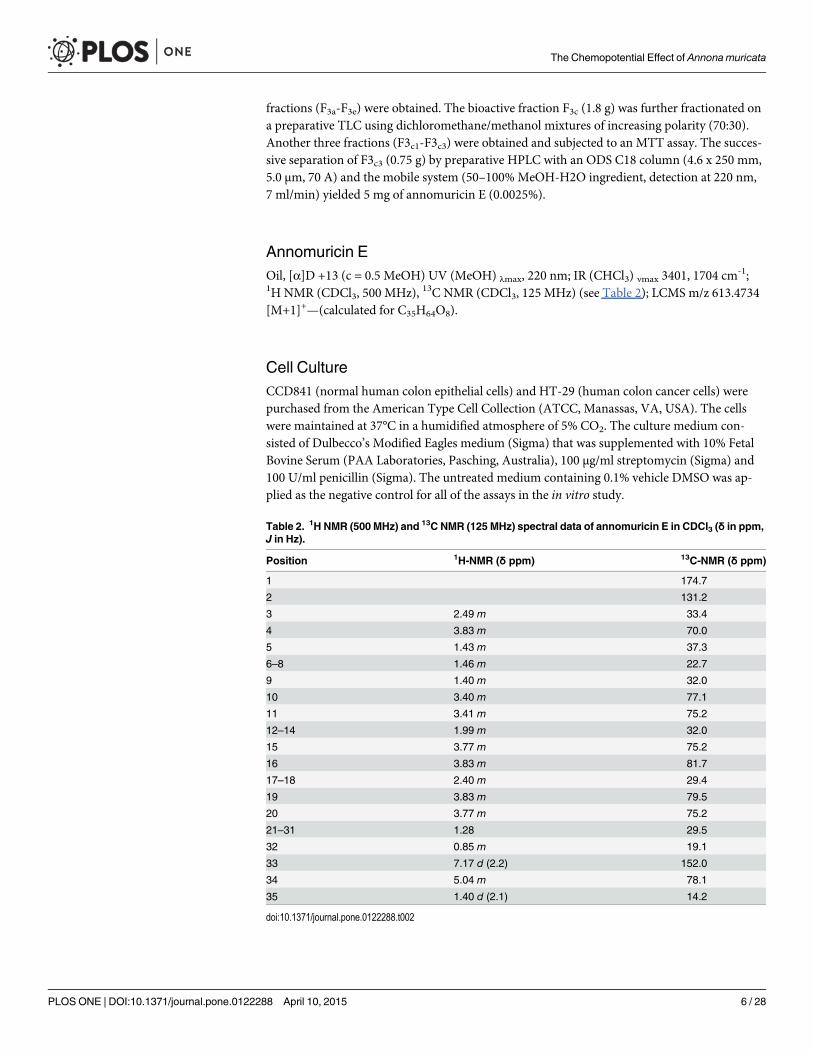
fractions (F3a-F3e) were obtained. The bioactive fraction F3c (1.8 g) was further fractionated ona preparative TLC using dichloromethane/methanol mixtures of increasing polarity (70:30).Another three fractions (F3c1-F3c3) were obtained and subjected to an MTT assay. The succes-sive separation of F3c3 (0.75 g) by preparative HPLC with an ODS C18 column (4.6 x 250 mm,5.0 μm, 70 A) and the mobile system (50–100%MeOH-H2O ingredient, detection at 220 nm,7 ml/min) yielded 5 mg of annomuricin E (0.0025%).
Annomuricin EOil, [α]D +13 (c = 0.5 MeOH) UV (MeOH) λmax, 220 nm; IR (CHCl3) νmax 3401, 1704 cm
-1;1H NMR (CDCl3, 500 MHz), 13C NMR (CDCl3, 125 MHz) (see Table 2); LCMS m/z 613.4734[M+1]+—(calculated for C35H64O8).
Cell CultureCCD841 (normal human colon epithelial cells) and HT-29 (human colon cancer cells) werepurchased from the American Type Cell Collection (ATCC, Manassas, VA, USA). The cellswere maintained at 37°C in a humidified atmosphere of 5% CO2. The culture medium con-sisted of Dulbecco’s Modified Eagles medium (Sigma) that was supplemented with 10% FetalBovine Serum (PAA Laboratories, Pasching, Australia), 100 μg/ml streptomycin (Sigma) and100 U/ml penicillin (Sigma). The untreated medium containing 0.1% vehicle DMSO was ap-plied as the negative control for all of the assays in the in vitro study.
Table 2. 1H NMR (500 MHz) and 13C NMR (125 MHz) spectral data of annomuricin E in CDCl3 (δ in ppm,J in Hz).
Position 1H-NMR (δ ppm) 13C-NMR (δ ppm)
1 174.7
2 131.2
3 2.49 m 33.4
4 3.83 m 70.0
5 1.43 m 37.3
6–8 1.46 m 22.7
9 1.40 m 32.0
10 3.40 m 77.1
11 3.41 m 75.2
12–14 1.99 m 32.0
15 3.77 m 75.2
16 3.83 m 81.7
17–18 2.40 m 29.4
19 3.83 m 79.5
20 3.77 m 75.2
21–31 1.28 29.5
32 0.85 m 19.1
33 7.17 d (2.2) 152.0
34 5.04 m 78.1
35 1.40 d (2.1) 14.2
doi:10.1371/journal.pone.0122288.t002
The Chemopotential Effect of Annona muricata
PLOSONE | DOI:10.1371/journal.pone.0122288 April 10, 2015 6 / 28
MTT AssayCell viability analysis was performed using the MTT assay as described previously [36]. Inbrief, cells (5 × 10⁴ cells/ml) at the exponential phase of growth were seeded in a 96-well plateand treated with serial concentrations of the tested agent (0.62, 1.25, 2.5, 5, 10, 20, 40 and80 μg/ml) for 12, 24 and 48 h. 5-FU, a standard anticancer drug, was used as a positive controlin this assay. After incubation, 20 μl of the MTT solution (5.0 mg/ml, Sigma) was loaded intoeach well, and the cells were further incubated at 37°C for 4 h. DMSO (150 μl) was then used todissolve the formazan crystals. The cytotoxicity against cancer and normal cells was measuredat the absorbance of 570 nm using an ELISA reader (Asys UVM340, Eugendorf, Austria). Thedata were then processed, and the antiproliferative potential of the tested agents was expressedas IC50 values, the concentration that causes a 50% inhibition of cell growth.
Lactate Dehydrogenase (LDH) Release AssayTo further confirm the cytotoxic effects of annomuricin E on HT-29 cells, the LDH releaseassay was performed using the Pierce LDH Cytotoxicity Assay Kit (Thermo Scientific, Pitts-burgh, PA, USA) as previously described [28]. Briefly, HT-29 cells at the exponential phase ofgrowth were treated with different concentrations of annomuricin E and Triton X-100 (posi-tive control) for 24 h. After the incubation, the treated HT-29 cells were exposed to the LDHreaction solution (100 μl) for 30 min. The red color intensity, representing the level of releasedLDH, was then measured at 490 nm using the Tecan Infinite 200 Pro (Tecan, Männedorf, Swit-zerland) microplate reader. The result of LDH release was calculated as a percentage of thepositive control.
Cell Cycle AssayTo determine the effect of annomuricin E on the cell cycle distribution, flow cytometric analy-sis was performed as described previously [37]. In brief, HT-29 cells (1 × 10⁶ cells/ml) at the ex-ponential phase of growth were seeded in 6-well plates and treated with annomuricin E at theIC50 concentration for 12, 24 and 48 h. After incubation, the treated HT-29 cells were har-vested, washed twice with ice-cold PBS and fixed overnight at 4°C with 90% ethanol. The fol-lowing day, the cells were washed and stained with propidium iodide (PI, 100 μl, 1 mg/ml).The cellular RNA was degraded using the enzyme RNAse A (200 μg/ml, Sigma). The stainedcells were instantly examined using a BD FACSCanto II flow cytometer (BD Biosciences, SanJose, CA, USA) by analyzing 10,000 cells per sample. The data were processed using ModFitLT software (Verity Software House, Inc., Topsham, ME, USA).
Quantitative Detection of Early and Late ApoptosisFlow cytometric analysis was performed to quantify early and late apoptosis in treated HT-29cells using the commercial BD Pharmingen Annexin V-FITC Apoptosis Detection kit (APOA-lert Annexin V; Clontech, Mountain View, CA, USA). Briefly, HT-29 cells (1 × 10⁵ cells/ml) atthe exponential phase of growth were incubated with annomuricin E at the IC50 concentrationfor 12, 24 and 48 h. After incubation, the treated cells were harvested, washed twice with PBSand suspended in the Annexin-V binding buffer. The cells were then supplemented withAnnexin-V-FITC and PI, according to the vendor’s instructions. The stained cells were exam-ined using a BD FACSCanto II flow cytometer. Early and late apoptotic cells and necrotic cellswere quantitatively detected using a quadrant statistics analysis [38].
The Chemopotential Effect of Annona muricata
PLOSONE | DOI:10.1371/journal.pone.0122288 April 10, 2015 7 / 28

Detection of Caspases ActivationA luminescence-based analysis was performed to investigate the activity of caspase 3/7 and cas-pase 9 using the Caspase-Glo 9 Assay and Caspase-Glo 3/7 Assay commercial kits (PromegaCorporation, Fitchburg, WI, USA) as described previously [39]. Briefly, HT-29 cells (2 × 10⁵
cells/ml) were seeded overnight in a white-walled 96-well plate and treated with an IC50 doseof annomuricin E for 3, 6, 12, 24 and 48 h. After incubation, 100 μl of the caspase-Glo reagentwas added to each well according to the manufacturer’s protocol. Luminescence, which repre-sents the caspase activities, was measured using a luminescence microplate reader (Tecan Infi-nite 200 Pro).
Multiple Cytotoxicity AssayThe simultaneous analysis of critical apoptosis markers, namely cell membrane permeability,cytochrome c leakage from the mitochondria, mitochondrial membrane potential (MMP) andtotal nuclear intensity, in HT-29 cells was performed using the Cellomics Multiparameter Cy-totoxicity 3 Kit (Cellomics, Pittsburgh, PA, USA) as previously described in detail [40]. Inbrief, HT-29 cells (1 × 10⁵ cells/ml) were plated overnight in a 96-well plate and were exposedto an IC50 dose of annomuricin E for 12, 24 and 48 h. After incubation, the treated cells werestained with a cell permeability dye (FITC), a cytochrome c dye (Cy3), a mitochondrial mem-brane potential dye (Cy5) and a nuclear dye (Hoechst 33342), according to the vendor’s proto-col. The plates were analyzed using a Cell Reporter cytofluorimeter system (Gentix/MolecularDevices, United Kingdom).
Gene Expression Analysis of Bcl-2/BaxThe mRNA expression of two proteins, Bcl-2 and Bax, was quantified using real-time Q-PCRanalysis as described previously with some modifications [41]. In brief, HT-29 cells at the expo-nential phase of growth were treated with annomuricin E at the IC50 concentration for 12, 24and 48 h. The total RNA of treated cells was isolated using the RNeasy Plus Mini kit (Qiagen,Hilden, Germany) followed by the synthesis of the complementary DNA using the iScriptcDNA synthesis kit (Biorad, Hercules, CA, USA). Q-PCR was performed on the StepOnePLUS real-time PCR machine (Applied Biosystems, Carlsbad, CA, USA). The β-actin house-keeping gene was used as a positive reference and was applied to normalize the target mRNA.The Q-PCR master mix was provided by Solaris Q-PCR Expression Assays (Thermo FisherScientific, Waltham, MA, USA) for the gene expression analysis of Bcl-2, AX-003307-00-0100;Bax, AX-003308-00-0100; and β-actin, AX-003451-00-0100.
Immunofluorescence Analysis of Bcl-2/BaxThe perturbation in the protein expressions of Bcl-2 and Bax was investigated using immuno-fluorescence analysis as previously described in detail [37]. In brief, the HT-29 cells (5 × 10⁴
cells/ml) were seeded in a 96-well plate and exposed to the IC50 dose of annomuricin E for 12,24 and 48 h. After washing the cells twice with PBS, they were fixed in 4% paraformaldehyde at25°C for 20 min prior to blocking with blocking buffer (0.03% Triton X-100/PBS and normalserum) for 1 h. The cells were then supplemented with a primary antibody solution and incu-bated overnight at 4°C. After incubation, the cells were treated with Bcl-2 and Bax fluro-chrome-conjugated secondary antibody (Santa Cruz Biotechnology, Santa Cruz, CA, USA) for1 h. The cells were then washed twice with PBS prior to staining with DAPI. The stained cellswere examined using the Cell Reporter cytofluorimeter system.
The Chemopotential Effect of Annona muricata
PLOSONE | DOI:10.1371/journal.pone.0122288 April 10, 2015 8 / 28
Statistical AnalysisData from the rat study were reported as the means ± standard error of n animals per group.The experimental data were analyzed with one-way analysis of variance, followed by Tukey’spost hoc test using the SAS 9.1 statistical program (SAS Institute Inc., Cary, NC, USA). In vitroresults were presented as the means ± standard error of the mean from three independent ex-periments. Statistical analysis was performed using the statistical package GraphPad PrismVersion 5 (GraphPad Software Inc., San Diego, USA). One-way analysis of variance (Dunnett’smultiple comparison test) was used to distinguish the difference among groups. All values atP<0.05 were considered significant.
Results and Discussion
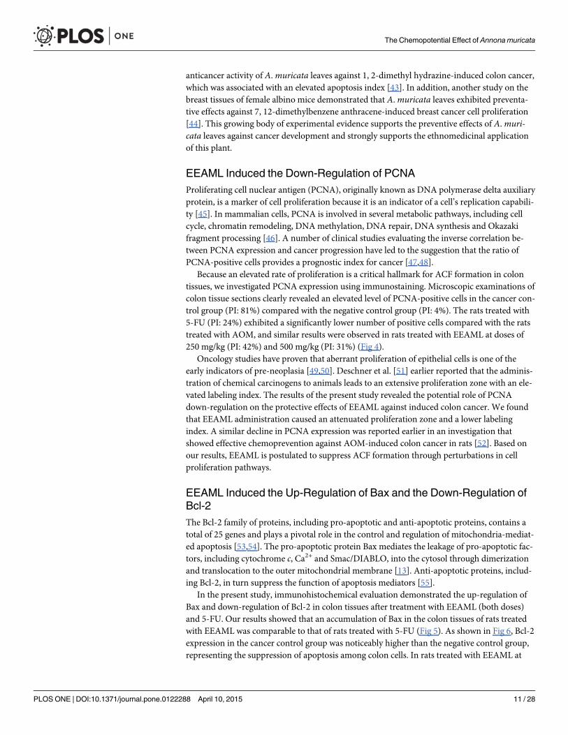
ACF FrequencyTo evaluate the effect of EEAML on suppressing colon carcinogenesis, ACF were employed asa biomarker to assess early stage AOM-induced colon cancer in rats. The incidence of ACF onthe proximal and distal parts of the colon mucosa were analyzed with methylene blue stainingimmediately after the sacrifice of animals, and these data are shown in Fig 2 and Table 3. ACFwere characterized by crypts with elevated sizes, altered luminal epithelia and easily discerniblepericryptal zones. Topographical views of the stained colon specimens did not elicit any micro-scopic changes in the negative control group (Fig 3). Meanwhile, all rats injected with AOM de-veloped ACF containing different numbers of crypts (Table 3). In agreement with previouslypublished findings, ACF formation in the distal colon was significantly higher than the proxi-mal colon [20,42]. Compared with the cancer control group, the administration of 5-FU orEEAML at 250 mg/kg or 500 mg/kg significantly suppressed the formation of ACF (79.5%,61.2% and 72.5%, respectively). The doses used in this experiment were chosen based on theprevious studies on the effect of different plant extracts against AOM-induced ACF formation[21,32]. A recent investigation on anticancer activity of A.muricata at a single dose of 300 mg/kg confirmed that these two doses would be appropriate for this study [43]. The respective in-vestigation reported a similar reduction in ACF formation that demonstrated a potent
Fig 2. The number of ACF formed in proximal and distal parts of the colon. Tissue specimens werecollected from five groups of rats: (A) negative control, (B) cancer control, (C) low dose of EEAML, (D) highdose of EEAML and (E) treatment control. Data are expressed as the means ± SEM of (n = 6/group).*P<0.05 compared with cancer control.
doi:10.1371/journal.pone.0122288.g002
The Chemopotential Effect of Annona muricata
PLOSONE | DOI:10.1371/journal.pone.0122288 April 10, 2015 9 / 28

Table 3. Distribution of aberrant crypt categories (1, 2, 3, 4 andmore) in the colons of five groups of rats: (A) negative control, (B) cancer control, (C)low dose of EEAML, (D) high dose of EEAML and (E) treatment control.
Group No. of crypts per ACF
1 crypt 2 crypt 3 crypt 4 crypt and more Total Inhibition (%)
A 0 0 0 0 0 -
B 33 ± 2.46 29 ± 1.89 48 ± 2.49 32 ± 2.32 142 ± 7.88 -
C 18 ± 0.92* 17 ± 0.68* 11 ± 0.66* 9 ± 0.48* 55 ± 2.32* 61.2
D 10 ± 0.65* 14 ± 0.59* 9 ± 0.52* 6 ± 0.25* 39 ± 1.48* 72.5
E 12 ± 0.26* 6 ± 0.45* 7 ± 0.78* 4 ± 0.18* 29 ± 1.40* 79.5
Data expressed as the means ± SEM of (n = 6/group).
*P<0.05 compared with cancer control.
doi:10.1371/journal.pone.0122288.t003
Fig 3. Topographical views of the colonmucosa. Tissue specimens were collected from five groups of rats: (A) negative control, (B) cancer control, (C)low dose of EEAML, (D) high dose of EEAML and (E) treatment control and were stained with methylene blue dye. The red arrows depict ACF in the colonmucosa. Scale bar: 500 μm.
doi:10.1371/journal.pone.0122288.g003
The Chemopotential Effect of Annona muricata
PLOSONE | DOI:10.1371/journal.pone.0122288 April 10, 2015 10 / 28
anticancer activity of A.muricata leaves against 1, 2-dimethyl hydrazine-induced colon cancer,which was associated with an elevated apoptosis index [43]. In addition, another study on thebreast tissues of female albino mice demonstrated that A.muricata leaves exhibited preventa-tive effects against 7, 12-dimethylbenzene anthracene-induced breast cancer cell proliferation[44]. This growing body of experimental evidence supports the preventive effects of A.muri-cata leaves against cancer development and strongly supports the ethnomedicinal applicationof this plant.
EEAML Induced the Down-Regulation of PCNAProliferating cell nuclear antigen (PCNA), originally known as DNA polymerase delta auxiliaryprotein, is a marker of cell proliferation because it is an indicator of a cell’s replication capabili-ty [45]. In mammalian cells, PCNA is involved in several metabolic pathways, including cellcycle, chromatin remodeling, DNAmethylation, DNA repair, DNA synthesis and Okazakifragment processing [46]. A number of clinical studies evaluating the inverse correlation be-tween PCNA expression and cancer progression have led to the suggestion that the ratio ofPCNA-positive cells provides a prognostic index for cancer [47,48].
Because an elevated rate of proliferation is a critical hallmark for ACF formation in colontissues, we investigated PCNA expression using immunostaining. Microscopic examinations ofcolon tissue sections clearly revealed an elevated level of PCNA-positive cells in the cancer con-trol group (PI: 81%) compared with the negative control group (PI: 4%). The rats treated with5-FU (PI: 24%) exhibited a significantly lower number of positive cells compared with the ratstreated with AOM, and similar results were observed in rats treated with EEAML at doses of250 mg/kg (PI: 42%) and 500 mg/kg (PI: 31%) (Fig 4).
Oncology studies have proven that aberrant proliferation of epithelial cells is one of theearly indicators of pre-neoplasia [49,50]. Deschner et al. [51] earlier reported that the adminis-tration of chemical carcinogens to animals leads to an extensive proliferation zone with an ele-vated labeling index. The results of the present study revealed the potential role of PCNAdown-regulation on the protective effects of EEAML against induced colon cancer. We foundthat EEAML administration caused an attenuated proliferation zone and a lower labelingindex. A similar decline in PCNA expression was reported earlier in an investigation thatshowed effective chemoprevention against AOM-induced colon cancer in rats [52]. Based onour results, EEAML is postulated to suppress ACF formation through perturbations in cellproliferation pathways.
EEAML Induced the Up-Regulation of Bax and the Down-Regulation ofBcl-2The Bcl-2 family of proteins, including pro-apoptotic and anti-apoptotic proteins, contains atotal of 25 genes and plays a pivotal role in the control and regulation of mitochondria-mediat-ed apoptosis [53,54]. The pro-apoptotic protein Bax mediates the leakage of pro-apoptotic fac-tors, including cytochrome c, Ca2+ and Smac/DIABLO, into the cytosol through dimerizationand translocation to the outer mitochondrial membrane [13]. Anti-apoptotic proteins, includ-ing Bcl-2, in turn suppress the function of apoptosis mediators [55].
In the present study, immunohistochemical evaluation demonstrated the up-regulation ofBax and down-regulation of Bcl-2 in colon tissues after treatment with EEAML (both doses)and 5-FU. Our results showed that an accumulation of Bax in the colon tissues of rats treatedwith EEAML was comparable to that of rats treated with 5-FU (Fig 5). As shown in Fig 6, Bcl-2expression in the cancer control group was noticeably higher than the negative control group,representing the suppression of apoptosis among colon cells. In rats treated with EEAML at
The Chemopotential Effect of Annona muricata
PLOSONE | DOI:10.1371/journal.pone.0122288 April 10, 2015 11 / 28
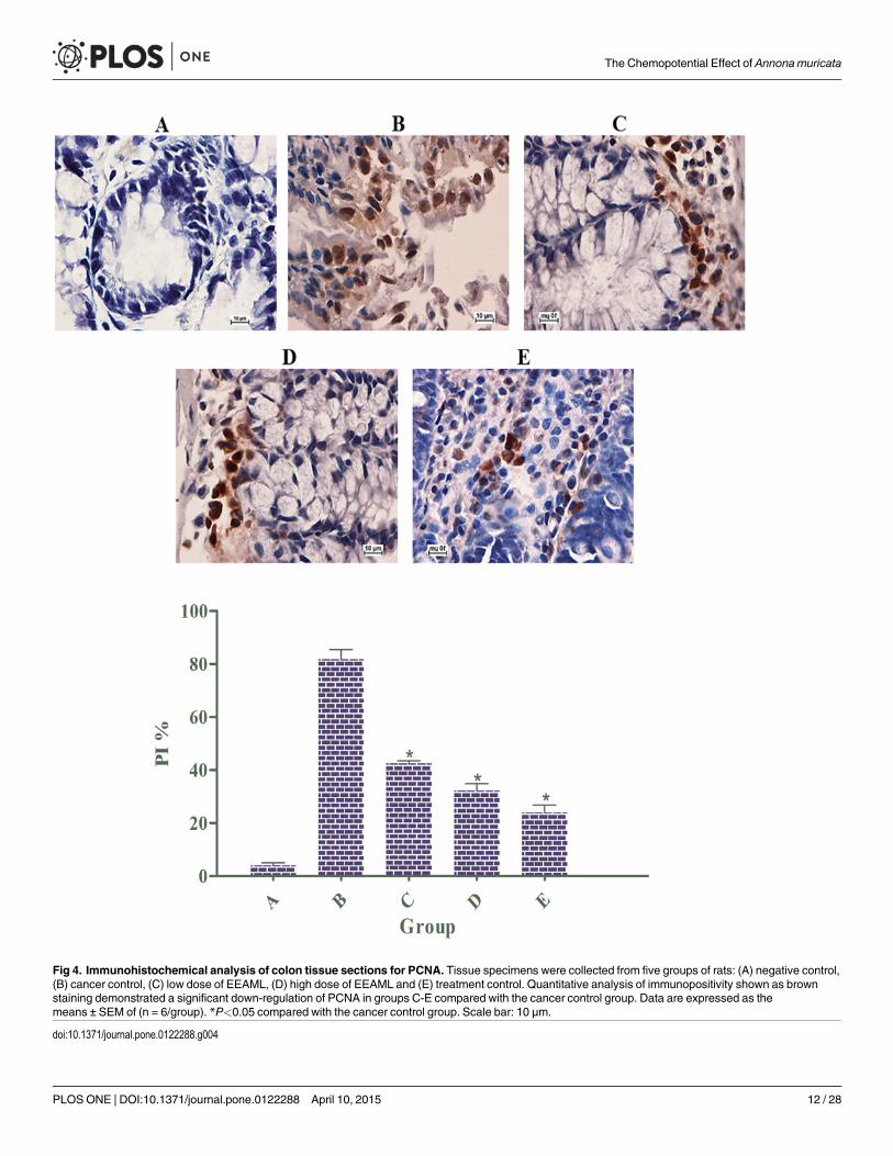
Fig 4. Immunohistochemical analysis of colon tissue sections for PCNA. Tissue specimens were collected from five groups of rats: (A) negative control,(B) cancer control, (C) low dose of EEAML, (D) high dose of EEAML and (E) treatment control. Quantitative analysis of immunopositivity shown as brownstaining demonstrated a significant down-regulation of PCNA in groups C-E compared with the cancer control group. Data are expressed as themeans ± SEM of (n = 6/group). *P<0.05 compared with the cancer control group. Scale bar: 10 μm.
doi:10.1371/journal.pone.0122288.g004
The Chemopotential Effect of Annona muricata
PLOSONE | DOI:10.1371/journal.pone.0122288 April 10, 2015 12 / 28

doses of 250 mg/kg and 500 mg/kg and 5-FU, an accumulation of Bcl-2 protein in colon tissueswas markedly decreased. Administration of EEAML (500 mg/kg) and 5-FU decreased Bcl-2protein expression to approximately the level of the negative control group.
Previous studies have reported that high Bax protein expression may augment the mediansurvival among cancer patients [56]. In addition, a deficiency in Bax protein has a strong im-pact on tumor clonal evolution [57]. The results of our present study demonstrate that EEAMLhas the potential to induce apoptosis in colon cells that are susceptible to AOM damage. Thisin vivo observation agreed with our previous in vitro study by illustrating the up-regulation ofBax and the down-regulation of Bcl-2 in HT-29 cells treated with EEAML [30].
EEAML Augmented Enzymatic Antioxidants ActivitiesAs an aggressive factor, reactive oxygen species (ROS) play a pivotal role in the pathogenesis ofcolorectal cancer [58]. The production of reactive oxygen species (ROS) are part of the normalmetabolism in the human body, and cellular antioxidants containing enzymatic and non-enzy-matic scavengers maintain ROS at their physiological levels [59]. Nonetheless, an extensivegeneration of ROS, including hydrogen radicals, hydrogen peroxide and superoxide anions,causes oxidative stress, which leads to metabolic impairments and irreversible cell damages
Fig 5. Expression of Bax in colon tissue sections. Tissue specimens were collected from five groups of rats (n = 6/group) and were analyzed usingimmunohistochemistry: (A) negative control, (B) cancer control, (C) low dose of EEAML, (D) high dose of EEAML and (E) treatment control. The up-regulation of Bax in groups C-E is shown as brown staining. Scale bar: 10 μm.
doi:10.1371/journal.pone.0122288.g005
The Chemopotential Effect of Annona muricata
PLOSONE | DOI:10.1371/journal.pone.0122288 April 10, 2015 13 / 28

[60]. SOD, the first scavenging barrier against ROS, converts the superoxide to hydrogen per-oxide, which is subsequently degraded to water and oxygen by CAT [61]. The degradation oflipid peroxides to hydroxyl lipids and water is mediated by GPx through oxidation of glutathi-one to glutathione disulfide [62,63].
The activities of antioxidant enzymes were significantly reduced in the AOM-treated groupcompared with the negative control group (Fig 7). However, EEAML supplementation at bothdoses significantly restored the levels of these enzymes towards normal values. As expected,EEAML showed a greater antioxidant defense than 5-FU. A number of earlier in vitro and invivo studies have demonstrated that the leaves of A.muricata possess significant antioxidantpotential [64–66]. Moreover, the leaves elicited noticeable defensive activities against acute andchronic inflammation in rats through suppressive effects on the secretion of proinflammatorycytokines [67]. Immunological studies have led to the suggestion that concomitant administra-tion of chemotherapeutic agents and antioxidant drugs counteract chemotherapy toxicity andenhance the survival rate among cancer patients [68,69]. Therefore, an establishment of anti-cancer agents with innate antioxidant defense may result in the discovery of new generationsof anticancer drugs.
Fig 6. Immunohistochemical analysis of colon tissue sections for Bcl-2. Tissue specimens were collected from five groups of rats (n = 6/group): (A)negative control, (B) cancer control, (C) low dose of EEAML, (D) high dose of EEAML and (E) treatment control. Immunopositivity shown as brown stainingrevealed the down-regulation of Bcl-2 in groups C-E. Scale bar: 10 μm.
doi:10.1371/journal.pone.0122288.g006
The Chemopotential Effect of Annona muricata
PLOSONE | DOI:10.1371/journal.pone.0122288 April 10, 2015 14 / 28

EEAML Suppressed Lipid PeroxidationExcessive ROS generation results in the production of lipid radicals and rearrangements of un-saturated lipids, leading to the formation of different degraded metabolites, including alkenes,lipid hydroperoxides and MDA, which eventually disrupt the integrity of membrane lipids[70,71]. MDA, a major metabolite of this process, is an easy indicator of lipid peroxidation andoxidative stress [72]. As a carcinogenic agent, AOM causes lipid peroxidation as a result of oxi-dative stress [73], which was observed in our study after administration of AOM to the cancercontrol group (Fig 7). This result appears to be in line with previous reports that plasma andtissue MDA concentrations are markedly elevated in patients suffering from colorectal cancer[74,75]. As expected, because of the augmentation in the enzymatic and antioxidant activities,EEAML treatment at both doses significantly reduced MDA formation in colon tissues, andthis reduction was stronger than the reduction found after treatment with 5-FU. This resultconfirmed the protective effects of EEAML against oxidative stress in colon tissues, which wasreflected by reduced MDA production.
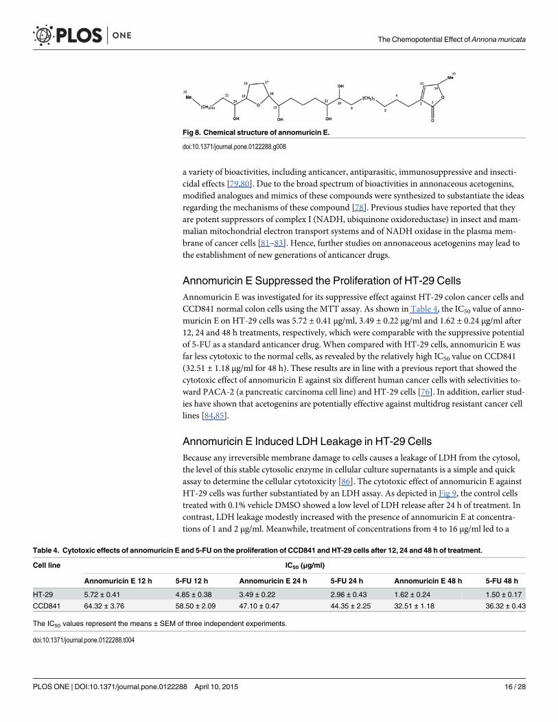
Isolation of the Bioactive Compound, Annomuricin EThe dried leaves of A.muricata were extracted with ethyl acetate at room temperature. Afterconcentrating the solution until dry, the ethyl acetate extract was obtained. The ethyl acetateextract was fractionated by chromatography on a silica gel 60 column, which yielded six frac-tions. Fraction 3 (3.9 g) was further purified on a micro column followed by preparative TLCand finally preparative HPLC using an ODS C-18 column and a PDA detector to obtain anno-muricin E (Fig 8), which was identified by 1D and 2D NMR, mass spectrometry and otherphysical properties that were then compared with reported data [76].
The annonaceous acetogenins, a series of C-35/C-37 fatty acid derivatives, are a class of nat-ural products that are uniquely isolated from the Annonaceae family [77]. The isolation ofmore than 500 annonaceous acetogenins from different parts of plants in this family has beenperformed for more than 27 years [78]. This hyperbioactive group of natural products exhibits
Fig 7. Level of CAT, GPx, MDA and SOD in colon tissue homogenates. Samples were collected from fivegroups of rats: (A) negative control, (B) cancer control, (C) low dose of EEAML, (D) high dose of EEAML and(E) treatment control. Data are expressed as the means ± SEM of (n = 6/group). *P<0.05 compared withcancer control.
doi:10.1371/journal.pone.0122288.g007
The Chemopotential Effect of Annona muricata
PLOSONE | DOI:10.1371/journal.pone.0122288 April 10, 2015 15 / 28
a variety of bioactivities, including anticancer, antiparasitic, immunosuppressive and insecti-cidal effects [79,80]. Due to the broad spectrum of bioactivities in annonaceous acetogenins,modified analogues and mimics of these compounds were synthesized to substantiate the ideasregarding the mechanisms of these compound [78]. Previous studies have reported that theyare potent suppressors of complex I (NADH, ubiquinone oxidoreductase) in insect and mam-malian mitochondrial electron transport systems and of NADH oxidase in the plasma mem-brane of cancer cells [81–83]. Hence, further studies on annonaceous acetogenins may lead tothe establishment of new generations of anticancer drugs.
Annomuricin E Suppressed the Proliferation of HT-29 CellsAnnomuricin E was investigated for its suppressive effect against HT-29 colon cancer cells andCCD841 normal colon cells using the MTT assay. As shown in Table 4, the IC50 value of anno-muricin E on HT-29 cells was 5.72 ± 0.41 μg/ml, 3.49 ± 0.22 μg/ml and 1.62 ± 0.24 μg/ml after12, 24 and 48 h treatments, respectively, which were comparable with the suppressive potentialof 5-FU as a standard anticancer drug. When compared with HT-29 cells, annomuricin E wasfar less cytotoxic to the normal cells, as revealed by the relatively high IC50 value on CCD841(32.51 ± 1.18 μg/ml for 48 h). These results are in line with a previous report that showed thecytotoxic effect of annomuricin E against six different human cancer cells with selectivities to-ward PACA-2 (a pancreatic carcinoma cell line) and HT-29 cells [76]. In addition, earlier stud-ies have shown that acetogenins are potentially effective against multidrug resistant cancer celllines [84,85].
Annomuricin E Induced LDH Leakage in HT-29 CellsBecause any irreversible membrane damage to cells causes a leakage of LDH from the cytosol,the level of this stable cytosolic enzyme in cellular culture supernatants is a simple and quickassay to determine the cellular cytotoxicity [86]. The cytotoxic effect of annomuricin E againstHT-29 cells was further substantiated by an LDH assay. As depicted in Fig 9, the control cellstreated with 0.1% vehicle DMSO showed a low level of LDH release after 24 h of treatment. Incontrast, LDH leakage modestly increased with the presence of annomuricin E at concentra-tions of 1 and 2 μg/ml. Meanwhile, treatment of concentrations from 4 to 16 μg/ml led to a
Fig 8. Chemical structure of annomuricin E.
doi:10.1371/journal.pone.0122288.g008
Table 4. Cytotoxic effects of annomuricin E and 5-FU on the proliferation of CCD841 and HT-29 cells after 12, 24 and 48 h of treatment.
Cell line IC50 (μg/ml)
Annomuricin E 12 h 5-FU 12 h Annomuricin E 24 h 5-FU 24 h Annomuricin E 48 h 5-FU 48 h
HT-29 5.72 ± 0.41 4.85 ± 0.38 3.49 ± 0.22 2.96 ± 0.43 1.62 ± 0.24 1.50 ± 0.17
CCD841 64.32 ± 3.76 58.50 ± 2.09 47.10 ± 0.47 44.35 ± 2.25 32.51 ± 1.18 36.32 ± 0.43
The IC50 values represent the means ± SEM of three independent experiments.
doi:10.1371/journal.pone.0122288.t004
The Chemopotential Effect of Annona muricata
PLOSONE | DOI:10.1371/journal.pone.0122288 April 10, 2015 16 / 28

significant LDH release compared with the control. The significant LDH leakage from HT-29cells was shown at concentrations as low as 4 μg/ml, which was compatible with the 24-h IC50
value of annomuricin E (3.49 ± 0.22 μg/ml) against HT-29 cells.
Cell Cycle Arrest at G1 Induced by Annomuricin ECancer progression is often associated with irregularities in cell cycle function [87]. A growingbody of experimental evidence supporting the concomitant involvement of cell cycle suppres-sion and apoptosis has stimulated widespread attention to phytochemicals with cell-cycle mod-ulatory effects [15,88]. Hence, we first evaluated whether the suppressive effect of annomuricinE was accompanied by a block in the cell cycle using PI staining and flow cytometry analysis.As illustrated in Fig 10, the augmented accumulation of HT-29 cells in the G1 phase was initiat-ed after 12 h of treatment with annomuricin E, and this accumulation of cells in the G1 phasecontinued in a time-dependent manner. After 24 and 48 h, the percentage of HT-29 cells treat-ed with annomuricin E that were arrested at the G1 phase reached 89.65% and 94.60%, respec-tively. This was accompanied by a concurrent decline in the S and G2/M cell populationscompared with the control. These results indicated that annomuricin E arrested HT-29 cells at
Fig 9. Effects of annomuricin E on LDH leakage formation in HT-29 cells.Cells were exposed to 0.1%vehicle DMSO (control) and annomuricin E at different concentrations for 24 h. The treated HT-29 cellsshowed a significant LDH release at 4 to 16 μg/ml concentrations compared with the control. The datarepresent the means ± SEM of three independent experiments. *P<0.05 compared with the control.
doi:10.1371/journal.pone.0122288.g009
The Chemopotential Effect of Annona muricata
PLOSONE | DOI:10.1371/journal.pone.0122288 April 10, 2015 17 / 28

the G1 phase. An earlier study on annonacin, an annonaceous acetogenin from the seeds ofAnnona reticulata, also showed the induction of cell cycle arrest in T24 bladder cancer cells atthe G1 phase through the activation of p21 [89].
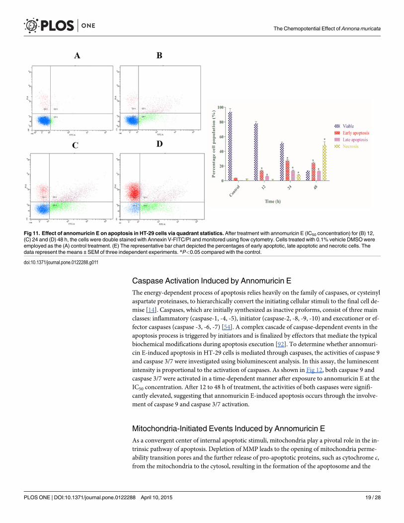
Phosphatidylserine Externalization Induced by Annomuricin EAs one of the biochemical characterizations of apoptosis, a transverse redistribution of phos-phatidylserine (PS) on the outer plasma membrane arises during early apoptosis [90]. A fluo-rescent probe of Annexin V-FITC is a recombinant protein with a high affinity for externalizedPS [91]. To gain insight into the mechanism through which annomuricin E induces its cytotox-ic effects, HT-29 cells were stained with Annexin V-FITC/PI and analyzed using flow cytome-try. In cells treated with 0.1% vehicle DMSO (control), only 3.6% and 0.5% of cells were inearly (Annexin V+/PI−) and late (Annexin V+/PI+) apoptosis after 48 h, respectively (Fig 11).However, the percentage of early and late apoptotic cells were significantly increased to 13.9%and 6.9%, respectively, after being treated with annomuricin E (IC50 concentration) for 12 h.The percentages of early and late apoptotic populations peaked at 24 h with values of 27.3%and 13.6%, respectively, and were reduced slightly at 48 h. This was reduction was associatedwith a significant elevation in the number of necrotic cells (Annexin V−/PI+) at 24 and 48 h.This increase in necrotic cells can be explained by the long exposure of annomuricin E to HT-29 cells that allowed the cells to enter secondary necrosis from primary apoptosis, increasingthe number of dead cells. These data showed that annomuricin E caused its cytotoxic effectsthrough the induction of apoptosis in HT-29 cells.
Fig 10. Effect of annomuricin E on cell cycle distribution in HT-29 cells.Cells were treated with (A) 0.1% vehicle DMSO (control) for 48 h andannomuricin E at the IC50 concentration for (B) 12, (C) 24 and (D) 48 h. After staining the cells with PI, the DNA contents were monitored using flowcytometry. (E) The representative bar chart shows the significant induction of G1 cell cycle arrest by annomuricin E after 12 h of treatment. The data representthe means ± SEM of three independent experiments. *P<0.05 compared with the control.
doi:10.1371/journal.pone.0122288.g010
The Chemopotential Effect of Annona muricata
PLOSONE | DOI:10.1371/journal.pone.0122288 April 10, 2015 18 / 28
Caspase Activation Induced by Annomuricin EThe energy-dependent process of apoptosis relies heavily on the family of caspases, or cysteinylaspartate proteinases, to hierarchically convert the initiating cellular stimuli to the final cell de-mise [14]. Caspases, which are initially synthesized as inactive proforms, consist of three mainclasses: inflammatory (caspase-1, -4, -5), initiator (caspase-2, -8, -9, -10) and executioner or ef-fector caspases (caspase -3, -6, -7) [54]. A complex cascade of caspase-dependent events in theapoptosis process is triggered by initiators and is finalized by effectors that mediate the typicalbiochemical modifications during apoptosis execution [92]. To determine whether annomuri-cin E-induced apoptosis in HT-29 cells is mediated through caspases, the activities of caspase 9and caspase 3/7 were investigated using bioluminescent analysis. In this assay, the luminescentintensity is proportional to the activation of caspases. As shown in Fig 12, both caspase 9 andcaspase 3/7 were activated in a time-dependent manner after exposure to annomuricin E at theIC50 concentration. After 12 to 48 h of treatment, the activities of both caspases were signifi-cantly elevated, suggesting that annomuricin E-induced apoptosis occurs through the involve-ment of caspase 9 and caspase 3/7 activation.
Mitochondria-Initiated Events Induced by Annomuricin EAs a convergent center of internal apoptotic stimuli, mitochondria play a pivotal role in the in-trinsic pathway of apoptosis. Depletion of MMP leads to the opening of mitochondria perme-ability transition pores and the further release of pro-apoptotic proteins, such as cytochrome c,from the mitochondria to the cytosol, resulting in the formation of the apoptosome and the
Fig 11. Effect of annomuricin E on apoptosis in HT-29 cells via quadrant statistics. After treatment with annomuricin E (IC50 concentration) for (B) 12,(C) 24 and (D) 48 h, the cells were double stained with Annexin V-FITC/PI and monitored using flow cytometry. Cells treated with 0.1% vehicle DMSOwereemployed as the (A) control treatment. (E) The representative bar chart depicted the percentages of early apoptotic, late apoptotic and necrotic cells. Thedata represent the means ± SEM of three independent experiments. *P<0.05 compared with the control.
doi:10.1371/journal.pone.0122288.g011
The Chemopotential Effect of Annona muricata
PLOSONE | DOI:10.1371/journal.pone.0122288 April 10, 2015 19 / 28
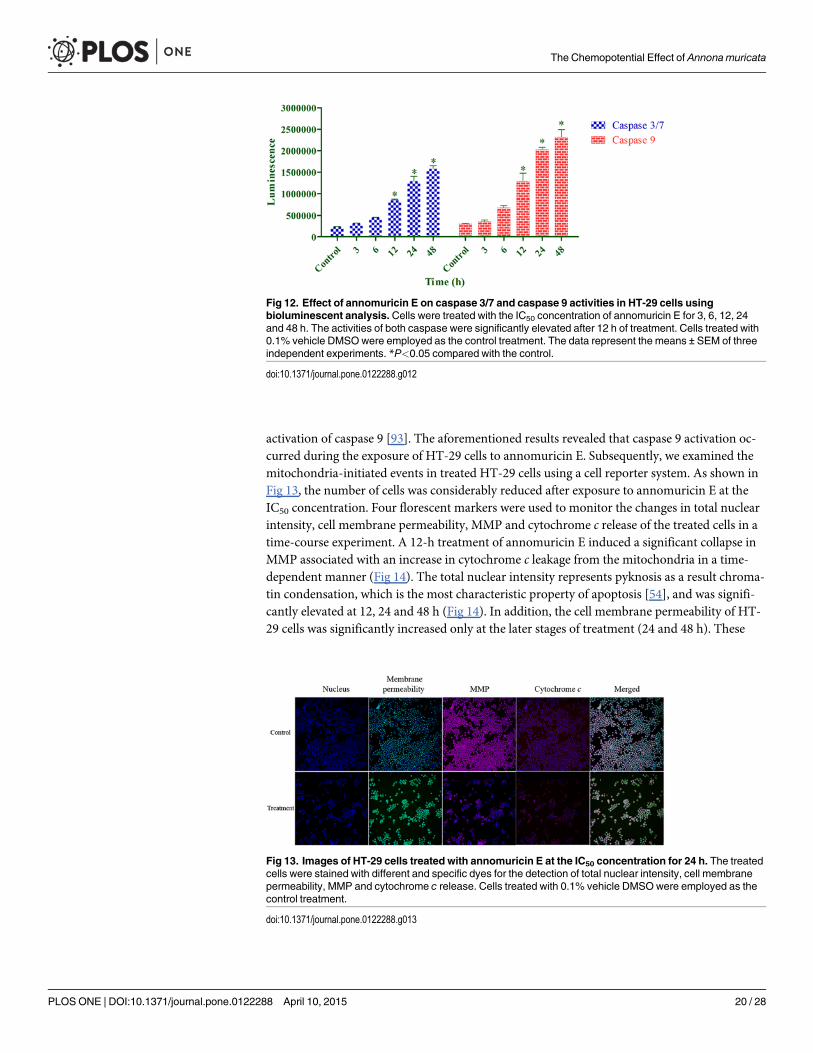
activation of caspase 9 [93]. The aforementioned results revealed that caspase 9 activation oc-curred during the exposure of HT-29 cells to annomuricin E. Subsequently, we examined themitochondria-initiated events in treated HT-29 cells using a cell reporter system. As shown inFig 13, the number of cells was considerably reduced after exposure to annomuricin E at theIC50 concentration. Four florescent markers were used to monitor the changes in total nuclearintensity, cell membrane permeability, MMP and cytochrome c release of the treated cells in atime-course experiment. A 12-h treatment of annomuricin E induced a significant collapse inMMP associated with an increase in cytochrome c leakage from the mitochondria in a time-dependent manner (Fig 14). The total nuclear intensity represents pyknosis as a result chroma-tin condensation, which is the most characteristic property of apoptosis [54], and was signifi-cantly elevated at 12, 24 and 48 h (Fig 14). In addition, the cell membrane permeability of HT-29 cells was significantly increased only at the later stages of treatment (24 and 48 h). These
Fig 13. Images of HT-29 cells treated with annomuricin E at the IC50 concentration for 24 h. The treatedcells were stained with different and specific dyes for the detection of total nuclear intensity, cell membranepermeability, MMP and cytochrome c release. Cells treated with 0.1% vehicle DMSOwere employed as thecontrol treatment.
doi:10.1371/journal.pone.0122288.g013
Fig 12. Effect of annomuricin E on caspase 3/7 and caspase 9 activities in HT-29 cells usingbioluminescent analysis. Cells were treated with the IC50 concentration of annomuricin E for 3, 6, 12, 24and 48 h. The activities of both caspase were significantly elevated after 12 h of treatment. Cells treated with0.1% vehicle DMSOwere employed as the control treatment. The data represent the means ± SEM of threeindependent experiments. *P<0.05 compared with the control.
doi:10.1371/journal.pone.0122288.g012
The Chemopotential Effect of Annona muricata
PLOSONE | DOI:10.1371/journal.pone.0122288 April 10, 2015 20 / 28

results suggest that annomuricin E caused the dissipation of MMP and the leakage of cyto-chrome c from mitochondria, which resulted in the activation of caspase 9.
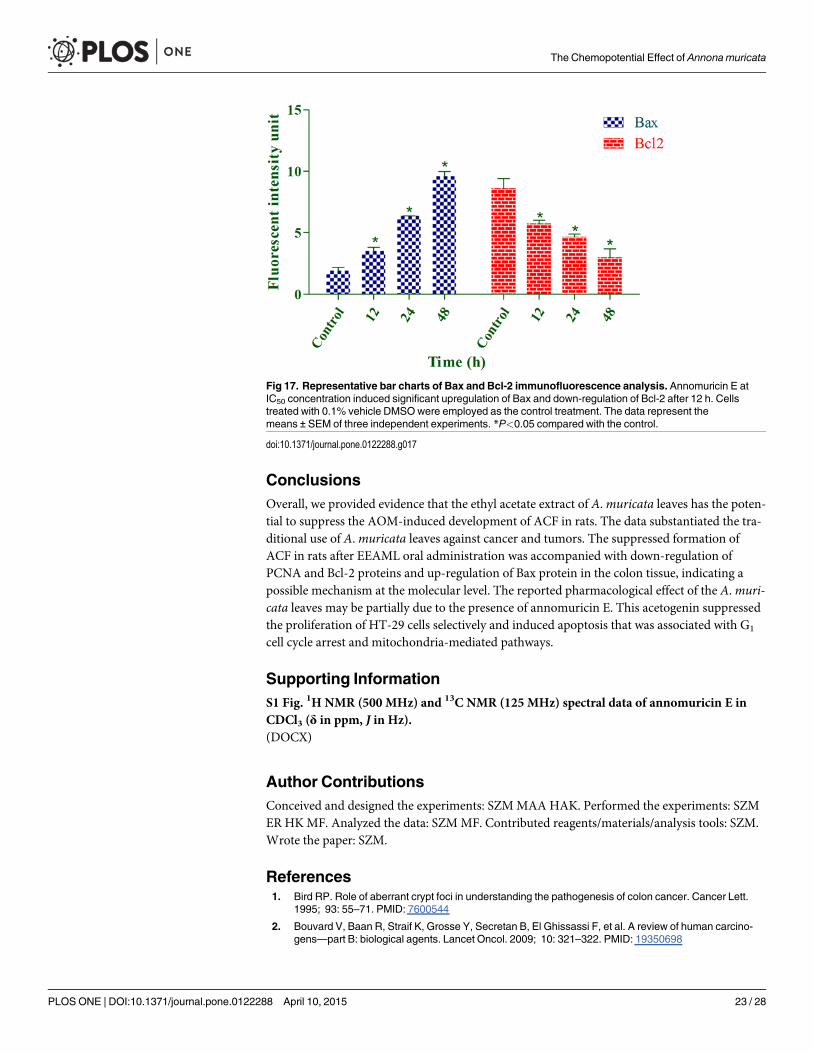
Bax Up-Regulation and Bcl-2 Down-Regulation Induced by AnnomuricinEBecause annomuricin E elicited the ability to interfere with MMP in HT-29 cells, we raised thepossibility of Bax and Bcl-2 involvement in annomuricin E-induced apoptosis. Hence, the ex-pression of Bax and Bcl-2 was investigated at both the mRNA and protein levels using Q-PCRand immunofluorescence analysis, respectively. As shown in Fig 15, the mRNA expression ofthe Bax protein was significantly and time-dependently elevated after 12 h treatment andreached an approximately 5-fold higher level after 48 h. In spite of Bax up-regulation, themRNA expression of the anti-apoptotic protein Bcl-2 was time-dependently reduced from 12to 48 h. Immunofluorescence analysis demonstrated that the number of HT-29 cells treatedwith annomuricin E decreased in a time-dependent manner after 12, 24 and 48 h, as illustratedby the blue fluorescent staining of DAPI, which identifies all cell nuclei (Fig 16). The time-de-pendent reduction in the number of surviving cells was accompanied with a distinct increase inthe fluorescent intensity of FITC dye (green) that represented Bax protein expression, whichreached a value approximately 10-fold higher than the control after 48 h (Fig 17). Bcl-2 proteinexpression also significantly and dose-dependently reduced compared with the control. Theperturbations in Bax and Bcl-2 expression at the mRNA and protein level substantiated theidea that annomuricin E-induced apoptosis was through the mitochondria-mediated pathway.
Fig 14. Representative bar charts of the multiple cytotoxicity assay. After 12 h of treatment withannomuricin E at the IC50 concentration, the total nuclear intensity, MMP and cytochrome c release weresignificantly elevated compared with the control. However, cell membrane permeability showed a significantincrease only after 24 h. Cells treated with 0.1% vehicle DMSOwere employed as the control treatment. Thedata represent the means ± SEM of three independent experiments. *P<0.05 compared with the control.
doi:10.1371/journal.pone.0122288.g014
The Chemopotential Effect of Annona muricata
PLOSONE | DOI:10.1371/journal.pone.0122288 April 10, 2015 21 / 28
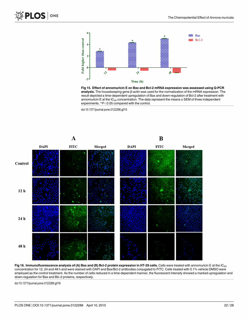
Fig 15. Effect of annomuricin E on Bax and Bcl-2 mRNA expression was assessed using Q-PCRanalysis. The housekeeping gene β-actin was used for the normalization of the mRNA expression. Theresult depicted a time-dependent upregulation of Bax and down-regulation of Bcl-2 after treatment withannomuricin E at the IC50 concentration. The data represent the means ± SEM of three independentexperiments. *P<0.05 compared with the control.
doi:10.1371/journal.pone.0122288.g015
Fig 16. Immunofluorescence analysis of (A) Bax and (B) Bcl-2 protein expression in HT-29 cells. Cells were treated with annomuricin E at the IC50
concentration for 12, 24 and 48 h and were stained with DAPI and Bax/Bcl-2 antibodies conjugated to FITC. Cells treated with 0.1% vehicle DMSOwereemployed as the control treatment. As the number of cells reduced in a time-dependent manner, the fluorescent intensity showed a marked upregulation anddown-regulation for Bax and Blc-2 proteins, respectively.
doi:10.1371/journal.pone.0122288.g016
The Chemopotential Effect of Annona muricata
PLOSONE | DOI:10.1371/journal.pone.0122288 April 10, 2015 22 / 28
ConclusionsOverall, we provided evidence that the ethyl acetate extract of A.muricata leaves has the poten-tial to suppress the AOM-induced development of ACF in rats. The data substantiated the tra-ditional use of A.muricata leaves against cancer and tumors. The suppressed formation ofACF in rats after EEAML oral administration was accompanied with down-regulation ofPCNA and Bcl-2 proteins and up-regulation of Bax protein in the colon tissue, indicating apossible mechanism at the molecular level. The reported pharmacological effect of the A.muri-cata leaves may be partially due to the presence of annomuricin E. This acetogenin suppressedthe proliferation of HT-29 cells selectively and induced apoptosis that was associated with G1
cell cycle arrest and mitochondria-mediated pathways.
Supporting InformationS1 Fig. 1H NMR (500 MHz) and 13C NMR (125 MHz) spectral data of annomuricin E inCDCl3 (δ in ppm, J in Hz).(DOCX)
Author ContributionsConceived and designed the experiments: SZMMAA HAK. Performed the experiments: SZMER HKMF. Analyzed the data: SZMMF. Contributed reagents/materials/analysis tools: SZM.Wrote the paper: SZM.
References1. Bird RP. Role of aberrant crypt foci in understanding the pathogenesis of colon cancer. Cancer Lett.
1995; 93: 55–71. PMID: 7600544
2. Bouvard V, Baan R, Straif K, Grosse Y, Secretan B, El Ghissassi F, et al. A review of human carcino-gens—part B: biological agents. Lancet Oncol. 2009; 10: 321–322. PMID: 19350698
Fig 17. Representative bar charts of Bax and Bcl-2 immunofluorescence analysis. Annomuricin E atIC50 concentration induced significant upregulation of Bax and down-regulation of Bcl-2 after 12 h. Cellstreated with 0.1% vehicle DMSOwere employed as the control treatment. The data represent themeans ± SEM of three independent experiments. *P<0.05 compared with the control.
doi:10.1371/journal.pone.0122288.g017
The Chemopotential Effect of Annona muricata
PLOSONE | DOI:10.1371/journal.pone.0122288 April 10, 2015 23 / 28

3. Kumar S, Weaver VM. Mechanics, malignancy, and metastasis: the force journey of a tumor cell. Can-cer Metast Rev. 2009; 28: 113–127.
4. Giovannucci E. Metabolic syndrome, hyperinsulinemia, and colon cancer: a review. Am J Clin Nutr.2007; 86: 836S–842S.
5. Ricci-Vitiani L, Lombardi DG, Pilozzi E, Biffoni M, Todaro M, Peschle C, et al. Identification and expan-sion of human colon-cancer-initiating cells. Nature. 2006; 445: 111–115. PMID: 17122771
6. Alrawi SJ, Schiff M, Carroll RE, Dayton M, Gibbs JF, Kulavlat M, et al. Aberrant crypt foci. AnticancerRes. 2006; 26: 107–119. PMID: 16475686
7. Fenoglio-Preiser CM, Noffsinger A. Review article: Aberrant crypt foci: A Review. Toxicol Pathol. 1999;27: 632–642. PMID: 10588543
8. Raju J. Azoxymethane-induced rat aberrant crypt foci: relevance in studying chemoprevention of coloncancer. World J Gastroentero. 2008; 14: 6632. PMID: 19034964
9. Fulda S. Modulation of apoptosis by natural products for cancer therapy. Planta Med. 2010; 76: 1075–1079. doi: 10.1055/s-0030-1249961 PMID: 20486070
10. Fulda S. Tumor resistance to apoptosis. Int J Cancer. 2009; 124: 511–515. doi: 10.1002/ijc.24064PMID: 19003982
11. Gutschner T, Diederichs S. The hallmarks of cancer. RNA Biol. 2012; 9: 703–719. doi: 10.4161/rna.20481 PMID: 22664915
12. Wong R. Apoptosis in cancer: from pathogenesis to treatment. J Exp Clin Canc Res. 2011; 30: 87. doi:10.1186/1756-9966-30-87 PMID: 21943236
13. Ocker M, Höpfner M. Apoptosis-modulating drugs for improved cancer therapy. Eur Surg Res. 2012;48: 111–120. doi: 10.1159/000336875 PMID: 22538523
14. Fan T-J, Han L-H, Cong R-S, Liang J. Caspase family proteases and apoptosis. Acta Bioch Bioph Sin.2005; 37: 719–727.
15. Zorofchian Moghadamtousi S, Karimian H, Khanabdali R, Razavi M, Firoozinia M, Keivan Z, et al. Anti-cancer and antitumor potential of fucoidan and fucoxanthin, two main metabolites isolated from brownalgae. Sci World J. 2014; 2014: doi: 10.1155/2014/768323
16. Mishra BB, Tiwari VK. Natural products: an evolving role in future drug discovery. Eur J Med Chem.2011; 46: 4769–4807. doi: 10.1016/j.ejmech.2011.07.057 PMID: 21889825
17. Taylor P, Colman L, Bajoon J. The search for plants with anticancer activity: Pitfalls at the early stages.J Ethnopharmacol. 2014; 158: 246–254. doi: 10.1016/j.jep.2014.10.034 PMID: 25446637
18. Thomson M, Ali M. Garlic [Allium sativum]: A Review of its Potential Use as an Anti-Cancer Agent. CurrCancer Drug Targets. 2003; 3: 67–81. PMID: 12570662
19. Bardi DA, Halabi MF, Hassandarvish P, Rouhollahi E, Paydar M, Moghadamtousi SZ, et al. Androgra-phis paniculata Leaf Extract Prevents Thioacetamide-Induced Liver Cirrhosis in Rats. Plos One. 2014;9: e109424. doi: 10.1371/journal.pone.0109424 PMID: 25280007
20. Gourineni V, Verghese M, Boateng J, Shackelford L, Bhat K. Chemopreventive potential of synergy1and soybean in reducing azoxymethane-induced aberrant crypt foci in fisher 344 male rats. J NutrMetab. 2011; 2011: doi: 10.1155/2011/983038
21. Shwter AN, Abdullah NA, Alshawsh MA, Alsalahi A, Hajrezaei M, Almaqrami AA, et al. Chemopreven-tion of colonic aberrant crypt foci byGynura procumbens in rats. J Ethnopharmacol. 2014; 151: 1194–1201. doi: 10.1016/j.jep.2013.12.044 PMID: 24393787
22. Shin HR, Kim JY, Yun TK, Morgan G, Vainio H. The cancer-preventive potential of Panax ginseng: a re-view of human and experimental evidence. Cancer Causes Control. 2000; 11: 565–576. PMID:10880039
23. Abdulaziz Bardi D, Halabi MF, Abdullah NA, Rouhollahi E, Hajrezaie M, Abdulla MA. In vivo evaluationof ethanolic extract of Zingiber officinale rhizomes for its protective effect against liver cirrhosis. BioMedRes Int. 2013; 2013: doi: 10.1155/2013/918460
24. George VC, Kumar DN, Rajkumar V, Suresh P, Ashok R. Quantitative assessment of the relative anti-neoplastic potential of the n-butanolic leaf extract of Annona Muricata Linn. in normal and immortalizedhuman cell lines. Asian Pac J Cancer Prev. 2012; 13: 699–704. PMID: 22524847
25. Moghadamtousi SZ, Rouhollahi E, Karimian H, Fadaeinasab M, Abdulla MA, Kadir HA. Gastroprotec-tive activity of Annona muricata leaves against ethanol-induced gastric injury in rats via Hsp70/Bax in-volvement. Drug Des Dev Ther. 2014; 8: 2099–2111. doi: 10.2147/DDDT.S70096 PMID: 25378912
26. Mishra S, Ahmad S, Kumar N, Sharma BK. Annona muricata (The cancer killer): A review. Glob JPharma Res. 2013; 2: 1613–1618.
The Chemopotential Effect of Annona muricata
PLOSONE | DOI:10.1371/journal.pone.0122288 April 10, 2015 24 / 28
27. Adewole S, Ojewole J. Protective effects of Annona muricata Linn. (Annonaceae) leaf aqueous extracton serum lipid profiles and oxidative stress in hepatocytes of streptozotocin-treated diabetic rats. Afr JTradit Complement Altern Med. 2009; 6: 30–41.
28. Moghadamtousi SZ, Karimian H, Rouhollahi E, Paydar M, Kadir HA, Fadaeinasab M, et al. Annonamuricata leaves induce G1 cell cycle arrest and apoptosis through mitochondria-mediated pathway inHuman HCT-116 and HT-29 colon Cancer cells. J Ethnopharmacol. 2014; 156: 277–289. doi: 10.1016/j.jep.2014.08.011 PMID: 25195082
29. Ezirim A, Okachi V, James A, Adebeshi O, Ogunnowo S, OdegheOB. Induction of apoptosis in myelog-enous leukemic K562 cells by ethanolic leaf extract of Annona muricata. Indian J Drugs Dis. 2013; 2:241–247.
30. Moghadamtousi SZ, Kadir HA, Paydar M, Rouhollahi E, Karimian H. Annona muricata leaves inducedapoptosis in A549 cells through mitochondrial-mediated pathway and involvement of NF-kappaB. BMCComplem Altern M. 2014; 14: 299.
31. Garber JC, Barbee RW, Bielitzki JT, Clayton LA, Donovan JC, Hendriksen CFM, et al. (Guide for thecare and use of laboratory animals. 2011; Washington, DC: The National Academies Press.
32. Almagrami AA, AlshawshMA, Saif-Ali R, Shwter A, Salem SD, Abdulla MA. (2014) Evaluation of Che-mopreventive Effects of Acanthus ilicifolius against Azoxymethane-Induced Aberrant Crypt Foci in theRat Colon. 2014; Plos One 9: e96004. doi: 10.1371/journal.pone.0096004 PMID: 24819728
33. Bird RP. Observation and quantification of aberrant crypts in the murine colon treated with a colon car-cinogen: preliminary findings. Cancer Lett. 1987; 37: 147–151. PMID: 3677050
34. Hajrezaie M, Hassandarvish P, Moghadamtousi SZ, Gwaram NS, Golbabapour S, NajiHussien A, et al.Chemopreventive Evaluation of a Schiff Base Derived Copper (II) Complex against Azoxymethane-In-duced Colorectal Cancer in Rats. 2014; Plos One 9: e91246. doi: 10.1371/journal.pone.0091246PMID: 24618844
35. Fraga CG, Leibovitz BE, Tappel AL. Lipid peroxidation measured as thiobarbituric acid-reactive sub-stances in tissue slices: characterization and comparison with homogenates and microsomes. FreeRadic Biol Med. 1988; 4: 155–161. PMID: 3356355
36. Mosmann T. Rapid colorimetric assay for cellular growth and survival: application to proliferation andcytotoxicity assays. J Immunol Methods. 1983; 65: 55–63. PMID: 6606682
37. Karimian H, Mohan S, Moghadamtousi SZ, Fadaeinasab M, Razavi M, Arya A, et al. Tanacetum poly-cephalum (L.) Schultz-Bip. Induces Mitochondrial-Mediated Apoptosis and Inhibits Migration and Inva-sion in MCF7 Cells. Molecules. 2014; 19: 9478–9501. doi: 10.3390/molecules19079478 PMID:24995928
38. Karimian H, Moghadamtousi Zorofchian S, Fadaeinasab M, Golbabapour S, Razavi M, Hajrezaie M,et al. Ferulago angulata activates intrinsic pathway of apoptosis in MCF-7 cells associated with G1 cellcycle arrest via involvement of p21/p27. 2014; Drug Des Dev Ther. 2014; 8: 1481–1497. doi: 10.2147/DDDT.S68818 PMID: 25278746
39. Hajrezaie M, Paydar M, Zorofchian Moghadamtousi S, Hassandarvish P, Gwaram NS, Zahedifard M,et al. A Schiff Base-Derived Copper (II) Complex Is a Potent Inducer of Apoptosis in Colon CancerCells by Activating the Intrinsic Pathway. Sci World J. 2014; 2014: doi: 10.1155/2014/540463
40. Lövborg H, Nygren P, Larsson R. Multiparametric evaluation of apoptosis: effects of standard cytotoxicagents and the cyanoguanidine CHS 828. Mol Cancer Ther. 2004; 3: 521–526. PMID: 15141009
41. Liew SY, Looi CY, Paydar M, Cheah FK, Leong KH, WongWF, et al. Subditine, a new monoterpenoidindole Alkaloid from bark of Nauclea subdita (Korth.) Steud. Induces apoptosis in human prostate can-cer cells. 2014; Plos One 9: e87286. doi: 10.1371/journal.pone.0087286 PMID: 24551054
42. Guo Y-W, Chen Y-H, Chiu W-C, Liao H, Lin S-H. Soy Saponins Meditate the Progression of Colon Can-cer in Rats by Inhibiting the Activity of β-Glucuronidase and the Number of Aberrant Crypt Foci but NotCyclooxygenase-2 Activity. ISRN Oncol. 2013; 2013: doi: 10.1155/2013/645817
43. Eggadi V, Gundamedi S, Sheshagiri SBB, Revoori SK, Jupally VR, Kulandaivelu U. Evaluation of Anti-cancer Activity of Annona muricata in 1, 2-Dimethyl Hydrazine Induced Colon Cancer. World Appl SciJ. 2014; 32: 444–450.
44. Minari J, Okeke U. Chemopreventive effect of Annona muricata on DMBA-induced cell proliferation inthe breast tissues of female albino mice. Egypt J Med HumGenet. 2014; 15: 327–334.
45. Mayer A, Takimoto M, Fritz E, Schellander G, Kofler K, Ludwig H. The prognostic significance of prolif-erating cell nuclear antigen, epidermal growth factor receptor, and mdr gene expression in colorectalcancer. Cancer. 1993; 71: 2454–2460. PMID: 8095852
46. Maga G, Hübscher U. Proliferating cell nuclear antigen (PCNA): a dancer with many partners. J CellSci. 2013; 116: 3051–3060.
The Chemopotential Effect of Annona muricata
PLOSONE | DOI:10.1371/journal.pone.0122288 April 10, 2015 25 / 28

47. Isozaki H, Okajima K, Ichinona T, Tanimura M, Morita S, Takada Y, et al. The significance of proliferat-ing cell nuclear antigen (PCNA) expression in cancer of the ampulla of Vater in terms of prognosis.Surg Today. 1994; 24: 494–499. PMID: 7919730
48. Naryzhny SN, Lee H. Characterization of proliferating cell nuclear antigen (PCNA) isoforms in normaland cancer cells: there is no cancer-associated form of PCNA. FEBS Lett. 2007; 581: 4917–4920.PMID: 17900571
49. Lipkin M, Blattner WE, Fraumeni JF, Lynch HT, Deschner E, Winawer S. Tritiated thymidine (φp, φh) la-beling distribution as a marker for hereditary predisposition to colon cancer. Cancer Res. 1983; 43:1899–1904. PMID: 6831425
50. De Leon MP, Roncucci L, Di Donato P, Tassi L, Smerieri O, Amorico MG, et al. Pattern of epithelial cellproliferation in colorectal mucosa of normal subjects and of patients with adenomatous polyps or can-cer of the large bowel. Cancer Res. 1988; 48: 4121–4126. PMID: 3383201
51. Deschner EE, Long FC, Hakissian M, Herrmann SL. Differential susceptibility of AKR, C57BL/6J, andCF1 mice to 1, 2-dimethylhydrazine-induced colonic tumor formation predicted by proliferative charac-teristics of colonic epithelial cells. J Natl Cancer Inst. 1983; 70: 279–282. PMID: 6571937
52. Velmurugan B, Singh RP, Agarwal R, Agarwal C. Dietary-feeding of grape seed extract prevents azoxy-methane-induced colonic aberrant crypt foci formation in fischer 344 rats. Mol Carcinogen. 2010; 49:641–652.
53. Cory S, Adams JM. The Bcl2 family: regulators of the cellular life-or-death switch. Nat Rev Cancer.2002; 2: 647–656. PMID: 12209154
54. Elmore S. Apoptosis: a review of programmed cell death. Toxicol Pathol. 2007; 35: 495–516. PMID:17562483
55. Green DR, Kroemer G. The pathophysiology of mitochondrial cell death. Science. 2004; 305: 626–629. PMID: 15286356
56. Sturm I, Petrowsky H, Volz R, Lorenz M, Radetzki S, Hillebrand T, et al. Analysis of p53/BAX/p16ink4a/CDKN2 in esophageal squamous cell carcinoma: High BAX and p16ink4a/CDKN2 identifies patientswith good prognosis. J Clin Oncol. 2001; 19: 2272–2281. PMID: 11304781
57. Ionov Y, Yamamoto H, Krajewski S, Reed JC, Perucho M. Mutational inactivation of the proapoptoticgene BAX confers selective advantage during tumor clonal evolution. Proc Natl Acad Sci. 2000; 97:10872–10877. PMID: 10984511
58. Nirmala P, Ramanathan M. Effect of kaempferol on lipid peroxidation and antioxidant status in 1, 2-di-methyl hydrazine induced colorectal carcinoma in rats. Eur J Pharmacol. 2011; 654: 75–79. doi: 10.1016/j.ejphar.2010.11.034 PMID: 21172346
59. Liochev SI. Reactive oxygen species and the free radical theory of aging. Free Radic Biol Med. 2011;60: 1–4.
60. ChenW,Weng Y-M, Tseng C-Y. Antioxidative and antimutagenic activities of healthy herbal drinksfrom Chinese medicinal herbs. Am J Chin Med. 2003; 31: 523–532. PMID: 14587875
61. Naziroglu M. Molecular role of catalase on oxidative stress-induced Ca2+ signaling and TRP cationchannel activation in nervous system. J Recept Sig Transd. 2012; 32: 134–141. doi: 10.3109/10799893.2012.672994 PMID: 22475023
62. Blokhina O, Virolainen E, Fagerstedt KV. Antioxidants, oxidative damage and oxygen deprivationstress: a review. Ann Bot. 2003; 91: 179–194. PMID: 12509339
63. Meguid NA, Dardir AA, Abdel-Raouf ER, Hashish A. Evaluation of oxidative stress in autism: defectiveantioxidant enzymes and increased lipid peroxidation. Biol Trace Elem Res. 2011; 143: 58–65. doi: 10.1007/s12011-010-8840-9 PMID: 20845086
64. George VC, Kumar DN, Suresh P, Kumar RA (2014) Antioxidant, DNA protective efficacy and HPLCanalysis of Annona muricata (soursop) extracts. J Food Sci Technol. 2014; 1–8.
65. Baskar R, Rajeswari V, Kumar TS. In vitro antioxidant studies in leaves of Annona species. Indian JExp Biol. 2007; 45: 480–485. PMID: 17569293
66. Adewole SO, Caxton-Martins EA. Morphological changes and hypoglycemic effects of Annona muri-cata linn. (annonaceae) leaf aqueous extract on pancreatic β-cells of streptozotocin-treated diabeticrats. Afr J Biomed Res. 2006; 9: 173–180.
67. Foong CP, Hamid RA. Evaluation of anti-inflammatory activities of ethanolic extract of Annona muricataleaves. Rev Bras Farmacogn. 2012; 22: 1301–1307.
68. Lissoni P, Barni S, Mandala M, Ardizzoia A, Paolorossi F, Vaghi M, et al. Decreased toxicity and in-creased efficacy of cancer chemotherapy using the pineal hormone melatonin in metastatic solid tu-mour patients with poor clinical status. Eur J Cancer. 1999; 35: 1688–1692. PMID: 10674014
The Chemopotential Effect of Annona muricata
PLOSONE | DOI:10.1371/journal.pone.0122288 April 10, 2015 26 / 28
69. Cerea G, Vaghi M, Ardizzoia A, Villa S, Bucovec R, Mengo S, et al. Biomodulation of cancer chemo-therapy for metastatic colorectal cancer: a randomized study of weekly low-dose irinotecan alone ver-sus irinotecan plus the oncostatic pineal hormone melatonin in metastatic colorectal cancer patientsprogressing on 5-fluorouracil-containing combinations. Anticancer Res. 2002; 23: 1951–1954.
70. Vaca C, Wilhelm J, Harms-Ringdahl M. Interaction of lipid peroxidation products with DNA. A review.Mutat Res-Rev Mutat. 1988; 195: 137–149.
71. Pandurangan AK, Dharmalingam P, Anandasadagopan S, Ganapasam S (2012) Effect of luteolin onthe levels of glycoproteins during azoxymethane-induced colon carcinogenesis in mice. Asian Pac JCancer Prev. 2012; 13: 1569–1573. PMID: 22799368
72. Demircan B, Çelik G, Süleyman H, Akçay F. Effects of indomethacin, celecoxib and meloxicam on glu-tathione, malondialdehyde and myeloperoxidase in rat gastric tissue. Pain Clinic. 2005; 17: 383–388.
73. Komiya M, Fujii G, Takahashi M, Iigo M, Mutoh M. Prevention and intervention trials for colorectal can-cer. Jpn J Clin Oncol. 2013; 43: 685–694. doi: 10.1093/jjco/hyt053 PMID: 23613189
74. Hendrickse C, Kelly R, Radley S, Donovan I, Keighley M, Neoptolemos JP. Lipid peroxidation and pros-taglandins in colorectal cancer. Br J Surg. 1994; 81: 1219–1223. PMID: 7953368
75. Skrzydlewska E, Sulkowski S, Koda M, Zalewski B, Kanczuga-Koda L, Sulkowska M. Lipid peroxida-tion and antioxidant status in colorectal cancer. World J Gastroenterol. 2005; 11: 403–406. PMID:15637754
76. Kim G-s, Zeng L, Alali F, Rogers LL, Wu F-E, McLaughlin JL, et al. Two newmono-tetrahydrofuran ringacetogenins, annomuricin E and muricapentocin, from the leaves of Annona muricata. J Nat Prod.1998; 61: 432–436. PMID: 9584396
77. Rupprecht JK, Hui Y-H, McLaughlin JL. Annonaceous acetogenins: a review. J Nat Prod. 1990; 53:237–278. PMID: 2199608
78. Liaw C-C, Wu T-Y, Chang F-R, Wu Y-C. Historic perspectives on Annonaceous acetogenins from thechemical bench to preclinical trials. Planta Med. 2010; 76: 1390–1404. doi: 10.1055/s-0030-1250006PMID: 20577943
79. Bermejo A, Figadère B, Zafra-Polo M-C, Barrachina I, Estornell E, Cortes D. Acetogenins from Annona-ceae: recent progress in isolation, synthesis and mechanisms of action. Nat Prod Rep. 2005; 22: 269–303. PMID: 15806200
80. Alali FQ, Liu X-X, McLaughlin JL. Annonaceous acetogenins: recent progress. J Nat Prod. 1999; 62:504–540. PMID: 10096871
81. Nakanishi Y, Chang F-R, Liaw C-C, Wu Y-C, Bastow KF, Lee K-H. Acetogenins as Selective Inhibitorsof the Human Ovarian 1A9 Tumor Cell Line 1. J Med Chem. 2003; 46: 3185–3188. PMID: 12852747
82. James Morré D, de Cabo R, Farley C, Oberlies NH, McLaughlin JL. Mode of action of bullatacin, a po-tent antitumor acetogenin: inhibition of NADH oxidase activity of HeLa and HL-60, but not liver, plasmamembranes. Life Sci. 1994; 56: 343–348.
83. Lewis MA, Arnason J, Philogene B, Rupprecht J, McLaughlin J. Inhibition of Respiration at Site I by Asi-micin, an Insecticidal Acetogenin of the Pawpaw, Asimina triloba(Annonaceae). Pestic Biochem Phys.1993; 45: 15–23.
84. Oberlies NH, Chang C-j, McLaughlin JL. Structure-activity relationships of diverse Annonaceous aceto-genins against multidrug resistant human mammary adenocarcinoma (MCF-7/Adr) cells. J Med Chem.1997; 40: 2102–2106. PMID: 9207950
85. Oberlies NH, Croy VL, Harrison ML, McLaughlin JL. The Annonaceous acetogenin bullatacin is cyto-toxic against multidrug-resistant human mammary adenocarcinoma cells. Cancer Lett. 1997. 115: 73–79. PMID: 9097981
86. Abe K, Matsuki N. Measurement of cellular 3-(4, 5-dimethylthiazol-2-yl)-2, 5-diphenyltetrazolium bro-mide (MTT) reduction activity and lactate dehydrogenase release using MTT. Neurosci Res. 2000; 38:325–329. PMID: 11164558
87. Park M-T, Lee S-J. Cell cycle and cancer. J BiochemMol Biol. 2003; 36: 60–65. PMID: 12542976
88. Wang Z-Y, Wang D-M, Loo TY, Cheng Y, Chen L-L, Shen J-G, et al. Spatholobus suberectus inhibitscancer cell growth by inducing apoptosis and arresting cell cycle at G2/M checkpoint. J Ethnopharma-col. 2011; 133: 751–758. doi: 10.1016/j.jep.2010.11.004 PMID: 21073941
89. Yuan S-SF, Chang H-L, Chen H-W, Yeh Y-T, Kao Y-H, Lin K-H, et al. Annonacin, a mono-tetrahydrofu-ran acetogenin, arrests cancer cells at the G1 phase and causes cytotoxicity in a Bax-and caspase-3-related pathway. Life Sci. 2003; 72: 2853–2861. PMID: 12697268
90. Wu Y, Tibrewal N, Birge RB. Phosphatidylserine recognition by phagocytes: a view to a kill. Trends CellBiol. 2006; 16: 189–197. PMID: 16529932
The Chemopotential Effect of Annona muricata
PLOSONE | DOI:10.1371/journal.pone.0122288 April 10, 2015 27 / 28

91. Brumatti G, Sheridan C, Martin SJ. Expression and purification of recombinant annexin V for the detec-tion of membrane alterations on apoptotic cells. Methods. 2008; 44: 235–240. doi: 10.1016/j.ymeth.2007.11.010 PMID: 18314054
92. Lakhani SA, Masud A, Kuida K, Porter GA, Booth CJ, Mehal WZ, et al. Caspases 3 and 7: key media-tors of mitochondrial events of apoptosis. Science. 2006; 311: 847–851. PMID: 16469926
93. Martinou J-C, Youle RJ. Mitochondria in Apoptosis: Bcl-2 Family Members and Mitochondrial Dynam-ics. Dev Cell. 2011; 21: 92–101. doi: 10.1016/j.devcel.2011.06.017 PMID: 21763611
The Chemopotential Effect of Annona muricata
PLOSONE | DOI:10.1371/journal.pone.0122288 April 10, 2015 28 / 28