Embed Size (px)
Citation preview
Mayo Clinic & British Cardiovascular Society
at the Royal College of Physicians, London : 21 - 23-October 2013
Cases-Controversies-Updates 2013
The Chest X-ray for Cardiologists
Michael Rubens
Royal Brompton Hospital
London, UK
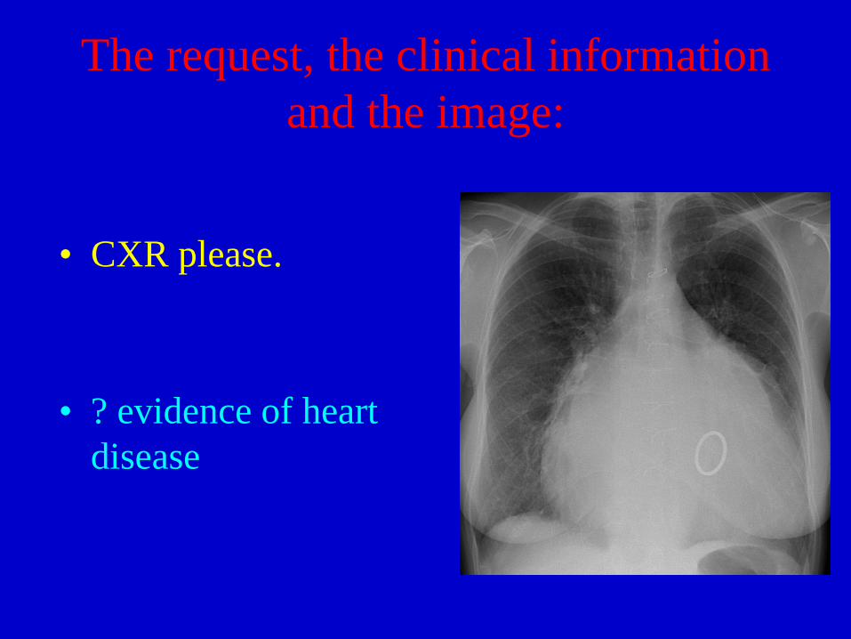
The request, the clinical information
and the image:
• CXR please.
• ? evidence of heart
disease
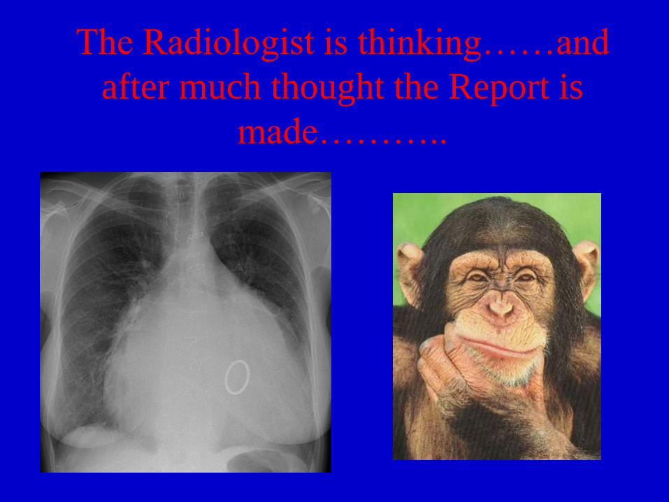
The Radiologist is thinking……and
after much thought the Report is
made………..
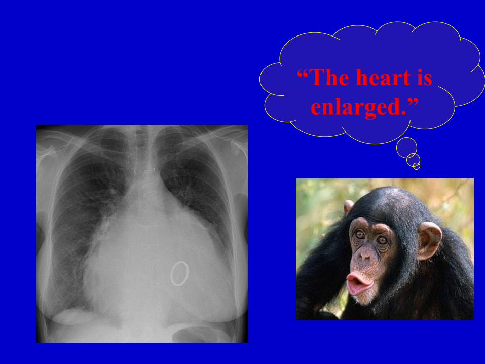
“The heart is
enlarged.”
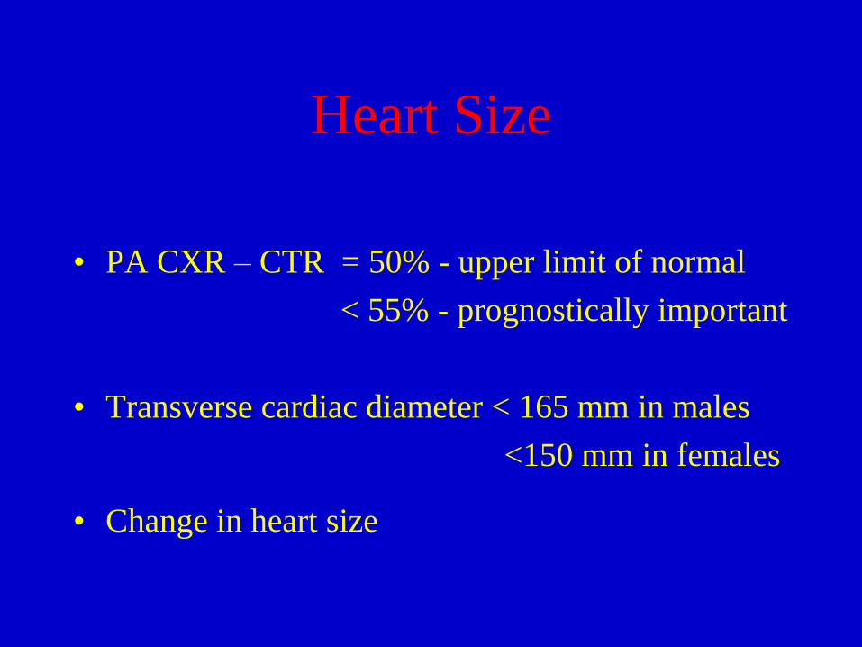
Heart Size
• PA CXR – CTR = 50% - upper limit of normal
< 55% - prognostically important
• Transverse cardiac diameter < 165 mm in males
<150 mm in females
• Change in heart size

Ebstein’s Anomaly
CTR = 49%

Ebstein’s Anomaly – Enlarging heart
CTR = 43% CTR = 49%
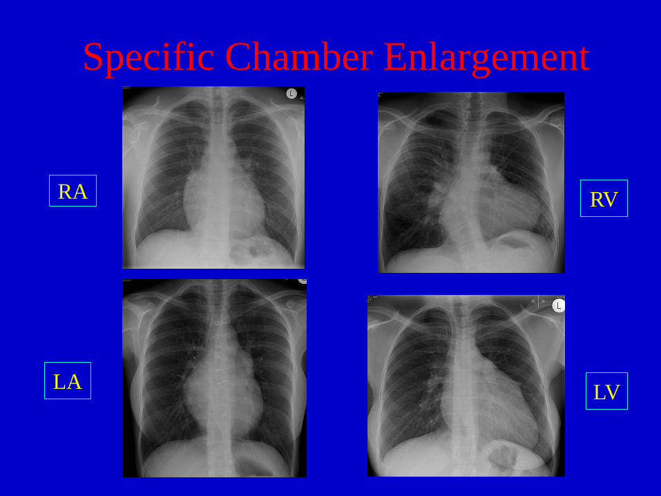
Specific Chamber Enlargement
RA
LA
RV
LV

Heart Enlarged – Why?
APPROACHES TO THE CXR
• Inspirational (a.k.a. the Aunt Minnie) approach
• Organised (a.k.a. the Larry Elliott*) approach
*“The X-ray Diagnosis of Congenital Heart Disease in Infants,
Children and Adults – pathologic, hemodynamic and clinical correlations as related to the chest film” by Larry P Elliott & Gerold L Schiebler. 2nd Edition. 1968

AN ORGANISEDAPPROACH TO
THE CXR
• Technical analysis
• Extra-cardiac analysis
• Physiological analysis
• Anatomic analysis

TECHNICAL ANALYSIS
• Image quality
• Depth of inspiration
• Alignment

EXTRA-CARDIAC ANALYSIS
• Bones & Soft tissues
• Abdomen
• Aortic arch
• Pulmonary trunk
• Azygous arch
BONES & SOFT TISSUES
• Evidence of surgery
• Evidence of inherited syndromes which
may be associated with heart disease
• Skeletal abnormalities which may have an
association with heart disease
• Skeletal abnormalities which may be caused
by cardiovascular disease

THORACOTOMY • Non-cardiac operation (pulmonary/oesophageal)
• Anastomotic shunt e.g. Blalock-Taussig (L/R), Glenn (R), Waterston (R), Pott’s (L)
• Closure of PDA (L)
• Repair of aortic coarctation (L)
• Pulmonary artery banding (L)
• Mitral valvotomy
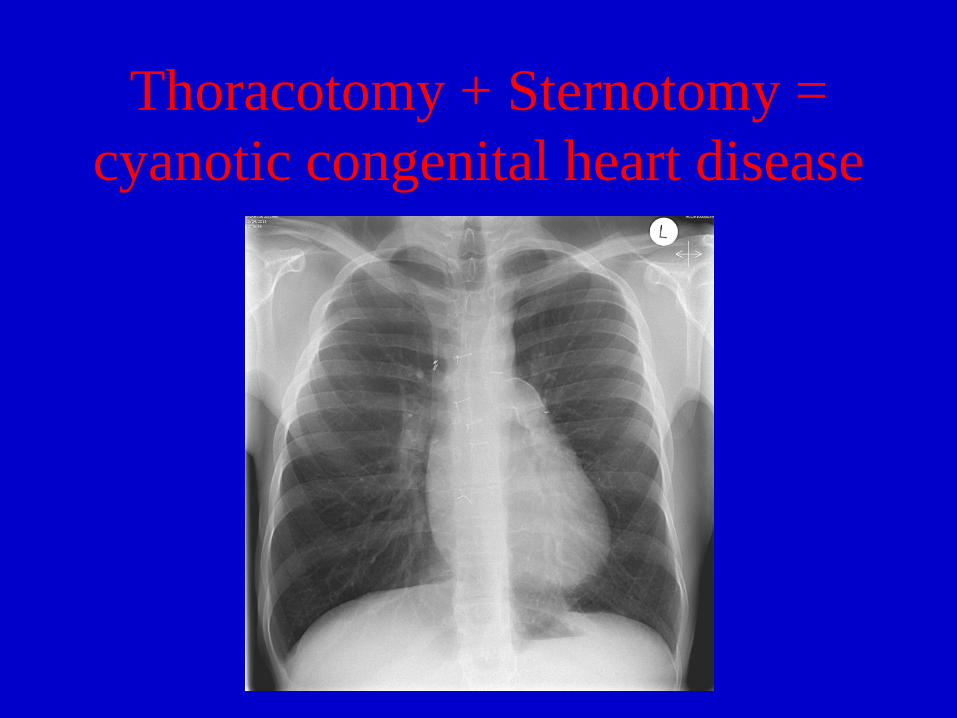
Thoracotomy + Sternotomy =
cyanotic congenital heart disease
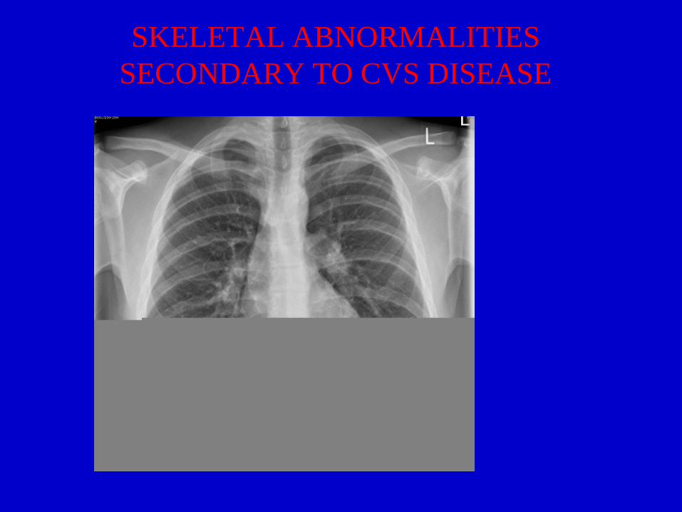
SKELETAL ABNORMALITIES
SECONDARY TO CVS DISEASE
ABDOMEN
• Liver
• Spleen
• Stomach
• Hiatus hernia
• Gallstones

Situs (1) - Dextrocardia
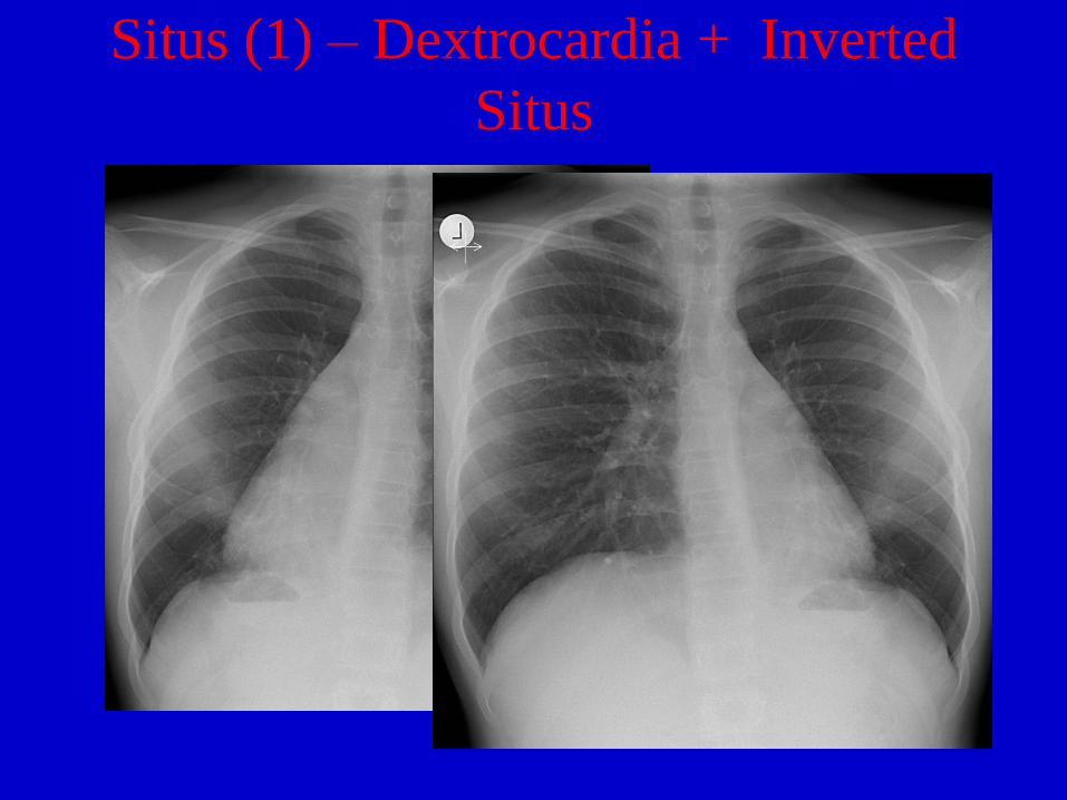
Situs (1) – Dextrocardia + Inverted
Situs
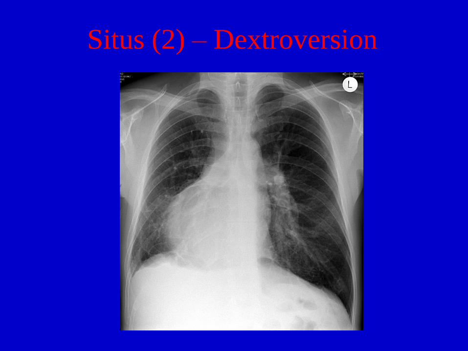
Situs (2) – Dextroversion
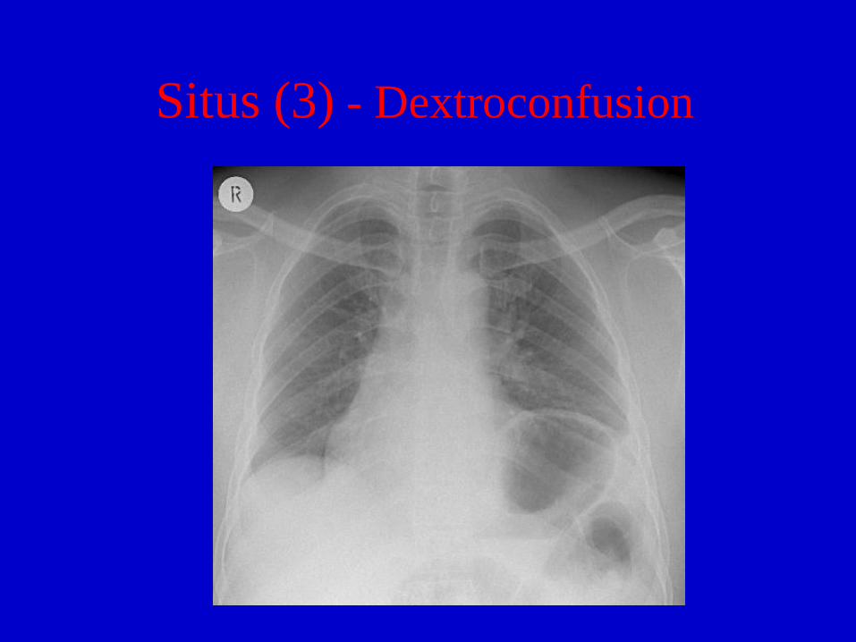
Situs (3) - Dextroconfusion
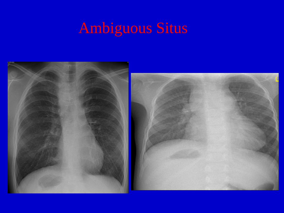
Ambiguous Situs

SUPERIOR MEDIASTINUM
• Azygous arch
• Aortic arch
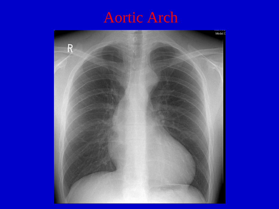
Aortic Arch
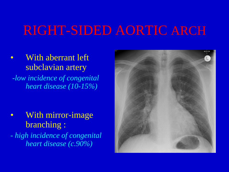
RIGHT-SIDED AORTIC ARCH
• With aberrant left subclavian artery
-low incidence of congenital heart disease (10-15%)
• With mirror-image branching :
- high incidence of congenital heart disease (c.90%)
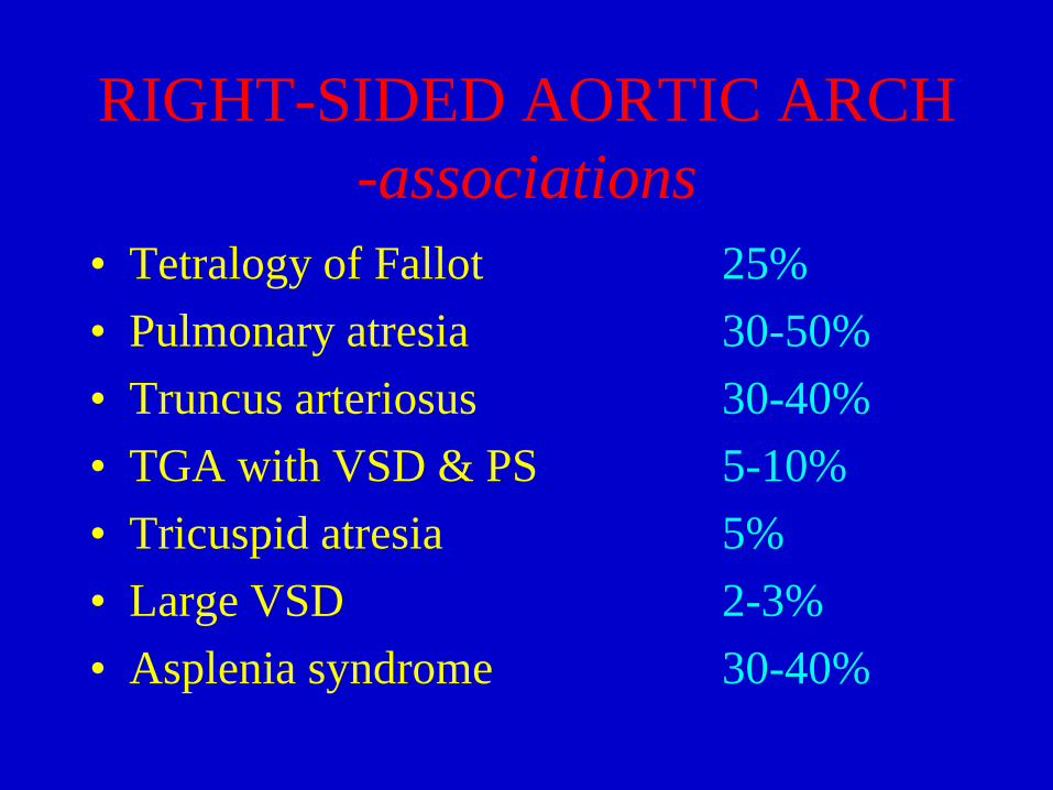
RIGHT-SIDED AORTIC ARCH
-associations
• Tetralogy of Fallot 25%
• Pulmonary atresia 30-50%
• Truncus arteriosus 30-40%
• TGA with VSD & PS 5-10%
• Tricuspid atresia 5%
• Large VSD 2-3%
• Asplenia syndrome 30-40%
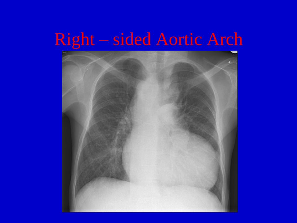
Right – sided Aortic Arch
CXR – PHYSIOLOGY
PULMONARY VASCULAR PATTERNS
1. Normal
2. Increased
i) PVH
ii) PAH
iii) pulmonary plethora
iv) systemic supply to lungs
3. Decreased
4. Uneven
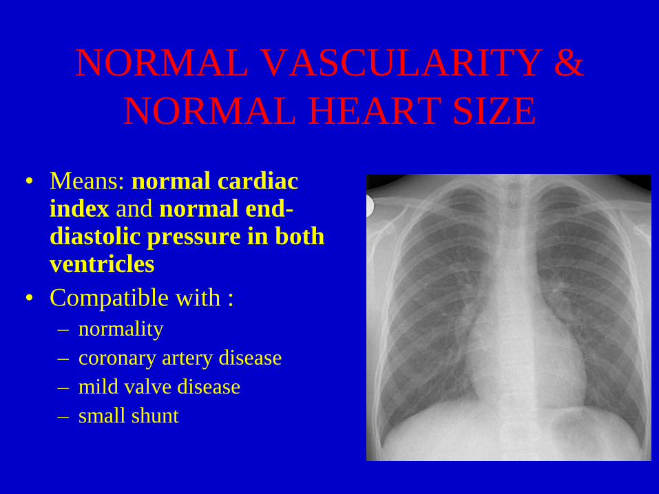
NORMAL VASCULARITY &
NORMAL HEART SIZE
• Means: normal cardiac index and normal end-diastolic pressure in both ventricles
• Compatible with :
– normality
– coronary artery disease
– mild valve disease
– small shunt
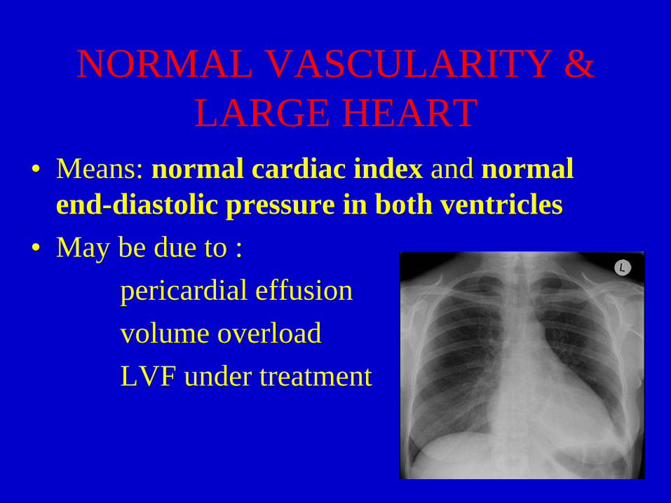
NORMAL VASCULARITY &
LARGE HEART
• Means: normal cardiac index and normal
end-diastolic pressure in both ventricles
• May be due to :
pericardial effusion
volume overload
LVF under treatment
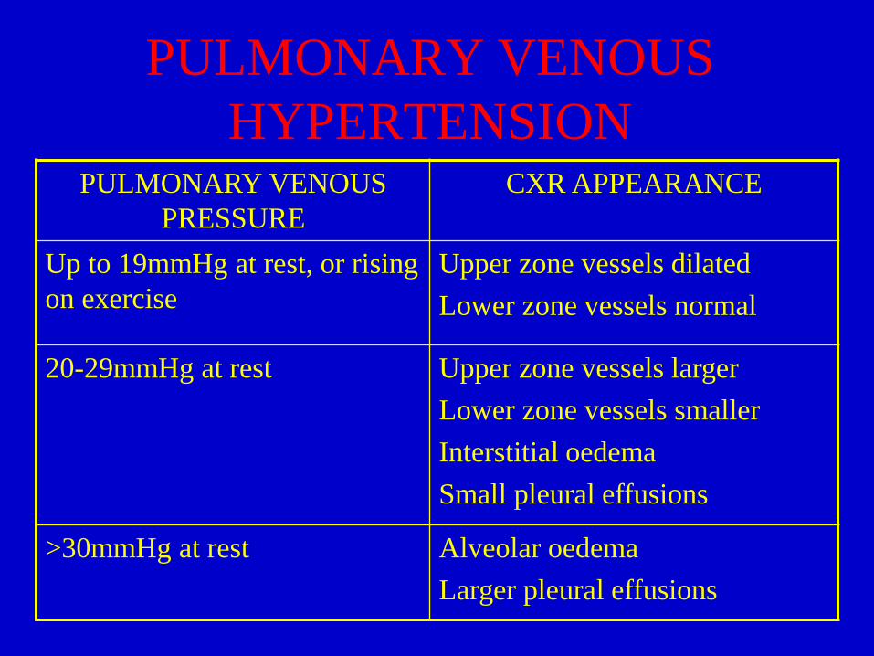
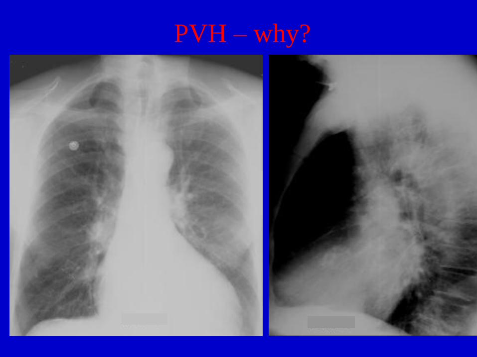
PULMONARY VENOUS
HYPERTENSION PULMONARY VENOUS
PRESSURE
CXR APPEARANCE
Up to 19mmHg at rest, or rising
on exercise
Upper zone vessels dilated
Lower zone vessels normal
20-29mmHg at rest Upper zone vessels larger
Lower zone vessels smaller
Interstitial oedema
Small pleural effusions
>30mmHg at rest Alveolar oedema
Larger pleural effusions
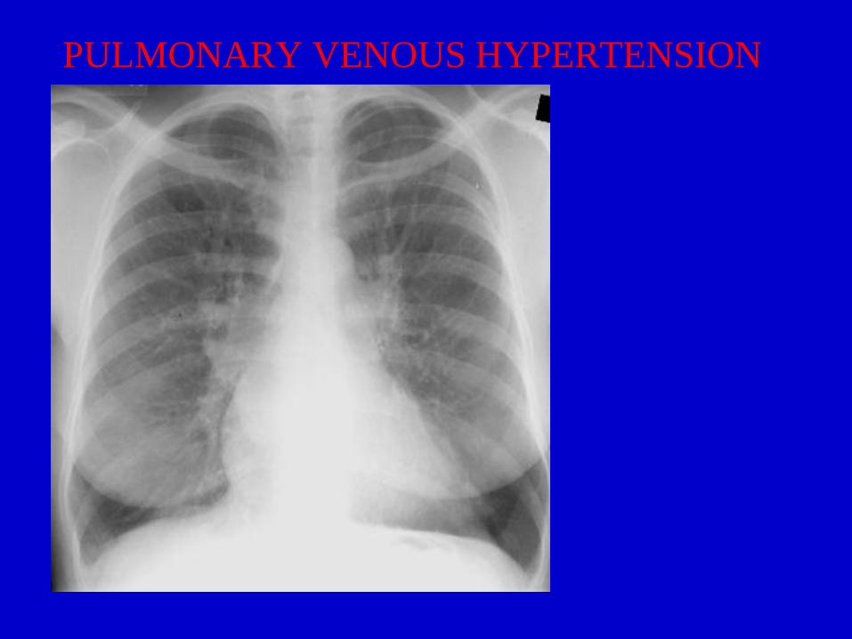
PULMONARY VENOUS HYPERTENSION
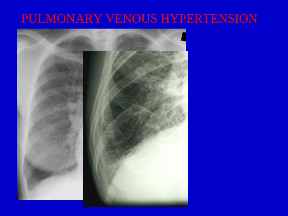
PULMONARY VENOUS HYPERTENSION

PULMONARY VENOUS HYPERTENSION
PULMONARY VENOUS
HYPERTENSION
= post-capillary problem
May be due to :
• obstruction at mitral valve level
• obstruction proximal to the mitral valve
• LV failure from any cause
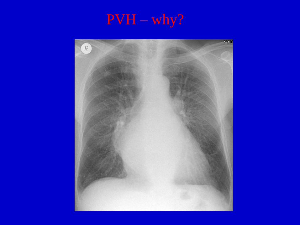
PVH – why?
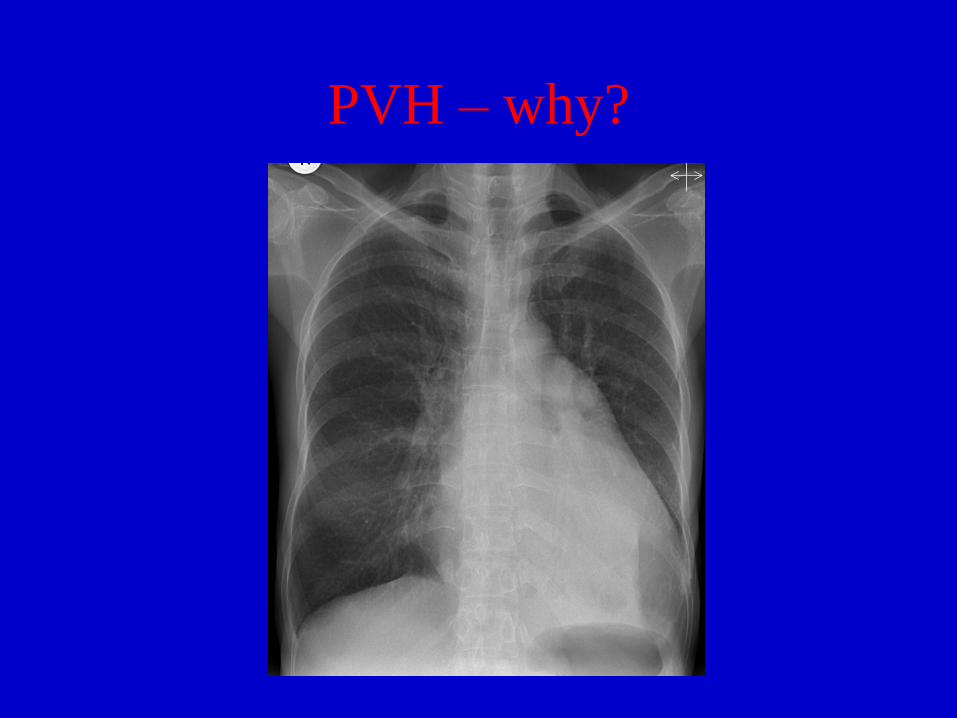
PVH – why?

PVH – why?
PVH – why?

PULMONARY ARTERIAL
HYPERTENSION
Systolic PA pressure > 30 mm.Hg
CXR Signs :
1. Dilatation of the central
(elastic) pulmonary arteries
2. Narrowing of the peripheral
(muscular) pulmonary arteries
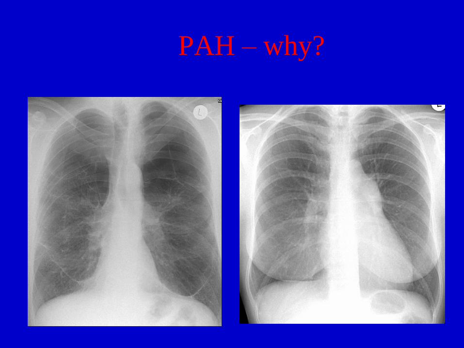
PAH – why?
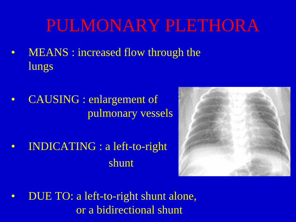
PULMONARY PLETHORA
• MEANS : increased flow through the
lungs
• CAUSING : enlargement of
pulmonary vessels
• INDICATING : a left-to-right
shunt
• DUE TO: a left-to-right shunt alone,
or a bidirectional shunt
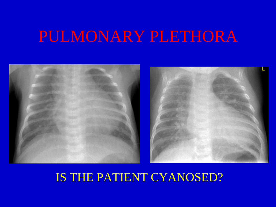
PULMONARY PLETHORA
IS THE PATIENT CYANOSED?
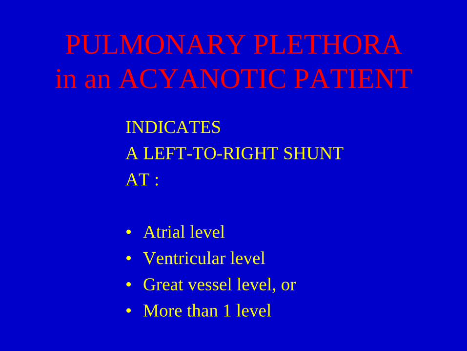
PULMONARY PLETHORA
in an ACYANOTIC PATIENT
INDICATES
A LEFT-TO-RIGHT SHUNT
AT :
• Atrial level
• Ventricular level
• Great vessel level, or
• More than 1 level
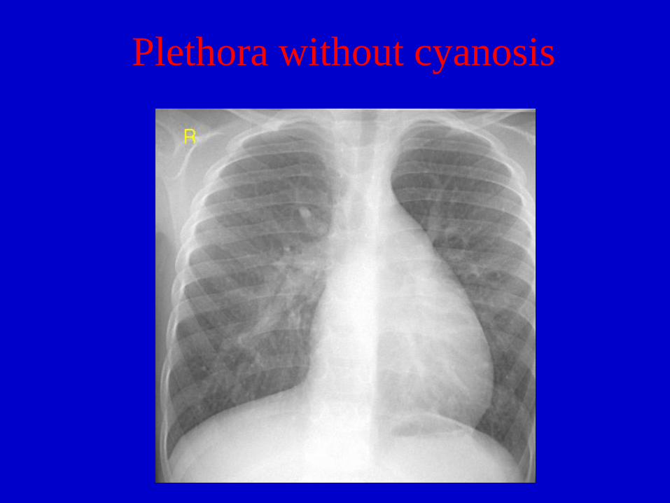
Plethora without cyanosis

Plethora without cyanosis
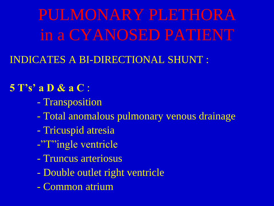
PULMONARY PLETHORA
in a CYANOSED PATIENT
INDICATES A BI-DIRECTIONAL SHUNT :
5 T’s’ a D & a C :
- Transposition
- Total anomalous pulmonary venous drainage
- Tricuspid atresia
-”T”ingle ventricle
- Truncus arteriosus
- Double outlet right ventricle
- Common atrium
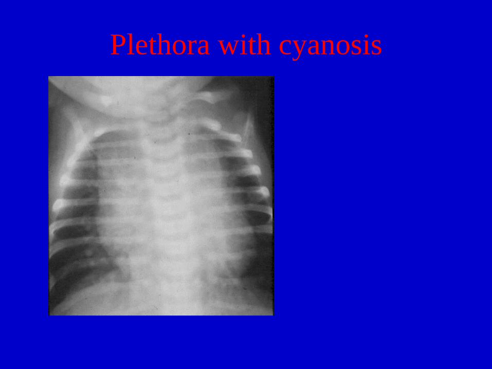
Plethora with cyanosis
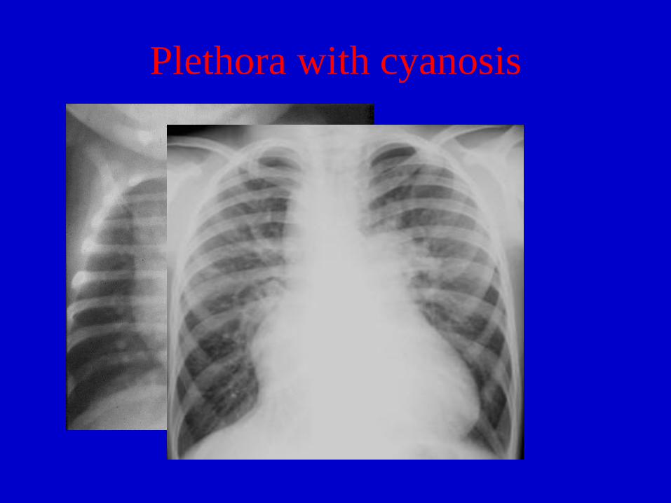
Plethora with cyanosis
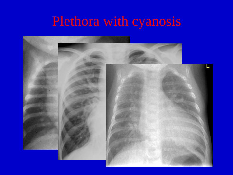
Plethora with cyanosis
PULMONARY OLIGAEMIA
Causes
• RV outflow obstruction with right-to-left shunt (at atrial* or ventricular** level)
• RV outflow obstruction without a shunt
• RV inflow obstruction
• RV failure
* heart often enlarged, sometimes huge
** heart size usually normal
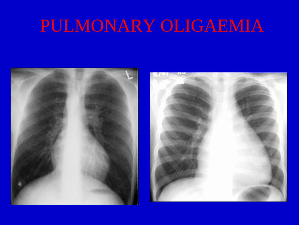
PULMONARY OLIGAEMIA
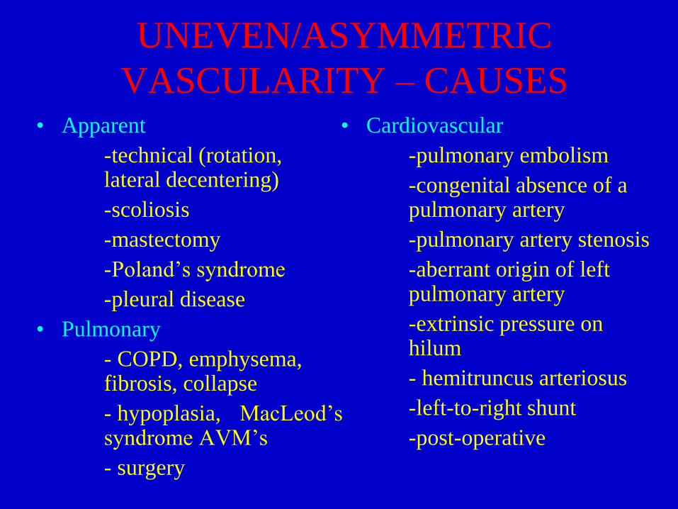
UNEVEN/ASYMMETRIC
VASCULARITY – CAUSES • Apparent
-technical (rotation, lateral decentering)
-scoliosis
-mastectomy
-Poland’s syndrome
-pleural disease
• Pulmonary
- COPD, emphysema, fibrosis, collapse
- hypoplasia, MacLeod’s syndrome AVM’s
- surgery
• Cardiovascular
-pulmonary embolism
-congenital absence of a pulmonary artery
-pulmonary artery stenosis
-aberrant origin of left pulmonary artery
-extrinsic pressure on hilum
- hemitruncus arteriosus
-left-to-right shunt
-post-operative

Uneven Vascularity
How I report the CXR
1. Is it a good quality radiograph?
2. Anything noteworthy in the bones, soft tissues, diaphragm or upper abdomen?
3. Anything noteworthy in the mediastinum and hila? (i.e.aorta, central PA’s and azygous v.)
4. What is the vascular pattern?
5. Does the cardiac shadow tell me anything else?
6. Cardiothoracic ratio.
7. Any change from previous CXR.

SUMMARY
• Use a disciplined approach
• The pulmonary vascular pattern is the key
to diagnosis
• If physiology & anatomy seem not to
correlate then go with the physiology