Embed Size (px)
Citation preview
THE CHICK CHORIOALLANTOICMEMBRANE AS A MODEL TISSUEFOR SURGICAL RETINAL RESEARCHAND SIMULATIONTHEODORE LENG, MS, JASON M. MILLER, BS,KALAYAAN V. BILBAO, MD, DANIEL V. PALANKER, PHD,*PHILIP HUIE, MA, MARK S. BLUMENKRANZ, MD
Purpose: We describe the use of chick chorioallantoic membrane (CAM) as a modelsystem for the study of the precision and safety of vitreoretinal microsurgical instrumentsand techniques.
Methods: The CAM was prepared for experimentation with and without its inner shellmembrane (ISM) attached for in vivo and in vitro experiments that simulated medical andsurgical interventions on the retina.
Results: The CAM’s ease of use, low cost, and anatomic structure make it a convenientmodel for surgical retinal and retinal vascular modeling.
Conclusion: While CAM has been used extensively in the past for ocular angiogenesisstudies, we describe the tissue as a useful tool for a variety of other applications, including(1) testing of novel surgical tools and techniques for cutting and coagulating retina and itsvasculature, (2) testing vessel cannulation and injection techniques, (3) angiographicstudies, and (4) endoscopic surgery.
RETINA 24:427–434, 2004
Cancer biologists, developmental biologists, andophthalmologists have described the chick cho-
rioallantoic membrane (CAM) as a model system forstudying development,1 cancer behavior,2–3 propertiesof biomaterials,4 angiogenesis,5–10 and photodynamictherapy.11 We propose several new applications forCAM in the study of retina and its vasculature withrespect to microsurgical interventions. Herein we de-
scribe the anatomic features of the CAM and some ofthe types of microsurgical interventions that can betested on it.
The chick CAM, a part of the extraembryonic tis-sue, begins to develop 7 days after initial incubationfrom the fusion of the chorion and the allantois.12
Structurally, the outer epithelial layer of the chorion isderived from the trophoblast, which opposes the al-lantois. This structure forms a supportive matrix forthe extensive vascular network that courses throughthe CAM, analogous to the retina and its vasculature(Figure 1, inset). Overall, mature chick CAM (20–100�m) and human retina (approximately 100–300 �m)are of roughly comparable thickness.13 Mature chickCAM (incubation day 12 and on) can be divided intothree anatomically distinct layers: (1) primary stratum;(2) capillary plexus, or blood sinus; and (3) a thin
From the Department of Ophthalmology, Stanford UniversitySchool of Medicine, and the *Hansen Experimental Physics Lab,Stanford University, Stanford, California.
This research was funded in part by the National Institutes ofHealth and by Carl Zeiss, Inc. This article has never been presentedat a meeting.
Reprint requests: Mark S. Blumenkranz, MD, Chair, Departmentof Ophthalmology, Stanford University School of Medicine, 300Pasteur Drive, Room A157, Stanford, CA 94305; e-mail:[email protected]
427
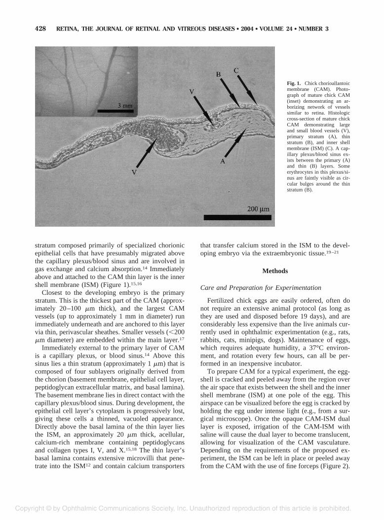
stratum composed primarily of specialized chorionicepithelial cells that have presumably migrated abovethe capillary plexus/blood sinus and are involved ingas exchange and calcium absorption.14 Immediatelyabove and attached to the CAM thin layer is the innershell membrane (ISM) (Figure 1).15,16
Closest to the developing embryo is the primarystratum. This is the thickest part of the CAM (approx-imately 20–100 �m thick), and the largest CAMvessels (up to approximately 1 mm in diameter) runimmediately underneath and are anchored to this layervia thin, perivascular sheathes. Smaller vessels (�200�m diameter) are embedded within the main layer.17
Immediately external to the primary layer of CAMis a capillary plexus, or blood sinus.14 Above thissinus lies a thin stratum (approximately 1 �m) that iscomposed of four sublayers originally derived fromthe chorion (basement membrane, epithelial cell layer,peptidoglycan extracellular matrix, and basal lamina).The basement membrane lies in direct contact with thecapillary plexus/blood sinus. During development, theepithelial cell layer’s cytoplasm is progressively lost,giving these cells a thinned, vacuoled appearance.Directly above the basal lamina of the thin layer liesthe ISM, an approximately 20 �m thick, acellular,calcium-rich membrane containing peptidoglycansand collagen types I, V, and X.15,18 The thin layer’sbasal lamina contains extensive microvilli that pene-trate into the ISM12 and contain calcium transporters
that transfer calcium stored in the ISM to the devel-oping embryo via the extraembryonic tissue.19–21
Methods
Care and Preparation for Experimentation
Fertilized chick eggs are easily ordered, often donot require an extensive animal protocol (as long asthey are used and disposed before 19 days), and areconsiderably less expensive than the live animals cur-rently used in ophthalmic experimentation (e.g., rats,rabbits, cats, minipigs, dogs). Maintenance of eggs,which requires adequate humidity, a 37°C environ-ment, and rotation every few hours, can all be per-formed in an inexpensive incubator.
To prepare CAM for a typical experiment, the egg-shell is cracked and peeled away from the region overthe air space that exists between the shell and the innershell membrane (ISM) at one pole of the egg. Thisairspace can be visualized before the egg is cracked byholding the egg under intense light (e.g., from a sur-gical microscope). Once the opaque CAM-ISM duallayer is exposed, irrigation of the CAM-ISM withsaline will cause the dual layer to become translucent,allowing for visualization of the CAM vasculature.Depending on the requirements of the proposed ex-periment, the ISM can be left in place or peeled awayfrom the CAM with the use of fine forceps (Figure 2).
Fig. 1. Chick chorioallantoicmembrane (CAM). Photo-graph of mature chick CAM(inset) demonstrating an ar-borizing network of vesselssimilar to retina. Histologiccross-section of mature chickCAM demonstrating largeand small blood vessels (V),primary stratum (A), thinstratum (B), and inner shellmembrane (ISM) (C). A cap-illary plexus/blood sinus ex-ists between the primary (A)and thin (B) layers. Someerythrocytes in this plexus/si-nus are faintly visible as cir-cular bulges around the thinstratum (B).
428 RETINA, THE JOURNAL OF RETINAL AND VITREOUS DISEASES ● 2004 ● VOLUME 24 ● NUMBER 3
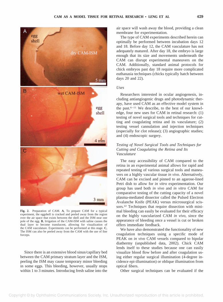
Since there is an extensive blood sinus/capillary bedbetween the CAM primary stratum layer and the ISM,peeling the ISM may cause temporary minor bleedingin some eggs. This bleeding, however, usually stopswithin 1 to 3 minutes. Introducing fresh saline into the
air space will wash away the blood, providing a cleanmembrane for experimentation.
The type of CAM experiments described herein canoptimally be performed between incubation days 12and 18. Before day 12, the CAM vasculature has notadequately matured. After day 18, the embryo is largeenough that its size and movements underneath theCAM can disrupt experimental maneuvers on theCAM. Additionally, standard animal protocols forchick embryos past day 18 require more complicatedeuthanasia techniques (chicks typically hatch betweendays 20 and 22).
Uses
Researchers interested in ocular angiogenesis, in-cluding antiangiogenic drugs and photodynamic ther-apy, have used CAM as an effective model system inthe past.6–11 We describe, to the best of our knowl-edge, four new uses for CAM in retinal research: (1)testing of novel surgical tools and techniques for cut-ting and coagulating retina and its vasculature; (2)testing vessel cannulation and injection techniques(especially for clot release); (3) angiographic studies;and (4) endoscopic surgery.
Testing of Novel Surgical Tools and Techniques forCutting and Coagulating the Retina and ItsVasculature
The easy accessibility of CAM compared to theretina in an experimental animal allows for rapid andrepeated testing of various surgical tools and maneu-vers on a highly vascular tissue in vivo. Alternatively,CAM can be excised and pinned to an agarose-linedPetri dish to allow for in vitro experimentation. Ourgroup has used both in vivo and in vitro CAM forcomparative testing of the cutting capacity of a novelplasma-mediated dissector called the Pulsed ElectronAvalanche Knife (PEAK) versus microsurgical scis-sors.22 Techniques that require dissection with mini-mal bleeding can easily be evaluated for their efficacyon the highly vascularized CAM in vivo, since theappearance of bleeding once a vessel is cut or brokenoffers immediate feedback.
We have also demonstrated the functionality of newcoagulation techniques using a specific mode ofPEAK on in vivo CAM vessels compared to bipolardiathermy (unpublished data, 2002). Chick CAMlends itself to these studies because one can easilyvisualize blood flow before and after coagulation us-ing either regular surgical illumination (4-degree in-cidence epi-illumination) or oblique illumination fromoptical fibers.
Other surgical techniques can be evaluated if the
Fig. 2. Preparation of CAM. A, To prepare CAM for a typicalexperiment, the eggshell is cracked and peeled away from the regionover the air space that exists between the shell and the ISM near onepole of the egg. B, Irrigation of the CAM-ISM with saline causes thedual layer to become translucent, allowing for visualization ofthe CAM vasculature. Experiments can be performed at this stage. C,The ISM can also be peeled away from the CAM with the use of fineforceps.
429CAM AS A MODEL TISSUE FOR RETINAL RESEARCH • LENG ET AL
ISM that naturally covers the CAM is not removed.Between the ISM and the CAM lies an extensivecapillary plexus/blood sinus. Any procedure practicedon the ISM that causes too much mechanical stress tothe CAM below will create a noticeable hemorrhagewithin this plexus/sinus, manifesting itself as a hema-toma trapped between the ISM and CAM. This prop-erty of the CAM-ISM complex is useful because of itscorrelative implications for retinal dissection.
Tissue Viability Studies: Simulating RetinalResponse to Various Insults
Determining retinal response to various insults iscritical for establishing safety data for novel instru-ments or therapies. One way to collect these data is toexpose a suspension of cells to an insult and thenmeasure their response through cell viability markers(e.g., LIVE/DEAD staining).23–25 This method is notuseful, however, if one is interested in tissue viabilityas a function of distance from the insult. For example,if one were testing a drug or therapy delivered locallyto a single point of tissue, one would be interested inthe reaction of both the proximally exposed tissue andthe surrounding distal tissue. In testing a new surgicalinstrument, it is important to know how tissue adjacent tothe instrument responds as a function of distance fromthe insult and at various instrument settings.
One way to solve this “viability versus distance”problem is to suspend cells in a three-dimensional gelmatrix. However, this method carries its own set ofdisadvantages and difficulties—preparing gels so thatcells are uniformly dispersed can be time and laborintensive. Furthermore, the gel matrix may not en-
tirely mimic the physical properties of tissue andtherefore may falsely represent the actual effects ofthe insult of interest. Finally, extracellular cell viabil-ity markers (e.g., trypan blue or propidium iodide)26,27
do not readily diffuse through a gel matrix, therebypreventing optimal staining of compromised cells.Preloading cells with an intracellular marker (e.g.,Calcein AM) before casting the matrix is possible, butis more complex to prepare and to image.
One solution to the viability versus distance ques-tion is to simply perform tissue viability tests on liveanimal eyes. This is not ideal because extensive test-ing in animals is expensive, is time consuming, andraises ethical issues, particularly in preliminary eval-uative studies.
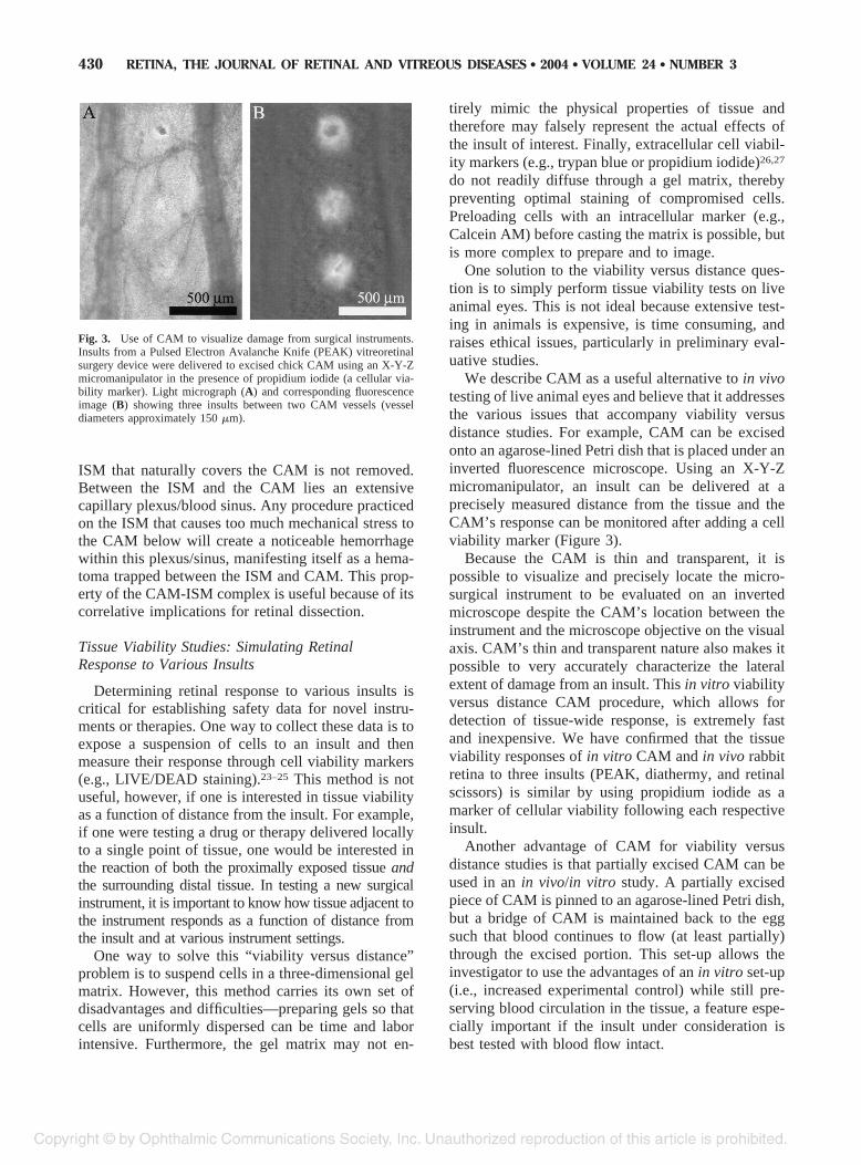
We describe CAM as a useful alternative to in vivotesting of live animal eyes and believe that it addressesthe various issues that accompany viability versusdistance studies. For example, CAM can be excisedonto an agarose-lined Petri dish that is placed under aninverted fluorescence microscope. Using an X-Y-Zmicromanipulator, an insult can be delivered at aprecisely measured distance from the tissue and theCAM’s response can be monitored after adding a cellviability marker (Figure 3).
Because the CAM is thin and transparent, it ispossible to visualize and precisely locate the micro-surgical instrument to be evaluated on an invertedmicroscope despite the CAM’s location between theinstrument and the microscope objective on the visualaxis. CAM’s thin and transparent nature also makes itpossible to very accurately characterize the lateralextent of damage from an insult. This in vitro viabilityversus distance CAM procedure, which allows fordetection of tissue-wide response, is extremely fastand inexpensive. We have confirmed that the tissueviability responses of in vitro CAM and in vivo rabbitretina to three insults (PEAK, diathermy, and retinalscissors) is similar by using propidium iodide as amarker of cellular viability following each respectiveinsult.
Another advantage of CAM for viability versusdistance studies is that partially excised CAM can beused in an in vivo/in vitro study. A partially excisedpiece of CAM is pinned to an agarose-lined Petri dish,but a bridge of CAM is maintained back to the eggsuch that blood continues to flow (at least partially)through the excised portion. This set-up allows theinvestigator to use the advantages of an in vitro set-up(i.e., increased experimental control) while still pre-serving blood circulation in the tissue, a feature espe-cially important if the insult under consideration isbest tested with blood flow intact.
Fig. 3. Use of CAM to visualize damage from surgical instruments.Insults from a Pulsed Electron Avalanche Knife (PEAK) vitreoretinalsurgery device were delivered to excised chick CAM using an X-Y-Zmicromanipulator in the presence of propidium iodide (a cellular via-bility marker). Light micrograph (A) and corresponding fluorescenceimage (B) showing three insults between two CAM vessels (vesseldiameters approximately 150 �m).
430 RETINA, THE JOURNAL OF RETINAL AND VITREOUS DISEASES ● 2004 ● VOLUME 24 ● NUMBER 3
Testing Vessel Cannulation and InjectionTechniques
The accessibility of CAM vessels and the ease ofimaging these vessels noninvasively makes CAM anideal tissue for testing vessel cannulation/injection
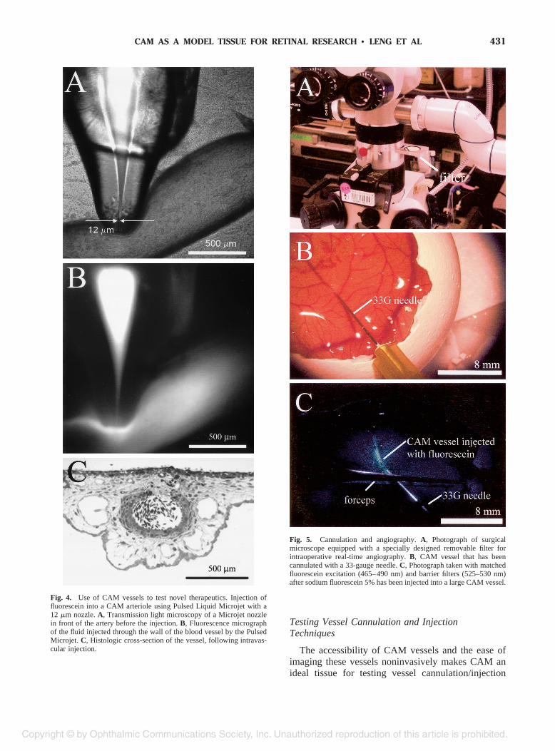
Fig. 4. Use of CAM vessels to test novel therapeutics. Injection offluorescein into a CAM arteriole using Pulsed Liquid Microjet with a12 �m nozzle. A, Transmission light microscopy of a Microjet nozzlein front of the artery before the injection. B, Fluorescence micrographof the fluid injected through the wall of the blood vessel by the PulsedMicrojet. C, Histologic cross-section of the vessel, following intravas-cular injection.
Fig. 5. Cannulation and angiography. A, Photograph of surgicalmicroscope equipped with a specially designed removable filter forintraoperative real-time angiography. B, CAM vessel that has beencannulated with a 33-gauge needle. C, Photograph taken with matchedfluorescein excitation (465–490 nm) and barrier filters (525–530 nm)after sodium fluorescein 5% has been injected into a large CAM vessel.
431CAM AS A MODEL TISSUE FOR RETINAL RESEARCH • LENG ET AL

techniques. Although CAM vessels vary in diameter,from approximately 1 mm to capillary size, a sizableportion of vessels embedded in the main CAM layerare similar in size to relevant retinal vessels (approx-imately 40 �m to 350 �m).
For cannulation/injection studies on in vivo CAM,observation of individual red blood cell movementand direction of blood flow is easily achieved using asurgical microscope and fiber optics. Distinguishingarteries from veins is possible by observing whetherflow in a vessel is pulsatile and ligation is facile whena particular procedure or technique requires it. Thecannulation/injection studies we have pursued include(1) testing of a novel Pulsed Liquid Microjet fordelivery of microliter quantities of fibrinolytics di-rectly to the site of green-argon laser inducedclots28–32 and (2) the creation of experimental aneu-rysms and vessel cannulation with a standard 41-gauge needle for fibrinolytic drug infusion33 (Figure4). We have found CAM vessels to be an effectivemodel for evaluating new methods for treating clots inthe retinal vasculature.
Angiographic Studies
Although it is sometimes difficult to inject fluores-cein into a CAM vessel without a small amount of dyeleaking out, it is possible to manually cannulate CAMvessels using small-gauge surgical instrumentation(33–41 G). CAM can be effective in evaluating newdyes and barrier/excitatory filter systems for retinalimaging. It is of value in teaching and practicingmanual dexterity to young surgeons or in evaluatingnew instrumentation and techniques for treatment ofretinal vascular disorders. Angiography in CAM ves-sels may be useful in assessing the effectiveness of
Fig. 6. Endoscopy. A, Photograph showing illumination from anendoscope fitted with fluorescein excitation and barrier filters. B, Viewthrough the endoscope after a solution of sodium fluorescein 5% hasbeen injected into a large CAM vessel.
Fig. 7. Endoscopy and instrumentation. A, View through a surgicalendoscope with a PEAK probe attachment, showing CAM inner shellmembrane. B, Photograph showing incision made with the PEAKprobe using endoscopic visualization.
432 RETINA, THE JOURNAL OF RETINAL AND VITREOUS DISEASES ● 2004 ● VOLUME 24 ● NUMBER 3
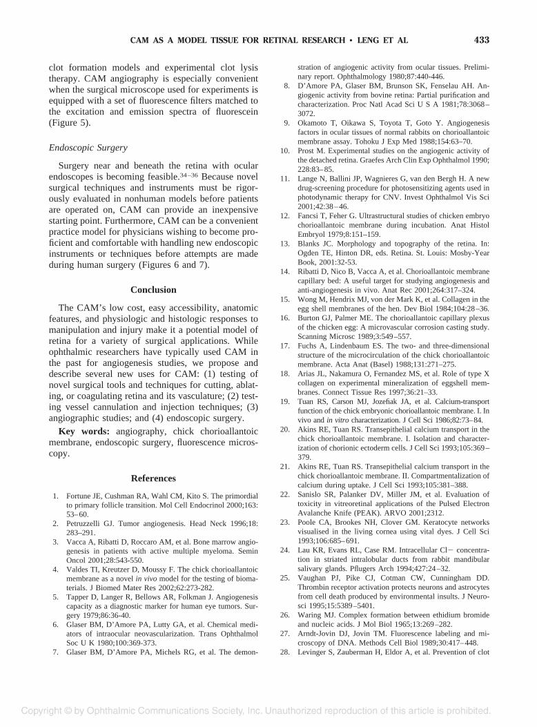
clot formation models and experimental clot lysistherapy. CAM angiography is especially convenientwhen the surgical microscope used for experiments isequipped with a set of fluorescence filters matched tothe excitation and emission spectra of fluorescein(Figure 5).
Endoscopic Surgery
Surgery near and beneath the retina with ocularendoscopes is becoming feasible.34–36 Because novelsurgical techniques and instruments must be rigor-ously evaluated in nonhuman models before patientsare operated on, CAM can provide an inexpensivestarting point. Furthermore, CAM can be a convenientpractice model for physicians wishing to become pro-ficient and comfortable with handling new endoscopicinstruments or techniques before attempts are madeduring human surgery (Figures 6 and 7).
Conclusion
The CAM’s low cost, easy accessibility, anatomicfeatures, and physiologic and histologic responses tomanipulation and injury make it a potential model ofretina for a variety of surgical applications. Whileophthalmic researchers have typically used CAM inthe past for angiogenesis studies, we propose anddescribe several new uses for CAM: (1) testing ofnovel surgical tools and techniques for cutting, ablat-ing, or coagulating retina and its vasculature; (2) test-ing vessel cannulation and injection techniques; (3)angiographic studies; and (4) endoscopic surgery.
Key words: angiography, chick chorioallantoicmembrane, endoscopic surgery, fluorescence micros-copy.
References
1. Fortune JE, Cushman RA, Wahl CM, Kito S. The primordialto primary follicle transition. Mol Cell Endocrinol 2000;163:53–60.
2. Petruzzelli GJ. Tumor angiogenesis. Head Neck 1996;18:283–291.
3. Vacca A, Ribatti D, Roccaro AM, et al. Bone marrow angio-genesis in patients with active multiple myeloma. SeminOncol 2001;28:543-550.
4. Valdes TI, Kreutzer D, Moussy F. The chick chorioallantoicmembrane as a novel in vivo model for the testing of bioma-terials. J Biomed Mater Res 2002;62:273-282.
5. Tapper D, Langer R, Bellows AR, Folkman J. Angiogenesiscapacity as a diagnostic marker for human eye tumors. Sur-gery 1979;86:36-40.
6. Glaser BM, D’Amore PA, Lutty GA, et al. Chemical medi-ators of intraocular neovascularization. Trans OphthalmolSoc U K 1980;100:369-373.
7. Glaser BM, D’Amore PA, Michels RG, et al. The demon-
stration of angiogenic activity from ocular tissues. Prelimi-nary report. Ophthalmology 1980;87:440-446.
8. D’Amore PA, Glaser BM, Brunson SK, Fenselau AH. An-giogenic activity from bovine retina: Partial purification andcharacterization. Proc Natl Acad Sci U S A 1981;78:3068–3072.
9. Okamoto T, Oikawa S, Toyota T, Goto Y. Angiogenesisfactors in ocular tissues of normal rabbits on chorioallantoicmembrane assay. Tohoku J Exp Med 1988;154:63–70.
10. Prost M. Experimental studies on the angiogenic activity ofthe detached retina. Graefes Arch Clin Exp Ophthalmol 1990;228:83–85.
11. Lange N, Ballini JP, Wagnieres G, van den Bergh H. A newdrug-screening procedure for photosensitizing agents used inphotodynamic therapy for CNV. Invest Ophthalmol Vis Sci2001;42:38–46.
12. Fancsi T, Feher G. Ultrastructural studies of chicken embryochorioallantoic membrane during incubation. Anat HistolEmbryol 1979;8:151–159.
13. Blanks JC. Morphology and topography of the retina. In:Ogden TE, Hinton DR, eds. Retina. St. Louis: Mosby-YearBook, 2001:32-53.
14. Ribatti D, Nico B, Vacca A, et al. Chorioallantoic membranecapillary bed: A useful target for studying angiogenesis andanti-angiogenesis in vivo. Anat Rec 2001;264:317–324.
15. Wong M, Hendrix MJ, von der Mark K, et al. Collagen in theegg shell membranes of the hen. Dev Biol 1984;104:28–36.
16. Burton GJ, Palmer ME. The chorioallantoic capillary plexusof the chicken egg: A microvascular corrosion casting study.Scanning Microsc 1989;3:549–557.
17. Fuchs A, Lindenbaum ES. The two- and three-dimensionalstructure of the microcirculation of the chick chorioallantoicmembrane. Acta Anat (Basel) 1988;131:271–275.
18. Arias JL, Nakamura O, Fernandez MS, et al. Role of type Xcollagen on experimental mineralization of eggshell mem-branes. Connect Tissue Res 1997;36:21–33.
19. Tuan RS, Carson MJ, Jozefiak JA, et al. Calcium-transportfunction of the chick embryonic chorioallantoic membrane. I. Invivo and in vitro characterization. J Cell Sci 1986;82:73–84.
20. Akins RE, Tuan RS. Transepithelial calcium transport in thechick chorioallantoic membrane. I. Isolation and character-ization of chorionic ectoderm cells. J Cell Sci 1993;105:369–379.
21. Akins RE, Tuan RS. Transepithelial calcium transport in thechick chorioallantoic membrane. II. Compartmentalization ofcalcium during uptake. J Cell Sci 1993;105:381–388.
22. Sanislo SR, Palanker DV, Miller JM, et al. Evaluation oftoxicity in vitreoretinal applications of the Pulsed ElectronAvalanche Knife (PEAK). ARVO 2001;2312.
23. Poole CA, Brookes NH, Clover GM. Keratocyte networksvisualised in the living cornea using vital dyes. J Cell Sci1993;106:685–691.
24. Lau KR, Evans RL, Case RM. Intracellular Cl� concentra-tion in striated intralobular ducts from rabbit mandibularsalivary glands. Pflugers Arch 1994;427:24–32.
25. Vaughan PJ, Pike CJ, Cotman CW, Cunningham DD.Thrombin receptor activation protects neurons and astrocytesfrom cell death produced by environmental insults. J Neuro-sci 1995;15:5389–5401.
26. Waring MJ. Complex formation between ethidium bromideand nucleic acids. J Mol Biol 1965;13:269–282.
27. Arndt-Jovin DJ, Jovin TM. Fluorescence labeling and mi-croscopy of DNA. Methods Cell Biol 1989;30:417–448.
28. Levinger S, Zauberman H, Eldor A, et al. Prevention of clot
433CAM AS A MODEL TISSUE FOR RETINAL RESEARCH • LENG ET AL
formation in cat retinal vein by systemic and subconjunctivalurokinase. Arch Ophthalmol 1987;105:554–558.
29. Oncel M, Peyman GA, Khoobehi B. Tissue plasminogenactivator in the treatment of experimental retinal vein occlu-sion. Retina 1989;9:1–7.
30. Larsson J, Carlson J, Olsson SB. Ultrasound enhanced throm-bolysis in experimental retinal vein occlusion in the rabbit.Br J Ophthalmol 1998;82:1438–1440.
31. Tamura M. Neovascularization in experimental retinal venousobstruction in rabbits. Jpn J Ophthalmol 2001;45:144–150.
32. Fletcher DA, Palanker DV, Huie P, et al. Intra-vascular drugdelivery with a pulsed liquid microjet. Arch Ophthalmol2002;120:1206–1208.
33. Miller JM, Castelo-Branco A, Huie P, et al. Application ofthe Pulsed Electron Avalanche Knife (PEAK) to treatment ofretinal vascular occlusions. ARVO 2002;1870.
34. Koch FH, Luloh KP, Augustin AJ, et al. Subretinal micro-surgery with gradient index endoscopes. Ophthalmologica1997;211:283–287.
35. Terasaki H, Miyake Y, Mori M, et al. Fluorescein angiogra-phy of extreme peripheral retina and rubeosis iridis in pro-liferative diabetic retinopathy. Retina 1999;19:302–308.
36. Quiroz-Mercado H, Yeshurun I, Sanchez-Buefil E, et al. Sub-retinal, viscoelastic-assisted, endoscope-guided photothermalablation of choroidal neovascular membranes by Erbium:YAGlaser. Ophthalmic Surg Lasers 2001;32:456–463.
434 RETINA, THE JOURNAL OF RETINAL AND VITREOUS DISEASES ● 2004 ● VOLUME 24 ● NUMBER 3