Embed Size (px)
Citation preview
ARTICLE
Received 21 Apr 2014 | Accepted 22 Jan 2015 | Published 27 Feb 2015
The cholesterol transporter ABCG1 links cholesterolhomeostasis and tumour immunityDuygu Sag1,*, Caglar Cekic2,*, Runpei Wu1, Joel Linden3 & Catherine C. Hedrick1
ATP-binding cassette transporter G1 (ABCG1) promotes cholesterol efflux from cells and
regulates intracellular cholesterol homeostasis. Here we demonstrate a role of ABCG1 as a
mediator of tumour immunity. Abcg1� /� mice have dramatically suppressed subcutaneous
MB49-bladder carcinoma and B16-melanoma growth and prolonged survival. We show that
reduced tumour growth in Abcg1� /� mice is myeloid cell intrinsic and is associated with a
phenotypic shift of the macrophages from a tumour-promoting M2 to a tumour-fighting M1
within the tumour. Abcg1� /� macrophages exhibit an intrinsic bias towards M1 polarization
with increased NF-kB activation and direct cytotoxicity for tumour cells in vitro. Overall, our
study demonstrates that the absence of ABCG1 inhibits tumour growth through modulation
of macrophage function within the tumour, and illustrates a link between cholesterol
homeostasis and cancer.
DOI: 10.1038/ncomms7354
1 Division of Inflammation Biology, La Jolla Institute for Allergy and Immunology, La Jolla, California 92037, USA. 2 Department of Molecular Biology andGenetics, Bilkent University, Ankara 06800, Turkey. 3 Division of Developmental Immunology, La Jolla Institute for Allergy and Immunology, La Jolla,California 92037, USA. * These authors contributed equally to this work. Correspondence and requests for materials should be addressed to D.S.(email: [email protected]) or to C.C.H. (email: [email protected]).
NATURE COMMUNICATIONS | 6:6354 | DOI: 10.1038/ncomms7354 | www.nature.com/naturecommunications 1
& 2015 Macmillan Publishers Limited. All rights reserved.
In addition to the cancer cells and their surrounding stroma,the tumour microenvironment contains innate and adaptiveimmune cells that can recognize and destroy tumours1.
However, the tumour not only manages to evade the immunesystem through various mechanisms, but also it contrives tobenefit from infiltrating immune cells by modifying theirfunctions to create a microenvironment favourable to tumourprogression2. Macrophages are major players of tumourimmunity. Monocyte-derived macrophages can polarize intoeither M1 (classically activated) or M2 (alternatively activated)macrophage subtypes in the presence of specific polarizationfactors, including cytokines, growth factors and bioactive lipids,when recruited into peripheral tissues3,4. In general, M1macrophages are potent tumour-fighting cells, whereas M2macrophages display protumoral functions. The tumour recruitsblood monocytes and promotes their differentiation mostly intoM2-like macrophages5. M2-like tumour-associated macrophages(TAMs) play a key role in tumour growth and progression byproducing molecules to promote angiogenesis, as well as survivaland metastasis of tumour cells6–9. Moreover, TAMs affectadaptive immune responses by recruiting T regulatory cells(Tregs), which in turn suppress antitumour effector cells such asNK cells and CD4þ /CD8þ T cells10. Several studies havereported a positive correlation between high TAM density andpoor prognosis in human tumours, including bladder, breast andprostate7,11. Furthermore, it has been shown in different murinetumour models that either depletion of macrophages12–16 orswitching the phenotype of macrophages into tumour-fightingM1 macrophages17–19 results in a significant reduction in tumourgrowth.
ATP-binding cassette transporter G1 (ABCG1) is a member ofthe ABC transporter family that regulates cellular cholesterolhomeostasis20. Cholesterol homeostasis is crucial for the survivaland function of cells21. ABCG1 effluxes excess cholesterol fromcells to high-density lipoprotein (HDL) particles for reversecholesterol transport, which is the only path for elimination ofcholesterol from the body22,23. ABCG1 is also important for theintracellular transport of cholesterol24,25. It is ubiquitouslyexpressed in many cell types including myeloid cells,lymphocytes and endothelial cells20.
ABCG1 is known to regulate several aspects of macrophagebiology. Abcg1� /� mice fed a Western-like diet display excessivelipid accumulation in macrophages22. Atherosclerosis studiesdemonstrated that ABCG1-deficient macrophages were moresusceptible to apoptosis compared with wild-type (WT)macrophages under Western-like diet conditions in vivo26,27.Furthermore, Abcg1� /� macrophages have been shown todisplay enhanced proinflammatory cytokine production at basallevel28,29, in response to lipopolysaccharide (LPS)30 and whenloaded with cholesterol31. ABCG1 also plays a role in T-cellbiology. We and others have reported that alterations inintracellular cholesterol homeostasis in the absence of ABCG1increase proliferation of CD4þ T cells32,33 and impairsdevelopment of invariant natural killer T cells in thymus34.
Overall, changes in cholesterol homeostasis by the absence ofABCG1 modulate immune cell function; however, the role ofABCG1 in antitumour immune responses is unknown.
In this study, we demonstrate that the in vivo deficiencyof ABCG1 reduces tumour growth and increases the survival ofmice. Reduced tumour growth in the absence of ABCG1 ismediated by myeloid cell intrinsic mechanisms and is associatedwith a shift of macrophages to a tumour-fighting M1 phenotypewithin the tumour, which results in the direct killing oftumour cells.
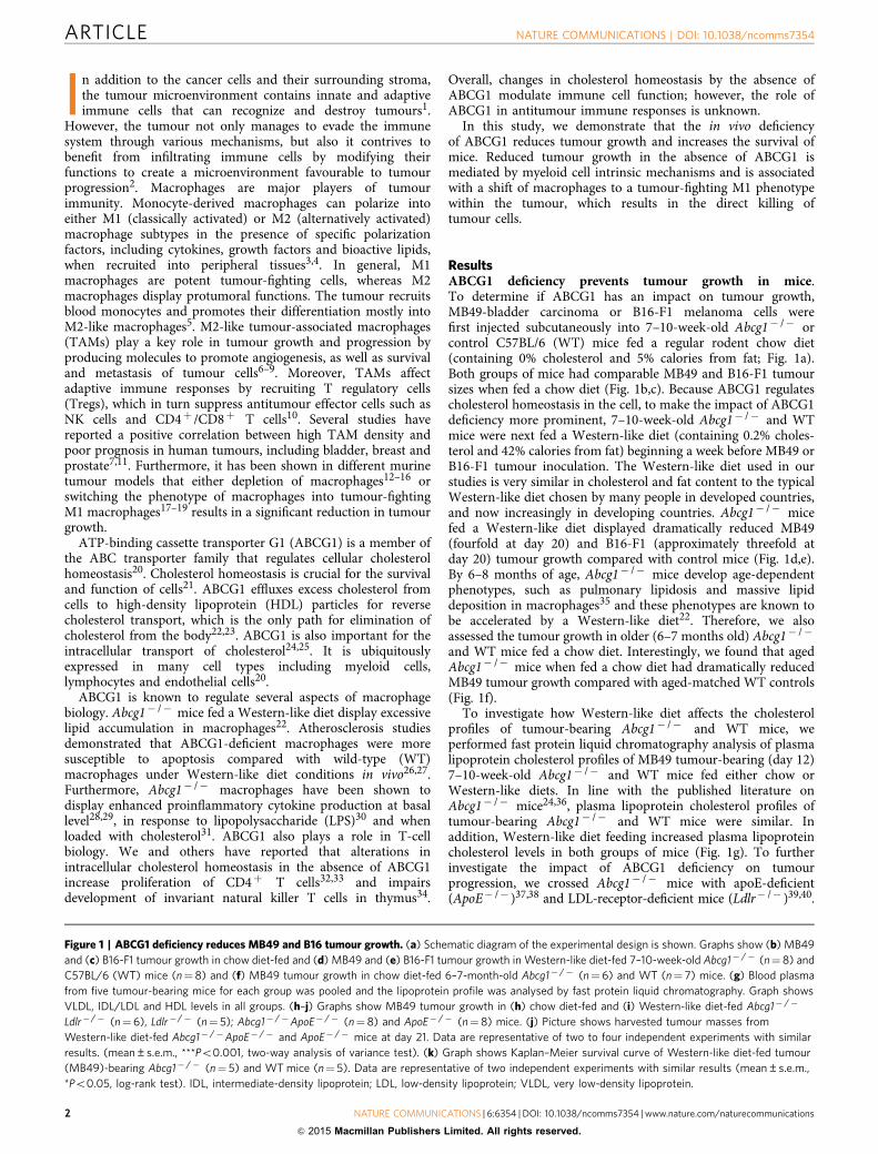
ResultsABCG1 deficiency prevents tumour growth in mice.To determine if ABCG1 has an impact on tumour growth,MB49-bladder carcinoma or B16-F1 melanoma cells werefirst injected subcutaneously into 7–10-week-old Abcg1� /� orcontrol C57BL/6 (WT) mice fed a regular rodent chow diet(containing 0% cholesterol and 5% calories from fat; Fig. 1a).Both groups of mice had comparable MB49 and B16-F1 tumoursizes when fed a chow diet (Fig. 1b,c). Because ABCG1 regulatescholesterol homeostasis in the cell, to make the impact of ABCG1deficiency more prominent, 7–10-week-old Abcg1� /� and WTmice were next fed a Western-like diet (containing 0.2% choles-terol and 42% calories from fat) beginning a week before MB49 orB16-F1 tumour inoculation. The Western-like diet used in ourstudies is very similar in cholesterol and fat content to the typicalWestern-like diet chosen by many people in developed countries,and now increasingly in developing countries. Abcg1� /� micefed a Western-like diet displayed dramatically reduced MB49(fourfold at day 20) and B16-F1 (approximately threefold atday 20) tumour growth compared with control mice (Fig. 1d,e).By 6–8 months of age, Abcg1� /� mice develop age-dependentphenotypes, such as pulmonary lipidosis and massive lipiddeposition in macrophages35 and these phenotypes are known tobe accelerated by a Western-like diet22. Therefore, we alsoassessed the tumour growth in older (6–7 months old) Abcg1� /�
and WT mice fed a chow diet. Interestingly, we found that agedAbcg1� /� mice when fed a chow diet had dramatically reducedMB49 tumour growth compared with aged-matched WT controls(Fig. 1f).
To investigate how Western-like diet affects the cholesterolprofiles of tumour-bearing Abcg1� /� and WT mice, weperformed fast protein liquid chromatography analysis of plasmalipoprotein cholesterol profiles of MB49 tumour-bearing (day 12)7–10-week-old Abcg1� /� and WT mice fed either chow orWestern-like diets. In line with the published literature onAbcg1� /� mice24,36, plasma lipoprotein cholesterol profiles oftumour-bearing Abcg1� /� and WT mice were similar. Inaddition, Western-like diet feeding increased plasma lipoproteincholesterol levels in both groups of mice (Fig. 1g). To furtherinvestigate the impact of ABCG1 deficiency on tumourprogression, we crossed Abcg1� /� mice with apoE-deficient(ApoE� /� )37,38 and LDL-receptor-deficient mice (Ldlr� /� )39,40.
Figure 1 | ABCG1 deficiency reduces MB49 and B16 tumour growth. (a) Schematic diagram of the experimental design is shown. Graphs show (b) MB49
and (c) B16-F1 tumour growth in chow diet-fed and (d) MB49 and (e) B16-F1 tumour growth in Western-like diet-fed 7–10-week-old Abcg1� /� (n¼ 8) and
C57BL/6 (WT) mice (n¼8) and (f) MB49 tumour growth in chow diet-fed 6–7-month-old Abcg1� /� (n¼6) and WT (n¼ 7) mice. (g) Blood plasma
from five tumour-bearing mice for each group was pooled and the lipoprotein profile was analysed by fast protein liquid chromatography. Graph shows
VLDL, IDL/LDL and HDL levels in all groups. (h–j) Graphs show MB49 tumour growth in (h) chow diet-fed and (i) Western-like diet-fed Abcg1� /�
Ldlr�/� (n¼6), Ldlr�/� (n¼ 5); Abcg1� /�ApoE� /� (n¼ 8) and ApoE� /� (n¼ 8) mice. (j) Picture shows harvested tumour masses from
Western-like diet-fed Abcg1� /�ApoE� /� and ApoE� /� mice at day 21. Data are representative of two to four independent experiments with similar
results. (mean±s.e.m., ***Po0.001, two-way analysis of variance test). (k) Graph shows Kaplan–Meier survival curve of Western-like diet-fed tumour
(MB49)-bearing Abcg1� /� (n¼ 5) and WT mice (n¼ 5). Data are representative of two independent experiments with similar results (mean±s.e.m.,
*Po0.05, log-rank test). IDL, intermediate-density lipoprotein; LDL, low-density lipoprotein; VLDL, very low-density lipoprotein.
ARTICLE NATURE COMMUNICATIONS | DOI: 10.1038/ncomms7354
2 NATURE COMMUNICATIONS | 6:6354 | DOI: 10.1038/ncomms7354 | www.nature.com/naturecommunications
& 2015 Macmillan Publishers Limited. All rights reserved.
The ApoE� /� and Ldlr� /� models are two hypercholesterolemicmouse models that are widely used to study atherosclerosis. Bothgenotypes have high plasma cholesterol levels when fed a chowdiet and show profoundly increased plasma cholesterol levels when
fed a Western-like diet37–41. We first compared the plasmalipoprotein cholesterol profiles of MB49 tumour-bearing (day 12)7–10-week-old Abcg1� /� ApoE� /� mice with ApoE� /� miceand Abcg1� /� Ldlr� /� mice with Ldlr� /� mice, all fed a chow
0 2 4 6 8 10 12 14 16 18 20 22 24 26 28
0
1
2
3
4
5
6
7
8
Fraction number
mg
dl–1
cho
lest
erol
per
frac
tion
WT chow
Abcg1–/– chow
WT western
Abcg1–/– western
VLDL IDL/ LDL
HDL
MB49-bladder carcinoma
Days
Tum
our
volu
me
(mm
3 )
Chow diet
Western-like diet
8 10 12 14 16 18 20
0
500
1,000
1,500
2,000
2,500 Ldlr –/–
Abcg1 –/– Ldlr –/–
***
8 10 12 14 16 18 20
0
500
1,000
1,500
2,000
2,500 Ldlr –/–
Abcg1–/– Ldlr –/–
***
8 10 12 14 16 18 20
0
500
1,000
1,500
2,000
2,500 ApoE –/–
Abcg1 –/– ApoE –/–
***
8 10 12 14 16 18 20
0
500
1,000
1,500
2,000
2,500 ApoE –/–
Abcg1 –/– ApoE –/–
***
Old mice
8 10 12 14 16 18 20
0
500
1,000
1,500
2,000
2,500
Days
Lipoprotein profile
Tum
our
volu
me
(mm
3 )
WT
Abcg1–/–
Chow diet
***
0 10 20 30 40 500
20
40
60
80
100
Days
Sur
viva
l (%
)
WTAbcg1–/–*Abcg1–/–
ApoE–/–ApoE–/–
Day 21
Chow diet
8 10 12 14 16 18 20
0
500
1,000
1,500
2,000
2,500WT
Abcg1–/–
MB49-bladder carcinoma B16-F1 melanoma
Tum
our
volu
me
(mm
3 )
Tum
our
volu
me
(mm
3 )
Days Days
Chow diet
10 14 16 18 20
0
1,000
2,000
3,000
4,000
5,000WT
Abcg1–/–
Tumour measurement
105 MB49 or B16-F1subcutaneous
injection
Chow dietor
Western-like diet
8 10 12 14 16 18 20
0
500
1,000
1,500
2,000
2,500 Western-like dietWT
Abcg1–/–
***
Western-like diet
10 14 16 18 20
0
1,000
2,000
3,000
4,000
5,000WT
Abcg1–/–
***
–7 0 8 10 12 14 16 18 20Day
NATURE COMMUNICATIONS | DOI: 10.1038/ncomms7354 ARTICLE
NATURE COMMUNICATIONS | 6:6354 | DOI: 10.1038/ncomms7354 | www.nature.com/naturecommunications 3
& 2015 Macmillan Publishers Limited. All rights reserved.
diet. We found that the loss of ABCG1 had no impact on plasmalipoprotein profiles in tumour-bearing hypercholesterolemicmice (Supplementary Fig. 1). We next measured subcutaneoustumour growth in Abcg1� /� mice crossed with these hyper-cholesterolemic mouse models. Interestingly, both 7–10-week-oldAbcg1� /� Ldlr� /� and Abcg1� /�ApoE� /� chow-fed miceshowed dramatically reduced MB49 tumour growth comparedwith chow-fed control Ldlr� /� and ApoE� /� mice, respectively(Fig. 1h). Both genotypes also displayed a profound reduction intumour growth when fed a Western-like diet (Fig. 1i,j).Collectively, these data show that Western-like diet feeding orcrossing with hypercholesterolemic mice is necessary to observethe changes in tumour growth in young Abcg1� /� mice, whilethis tumour phenotype is evident in aged Abcg1� /� mice fed achow diet.
To investigate the impact of ABCG1 deficiency on spontaneoustumour metastasis, we utilized luciferase-expressing B16-F10 cells(B16-F10-luc2). B16-F10-luc2 cells were injected subcutaneouslyinto Western-like diet-fed Abcg1� /� or WT mice. B16-F10-luc2tumours grew aggressively and by day 28, the difference intumour growth between Abcg1� /� and WT mice was significant,but not very prominent (Supplementary Fig. 2). This aspect ofB16-F10-luc2 tumour growth allowed us to choose mice for studythat had similar-sized tumours. Lungs from mice with similar,but average, tumour sizes in both groups were analysed forspontaneous metastases of B16-F10 melanoma by biolumines-cence imaging ex vivo (Supplementary Fig. 2). Subcutaneous B16
transplants have been shown to spontaneously metastasize tolung42,43. Abcg1� /� mice had significantly diminished tumourmetastasis compared with WT mice (Supplementary Fig. 2).Subsequently, we examined the impact of ABCG1 deficiencyon survival of tumour-bearing mice. MB49 tumour-bearingAbcg1� /� mice showed prolonged survival compared with WTmice when fed a Western-like diet (Fig. 1k). Collectively, thesedata demonstrate that in vivo deficiency of ABCG1 impairstumour growth and increases animal survival.
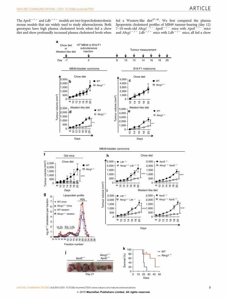
Reduction in tumours in Abcg1� /� mice is immune mediated.To determine if the impact of ABCG1 deficiency on tumourgrowth is mediated by immune cells, we used a bone marrowchimera approach. We measured MB49 tumour growth inWestern-like diet-fed irradiated CD45.1þ B6.SJL (WT) mice,which were reconstituted with CD45.2þ Abcg1� /� or CD45.1þ
B6.SJL bone marrow (Fig. 2a). WT mice reconstituted withAbcg1� /� bone marrow had a significant reduction in tumourgrowth over time compared with WT mice reconstituted withWT bone marrow (Fig. 2b), demonstrating that the impact ofABCG1 deficiency on tumour growth is immune cell mediated.
The tumour microenvironment contains innate and adaptiveimmune cells, which display pro or antitumour functions. WhileNK cells, M1 macrophages, CD4þ Th1 cells and CD8þ T havebeen shown to act as tumour-fighting cells, M2 macrophages andTregs in tumour are known to support tumour progression.
0
10
20
30
40
0.0
0.5
1.0
1.5
2.0
0.00.51.01.52.02.5
0
10
20
30
40
0.0
0.5
1.0
1.5
2.0
0.00.51.01.52.02.5 **
012345
012345
*
WT
Abcg1–/–
Macrophages
Chow
Neutrophils
Dendritic cells
NK cells
0.0
0.5
1.0
1.5
2.0
0.0
0.5
1.0
1.5
2.0*
0
1
2
3
4
0
1
2
3
4**
0.0
0.2
0.4
0.6
0.0
0.2
0.4
0.6
0
10
20
30
40
0
10
20
30
40
**
CD4+ T cells
CD8+ T cells
NKT cells
Tregs
Chow
Fre
quen
cy (
%)
8 10 12 14 16 180
200
400
600
800WT to WT
Abcg1–/– to WT
BM chimera
Days
Tum
our
volu
me
(mm
3 )
***
Irradiation
CD45.1 WTbone marrow
CD45.2 Abcg1–/–
bone marrow
Tumour measurement
Start Western-like diet
6 weeks
MB49 tumour injection
1 week
WT mice
Western Western
Figure 2 | Impact of ABCG1 deficiency on tumour growth is immune cell mediated. (a,b) Bone marrow chimeras were generated by reconstituting
irradiated B6.SJL mice (n¼ 16 total) with bone marrow cells from CD45.1þ B6.SJL (WT) or CD45.2þ Abcg1� /� donor mice. (a) Schematic diagram of the
experimental design is shown. (b) Graph shows MB49 tumour growth in the chimeric mice. Data are representative of two independent experiments with
similar results (***Po0.001, two-way analysis of variance test). (c) Tumour cells from Abcg1� /� and C57BL/6 (WT) mice (n¼ 5–7 per group) were
analysed by flow cytometry 20 days after injection of MB49 cells. Bar graphs show the frequencies of macrophages, neutrophils, DCs, NK cells, CD4þ
T cells, CD8þ T cells, NKT cells (% of live cells) and Tregs (% of CD4þ T cells) in the tumour. (See methods and Supplementary Fig. 2 for gating
strategies). Data are pooled from two independent experiments with similar results (mean±s.e.m., *Po0.05, **Po0.01, two-tailed Student’s t-test).
ARTICLE NATURE COMMUNICATIONS | DOI: 10.1038/ncomms7354
4 NATURE COMMUNICATIONS | 6:6354 | DOI: 10.1038/ncomms7354 | www.nature.com/naturecommunications
& 2015 Macmillan Publishers Limited. All rights reserved.
Dendritic cells (DCs), neutrophils and NKT cells have beenshown to exert both tumour-suppressive and -promotingeffects1,44. Next we wanted to define the primary immune cellpopulations in the tumour microenvironment that are affected bythe absence of ABCG1 under Western-like diet conditions. MB49tumour cells were injected subcutaneously into either Western-like diet-fed or chow diet-fed Abcg1� /� and WT mice andtumour-infiltrating immune cells were analysed by flowcytometry (For gating strategy, see Supplementary Fig. 3). Wefound that the frequencies of macrophages and Tregs significantlydecreased, whereas the frequencies of NK cells, CD4þ T cells andCD8þ T cells significantly increased in the tumours of Western-like diet-fed Abcg1� /� mice compared with WT mice (Fig. 2c).No significant differences were observed in the frequencies oftumour-infiltrating neutrophils, DCs or NKT cells in chow diet-fed or Western-like diet-fed Abcg1� /� or WT mice (Fig. 2c).These results demonstrate that ABCG1 deficiency changesthe balance between tumour-promoting and tumour-fightingimmune cells within the tumour microenvironment.
Tumour reduction in Abcg1� /� mice is myeloid cell mediated.To determine which cell type(s) were intrinsically affected by the
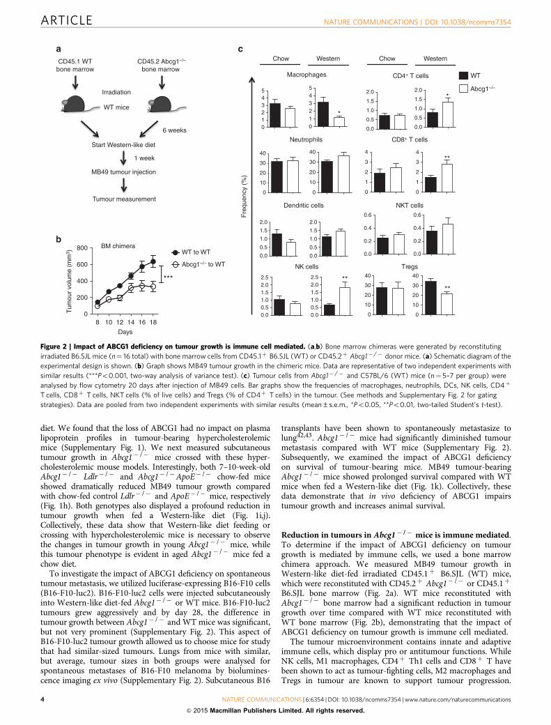
absence of ABCG1 to impact tumour growth, we deleted ABCG1selectively in either myeloid cells or T cells using Cre/loxPtechnology. We generated conditional knockout mice (Abcg1fl/fl)in which loxP sites flank the Walker domain of exon 3 of Abcg1and crossed them with either LysM-Cre or Lck-Cre mice forselective deletion of ABCG1 in myeloid cells and T cells,respectively45,46. We observed B95% deletion of ABCG1in macrophages from Abcg1fl/fl-LysM-Creþ mice and 70%deletion of ABCG1 in T cells from Abcg1fl/fl-Lck-Creþ mice(Supplementary Fig. 4).
We32 and others33 have previously reported that ABCG1deficiency increases proliferation of CD4þ T cells. Therefore, it ispossible that the impact of ABCG1 deficiency on tumour growthmight be mediated directly through T-cell intrinsic mechanisms.To determine the impact of selective ABCG1 deletion inT cells on tumour growth, we injected MB49 tumour cellssubcutaneously into Western-like diet-fed Abcg1fl/fl-Lck-Creþ
and control Abcg1fl/fl-Lck-Cre� mice. The tumour growth in theAbcg1fl/fl-Lck-Creþ mice was comparable to control (Fig. 3a),indicating that the inhibition of tumour growth in the absence ofABCG1 is not mediated directly through T cells. However, wefound that the tumour growth in Abcg1fl/fl-LysM-Creþ mice,which have selective ABCG1 deletion in myeloid cells, was
Fre
quen
cy (
%)
MF
I
0
200
400
600
800
CD4+ T cellsCD69
0100200300400500
DCsCD86
0
200
400
600
CD8+ T cellsCD69
0
200
400
600
800
NK cellsCD69
0
100
200
300
400 *
0200400600800
1,000
0
5,000
10,000
15,000
8 10 12 14 16 18 200
500
1,000
1,500
2,000
2,500Abcg1fl/fl-Lck-Cre–
Abcg1fl/fl-Lck-Cre+
Days
Tum
our
volu
me
(mm
3 )
8 10 12 14 16 18 200
500
1,000
1,500
2,000
2,500Abcg1fl/fl-LysM-Cre–
Abcg1fl/fl-LysM-Cre+
Days
Tum
our
volu
me
(mm
3 )
***
0
1
2
3
**
05
10152025
NK cells
0
2
4
6
8***
05
10152025
*
0.0
0.5
1.0
1.5
2.0CD4+ T cells
012345 *
CD8+ T cells
0
5
10
15
0.0
0.5
1.0
1.5
Macrophages Neutrophils DCs
Abcg1fl/fl-LysM-Cre–
Abcg1fl/fl-LysM-Cre+
Abcg1fl/fl-LysM-Cre–
Abcg1fl/fl-LysM-Cre+
NKT cells Tregs
MacrophagesCD86
NeutrophilsCD11b
NKT cellsCD69
Figure 3 | Reduced tumour growth in Abcg1� /� mice is myeloid cell intrinsic. Graphs show MB49 tumour growth in Western-like diet-fed
(a) Abcg1fl/fl-Lck-Creþ (n¼ 7) and Abcg1fl/fl-Lck-Cre� (n¼ 6) mice, (b) Abcg1fl/fl-LysM-Creþ (n¼ 5) and Abcg1fl/fl-LysM-Cre� (n¼ 7) mice.
Data are representative of two independent experiments with similar results (***Po0.001, two-way analysis of variance test). Tumour cells from
Western-like diet-fed Abcg1fl/fl-LysM-Creþ (n¼ 5) and Abcg1fl/fl-LysM-Cre� mice (n¼ 5) were analysed by flow cytometry 20 days after injection
of MB49 cells. (c) Bar graphs show the frequencies of macrophages, neutrophils, DCs, NK cells, CD4þ T cells, CD8þ T cells, NKT cells (% of live cells)
and Tregs (% of CD4þ T cells) in tumour (See methods and Supplementary Fig. 2 for gating strategies). (d) Bar graphs show the MFI of indicated
activation markers on immune cells in the tumour. Data are representative of two independent experiments with similar results (mean±s.e.m., *Po0.05,
**Po0.01, ***Po0.001, two-tailed Student’s t-test).
NATURE COMMUNICATIONS | DOI: 10.1038/ncomms7354 ARTICLE
NATURE COMMUNICATIONS | 6:6354 | DOI: 10.1038/ncomms7354 | www.nature.com/naturecommunications 5
& 2015 Macmillan Publishers Limited. All rights reserved.
dramatically reduced compared with control Abcg1fl/fl-LysM-Cre� mice (Fig. 3b). These data indicate that the reduced tumourgrowth in the absence of ABCG1 is mediated directly throughmyeloid cell intrinsic mechanisms.
Subsequently, we analysed the tumour-infiltrating immunecells in MB49 tumours from Western-like diet-fed Abcg1fl/fl-LysM-Creþ and control mice by flow cytometry. In line with thechanges in the frequencies of tumour-infiltrating immune cells inWestern-like diet-fed Abcg1� /� mice (Fig. 2c), the frequenciesof tumour-infiltrating macrophages and Tregs in Abcg1fl/fl-LysM-Creþ mice were significantly lower, whereas the frequencies ofNK cells and CD4þ T cells were significantly higher comparedwith control (Fig. 3c). The frequencies of neutrophils, DCs,CD8þ cells and NKT cells in Abcg1fl/fl-LysM-Creþ mice andcontrol mice were comparable (Fig. 3c). We also analysedactivation markers on tumour-infiltrating immune cells. Weobserved that the mean fluorescence intensity (MFI) of CD69expression on NK cells was significantly higher in Abcg1fl/fl-LysM-Creþ mice compared with control (Fig. 3d). The MFI ofCD69 expression on CD4þ T cells, CD8þ T cells and NKT cells,the MFI of CD86 expression on macrophages and DCs and theMFI of CD11b expression on neutrophils were similar betweenboth genotypes (Fig. 3d). LysM-Cre mice have been shown todisplay Cre-mediated deletion of loxP-flanked target genes inmyeloid cells; mainly in macrophages and neutrophils and
partially in DCs46. In both Western-like diet-fed Abcg1� /�
mice and Abcg1fl/fl-LysM-Creþ mice, the frequency ofmacrophages was decreased, whereas no significant differenceswere observed in the frequencies or the activation of tumour-infiltrating neutrophils and DCs (Figs 2c and 3c,d). Therefore,our data suggest that ABCG1 deficiency in macrophages likelypromotes multiple antitumour immune responses.
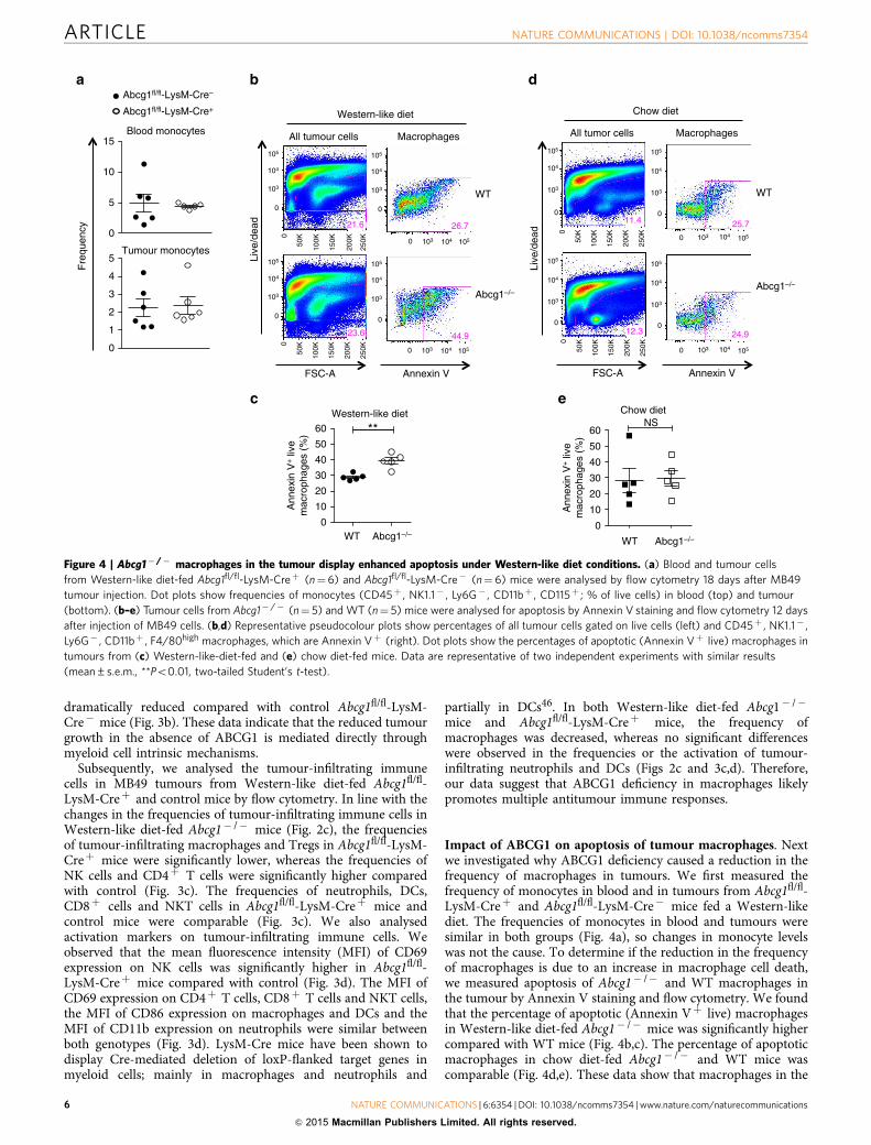
Impact of ABCG1 on apoptosis of tumour macrophages. Nextwe investigated why ABCG1 deficiency caused a reduction in thefrequency of macrophages in tumours. We first measured thefrequency of monocytes in blood and in tumours from Abcg1fl/fl-LysM-Creþ and Abcg1fl/fl-LysM-Cre� mice fed a Western-likediet. The frequencies of monocytes in blood and tumours weresimilar in both groups (Fig. 4a), so changes in monocyte levelswas not the cause. To determine if the reduction in the frequencyof macrophages is due to an increase in macrophage cell death,we measured apoptosis of Abcg1� /� and WT macrophages inthe tumour by Annexin V staining and flow cytometry. We foundthat the percentage of apoptotic (Annexin Vþ live) macrophagesin Western-like diet-fed Abcg1� /� mice was significantly highercompared with WT mice (Fig. 4b,c). The percentage of apoptoticmacrophages in chow diet-fed Abcg1� /� and WT mice wascomparable (Fig. 4d,e). These data show that macrophages in the
Live
/dea
d
WT
Abcg1–/–
All tumour cells Macrophages
Western-like diet
FSC-A Annexin V
Fre
quen
cy
0
5
10
15Blood monocytes
0
1
2
3
4
5Tumour monocytes
Chow diet
WT Abcg1–/–Abcg1–/–0
10
20
30
40
50
60NS
Abcg1–/–
FSC-A Annexin V
All tumor cells Macrophages
Chow diet
WT
Live
/dea
d WT
0
10
20
30
40
50
60 **Western-like diet
Abcg1fl/fl-LysM-Cre–
Abcg1fl/fl-LysM-Cre+
Ann
exin
V+ li
vem
acro
phag
es (
%)
Ann
exin
V+ li
vem
acro
phag
es (
%)
105
104
103
0
105
104
103
0
105
104
103
0
105
105
104
104
103
103
0
26.721.6
23.6 44.9
11.4
12.3 24.9
25.7
0
1051041030 1051041030
1051041030
105
104
103
0
105
104
103
0
105
104
103
0
105
104
103
0
0
50K
100K
150K
200K
0
50K
100K
150K
200K
250K
250K
0
50K
100K
150K
200K
250K
0
50K
100K
150K
200K
250K
Figure 4 | Abcg1� /� macrophages in the tumour display enhanced apoptosis under Western-like diet conditions. (a) Blood and tumour cells
from Western-like diet-fed Abcg1fl/fl-LysM-Creþ (n¼6) and Abcg1fl/fl-LysM-Cre� (n¼ 6) mice were analysed by flow cytometry 18 days after MB49
tumour injection. Dot plots show frequencies of monocytes (CD45þ , NK1.1� , Ly6G� , CD11bþ , CD115þ ; % of live cells) in blood (top) and tumour
(bottom). (b–e) Tumour cells from Abcg1� /� (n¼ 5) and WT (n¼ 5) mice were analysed for apoptosis by Annexin V staining and flow cytometry 12 days
after injection of MB49 cells. (b,d) Representative pseudocolour plots show percentages of all tumour cells gated on live cells (left) and CD45þ , NK1.1� ,
Ly6G� , CD11bþ , F4/80high macrophages, which are Annexin Vþ (right). Dot plots show the percentages of apoptotic (Annexin Vþ live) macrophages in
tumours from (c) Western-like-diet-fed and (e) chow diet-fed mice. Data are representative of two independent experiments with similar results
(mean±s.e.m., **Po0.01, two-tailed Student’s t-test).
ARTICLE NATURE COMMUNICATIONS | DOI: 10.1038/ncomms7354
6 NATURE COMMUNICATIONS | 6:6354 | DOI: 10.1038/ncomms7354 | www.nature.com/naturecommunications
& 2015 Macmillan Publishers Limited. All rights reserved.
tumour of Abcg1� /� mice fed a Western-like diet displayincreased apoptosis.
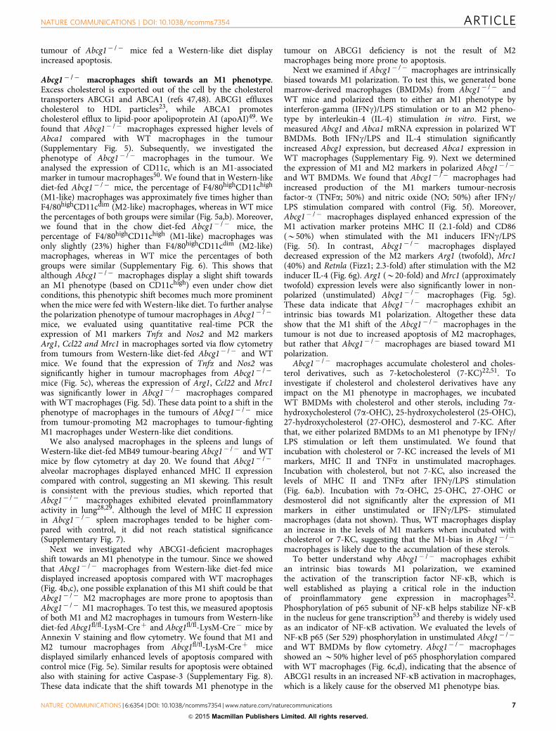
Abcg1� /� macrophages shift towards an M1 phenotype.Excess cholesterol is exported out of the cell by the cholesteroltransporters ABCG1 and ABCA1 (refs 47,48). ABCG1 effluxescholesterol to HDL particles23, while ABCA1 promotescholesterol efflux to lipid-poor apolipoprotein AI (apoAI)49. Wefound that Abcg1� /� macrophages expressed higher levels ofAbca1 compared with WT macrophages in the tumour(Supplementary Fig. 5). Subsequently, we investigated thephenotype of Abcg1� /� macrophages in the tumour. Weanalysed the expression of CD11c, which is an M1-associatedmarker in tumour macrophages50. We found that in Western-likediet-fed Abcg1� /� mice, the percentage of F4/80highCD11chigh
(M1-like) macrophages was approximately five times higher thanF4/80highCD11cdim (M2-like) macrophages, whereas in WT micethe percentages of both groups were similar (Fig. 5a,b). Moreover,we found that in the chow diet-fed Abcg1� /� mice, thepercentage of F4/80highCD11chigh (M1-like) macrophages wasonly slightly (23%) higher than F4/80highCD11cdim (M2-like)macrophages, whereas in WT mice the percentages of bothgroups were similar (Supplementary Fig. 6). This shows thatalthough Abcg1� /� macrophages display a slight shift towardsan M1 phenotype (based on CD11chigh) even under chow dietconditions, this phenotypic shift becomes much more prominentwhen the mice were fed with Western-like diet. To further analysethe polarization phenotype of tumour macrophages in Abcg1� /�
mice, we evaluated using quantitative real-time PCR theexpression of M1 markers Tnfa and Nos2 and M2 markersArg1, Ccl22 and Mrc1 in macrophages sorted via flow cytometryfrom tumours from Western-like diet-fed Abcg1� /� and WTmice. We found that the expression of Tnfa and Nos2 wassignificantly higher in tumour macrophages from Abcg1� /�
mice (Fig. 5c), whereas the expression of Arg1, Ccl22 and Mrc1was significantly lower in Abcg1� /� macrophages comparedwith WT macrophages (Fig. 5d). These data point to a shift in thephenotype of macrophages in the tumours of Abcg1� /� micefrom tumour-promoting M2 macrophages to tumour-fightingM1 macrophages under Western-like diet conditions.
We also analysed macrophages in the spleens and lungs ofWestern-like diet-fed MB49 tumour-bearing Abcg1� /� and WTmice by flow cytometry at day 20. We found that Abcg1� /�
alveolar macrophages displayed enhanced MHC II expressioncompared with control, suggesting an M1 skewing. This resultis consistent with the previous studies, which reported thatAbcg1� /� macrophages exhibited elevated proinflammatoryactivity in lung28,29. Although the level of MHC II expressionin Abcg1� /� spleen macrophages tended to be higher com-pared with control, it did not reach statistical significance(Supplementary Fig. 7).
Next we investigated why ABCG1-deficient macrophagesshift towards an M1 phenotype in the tumour. Since we showedthat Abcg1� /� macrophages from Western-like diet-fed micedisplayed increased apoptosis compared with WT macrophages(Fig. 4b,c), one possible explanation of this M1 shift could be thatAbcg1� /� M2 macrophages are more prone to apoptosis thanAbcg1� /� M1 macrophages. To test this, we measured apoptosisof both M1 and M2 macrophages in tumours from Western-likediet-fed Abcg1fl/fl-LysM-Creþ and Abcg1fl/fl-LysM-Cre� mice byAnnexin V staining and flow cytometry. We found that M1 andM2 tumour macrophages from Abcg1fl/fl-LysM-Creþ micedisplayed similarly enhanced levels of apoptosis compared withcontrol mice (Fig. 5e). Similar results for apoptosis were obtainedalso with staining for active Caspase-3 (Supplementary Fig. 8).These data indicate that the shift towards M1 phenotype in the
tumour on ABCG1 deficiency is not the result of M2macrophages being more prone to apoptosis.
Next we examined if Abcg1� /� macrophages are intrinsicallybiased towards M1 polarization. To test this, we generated bonemarrow-derived macrophages (BMDMs) from Abcg1� /� andWT mice and polarized them to either an M1 phenotype byinterferon-gamma (IFNg)/LPS stimulation or to an M2 pheno-type by interleukin-4 (IL-4) stimulation in vitro. First, wemeasured Abcg1 and Abca1 mRNA expression in polarized WTBMDMs. Both IFNg/LPS and IL-4 stimulation significantlyincreased Abcg1 expression, but decreased Abca1 expression inWT macrophages (Supplementary Fig. 9). Next we determinedthe expression of M1 and M2 markers in polarized Abcg1� /�
and WT BMDMs. We found that Abcg1� /� macrophages hadincreased production of the M1 markers tumour-necrosisfactor-a (TNFa; 50%) and nitric oxide (NO; 50%) after IFNg/LPS stimulation compared with control (Fig. 5f). Moreover,Abcg1� /� macrophages displayed enhanced expression of theM1 activation marker proteins MHC II (2.1-fold) and CD86(B50%) when stimulated with the M1 inducers IFNg/LPS(Fig. 5f). In contrast, Abcg1� /� macrophages displayeddecreased expression of the M2 markers Arg1 (twofold), Mrc1(40%) and Retnla (Fizz1; 2.3-fold) after stimulation with the M2inducer IL-4 (Fig. 6g). Arg1 (B20-fold) and Mrc1 (approximatelytwofold) expression levels were also significantly lower in non-polarized (unstimulated) Abcg1� /� macrophages (Fig. 5g).These data indicate that Abcg1� /� macrophages exhibit anintrinsic bias towards M1 polarization. Altogether these datashow that the M1 shift of the Abcg1� /� macrophages in thetumour is not due to increased apoptosis of M2 macrophages,but rather that Abcg1� /� macrophages are biased toward M1polarization.
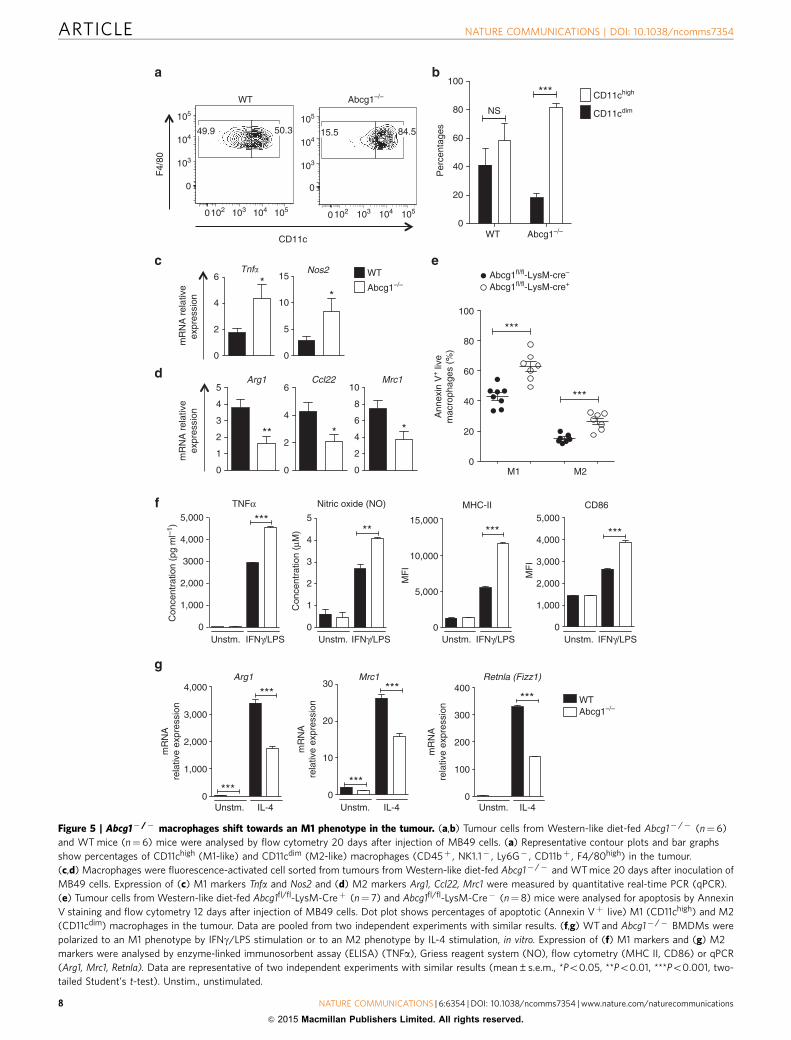
Abcg1� /� macrophages accumulate cholesterol and choles-terol derivatives, such as 7-ketocholesterol (7-KC)22,51. Toinvestigate if cholesterol and cholesterol derivatives have anyimpact on the M1 phenotype in macrophages, we incubatedWT BMDMs with cholesterol and other sterols, including 7a-hydroxycholesterol (7a-OHC), 25-hydroxycholesterol (25-OHC),27-hydroxycholesterol (27-OHC), desmosterol and 7-KC. Afterthat, we either polarized BMDMs to an M1 phenotype by IFNg/LPS stimulation or left them unstimulated. We found thatincubation with cholesterol or 7-KC increased the levels of M1markers, MHC II and TNFa in unstimulated macrophages.Incubation with cholesterol, but not 7-KC, also increased thelevels of MHC II and TNFa after IFNg/LPS stimulation(Fig. 6a,b). Incubation with 7a-OHC, 25-OHC, 27-OHC ordesmosterol did not significantly alter the expression of M1markers in either unstimulated or IFNg/LPS- stimulatedmacrophages (data not shown). Thus, WT macrophages displayan increase in the levels of M1 markers when incubated withcholesterol or 7-KC, suggesting that the M1-bias in Abcg1� /�
macrophages is likely due to the accumulation of these sterols.To better understand why Abcg1� /� macrophages exhibit
an intrinsic bias towards M1 polarization, we examinedthe activation of the transcription factor NF-kB, which iswell established as playing a critical role in the inductionof proinflammatory gene expression in macrophages52.Phosphorylation of p65 subunit of NF-kB helps stabilize NF-kBin the nucleus for gene transcription53 and thereby is widely usedas an indicator of NF-kB activation. We evaluated the levels ofNF-kB p65 (Ser 529) phosphorylation in unstimulated Abcg1� /�
and WT BMDMs by flow cytometry. Abcg1� /� macrophagesshowed an B50% higher level of p65 phosphorylation comparedwith WT macrophages (Fig. 6c,d), indicating that the absence ofABCG1 results in an increased NF-kB activation in macrophages,which is a likely cause for the observed M1 phenotype bias.
NATURE COMMUNICATIONS | DOI: 10.1038/ncomms7354 ARTICLE
NATURE COMMUNICATIONS | 6:6354 | DOI: 10.1038/ncomms7354 | www.nature.com/naturecommunications 7
& 2015 Macmillan Publishers Limited. All rights reserved.
0
20
40
60
80
100
M2M1
***
***
mR
NA
rel
ativ
eex
pres
sion
F4/
80
CD11c
0102 103 104 1050102 103 104 105
0
103
104
105
0
103
104
105
50.349.9
WT
84.515.5
Tnf�
0
2
4
6 *Nos2
0
5
10
15
*
WT
Abcg1–/–
mR
NA
rel
ativ
e e
xpre
ssio
n
WT
Arg1
0
1,000
2,000
3,000
4,000
mR
NA
rela
tive
expr
essi
on
***
***
Unstm. IL-4
Mrc1
0
10
20
30
mR
NA
rela
tive
expr
essi
on
***
***
Unstm. IL-4
Retnla (Fizz1)
0
100
200
300
400
mR
NA
rela
tive
expr
essi
on
***
Unstm. IL-4
TNFα
0
1,000
2,000
3000
4,000
5,000
Con
cent
ratio
n (p
g m
l–1)
Unstm.
***
0
1
2
3
4
5**
Unstm.
MHC-II
0
5,000
10,000
15,000
MF
I
Unstm.
***
CD86
0
1,000
2,000
3,000
4,000
5,000M
FI
***
Unstm.
Arg1
0
1
2
3
4
5
**
Ccl22
0
2
4
6
*
Mrc1
0
2
4
6
8
10
*P
erce
ntag
es
WT Abcg1–/–0
20
40
60
80
100
NS
*** CD11chigh
CD11cdim
Con
cent
ratio
n (μ
M)
Nitric oxide (NO)
Abcg1–/–
Abcg1fl/fl-LysM-cre+Abcg1fl/fl-LysM-cre–
Ann
exin
V+ li
vem
acro
phag
es (
%)
Abcg1–/–
IFNγ/LPS IFNγ/LPS IFNγ/LPS IFNγ/LPS
Figure 5 | Abcg1� /� macrophages shift towards an M1 phenotype in the tumour. (a,b) Tumour cells from Western-like diet-fed Abcg1� /� (n¼ 6)
and WT mice (n¼6) mice were analysed by flow cytometry 20 days after injection of MB49 cells. (a) Representative contour plots and bar graphs
show percentages of CD11chigh (M1-like) and CD11cdim (M2-like) macrophages (CD45þ , NK1.1� , Ly6G� , CD11bþ , F4/80high) in the tumour.
(c,d) Macrophages were fluorescence-activated cell sorted from tumours from Western-like diet-fed Abcg1� /� and WT mice 20 days after inoculation of
MB49 cells. Expression of (c) M1 markers Tnfa and Nos2 and (d) M2 markers Arg1, Ccl22, Mrc1 were measured by quantitative real-time PCR (qPCR).
(e) Tumour cells from Western-like diet-fed Abcg1fl/fl-LysM-Creþ (n¼ 7) and Abcg1fl/fl-LysM-Cre� (n¼ 8) mice were analysed for apoptosis by Annexin
V staining and flow cytometry 12 days after injection of MB49 cells. Dot plot shows percentages of apoptotic (Annexin Vþ live) M1 (CD11chigh) and M2
(CD11cdim) macrophages in the tumour. Data are pooled from two independent experiments with similar results. (f,g) WT and Abcg1� /� BMDMs were
polarized to an M1 phenotype by IFNg/LPS stimulation or to an M2 phenotype by IL-4 stimulation, in vitro. Expression of (f) M1 markers and (g) M2
markers were analysed by enzyme-linked immunosorbent assay (ELISA) (TNFa), Griess reagent system (NO), flow cytometry (MHC II, CD86) or qPCR
(Arg1, Mrc1, Retnla). Data are representative of two independent experiments with similar results (mean±s.e.m., *Po0.05, **Po0.01, ***Po0.001, two-
tailed Student’s t-test). Unstim., unstimulated.
ARTICLE NATURE COMMUNICATIONS | DOI: 10.1038/ncomms7354
8 NATURE COMMUNICATIONS | 6:6354 | DOI: 10.1038/ncomms7354 | www.nature.com/naturecommunications
& 2015 Macmillan Publishers Limited. All rights reserved.
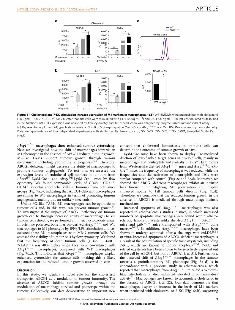
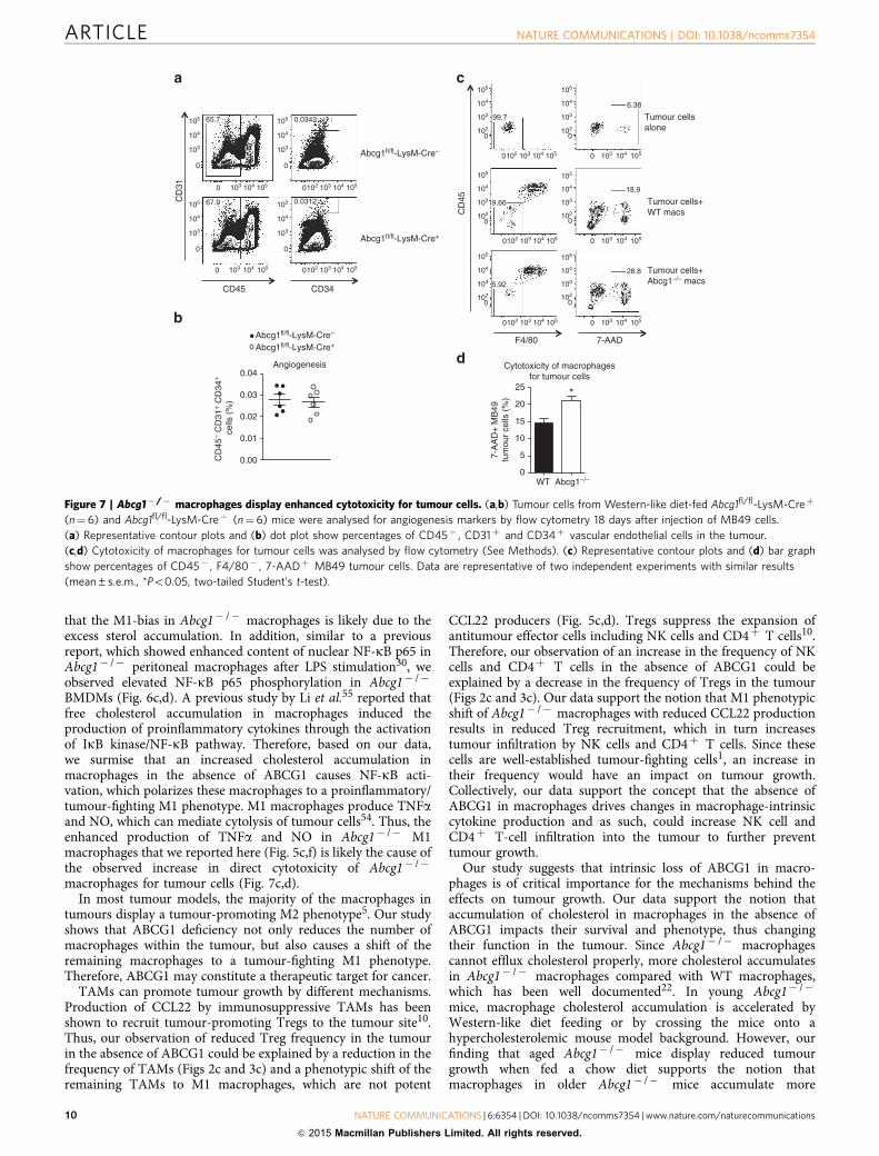
Abcg1� /� macrophages show enhanced tumour cytotoxicity.Next we investigated how the shift of macrophages towards anM1 phenotype in the absence of ABCG1 reduces tumour growth.M2-like TAMs support tumour growth through variousmechanisms including promoting angiogenesis6–9 Therefore,ABCG1 deficiency might decrease the ability of macrophages topromote tumour angiogenesis. To test this, we assessed theexpression levels of endothelial cell markers in tumours fromAbcg1fl/fl-LysM-Creþ and Abcg1fl/fl-LysM-Cre� mice by flowcytometry. We found comparable levels of CD45� CD31þ
CD34þ vascular endothelial cells in tumours from both micegroups (Fig. 7a,b), indicating that ABCG1-deficient macrophagesare similar to WT macrophages in terms of promoting tumourangiogenesis, making this an unlikely mechanism.
Unlike M2-like TAMs, M1 macrophages can be cytotoxic totumour cells and, in this way, can prevent tumour growth54.To investigate if the impact of ABCG1 deficiency on tumourgrowth can be through increased ability of macrophages to killtumour cells directly, we performed an in vitro cytotoxicity assay.In brief, we polarized bone marrow-derived Abcg1� /� and WTmacrophages to M1 phenotype by IFNg/LPS stimulation and co-cultured these M1 macrophages with MB49 tumour cells. Weassessed the viability of tumour cells by flow cytometry. We foundthat the frequency of dead tumour cells (CD45� F4/80�
7-AADþ ) was 40% higher when they were co-cultured withAbcg1� /� macrophages, compared with WT macrophages(Fig. 7c,d). This indicates that Abcg1� /� macrophages displayenhanced cytotoxicity for tumour cells, making this a likelyexplanation for the reduced tumour growth observed in vivo.
DiscussionIn this study, we identify a novel role for the cholesteroltransporter ABCG1 as a modulator of tumour immunity. Theabsence of ABCG1 inhibits tumour growth through themodulation of macrophage survival and phenotype within thetumour. Collectively, our data demonstrate an important new
concept that cholesterol homeostasis in immune cells candetermine the outcome of tumour growth in vivo.
LysM-Cre mice have been shown to display Cre-mediateddeletion of loxP-flanked target genes in myeloid cells, mainly inmacrophages and neutrophils and partially in DCs46. In tumoursfrom Western-like diet-fed Abcg1� /� mice and Abcg1fl/fl-LysM-Creþ mice, the frequency of macrophages was reduced, while thefrequencies and the activation of neutrophils and DCs weresimilar compared with control (Figs 2c and 3c,d). Moreover, weshowed that ABCG1-deficient macrophages exhibit an intrinsicbias toward tumour-fighting M1 polarization and displayenhanced ability to kill tumour cells directly (Fig. 7c,d).Therefore, we conclude that the reduced tumour growth in theabsence of ABCG1 is mediated through macrophage-intrinsicmechanisms.
Increased apoptosis of Abcg1� /� macrophages was alsoreported in atherosclerosis studies in mice, in which increasednumbers of apoptotic macrophages were found within athero-sclerotic lesions of Western-like diet-fed Abcg1� /� ApoE� /�
mice and Ldlr� /� mice transplanted with Abcg1� /� bonemarrow26,27. In addition, Abcg1� /� macrophages have beenshown to undergo apoptosis after a challenge with oxLDL26,51
in vitro. Increased apoptosis of ABCG1-deficient macrophages isa result of the accumulation of specific toxic oxysterols, including7-KC, which are known to induce apoptosis27,51. 7-KC andrelated oxysterols have been shown to be selectively exported outof the cell by ABCG1, but not by ABCA1 (ref. 51). Furthermore,the observed shift of Abcg1� /� macrophages in the tumourtowards a proinflammatory M1 phenotype (Fig. 5a–d) is inconcordance with a previous study in atherosclerosis, whichreported that macrophages from Abcg1� /� mice fed a Western-like/high-cholesterol diet exhibited elevated proinflammatoryactivity31. Macrophages are known to accumulate cholesterol inthe absence of ABCG1 (ref. 22). Our data demonstrate thatmacrophages display an increase in the levels of M1 markerswhen incubated with cholesterol or 7-KC (Fig. 6a,b), suggesting
0
1,000
2,000
3,000
4,000
5,000MHC-II
MF
ICholesterol
IFNγ /LPS IFNγ /LPS
IFNγ /LPS IFNγ /LPS
–
– –
+
+ +
– +
–
– –
+
+ +
– +
–
– –
+
+ +
– +
–
– –
+
+ +
– +
**
*
0102030
3,000
4,000
5,000
6,000
pg m
l–1
TNFα
**
*
Cholesterol
0
10
20
302,000
3,000
4,000
5,000
pg m
l–1
TNFα
7-KC
**
0
1,000
2,000
3,000
4,000
5,000
MF
I
MHC-II
***
**
7-KC
Cholesterol
7-KC
MF
I
WT 0
1,000
2,000
3,000 *
WT
% O
f max
NF-κB p-p65
Abcg1–/–
NF-κB p-p65
Abcg1–/–
Figure 6 | Cholesterol and 7-KC stimulation increase expression of M1 markers in macrophages. (a,b) WT BMDMs were preincubated with cholesterol
(20mg ml� 1) or 7-KC (5mM) for 2 h. After that, the cells were stimulated with IFNg (20 ng ml� 1) and LPS (100 ng ml� 1) or left unstimulated as described
in the Methods. MHC II expression was analysed by flow cytometry and TNFa production was analysed by enzyme-linked immunosorbent assay.
(c) Representative plot and (d) graph show levels of NF-kB p65 phosphorylation (Ser 529) in Abcg1� /� and WT BMDMs analysed by flow cytometry.
Data are representative of two independent experiments with similar results. (mean±s.e.m., *Po0.05, **Po0.01, ***Po0.001, two-tailed Student’s
t-test).
NATURE COMMUNICATIONS | DOI: 10.1038/ncomms7354 ARTICLE
NATURE COMMUNICATIONS | 6:6354 | DOI: 10.1038/ncomms7354 | www.nature.com/naturecommunications 9
& 2015 Macmillan Publishers Limited. All rights reserved.
that the M1-bias in Abcg1� /� macrophages is likely due to theexcess sterol accumulation. In addition, similar to a previousreport, which showed enhanced content of nuclear NF-kB p65 inAbcg1� /� peritoneal macrophages after LPS stimulation30, weobserved elevated NF-kB p65 phosphorylation in Abcg1� /�
BMDMs (Fig. 6c,d). A previous study by Li et al.55 reported thatfree cholesterol accumulation in macrophages induced theproduction of proinflammatory cytokines through the activationof IkB kinase/NF-kB pathway. Therefore, based on our data,we surmise that an increased cholesterol accumulation inmacrophages in the absence of ABCG1 causes NF-kB acti-vation, which polarizes these macrophages to a proinflammatory/tumour-fighting M1 phenotype. M1 macrophages produce TNFaand NO, which can mediate cytolysis of tumour cells54. Thus, theenhanced production of TNFa and NO in Abcg1� /� M1macrophages that we reported here (Fig. 5c,f) is likely the cause ofthe observed increase in direct cytotoxicity of Abcg1� /�
macrophages for tumour cells (Fig. 7c,d).In most tumour models, the majority of the macrophages in
tumours display a tumour-promoting M2 phenotype5. Our studyshows that ABCG1 deficiency not only reduces the number ofmacrophages within the tumour, but also causes a shift of theremaining macrophages to a tumour-fighting M1 phenotype.Therefore, ABCG1 may constitute a therapeutic target for cancer.
TAMs can promote tumour growth by different mechanisms.Production of CCL22 by immunosuppressive TAMs has beenshown to recruit tumour-promoting Tregs to the tumour site10.Thus, our observation of reduced Treg frequency in the tumourin the absence of ABCG1 could be explained by a reduction in thefrequency of TAMs (Figs 2c and 3c) and a phenotypic shift of theremaining TAMs to M1 macrophages, which are not potent
CCL22 producers (Fig. 5c,d). Tregs suppress the expansion ofantitumour effector cells including NK cells and CD4þ T cells10.Therefore, our observation of an increase in the frequency of NKcells and CD4þ T cells in the absence of ABCG1 could beexplained by a decrease in the frequency of Tregs in the tumour(Figs 2c and 3c). Our data support the notion that M1 phenotypicshift of Abcg1� /� macrophages with reduced CCL22 productionresults in reduced Treg recruitment, which in turn increasestumour infiltration by NK cells and CD4þ T cells. Since thesecells are well-established tumour-fighting cells1, an increase intheir frequency would have an impact on tumour growth.Collectively, our data support the concept that the absence ofABCG1 in macrophages drives changes in macrophage-intrinsiccytokine production and as such, could increase NK cell andCD4þ T-cell infiltration into the tumour to further preventtumour growth.
Our study suggests that intrinsic loss of ABCG1 in macro-phages is of critical importance for the mechanisms behind theeffects on tumour growth. Our data support the notion thataccumulation of cholesterol in macrophages in the absence ofABCG1 impacts their survival and phenotype, thus changingtheir function in the tumour. Since Abcg1� /� macrophagescannot efflux cholesterol properly, more cholesterol accumulatesin Abcg1� /� macrophages compared with WT macrophages,which has been well documented22. In young Abcg1� /�
mice, macrophage cholesterol accumulation is accelerated byWestern-like diet feeding or by crossing the mice onto ahypercholesterolemic mouse model background. However, ourfinding that aged Abcg1� /� mice display reduced tumourgrowth when fed a chow diet supports the notion thatmacrophages in older Abcg1� /� mice accumulate more
0.00
0.01
0.02
0.03
0.04Angiogenesis
CD45 CD34
CD
31
Abcg1fl/fl-LysM-Cre–
0
103
104
105
0
103
104
105
0
103
102
104
105
0
103
102
104
105
0
103
102
104
105
0
103
102
104
105
0
103
102
104
105
0
103
102
104
1050
103
104
105
0
103
104
105
0 103102 104 105
0 103102 104 105 0 103 104 105
0 103102 104 105 0 103 104 105
0 103102 104 105 0 103 104 105
0 103 104 105
0 103102 104 1050 103 104 105
65.7
Abcg1fl/fl-LysM-Cre+
CD
45
Tumour cellsalone
F4/80 7-AAD
Tumour cells+WT macs
Tumour cells+Abcg1–/– macs
7-A
AD
+ M
B49
tum
our
cells
(%
)
WT0
5
10
15
20
25*
Cytotoxicity of macrophagesfor tumour cells
0.0342
0.031267.9 9.66
5.92
28.8
18.9
6.38
99.7
Abcg1–/–
CD
45–
CD
31+ C
D34
+
cel
ls (
%)
Abcg1fl/fl-LysM-Cre–
Abcg1fl/fl-LysM-Cre+
Figure 7 | Abcg1� /� macrophages display enhanced cytotoxicity for tumour cells. (a,b) Tumour cells from Western-like diet-fed Abcg1fl/fl-LysM-Creþ
(n¼6) and Abcg1fl/fl-LysM-Cre� (n¼6) mice were analysed for angiogenesis markers by flow cytometry 18 days after injection of MB49 cells.
(a) Representative contour plots and (b) dot plot show percentages of CD45� , CD31þ and CD34þ vascular endothelial cells in the tumour.
(c,d) Cytotoxicity of macrophages for tumour cells was analysed by flow cytometry (See Methods). (c) Representative contour plots and (d) bar graph
show percentages of CD45� , F4/80� , 7-AADþ MB49 tumour cells. Data are representative of two independent experiments with similar results
(mean±s.e.m., *Po0.05, two-tailed Student’s t-test).
ARTICLE NATURE COMMUNICATIONS | DOI: 10.1038/ncomms7354
10 NATURE COMMUNICATIONS | 6:6354 | DOI: 10.1038/ncomms7354 | www.nature.com/naturecommunications
& 2015 Macmillan Publishers Limited. All rights reserved.
cholesterol over time44. Thus, in the aged Abcg1� /� mice, thetumour phenotype becomes evident even on a chow diet.
Enhanced expression of ABCG1 but reduced expression ofABCA1 after LPS/IFNg or IL-4 stimulation in WT macrophages(Supplementary Fig. 9) point to an importance of ABCG1 inmacrophage activation. Increased expression of ABCA1 inAbcg1� /� TAMs is not surprising (Supplementary Fig. 5), sincewe32,34 and others36,56 have previously shown that geneticdeletion of one cholesterol transporter, either ABCG1 orABCA1, is compensated for by an upregulation of the othertransporter. Nevertheless, neither ABCG1 nor ABCA1 can fullycompensate for the loss of the other20. However, we cannot ruleout the possibility that changes in ABCA1 expression contributedto our observed findings of reduced tumour growth in Abcg1� /�
mice. Future studies using ABCA1-deficient mice will beuseful to delineate the roles of these two transporters in tumourimmunity.
From many perspectives, atherosclerosis and cancer arefundamentally different. However, the immune system plays amajor role in the progression of both diseases. Human populationand animal studies clearly demonstrate that HDL protects againstatherosclerosis57. Interestingly, meta-analysis of lipid-alteringtherapies has indicated an inverse relationship between plasmaHDL levels and incidence of cancer58. In concordance with thisreport, a recent study has shown that apoAI, the major proteincomponent of HDL, suppresses tumour growth and metastasis inmice via the modulation of immune responses59. Collectively,these studies suggest that HDL has a role both in atherosclerosisand cancer. ABCG1 deficiency in immune cells has been shownto protect mice from atherosclerosis development26,36. Bydemonstrating that myeloid cell-specific ABCG1 deletionsuppresses tumour growth, our present study suggests thatABCG1 might link immunity in atherosclerosis and cancer.
In sum, our study identifies ABCG1 as a novel mediator ofantitumour immune responses. It defines an important role forcholesterol transporters in tumour immunity and provides a linkbetween lipid homeostasis and cancer. Understanding howABCG1 and cholesterol metabolism in immune cells impactsantitumour immune responses could lead to development ofentirely new therapeutic approaches for cancer immunotherapy.
MethodsMice. C57BL/6J mice (000664), Ldlr� /� mice (002207) and ApoE� /� mice(002052) were purchased from The Jackson Laboratory (Bar Harbor, ME).Abcg1� /� /lacZ knock-in mice were purchased from Deltagen (San Mateo, CA)and are congenic to a C57BL/6J background (backcrossed 14 generations).B6.SJL-Ptprca/BoyAiTac mice (CD45.1 congenic, 004007) were purchased fromTaconic Farms (Germantown, NY). Abcg1� /� mice were crossed with Ldlr� /�
and ApoE� /� mice to obtain Abcg1� /� Ldlr� /� and Abcg1� /�ApoE� /�
mice, respectively. Conditional knockout Abcg1fl/fl mice (C57BL/6J background) inwhich loxP sites flank the Walker domain of exon 3 of Abcg1 were generated forour laboratory using InGenious Targeting Laboratory (New York). An B10.3-kbregion used to construct the targeting vector was first subcloned from a positivelyidentified BAC clone using homologous recombination. The region was designedsuch that the short homology arm extends 2.2 kb 50 to lox P/FRT-flanked Neocassette. The long homology arm ends on the 30 side of lox P/FRT-flanked Neocassette and is B8.1-kb long. The single lox P site is inserted upstream of exon 3,and the lox P/FRT-flanked Neo cassette is inserted downstream of exon 3. Thetarget region is 1.5 kb including exon 3. The targeting vector is confirmed byrestriction analysis after each modification step and by sequencing using primersdesigned to read from the selection cassette into the 30 end of the long homologyarm (N7) and the 50 end of the short homology arm (N1), or from primers thatanneal to the vector sequence, P6 and T7, and read into the 50 and 30 ends of theBAC sub clone. Abcg1fl/fl mice were crossed with Lck-Cre mice (003802, TheJackson Laboratory) to obtain Abcg1fl/fl-Lck-Creþ and control Abcg1fl/fl-Lck-Cre�
mice, and crossed with LysM-Cre mice (004781, The Jackson Laboratory) to obtainAbcg1fl/fl-LysM-Creþ and Abcg1fl/fl-LysM-Cre� mice. All the mice used in thisstudy were female and 7–10 weeks old, except for the tumour growth experiment inthe aged Abcg1� /� and C57BL/6 (WT) mice, which were 6–7 months old. Micewere fed a standard rodent chow diet containing 0% cholesterol and 5% caloriesfrom fat (Pico lab, #5053) or Western-like diet containing 0.2% cholesterol and
42% calories from fat (Harlan Laboratories, #TD88137). The mice were housed inmicroisolator cages in a pathogen-free animal facility of the La Jolla Institute forAllergy and Immunology.
The plasma lipid profile analyses were performed at day 12, because this is thevery earliest time that significant differences were observed in tumour growth. Day12 is also the time when differences in macrophage apoptosis were observed, whichthen later on leads to a more prominent difference in tumour growth. Tumourgrowth was measured until days 18–20 and that is the time when the frequency andphenotype of the immune cells within the tumour were analysed.
All the experiments followed the guidelines of the La Jolla Institute for Allergyand Immunology Animal Care and Use Committee and approval for use of rodentswas obtained from the La Jolla Institute for Allergy and Immunology according tocriteria outlined in the Guide for the Care and Use of Laboratory Animals from theNational Institutes of Health. Mice were euthanized by CO2 inhalation.
Cell lines and reagents. MB49-bladder carcinoma and B16-F1 melanoma cellswere derived from C57BL/6 mice and obtained from American Type CultureCollection. The B16-F10-luc2 cell line was established by Caliper Life Sciencesby transduction of lentivirus containing luciferase 2 gene under the control ofhuman ubiquitin C promoter. Tumour cells were cultured in R5 medium con-taining RPMI 1640, 5% heat-inactivated fetal bovine serum, 50 U ml� 1 penicillinand 50mg ml� 1 streptomycin. Cells were injected into mice after reaching 60–80%confluency.
Flow cytometry antibodies including anti-mouse APC-F4/80 (BM8; 1/100),FITC-Ly6G (RB6-8C5; 1/200), APC/Cy7 or AF700-CD45 (30-F11; 1/200),AF700-CD45.2 (104; 1/200), PerCP/Cy5.5-NK1.1 (PK136; 1/100), e-Fluor 450-CD4 (RM4-5; 1/200), FITC-TCRb (H57-597; 1/400), PE-CD25 (PC61.5; 1/100),APC-Foxp3 (FJK-16s; 1/100), AF700-CD34 (RAM34; 1/200) and e-Fluor 450-MHC II (M5/114.15.2; 1/200), were purchased from eBioscience (San Diego, CA);PE-Cy7-CD11b (M1/70; 1/800), PE-CD11c (HL3; 1/300), FITC-CD45.1 (A20;1/200), APC/Cy7-CD8a (53-6.7; 1/200), PE-Cy7-CD69 (H1.2F3; 1/400), PE-CD31(MEC 13.3; 1/300) and PE-phospho-p65 (S529; K10-895.12.50; 1/10), werepurchased from BD Biosciences (San Jose, California); PerCP/Cy5.5-CD19 (6D5;1/100), PE-CD115 (AFS98; 1/100) and AF700-CD86 (GL-1; 1/200) were purchasedfrom Biolegend (San Diego, CA). CD16/CD32 (2.4G2; 1/200) antibody waspurchased from BD Biosciences. Ultrapure LPS (Escherichia coli 0111:B4) waspurchased from InvivoGen (San Diego, CA), murine rIFNg and rIL-4 werepurchased from R&D Systems (Minneapolis, MN), murine macrophage colony-stimulating factor (M-CSF) was purchased from PeproTech (Rocky Hill, NJ) andRPMI 1640 medium was purchased from Invitrogen (Carlsbad, CA). Fetal bovineserum, Collagenase IV, water-soluble cholesterol, 7a-OHC, 25-OHC, 27-OHC,desmosterol and 7-KC were purchased from Sigma-Aldrich (St Louis, MO). DNaseI was purchased from Roche (Basel, Switzerland), PBS was purchased from ThermoScientific (Rockford, IL) and Ficoll-Paque plus was purchased from GE Healthcare(Pittsburgh, PA).
Measurement of tumour growth/metastasis and survival. MB49 or B16-F1cells (105) in 100ml PBS were injected subcutaneously into the right flanks offemale age-matched 7–10-week-old Abcg1� /� , C57BL/6 (WT), Abcg1� /�
Ldlr� /� , Ldlr� /� , Abcg1� /�ApoE� /� , ApoE� /� , Abcg1fl/fl-Lck-Creþ ,Abcg1fl/fl-Lck-Cre� , Abcg1fl/fl-LysM-Creþ and Abcg1fl/fl-LysM-Cre� mice or6–7-month-old Abcg1� /� and WT mice. Mice were fed with either Western-likediet or chow diet beginning a week before injection of tumour cells. Tumourdiameters were measured using a digital caliper and tumour volume was calculatedusing the formula V¼D� d2/2, where V is the tumour volume, D is the largestmeasured tumour diameter and d is the smallest measured tumour diameter. Forthe survival experiments, the mice with tumour volume reaching 2,000 mm3 wereconsidered as dead and euthanized. To measure spontaneous lung metastasis, 105
luciferase-expressing B16-F10 cells (B16-F10-luc2) in 100ml PBS were injected intofemale age-matched Abcg1� /� and C57BL/6 mice subcutaneously. Mice were fedwith Western-like diet beginning a week before injection of tumour cells. B16-F10-luc2 tumours grow more aggressively than B16-F1 tumours, which allowed us tochoose mice for study that had similar-sized tumours. At 28 days after B16-F10-luc2 tumour inoculation, mice with similar, but average, tumour sizes in bothgroups were anaesthetized by inhalation of isoflurane (Butler Animal HealthSupply) and 1 mg D-Luciferin (Caliper Life Sciences, Waltham, MA) in 100 ml PBSwas delivered into each mouse retro-orbitally. Mice were euthanized 2 min after D-Luciferin injection, lungs were harvested and lung metastases were measured usingan IVIS 200 Bioluminescence Imager (Caliper Life Sciences, Hopkinton, MA).
Measurement of plasma lipoproteins. Mice were fed with either chow diet orWestern-like diet beginning a week before injection of tumour cells. Blood(in EDTA) was collected from chow diet-fed or Western-like diet-fed mice 12 daysafter MB49 tumour inoculation. Plasma lipoprotein profiles were obtained by fastprotein liquid chromatography as described previously60,61. In brief, equal volumesof plasma from five mice per group were pooled and 200ml of this pooled plasmawas applied to a set of 2 Superose 6 (HR 10/30) columns linked in series.Lipoproteins were eluted by size exclusion into 0.5-ml fractions in EDTA/NaCl/NaN3 (1 mmol l� 1; 0.154 mol l� 1; 0.02%) at a flow rate of 0.5 ml min� 1.
NATURE COMMUNICATIONS | DOI: 10.1038/ncomms7354 ARTICLE
NATURE COMMUNICATIONS | 6:6354 | DOI: 10.1038/ncomms7354 | www.nature.com/naturecommunications 11
& 2015 Macmillan Publishers Limited. All rights reserved.
Cholesterol was measured in each fraction using an enzymatic cholesterol kit(Wako) according to the manufacturers’ instructions.
Generation of bone marrow chimeras. Recipient B6.SJL mice were irradiated intwo doses of 500 rad each (for a total of 1,000 rad) 4 h apart. Bone marrow cellsfrom both femurs and tibias of B6.SJL (CD45.1) and Abcg1� /� (CD45.2) donormice were collected under sterile conditions. Bones were centrifuged for the col-lection of marrow and the cells were washed and resuspended in PBS for injection.Bone marrow cells (107) from B6.SJL or Abcg1� /� mice in 200ml PBS weredelivered retro-orbitally into each recipient mouse. Recipient mice were housed ina barrier facility under pathogen-free conditions and were provided autoclavedacidified water with antibiotics (trimethoprim-sulfamethoxazole) and were fedautoclaved food. The chimeric mice were fed with Western-like diet starting6 weeks after bone marrow reconstitution.
Flow cytometry. Tumours were meshed through a 100-mm strainer (FisherScientific, Pittsburg, PA) and then filtered through a 40-mm strainer. Single-cellsuspension was resuspended in 100 ml flow cytometry staining buffer (1% bovineserum albumin plus 0.1% sodium azide in PBS). Fcg receptors were blocked withCD16/32-blocking antibody for 10 min and surface antigens on cells were stainedfor 30 min at 4 �C. LIVE/DEAD Fixable Dead Cell Stain (Invitrogen) was used foranalysis of viability, and forward- and side-scatter parameters were used forexclusion of doublets from analysis. Antibody clones and dilutions used are listedabove. For intracellular staining, cells were fixed and permeabilized with theCytofix/Cytoperm Fixation/Permeabilization Solution Kit (BD Biosciences; forcytoplamis proteins) or Foxp3 Staining Buffer Set (eBioscience; for nuclear pro-teins) after the cell surface staining. Cells were stained with directly conjugatedfluorescent of Foxp3 antibody for 30 min at 4 �C and with directly conjugatedfluorescent of NF-kB phospho-p65 (Ser 529) antibody for 30 min at RT. Apoptosisof macrophages in tumour was measured by flow cytometry using a PE AnnexinV Apoptosis Detection Kit 1 or a FITC active Caspase-3 Apoptosis Kit (BDBiosciences) according to the manufacturer’s instructions.
Cell fluorescence was assessed using LSR-II (BD Biosciences) and data wereanalysed with FlowJo software (TreeStar, Ashland, OR). Macrophages (CD45þ ,NK1.1� , Ly6G� , CD11bþ , F4/80high), neutrophils (CD45þ , NK1.1� , Ly6Gþ ,CD11bþ ), myeloid DCs (CD45þ , NK1.1� , Ly6G� , F4/80� , CD11bþ
CD11cþ ), monocytes (CD45þ , NK1.1� , Ly6G� , CD11bþ , CD115þ ), NK cells(CD45þ , TCRb� , NK1.1þ ), CD4þ T cells (CD45þ , TCRbþ , NK1.1� , CD4þ ),CD8þ T cells (CD45þ , TCRbþ , NK1.1� , CD8þ ), NKT cells (CD45þ , TCRbþ ,NK1.1þ ) and Tregs (CD45þ , TCRbþ , NK1.1� , CD4þ ,CD25þ , Foxp3þ ) wereidentified with the appropriate gating.
Cell sorting. Tumours from WT and Abcg1� /� mice at day 20 were enriched forCD11bþ cells by positive selection with mouse CD11bþ positive selection kit(Stem Cell Technologies, Vancouver, Canada) according to the manufacturer’sinstructions, before cell sorting. Surface antigens on enriched CD11bþ cells werethen stained as described above, followed by macrophage (Ly6G� , NK1.1� ,CD11bþ , F4/80high) sorting with a FACSAria cytometer (BD Biosciences).Peritoneal lavage from Abcg1fl/fl-LysM-Creþ and Abcg1fl/fl-LysM-Cre� mice5 days post thioglycollate injection was sorted for macrophages (F4/80high) andsplenocytes from Abcg1fl/fl-Lck-Creþ and Abcg1fl/fl-Lck-Cre� mice were sortedfor T cells (CD3þ ) using FACSAria cytometer.
Tumour angiogenesis. Tumours were minced and digested with Collagenase IV(400 U ml� 1) in the presence of DNase I (20 mg ml� 1) in RPMI medium at 37 �Cfor 30 min. Cell suspension was filtered through a 40-mm strainer and resuspendedin warm R5 medium and incubated at 37 �C for 30 min. The cells were stained withfluorophore-conjugated antibodies against CD45, CD31, CD34 and analysed byflow cytometry.
Generation and M1/M2 polarization of BMDMs. BMDMs were prepared asdescribed previously62,63. In brief, bone marrow cells were cultured in standardtissue culture plates in the presence of 10 ng ml� 1 M-CSF overnight. Non-adherent cells from this initial culture were then transferred to low-attachment six-well plates (Corning Life Sciences, Tewksbury MA) in 4 ml R5 medium containing30% L929 conditioned medium and 10 ng ml� 1 M-CSF per well for 7 days, addingmore medium on days 3 and 6. After that, the cells were purified by centrifugationover Ficoll-Paque plus. Cells were verified to be 98% CD11bþ , F4/80þ , MHCIIlow, CD80low, CD86low by flow cytometry.
WT and Abcg1� /� BMDMs were allowed to rest at 37 �C over night beforestimulation. For M1 polarization, macrophages were either stimulated with IFNg(20 ng ml� 1) for 12 h followed by LPS (100 ng ml� 1) stimulation for 4 h (for MHCII and CD86 analysis) or stimulated with IFNg (20 ng ml� 1)þ LPS (100 ng ml� 1)overnight (for TNFa and NO analysis). For M2 polarization, macrophages werestimulated with IL-4 (20 ng ml� 1) for 16 h.
Stimulation of macrophages with cholesterol and -derivatives. WT BMDMswere allowed to rest at 37 �C over night before stimulation. The next day,macrophages were preincubated with water-soluble cholesterol (20 mg ml� 1),7a-OHC (1mM), 25-OHC (1mM), 27-OHC (1mM), desmosterol (5 mM) or 7-KC(5 mM) for 2 h. Next, macrophages were either left unstimulated or stimulated withIFNg (20 ng ml� 1) for 12 h followed by LPS (100 ng ml� 1) stimulation for 4 h(for MHC II analysis) or stimulated with IFNg (20 ng ml� 1)þ LPS (100 ng ml� 1)overnight (for TNFa analysis).
Cytokine and NO measurements. The supernatants were collected and TNFawas measured by ELISA (eBioscience) and NO (as nitrite) was measured by theGriess Reagent System (Promega, Madison, WI) according to the manufacturers’instructions.
In vitro tumour cytotoxicity assay. WT and Abcg1� /� BMDMs were plated inround well 96-well plates as 4� 105 cells per well in 200 ml R5 and stimulated withIFNg (20 ng ml� 1) and LPS (100 ng ml� 1) for 24 h. The cells were washed withR5 twice and co-cultured with 104 MB49 tumour cells (40:1 ratio of effector(macrophages): target cells (tumour cells) ). Twenty four hours later, the cellswere washed with PBS and treated with Accutase cell detachment solution (BDBiosciences). Tumour cell viability was determined with 7-aminoactinomycin D(7-AAD) staining by flow cytometry. Tumour cytotoxicity was calculated as % of7-AADþ tumour cells (CD45� , F4/80� ) co-cultured with macrophages (CD45þ ,F4/80þ )—% 7-AADþ of tumour cells alone.
Quantitative real-time PCR. Total cellular RNA of macrophages was collectedwith an RNeasy Plus Micro Kit according to the manufacturer’s protocol (Qiagen,Valencia, CA). RNA purity and quantity was measured with a nanodrop spec-trophotometer (Thermo Scientific). Approximately 500 ng RNA was used forsynthesis of cDNA with an Iscript cDNA Synthesis Kit (Bio-Rad, Hercules, CA).Total cDNA was diluted 1:20 in H2O and a volume of 9 ml was used for eachreal-time condition with a MyIQ Single-Color Real-Time PCR DetectionSystem (Bio-Rad) and TaqMan Gene Expression Mastermix and Arg1(# Mm00475988_m1), Ccl22 (# Mm00436439_m1), Mrc1 (# Mm00485148_m1),Tnfa (# Mm00443258_m1), Rtnla (# Mm00445109_m1), Nos2 (# Mm00440502_m1), Abcg1 (# Mm01348250_m1) and Abca1 (# Mm01350760_m1) TaqManprimers (Applied Biosystems). Data were analysed and presented on the basis ofthe relative expression method64. The formula for this calculation is as follows:relative expression¼ 2� (SD Ct–CDC t) where DCt is the difference in the thresholdcycle between the gene of interest and the housekeeping gene (18S), S is theAbcg1� /� mouse and C is the WT mouse.
Statistical analyses. Data for all experiments were analysed with Prism software(GraphPad). Two-way analysis of variance test, long-rank test, unpaired Student’st-test and Wilcoxon-matched-pairs signed rank test were used for comparison ofexperimental groups when appropriate. The data shown are the means±s.e.m.P values of less than 0.05 were considered statistically significant.
References1. Grivennikov, S. I., Greten, F. R. & Karin, M. Immunity, inflammation, and
cancer. Cell 140, 883–899.2. Whiteside, T. L. The tumor microenvironment and its role in promoting tumor
growth. Oncogene 27, 5904–5912 (2008).3. Mantovani, A., Sica, A. & Locati, M. Macrophage polarization comes of age.
Immunity 23, 344–346 (2005).4. Martinez, F. O., Helming, L. & Gordon, S. Alternative activation of
macrophages: an immunologic functional perspective. Annu. Rev. Immunol.27, 451–483 (2009).
5. Solinas, G., Germano, G., Mantovani, A. & Allavena, P. Tumor-associatedmacrophages (TAM) as major players of the cancer-related inflammation.J. Leukoc. Biol. 86, 1065–1073 (2009).
6. Coussens, L. M. & Werb, Z. Inflammation and cancer. Nature 420, 860–867(2002).
7. Lewis, C. E. & Pollard, J. W. Distinct role of macrophages in different tumormicroenvironments. Cancer Res. 66, 605–612 (2006).
8. Mantovani, A., Sozzani, S., Locati, M., Allavena, P. & Sica, A. Macrophagepolarization: tumor-associated macrophages as a paradigm for polarized M2mononuclear phagocytes. Trends Immunol. 23, 549–555 (2002).
9. Pollard, J. W. Tumour-educated macrophages promote tumour progressionand metastasis. Nat. Rev. Cancer 4, 71–78 (2004).
10. Nishikawa, H. & Sakaguchi, S. Regulatory T cells in tumor immunity. Int. J.Cancer 127, 759–767.
11. Sica, A. et al. Macrophage polarization in tumour progression. Semin. CancerBiol. 18, 349–355 (2008).
12. Hiraoka, K. et al. Inhibition of bone and muscle metastases of lung cancercells by a decrease in the number of monocytes/macrophages. Cancer Sci. 99,1595–1602 (2008).
ARTICLE NATURE COMMUNICATIONS | DOI: 10.1038/ncomms7354
12 NATURE COMMUNICATIONS | 6:6354 | DOI: 10.1038/ncomms7354 | www.nature.com/naturecommunications
& 2015 Macmillan Publishers Limited. All rights reserved.
13. Lin, E. Y. et al. Macrophages regulate the angiogenic switch in a mouse modelof breast cancer. Cancer Res. 66, 11238–11246 (2006).
14. Lin, E. Y., Nguyen, A. V., Russell, R. G. & Pollard, J. W. Colony-stimulatingfactor 1 promotes progression of mammary tumors to malignancy. J. Exp. Med.193, 727–740 (2001).
15. Miselis, N. R., Wu, Z. J., Van Rooijen, N. & Kane, A. B. Targeting tumor-associated macrophages in an orthotopic murine model of diffuse malignantmesothelioma. Mol. Cancer Ther. 7, 788–799 (2008).
16. Zeisberger, S. M. et al. Clodronate-liposome-mediated depletion of tumour-associated macrophages: a new and highly effective antiangiogenic therapyapproach. Br. J. Cancer 95, 272–281 (2006).
17. Colombo, M. P. & Mantovani, A. Targeting myelomonocytic cells to revertinflammation-dependent cancer promotion. Cancer Res. 65, 9113–9116 (2005).
18. Guiducci, C., Vicari, A. P., Sangaletti, S., Trinchieri, G. & Colombo, M. P.Redirecting in vivo elicited tumor infiltrating macrophages and dendritic cellstowards tumor rejection. Cancer Res. 65, 3437–3446 (2005).
19. Ostrand-Rosenberg, S., Grusby, M. J. & Clements, V. K. Cutting edge: STAT6-deficient mice have enhanced tumor immunity to primary and metastaticmammary carcinoma. J. Immunol. 165, 6015–6019 (2000).
20. Tarr, P. T., Tarling, E. J., Bojanic, D. D., Edwards, P. A. & Baldan, A. Emergingnew paradigms for ABCG transporters. Biochim. Biophys. Acta 1791, 584–593(2009).
21. Ikonen, E. Cellular cholesterol trafficking and compartmentalization. Nat. Rev.Mol. Cell Biol. 9, 125–138 (2008).
22. Kennedy, M. A. et al. ABCG1 has a critical role in mediating cholesterol efflux toHDL and preventing cellular lipid accumulation. Cell Metab. 1, 121–131 (2005).
23. Wang, N., Lan, D., Chen, W., Matsuura, F. & Tall, A. R. ATP-binding cassettetransporters G1 and G4 mediate cellular cholesterol efflux to high-densitylipoproteins. Proc. Natl Acad. Sci. USA 101, 9774–9779 (2004).
24. Sturek, J. M. et al. An intracellular role for ABCG1-mediated cholesteroltransport in the regulated secretory pathway of mouse pancreatic beta cells.J. Clin. Invest. 120, 2575–2589 (2010).
25. Tarling, E. J. & Edwards, P. A. ATP binding cassette transporter G1 (ABCG1) isan intracellular sterol transporter. Proc. Natl Acad. Sci. USA 108, 19719–19724(2011).
26. Baldan, A. et al. Impaired development of atherosclerosis in hyperlipidemicLdlr� /� and ApoE� /� mice transplanted with Abcg1� /� bone marrow.Arterioscler. Thromb. Vasc. Biol. 26, 2301–2307 (2006).
27. Tarling, E. J. et al. Impaired development of atherosclerosis in Abcg1� /�Apoe� /� mice: identification of specific oxysterols that both accumulate inAbcg1� /� Apoe� /� tissues and induce apoptosis. Arterioscler. Thromb.Vasc. Biol. 30, 1174–1180.
28. Baldan, A., Gomes, A. V., Ping, P. & Edwards, P. A. Loss of ABCG1 results inchronic pulmonary inflammation. J. Immunol. 180, 3560–3568 (2008).
29. Wojcik, A. J., Skaflen, M. D., Srinivasan, S. & Hedrick, C. C. A critical role forABCG1 in macrophage inflammation and lung homeostasis. J. Immunol. 180,4273–4282 (2008).
30. Yvan-Charvet, L. et al. Increased inflammatory gene expression in ABCtransporter-deficient macrophages: free cholesterol accumulation, increasedsignaling via toll-like receptors, and neutrophil infiltration of atheroscleroticlesions. Circulation 118, 1837–1847 (2008).
31. Yvan-Charvet, L. et al. Combined deficiency of ABCA1 and ABCG1 promotesfoam cell accumulation and accelerates atherosclerosis in mice. J. Clin. Invest.117, 3900–3908 (2007).
32. Armstrong, A. J., Gebre, A. K., Parks, J. S. & Hedrick, C. C. ATP-bindingcassette transporter G1 negatively regulates thymocyte and peripherallymphocyte proliferation. J. Immunol. 184, 173–183.
33. Bensinger, S. J. et al. LXR signaling couples sterol metabolism to proliferation inthe acquired immune response. Cell 134, 97–111 (2008).
34. Sag, D. et al. ATP-binding cassette transporter G1 intrinsically regulatesinvariant NKT cell development. J. Immunol. 189, 5129–5138 (2012).
35. Baldan, A. et al. Deletion of the transmembrane transporter ABCG1 results inprogressive pulmonary lipidosis. J. Biol. Chem. 281, 29401–29410 (2006).
36. Ranalletta, M. et al. Decreased atherosclerosis in low-density lipoproteinreceptor knockout mice transplanted with Abcg1� /� bone marrow.Arterioscler. Thromb. Vasc. Biol. 26, 2308–2315 (2006).
37. Plump, A. S. et al. Severe hypercholesterolemia and atherosclerosis inapolipoprotein E-deficient mice created by homologous recombination in EScells. Cell 71, 343–353 (1992).
38. Zhang, S. H., Reddick, R. L., Piedrahita, J. A. & Maeda, N. Spontaneoushypercholesterolemia and arterial lesions in mice lacking apolipoprotein E.Science 258, 468–471 (1992).
39. Ishibashi, S. et al. Hypercholesterolemia in low density lipoprotein receptorknockout mice and its reversal by adenovirus-mediated gene delivery. J. Clin.Invest. 92, 883–893 (1993).
40. Ishibashi, S., Goldstein, J. L., Brown, M. S., Herz, J. & Burns, D. K. Massivexanthomatosis and atherosclerosis in cholesterol-fed low density lipoproteinreceptor-negative mice. J. Clin. Invest. 93, 1885–1893 (1994).
41. Ishibashi, S., Herz, J., Maeda, N., Goldstein, J. L. & Brown, M. S. The two-receptor model of lipoprotein clearance: tests of the hypothesis in ‘knockout’mice lacking the low density lipoprotein receptor, apolipoprotein E, or bothproteins. Proc. Natl Acad. Sci. USA 91, 4431–4435 (1994).
42. Nathanson, S. D., Haas, G. P., Mead, M. J. & Lee, M. Spontaneous regionallymph node metastases of three variants of the B16 melanoma: relationshipto primary tumor size and pulmonary metastases. J. Surg. Oncol. 33, 41–45(1986).
43. Stackpole, C. W. Intrapulmonary spread of established B16 melanoma lungmetastases and lung colonies. Invasion Metastasis 10, 267–280 (1990).
44. Swann, J., Crowe, N. Y., Hayakawa, Y., Godfrey, D. I. & Smyth, M. J. Regulationof antitumour immunity by CD1d-restricted NKT cells. Immunol. Cell Biol. 82,323–331 (2004).
45. Hennet, T., Hagen, F. K., Tabak, L. A. & Marth, J. D. T-cell-specific deletion ofa polypeptide N-acetylgalactosaminyl-transferase gene by site-directedrecombination. Proc. Natl Acad. Sci. USA 92, 12070–12074 (1995).
46. Clausen, B. E., Burkhardt, C., Reith, W., Renkawitz, R. & Forster, I. Conditionalgene targeting in macrophages and granulocytes using LysMcre mice.Transgenic Res. 8, 265–277 (1999).
47. Gelissen, I. C. et al. ABCA1 and ABCG1 synergize to mediate cholesterol exportto apoA-I. Arterioscler. Thromb. Vasc. Biol. 26, 534–540 (2006).
48. Vaughan, A. M. & Oram, J. F. ABCA1 and ABCG1 or ABCG4 act sequentiallyto remove cellular cholesterol and generate cholesterol-rich HDL. J. Lipid Res.47, 2433–2443 (2006).
49. Vedhachalam, C. et al. Mechanism of ATP-binding cassette transporterA1-mediated cellular lipid efflux to apolipoprotein A-I and formation of highdensity lipoprotein particles. J. Biol. Chem. 282, 25123–25130 (2007).
50. Movahedi, K. et al. Different tumor microenvironments contain functionallydistinct subsets of macrophages derived from Ly6C(high) monocytes. CancerRes. 70, 5728–5739 (2010).
51. Terasaka, N., Wang, N., Yvan-Charvet, L. & Tall, A. R. High-densitylipoprotein protects macrophages from oxidized low-density lipoprotein-induced apoptosis by promoting efflux of 7-ketocholesterol via ABCG1. Proc.Natl Acad. Sci. USA 104, 15093–15098 (2007).
52. Karin, M. & Delhase, M. The I kappa B kinase (IKK) and NF-kappa B: keyelements of proinflammatory signalling. Semin. Immunol. 12, 85–98 (2000).
53. Hu, J., Nakano, H., Sakurai, H. & Colburn, N. H. Insufficient p65phosphorylation at S536 specifically contributes to the lack of NF-kappaBactivation and transformation in resistant JB6 cells. Carcinogenesis 25,1991–2003 (2004).
54. Escorcio-Correia, M. & Hagemann, T. Measurement of tumor cytolysis bymacrophages. Curr. Protoc. Immunol. Chapter 14, Unit 14 18 11-11 (2011).
55. Li, Y. et al. Free cholesterol-loaded macrophages are an abundant source oftumor necrosis factor-alpha and interleukin-6: model of NF-kappaB- and mapkinase-dependent inflammation in advanced atherosclerosis. J. Biol. Chem. 280,21763–21772 (2005).
56. Brunham, L. R. et al. Beta-cell ABCA1 influences insulin secretion, glucosehomeostasis and response to thiazolidinedione treatment. Nat. Med. 13,340–347 (2007).
57. Rye, K. A., Bursill, C. A., Lambert, G., Tabet, F. & Barter, P. J. The metabolismand anti-atherogenic properties of HDL. J. Lipid Res. 50 Suppl, S195–S200(2009).
58. Jafri, H., Alsheikh-Ali, A. A. & Karas, R. H. Baseline and on-treatment high-density lipoprotein cholesterol and the risk of cancer in randomized controlledtrials of lipid-altering therapy. J. Am. Coll. Cardiol. 55, 2846–2854 (2010).
59. Zamanian-Daryoush, M. et al. The cardioprotective protein apolipoprotein A1promotes potent anti-tumorigenic effects. J. Biol. Chem. 288, 21237–21252(2013).
60. Castellani, L. W. et al. Overexpression of apolipoprotein AII in transgenic miceconverts high density lipoproteins to proinflammatory particles. J. Clin. Invest.100, 464–474 (1997).
61. Warden, C. H., Hedrick, C. C., Qiao, J. H., Castellani, L. W. & Lusis, A. J.Atherosclerosis in transgenic mice overexpressing apolipoprotein A-II. Science261, 469–472 (1993).
62. Cekic, C. et al. MyD88-dependent SHIP1 regulates proinflammatory signalingpathways in dendritic cells after monophosphoryl lipid A stimulation of TLR4.J. Immunol. 186, 3858–3865 (2011).
63. Sag, D., Carling, D., Stout, R. D. & Suttles, J. Adenosine 50-monophosphate-activated protein kinase promotes macrophage polarization to an anti-inflammatory functional phenotype. J. Immunol. 181, 8633–8641 (2008).
64. Livak, K. J. & Schmittgen, T. D. Analysis of relative gene expression data usingreal-time quantitative PCR and the 2(-Delta Delta C(T)) Method. Methods 25,402–408 (2001).
AcknowledgementsWe thank Amy Blatchley, Debbi Yoakum, Fernando Vazquez, Dr Iftach Shaked, theDepartment of Laboratory Animal Care and the Flow Cytometry Facility at the La JollaInstitute for Allergy and Immunology (LJI) for excellent technical assistance. We thank
NATURE COMMUNICATIONS | DOI: 10.1038/ncomms7354 ARTICLE
NATURE COMMUNICATIONS | 6:6354 | DOI: 10.1038/ncomms7354 | www.nature.com/naturecommunications 13
& 2015 Macmillan Publishers Limited. All rights reserved.

Dr Gerhard Wingender and Dr Klaus Ley at LJI for valuable scientific contributions.We also thank Jennifer Pattison and Dr Joseph Witztum at University of California SanDiego for assistance with plasma lipoprotein analysis. This work was supported byNIH R01 HL097368 (to C.C.H.) and NIH P01 HL55798 (to C.C.H.).
Author contributionsD.S and C.C. designed and performed the experiments and analysed the data; R.W.provided technical assistance; J.L. provided intellectual input; C.C.H. and D.S. supervisedthe overall study and advised on study design and data interpretation; D.S. and C.C.H.wrote the manuscript.
Additional informationSupplementary Information accompanies this paper at http://www.nature.com/naturecommunications
Competing financial interests: The authors declare no competing financial interests.
Reprints and permission information is available online at http://npg.nature.com/reprintsandpermissions/
How to cite this article: Sag, D. et al. The cholesterol transporter ABCG1 linkscholesterol homeostasis and tumour immunity. Nat. Commun. 6:6354doi: 10.1038/ncomms7354 (2015).
ARTICLE NATURE COMMUNICATIONS | DOI: 10.1038/ncomms7354
14 NATURE COMMUNICATIONS | 6:6354 | DOI: 10.1038/ncomms7354 | www.nature.com/naturecommunications
& 2015 Macmillan Publishers Limited. All rights reserved.