Embed Size (px)
Citation preview
The Chthonomonas calidiroseaGenome Is Highly Conserved acrossGeographic Locations and Distinct
Chemical and Microbial Environmentsin New Zealand's Taupō Volcanic Zone
The Harvard community has made thisarticle openly available. Please share howthis access benefits you. Your story matters
Citation Lee, Kevin C., Matthew B. Stott, Peter F. Dunfield, CurtisHuttenhower, Ian R. McDonald, and Xochitl C. Morgan. 2016.“The Chthonomonas calidirosea Genome Is Highly Conservedacross Geographic Locations and Distinct Chemical and MicrobialEnvironments in New Zealand's Taupō Volcanic Zone.” Appliedand Environmental Microbiology 82 (12): 3572-3581. doi:10.1128/AEM.00139-16. http://dx.doi.org/10.1128/AEM.00139-16.
Published Version doi:10.1128/AEM.00139-16
Citable link http://nrs.harvard.edu/urn-3:HUL.InstRepos:27822303
Terms of Use This article was downloaded from Harvard University’s DASHrepository, and is made available under the terms and conditionsapplicable to Other Posted Material, as set forth at http://nrs.harvard.edu/urn-3:HUL.InstRepos:dash.current.terms-of-use#LAA
The Chthonomonas calidirosea Genome Is Highly Conserved acrossGeographic Locations and Distinct Chemical and MicrobialEnvironments in New Zealand’s Taupo� Volcanic Zone
Kevin C. Lee,a,b Matthew B. Stott,a Peter F. Dunfield,a,c Curtis Huttenhower,d,e Ian R. McDonald,b Xochitl C. Morgana,d,e,f
GNS Science, Extremophiles Research Group, Wairakei Research Centre, Taupo� , New Zealanda; School of Science, University of Waikato, Hamilton, New Zealandb;Department of Biological Sciences, University of Calgary, Calgary, Alberta, Canadac; Department of Biostatistics, Harvard T. H. Chan School of Public Health, Boston,Massachusetts, USAd; Broad Institute, Cambridge, Massachusetts, USAe; Department of Microbiology and Immunology, University of Otago, Dunedin, New Zealandf
ABSTRACT
Chthonomonas calidirosea T49T is a low-abundance, carbohydrate-scavenging, and thermophilic soil bacterium with a seem-ingly disorganized genome. We hypothesized that the C. calidirosea genome would be highly responsive to local selection pres-sure, resulting in the divergence of its genomic content, genome organization, and carbohydrate utilization phenotype acrossenvironments. We tested this hypothesis by sequencing the genomes of four C. calidirosea isolates obtained from four separategeothermal fields in the Taupo� Volcanic Zone, New Zealand. For each isolation site, we measured physicochemical attributesand defined the associated microbial community by 16S rRNA gene sequencing. Despite their ecological and geographical isola-tion, the genome sequences showed low divergence (maximum, 1.17%). Isolate-specific variations included single-nucleotidepolymorphisms (SNPs), restriction-modification systems, and mobile elements but few major deletions and no major rearrange-ments. The 50-fold variation in C. calidirosea relative abundance among the four sites correlated with site environmental char-acteristics but not with differences in genomic content. Conversely, the carbohydrate utilization profiles of the C. calidiroseaisolates corresponded to the inferred isolate phylogenies, which only partially paralleled the geographical relationships amongthe sample sites. Genomic sequence conservation does not entirely parallel geographic distance, suggesting that stochastic dis-persal and localized extinction, which allow for rapid population homogenization with little restriction by geographical barriers,are possible mechanisms of C. calidirosea distribution. This dispersal and extinction mechanism is likely not limited to C. calid-irosea but may shape the populations and genomes of many other low-abundance free-living taxa.
IMPORTANCE
This study compares the genomic sequence variations and metabolisms of four strains of Chthonomonas calidirosea, a rare ther-mophilic bacterium from the phylum Armatimonadetes. It additionally compares the microbial communities and chemistry ofeach of the geographically distinct sites from which the four C. calidirosea strains were isolated. C. calidirosea was previouslyreported to possess a highly disorganized genome, but it was unclear whether this reflected rapid evolution. Here, we show thateach isolation site has a distinct chemistry and microbial community, but despite this, the C. calidirosea genome is highly con-served across all isolation sites. Furthermore, genomic sequence differences only partially paralleled geographic distance, sug-gesting that C. calidirosea genotypes are not primarily determined by adaptive evolution. Instead, the presence of C. calidiroseamay be driven by stochastic dispersal and localized extinction. This ecological mechanism may apply to many other low-abun-dance taxa.
To date, the poorly characterized phylum Armatimonadetes isdescribed primarily by environmental 16S rRNA marker gene
data (1–4) and has only three described type species (5–7), fromwhich two genomes have been sequenced (8, 9). Analysis of thesegenome sequences shows that Armatimonadetes are most closelyrelated to the candidate lineages FBP and WS1 and to the phylumChloroflexi (4, 8). Armatimonadetes has three described classes,each represented by a single type strain: Armatimonadia (Armati-monas rosea YO-36T) (6), Fimbriimonadia (Fimbriimonas ginsen-gisoli Gsoil 348T) (7), and Chthonomonadetes (Chthonomonas ca-lidirosea T49T) (5). The phylum contains at least eight additionalclass-level phylogenetic lineages without cultivated representa-tives (3).
Chthonomonas calidirosea T49T is an aerobic and moderatelyacidophilic thermophile isolated from geothermal soil within theTaupo� Volcanic Zone (TVZ), New Zealand (10), an area rich ingeothermal systems and surface hydrothermal features. In addi-
tion to extracellular polymeric substances, the strain produces ex-tracellular saccharolytic enzymes, which allow it to utilize many
Received 14 January 2016 Accepted 4 April 2016
Accepted manuscript posted online 8 April 2016
Citation Lee KC, Stott MB, Dunfield PF, Huttenhower C, McDonald IR, Morgan XC.2016. The Chthonomonas calidirosea genome is highly conserved acrossgeographic locations and distinct chemical and microbial environments in NewZealand’s Taupo� Volcanic Zone. Appl Environ Microbiol 82:3572–3581.doi:10.1128/AEM.00139-16.
Editor: M. J. Pettinari, University of Buenos Aires
Address correspondence to Xochitl C. Morgan, [email protected].
Supplemental material for this article may be found at http://dx.doi.org/10.1128/AEM.00139-16.
Copyright © 2016 Lee et al. This is an open-access article distributed under theterms of the Creative Commons Attribution 4.0 International license.
crossmark
3572 aem.asm.org June 2016 Volume 82 Number 12Applied and Environmental Microbiology
diverse carbohydrates, with the exception of crystalline insolublepolymers (e.g., cellulose) (5). Genomic analysis of strain T49T
identified a wide range of glycosyl hydrolases and carbohydrateATP-binding cassette transporters, as well as many extracytoplas-mic function sigma factors (8). Based on genomic sequence andphysiological characterization, the ecological role of C. calidiroseaT49T was proposed to be that of a scavenger, utilizing heteroge-neous carbohydrates from degraded biomass within the environ-ment (5, 8). Notably, no C. calidirosea-like phylotypes have beenreported from outside the TVZ (3, 11, 12). The most similar phy-lotypes (based on pairwise comparison, GenBank accession num-bers KM102610 and KM102602 have 93% 16S rRNA gene se-quence identity to strain T49T) originated from volcanic soils atParicutin Volcano, Mexico (see also the supplemental material).
One remarkable feature of strain T49T is its apparent genomicdisorganization. Many functionally related genes that are typicallyorganized into operons in other bacterial genomes (e.g., histidine,tryptophan, and purine biosynthesis) are instead individually dis-tributed throughout the C. calidirosea genome. This genomic dis-organization complicates metabolic pathway predictions, al-though the abundant sigma factors observed in the genomeprovide a potential mechanism for gene regulation (8, 13). Thehigh dispersal of functionally related genes observed in C. calid-irosea appears to be uncommon. High genomic disorganization isobserved in some cyanobacterial species (14–16), as well as in theArmatimonadetes species F. ginsengisoli (1) (see Fig. S1 in the sup-plemental material). Among the cyanobacteria, Prochlorococcusgenomes are highly plastic and frequently reorganized by bacte-riophages in response to their environment (17). Prochlorococcusgenomes often display shorter and more dispersed operons thanare found in other bacterial genomes (14, 18, 19). We thereforehypothesized that the nonoperonic genome organization of strain
T49T would likewise be highly plastic and responsive to the selec-tion pressure of the immediate environment, and this plasticitywould be phenotypically reflected in traits such as the repertoire ofcarbohydrate utilization in distinct environments.
In order to assess the degree of genomic flux within the C.calidirosea genome in response to environmental selection pres-sure, we compared the genome sequences of strain T49T to thoseof three additional C. calidirosea isolates cultured from geograph-ically isolated sites, averaging 46.2 km from the site of strain T49T
isolation. In addition, we collected environmental geochemistrydata and defined the microbial community structures by assessingthe 16S rRNA gene sequence diversity for each sample site. Withthese data, we evaluated the relationships among geographic dis-tance, geochemistry, community diversity, and genomic sequencevariation in C. calidirosea.
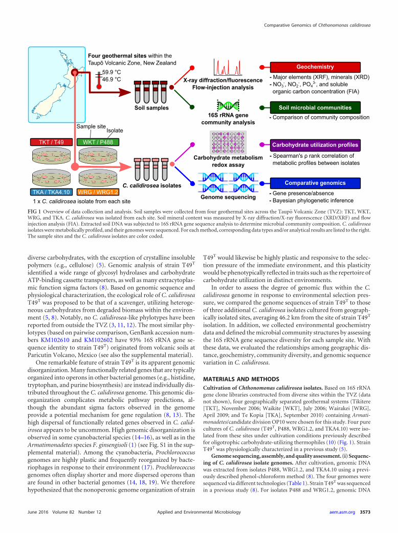
MATERIALS AND METHODSCultivation of Chthonomonas calidirosea isolates. Based on 16S rRNAgene clone libraries constructed from diverse sites within the TVZ (datanot shown), four geographically separated geothermal systems (Tikitere[TKT], November 2006; Waikite [WKT], July 2006; Wairakei [WRG],April 2009; and Te Kopia [TKA], September 2010) containing Armati-monadetes/candidate division OP10 were chosen for this study. Four purecultures of C. calidirosea (T49T, P488, WRG1.2, and TKA4.10) were iso-lated from these sites under cultivation conditions previously describedfor oligotrophic carbohydrate-utilizing thermophiles (10) (Fig. 1). StrainT49T was physiologically characterized in a previous study (5).
Genome sequencing, assembly, and quality assessment. (i) Sequenc-ing of C. calidirosea isolate genomes. After cultivation, genomic DNAwas extracted from isolates P488, WRG1.2, and TKA4.10 using a previ-ously described phenol-chloroform method (8). The four genomes weresequenced via different technologies (Table 1). Strain T49T was sequencedin a previous study (8). For isolates P488 and WRG1.2, genomic DNA
Four geothermal sites within theTaupō Volcanic Zone, New Zealand
C. calidirosea isolates
Carbohydrate metabolismredox assay
16S rRNA genecommunity analysis
Soil samples
TKT / T49 WKT / P488
TKA / TKA4.10 WRG / WRG1.21 x C. calidirosea isolate from each site
Genome sequencing
X-ray diffraction/fluorescenceFlow-injection analysis
Geochemistry
- Major elements (XRF), minerals (XRD)- NO3
-, NO2-, PO4
3-, and soluble organic carbon concentration (FIA)
Soil microbial communities- Comparison of community composition
Carbohydrate utilization profiles
- Spearman's ρ rank correlation of metabolic profiles between isolates
Comparative genomics
- Gene presence/absence- Bayesian phylogenetic inference
Sample siteIsolate
59.9 °C46.9 °C
FIG 1 Overview of data collection and analysis. Soil samples were collected from four geothermal sites across the Taupo� Volcanic Zone (TVZ): TKT, WKT,WRG, and TKA. C. calidirosea was isolated from each site. Soil mineral content was measured by X-ray diffraction/X-ray fluorescence (XRD/XRF) and flowinjection analysis (FIA). Extracted soil DNA was subjected to 16S rRNA gene sequence analysis to determine microbial community composition. C. calidiroseaisolates were metabolically profiled, and their genomes were sequenced. For each method, corresponding data types and/or analytical results are listed to the right.The sample sites and the C. calidirosea isolates are color coded.
Comparative Genomics of Chthonomonas calidirosea
June 2016 Volume 82 Number 12 aem.asm.org 3573Applied and Environmental Microbiology
libraries were constructed using the Nextera XT DNA sample preparationkit (Illumina, Inc., San Diego, CA, USA), according to the manufacturer’sprotocol, and 2 � 150-bp paired-end sequencing was performed using a300-cycle sequencing kit (version 1.0) on an Illumina MiSeq. For isolateTKA4.10, a genomic DNA library was constructed using the Ion XpressPlus fragment library kit with 100 ng of input DNA. The beads wereprepared using the Ion One Touch 200-bp version 2 DL kit, and sequenc-ing was performed with the Ion PGM 300-bp sequencing kit on an IonTorrent (Thermo Fisher Scientific, Inc., Waltham, MA, USA).
(ii) Assembly and annotation of isolate genomes. MIRA version4.0rc3 (20) was used to assemble the genomes of P488, WRG1.2, andTKA4.10. The strain T49T genome (8) was used as a reference duringassembly. This resulted in a single scaffold for each genome, in whichambiguous regions were indicated with repeating Ns (IUPAC notation).The resulting assemblies were aligned using progressiveMauve (21). As-sembly coverage (Table 1) was calculated from contigs of �5,000 bp toavoid inflation of the figures. Gene prediction and annotation of the ge-nomes were performed using the Integrated Microbial Genome-ExpertReview (IMG-ER) pipeline (22). Further details regarding sequencing,assembly, and quality control of genome assemblies are described in Fig.S2 and S3 in the supplemental material.
Genome comparison and phylogenetic analysis. (i) Identification ofgene homologs across C. calidirosea isolates. To determine whether an-notated genes were conserved between strains, we calculated the readcoverage for each gene in each strain using Bowtie2 (23) and BEDtools(24). The complete code for calculating gene coverage is available at http://bitbucket.org/biobakery/chthonomonas. The identification of con-served homologs between genomes with UCLUST (25) and the identifi-cation of putative genes in unmapped reads are described in thesupplemental material and are summarized in Fig. S4.
(ii) Determination of isolate phylogeny. The PhyloPhlAn gene col-lection of 400 ubiquitous and phylogenetically informative genes (26) wasused for phylogenetic analysis of the isolates. The C. calidirosea genomescontained 327 of these genes (see Table S1 in the supplemental material).Due to the high similarity between the isolates, we conducted Bayesianphylogeny inference using nucleic acid sequences rather than the filteredprotein sequences used by PhyloPhlAn; this increased the isolate-levelphylogenetic resolution and enabled the assessment of nucleic acid se-quence divergence (substitutions per site). Each gene was first separatelyaligned (aligning the four homologs of the gene from the four isolates)using MUSCLE (27) without the removal of invariant sites. The multiplealignments were then concatenated and used for phylogenetic inferencewith MrBayes (28). The genes were divided into partitions and were un-linked so that each partition had its own set of parameters for estimationduring the Markov chain Monte Carlo (MCMC) process. Each partitionused the general time-reversible substitution model, with the hetero-geneity rate determined by the invariable site plus gamma distribution(GTR � I � �). The model was selected using jModelTest (29) with theAkaike information criterion (AIC). Two MCMC runs were conducted.Each MCMC process ran for 140 million generations, sampling every
1,000 generations, with 35 million (25%) generations as burn-in. MCMCperformance diagnostics were conducted via Tracer (http://tree.bio.ed.ac.uk/software/tracer/).
(iii) Genomic rearrangement comparison with other thermophiles.We compared the degree of genomic sequence rearrangement observed inC. calidirosea to that observed in other thermophilic species isolated fromdefined geographic locations. The genomic sequences of Sulfolobus islan-dicus (strains Y.N.15.51 and Y.G.57.14) and Thermus thermophilus(strains HB-8, HB-27, and JL-18) were downloaded from NCBI GenBank,and the genomic rearrangement of each species was assessed by aligningall of its strains (including those of the C. calidirosea isolates) using pro-gressiveMauve (21). PhyloPhlAn and MUSCLE were used to select andalign conserved genes within the genome sequence of each comparedspecies (i.e., C. calidirosea, T. thermophilus, and S. islandicus). Sequenceidentities/dissimiliarities were calculated from the concatenated alignedgene sequences with SIAS (http://imed.med.ucm.es/Tools/sias.html).Due to an incomplete assembly (�100 contigs), T. thermophilus strainATCC 33923 could not be analyzed for genomic sequence rearrangement,but the genome sequence was used for assessment of the divergence ofconserved genes. Further details are described in the supplemental mate-rial and are summarized in Fig. S5.
Characterization of C. calidirosea isolate metabolisms with Biologphenotype microarrays. The substrate utilization phenotypes of the fourC. calidirosea isolates were compared using Biolog phenotype microarrays(Biolog, Inc., Hayward, CA, USA). All isolates were maintained using4.5NZS solid medium (10). Colonies were collected from the mediumafter 1 week of incubation at 60°C and were used to inoculate 250 ml of4.5NZS liquid medium (pH 5.5), and 3 g/liter maltose and 0.2 g/literCasamino Acids were added in 500-ml containers under an air headspace.The liquid cultures were incubated at 60°C for �40 h. Cells from 50 ml ofculture were pelleted by centrifugation (5,190 � g for 15 min) and washedwith sterile water three times prior to resuspension in a maltose-free4.5NZS liquid medium at an optical density at 600 nm (OD600) of 0.6 to0.8. A 100-�l aliquot of the resuspended culture and 5 �l (5 g/liter) ofMTT [3-(4,5-dimethylthiazol-2-yl)-2,5-diphenyltetrazolium bromide]were added to each of the 96 wells on a Biolog PM1 plate (Biolog, Inc.).The plates were sealed and incubated in darkness at 60°C for �23 h. Afterincubation, 900 �l of dimethyl sulfoxide was added to each well (30), andthe OD540 was recorded. Each isolate was tested twice, using a separatecultivation batch for each experimental replicate. Mean responses werecalculated from the corresponding wells of each isolate, and tie-correctedSpearman rank-order correlations between isolates were calculated withthe R function cor.test (31) (see Fig. S6 and Table S9 in the supplementalmaterial).
Analysis of soil chemistry and mineral content. Soil temperature wasmeasured on site using a Fluke 50S thermocouple. Sample pH was mea-sured in the laboratory at 25°C by mixing 1 g of the sample in 10 ml ofdeionized water. Soil moisture content and major oxides were analyzedusing X-ray fluorescence (XRF). Major oxides were detected by XRF withborate fusion of the samples. The total soil carbon content was measured
TABLE 1 Genome sequencing statistics of the four C. calidirosea isolates
Isolate SourceSequencingtechnology
No. of readsassembled
Coverage(�)
Genomesize (bp)
G�Ccontent (%)
No. of assembledprotein-codinggenes
No. of 5S, 16S,23S rRNAgenes
No. oftRNAgenes
T49T Hell’s Gate, Tikitere,North Island, NZ
454 Titanium �Sanger
171,649 20 3,437,861 54.41 2,877 1, 2, 1 46
P488 Waikite Valley,North Island, NZ
Illumina MiSeq 2,533,602 88 3,438,278 53.93 2,876 1, 2, 1 46
WRG1.2 Wairakei, NorthIsland, NZ
Illumina MiSeq 3,272,279 112 3,438,088 53.57 2,855 1, 2, 1 46
TKA4.10 Te Kopia, NorthIsland, NZ
Ion Torrent 954,245 39 3,437,861 54.41 2,885 1, 2, 1 46
Lee et al.
3574 aem.asm.org June 2016 Volume 82 Number 12Applied and Environmental Microbiology
by mixing weighed soil aliquots with 25 ml of 0.05 M K2SO4 and filteringto extract total soluble carbon. Total soluble carbon was then measured bycombusting the samples and measuring the CO2 evolution using a multiN/C 3100 analyzer (Analytik Jena AG, Jena, Germany). The inorganiccarbon content of the sample was measured separately by quantifying theCO2 generated by the acidification of an aliquot of the original sample(32). Mineral content was determined by X-ray diffraction (XRD) (PhilipX’Pert Pro with Co K� radiation source) with the software X’Pert High-Score (Spectris plc, England) and Siroquant (Sietronics Pty. Ltd., Can-berra, Australia) (see Fig. S7 in the supplemental material). Soil nitrate,nitrite, ammonia, and dissolved reactive phosphate levels were deter-mined by flow injection analysis (FIA) (Hach Company, Loveland, CO,USA). Analytes were extracted by mixing 1 g of soil sample with 50 ml of1 M KCl. The mixture was centrifuged, and the resulting supernatant waspassed through a 0.2-�m-pore-size filter prior to analysis.
Community 16S rRNA gene-targeted sequencing and bioinformaticprocessing. Total DNA was then extracted from each soil sample. Prior toDNA extraction, each soil sample from each site was mixed with 500 �l ofa 50-g/liter sterile skim milk solution (Becton, Dickinson and Company,NJ, USA) to reduce DNA binding to clay. DNA was then extracted with aNucleoSpin soil DNA kit (Macherey-Nagel GmbH & Co. KG, Düren,Germany) according to the manufacturer’s instructions. The V4 hyper-variable region of the 16S rRNA gene was amplified by PCR and se-quenced using the Ion Torrent platform, according to standardized meth-ods, as described in detail in the supplemental material. Quality controlwas performed with UPARSE (33), as described in detail in the supple-mental material, resulting in a mean of �20,700 sequences per sample.These sequences were clustered de novo into operational taxonomic units(OTUs), with a minimum identity of 97%, and the representative se-quences were taxonomically assigned using the UCLUST (25) consensustaxonomy assigner in QIIME 1.8.0 (34). Greengenes release 13_8 (35) wasused as the reference taxonomic database. The OTU table was rarefied to17,941 sequences per sample to remove sample heterogeneity; rarefactionanalysis indicated that the sequencing depth was sufficient to identify themajority of OTUs within each community (see Fig. S8 in the supplementalmaterial). QIIME (34) was used for -diversity calculations and down-stream analyses; -diversity was measured by Bray-Curtis dissimilarityand by weighted and unweighted UniFrac indices. A weighted-UniFrachierarchical clustering tree was constructed using the unweighted pairgroup method using average linkages (UPGMA). Jackknife analysis withthe UPGMA tree was conducted with 200 repetitions and 12,000 se-quences per sample. PyNAST alignment (36) and FastTree (37) wereused to generate phylogenetic trees.
Accession numbers. The genome sequence and assembly data of theC. calidirosea isolates were deposited at EMBL/GenBank with the fol-lowing accession numbers: PRJEB1573 (strain T49T), PRJEB4907 (iso-late P488), PRJEB4936 (isolate WRG1.2), and PRJEB4937 (isolateTKA4.10). Community sequencing data were deposited under acces-sion number PRJEB13454.
RESULTS
The aim of this research was to understand how the ecosystemshapes the evolution of the C. calidirosea genome and phenotype.
To connect genome dynamics with environmental factors andobserved phenotypes, we isolated four C. calidirosea strains fromfour geographically distinct geothermal soil environments. Foreach site, we determined the soil chemistry and microbial com-munity composition by 16S rRNA gene sequence analysis. Wesequenced each isolate genome and performed nucleic acid se-quence divergence assessment, phylogenetic inference, and genecontent and organization analysis. In addition, we characterizedthe metabolic profile of each isolate (Fig. 1).
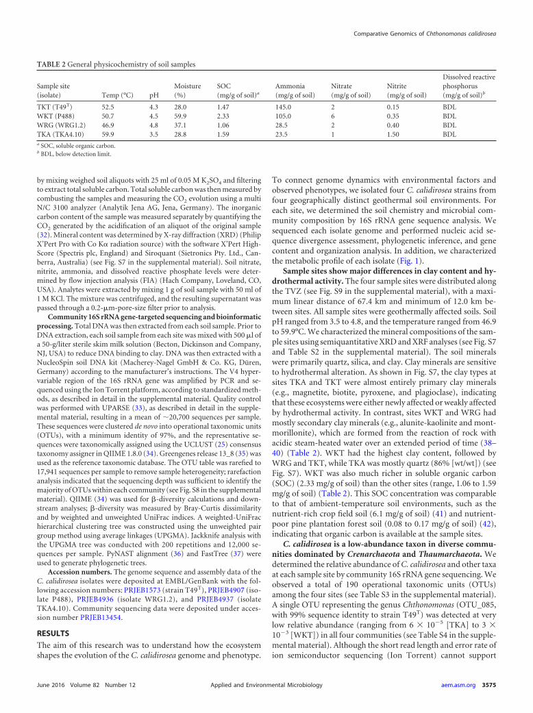
Sample sites show major differences in clay content and hy-drothermal activity. The four sample sites were distributed alongthe TVZ (see Fig. S9 in the supplemental material), with a maxi-mum linear distance of 67.4 km and minimum of 12.0 km be-tween sites. All sample sites were geothermally affected soils. SoilpH ranged from 3.5 to 4.8, and the temperature ranged from 46.9to 59.9°C. We characterized the mineral compositions of the sam-ple sites using semiquantitative XRD and XRF analyses (see Fig. S7and Table S2 in the supplemental material). The soil mineralswere primarily quartz, silica, and clay. Clay minerals are sensitiveto hydrothermal alteration. As shown in Fig. S7, the clay types atsites TKA and TKT were almost entirely primary clay minerals(e.g., magnetite, biotite, pyroxene, and plagioclase), indicatingthat these ecosystems were either newly affected or weakly affectedby hydrothermal activity. In contrast, sites WKT and WRG hadmostly secondary clay minerals (e.g., alunite-kaolinite and mont-morillonite), which are formed from the reaction of rock withacidic steam-heated water over an extended period of time (38–40) (Table 2). WKT had the highest clay content, followed byWRG and TKT, while TKA was mostly quartz (86% [wt/wt]) (seeFig. S7). WKT was also much richer in soluble organic carbon(SOC) (2.33 mg/g of soil) than the other sites (range, 1.06 to 1.59mg/g of soil) (Table 2). This SOC concentration was comparableto that of ambient-temperature soil environments, such as thenutrient-rich crop field soil (6.1 mg/g of soil) (41) and nutrient-poor pine plantation forest soil (0.08 to 0.17 mg/g of soil) (42),indicating that organic carbon is available at the sample sites.
C. calidirosea is a low-abundance taxon in diverse commu-nities dominated by Crenarchaeota and Thaumarchaeota. Wedetermined the relative abundance of C. calidirosea and other taxaat each sample site by community 16S rRNA gene sequencing. Weobserved a total of 190 operational taxonomic units (OTUs)among the four sites (see Table S3 in the supplemental material).A single OTU representing the genus Chthonomonas (OTU_085,with 99% sequence identity to strain T49T) was detected at verylow relative abundance (ranging from 6 � 105 [TKA] to 3 �103 [WKT]) in all four communities (see Table S4 in the supple-mental material). Although the short read length and error rate ofion semiconductor sequencing (Ion Torrent) cannot support
TABLE 2 General physicochemistry of soil samples
Sample site(isolate) Temp (°C) pH
Moisture(%)
SOC(mg/g of soil)a
Ammonia(mg/g of soil)
Nitrate(mg/g of soil)
Nitrite(mg/g of soil)
Dissolved reactivephosphorus(mg/g of soil)b
TKT (T49T) 52.5 4.3 28.0 1.47 145.0 2 0.15 BDLWKT (P488) 50.7 4.5 59.9 2.33 105.0 6 0.35 BDLWRG (WRG1.2) 46.9 4.8 37.1 1.06 28.5 2 0.40 BDLTKA (TKA4.10) 59.9 3.5 28.8 1.59 23.5 1 1.50 BDLa SOC, soluble organic carbon.b BDL, below detection limit.
Comparative Genomics of Chthonomonas calidirosea
June 2016 Volume 82 Number 12 aem.asm.org 3575Applied and Environmental Microbiology
high-resolution comparison, these data confirm the detection ofChthonomonas with limited phylogenetic diversity (within the97% OTU clustering criterion). The C. calidirosea OTU was mostabundant in the more nutrient-rich soil at WKT, which hadhigher soluble organic carbon content, nitrate concentration, andoverall clay mineral content (Table 2; see also Fig. S7 and Tables S3and S4 in the supplemental material). In contrast, C. calidiroseawas less abundant in sites consisting mainly of fresh hydrothermaldeposits lacking in soluble organic carbon but rich in quartz andamorphous silica, as represented by the SiO2 concentration (seeTable S2 in the supplemental material). Clay and organic matterincrease the buffering capacity of soil (43), which may facilitatethe survival of C. calidirosea, a species with a very limited pH rangefor growth (5, 8).
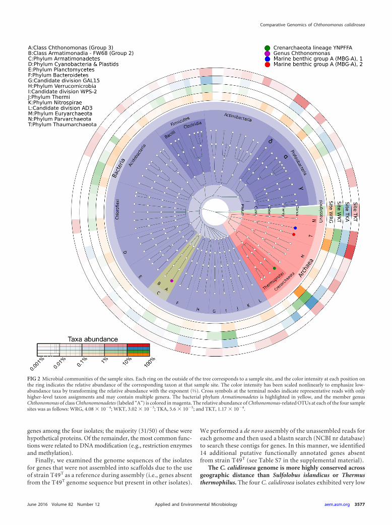
We observed only 21 archaeal OTUs, but these OTUs com-prised the majority of the community at all four sites (56% [WKT]to 75% [TKA]). Five archaeal OTUs were present at all sites.Nearly half of observed archaeal OTUs (10/21) were Euryar-chaeota, all belonging to the thermophilic acidophilic class Ther-moplasmata (44). However, these represented �5% of the totalsequence reads from any community. In contrast, the Crenar-chaeota and Thaumarchaeota comprised the majority of readsfrom each community (Fig. 2). The Crenarchaeota, which weremainly Thermoprotei (nine OTUs [39 to 58% of total sequencereads]) (45), dominated all communities except TKT. The deep-branching MBG-A group (45–48) (12 OTUs [13 to 28% of totalsequence reads]) dominated the TKT community (57% ofOTUs). In this study, the class Thermoprotei was predominantlyrepresented by the lineage YNPFFA, which is also associated withgeothermal features at Yellowstone National Park, USA (49). Un-fortunately, the lack of cultivated representatives within these Cre-narchaeota and Thaumarchaeota lineages impedes any attempt toinfer their ecological roles.
The four communities were distinctly different at a finer taxo-nomic resolution. Only 25 of the 128 total bacterial OTUs wereobserved at all four sites, and none of these were highly abundant(see Table S3 in the supplemental material). These taxa includedSulfobacillaceae, Rhodospirillales, and Acidimicrobiaceae. Based ontheir affiliations to characterized type strains, we speculate thatthese bacteria are moderately acidophilic, chemolithotrophic, andaerobic, and they grow via mechanisms, such as iron and sulfuroxidation (Sulfobacillaceae [50]), iron oxidation (Acidimicrobi-aceae [51]), or methanotrophy (“Candidatus Methylacidiphilum”[52]). In contrast, many predominant OTUs were detected in onlyone or two sites. For example, an archaeal OTU from clade SK322(OTU_0003) comprised 10.3% of the total reads from WKT butwas not detected at TKT, while an Acinetobacter OTU (OTU_158)comprised 8.5% of the total reads from WKT but was undetectedat the other sites (see Table S3). These observations indicate thatalthough the four C. calidirosea-hosting geothermal systems werebroadly similar (in pH, temperature, and hydrothermally affectedclays), they did not support similar microbial community compo-sitions. This suggests that other ecological or stochastic differencesdetermine the abundance of Chthonomonas spp. within these eco-systems.
C. calidirosea genomic content and organization are highlyconserved between isolates. The genomes of C. calidirosea iso-lates P488, WRG1.2, and TKA4.10 were sequenced using the Illu-mina MiSeq or Ion Torrent platform (Table 1) and assembledusing the previously sequenced T49T genome (8) as a template.
MIRA (20) and SPAdes (53) produced very similar assemblies (seethe supplemental material), indicating little bias due to assemblerchoice. The length and G�C content of the TKA4.10 genome wereidentical to those of T49T; this was potentially influenced by theinefficient assembly of single-ended Ion Torrent reads beyond ref-erence contig boundaries. Isolates P488 and WRG1.2 had slightlylonger genome assemblies and lower G�C content than T49T andTKA4.10 (Table 1).
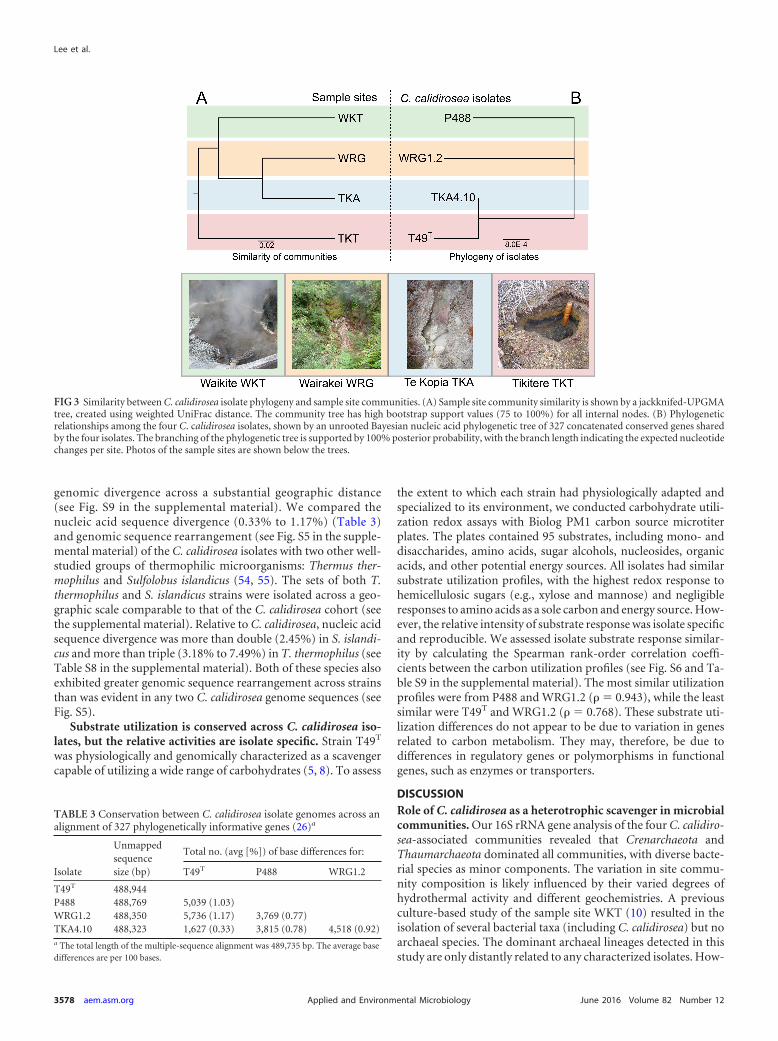
We determined the phylogeny of the strains by aligning theconcatenated nucleic acid sequences of 327 phylogenetically in-formative genes (26) (see Table S1 in the supplemental material)and building a phylogenetic tree using Bayesian inference (Fig. 3).The pairwise sequence identity of these genes was very high (av-erage, 99.2%) between the isolates. Based on these data, strainT49T shares the highest similarity with TKA, followed by P488 andWRG1.2. The posterior probability support for this unrooted treetopology was �99%. Within this tree, TKA4.10 showed a veryshort branch length from the internal node shared with T49T.WRG1.2 had the longest terminal branch length. This is in slightdisagreement with strain-level conservation of the 16S rRNA genesequences; each genome had two copies of the 16S rRNA gene, andall are identical except in strain P488, in which the two copiesdiffer by 1 base.
While the four C. calidirosea genome sequences showed a highdegree of synteny in most genomic regions (see Fig. S2 and S5in the supplemental material), a previous study identified twoputative horizontally transferred regions, A (between the lociCCALI_00447 and CCALI_00449) and B (between the lociCCALI_00804 and CCALI_00807) (8) (see Fig. S10 in the supple-mental material). These two regions were conserved between iso-lates T49T and TKA4.10 but are mostly absent in isolate WRG1.2.Isolate P488 contained only a small deletion in region B, and thesequences in both regions were slightly less conserved. The vari-ability in these regions is consistent with our inferred strain phy-logeny (Fig. 3); there is low divergence between T49T andTKA4.10, more divergence in P488 (e.g., deletions, truncations,and frameshift mutations in the loci CCALI_00449 andCCALI_00805), and the regions were either never acquired ordeleted from WRG1.2.
Finally, we compared our Bayesian-inferred strain phylogenyto the structure of the microbial community from each samplesite, as measured by -diversity (Fig. 3; see also Table S5 in thesupplemental material). When communities were compared bymetrics that consider OTU abundance (e.g., weighted UniFracand Bray-Curtis dissimilarity), sites WRG and TKA had the mostsimilar communities, followed by WKT and then TKT. In con-trast, isolates TKA4.10 and T49T are phylogenetically the mostsimilar, indicating that the differences in genome sequence phy-logeny of C. calidirosea isolates are not reflected by differences inthe community composition of their respective isolation sites.
Functionally annotated isolate-variant genes are primarilyDNA-interacting enzymes. We next assessed the similarity of ge-netic contents between the genomic assemblies by aligning thereads from each isolate to the genes from all four isolates and thenassessing the coverage of each gene in each strain (see the supple-mental material). Most gene homologs (�2,600) were present inall four C. calidirosea isolates, but we identified 769 putative iso-late-variant genes, many of which encoded hypothetical proteinsor domains of unknown function (see Table S6 in the supplemen-tal material). There were a total of 50 apparently isolate-unique
Lee et al.
3576 aem.asm.org June 2016 Volume 82 Number 12Applied and Environmental Microbiology
genes among the four isolates; the majority (31/50) of these werehypothetical proteins. Of the remainder, the most common func-tions were related to DNA modification (e.g., restriction enzymesand methylation).
Finally, we examined the genome sequences of the isolatesfor genes that were not assembled into scaffolds due to the useof strain T49T as a reference during assembly (i.e., genes absentfrom the T49T genome sequence but present in other isolates).
We performed a de novo assembly of the unassembled reads foreach genome and then used a blastn search (NCBI nr database)to search these contigs for genes. In this manner, we identified14 additional putative functionally annotated genes absentfrom strain T49T (see Table S7 in the supplemental material).
The C. calidirosea genome is more highly conserved acrossgeographic distance than Sulfolobus islandicus or Thermusthermophilus. The four C. calidirosea isolates exhibited very low
Taxa abundance
0.001
%0.0
1%1
FIG 2 Microbial communities of the sample sites. Each ring on the outside of the tree corresponds to a sample site, and the color intensity at each position onthe ring indicates the relative abundance of the corresponding taxon at that sample site. The color intensity has been scaled nonlinearly to emphasize low-abundance taxa by transforming the relative abundance with the exponent (⅓). Cross symbols at the terminal nodes indicate representative reads with onlyhigher-level taxon assignments and may contain multiple genera. The bacterial phylum Armatimonadetes is highlighted in yellow, and the member genusChthonomonas of class Chthonomonadetes (labeled “A”) is colored in magenta. The relative abundance of Chthonomonas-related OTUs at each of the four samplesites was as follows: WRG, 4.08 � 104; WKT, 3.02 � 103; TKA, 5.6 � 105; and TKT, 1.17 � 104.
Comparative Genomics of Chthonomonas calidirosea
June 2016 Volume 82 Number 12 aem.asm.org 3577Applied and Environmental Microbiology
genomic divergence across a substantial geographic distance(see Fig. S9 in the supplemental material). We compared thenucleic acid sequence divergence (0.33% to 1.17%) (Table 3)and genomic sequence rearrangement (see Fig. S5 in the supple-mental material) of the C. calidirosea isolates with two other well-studied groups of thermophilic microorganisms: Thermus ther-mophilus and Sulfolobus islandicus (54, 55). The sets of both T.thermophilus and S. islandicus strains were isolated across a geo-graphic scale comparable to that of the C. calidirosea cohort (seethe supplemental material). Relative to C. calidirosea, nucleic acidsequence divergence was more than double (2.45%) in S. islandi-cus and more than triple (3.18% to 7.49%) in T. thermophilus (seeTable S8 in the supplemental material). Both of these species alsoexhibited greater genomic sequence rearrangement across strainsthan was evident in any two C. calidirosea genome sequences (seeFig. S5).
Substrate utilization is conserved across C. calidirosea iso-lates, but the relative activities are isolate specific. Strain T49T
was physiologically and genomically characterized as a scavengercapable of utilizing a wide range of carbohydrates (5, 8). To assess
the extent to which each strain had physiologically adapted andspecialized to its environment, we conducted carbohydrate utili-zation redox assays with Biolog PM1 carbon source microtiterplates. The plates contained 95 substrates, including mono- anddisaccharides, amino acids, sugar alcohols, nucleosides, organicacids, and other potential energy sources. All isolates had similarsubstrate utilization profiles, with the highest redox response tohemicellulosic sugars (e.g., xylose and mannose) and negligibleresponses to amino acids as a sole carbon and energy source. How-ever, the relative intensity of substrate response was isolate specificand reproducible. We assessed isolate substrate response similar-ity by calculating the Spearman rank-order correlation coeffi-cients between the carbon utilization profiles (see Fig. S6 and Ta-ble S9 in the supplemental material). The most similar utilizationprofiles were from P488 and WRG1.2 (� 0.943), while the leastsimilar were T49T and WRG1.2 (� 0.768). These substrate uti-lization differences do not appear to be due to variation in genesrelated to carbon metabolism. They may, therefore, be due todifferences in regulatory genes or polymorphisms in functionalgenes, such as enzymes or transporters.
DISCUSSIONRole of C. calidirosea as a heterotrophic scavenger in microbialcommunities. Our 16S rRNA gene analysis of the four C. calidiro-sea-associated communities revealed that Crenarchaeota andThaumarchaeota dominated all communities, with diverse bacte-rial species as minor components. The variation in site commu-nity composition is likely influenced by their varied degrees ofhydrothermal activity and different geochemistries. A previousculture-based study of the sample site WKT (10) resulted in theisolation of several bacterial taxa (including C. calidirosea) but noarchaeal species. The dominant archaeal lineages detected in thisstudy are only distantly related to any characterized isolates. How-
FIG 3 Similarity between C. calidirosea isolate phylogeny and sample site communities. (A) Sample site community similarity is shown by a jackknifed-UPGMAtree, created using weighted UniFrac distance. The community tree has high bootstrap support values (75 to 100%) for all internal nodes. (B) Phylogeneticrelationships among the four C. calidirosea isolates, shown by an unrooted Bayesian nucleic acid phylogenetic tree of 327 concatenated conserved genes sharedby the four isolates. The branching of the phylogenetic tree is supported by 100% posterior probability, with the branch length indicating the expected nucleotidechanges per site. Photos of the sample sites are shown below the trees.
TABLE 3 Conservation between C. calidirosea isolate genomes across analignment of 327 phylogenetically informative genes (26)a
Isolate
Unmappedsequencesize (bp)
Total no. (avg [%]) of base differences for:
T49T P488 WRG1.2
T49T 488,944P488 488,769 5,039 (1.03)WRG1.2 488,350 5,736 (1.17) 3,769 (0.77)TKA4.10 488,323 1,627 (0.33) 3,815 (0.78) 4,518 (0.92)a The total length of the multiple-sequence alignment was 489,735 bp. The average basedifferences are per 100 bases.
Lee et al.
3578 aem.asm.org June 2016 Volume 82 Number 12Applied and Environmental Microbiology
ever, based on the metabolic capabilities of described strainswithin related lineages (e.g., Sulfolobales, Nitrososphaerales, andNitrosopumilales), lithoautotrophic lifestyles would seem likely(56–59). We therefore speculate that the dominant crenarchaeotaland thaumarchaeal phylotypes may occupy the niche of au-totrophic primary producers, supporting diverse, yet low-abun-dance, chemoheterotrophic bacterial species, like C. calidirosea.The scavenger phenotype of C. calidirosea may be well suited topersist in an ecosystem with minimal or inconsistent saccharidesources (5, 8). A recent study of bacterial communities in a Thai-land hot spring also indicated a potential relationship betweenArmatimonadetes and autotrophs. This survey detected abundantArmatimonadetes and Chloroflexi OTUs associated with cyano-bacterial mats (60). Pairwise discontiguous megaBLAST analysesbetween the Thailand hot springs Armatimonadetes OTUs and C.calidirosea showed that the Thailand OTUs had low sequence sim-ilarities (75 to 84%) to the type strains of the three Armatimon-adetes classes (see Table S10 in the supplemental material).
Low genomic diversity in the face of geographical isolation.Although the four C. calidirosea isolates were cultured from geo-graphically distant sites across the TVZ, their genome sequenceswere highly conserved, particularly compared to other thermo-philic microorganisms recovered across similar geographicalscales (see Fig. S5 and Table S8 in the supplemental material).Phylogeny inferred from single-nucleotide polymorphisms(SNPs) and horizontal gene transfer indicated a close relation-ship between all isolates, particularly between T49T and TKA4.10.Gene presence/absence analysis identified relatively few variantgenes (see Tables S6 and S7 in the supplemental material), such asrestriction-modification systems. These genes are known to berapidly evolving (61) and mobile (62), which may explain theirpresence in an otherwise largely conserved pangenome. Physio-logically, all four C. calidirosea isolates had similar carbohydratemetabolism. Future comparative transcriptomic and proteomicanalyses of the isolates may shed light on subtle underlying regu-latory processes resulting in the variations of phenotypic responsewithin these highly conserved genome sequences.
Potential mechanisms underpinning genomic conservationacross geographic distance. The divergence between genome se-quences of C. calidirosea isolates consisted primarily of SNPs,which may accumulate by genetic drift during geographic isola-tion. However, the phylogenetic distances of the isolate genomesequences do not reflect the geographic relationships of the sam-ple sites (see Fig. S9 in the supplemental material). The reason forthe low genomic sequence divergence between the C. calidiroseaisolates despite their geographical and ecological isolation is notimmediately clear. One possible explanation for the high genomicsequence conservation is a relatively recent sympatric determina-tion of species of C. calidirosea from a mesophilic relative, leadingto its occupation of a new niche in geothermal environments. Areview of 16S rRNA gene data sets available in the NCBI and EMPdatabases indicates that C. calidirosea can presently be consideredendemic to New Zealand, with no genus-level phylotypes detectedelsewhere (see the supplemental material). A recent review of theenvironmental distribution of Armatimonadetes phylotypes indi-cated that temperate soil is the most dominant environment forthe phylum (3). Of 39 phylotypes identified representing the classChthonomonadetes (group 2), only two phylogenetically distantclones were associated with thermophilic environments. In con-trast, group 10 (A and B) consists entirely of phylotypes from
geographically disparate geothermal environments. Thus, the oc-casional occurrence of thermophily in the Chthonomonadetes mayindicate either recently acquired thermophilic adaptation or sam-pling bias. The identification of more closely related phylotypes inthe future will help clarify the evolutionary history and environ-mental distribution of Chthonomonas.
Although this hypothesis may explain the low diversity from anevolution and sampling perspective, it is inadequate to explain thevery high similarity in the genome sequences from genetic drift,unless the dispersal of C. calidirosea was relatively recent. The highsimilarity between strain T49T and isolate TKA4.10 suggests re-cent dispersal between the two sites. A recent study found thatthermophilic microbes can be globally dispersed via aeolian trans-port but are selected by their environments (63). In dynamic geo-thermal environments, taxa with very low abundance, such as C.calidirosea, may become locally extinct but subsequently recolo-nized from another site, resulting in high genetic similarity be-tween two populations. Over a sufficiently rapid timespan of ex-tinction and colonization, the genome sequences would reflectstochastic aeolian dispersal rather than adaptive responses to se-lection pressures of specific environments. In contrast, an abun-dant species would be less sensitive to the stochastic process oflocalized extinction and repeated seeding and thus more likely topresent a clearer relationship between geography and phyloge-netic distance. We believe the low diversity of C. calidirosea ge-nome sequences potentially reflects these scenarios. While surfaceand groundwater water flow presents an alternative mechanism ofdispersal between sites, this is unlikely due to the arrangement ofriver catchments and aquifers flowing away from individual sites(see Fig. S9). The environmental persistence of C. calidirosea isfurther restricted by its inability to sporulate and by its fastidiousgrowth requirements (limited pH and temperature growthranges). Thus, rapid dispersal between suitable habitats is a possi-ble mechanism for maintenance of local populations and genomeconsistency.
Based on analysis of the genome sequences of C. calidiroseaisolates, we reject our initial hypothesis that C. calidirosea genomicdisorganization reflects plasticity and niche-specific adaptation.Instead, the genome sequence is highly conserved and shows asmall degree of genetic drift rather than response to different eco-logical selection pressures at different sample sites. To date, C.calidirosea has been detected only within New Zealand’s TVZ, andits global distribution is currently unknown. If additional isolatescan be cultured from greater geographic distances (such as thefumarolic/volcanic-associated phylotypes of Paricutin Volcano,Mexico, which share 93% 16S rRNA gene sequence identity), theywill continue to improve our understanding of how the C. calid-irosea genome is shaped by genetic drift. Rather than possessing adynamic and highly plastic genome, the conservation of gene con-tent and order (synteny) of C. calidirosea may be comparable tothose of prokaryotes such as Haloquadratum walsbyi (64) andmembers of the SAR11 (Pelagibacterales) clade (65), which areknown for high genomic conservation.
Conclusion. In order to investigate the relationship betweenthe C. calidirosea genome and its environment, we performedcomparative genomics and determined the physiology of isolatesobtained from four distinct sites. These data were integrated withmicrobial community analyses and environmental geochemistrydata for the respective sites. We have shown that C. calidirosea, athermophilic non-spore-forming bacterium, exhibits detectable,
Comparative Genomics of Chthonomonas calidirosea
June 2016 Volume 82 Number 12 aem.asm.org 3579Applied and Environmental Microbiology
albeit minor, genome sequence differences across a geographicdistance. We propose that this bacterium is capable of dispersalacross geographical barriers, and that the resulting genome con-servation over space is potentially applicable to many low-abun-dance microbial taxa. Our approach shows the value of augment-ing amplicon profiling of poorly characterized communities withadditional data, such as the physiological characterization of iso-lates and geochemical analysis, in order to provide context andvalidation of ecological inferences.
ACKNOWLEDGMENTS
We thank Jean Power, Heike Anders, and Michelle Crowe for isolatingstrains used in this study. K.C.L. thanks Alexander Kmoch, MichaelRosenberg, Nellie Olsen, and Timothy Tickle for valuable comments onthis paper and study, as well as Magali Moreau for assistance in GIS visu-alization, and Georgia Wakerley and Jean Power for assistance with com-munity sequencing. We thank Tikitere Trust, Nga�ti Tahu-Nga�ti WhaoaRunanga Trust and Tauhara 2 Trust for their ongoing support and foraccess to sample sites.
FUNDING INFORMATIONK.C.L. acknowledges the Sarah Beanland Memorial Scholarship (GNSScience), M.B.S. acknowledges the GNS Science DCF program grantGRN5, and X.C.M. and C.H. acknowledge grant DBI-1053486 from theU.S. National Science Foundation for assistance in funding of this re-search.
REFERENCES1. Hugenholtz P, Pitulle C, Hershberger KL, Pace NR. 1998. Novel division
level bacterial diversity in a Yellowstone hot spring. J Bacteriol 180:366 –376.
2. Portillo MC, Gonzalez JM. 2008. Members of the Candidate DivisionOP10 are spread in a variety of environments. World J Microbiol Biotech-nol 25:347–353. http://dx.doi.org/10.1007/s11274-008-9895-z.
3. Dunfield PF, Tamas I, Lee KC, Morgan XC, McDonald IR, Stott MB.2012. Electing a candidate: a speculative history of the bacterial phylumOP10. Environ Microbiol 14:3069 –3080. http://dx.doi.org/10.1111/j.1462-2920.2012.02742.x.
4. Lee KCY, Herbold CW, Dunfield PF, Morgan XC, McDonald IR, StottMB. 2013. Phylogenetic delineation of the novel phylum Armatimon-adetes (former candidate division OP10) and definition of two novel can-didate divisions. Appl Environ Microbiol 79:2484 –2487. http://dx.doi.org/10.1128/AEM.03333-12.
5. Lee KC-Y, Dunfield PF, Morgan XC, Crowe MA, Houghton KM,Vyssotski M, Ryan JLJ, Lagutin K, McDonald IR, Stott MB. 2011.Chthonomonas calidirosea gen. nov., sp. nov., an aerobic, pigmented, ther-mophilic micro-organism of a novel bacterial class, Chthonomonadetesclassis nov., of the newly described phylum Armatimonadetes originallydesignated candidate division OP10. Int J Syst Evol Microbiol 61:2482–2490. http://dx.doi.org/10.1099/ijs.0.027235-0.
6. Tamaki H, Tanaka Y, Matsuzawa H, Muramatsu M, Meng X-Y, HanadaS, Mori K, Kamagata Y. 2011. Armatimonas rosea gen. nov., sp. nov., of anovel bacterial phylum, Armatimonadetes phyl. nov, formally called thecandidate phylum OP10. Int J Syst Evol Microbiol 61:1442–1447.
7. Im W-T, Hu Z-Y, Kim K-H, Rhee S-K, Meng H, Lee S-T, Quan Z-X.2012. Description of Fimbriimonas ginsengisoli gen. nov., sp. nov. withinthe Fimbriimonadia class nov., of the phylum Armatimonadetes. AntonieVan Leeuwenhoek 102:307–317. http://dx.doi.org/10.1007/s10482-012-9739-6.
8. Lee KC, Morgan XC, Dunfield PF, Tamas I, McDonald IR, Stott MB.2014. Genomic analysis of Chthonomonas calidirosea, the first sequencedisolate of the phylum Armatimonadetes. ISME J 8:1522–1533. http://dx.doi.org/10.1038/ismej.2013.251.
9. Hu Z-Y, Wang Y-Z, Im W-T, Wang S-Y, Zhao G-P, Zheng H-J, QuanZ-X. 2014. The first complete genome sequence of the class Fimbriimo-nadia in the phylum Armatimonadetes. PLoS One 9:e100794. http://dx.doi.org/10.1371/journal.pone.0100794.
10. Stott MB, Crowe MA, Mountain BW, Smirnova AV, Hou S, Alam M,
Dunfield PF. 2008. Isolation of novel bacteria, including a candidatedivision, from geothermal soils in New Zealand. Environ Microbiol 10:2030 –2041. http://dx.doi.org/10.1111/j.1462-2920.2008.01621.x.
11. Pruesse E, Quast C, Knittel K, Fuchs BM, Ludwig W, Peplies J, Glöck-ner FO. 2007. SILVA: a comprehensive online resource for qualitychecked and aligned ribosomal RNA sequence data compatible with ARB.Nucleic Acids Res 35:7188 –7196. http://dx.doi.org/10.1093/nar/gkm864.
12. Benson DA, Karsch-Mizrachi I, Lipman DJ, Ostell J, Sayers EW. 2011.GenBank. Nucleic Acids Res 39:D32–D37. http://dx.doi.org/10.1093/nar/gkq1079.
13. Nuñez PA, Romero H, Farber MD, Rocha EPC. 2013. Natural selectionfor operons depends on genome size. Genome Biol Evol 5:2242–2254.http://dx.doi.org/10.1093/gbe/evt174.
14. Memon D, Singh AK, Pakrasi HB, Wangikar PP. 2013. A global analysisof adaptive evolution of operons in cyanobacteria. Antonie Van Leeuwen-hoek 103:331–346. http://dx.doi.org/10.1007/s10482-012-9813-0.
15. Xie G, Bonner CA, Brettin T, Gottardo R, Keyhani NO, Jensen RA.2003. Lateral gene transfer and ancient paralogy of operons containingredundant copies of tryptophan-pathway genes in Xylella species and inheterocystous cyanobacteria. Genome Biol 4:R14. http://dx.doi.org/10.1186/gb-2003-4-2-r14.
16. Itoh T, Takemoto K, Mori H, Gojobori T. 1999. Evolutionary instabilityof operon structures disclosed by sequence comparisons of complete mi-crobial genomes. Mol Biol Evol 16:332–346. http://dx.doi.org/10.1093/oxfordjournals.molbev.a026114.
17. Coleman ML, Sullivan MB, Martiny AC, Steglich C, Barry K, DelongEF, Chisholm SW. 2006. Genomic islands and the ecology and evolutionof Prochlorococcus. Science 311:1768 –1770. http://dx.doi.org/10.1126/science.1122050.
18. Wang B, Lu L, Lv H, Jiang H, Qu G, Tian C, Ma Y. 2014. Thetranscriptome landscape of Prochlorococcus MED4 and the factors for sta-bilizing the core genome. BMC Microbiol 14:11. http://dx.doi.org/10.1186/1471-2180-14-11.
19. Zheng Y, Szustakowski JD, Fortnow L, Roberts RJ, Kasif S. 2002.Computational identification of operons in microbial genomes. GenomeRes 12:1221–1230. http://dx.doi.org/10.1101/gr.200601.
20. Chevreux B, Wetter T, Suhai S. 1999. Genome sequence assembly usingtrace signals and additional sequence information, p 45–56. In Computerscience and biology. Proceedings of the German Conference on Bioinfor-matics, GCB ’99. GCB, Hannover, Germany.
21. Darling AE, Mau B, Perna NT. 2010. progressiveMauve: multiple ge-nome alignment with gene gain, loss and rearrangement. PLoS One5:e11147. http://dx.doi.org/10.1371/journal.pone.0011147.
22. Markowitz VM, Chen I-MA, Palaniappan K, Chu K, Szeto E, GrechkinY, Ratner A, Anderson I, Lykidis A, Mavromatis K, Ivanova NN,Kyrpides NC. 2010. The integrated microbial genomes system: an ex-panding comparative analysis resource. Nucleic Acids Res 38:D382–D390.http://dx.doi.org/10.1093/nar/gkp887.
23. Langmead B, Salzberg SL. 2012. Fast gapped-read alignment with Bowtie2. Nat Methods 9:357–359. http://dx.doi.org/10.1038/nmeth.1923.
24. Quinlan AR. 2014. BEDTools: the Swiss-army tool for genome featureanalysis. Curr Protoc Bioinformatics 47:11.12.1–11.12.34.
25. Edgar RC. 2010. Search and clustering orders of magnitude faster thanBLAST. Bioinformatics 26:2460 –2461. http://dx.doi.org/10.1093/bioinformatics/btq461.
26. Segata N, Börnigen D, Morgan XC, Huttenhower C. 2013. PhyloPhlAnis a new method for improved phylogenetic and taxonomic placement ofmicrobes. Nat Commun 4:2304.
27. Edgar RC. 2004. MUSCLE: multiple sequence alignment with high accu-racy and high throughput. Nucleic Acids Res 32:1792–1797. http://dx.doi.org/10.1093/nar/gkh340.
28. Huelsenbeck JP, Ronquist F. 2001. MRBAYES: Bayesian inference ofphylogenetic trees. Bioinformatics 17:754 –755. http://dx.doi.org/10.1093/bioinformatics/17.8.754.
29. Posada D. 2008. jModelTest: phylogenetic model averaging. Mol BiolEvol 25:1253–1256. http://dx.doi.org/10.1093/molbev/msn083.
30. Mosmann T. 1983. Rapid colorimetric assay for cellular growth and sur-vival: application to proliferation and cytotoxicity assays. J ImmunolMethods 65:55– 63. http://dx.doi.org/10.1016/0022-1759(83)90303-4.
31. R Development Core Team. 2013. R: a language and environment forstatistical computing. R Foundation for Statistical Computing, Vienna,Austria.
Lee et al.
3580 aem.asm.org June 2016 Volume 82 Number 12Applied and Environmental Microbiology
32. American Public Health Association. 2005. Standard methods for theexamination of waste & wastewater, centennial edition, 21 Har/Cdr ed.American Public Health Association, Washington, DC.
33. Edgar RC. 2013. UPARSE: highly accurate OTU sequences from micro-bial amplicon reads. Nat Methods 10:996 –998. http://dx.doi.org/10.1038/nmeth.2604.
34. Caporaso JG, Kuczynski J, Stombaugh J, Bittinger K, Bushman FD,Costello EK, Fierer N, Peña AG, Goodrich JK, Gordon JI, Huttley GA,Kelley ST, Knights D, Koenig JE, Ley RE, Lozupone CA, McDonald D,Muegge BD, Pirrung M, Reeder J, Sevinsky JR, Turnbaugh PJ, WaltersWA, Widmann J, Yatsunenko T, Zaneveld J, Knight R. 2010. QIIMEallows analysis of high-throughput community sequencing data. NatMethods 7:335–336. http://dx.doi.org/10.1038/nmeth.f.303.
35. DeSantis TZ, Hugenholtz P, Larsen N, Rojas M, Brodie EL, Keller K,Huber T, Dalevi D, Hu P, Andersen GL. 2006. Greengenes, a chimera-checked 16S rRNA gene database and workbench compatible with ARB.Appl Environ Microbiol 72:5069 –5072. http://dx.doi.org/10.1128/AEM.03006-05.
36. Caporaso JG, Bittinger K, Bushman FD, DeSantis TZ, Andersen GL,Knight R. 2010. PyNAST: a flexible tool for aligning sequences to a tem-plate alignment. Bioinformatics 26:266 –267. http://dx.doi.org/10.1093/bioinformatics/btp636.
37. Price MN, Dehal PS, Arkin AP. 2010. FastTree 2—approximately max-imum-likelihood trees for large alignments. PLoS One 5:e9490. http://dx.doi.org/10.1371/journal.pone.0009490.
38. Steiner A. 1977. The Wairakei geothermal area, North Island, New Zea-land: its subsurface geology and hydrothermal rock alteration, p 136. InNew Zealand geological survey bulletin 90. New Zealand Department ofScientific and Industrial Research, Wellington, New Zealand.
39. Browne PRL. 1978. Hydrothermal alteration in active geothermal fields.Annu Rev Earth Planet Sci 6:229 –250. http://dx.doi.org/10.1146/annurev.ea.06.050178.001305.
40. Reyes AG. 1990. Petrology of Philippine geothermal systems and the appli-cation of alteration mineralogy to their assessment. J Volcanol Geotherm Res43:279–309. http://dx.doi.org/10.1016/0377-0273(90)90057-M.
41. Kanchikerimath M, Singh D. 2001. Soil organic matter and biologicalproperties after 26 years of maize-wheat-cowpea cropping as affectedby manure and fertilization in a Cambisol in semiarid region of India.Agric Ecosyst Environ 86:155–162. http://dx.doi.org/10.1016/S0167-8809(00)00280-2.
42. Li W, Pan KW, Wu N, Wang JC, Wang YJ, Zhang L. 2014. Effect of littertype on soil microbial parameters and dissolved organic carbon in a lab-oratory microcosm experiment. Plant Soil Environ 60:170 –176.
43. Osman KT. 2013. Soils: principles, properties, and management. SpringerNetherlands, Dordrecht, the Netherlands.
44. Reysenbach AL. 2001. Class IV. Thermoplasmata class nov., p 335–338. InBoone DR, Castenholz RW (ed), Bergey’s manual of systematic bacteriol-ogy, vol 1: the Archaea and the deeply branching and phototrophic bacte-ria, 2nd ed. Springer-Verlag, New York, NY.
45. Vetriani C, Jannasch HW, MacGregor BJ, Stahl DA, Reysenbach AL.1999. Population structure and phylogenetic characterization of marinebenthic Archaea in deep-sea sediments. Appl Environ Microbiol 65:4375–4384.
46. Stieglmeier M, Klingl A, Alves RJE, Rittmann SK-MR, Melcher M,Leisch N, Schleper C. 2014. Nitrososphaera viennensis gen. nov., sp. nov.,an aerobic and mesophilic, ammonia-oxidizing archaeon from soil and amember of the archaeal phylum Thaumarchaeota. Int J Syst Evol Micro-biol 64:2738 –2752. http://dx.doi.org/10.1099/ijs.0.063172-0.
47. Yakimov MM, La Cono V, Slepak VZ, La Spada G, Arcadi E, MessinaE, Borghini M, Monticelli LS, Rojo D, Barbas C, Golyshina OV, FerrerM, Golyshin PN, Giuliano L. 2013. Microbial life in the Lake Medee, thelargest deep-sea salt-saturated formation. Sci Rep 3:3554.
48. Kato S, Ohkuma M, Yamagishi A. 2015. Intra-field variation of prokary-otic communities on and below the seafloor in the back-arc hydrothermalsystem of the southern Mariana trough, p 301–311. In Ishibashi J, OkinoK, Sunamura M (ed), Subseafloor biosphere linked to hydrothermal sys-tems. Springer Japan, Tokyo, Japan.
49. Kan J, Clingenpeel S, Macur RE, Inskeep WP, Lovalvo D, Varley J,Gorby Y, McDermott TR, Nealson K. 2011. Archaea in Yellowstone Lake.ISME J 5:1784 –1795. http://dx.doi.org/10.1038/ismej.2011.56.
50. Norris PR, Clark DA, Owen JP, Waterhouse S. 1996. Characteristics ofSulfobacillus acidophilus sp. nov. and other moderately thermophilic min-eral-sulphide-oxidizing bacteria. Microbiology 142:775–783. http://dx.doi.org/10.1099/00221287-142-4-775.
51. Cleaver AA, Burton NP, Norris PR. 2007. A novel Acidimicrobium spe-cies in continuous cultures of moderately thermophilic, mineral-sulfide-oxidizing acidophiles. Appl Environ Microbiol 73:4294 – 4299. http://dx.doi.org/10.1128/AEM.02658-06.
52. Dunfield PF, Yuryev A, Senin P, Smirnova AV, Stott MB, Hou S, Ly B,Saw JH, Zhou Z, Ren Y, Wang J, Mountain BW, Crowe MA, WeatherbyTM, Bodelier PLE, Liesack W, Feng L, Wang L, Alam M. 2007. Methaneoxidation by an extremely acidophilic bacterium of the phylum Verruco-microbia. Nature 450:879 – 882. http://dx.doi.org/10.1038/nature06411.
53. Bankevich A, Nurk S, Antipov D, Gurevich AA, Dvorkin M, KulikovAS, Lesin VM, Nikolenko SI, Pham S, Prjibelski AD, Pyshkin AV,Sirotkin AV, Vyahhi N, Tesler G, Alekseyev MA, Pevzner PA. 2012.SPAdes: a new genome assembly algorithm and its applications to single-cell sequencing. J Comput Biol 19:455– 477. http://dx.doi.org/10.1089/cmb.2012.0021.
54. Guo L, Brügger K, Liu C, Shah SA, Zheng H, Zhu Y, Wang S, LillestølRK, Chen L, Frank J, Prangishvili D, Paulin L, She Q, Huang L, GarrettRA. 2011. Genome analyses of Icelandic strains of Sulfolobus islandicus,model organisms for genetic and virus-host interaction studies. J Bacteriol193:1672–1680. http://dx.doi.org/10.1128/JB.01487-10.
55. Cava F, Hidalgo A, Berenguer J. 2009. Thermus thermophilus as biolog-ical model. Extremophiles 13:213–231. http://dx.doi.org/10.1007/s00792-009-0226-6.
56. Huber H, Prangishvili D. 2006. Sulfolobales, p 23–51. In Dworkin M,Falkow S, Rosenberg E, Schleifer K-H, Stackebrandt E (ed), The pro-karyotes: vol 3: Archaea. Bacteria: Firmicutes, Actinomycetes. Springer NewYork, New York, NY.
57. Villanueva L, Sinninghe Damsté JS, Schouten S. 2014. A re-evaluation ofthe archaeal membrane lipid biosynthetic pathway. Nat Rev Microbiol12:438 – 448. http://dx.doi.org/10.1038/nrmicro3260.
58. Berg IA, Kockelkorn D, Ramos-Vera WH, Say RF, Zarzycki J, HüglerM, Alber BE, Fuchs G. 2010. Autotrophic carbon fixation in archaea. NatRev Microbiol 8:447– 460. http://dx.doi.org/10.1038/nrmicro2365.
59. Könneke M, Schubert DM, Brown PC, Hügler M, Standfest S, Schwan-der T, Schada von Borzyskowski L, Erb TJ, Stahl DA, Berg IA. 2014.Ammonia-oxidizing archaea use the most energy-efficient aerobic path-way for CO2 fixation. Proc Natl Acad Sci U S A 111:8239 – 8244. http://dx.doi.org/10.1073/pnas.1402028111.
60. Cuecas A, Portillo MC, Kanoksilapatham W, Gonzalez JM. 2014. Bac-terial distribution along a 50°C temperature gradient reveals a parceledout hot spring environment. Microb Ecol 68:729 –739. http://dx.doi.org/10.1007/s00248-014-0437-y.
61. Stern A, Sorek R. 2011. The phage-host arms race: shaping the evo-lution of microbes. Bioessays 33:43–51. http://dx.doi.org/10.1002/bies.201000071.
62. Kobayashi I. 2001. Behavior of restriction-modification systems as selfishmobile elements and their impact on genome evolution. Nucleic Acids Res29:3742–3756. http://dx.doi.org/10.1093/nar/29.18.3742.
63. Herbold CW, Lee CK, McDonald IR, Cary SC. 2014. Evidence ofglobal-scale aeolian dispersal and endemism in isolated geothermal mi-crobial communities of Antarctica. Nat Commun 5:3875.
64. Dyall-Smith ML, Pfeiffer F, Klee K, Palm P, Gross K, Schuster SC,Rampp M, Oesterhelt D. 2011. Haloquadratum walsbyi: limited diversityin a global pond. PLoS One 6:e20968. http://dx.doi.org/10.1371/journal.pone.0020968.
65. Grote J, Thrash JC, Huggett MJ, Landry ZC, Carini P, Giovannoni SJ,Rappé MS. 2012. Streamlining and core genome conservation amonghighly divergent members of the SAR11 clade. mBio 3(5):e00252-12. http://dx.doi.org/10.1128/mBio.00252-12.
Comparative Genomics of Chthonomonas calidirosea
June 2016 Volume 82 Number 12 aem.asm.org 3581Applied and Environmental Microbiology