Embed Size (px)
Citation preview
ANRV285-GG07-06 ARI 12 August 2006 17:5
The Ciliopathies: AnEmerging Class of HumanGenetic DisordersJose L. Badano,1 Norimasa Mitsuma,1
Phil L. Beales,3 and Nicholas Katsanis1,2
1McKusick-Nathans Institute of Genetic Medicine, 2Wilmer Eye Institute, JohnsHopkins University, Baltimore, Maryland 21205; email: [email protected] Medicine Unit, Institute of Child Health, University College London,London WC1N 1EH, United Kingdom
Annu. Rev. Genomics Hum. Genet. 2006.7:125–48
First published online as a Review inAdvance on May 24, 2006
The Annual Review of Genomics and HumanGenetics is online atgenom.annualreviews.org
This article’s doi:10.1146/annurev.genom.7.080505.115610
Copyright c© 2006 by Annual Reviews.All rights reserved
1527-8204/06/0922-0125$20.00
Key Words
cilia, flagella, cystic disease, retinal dystrophy, polydactyly,exencephaly
AbstractCilia and flagella are ancient, evolutionarily conserved organellesthat project from cell surfaces to perform diverse biological roles,including whole-cell locomotion; movement of fluid; chemo-,mechano-, and photosensation; and sexual reproduction. Consis-tent with their stringent evolutionary conservation, defects in ciliaare associated with a range of human diseases, such as primary cil-iary dyskinesia, hydrocephalus, polycystic liver and kidney disease,and some forms of retinal degeneration. Recent evidence indicatesthat ciliary defects can lead to a broader set of developmental andadult phenotypes, with mutations in ciliary proteins now associatedwith nephronophthisis, Bardet-Biedl syndrome, Alstrom syndrome,and Meckel-Gruber syndrome. The molecular data linking seem-ingly unrelated clinical entities are beginning to highlight a commontheme, where defects in ciliary structure and function can lead to apredictable phenotypic pattern that has potentially predictive andtherapeutic value.
125
Ann
u. R
ev. G
enom
. Hum
an G
enet
. 200
6.7:
125-
148.
Dow
nloa
ded
from
arj
ourn
als.
annu
alre
view
s.or
gby
Ins
titut
Pas
teur
- B
iblio
theq
ue C
entr
ale
on 1
2/19
/07.
For
per
sona
l use
onl
y.
ANRV285-GG07-06 ARI 12 August 2006 17:5
INTRODUCTION
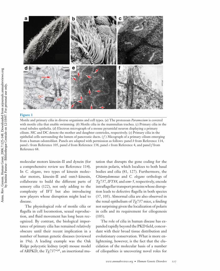
Since their first description in kidneys andthe thyroid gland (144), cilia have beenobserved in a number of organs, such asthe liver and pancreas, as well as numerouscell types, including endothelial cells, themyocardium, odontoblasts, photoreceptorsin the retina, and cortical and hypotha-lamic neurons (for examples, see 4, 21,25, 30, 68, 69, 82; for a comprehensivelist of cells and tissues containing cilia seehttp://members.global2000.net/bowser/cilialist.html) (Figure 1). Consistent withthe broad and varied tissue and cellulardistribution, dysfunction of cilia and theiranchoring structure, the basal body, has beenimplicated in numerous human diseases thatrange from organ-specific disorders such aspolycystic kidney disease to broad, pleiotropicphenotypes such as the Bardet-Biedl (BBS)and Alstrom (ALMS) syndromes.
In this review, we discuss the role of ciliain human disease, whose prominence has beenelaborated in recent years through the attri-bution of ciliary and basal body dysfunctionto a number of phenotypes. By examiningthe clinical manifestations and molecular ba-sis of seemingly diverse, yet overlapping hu-man conditions, we attempt to delineate thecommon phenotypes caused by ciliary dys-function, thus defining the hallmarks of a cil-iopathy, and then extend our observations toassign predictive value for disorders of un-known molecular etiology.
An Overview of the Cilium
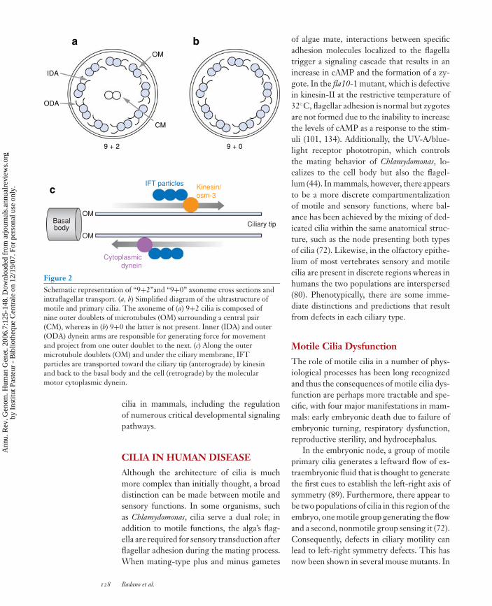
Cilia typically project from the apical sur-face of cells and are composed of a micro-tubule backbone (axoneme) ensheathed by amembrane contiguous with the plasma mem-brane (Figure 2a,b). Inner and outer dyneinarms extend from the A tubules (composedof 13 protofilaments) of each outer micro-tubule doublet and generate the force neededfor motility in an ATP-dependent process(Figure 2a,b). (For more information on
the structure of cilia and flagella, see Ref-erence 140.) Historically, the geometry andcomposition of microtubules within the cil-iary axoneme have defined the two mainciliary types: “9+2” (motile) and “9+0” (pri-mary, nonmotile) cilia, referring to the axone-mal organization of microtubule (mt) pairs.“9+2” cilia contain an axoneme that is formedof nine microtubule doublets surrounding acentral pair, whereas “9+0” lack the latter(Figure 2a,b). However, newer studies sug-gest that such distinctions might be naıve. Forexample, the organization of microtubulesalong the axoneme varies depending on theposition and at least two regions have beendistinguished in the cilia of sensory neuronsof C. elegans, the middle and distal segments,which are composed of nine microtubule dou-blets and singlets, respectively (75, 79, 109,122). Even the classic distinction of “9+2”and “9+0” as motile or sensory, respectively,seems to be simplistic and examples of motileprimary cilia as well as motile cilia and flagellawith sensory roles have been reported. Con-trary to the notion that cilia in the renal ep-ithelium are nonmotile and sensory in nature,motile cilia have been reported (95). Further-more, cilia in the pronephric kidney of ze-brafish are required for fluid movement andtheir dysfunction can lead to cyst formation(58). Additionally, it is likely that the role ofmotile cilia as sensory organelles has been un-derappreciated. For example, it was recentlyshown that transient receptor potential (TRP)channels involved in sensing environmentalstimuli of diverse forms localize to both motileand primary cilia in the female reproductivetract in mice (128).
The synthesis of structural and functionalcomponents of cilia occurs in the cytoplasmand a specialized system termed intraflag-ellar transport (IFT), which was first de-scribed in the algae Chlamydomonas reinhardtii(57), is responsible for moving cargo (IFTparticles) toward the axonemal tip or awayfrom it (anterograde and retrograde trans-port, respectively (Figure 2c). IFT parti-cles are transported by the microtubule-based
126 Badano et al.
Ann
u. R
ev. G
enom
. Hum
an G
enet
. 200
6.7:
125-
148.
Dow
nloa
ded
from
arj
ourn
als.
annu
alre
view
s.or
gby
Ins
titut
Pas
teur
- B
iblio
theq
ue C
entr
ale
on 1
2/19
/07.
For
per
sona
l use
onl
y.
ANRV285-GG07-06 ARI 12 August 2006 17:5
Figure 1Motile and primary cilia in diverse organisms and cell types. (a) The protozoan Paramecium is coveredwith motile cilia that enable swimming. (b) Motile cilia in the mammalian trachea. (c) Primary cilia in therenal tubules epithelia. (d) Electron micrograph of a mouse pyramidal neuron displaying a primarycilium. MC and DC denote the mother and daughter centrioles, respectively. (e) Primary cilia in theepithelial cells surrounding the lumen of pancreatic ducts. ( f ) Micograph of a primary cilium emergingfrom a human odontoblast. Panels are adapted with permission as follows: panel b from Reference 114,panel c from Reference 105, panel d from Reference 138, panel e from Reference 4, and panel f fromReference 68.
molecular motors kinesin-II and dynein (fora comprehensive review see Reference 114).In C. elegans, two types of kinesin molec-ular motors, kinesin-II and osm3-kinesin,collaborate to build the different parts ofsensory cilia (122), not only adding to thecomplexity of IFT but also introducingnew players whose disruption might lead todisease.
The physiological role of motile cilia orflagella in cell locomotion, sexual reproduc-tion, and fluid movement has long been rec-ognized. By contrast, the biological impor-tance of primary cilia has remained relativelyobscure until their recent implication in anumber of human genetic diseases (reviewedin 19a). A leading example was the OakRidge polycystic kidney (orpk) mouse modelof ARPKD, the Tg737orpk, an insertional mu-
tation that disrupts the gene coding for theprotein polaris, which localizes to both basalbodies and cilia (81, 127). Furthermore, theChlamydomonas and C. elegans orthologs ofTg737, IFT88, and osm-5, respectively, encodeintraflagellar transport proteins whose disrup-tion leads to defective flagella in both species(37, 105). Abnormal cilia are also observed inthe renal epithelium of Tg737 mice, a findingnot surprising given the localization of polarisin cells and its requirement for ciliogenesis(105).
The role of cilia in human disease has ex-panded rapidly beyond the PKD field, concor-dant with their broad tissue distribution andevolutionary conservation. What is more en-lightening, however, is the fact that the elu-cidation of the molecular basis of a numberof ciliopathies is uncovering novel roles for
www.annualreviews.org • Human Genetic Disorders 127
Ann
u. R
ev. G
enom
. Hum
an G
enet
. 200
6.7:
125-
148.
Dow
nloa
ded
from
arj
ourn
als.
annu
alre
view
s.or
gby
Ins
titut
Pas
teur
- B
iblio
theq
ue C
entr
ale
on 1
2/19
/07.
For
per
sona
l use
onl
y.
ANRV285-GG07-06 ARI 12 August 2006 17:5
9 + 2 9 + 0
OM
CM
IFT particles
OM
OM
Basalbody Ciliary tip
Cytoplasmicdynein
Kinesin/osm-3
c
a b
ODA
IDA
Figure 2Schematic representation of “9+2”and “9+0” axoneme cross sections andintraflagellar transport. (a, b) Simplified diagram of the ultrastructure ofmotile and primary cilia. The axoneme of (a) 9+2 cilia is composed ofnine outer doublets of microtubules (OM) surrounding a central pair(CM), whereas in (b) 9+0 the latter is not present. Inner (IDA) and outer(ODA) dynein arms are responsible for generating force for movementand project from one outer doublet to the next. (c) Along the outermicrotubule doublets (OM) and under the ciliary membrane, IFTparticles are transported toward the ciliary tip (anterograde) by kinesinand back to the basal body and the cell (retrograde) by the molecularmotor cytoplasmic dynein.
cilia in mammals, including the regulationof numerous critical developmental signalingpathways.
CILIA IN HUMAN DISEASE
Although the architecture of cilia is muchmore complex than initially thought, a broaddistinction can be made between motile andsensory functions. In some organisms, suchas Chlamydomonas, cilia serve a dual role; inaddition to motile functions, the alga’s flag-ella are required for sensory transduction afterflagellar adhesion during the mating process.When mating-type plus and minus gametes
of algae mate, interactions between specificadhesion molecules localized to the flagellatrigger a signaling cascade that results in anincrease in cAMP and the formation of a zy-gote. In the fla10-1 mutant, which is defectivein kinesin-II at the restrictive temperature of32◦C, flagellar adhesion is normal but zygotesare not formed due to the inability to increasethe levels of cAMP as a response to the stim-uli (101, 134). Additionally, the UV-A/blue-light receptor phototropin, which controlsthe mating behavior of Chlamydomonas, lo-calizes to the cell body but also the flagel-lum (44). In mammals, however, there appearsto be a more discrete compartmentalizationof motile and sensory functions, where bal-ance has been achieved by the mixing of ded-icated cilia within the same anatomical struc-ture, such as the node presenting both typesof cilia (72). Likewise, in the olfactory epithe-lium of most vertebrates sensory and motilecilia are present in discrete regions whereas inhumans the two populations are interspersed(80). Phenotypically, there are some imme-diate distinctions and predictions that resultfrom defects in each ciliary type.
Motile Cilia Dysfunction
The role of motile cilia in a number of phys-iological processes has been long recognizedand thus the consequences of motile cilia dys-function are perhaps more tractable and spe-cific, with four major manifestations in mam-mals: early embryonic death due to failure ofembryonic turning, respiratory dysfunction,reproductive sterility, and hydrocephalus.
In the embryonic node, a group of motileprimary cilia generates a leftward flow of ex-traembryonic fluid that is thought to generatethe first cues to establish the left-right axis ofsymmetry (89). Furthermore, there appear tobe two populations of cilia in this region of theembryo, one motile group generating the flowand a second, nonmotile group sensing it (72).Consequently, defects in ciliary motility canlead to left-right symmetry defects. This hasnow been shown in several mouse mutants. In
128 Badano et al.
Ann
u. R
ev. G
enom
. Hum
an G
enet
. 200
6.7:
125-
148.
Dow
nloa
ded
from
arj
ourn
als.
annu
alre
view
s.or
gby
Ins
titut
Pas
teur
- B
iblio
theq
ue C
entr
ale
on 1
2/19
/07.
For
per
sona
l use
onl
y.
ANRV285-GG07-06 ARI 12 August 2006 17:5
the inversus viscerum (iv/iv) mouse, disruptionof left-right dynein, an axonemal dynein heavychain important for ciliary motility (125), re-sults in immotile cilia and randomization ofthe left-right axis of symmetry, with 50% ofembryos being normal and 50% presentingsitus inversus (62). Complete absence of ciliain the node occurs when members of the het-erotrimeric kinesin complex, fundamental inIFT, are compromised. Targeting of KIF3Aand B in the mouse results in left-right de-fects, embryonic lethality, and developmentalproblems (71, 89, 126).
Primary ciliary dyskinesia (PCD) (OMIM:24,2650) is a group of heterogeneous disor-ders characterized by bronchiectasis, sinusitis,and infertility, with defects in body situs be-ing present in Kartagener syndrome (OMIM:24,4400). As first described by Afzelius (1976)while studying individuals with immotilesperm, cilia in PCD patients lack dynein arms,as shown by electron microscopy, but canalso present with other ultrastructural defectsthat result in impaired or inefficient motility(1). To date, mutations in a number of genesencoding components of the machinery re-quired for ciliary motility have been reportedin PCD and Kartagener syndrome. First, byfiltering a candidate gene list with Chlamy-domonas mutants that result in immotile an-imals with axonemal defects reminiscent ofPCD (absence of outer dynein arms), Pen-narun and colleagues (108) identified muta-tions in DNAI1, a gene encoding a dyneinintermediate chain. Mutations in DNAH5and DNAH11 encoding two axonemal dyneinheavy chains also cause PCD (9, 94).
Ciliary motility is also required for braindevelopment and function. Cilia in theependymal cell layer surrounding the ventri-cles maintain a flow of cerebrospinal fluid,the so-called “ependymal flow,” necessaryto maintain an open aqueduct (47). InMdnah5 mouse mutants, the murine orthologof DNAH5, a defect in the axonemal dyneinheavy chain that is expressed in ependymalcells, leads to a deficiency in outer dynein armsand results in impaired ciliary beating (47).
In the ependymal cell layer, this defective cil-iary function translates into failure to produce“ependymal flow” resulting in closure of thecerebral aqueduct and the development of hy-drocephalus (47), a condition associated withPCD in humans (49, 56, 110).
The autosomal recessive mouse model ofhydrocephalus (hy3) is caused by disruptionof the gene Hydin, which encodes a proteinexpressed in the ciliated ependymal cell layerlining the ventricles, the ciliated epithelialcells in the respiratory tract and oviduct, andspermatocytes in the testis (20). Hydin is anovel protein that, based on its expression pat-tern and mouse phenotype, is a potential can-didate for the pathogenesis of some humanciliopathies.
Sensory Cilia Defects
In contrast to the disorders of motile cilia,defects in sensory cilia appear to underliea broad range of phenotypes, probably dueto their nearly ubiquitous presence in al-most every cell type of the human body andtheir emerging role in morphogenetic signaltransduction.
The renal phenotype of ciliary dysfunc-tion. In addition to the Tg737orpk mousemodel of autosomal recessive PKD (ARPKD),other animal models support the link betweenciliary dysfunction and renal cyst formation.In the congenital polycystic kidney (cpk) mu-tant mouse, cystin, the protein product of cpk,is present in the cilia of renal epithelial cells(142). Additionally, the identification of genesmutated in various forms of human PKD isalso highlighting the central role that pri-mary cilia have in the pathomechanism of thedisease.
The process of cyst formation and distri-bution in the nephron varies, but invariablyinvolves a deregulation of the fine balance be-tween cell proliferation and cell differentia-tion. In ARPKD, cysts form from the col-lecting ducts, whereas in autosomal dominantPKD (ADPKD) they can arise in any part
www.annualreviews.org • Human Genetic Disorders 129
Ann
u. R
ev. G
enom
. Hum
an G
enet
. 200
6.7:
125-
148.
Dow
nloa
ded
from
arj
ourn
als.
annu
alre
view
s.or
gby
Ins
titut
Pas
teur
- B
iblio
theq
ue C
entr
ale
on 1
2/19
/07.
For
per
sona
l use
onl
y.
ANRV285-GG07-06 ARI 12 August 2006 17:5
of the nephron, but in both cases cells sur-rounding the cysts are usually less differenti-ated (64).
ARPKD (OMIM 26,3200) is a severe,early-onset form of PKD characterized bycystic, enlarged kidneys and hepatic fibrosisand is caused by mutations in PKHD1 (96,135, 141). PKHD1 encodes polyductin, alsonamed fibrocystin, a protein that localizes toprimary cilia in MDCK cells and that has beensuggested to be a receptor affecting the differ-entiation of collecting duct cells (64, 96, 135,136, 141).
In ADPKD, mutations have been foundin two genes, PKD1 and PKD2 (17, 76). Theproducts of these two genes are polycystin 1and 2, respectively, two novel proteins ableto interact with each other (112, 130) andthought to be part of a Ca2+ channel local-ized in the primary cilium of renal epithelialcells (31, 34, 106, 123, 142). It has been sug-gested that both polycystin 1 and 2 functionas mechanosensors of extracellular fluid flowsignaling to the interior of the cell by regulat-ing Ca2+ flux (86). These data have raised theintriguing possibility that primary cilia in therenal epithelium might act as environmentalsensors to regulate cell growth and differen-tiation; their failure results in abnormal cellproliferation and the consequent productionof renal cysts (86). Consistent with this hy-pothesis, polycystin 1 can regulate the expres-sion of p21, a tumor suppressor that inhibitscyclin-dependent kinases leading to cell cyclearrest (11).
Nephronophthisis (OMIM 25,6100) is anautosomal recessive cystic renal disease char-acterized by progressive wasting of the fil-tering unit of the kidney with or withoutmedullary involvement that can be presentin association with retinitis pigmentosa (RP)(Senior-Loken syndrome, see below) (40). Todate, five genes have been cloned (NPHP1-5 ),and analysis of their protein products has pro-vided a strong link between ciliary functionand the pathogenesis of this disease (41, 77,93, 98, 100, 118). Mutations in the humaninversin gene (INVS) cause nephronophthisis
type 2 (NPHP2) (100). An insertional eventin the mouse inversin gene, the inversion ofembryonic turning (inv) murine model, re-sults in pancreatic and renal cysts and a com-plete inversion of the left-right axis of sym-metry, which correlates specifically with aninversion in the expression patterns of genesnormally present asymmetrically, such as nodaland lefty (91, 143). It was shown recently thatprimary cilia in the node, which move in aclockwise vortical fashion, need to be tiltedposteriorly to achieve a net leftward flow (92).This finding highlights the importance of ori-enting and establishing the polarity of cellsin the plane of the tissue to coordinate thecorrect localization and angle of cilia, a pro-cess dependent on noncanonical Wnt signal-ing, the planar cell polarity (PCP) pathway(reviewed in 132). The nodal cilia in inv mu-tants are defective both in their orientationand movement, thus generating an abnor-mal, decreased nodal flow (91, 92). There-fore, it is potentially relevant that inversin hasbeen involved in controlling the balance be-tween canonical and noncanonical Wnt sig-naling cascades (119). Downstream of the re-ceptor frizzled, disheveled (Dsh) is thoughtto act as a switch between the “canonical,” b-catenin-dependent, and “noncanonical” Wntpathways (132). Inversin inhibits the canoni-cal pathway by targeting Dsh for degradationfavoring the use of the PCP pathway (119).
Nephrocystin-1 localizes to cell-cell junc-tions in polarized cells and interacts with focaladhesion proteins such as p130Cas and Pyk2,as well as N-cadherins and catenins, possiblyinfluencing cell polarity (10, 22, 23, 90). Fur-thermore, inversin can bind to the anaphase-promoting complex (APC), supporting theidea that the cilium plays a role in regulat-ing cell cycle and adding to the evidence fromthe polycystin 1–regulating p21 (78).
Ciliary dysfunction in the retina. Verte-brate photoreceptors are polarized sensoryneurons composed of an inner and an outersegment connected by a highly specialized9+0 cilium, the connecting cilium. Like other
130 Badano et al.
Ann
u. R
ev. G
enom
. Hum
an G
enet
. 200
6.7:
125-
148.
Dow
nloa
ded
from
arj
ourn
als.
annu
alre
view
s.or
gby
Ins
titut
Pas
teur
- B
iblio
theq
ue C
entr
ale
on 1
2/19
/07.
For
per
sona
l use
onl
y.
ANRV285-GG07-06 ARI 12 August 2006 17:5
types of cilia, the synthesis of materials re-quired for the formation, maintenance, andfunction of the outer segment occurs in theinner segment. Consequently, IFT is respon-sible for moving cargo across the connectingcilium, is critical for the survival of photore-ceptor cells, and underlies the pathogenesisof at least some forms of retinal degenera-tion (70, 104). For example, specific disrup-tion of kinesin-II in photoreceptors leads tothe accumulation of opsin and arrestin in theinner segment, resulting in an increased inci-dence of apoptotic cell death, a hallmark of RP(70). The requirement of delivering as manyas 2000 photopigment molecules per minuteto the mammalian outer segment might ex-plain the sensitivity of photoreceptors to IFTdefects.
RP is a genetically heterogeneous group ofretinal dystrophies that result in night blind-ness and progressive visual loss. The role ofIFT in photoreceptor survival suggests that anumber of candidate genes for RP would liein the still poorly characterized group of moi-eties involved in the process, including bothmotors as well as cargo.
Recent studies show that two proteins im-plicated in RP, RP1 and RPGR, localize pre-dominantly to the photoreceptor-connectingcilium (42, 66). RP1, commonly mutated insome forms of RP, shares a region of similar-ity with the microtubule-binding domain ofdoublecortin (DCX), a neuronal microtubule-associated protein involved in neuronal mi-gration (8, 111). RP1 is a microtubule-bindingprotein that localizes to the photoreceptor ax-oneme and helps control its length and stabil-ity in vivo (67). Rp1 mutant mice present withmisoriented outer segment discs, suggestingthat an axonemal protein is involved in theirorganization, adding another layer of com-plexity to the role of cilia-associated proteinsin the retina and the pathogenesis of RP (65).
The RP guanosine triphosphatase (GT-Pase) regulator (RPGR) is essential for pho-toreceptor maintenance and viability and mu-tations in the human RPGR gene cause RP3(74). RPGR is concentrated in the connecting
cilia of both cones and rods and its disruptionin mice leads to the mislocalization of opsins,suggesting that RPGR may be involved inprotein trafficking across the connecting cil-ium (42, 43). Some alleles of RPGR mightalso be involved in the function of motilecilia given that mutations in RPGR have beenfound in patients with RP and recurrent res-piratory infections, a phenotype characteristicof PCD, with cilia exhibiting ultrastructuralproblems in the dynein arms and microtubulebackbone (131, 133). This association of de-fects characteristic of motile and sensory ciliaare likely to be more common than expected,given the high overlap in protein content be-tween the two types of cilia.
PKD and retinal degeneration. Both reti-nal degeneration and kidney disease can becaused by defects in cilia formation, main-tenance, or function and thus it is not sur-prising to find an association of these twomajor phenotypes in human patients. Senior-Loken syndrome (SLSN; OMIM 266900) isa rare autosomal recessive disorder character-ized by nephronophthisis and progressive eyedisease. SLSN has been associated with mu-tations in several of the genes responsible fornephronophthisis (NPHP1, 3, 4) and in partic-ular with NPHP5/IQCB1 (99). Interestingly,NPHP5 interacts with RPGR in the retinaand is localized to the connecting cilia of pho-toreceptors and to primary cilia of renal ep-ithelial cells (99). These data highlight the factthat the dysfunction of cilia as an organellecan result in a group of related phenotypes.It is likely that a combination of the specificfunction of individual mutant proteins, theirpattern of expression, the level of redundancyin each tissue/cell type, the sensitivity of indi-vidual tissues, and the mutational load in addi-tional causative or modifier genes, determineswhich subset of defects are expressed in eachcase.
Such variability is better exemplified inmore pleiotropic disorders that include notonly kidney and retinal defects, but are alsodefined by defects in other tissues, such as the
www.annualreviews.org • Human Genetic Disorders 131
Ann
u. R
ev. G
enom
. Hum
an G
enet
. 200
6.7:
125-
148.
Dow
nloa
ded
from
arj
ourn
als.
annu
alre
view
s.or
gby
Ins
titut
Pas
teur
- B
iblio
theq
ue C
entr
ale
on 1
2/19
/07.
For
per
sona
l use
onl
y.
ANRV285-GG07-06 ARI 12 August 2006 17:5
limb and the nervous system, as is the casein BBS (see below) and Joubert Syndrome,an autosomal recessive disease ( JS; OMIM21,3300). In some JS cases, the character-istic features of cerebellar vermis hypopla-sia, mental retardation, hypotonia, breathing,and eye movement abnormalities are presentin conjunction with retinal degeneration andnephronophthisis. Some patients with JS seg-regate mutations in NPHP1 (102). Althoughit is possible that the NPHP1 deletion un-masks a recessive mutation in this genomicregion, the presence of the same deletion inboth JS and NPHP patients argues against thisalternative.
Cilia in other tissues and cell types. Al-though the role of cilia in the pathogenesisof cystic kidney disease and retinal degenera-tion has been well documented, the impact oftheir dysfunction in a number of other tis-sues and cell types is just beginning to beappreciated. The analysis of mouse mutantshave indicated that the cilium plays a key rolein the transduction of several paracrine sig-naling cascades, a finding supported by theenrichment for proteins involved in signal-ing observed in the flagellar and basal bodyproteome (FABB) (63). These signaling path-ways are important for diverse functions, in-cluding the establishment of cell polarity andaxis of symmetry, cell specification and differ-entiation, limb development, and neural tubeformation.
In the neural tube. Several recent lines of ev-idence indicate a crucial role for ciliary pro-teins in neural tube development. In a mousemutagenesis screen of embryonic patterningdefects, Huangfu and colleagues (48, 73, 139)identified two mutants with phenotypes char-acteristic of defective Sonic hedgehog (Shh)activity. Interestingly, the wimple (wim) andflexo (fxo) phenotypes, which include openneural tube, brain defects, and limb abnormal-ities, are caused by mutations in the IFT pro-teins IFT172 and polaris/IFT88, respectively,demonstrating that intact IFT, as well as the
molecular motor Kif3a (kinesin), are requiredin Shh signaling downstream of the recep-tor Patched 1 and the activator smoothened(Smo) (45, 46). Furthermore, Smo, a trans-membrane protein, localizes to the primarycilium and its presence in the organelle is re-quired for Shh signaling (18).
Recently, BBS mutant mice have beenshown to develop phenotypes characteristicof PCP mutant animals (a pathway requiredfor convergence and extension movementsduring gastrulation and neurulation in ver-tebrates), which include neural tube defects,open eyelids, and defective stereociliary bun-dles in the cochlea (115). Additional evidencefor the involvement of the BBS loci in the PCPpathway came from genetic crosses whereinthe BBS genes interacted genetically in bothmice and zebrafish with Vangl2, a known com-ponent of the noncanonical Wnt signalingcascade (115).
In the developing limb. Tight regulation ofcell growth and differentiation results in thecorrect patterning of digits. Perturbations inthe production, distribution, and interpreta-tion of morphogens can then result in a rangeof anatomical defects that include post- andpreaxial polydactyly (reviewed in 129). Sen-sory cilia are present in both ectodermal andmesenchymal cells in the limb bud (36), sug-gesting that they might play a role in sens-ing and transducing morphogenetic signals,thus offering a potential explanation for therecurrent presence of limb defects in a num-ber of ciliopathies (Table 1). The require-ment of functional cilia for normal Shh sig-naling is further supported by the recent workof Haycraft and colleagues in the developinglimb. The authors show that defects in po-laris/IFT88 result in the defective processingof the glioma (Gli) transcription factors, morespecifically Gli3 processing, and that all Gliproteins localize to both the nucleus as well asthe tip of the cilium (36).
Cilia and cognitive defects. Several pleio-tropic disorders caused by disruption of the
132 Badano et al.
Ann
u. R
ev. G
enom
. Hum
an G
enet
. 200
6.7:
125-
148.
Dow
nloa
ded
from
arj
ourn
als.
annu
alre
view
s.or
gby
Ins
titut
Pas
teur
- B
iblio
theq
ue C
entr
ale
on 1
2/19
/07.
For
per
sona
l use
onl
y.
ANRV285-GG07-06 ARI 12 August 2006 17:5
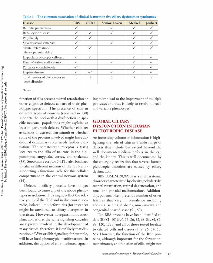
Table 1 The common association of clinical features in five ciliary dysfunction syndromes
Disease BBS OFD1 Senior-Loken Meckel JoubertRetinitis pigmentosa � � � �Renal cystic disease � � � � �Polydactyly � � � �Situs inversus/Isomerism � � � �Mental retardation/developmental delay
� � � �
Hypoplasia of corpus callosum � � � �Dandy-Walker malformation � � � �Posterior encephalocele �∗ � �Hepatic disease � �∗ � � �Total number of phenotypes ineach disorder
8 5 5 9 9
∗In mice.
function of cilia present mental retardation orother cognitive defects as part of their phe-notypic spectrum. The presence of cilia indifferent types of neurons (reviewed in 138)supports the notion that dysfunction in spe-cific neuronal populations might explain, atleast in part, such defects. Whether cilia actas sensors of extracellular stimuli or whethersome of the proteins involved might have ad-ditional extraciliary roles needs further eval-uation. The somatostatin receptor 3 (sst3)localizes to the cilia of neurons in the hip-pocampus, amygdala, cortex, and thalamus(35). Serotonin receptor 5-HT6 also localizesto cilia in different neurons of the rat brain,-supporting a functional role for this cellularcompartment in the central nervous system(14).
Defects in ciliary proteins have not yetbeen found to cause any of the above pheno-types in isolation. This might reflect the rela-tive youth of the field and in due course spo-radic, isolated limb deformities (for instance)might be attributed to ciliary disruption inthat tissue. However, a more parsimonious ex-planation is that the same signaling cascadesare typically involved in the development ofmany tissues; therefore, it is unlikely that dis-ruption of Wnt or Shh signaling, for example,will have local phenotypic manifestations. Inaddition, disruption of cilia-mediated signal-
ing might lead to the impairment of multiplepathways and thus is likely to result in broadand variable phenotypes.
GLOBAL CILIARYDYSFUNCTION IN HUMANPLEIOTROPIC DISEASE
An increasing volume of information is high-lighting the role of cilia in a wide range ofdefects that include but extend beyond thewell documented ciliary defects in the eyeand the kidney. This is well documented bythe emerging realization that several humanpleiotropic disorders are caused by ciliarydysfunction.
BBS (OMIM 20,9900) is a multisystemicdisorder characterized by obesity, polydactyly,mental retardation, retinal degeneration, andrenal and gonadal malformations. Addition-ally, patients often present a number of otherfeatures that vary in prevalence includinganosmia, asthma, diabetes, situs inversus, andcongenital heart disease (51, 60).
Ten BBS proteins have been identified todate (BBS1–10) (3, 6, 15, 26, 52, 63, 83, 84, 87,88, 120, 123a) and all of those tested localizeto ciliated cells and tissues (3, 7, 26, 54, 55,63). However, the function of the BBS pro-teins, although important for the formation,mantainance, and function of cilia, might not
www.annualreviews.org • Human Genetic Disorders 133
Ann
u. R
ev. G
enom
. Hum
an G
enet
. 200
6.7:
125-
148.
Dow
nloa
ded
from
arj
ourn
als.
annu
alre
view
s.or
gby
Ins
titut
Pas
teur
- B
iblio
theq
ue C
entr
ale
on 1
2/19
/07.
For
per
sona
l use
onl
y.
ANRV285-GG07-06 ARI 12 August 2006 17:5
be restricted to the biology of this organelle.Several of the BBS proteins localize to bothcentrosomes and basal bodies in ciliated cells(3, 54, 55). BBS4 interacts in mammalian cellswith the p150-glued subunit of dynactin, thusdirectly implying a role in microtubule trans-port. Furthermore, BBS4 is required, perhapsas an adaptor protein for the correct local-ization of pericentriolar material 1 (PCM1),the major component of pericentriolar satel-lites and a protein required for both centro-some function and ciliogenesis (19, 54, 59).BBS3 is a member of the RAS superfamilyof GTP-binding proteins that localizes to thecytoplasm and is thought to play a role in vesi-cle trafficking. However, studies in C. elegansdemonstrate that Bbs-3 localizes to the cyto-plasm and the basal body and is involved inIFT in the cilia of sensory neurons (26). Bbs-1, bbs-2, bbs-7, and bbs-8 in C. elegans localize tothe transition zones of cilia, basal bodies in theworm, and loss of bbs-7 and bbs-8 affects ciliaboth structurally (shortened) and functionally(impaired IFT) (13).
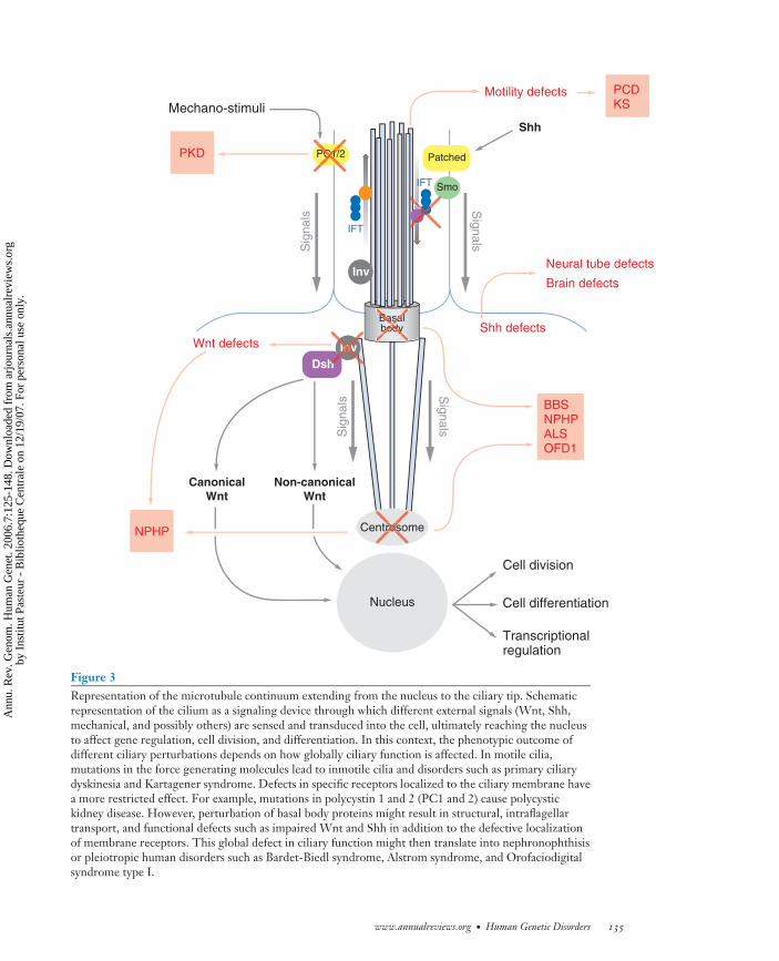
These data suggest that at least some ofthese proteins might have a dual or broaderfunction that includes, but is not limited to,their role in ciliary biology and could providea functional link between different structuresand subcellular compartments. Importantly, acentrosomal dysfunction might underlie someof the phenotypic aspects of BBS that are noteasily reconciled with a ciliary defect. Besidesits role during cell division, the centrosome isthought to have a role in diverse cellular pro-cesses that include protein degradation, neu-ronal migration, axonal guidance, and vesicu-lar transport (8). By affecting the function ofcilia and other microtubule-based processess,defects in the BBS proteins may thus result ina global impairment of those ciliary functionsthat depend both on the structure as well as theability of the organelle to sense and transducediverse extracellular signals, perhaps explain-ing, at least in part, the pleiotropy observed inthis syndrome (Figure 3).
A similar example is ALMS (OMIM20,3800), which is caused by mutations in
ALMS1 (16, 38). ALMS patients present witha number of phenotypes reminiscent of BBS,including RP, obesity, and diabetes, but aredistinguished from the latter in that they de-velop significant sensorineural deafness anddo not have polydactyly. ALMS1 was iden-tified in a proteomic analysis of the humancentrosome (2) and was shown to localize toboth centrosome and basal bodies in a patternhighly reminiscent of that of the BBS pro-teins (39). These data may indicate that BBSand ALMS could belong to a discrete groupof disorders based on both cilia and centroso-mal dysfunction that are distinct from otherciliopathies.
Orofaciodigital syndrome type I (OFD1;OMIM 31,1200), an X-linked disorder char-acterized by malformations of the face, oralcavity, and digits with PKD and variable in-volvement of the central nervous system, iscaused by mutations in OFD1 (27). OFD1 lo-calizes to both centrosomes and basal bodies,suggesting that this syndrome might also fallinto this broader category of ciliary diseases(2, 53, 113).
The most recent example of ciliopathyis Meckel-Gruber syndrome (MKS; OMIM24,9000). MKS is a lethal condition charac-terized by cleft palate, renal cysts, hepaticfibrosis, polydactyly, and central nervous sys-tem defects, including occipital encephalo-cele. Importantly, mutations in several of theBBS genes have been found in fetuses withMeckel-like phenotypes, raising the possibil-ity that the MKS and BBS loci might interactgenetically (50). Recently, the first two genesthat cause MKS were cloned (61, 121) andtheir encoded proteins are found in the pre-dicted ciliary proteome (63), supporting thehypothesis that ciliary dysfunction likely un-derlies the pathogenesis of MKS (see predict-ing ciliary diseases) (61, 121).
DISSECTING CILIARY DISEASES
A better understanding of ciliary structure andfunction is likely to have significant conse-quences both at the basic research and clinical
134 Badano et al.
Ann
u. R
ev. G
enom
. Hum
an G
enet
. 200
6.7:
125-
148.
Dow
nloa
ded
from
arj
ourn
als.
annu
alre
view
s.or
gby
Ins
titut
Pas
teur
- B
iblio
theq
ue C
entr
ale
on 1
2/19
/07.
For
per
sona
l use
onl
y.
ANRV285-GG07-06 ARI 12 August 2006 17:5
Centrosome
Nucleus
Basalbody
PC1/2
Transcriptionalregulation
Cell division
Cell differentiation
Mechano-stimuli
Shh
PKD
IFT
IFT
Neural tube defects
Brain defects
Smo
Patched
Sig
nals
Sig
nals
Shh defects
InvDsh
Non-canonicalWnt
CanonicalWnt
Inv
Wnt defects
NPHP
BBSNPHPALSOFD1
Motility defects PCDKS
Sig
nals
Sig
nals
CC1/PC1
roosros
Inh
nvInv
Figure 3Representation of the microtubule continuum extending from the nucleus to the ciliary tip. Schematicrepresentation of the cilium as a signaling device through which different external signals (Wnt, Shh,mechanical, and possibly others) are sensed and transduced into the cell, ultimately reaching the nucleusto affect gene regulation, cell division, and differentiation. In this context, the phenotypic outcome ofdifferent ciliary perturbations depends on how globally ciliary function is affected. In motile cilia,mutations in the force generating molecules lead to inmotile cilia and disorders such as primary ciliarydyskinesia and Kartagener syndrome. Defects in specific receptors localized to the ciliary membrane havea more restricted effect. For example, mutations in polycystin 1 and 2 (PC1 and 2) cause polycystickidney disease. However, perturbation of basal body proteins might result in structural, intraflagellartransport, and functional defects such as impaired Wnt and Shh in addition to the defective localizationof membrane receptors. This global defect in ciliary function might then translate into nephronophthisisor pleiotropic human disorders such as Bardet-Biedl syndrome, Alstrom syndrome, and Orofaciodigitalsyndrome type I.
www.annualreviews.org • Human Genetic Disorders 135
Ann
u. R
ev. G
enom
. Hum
an G
enet
. 200
6.7:
125-
148.
Dow
nloa
ded
from
arj
ourn
als.
annu
alre
view
s.or
gby
Ins
titut
Pas
teur
- B
iblio
theq
ue C
entr
ale
on 1
2/19
/07.
For
per
sona
l use
onl
y.
ANRV285-GG07-06 ARI 12 August 2006 17:5
levels. To this end, multiple groups have en-gaged in proteomic and comparative genomicstudies to elucidate the complete protein com-plement of cilia (5, 12, 24, 26, 63, 97, 103,124).
An in silico comparative approach betweenthe proteome of a nonflagellated/ciliated or-ganism such as Arabidopsis and that of cili-ated organisms such as humans and Chlamy-domonas resulted in the identification of agroup of 688 proteins likely involved in thebiology of cilia and basal bodies (63). Further-more, identification of all C. elegans genes andtheir human orthologs that contain an X box,the recognition site for the cilia-specific tran-scription factor Daf-19, resulted in additionalloci that overlap and expand the previous list(12, 24, 26). Additionally, transcriptional andmass spectrometry analysis in both C. rein-hardtii and human cells further increased thelist, resulting in a combined ciliary proteomedata set that contains more than 1300 genes(97, 103, 124; our unpublished data).
The availability of the ciliary proteome isproving to be a powerful resource to expeditethe cloning of suspected ciliopathies by prior-itizing positional candidates by their presencein the data set. This has facilitated the cloningof both novel causative and modifier genes.For example, in BBS, the cloning of BBS3,BBS5, and, more recently, the BBS modi-fier MGC1203, was achieved by sequencinga reduced set of positional or BBS-interactingcandidates, respectively (7, 15, 26, 63), as wasthe case for the cloning of MKS1 (61). Thisresource is not only facilitating the identifi-cation of novel human disease genes, but alsopromises to help unravel the genetic basis ofother suspected ciliopathies.
THE HUMAN CILIOPATHIES:PHENOTYPES ANDPREDICTIONS
The clinical manifestations of the known cil-iopathies have the potential to provide us witha set of phenotypic parameters that can beused as a training set to predict ciliary in-
volvement in disorders of unknown molec-ular etiology. By comparing the ontologi-cal descriptions of five syndromes for whichfunction has already been ascribed to dis-ordered cilia (Table 1), we can devise coresearch terms by which to query databasessuch as OMIM or the London Dysmorphol-ogy Database (LDDB, formerly known as theWinter Baraitser Dysmorphology Database).
As described earlier, phenotypes such asRP, renal cystic disease, left-right axis de-termination, and polydactyly can be causedby structural and functional abnormalities ofcilia. Additionally, the observation of globallevels of cognitive impairment or develop-mental delay in many of these syndromeshighlights the importance of primary cilia inneurological function, although their exactrole remains to be determined.
Perhaps more difficult to explain are thereports of specific structural changes withinthe brain, such as agenesis of the corpuscallosum and Dandy-Walker malformation(DWM) associated with four of the five condi-tions listed in Table 1. One possibility mightbe that the activity of ZIC genes (zinc fingersin the cerebellum) relies on the cilium. Mu-tations in ZIC genes were recently implicatedin a wide variety of congenital malformationsincluding DWM, holoprosencephaly, neuraltube defects, and heterotaxy (reviewed in 32).Mice doubly heterozygous for inactivatingmutations in both Zic1 and Zic4 give riseto cerebellar hypoplasia and foliation defectssimilar to DWM in human patients wherebythe cerebellar hemispheres are relatively un-affected (33). Additionally, loss of functionof ZIC2 has been associated with holopros-encephaly (HPE), a phenotype that can becaused by Shh defects, and neural tube de-fects (reviewed in 32). Furthermore, Zic2 mu-tants also display phenotypes reminiscent ofciliary dysfunction including neural tube clo-sure defects such as exencephaly, anencephaly,and spina bifida (85). Likewise, mutations inZIC3 cause heterotaxy and neural tube defects(29, 137). Although the exact relationship ofthe ZIC family of genes with cilia remains to
136 Badano et al.
Ann
u. R
ev. G
enom
. Hum
an G
enet
. 200
6.7:
125-
148.
Dow
nloa
ded
from
arj
ourn
als.
annu
alre
view
s.or
gby
Ins
titut
Pas
teur
- B
iblio
theq
ue C
entr
ale
on 1
2/19
/07.
For
per
sona
l use
onl
y.
ANRV285-GG07-06 ARI 12 August 2006 17:5
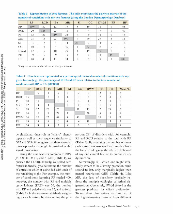
Table 2 Representation of core features. The table represents the pairwise analysis of thenumber of conditions with any two features (using the London Dysmorphology Database)
RP RCD Po MR SI CC DWM PE HFRP 409∗ 20 12 73 5 10 12 9 68RCD 20 128 22 16 6 4 9 9 40Po 12 22 119 22 7 5 10 9 13MR 73 16 22 599 7 49 29 5 34SI 5 6 4 4 27 3 4 2 8CC 10 4 5 49 3 62 19 2 5DWM 12 9 10 29 4 19 45 9 5PE 9 9 9 5 2 2 9 46 7HF 68 40 13 34 8 5 5 7 288
∗Gray box = total number of entries with given feature.
Table 3 Core features represented as a percentage of the total number of conditions with anygiven feature [e.g., the percentage of RCD and RP cases relative to the total number ofconditions with RP = 5% (20/409)]
RP RCD Po MR SI CC DWM PE HF Mean %RP 5 3 17 1 2 3 2 16 6RCD 15 17 12 4 3 7 7 31 12Po 10 18 18 6 4 8 7 11 10MR 12 3 4 1 8 5 1 5 5SI 18 22 26 26 11 15 7 29 16CC 16 6 8 79 5 30 3 8 19DWM 26 20 22 64 9 42 20 11 27PE 19 19 19 10 4 4 19 15 13HF 24 14 4 12 3 2 2 2 8
be elucidated, their role in “ciliary” pheno-types as well as their sequence similarity toGli1 and Gli3 (32) suggests that these encodedtranscription factors might be involved in Shhsignal transduction.
Using the nine features common to BBS,JS, OFD1, MKS, and SLSN (Table 1), wequeried the LDDB. Initially, we tested eachfeature individually to determine the numberof entries in which it coincided with each ofthe remaining eight. For example, the num-ber of conditions featuring RP totaled 409;however, the number with RP and multiplecystic kidneys (RCD) was 20, the numberwith RP and polydactyly was 12, and so forth(Table 2). In this way we established a weight-ing for each feature by determining the pro-
portion (%) of disorders with, for example,RP and RCD relative to the total with RP(Table 3). By averaging the number of timeseach feature was associated with another fromthe list we could gauge the relative likelihoodof any one clinical feature to predict ciliarydysfunction.
Surprisingly, RP, which one might intu-itively expect to be a strong predictor, camesecond to last, only marginally higher thanmental retardation (MR) (Table 4). LikeMR, this lack of specificity probably re-flects the multiple etiologies of retinal de-generation. Conversely, DWM scored as thegreatest predictor for ciliary dysfunction.To test these observations we took two ofthe highest-scoring features from different
www.annualreviews.org • Human Genetic Disorders 137
Ann
u. R
ev. G
enom
. Hum
an G
enet
. 200
6.7:
125-
148.
Dow
nloa
ded
from
arj
ourn
als.
annu
alre
view
s.or
gby
Ins
titut
Pas
teur
- B
iblio
theq
ue C
entr
ale
on 1
2/19
/07.
For
per
sona
l use
onl
y.
ANRV285-GG07-06 ARI 12 August 2006 17:5
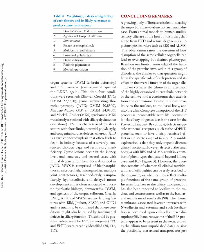
Table 4 Weighting (in descending order)of each feature and its likely relevance topredict ciliary involvement
1 Dandy-Walker Malformation2 Agenesis of Corpus Callosum3 Situs inversus4 Posterior encephalocele5 Multicystic renal disease6 Post-axial polydactyly7 Hepatic disease8 Retinitis pigmentosa9 Mental retardation
organ systems—DWM (a brain deformity)and situs inversus (cardiac)—and queriedthe LDDB again. This time four condi-tions were returned: Ellis-van Creveld (EVC;OMIM 22,5500), Jeune asphyxiating tho-racic dystrophy (JATD; OMIM 20,8500),Marden-Walker (MWS; OMIM 24,8700),and Meckel-Gruber (MKS) syndromes. MKSwas already associated with ciliary dysfunction(see above). EVC is characterized by shortstature with short limbs, postaxial polydactyly,and congenital cardiac defects, whereas JATDis a rare chondrodysplasia that often leads todeath in infancy because of a severely con-stricted thoracic cage and respiratory insuf-ficiency. Cystic lesions occur in the kidney,liver, and pancreas, and several cases withretinal degeneration have been described inJATD. MWS is comprised of blepharophi-mosis, microcephaly, micrognathia, multiplejoint contractures, arachnodactyly, campto-dactyly, kyphoscoliosis, and delayed motordevelopment and is often associated with cys-tic dysplastic kidneys, dextrocardia, DWM,and agenesis of the corpus callosum. Clearly,EVC, JATD, and MWS have overlapping fea-tures with BBS, Joubert, SLSN, and OFD1and it remains to be confirmed that these con-ditions might also be caused by fundamentaldefects in ciliary function. This should be pos-sible to determine for EVC as two genes (EVCand EVC2) were recently identified (28, 116,117).
CONCLUDING REMARKS
A growing body of literature is demonstratingthe impact of ciliary dysfunction in human dis-ease. From animal models to human studies,sensory cilia are at the heart of disorders thatrange from PKD and retinal degeneration topleiotropic disorders such as BBS and ALMS.This observation raises the question of howdisruption of the same cellular organelle canlead to overlapping but distinct phenotypes.Based on our limited knowledge of the func-tion of the proteins involved in this group ofdisorders, the answer to that question mightlie in the specific role of each protein and itseffect on the overall function of the organelle.
If we consider the cilium as an extensionof the highly organized microtubule networkof the cell, we find a continuum that extendsfrom the centrosome located in close prox-imity to the nucleus, to the basal body, andinto the cilia. Complete disruption of the IFTprocess is incompatible with life, because itblocks ciliary biogenesis, as is the case for thePolaris null mutant. By contrast, defects in spe-cific axonemal receptors, such as the ADPKDproteins, seem to have a fairly restricted ef-fect in a discrete range of tissues. A plausibleexplanation is that they only impede discreteciliary functions. However, defects at the basalbody, as with BBS and ALMS, result in a num-ber of phenotypes that extend beyond kidneycysts and RP (Figure 3). However, the ques-tion remains of whether all clinical manifes-tations of ciliopathies can be truly ascribed tothe organelle, or whether they reflect multi-ple functions of the same group of proteins.Inversin localizes to the ciliary axoneme, buthas also been reported to localize to the nu-cleus and centrosome as well as to the basolat-eral membrane of renal cells (90). The plasmamembrane–associated inversin interacts withN-cadherin and catenins and such localiza-tion is perturbed upon cell-cell contact dis-ruption (90). In neurons, some of the BBS pro-teins appear to be present in the axon, as wellas the cilium (our unpublished data), raisingthe possibility that axonal transport, not just
138 Badano et al.
Ann
u. R
ev. G
enom
. Hum
an G
enet
. 200
6.7:
125-
148.
Dow
nloa
ded
from
arj
ourn
als.
annu
alre
view
s.or
gby
Ins
titut
Pas
teur
- B
iblio
theq
ue C
entr
ale
on 1
2/19
/07.
For
per
sona
l use
onl
y.
ANRV285-GG07-06 ARI 12 August 2006 17:5
intraflagellar transport, might be involved inthe development of the complex cognitivephenotype characteristic of the syndrome.Generating mutants that selectively fail to lo-calize to some but not all physiologically rel-evant regions of the cell (e.g., the basal bodybut not the centrosome) will be required todissect the potential distinct roles of the sameprotein and better address these questions.In addition, in vivo models that enable usto specifically block and unblock ciliogenesisin a tissue- and cell-specific manner will beinvaluable.
Remarkable progress has been achieved inassigning function to cilia in different celltypes. However, there are still a large num-ber of tissues in which the role of these cel-lular structures is less clear and for which theidentification and study of pleiotropic humanciliary disorders promises to provide impor-tant clues. For example, in odontoblasts, ciliamight have a mechanosensory role sensingfluid forces within dentinal tubules and reg-ulating dentine deposition (68). Syndromessuch as BBS present dental crowding and
other dental anomalies as components of theirphenotypes, highlighting the importance andexpanding the putative role of cilia in this tis-sue. Likewise, the presence of different typesof receptors in neuronal cilia and the com-mon association of cognitive defects with cil-iary disease suggest an important role of ciliain the still poorly understood central nervoussystem.
Defining the hallmarks of a ciliopathyand classifying a human disease as such willbe important both for understanding thepathomechanism of the disease and for theclinical management of patients. Identifyingciliopathies in humans can be aided by the ad-ministration of noninvasive, yet informativetests, as exemplified by the identification ofanosmia and defective otoacoustic emissionsas additional features of BBS (60, 115). Finally,a unified view of the phenotypic characteris-tics of a ciliary dysfunction and the availabilityof the ciliary proteome are likely to facilitatethe identification of yet unrecognized ciliarydisorders and the genes and proteins involvedin their pathogenesis.
SUMMARY POINTS
1. Cilia and flagella are evolutionary conserved organelles involved in a number of cellu-lar processes that range from whole cell locomotion, chemotaxis, and fluid movementto signal transduction.
2. The identification of several animal models and human disorders caused by defectsin these organelles has highlighted the roles of cilia in a range of biological functionsincluding the intracellular interpretation of several paracrine morphogenetic signals.
3. The association of an increasing number of human disorders with ciliary dysfunctionis offering the possibility of unifying diverse phenotypic manifestations under thecommon theme of the ciliopathies, where each discrete clinical entity manifests asubset of overlapping clinical features.
4. The phenotypic parameters that define a ciliopathy can be used to both recognizethe cellular basis of a number of genetic disorders and to facilitate the diagnosis andtreatment of some diseases of unknown etiology.
FUTURE ISSUES
1. Although cilia have been found in almost every tissue and cell type in mammals, theirrole is typically obscure, as are the consequences of their dysfunction.
www.annualreviews.org • Human Genetic Disorders 139
Ann
u. R
ev. G
enom
. Hum
an G
enet
. 200
6.7:
125-
148.
Dow
nloa
ded
from
arj
ourn
als.
annu
alre
view
s.or
gby
Ins
titut
Pas
teur
- B
iblio
theq
ue C
entr
ale
on 1
2/19
/07.
For
per
sona
l use
onl
y.
ANRV285-GG07-06 ARI 12 August 2006 17:5
2. Do ciliary proteins have additional roles in the cell, and if so what are they?
3. How can ciliary dysfunction lead to variable and diverse phenotypes? Is it a question ofspecific protein dysfunction, the fact that some of the moieties involved are expressedin different cells and tissues, or the variable redundancy and/or buffering capacity ofindividual systems?
4. Despite a number of clues as to the function of several ciliary proteins, the exact roleof this group of molecules is largely unknown.
ACKNOWLEDGMENTS
The structure and function of cilia have been studied for more than 100 years and hence weapologize to those scientists whose contribution could not be properly cited in this work due tospace limitations. We also thank Jantje Gerdes and Erica Davis for their thoughtful commentson the manuscript. This work was supported by grant R01HD04260 from the National Instituteof Child Health and Development (N.K.), grant R01DK072301 from the National Institute ofDiabetes, Digestive and Kidney disorders (N.K.), a grant from the Polycystic Kidney DiseaseFoundation (J.L.B. and N.K.), and the Medical Research Council (P.L.B.). P.L.B. is a SeniorWellcome Trust Fellow.
LITERATURE CITED
1. Afzelius BA. 1976. A human syndrome caused by immotile cilia. Science 193:317–192. Andersen JS, Wilkinson CJ, Mayor T, Mortensen P, Nigg EA, Mann M. 2003. Pro-
teomic characterization of the human centrosome by protein correlation profiling. Na-ture 426:570–74
3. Presents the firstevidence likeningpleiotropicphenotypes tociliary dysfunction.
3. Ansley SJ, Badano JL, Blacque OE, Hill J, Hoskins BE, et al. 2003. Basal body dys-function is a likely cause of pleiotropic Bardet-Biedl syndrome. Nature 425:628–33.
4. Ashizawa N, Endoh H, Hidaka K, Watanabe M, Fukumoto S. 1997. Three-dimensionalstructure of the rat pancreatic duct in normal and inflammated pancreas. Microsc. Res.Tech. 37:543–56
5. Together withRef. 63, describesthe use ofcomparativegenomics to definethe proteinsimportant forciliary structureand function.
5. Avidor-Reiss T, Maer AM, Koundakjian E, Polyanovsky A, Keil T, et al. 2004. De-coding cilia function: defining specialized genes required for compartmentalizedcilia biogenesis. Cell 117:527–39
6. Badano JL, Ansley SJ, Leitch CC, Lewis RA, Lupski JR, Katsanis N. 2003. Identificationof a novel Bardet-Biedl syndrome protein, BBS7, that shares structural features withBBS1 and BBS2. Am. J. Hum. Genet. 72:650–58
7. Badano JL, Leitch CC, Ansley SJ, May-Simera H, Lawson S, et al. 2006. Dissection ofepistasis in oligogenic Bardet-Biedl syndrome. Nature 439:326–30
8. Badano JL, Teslovich TM, Katsanis N. 2005. The centrosome in human genetic disease.Nat. Rev. Genet. 6:194–205
9. Bartoloni L, Blouin JL, Pan Y, Gehrig C, Maiti AK, et al. 2002. Mutations in theDNAH11 (axonemal heavy chain dynein type 11) gene cause one form of situs inversustotalis and most likely primary ciliary dyskinesia. Proc. Natl. Acad. Sci. USA 99:10282–86
10. Benzing T, Gerke P, Hopker K, Hildebrandt F, Kim E, Walz G. 2001. Nephrocystininteracts with Pyk2, p130(Cas), and tensin and triggers phosphorylation of Pyk2. Proc.Natl. Acad. Sci. USA 98:9784–89
140 Badano et al.
Ann
u. R
ev. G
enom
. Hum
an G
enet
. 200
6.7:
125-
148.
Dow
nloa
ded
from
arj
ourn
als.
annu
alre
view
s.or
gby
Ins
titut
Pas
teur
- B
iblio
theq
ue C
entr
ale
on 1
2/19
/07.
For
per
sona
l use
onl
y.
ANRV285-GG07-06 ARI 12 August 2006 17:5
11. Bhunia AK, Piontek K, Boletta A, Liu L, Qian F, et al. 2002. PKD1 induces p21(waf1)and regulation of the cell cycle via direct activation of the JAK-STAT signaling pathwayin a process requiring PKD2. Cell 19:157–68
12. Blacque OE, Perens EA, Boroevich KA, Inglis PN, Li C, et al. 2005. Functional genomicsof the cilium, a sensory organelle. Curr. Biol. 15:935–41
13. Blacque OE, Reardon MJ, Li C, McCarthy J, Mahjoub MR, et al. 2004. Loss of C. elegansBBS-7 and BBS-8 protein function results in cilia defects and compromised intraflagellartransport. Genes Dev. 18:1630–42
14. Brailov I, Bancila M, Brisorgueil MJ, Miquel MC, Hamon M, Verge D. 2000. Local-ization of 5-HT(6) receptors at the plasma membrane of neuronal cilia in the rat brain.Brain Res. 872:271–75
15. Chiang AP, Nishimura D, Searby C, Elbedour K, Carmi R, et al. 2004. Comparativegenomic analysis identifies an ADP-ribosylation factor-like gene as the cause of Bardet-Biedl syndrome (BBS3). Am. J. Hum. Genet. 75:475–84
16. Collin GB, Marshall JD, Ikeda A, So WV, Russell-Eggit I, et al. 2002. Mutations inALMS1 cause obesity, type 2 diabetes and neurosensory degeneration in Alstrom syn-drome. Nat. Genet. 31:74–78
17. Consort. TEPKD. 1994. The polycystic kidney disease 1 gene encodes a 14 kb transcriptand lies within a duplicated region on chromosome 16. Cell 77:881–94
18. Demonstratesthat Smoothened, aprotein essential inthis pathway,localizes to primarycilia where it isessential for Shhsignal processing.
18. Corbit KC, Aanstad P, Singla V, Norman AR, Stainier DY, Reiter JF. 2005. Ver-tebrate Smoothened functions at the primary cilium. Nature 437:1018–21
19. Dammermann A, Merdes A. 2002. Assembly of centrosomal proteins and microtubuleorganization depends on PCM-1. J. Cell Biol. 159:255–66
19a. Davenport JR, Yoder BK. 2005. An incredible decade for the primary cilium: a look ata once-forgotten organelle. Am. J. Physiol. Renal Physiol. 289:F1159–69
20. Davy BE, Robinson ML. 2003. Congenital hydrocephalus in hy3 mice is caused by aframeshift mutation in Hydin, a large novel gene. Hum. Mol. Genet. 12:1163–70
21. De Robertis E. 1956. Morphogenesis of the retinal rods: an electron microscope study.J. Biophys. Biochem. Cytol. 2:209–18
22. Donaldson JC, Dempsey PJ, Reddy S, Bouton AH, Coffey RJ, Hanks SK. 2000. Crk-associated substrate p130(Cas) interacts with nephrocystin and both proteins localize tocell-cell contacts of polarized epithelial cells. Exp. Cell Res. 256:168–78
23. Donaldson JC, Dise RS, Ritchie MD, Hanks SK. 2002. Nephrocystin-conserved do-mains involved in targeting to epithelial cell-cell junctions, interaction with filamins,and establishing cell polarity. J. Biol. Chem. 277:29028–35
24. Efimenko E, Bubb K, Mak HY, Holzman T, Leroux MR, et al. 2005. Analysis of xbxgenes in C. elegans. Development 132:1923–34
25. Ekblom A, Hansson P. 1984. A thin-section and freeze-fracture study of the pulp bloodvessels in feline and human teeth. Arch. Oral Biol. 29:413–24
26. Fan Y, Esmail MA, Ansley SJ, Blacque OE, Boroevich K, et al. 2004. Mutations ina member of the Ras superfamily of small GTP-binding proteins causes Bardet-Biedlsyndrome. Nat. Genet. 36:989–93
27. Ferrante MI, Giorgio G, Feather SA, Bulfone A, Wright V, et al. 2001. Identificationof the gene for oral-facial-digital type I syndrome. Am. J. Hum. Genet. 68:569–76
28. Galdzicka M, Patnala S, Hirshman MG, Cai JF, Nitowsky H, et al. 2002. A new gene,EVC2, is mutated in Ellis-van Creveld syndrome. Mol. Genet. Metab. 77:291–95
29. Gebbia M, Ferrero GB, Pilia G, Bassi MT, Aylsworth A, et al. 1997. X-linked situsabnormalities result from mutations in ZIC3. Nat. Genet. 17:305–8
www.annualreviews.org • Human Genetic Disorders 141
Ann
u. R
ev. G
enom
. Hum
an G
enet
. 200
6.7:
125-
148.
Dow
nloa
ded
from
arj
ourn
als.
annu
alre
view
s.or
gby
Ins
titut
Pas
teur
- B
iblio
theq
ue C
entr
ale
on 1
2/19
/07.
For
per
sona
l use
onl
y.
ANRV285-GG07-06 ARI 12 August 2006 17:5
30. Geerts A, Bouwens L, Wisse E. 1990. Ultrastructure and function of hepatic fat-storingand pit cells. J. Electron Microsc. Tech. 14:247–56
31. Gonzalez-Perrett S, Kim K, Ibarra C, Damiano AE, Zotta E, et al. 2001. Polycystin-2,the protein mutated in autosomal dominant polycystic kidney disease (ADPKD), is aCa2+-permeable nonselective cation channel. Proc. Natl. Acad. Sci. USA 98:1182–87
32. Grinberg I, Millen KJ. 2005. The ZIC gene family in development and disease. Clin.Genet. 67:290–96
33. Grinberg I, Northrup H, Ardinger H, Prasad C, Dobyns WB, Millen KJ. 2004. Het-erozygous deletion of the linked genes ZIC1 and ZIC4 is involved in Dandy-Walkermalformation. Nat. Genet. 36:1053–55
34. Hanaoka K, Qian F, Boletta A, Bhunia AK, Piontek K, et al. 2000. Co-assembly ofpolycystin-1 and -2 produces unique cation-permeable currents. Nature 408:990–94
35. Handel M, Schulz S, Stanarius A, Schreff M, Erdtmann-Vourliotis M, et al. 1999. Se-lective targeting of somatostatin receptor 3 to neuronal cilia. Neuroscience 89:909–26
36. Haycraft CJ, Banizs B, Aydin-Son Y, Zhang Q, Michaud EJ, Yoder BK. 2005. Gli2 andGli3 localize to cilia and require the intraflagellar transport protein polaris for processingand function. PLoS Genet. 1:0480–88
37. Haycraft CJ, Swoboda P, Taulman PD, Thomas JH, Yoder BK. 2001. The C. elegans ho-molog of the murine cystic kidney disease gene Tg737 functions in a ciliogenic pathwayand is disrupted in osm-5 mutant worms. Development 128:1493–505
38. Hearn T, Renforth GL, Spalluto C, Hanley NA, Piper K, et al. 2002. Mutation ofALMS1, a large gene with a tandem repeat encoding 47 amino acids, causes Alstromsyndrome. Nat. Genet. 31:79–83
39. Hearn T, Spalluto C, Phillips VJ, Renforth GL, Copin N, et al. 2005. Subcellularlocalization of ALMS1 supports involvement of centrosome and basal body dysfunctionin the pathogenesis of obesity, insulin resistance, and type 2 diabetes. Diabetes 54:1581–87
40. Hildebrandt F. 2004. Nephronophthisis-medullary cystic kidney disease. In PediatricNephrology, ed. ED Avner, P Niaudet, pp. 665–73. Philadelphia: Lippincott, Williams& Wilkins
41. Hildebrandt F, Otto E, Rensing C, Nothwang HG, Vollmer M, et al. 1997. A novel geneencoding an SH3 domain protein is mutated in nephronophthisis type 1. Nat. Genet.17:149–53
42. Hong DH, Pawlyk B, Sokolov M, Strissel KJ, Yang J, et al. 2003. RPGR isoforms in pho-toreceptor connecting cilia and the transitional zone of motile cilia. Invest. Ophthalmol.Vis. Sci. 44:2413–21
43. Hong DH, Pawlyk BS, Shang J, Sandberg MA, Berson EL, Li T. 2000. A retinitispigmentosa GTPase regulator (RPGR)-deficient mouse model for X-linked retinitispigmentosa (RP3). Proc. Natl. Acad. Sci. USA 97:3649–54
44. Huang K, Kunkel T, Beck CF. 2004. Localization of the blue-light receptor phototropinto the flagella of the green alga Chlamydomonas reinhardtii. Mol. Biol. Cell 15:3605–14
45. Huangfu D, Anderson KV. 2005. Cilia and Hedgehog responsiveness in the mouse.Proc. Natl. Acad. Sci. USA 102:11325–30
46. Firstimplication of IFTproteins in thetransmission of Shhsignal.
46. Huangfu D, Liu A, Rakeman AS, Murcia NS, Niswander L, Anderson KV. 2003.Hedgehog signalling in the mouse requires intraflagellar transport proteins. Na-
ture 426:83–8747. Ibanez-Tallon I, Pagenstecher A, Fliegauf M, Olbrich H, Kispert A, et al. 2004. Dys-
function of axonemal dynein heavy chain Mdnah5 inhibits ependymal flow and revealsa novel mechanism for hydrocephalus formation. Hum. Mol. Genet. 13:2133–41
142 Badano et al.
Ann
u. R
ev. G
enom
. Hum
an G
enet
. 200
6.7:
125-
148.
Dow
nloa
ded
from
arj
ourn
als.
annu
alre
view
s.or
gby
Ins
titut
Pas
teur
- B
iblio
theq
ue C
entr
ale
on 1
2/19
/07.
For
per
sona
l use
onl
y.
ANRV285-GG07-06 ARI 12 August 2006 17:5
48. Ishibashi M, Saitsu H, Komada M, Shiota K. 2005. Signaling cascade coordinatinggrowth of dorsal and ventral tissues of the vertebrate brain, with special reference to theinvolvement of Sonic Hedgehog signaling. Anat. Sci. Int. 80:30–36
49. Jabourian Z, Lublin FD, Adler A, Gonzales C, Northrup B, Zwillenberg D. 1986.Hydrocephalus in Kartagener’s syndrome. Ear Nose Throat J. 65:468–72
50. Karmous-Benailly H, Martinovic J, Gubler MC, Sirot Y, Clech L, et al. 2005. Antenatalpresentation of Bardet-Biedl syndrome may mimic Meckel syndrome. Am. J. Hum.Genet. 76:493–504
51. Katsanis N. 2004. The oligogenic properties of Bardet-Biedl syndrome. Hum. Mol.Genet. 13:R65–R71
52. Katsanis N, Beales PL, Woods MO, Lewis RA, Green JS, et al. 2000. Mutations inMKKS cause obesity, retinal dystrophy and renal malformations associated with Bardet-Biedl syndrome. Nat. Genet. 26:67–70
53. Keller LC, Romijn EP, Zamora I, Yates JR 3rd, Marshall WF. 2005. Proteomic analysisof isolated chlamydomonas centrioles reveals orthologs of ciliary-disease genes. Cell15:1090–98
54. Kim JC, Badano JL, Sibold S, Esmail MA, Hill J, et al. 2004. The Bardet-Biedl pro-tein BBS4 targets cargo to the pericentriolar region and is required for microtubuleanchoring and cell cycle progression. Nat. Genet. 36:462–70
55. Kim JC, Ou YY, Badano JL, Esmail MA, Leitch CC, et al. 2005. MKKS/BBS6, adivergent chaperonin-like protein linked to the obesity disorder Bardet-Biedl syndrome,is a novel centrosomal component required for cytokinesis. J. Cell Sci. 118:1007–20
56. Kosaki K, Ikeda K, Miyakoshi K, Ueno M, Kosaki R, et al. 2004. Absent inner dyneinarms in a fetus with familial hydrocephalus-situs abnormality. Am. J. Med. Genet.129:308–11
57. Kozminski KG, Johnson KA, Forscher P, Rosenbaum JL. 1993. A motility in the eu-karyotic flagellum unrelated to flagellar beating. Proc. Natl. Acad. Sci. USA 90:5519–23
58. Kramer-Zucker AG, Olale F, Haycraft CJ, Yoder BK, Schier AF, Drummond IA. 2005.Cilia-driven fluid flow in the zebrafish pronephros, brain and Kupffer’s vesicle is requiredfor normal organogenesis. Development 132:1907–21
59. Kubo A, Sasaki H, Yuba-Kubo A, Tsukita S, Shiina N. 1999. Centriolar satellites: molec-ular characterization, ATP-dependent movement toward centrioles and possible in-volvement in ciliogenesis. J. Cell Biol. 147:969–79
60. Kulaga HM, Leitch CC, Eichers ER, Badano JL, Lesemann A, et al. 2004. Loss of BBSproteins causes anosmia in humans and defects in olfactory cilia structure and functionin the mouse. Nat. Genet. 36:994–98
61. Kyttala M, Tallila J, Salonen R, Kopra O, Kohlschmidt N, et al. 2006. MKS1, encodinga component of the flagellar apparatus basal body proteome, is mutated in Meckelsyndrome. Nat. Genet. 38:155–57
62. Layton WMJ. 1976. Random determination of a developmental process: reversal ofnormal visceral asymmetry in the mouse. J. Hered. 67:336–38
63. Together withRef. 5, describesthe use of an insilico comparativeapproach todetermine theproteincomplement ofcilia/flagella andbasal bodies,resulting in theidentification of688 proteins thatcompose the FABBproteome.
63. Li JB, Gerdes JM, Haycraft CJ, Fan Y, Teslovich TM, et al. 2004. Comparativegenomic identification of conserved flagellar and basal body proteins that includesa novel gene for Bardet-Biedl syndrome. Cell 117:541–52
64. Lin F, Satlin LM. 2004. Polycystic kidney disease: the cilium as a common pathway incystogenesis. Curr. Opin. Pediatr. 16:171–76
65. Liu Q, Lyubarsky A, Skalet JH, Pugh ENJ, Pierce EA. 2003. RP1 is required for thecorrect stacking of outer segment discs. Invest. Ophthalmol. Vis. Sci. 44:4171–83
www.annualreviews.org • Human Genetic Disorders 143
Ann
u. R
ev. G
enom
. Hum
an G
enet
. 200
6.7:
125-
148.
Dow
nloa
ded
from
arj
ourn
als.
annu
alre
view
s.or
gby
Ins
titut
Pas
teur
- B
iblio
theq
ue C
entr
ale
on 1
2/19
/07.
For
per
sona
l use
onl
y.
ANRV285-GG07-06 ARI 12 August 2006 17:5
66. Liu Q, Zhou J, Daiger SP, Farber DB, Heckenlively JR, et al. 2002. Identification andsubcellular localization of the RP1 protein in human and mouse photoreceptors. Invest.Ophthalmol. Vis. Sci. 43:22–32
67. Liu Q, Zuo J, Pierce EA. 2004. The retinitis pigmentosa 1 protein is a photoreceptormicrotubule-associated protein. J. Neurosci. 24:6427–36
68. Magloire H, Couble ML, Romeas A, Bleicher F. 2004. Odontoblast primary cilia: factsand hypotheses. Cell Biol. Int. 28:93–99
69. Mandl L, Megele R. 1989. Primary cilia in normal human neocortical neurons. Z.Mikrosk. Anat. Forsch. 103:425–30
70. Marszalek JR, Liu X, Roberts EA, Chui D, Marth JD, et al. 2000. Genetic evidence forselective transport of opsin and arrestin by kinesin-II in mammalian photoreceptors.Cell 102:175–87
71. Marszalek JR, Ruiz-Lozano P, Roberts E, Chien KR, Goldstein LS. 1999. Situs inversusand embryonic ciliary morphogenesis defects in mouse mutants lacking the KIF3Asubunit of kinesin-II. Proc. Natl. Acad. Sci. USA 96:5043–48
72. Shows that thepresence ofimmotile andmotile primary ciliain the node isrequired toestablish and sensefluid flow, resultingin the first cuesleading toembryonicleft-rightasymmetry.
72. McGrath J, Somlo S, Makova S, Tian X, Brueckner M. 2003. Two populations ofnode monocilia initiate left-right asymmetry in the mouse. Cell 114:61–73
73. Mehlen P, Mille F, Thibert C. 2005. Morphogens and cell survival during development.J. Neurobiol. 64:357–66
74. Meindl A, Dry K, Herrmann K, Manson F, Ciccodicola A, et al. 1996. A gene (RPGR)with homology to the RCC1 guanine nucleotide exchange factor is mutated in X-linkedretinitis pigmentosa (RP3). Nat. Genet. 13:35–42
75. Mesland DA, Hoffman JL, Caligor E, Goodenough UW. 1980. Flagellar tip activationstimulated by membrane adhesions in Chlamydomonas gametes. J. Cell Biol. 84:599–617
76. Mochizuki T, Wu G, Hayashi T, Xenophontos S, Veldhuisen B, et al. 1996. PKD2, agene for polycystic kidney disease that encodes an integral membrane protein. Science272:1339–42
77. Mollet G, Salomon R, Gribouval O, Silbermann F, Bacq D, et al. 2002. The genemutated in juvenile nephronophthisis type 4 encodes a novel protein that interacts withnephrocystin. Nat. Genet. 32:300–5
78. Morgan D, Eley L, Sayer J, Strachan T, Yates LM, et al. 2002. Expression analysesand interaction with the anaphase promoting complex protein Apc2 suggest a role forinversin in primary cilia and involvement in the cell cycle. Hum. Mol. Genet. 11:3345–50
79. Morris RL, Scholey JM. 1997. Heterotrimeric kinesin-II is required for the assemblyof motile 9+2 ciliary axonemes on sea urchin embryos. J. Cell Biol. 138:1009–22
80. Morrison EE, Costanzo RM. 1990. Morphology of the human olfactory epithelium. J.Comp. Neurol. 297:1–13
81. Moyer JH, Lee-Tischler MJ, Kwon HY, Schrick JJ, Avner ED, et al. 1994. Candidategene associated with a mutation causing recessive polycystic kidney disease in mice.Science 264:1329–33
82. Myklebust R, Engedal H, Saetersdal TS, Ulstein M. 1977. Primary 9+0 cilia in theembryonic and the adult human heart. Anat. Embryol. 151:127–39
83. Mykytyn K, Braun T, Carmi R, Haider NB, Searby CC, et al. 2001. Identification ofthe gene that, when mutated, causes the human obesity syndrome BBS4. Nat. Genet.28:188–91
84. Mykytyn K, Nishimura DY, Searby CC, Shastri M, Yen HJ, et al. 2002. Identification ofthe gene (BBS1) most commonly involved in Bardet-Biedl syndrome, a complex humanobesity syndrome. Nat. Genet. 31:435–38
144 Badano et al.
Ann
u. R
ev. G
enom
. Hum
an G
enet
. 200
6.7:
125-
148.
Dow
nloa
ded
from
arj
ourn
als.
annu
alre
view
s.or
gby
Ins
titut
Pas
teur
- B
iblio
theq
ue C
entr
ale
on 1
2/19
/07.
For
per
sona
l use
onl
y.
ANRV285-GG07-06 ARI 12 August 2006 17:5
85. Nagai T, Aruga J, Minowa O, Sugimoto T, Ohno Y, et al. 2000. Zic2 regulates thekinetics of neurulation. Proc. Natl. Acad. Sci. USA 97:1618–23
86. Proposes thatcilia act asmechanosensors offluid flow in therenal tubules,providing anintriguing modelfor cystic kidneypathogenesis.
86. Nauli SM, Alenghat FJ, Luo Y, Williams E, Vassilev P, et al. 2003. Polycystins1 and 2 mediate mechanosensation in the primary cilium of kidney cells. Nat.
Genet. 33:129–3787. Nishimura DY, Searby CC, Carmi R, Elbedour K, Maldergem LV, et al. 2001. Positional
cloning of a novel gene on chromosome 16q causing Bardet-Biedl syndrome (BBS2).Hum. Mol. Genet. 10:865–74
88. Nishimura DY, Swiderski RE, Searby CC, Berg EM, Ferguson AL, et al. 2005. Com-parative genomics and gene expression analysis identifies BBS9, a new Bardet-Biedlsyndrome gene. Am. J. Hum. Genet. 77:1021–33
89. Through theanalysis of mousemutants deficientfor the anterogradeIFT motor KIF3B,shows the role ofnodal cilia ingenerating aleftward flow ofextraembryonicfluid.
89. Nonaka S, Tanaka Y, Okada Y, Takeda S, Harada A, et al. 1998. Randomizationof left-right asymmetry due to loss of nodal cilia generating leftward flow ofextraembryonic fluid in mice lacking KIF3B motor protein. Cell 95:829–37
90. Nurnberger J, Bacallao RL, Phillips CL. 2002. Inversin forms a complex with cateninsand N-cadherin in polarized epithelial cells. Mol. Biol. Cell 13:3096–106
91. Okada Y, Nonaka S, Tanaka Y, Saijoh Y, Hamada H, Hirokawa N. 1999. Abnormalnodal flow precedes situs inversus in iv and inv mice. Mol. Cell 4:459–68
92. Shows thatnodal flow cancreate a gradient ofexogenously addedproteins andpresent datasupporting thenotion that nodalcilia have to betilted in a specificdirection toefficiently generatenodal flow.
92. Okada Y, Takeda S, Tanaka Y, Belmonte JC, Hirokawa N. 2005. Mechanism ofnodal flow: a conserved symmetry breaking event in left-right axis determination.Cell 121:633–44
93. Olbrich H, Fliegauf M, Hoefele J, Kispert A, Otto E, et al. 2003. Mutations in a novelgene, NPHP3, cause adolescent nephronophthisis, tapeto-retinal degeneration and hep-atic fibrosis. Nat. Genet. 34:455–59
94. Olbrich H, Haffner K, Kispert A, Volkel A, Volz A, et al. 2002. Mutations in DNAH5cause primary ciliary dyskinesia and randomization of left-right asymmetry. Nat. Genet.30:143–44
95. Ong AC, Wagner B. 2005. Detection of proximal tubular motile cilia in a patient withrenal sarcoidosis associated with hypercalcemia. Am. J. Kidney Dis. 45:1096–99
96. Onuchic LF, Furu L, Nagasawa Y, Hou X, Eggermann T, et al. 2002. PKHD1, thepolycystic kidney and hepatic disease 1 gene, encodes a novel large protein containingmultiple immunoglobulin-like plexin-transcription-factor domains and parallel beta-helix 1 repeats. Am. J. Hum. Genet. 70:1305–17
97. Ostrowski LE, Blackburn K, Radde KM, Moyer MB, Schlatzer DM, et al. 2002. A pro-teomic analysis of human cilia: identification of novel components. Mol. Cell Proteomics1:451–65
98. Otto E, Hoefele J, Ruf R, Mueller AM, Hiller KS, et al. 2002. A gene mutated innephronophthisis and retinitis pigmentosa encodes a novel protein, nephroretinin, con-served in evolution. Am. J. Hum. Genet. 71:1161–67
99. Otto EA, Loeys B, Khanna H, Hellemans J, Sudbrak R, et al. 2005. Nephrocystin-5,a ciliary IQ domain protein, is mutated in Senior-Loken syndrome and interacts withRPGR and calmodulin. Nat. Genet. 37:282–88
100. Otto EA, Schermer B, Obara T, O’Toole JF, Hiller KS, et al. 2003. Mutations in INVSencoding inversin cause nephronophthisis type 2, linking renal cystic disease to thefunction of primary cilia and left-right axis determination. Nat. Genet. 34:413–20
101. Pan J, Snell WJ. 2002. Kinesin-II is required for flagellar sensory transduction duringfertilization in Chlamydomonas. Mol. Biol. Cell 13:1417–26
www.annualreviews.org • Human Genetic Disorders 145
Ann
u. R
ev. G
enom
. Hum
an G
enet
. 200
6.7:
125-
148.
Dow
nloa
ded
from
arj
ourn
als.
annu
alre
view
s.or
gby
Ins
titut
Pas
teur
- B
iblio
theq
ue C
entr
ale
on 1
2/19
/07.
For
per
sona
l use
onl
y.
ANRV285-GG07-06 ARI 12 August 2006 17:5
102. Parisi MA, Bennett CL, Eckert ML, Dobyns WB, Gleeson JG, et al. 2004. The NPHP1gene deletion associated with juvenile nephronophthisis is present in a subset of indi-viduals with Joubert syndrome. Am. J. Hum. Genet. 75:82–91
103. Through theuse of massspectrometry,identifies 360proteins frompurified flagella ofChlamydomonas.
103. Pazour GJ, Agrin N, Leszyk J, Witman GB. 2005. Proteomic analysis of a eukary-otic cilium. J. Cell Biol. 170:103–13
104. Pazour GJ, Baker SA, Deane JA, Cole DG, Dickert BL, et al. 2002. The intraflag-ellar transport protein, IFT88, is essential for vertebrate photoreceptor assembly andmaintenance. J. Cell Biol. 157:103–13
105. Pazour GJ, Dickert BL, Vucica Y, Seeley ES, Rosenbaum JL, et al. 2000. Chlamy-domonas IFT88 and its mouse homologue, polycystic kidney disease gene tg737, arerequired for assembly of cilia and flagella. J. Cell Biol. 151:709–18
106. Pazour GJ, San Agustin JT, Follit JA, Rosenbaum JL, Witman GB. 2002. Polycystin-2localizes to kidney cilia and the ciliary level is elevated in ORPK mice with polycystickidney disease.Curr. Biol. 12:R378–R80
107. Pazour GJ, Witman GB. 2003. The vertebrate primary cilium is a sensory organelle.Curr. Opin. Cell Biol. 15:105–10
108. Pennarun G, Escudier E, Chapelin C, Bridoux AM, Cacheux V, et al. 1999. Loss-of-function mutations in a human gene related to Chlamydomonas reinhardtii dynein IC78result in primary ciliary dyskinesia. Am. J. Hum. Genet. 65:1508–19
109. Perkins LA, Hedgecock EM, Thomson JN, Culotti JG. 1986. Mutant sensory cilia inthe nematode Caenorhabditis elegans. Dev. Biol. 117:456–87
110. Picco P, Leveratto L, Cama A, Vigliarolo MA, Levato GL, et al. 1993. Immotile ciliasyndrome associated with hydrocephalus and precocious puberty: a case report. Eur. J.Pediatr. Surg. 3(Suppl. 1):20–21
111. Pierce EA, Quinn T, Meehan T, McGee TL, Berson EL, Dryja TP. 1999. Mutations ina gene encoding a new oxygen-regulated photoreceptor protein cause dominant retinitispigmentosa. Nat. Genet. 22:248–54
112. Qian F, Germino FJ, Cai Y, Zhang X, Somlo S, Germino GG. 1997. PKD1 interactswith PKD2 through a probable coiled-coil domain. Nat. Genet. 16:179–83
113. Romio L, Fry AM, Winyard PJ, Malcolm S, Woolf AS, Feather SA. 2004. OFD1 is acentrosomal/basal body protein expressed during mesenchymal-epithelial transition inhuman nephrogenesis. J. Am. Soc. Nephrol. 15:2556–68114.
Comprehensivereview of ciliastructure and themechanisms oftransport for ciliaryformation,maintenance, andfunction.
114. Rosenbaum JL, Witman GB. 2002. Intraflagellar transport. Nat. Rev. Mol. Cell
Biol. 3:813–25115. Ross AJ, May-Simera H, Eichers ER, Kai M, Hill J, et al. 2005. Disruption of Bardet-
Biedl syndrome ciliary proteins perturbs planar cell polarity in vertebrates. Nat. Genet.37:1135–40
116. Ruiz-Perez VL, Ide SE, Strom TM, Lorenz B, Wilson D, et al. 2000. Mutations in anew gene in Ellis-van Creveld syndrome and Weyers acrodental dysostosis. Nat. Genet.24:283–86
117. Ruiz-Perez VL, Tompson SW, Blair HJ, Espinoza-Valdez C, Lapunzina P, et al. 2003.Mutations in two nonhomologous genes in a head-to-head configuration cause Ellis-vanCreveld syndrome. Am. J. Hum. Genet. 72:728–32
118. Saunier S, Calado J, Heilig R, Silbermann F, Benessy F, et al. 1997. A novel gene thatencodes a protein with a putative src homology 3 domain is a candidate gene for familialjuvenile nephronophthisis. Hum. Mol. Genet. 6:2317–23
119. First evidenceimplicating ciliaryproteins in theregulation of Wntsignalling.
119. Simons M, Gloy J, Ganner A, Bullerkotte A, Bashkurov M, et al. 2005. Inversin,the gene product mutated in nephronophthisis type II, functions as a molecularswitch between Wnt signalling pathways. Nat. Genet. 37:537–43
146 Badano et al.
Ann
u. R
ev. G
enom
. Hum
an G
enet
. 200
6.7:
125-
148.
Dow
nloa
ded
from
arj
ourn
als.
annu
alre
view
s.or
gby
Ins
titut
Pas
teur
- B
iblio
theq
ue C
entr
ale
on 1
2/19
/07.
For
per
sona
l use
onl
y.
ANRV285-GG07-06 ARI 12 August 2006 17:5
120. Slavotinek AM, Stone EM, Mykytyn K, Heckenlively JR, Green JS, et al. 2000. Muta-tions in MKKS cause Bardet-Biedl syndrome. Nat. Genet. 26:15–16
121. Smith UM, Consugar M, Tee LJ, McKee BM, Maina EN, et al. 2006. The transmem-brane protein meckelin (MKS3) is mutated in Meckel-Gruber syndrome and the wpkrat. Nat. Genet. 38:191–96
122. Snow JJ, Ou G, Gunnarson AL, Walker MR, Zhou HM, et al. 2004. Two anterogradeintraflagellar transport motors cooperate to build sensory cilia on C. elegans neurons.Nat. Cell Biol. 6:1109–13
123. Somlo S, Ehrlich B. 2001. Human disease: calcium signaling in polycystic kidney disease.Curr. Biol. 11:R356–60
123a. Stoetzel C, Laurier V, Davis EE, Muller J, Rix S, et al. 2006. A novel gene encoding avertebrate-specific chaperonin is a major BBS locus. Nat. Genet. In press
124. Stolc V, Samanta MP, Tongprasit W, Marshall WF. 2005. Genome-wide transcriptionalanalysis of flagellar regeneration in Chlamydomonas reinhardtii identifies orthologs ofciliary disease genes. Proc. Natl. Acad. Sci. USA 102:3703–7
125. Supp DM, Witte DP, Potter SS, Brueckner M. 1997. Mutation of an axonemal dyneinaffects left-right asymmetry in inversus viscerum mice. Nature 389:963–66
126. Takeda S, Yonekawa Y, Tanaka Y, Okada Y, Nonaka S, Hirokawa N. 1999. Left-rightasymmetry and kinesin superfamily protein KIF3A: new insights in determination oflaterality and mesoderm induction by kif3A-/- mice analysis. J. Cell Biol. 145:825–36
127. Taulman PD, Haycraft CJ, Balkovetz DF, Yoder BK. 2001. Polaris, a protein involvedin left-right axis patterning, localizes to basal bodies and cilia. Mol. Biol. Cell 12:589–99
128. Teilmann SC, Byskov AG, Pedersen PA, Wheatley DN, Pazour GJ, Christensen ST.2005. Localization of transient receptor potential ion channels in primary and motilecilia of the female murine reproductive organs. Mol. Reprod. Dev. 71:444–52
129. Tickle C. 2003. Patterning systems–from one end of the limb to the other. Dev. Cell4:449–58
130. Tsiokas L, Kim E, Arnould T, Sukhatme VP, Walz G. 1997. Homo- and heterodimericinteractions between the gene products of PKD1 and PKD2. Proc. Natl. Acad. Sci. USA94:6965–70
131. van Dorp DB, Wright AF, Carothers AD, Bleeker-Wagemakers EM. 1992. A familywith RP3 type of X-linked retinitis pigmentosa: an association with ciliary abnormalities.Hum. Genet. 88:331–34
132. Veeman MT, Axelrod JD, Moon RT. 2003. A second canon. Functions and mechanismsof beta-catenin-independent Wnt signaling. Dev. Cell 5:367–77
133. Vervoort R, Wright AF. 2002. Mutations of RPGR in X-linked retinitis pigmentosa(RP3). Hum. Mutat. 19:486–500
134. Wang Q, Snell WJ. 2003. Flagellar adhesion between mating type plus and matingtype minus gametes activates a flagellar protein-tyrosine kinase during fertilization inChlamydomonas. J. Biol. Chem. 278:32936–42
135. Ward CJ, Hogan MC, Rossetti S, Walker D, Sneddon T, et al. 2002. The gene mutatedin autosomal recessive polycystic kidney disease encodes a large, receptor-like protein.Nat. Genet. 30:259–69
136. Ward CJ, Yuan D, Masyuk TV, Wang X, Punyashthiti R, et al. 2003. Cellular andsubcellular localization of the ARPKD protein; fibrocystin is expressed on primary cilia.Hum. Mol. Genet. 12:2703–10
137. Ware SM, Peng J, Zhu L, Fernbach S, Colicos S, et al. 2004. Identification and functionalanalysis of ZIC3 mutations in heterotaxy and related congenital heart defects. Am. J.Hum. Genet. 74:93–105
www.annualreviews.org • Human Genetic Disorders 147
Ann
u. R
ev. G
enom
. Hum
an G
enet
. 200
6.7:
125-
148.
Dow
nloa
ded
from
arj
ourn
als.
annu
alre
view
s.or
gby
Ins
titut
Pas
teur
- B
iblio
theq
ue C
entr
ale
on 1
2/19
/07.
For
per
sona
l use
onl
y.
ANRV285-GG07-06 ARI 12 August 2006 17:5
138. Whitfield JF. 2004. The neuronal primary cilium-an extrasynaptic signaling device. Cell.Signal. 16:763–67
139. Wilson L, Maden M. 2005. The mechanisms of dorsoventral patterning in the vertebrateneural tube. Dev. Biol. 282:1–13
140. Witman GB. 1990. Introduction to cilia and flagella. In Ciliary and Flagellar Membranes,ed. RA Bloodgood RA. pp. 1–30. Norwell, MA: Kluwer Acad.
141. Xiong H, Chen Y, Yi Y, Tsuchiya K, Moeckel G, et al. 2002. A novel gene encoding aTIG multiple domain protein is a positional candidate for autosomal recessive polycystickidney disease. Genomics 80:96–104
142. Yoder BK, Hou X, Guay-Woodford LM. 2002. The polycystic kidney disease proteins,polycystin-1, polycystin-2, polaris, and cystin, are co-localized in renal cilia. J. Am. Soc.Nephrol. 13:2508–16
143. Yokoyama T, Copeland NG, Jenkins NA, Montgomery CA, Elder FF, Overbeek PA.1993. Reversal of left-right asymmetry: a situs inversus mutation. Science 260:679–82
144. Zimmermann KW. 1898. Beitrage zur Kenntnis einiger Drusen und Epithelien. Arch.Mikrosk. Anat. 52:552–706
148 Badano et al.
Ann
u. R
ev. G
enom
. Hum
an G
enet
. 200
6.7:
125-
148.
Dow
nloa
ded
from
arj
ourn
als.
annu
alre
view
s.or
gby
Ins
titut
Pas
teur
- B
iblio
theq
ue C
entr
ale
on 1
2/19
/07.
For
per
sona
l use
onl
y.
Contents ARI 8 August 2006 1:10
Annual Review ofGenomics andHuman Genetics
Volume 7, 2006
Contents
A 60-Year Tale of Spots, Maps, and GenesVictor A. McKusick � � � � � � � � � � � � � � � � � � � � � � � � � � � � � � � � � � � � � � � � � � � � � � � � � � � � � � � � � � � � � � � � � � � � � � � � � � � � � 1
Transcriptional Regulatory Elements in the Human GenomeGlenn A. Maston, Sara K. Evans, and Michael R. Green � � � � � � � � � � � � � � � � � � � � � � � � � � � � � � � � � �29
Predicting the Effects of Amino Acid Substitutions on ProteinFunctionPauline C. Ng and Steven Henikoff � � � � � � � � � � � � � � � � � � � � � � � � � � � � � � � � � � � � � � � � � � � � � � � � � � � � � � � � � �61
Genome-Wide Analysis of Protein-DNA InteractionsTae Hoon Kim and Bing Ren � � � � � � � � � � � � � � � � � � � � � � � � � � � � � � � � � � � � � � � � � � � � � � � � � � � � � � � � � � � � � � � � �81
Protein Misfolding and Human DiseaseNiels Gregersen, Peter Bross, Søren Vang, and Jane H. Christensen � � � � � � � � � � � � � � � � � � � � � 103
The Ciliopathies: An Emerging Class of Human Genetic DisordersJose L. Badano, Norimasa Mitsuma, Phil L. Beales, and Nicholas Katsanis � � � � � � � � � � � � � 125
The Evolutionary Dynamics of Human Endogenous RetroviralFamiliesNorbert Bannert and Reinhard Kurth � � � � � � � � � � � � � � � � � � � � � � � � � � � � � � � � � � � � � � � � � � � � � � � � � � � � � 149
Genetic Disorders of Adipose Tissue Development, Differentiation,and DeathAnil K. Agarwal and Abhimanyu Garg � � � � � � � � � � � � � � � � � � � � � � � � � � � � � � � � � � � � � � � � � � � � � � � � � � � 175
Preimplantation Genetic Diagnosis: An Overview of Socio-Ethicaland Legal ConsiderationsBartha M. Knoppers, Sylvie Bordet, and Rosario M. Isasi � � � � � � � � � � � � � � � � � � � � � � � � � � � � � � � � 201
Pharmacogenetics and Pharmacogenomics: Development, Science,and TranslationRichard M. Weinshilboum and Liewei Wang � � � � � � � � � � � � � � � � � � � � � � � � � � � � � � � � � � � � � � � � � � � � � � 223
Mouse Chromosome Engineering for Modeling Human DiseaseLouise van der Weyden and Allan Bradley � � � � � � � � � � � � � � � � � � � � � � � � � � � � � � � � � � � � � � � � � � � � � � � � � 247
v
Ann
u. R
ev. G
enom
. Hum
an G
enet
. 200
6.7:
125-
148.
Dow
nloa
ded
from
arj
ourn
als.
annu
alre
view
s.or
gby
Ins
titut
Pas
teur
- B
iblio
theq
ue C
entr
ale
on 1
2/19
/07.
For
per
sona
l use
onl
y.
Contents ARI 8 August 2006 1:10
The Killer Immunoglobulin-Like Receptor Gene Cluster: Tuning theGenome for DefenseArman A. Bashirova, Maureen P. Martin, Daniel W. McVicar,and Mary Carrington � � � � � � � � � � � � � � � � � � � � � � � � � � � � � � � � � � � � � � � � � � � � � � � � � � � � � � � � � � � � � � � � � � � � � � � 277
Structural and Functional Dynamics of Human CentromericChromatinMary G. Schueler and Beth A. Sullivan � � � � � � � � � � � � � � � � � � � � � � � � � � � � � � � � � � � � � � � � � � � � � � � � � � � 301
Prediction of Genomic Functional ElementsSteven J.M. Jones � � � � � � � � � � � � � � � � � � � � � � � � � � � � � � � � � � � � � � � � � � � � � � � � � � � � � � � � � � � � � � � � � � � � � � � � � � � 315
Of Flies and Man: Drosophila as a Model for Human Complex TraitsTrudy F.C. Mackay and Robert R.H. Anholt � � � � � � � � � � � � � � � � � � � � � � � � � � � � � � � � � � � � � � � � � � � � � � 339
The Laminopathies: The Functional Architecture of the Nucleus andIts Contribution to DiseaseBrian Burke and Colin L. Stewart � � � � � � � � � � � � � � � � � � � � � � � � � � � � � � � � � � � � � � � � � � � � � � � � � � � � � � � � � 369
Structural Variation of the Human GenomeAndrew J. Sharp, Ze Cheng, and Evan E. Eichler � � � � � � � � � � � � � � � � � � � � � � � � � � � � � � � � � � � � � � � � 407
Resources for Genetic Variation StudiesDavid Serre and Thomas J. Hudson � � � � � � � � � � � � � � � � � � � � � � � � � � � � � � � � � � � � � � � � � � � � � � � � � � � � � � � 443
Indexes
Subject Index � � � � � � � � � � � � � � � � � � � � � � � � � � � � � � � � � � � � � � � � � � � � � � � � � � � � � � � � � � � � � � � � � � � � � � � � � � � � � � � � � � � 459
Cumulative Index of Contributing Authors, Volumes 1–7 � � � � � � � � � � � � � � � � � � � � � � � � � � � � � � 477
Cumulative Index of Chapter Titles, Volumes 1–7 � � � � � � � � � � � � � � � � � � � � � � � � � � � � � � � � � � � � � � � 480
Errata
An online log of corrections to Annual Review of Genomics and Human Genetics chaptersmay be found at http://genom.annualreviews.org/
vi Contents
Ann
u. R
ev. G
enom
. Hum
an G
enet
. 200
6.7:
125-
148.
Dow
nloa
ded
from
arj
ourn
als.
annu
alre
view
s.or
gby
Ins
titut
Pas
teur
- B
iblio
theq
ue C
entr
ale
on 1
2/19
/07.
For
per
sona
l use
onl
y.


![Einfach kochen! Einfach Gote!...KDGLDGI Oj YZc k^ZaZc edh^i^kZc GZV`i^dcZc Vj[ bZ^cZ :^c[VX]"](https://img.pdfslide.net/doc/110x75/5e8c20e2f6276811d42dbd95/einfach-kochen-einfach-gote-kdgldgi-oj-yzc-kzazc-edhikzc-gzvidczc-vj.jpg)


![Current insights into renal ciliopathies: what can genetics teach us? · 2017-08-25 · horseshoe kidneys [29, 30], lobulated kidneys [31], urinary tract infections and anomalies](https://img.pdfslide.net/doc/110x75/5f6d9095d840910a0d33c6b3/current-insights-into-renal-ciliopathies-what-can-genetics-teach-us-2017-08-25.jpg)

![Ppt0000000 [Lecture seule] - HÔPITAL NECKER · Sophie Saunier Tania Attié-Bitach U781 Valérie Cormier-Daire U781 Jean-Michel Rozet U781 Renal ciliopathies (NPH and associated syndromes)](https://img.pdfslide.net/doc/110x75/5babd0a709d3f279368c7d14/ppt0000000-lecture-seule-hopital-sophie-saunier-tania-attie-bitach-u781.jpg)