Embed Size (px)
Citation preview
The Circulatory System – The Blood Vessels
Part 4: Regulation & Maintenance
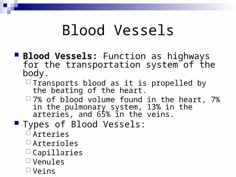
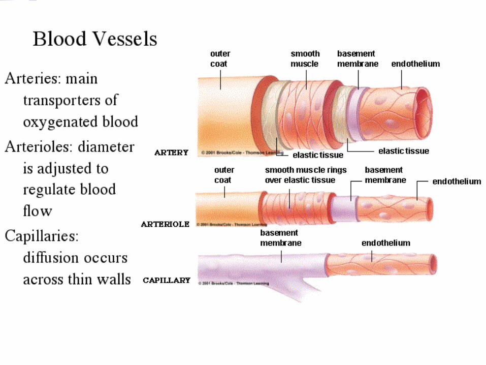
Blood Vessels
Blood Vessels: Function as highways for the transportation system of the body. Transports blood as it is propelled by the beating of
the heart. 7% of blood volume found in the heart, 7% in the
pulmonary system, 13% in the arteries, and 65% in the veins.
Types of Blood Vessels: Arteries Arterioles Capillaries Venules Veins
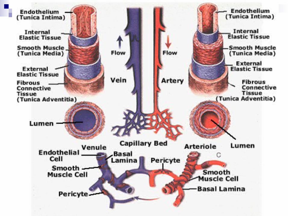
Blood Vessel Anatomy
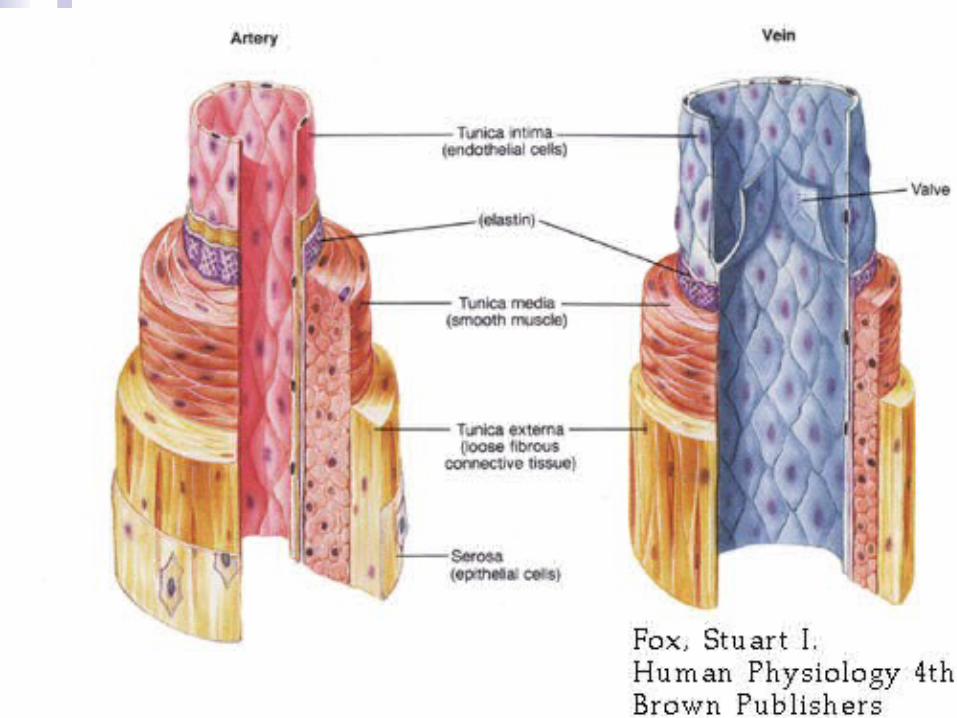
Tunics: The three layers that make up the walls of arteries & veins Tunica Adventitia: Outer layer composed of loose connective
tissue – keeps the vessels in place. Vasa Vasorum: Supplies nutrients to the larger arteries & veins.
Tunica Media: Middle layer composed of smooth muscles. Vasoconstriction: Contraction of this layer that narrows the blood
vessel. Vasodilation: Relaxation of this layer that widens the blood vessel.
Tunica Interna: Inner layer composed of smooth endothelium – allows blood to pass through easily without catching on the walls.
Lumen: The hollow center of the blood vessel through which blood flows.
Arteries & Arterioles
Arteries: Blood vessels responsible for carrying blood away from the heart – very strong to withstand the pressure surges! Elastic Arteries aka Conducting Arteries: The
largest diameter arteries, such as the aorta, that propel the blood through the vessels & provide a pressure reservoir to keep the blood flowing.
Muscular Arteries aka Distributing Arteries: Contain more smooth muscle & help to control blood flow – carry blood to all parts of the body.
Arterioles: The smallest blood vessels that regulate blood flow into capillaries. Resistance: The regulation arterioles uses in
opposition to blood flow to control total blood volume.
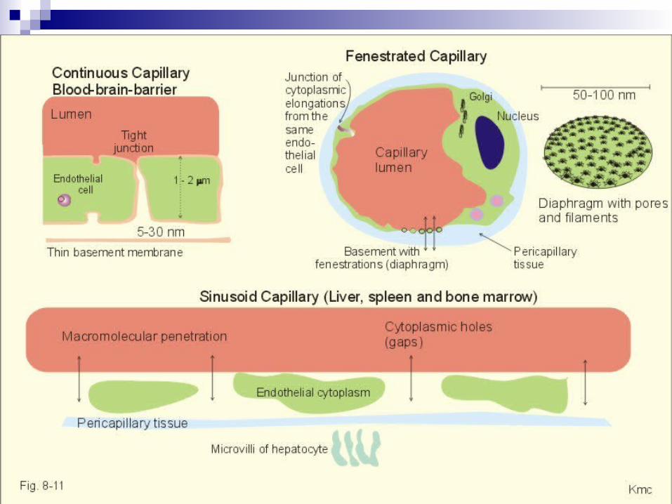
Capillaries
Capillaries: The smallest vessels in the body consisting of a single layer of endothelium. Found almost everywhere in the body, but not in tendons,
ligaments, or the cornea & lens of the eye. Three Types of Capillaries:
Continuous Capillaries: Continuous tubes formed of endothelial cells – intracellular cleft present for some materials to pass through.
Found in most connective tissue, smooth muscle, lungs, & skeleton. Fenestrated Capillaries: Fenestrations (holes) along the wall to
allow for quicker passage of materials. Found in organs that use rapid filtration - small intestine, endocrine
glands, kidneys. Sinusoids: Large fenestrations (holes) and large intracellular
lefts to allow for easiest passage of materials. Found in liver, spleen, some glands, red bone marrow.

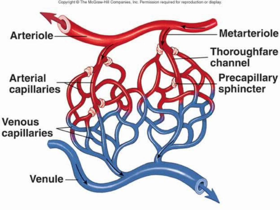
Capillary Beds
Capillary Beds: Organized “tangles” of capillaries that exchanges materials into body tissue – connect arterioles & venules.
Thoroughfare Channels: Bypasses the capillaries to carry blood directly from the arterioles to the venules.
Precapillary Sphincter: The ring of smooth muscle regulating blood flow into the capillaries.
Vasomotion: The intermittent flow of blood through capillaries.
Portal Systems: Two separate capillary beds in which blood flows through before heading toward the heart. Portal Vein: Transports blood between the two capillary beds. Found in kidneys, between the intestines & the liver, between
the hypothalamus & pituitary glands.
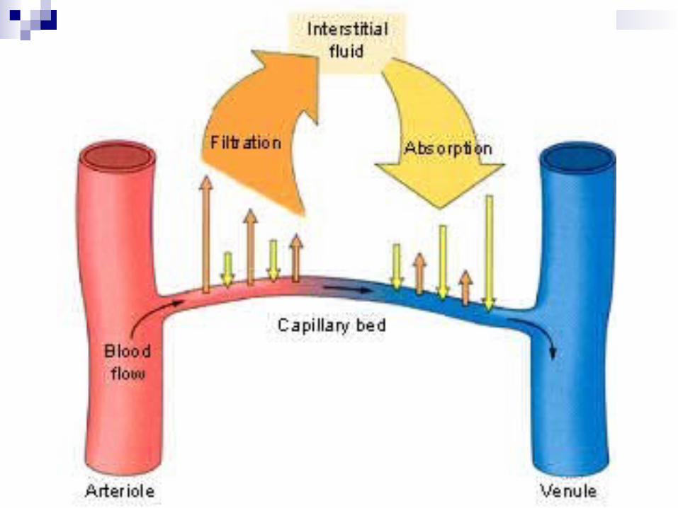
Capillary Exchange
Capillary Exchange: The movement of substances between the blood and interstitial fluid of the tissue. Mainly exchanges.. Oxygen Carbon Dioxide Glucose Amino Acids Hormones
Diffusion: The basic rule states that the more surface area there is for exchange, the faster the gases and nutrients will transfer into the tissue.
Capillary Exchance
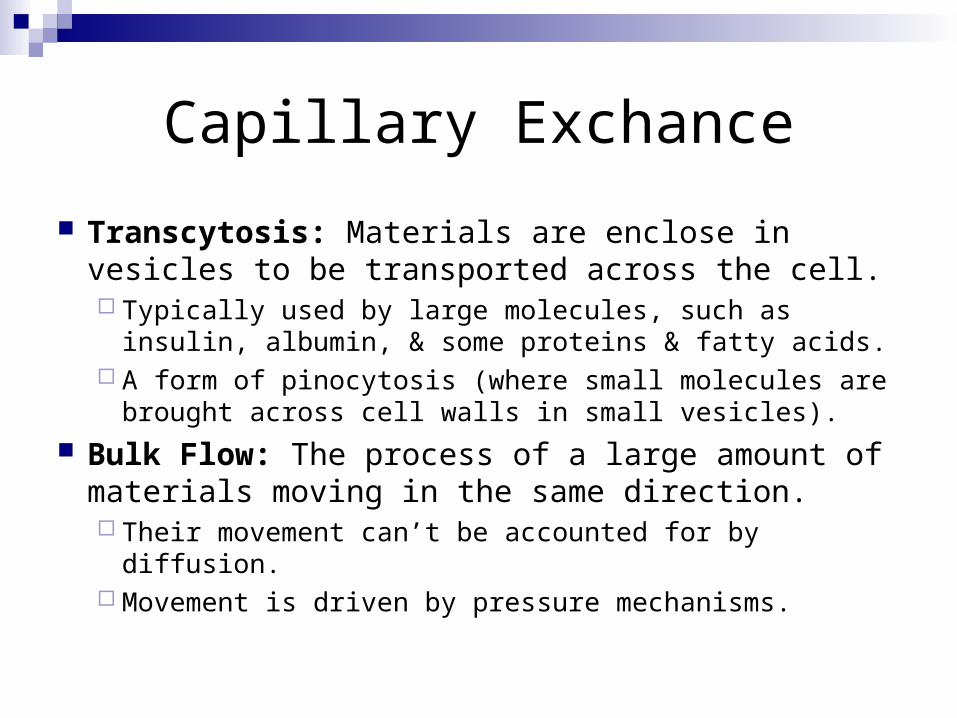
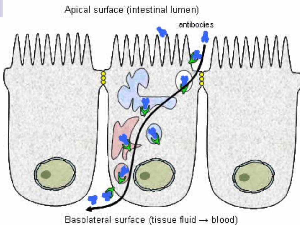
Transcytosis: Materials are enclose in vesicles to be transported across the cell. Typically used by large molecules, such as insulin,
albumin, & some proteins & fatty acids. A form of pinocytosis (where small molecules are
brought across cell walls in small vesicles).
Bulk Flow: The process of a large amount of materials moving in the same direction. Their movement can’t be accounted for by diffusion. Movement is driven by pressure mechanisms.
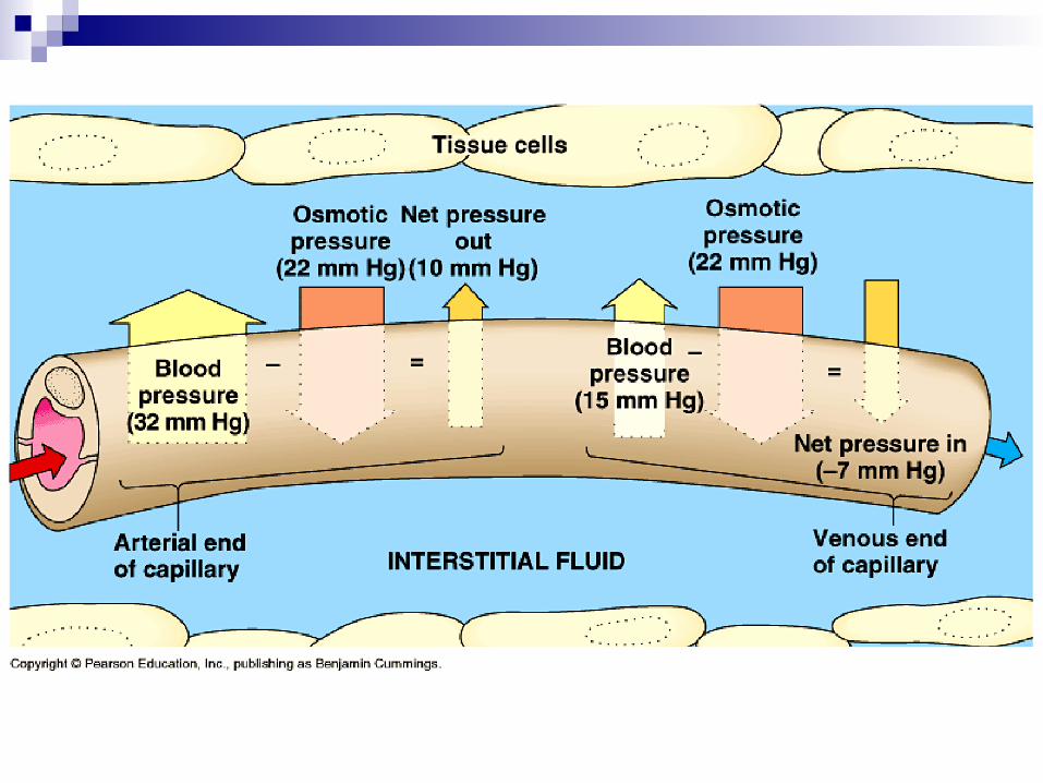
Capillary Exchange
Filtration: The movement of materials from capillaries into the interstitial fluid,.
Reabsorption: The movement of materials from the interstitial fluid into the capillaries.
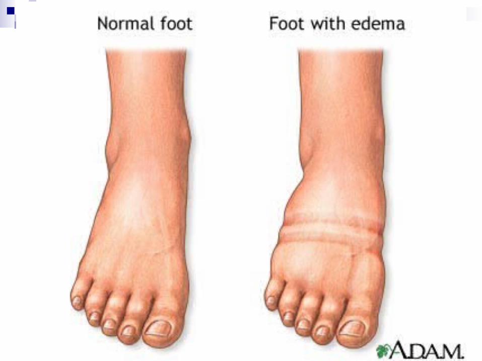
Edema: The increase in interstitial fluid volume caused by filtration rate exceeding reabsorption rate which results in bloated tissue! Can be caused… Increased capillary blood pressure Increased permeability of the capillaries Decreased concentrations of the plasma proteins
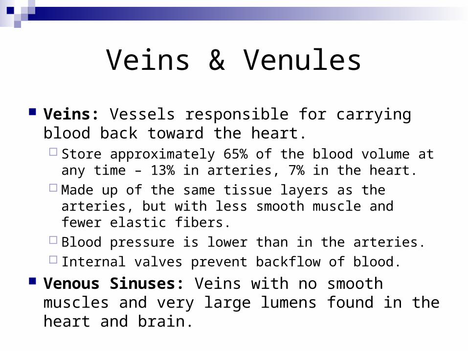
Veins & Venules
Veins: Vessels responsible for carrying blood back toward the heart. Store approximately 65% of the blood volume at any
time – 13% in arteries, 7% in the heart. Made up of the same tissue layers as the arteries, but
with less smooth muscle and fewer elastic fibers. Blood pressure is lower than in the arteries. Internal valves prevent backflow of blood.
Venous Sinuses: Veins with no smooth muscles and very large lumens found in the heart and brain.
Venous Return
Venous Return: The flow of blood back into the heart, controlled by valves within the veins.
5 Influencing Factors on Venus Return: Pressure Gradient: Blood flows from higher pressure (arteries)
to lower pressure (veins). Respiratory Pump: Breathing muscles help to compress and
decompress veins. Chordae Tendinae: Tendonlike fibrous cords that connect the
atrioventricular valves of the heart via papillary muscles & provides suction within the heart.
Gravity: Helps return blood from the higher areas of the body. Skeletal Muscle Pump: Contraction of the skeletal muscles
contract to push on the veins, causing the blood to flow toward the heart.
Hemodynamics
Hemodynamics: The regulation of blood flow via pressure & resistance.
Blood Flow: The volume of blood that flows through a given tissue during a given amount of time, measured in milliliters per minute (mL/min). “In general, blood flows from areas of high pressure to
areas of low pressure.” Blood Distribution throughout the body is
dependent on pressure differences and resistance to blood flow within the vessels. Distribution/Flow determines the amounts of nutrients
& oxygen being delivered to the tissue. Inadequate blood flow can cause all kinds of trouble!
Blood Pressure
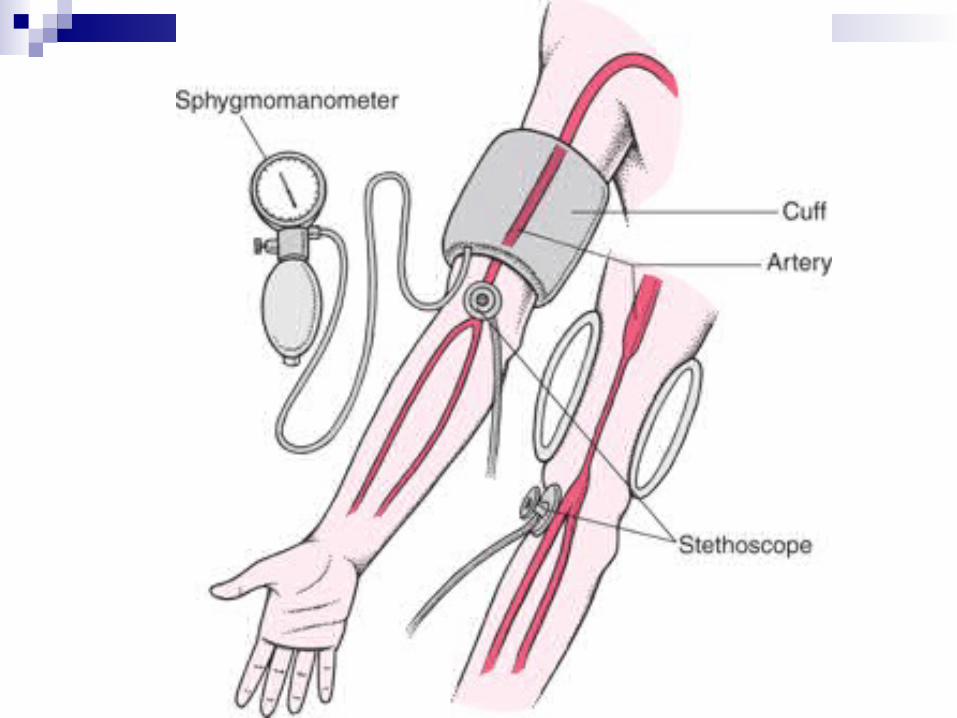
Blood Pressure: The measure of pressure inside the main arteries during systole and diastole.
Pulse Pressure: The difference between systolic & diastolic pressure. Can be used to measure the stress on the arteries
caused by the contractions of the heart.
Mean Arterial Pressure (MAP): The average blood pressure in the arteries. Measured as the 1/3 point between diastolic &
systolic pressure.
Blood Pressure
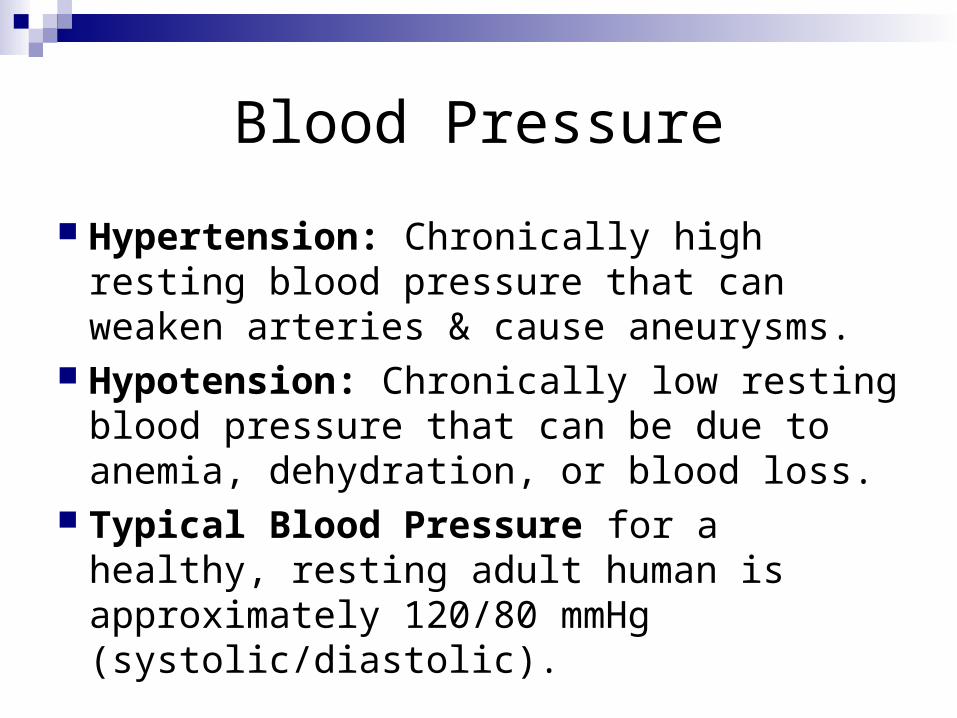
Hypertension: Chronically high resting blood pressure that can weaken arteries & cause aneurysms.
Hypotension: Chronically low resting blood pressure that can be due to anemia, dehydration, or blood loss.
Typical Blood Pressure for a healthy, resting adult human is approximately 120/80 mmHg (systolic/diastolic).
Vascular Resistance
Vascular Resistance: The opposition to normal blood flow within the vessels that slows the movement of the blood down.
Three factors that affect Vascular Resistance: Diameter of the Vessels: The size of the lumen can be controlled by
vasoconstriction & vasodilation to control blood flow. Length of Vessels: The longer the blood vessel, the greater the
resistance provided. Viscosity: The thicker the blood, the greater the resistance to blood
flow & the higher the blood pressure must be to move the blood. Viscosity is mainly due to the ratio of red blood cells to plasma, with some
effect from proteins. Systemic Vascular Resistance (SVR): The measure of the total
resistance offered by all of the systemic blood vessels. The smaller the vessel, the more it contributes to the SVR total.
Blood Flow Control
Autoregulation: The means by which tissue naturally regulate their own blood flow in response to metabolic demand.
Blood Flow Control
Neural Control: The nervous system controls blood flow & pressure through the cardiovascular center (CV center) of the medulla oblongata. Controls heart rate, stroke volume, and blood flow in specific organs. Cardiac Accelerator Nerves: Delivers sympathetic control to
speed up heart rate. Vagus Nerve: Delivers parasympathetic control to slow down
heart rate. Baroreceptors: Pressure-sensitive receptors in the arteries that
signal the CV center to control the carotid sinus reflex and the aortic reflex – also regulates the baroreflexes.
Chemoreceptors: Sensors in the aorta and the carotid sinus of the brain that detect changes in blood oxygen, carbon dioxide and hydrogen levels – also regulates the chemoreceptor reflexes.
Blood Flow Control
Hormonal Control: Several hormones regulate blood pressure & blood flow! Angiotensin II: Increases vasoconstriction to raise
blood pressure. Norepinephrine & Epinephrine: Increases
vasoconstriction & heart rate to increase blood pressure.
Artial Natriuretic Factor: Increases vasodilation to decrease blood pressure.
Nitric Oxide: Increases vasodilation to decrease blood pressure.
Aldosterone & ADh: Increase the amount of blood to increase blood pressure.
Circulatory Shock
Circulatory Shock: Occurs when the cardiovascular system fails to deliver adequate supplies of oxygen & nutrients to the cells via inadequate blood flow. 4 types! Cardiogenic Shock: Due to the heart failing to pump adequately,
typically due to heart attack. Hypovolemic Shock: Due to hemorrhage (sudden loss of blood), loss
of fluid or inadequate fluid intake (dehydration). Vascular Shock: Due to excessive pooling of the blood in the limbs,
which can be caused by brain injury, excessive time spent standing, allergic reactions (anaphylaxis).
Obstructed Shock: Due to the path of venous return being blocked, typically by an embolism (blood clot).
Response to Shock: The body attempts to return to homeostasis via hormones, local vasodilators, and sympathetic and autonomic nervous system activation.
Signs of Shock: Low systolic blood pressure; weak & rapid pulse; cool, pale, clammy skin; altered mental state; nausea; thirst; low blood pH.
Pulse Rates – Good to Know!
Pulse: The traveling wave of pressure created by the expansion & recoiling of large arteries after each ventricular systole. The closer the arteries to the heart the stronger the
pulse. Pulse Rate: The speed at which the pulse
occurs. Arterial Pressure Points: Brachial artery,
common carotid artery, facial artery, femoral artery, popliteal artery, & radial artery are good places to look for pulse rates!
Major Circulatory Routes
The ones you need to know: Systemic ArteriesSystemic VeinsHepatic Portal SystemPulmonary CirculationFetal Circulation
The rest tend to follow the names of the bones – 2-in-1 for studying!
The Systemic Arteries
Systemic Circuit: Supplies the cells all over the body with nutrients & oxygen while removing metabolic wastes.
The Systemic Arteries
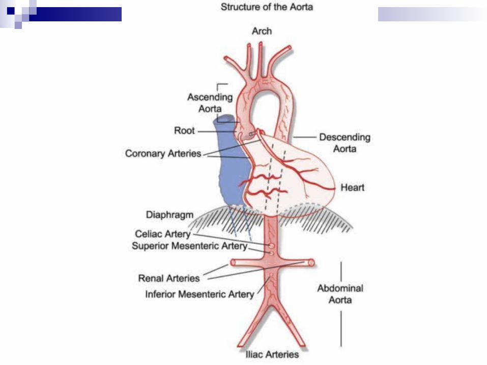
Ascending Aorta: The biggest blood vessel starting out the systemic circuit – leaves the left ventricle of the heart & extends to the arch of the aorta. Right & Left Coronary Arteries: First
branches of the ascending aorta that supply the heart muscle with blood.

The Systemic Arteries
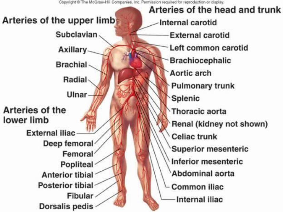
Aortic Arch: Follows the coronary arteries & has three branches…Brachiocephalic Trunk: Splits into the…
Right Common Carotid Artery to supply the right side of the head & neck
Right Subclavian Artery to supply the right upper limb
Left Common Carotid Artery: Supplies the left side of the head & neck
Left Subclavian Artery: Supplies the left upper limb

The Systemic Arteries
Descending Aorta: The portion of the aorta after the arch that descends through the chest and abdomen before branching into the external and internal iliac arteries that supply the legs. Thoracic Aorta: The name of the descending aorta when
serving the organs of the chest. Branches… Bronchial arteries that serve the bronchi of the lungs Arteries serving the pericardium, esophagus, chest muscles, &
diaphragms. Abdominal Aorta: The portion of the descending aorta that
serves the organs of the abdomen. Celiac Trunk: Splits into..
The common hepatic artery (serving the liver) The left gastric artery (serving the stomach) The splenic artery (serving the stomach, pancreas, & spleen)
Also serves the kidneys, gonads, colon, rectum, small intestine, and pancreas.
The Systemic Arteries
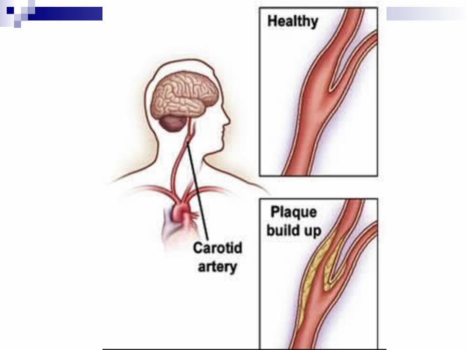
Right & Left Carotid Arteries: Serve the head & neck.Split into the internal and external cerebral
arteries, the facial arteries, & the basilar artery.
The Systemic Arteries
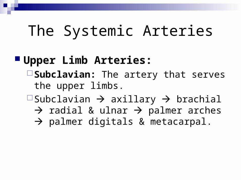
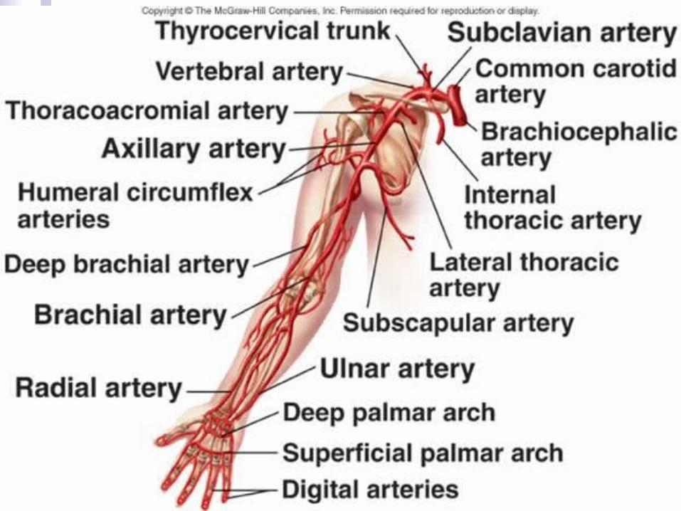
Upper Limb Arteries: Subclavian: The artery that serves the upper
limbs. Subclavian axillary brachial radial &
ulnar palmer arches palmer digitals & metacarpal.
The Systemic Arteries
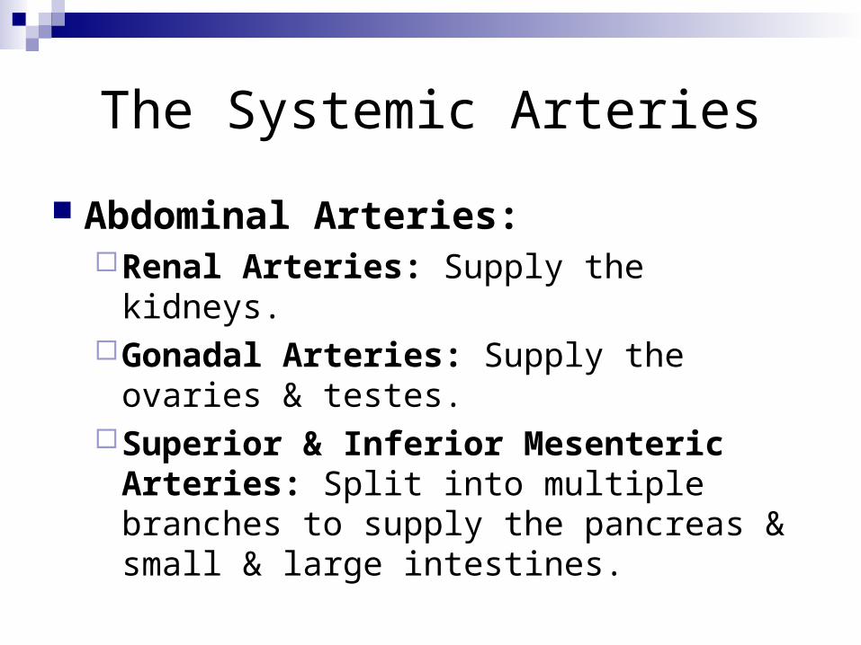
Abdominal Arteries: Renal Arteries: Supply the kidneys. Gonadal Arteries: Supply the ovaries &
testes. Superior & Inferior Mesenteric Arteries:
Split into multiple branches to supply the pancreas & small & large intestines.
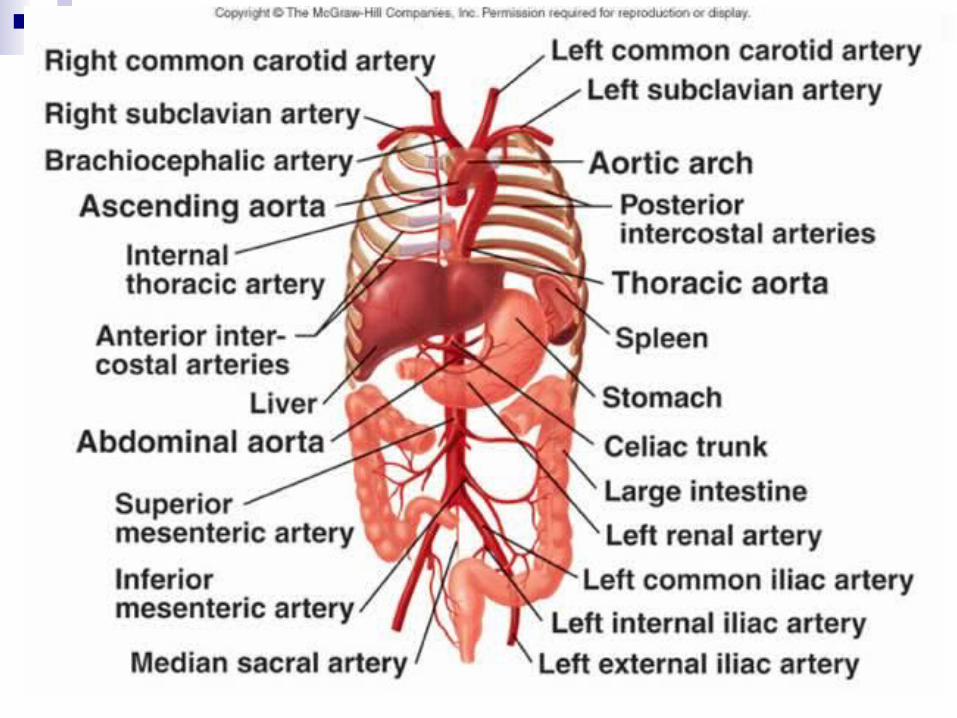
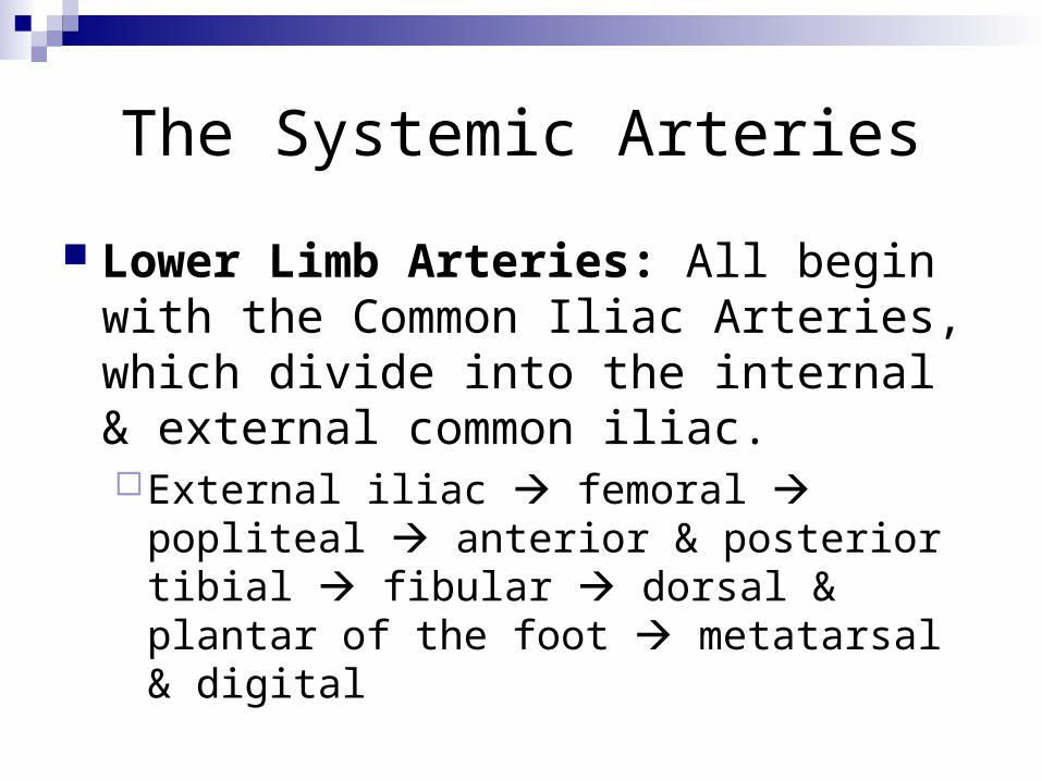
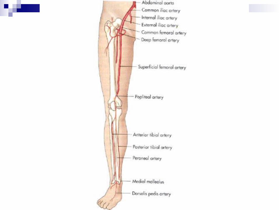
The Systemic Arteries
Lower Limb Arteries: All begin with the Common Iliac Arteries, which divide into the internal & external common iliac. External iliac femoral popliteal
anterior & posterior tibial fibular dorsal & plantar of the foot metatarsal & digital
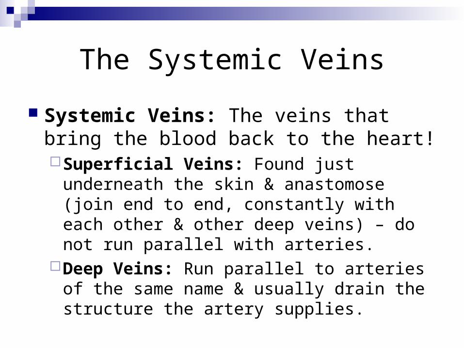
The Systemic Veins
Systemic Veins: The veins that bring the blood back to the heart! Superficial Veins: Found just underneath the
skin & anastomose (join end to end, constantly with each other & other deep veins) – do not run parallel with arteries.
Deep Veins: Run parallel to arteries of the same name & usually drain the structure the artery supplies.
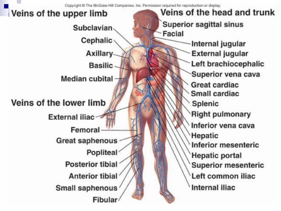
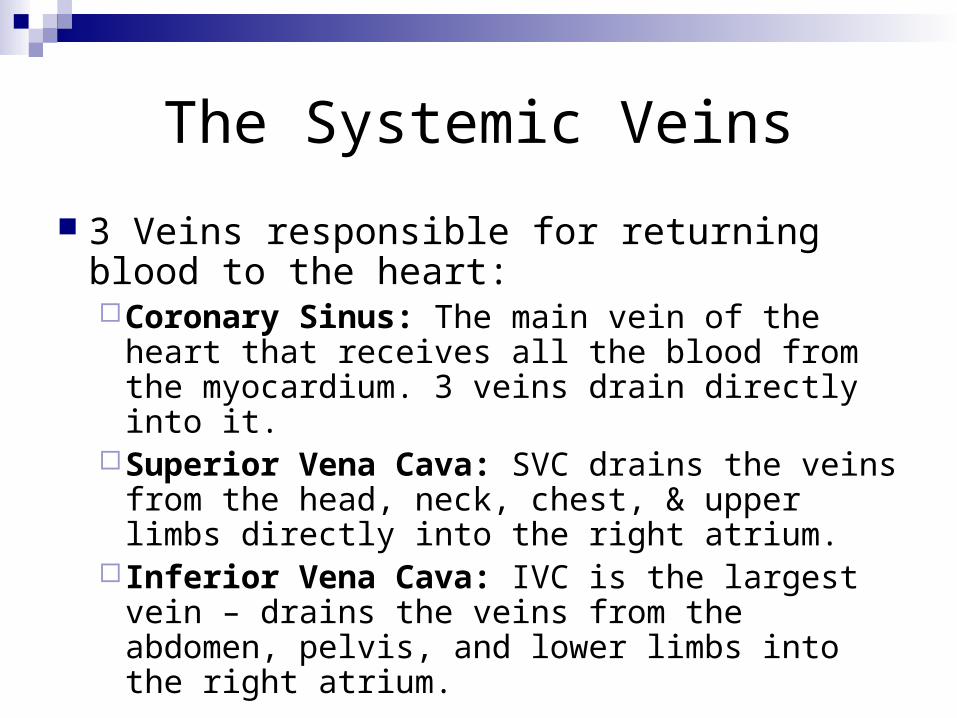
The Systemic Veins
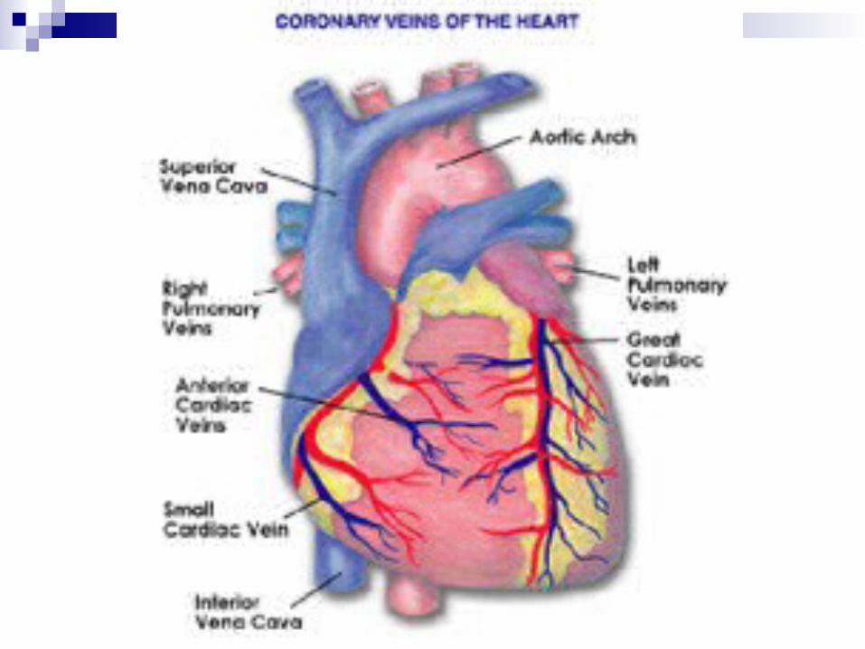
3 Veins responsible for returning blood to the heart: Coronary Sinus: The main vein of the heart
that receives all the blood from the myocardium. 3 veins drain directly into it.
Superior Vena Cava: SVC drains the veins from the head, neck, chest, & upper limbs directly into the right atrium.
Inferior Vena Cava: IVC is the largest vein – drains the veins from the abdomen, pelvis, and lower limbs into the right atrium.
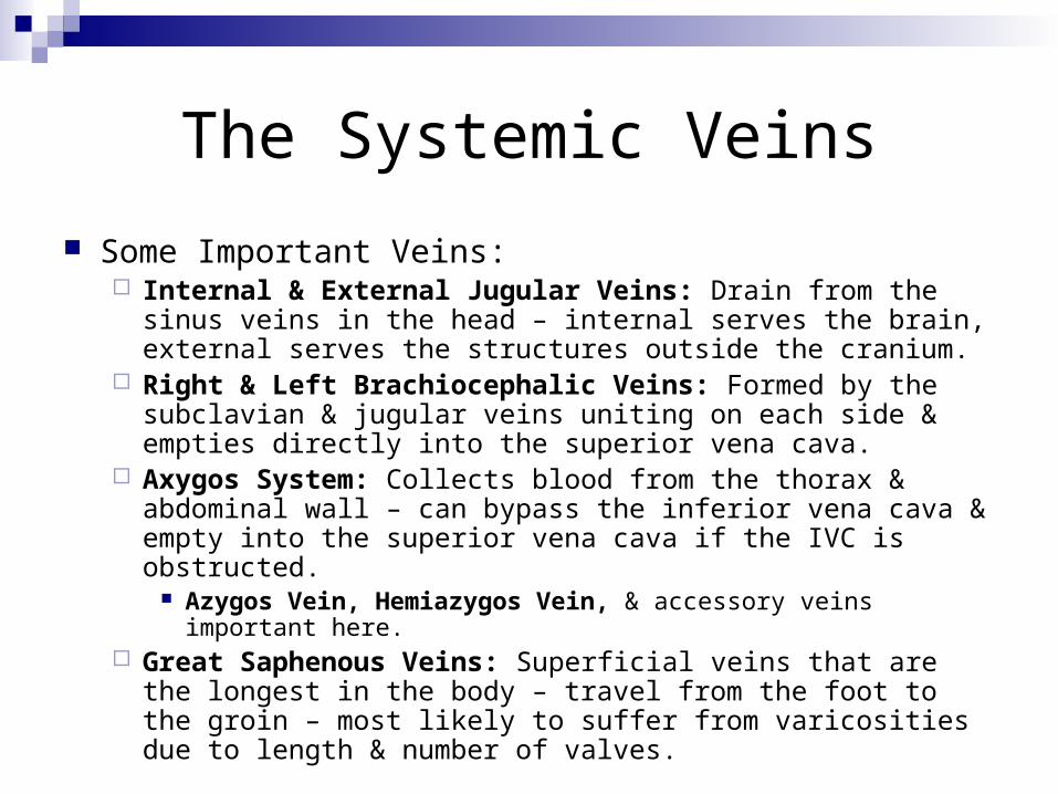
The Systemic Veins
Some Important Veins: Internal & External Jugular Veins: Drain from the sinus veins
in the head – internal serves the brain, external serves the structures outside the cranium.
Right & Left Brachiocephalic Veins: Formed by the subclavian & jugular veins uniting on each side & empties directly into the superior vena cava.
Axygos System: Collects blood from the thorax & abdominal wall – can bypass the inferior vena cava & empty into the superior vena cava if the IVC is obstructed.
Azygos Vein, Hemiazygos Vein, & accessory veins important here.
Great Saphenous Veins: Superficial veins that are the longest in the body – travel from the foot to the groin – most likely to suffer from varicosities due to length & number of valves.

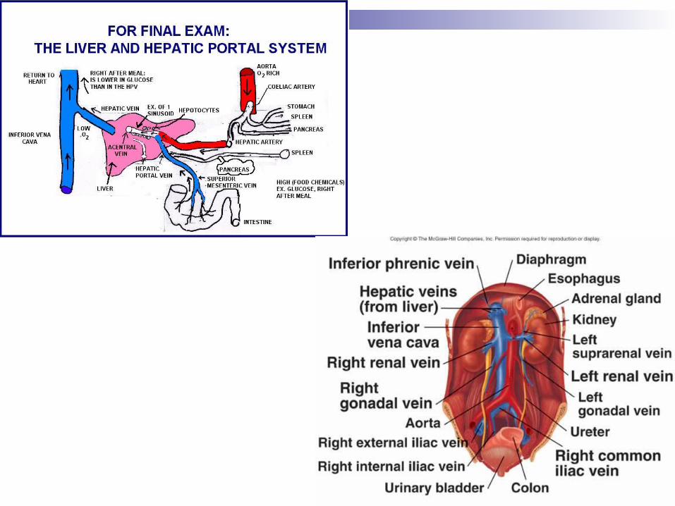
The Hepatic Portal System
Portal Vein: Any vein that transports blood from one network of capillaries to another rather than to anther vein or the heart.
Hepatic Portal Vein: Takes blood from the capillaries of the organs of the digestive system and transports it to the sinusoids of the liver. Formed by the joining of the splenic vein and superior
mesenteric vein. Hepatic Portal System: Absorbs blood directly from the
digestive tract and transports it to the liver to convert some of the rich nutrients into other substances to be used or stored & detoxifies other substances. Blood in this system is high in nutrients & low in oxygen content.
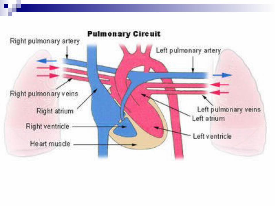
Pulmonary Circulation
Pulmonary Circulation: Takes deoxygenated blood from the right ventricle, oxygenates it, and returns it to the left atrium. Deoxygenated blood enters the right atrium Blood flows from the right atrium into the right ventricle & is
forced out into the pulmonary arteries. Blood travels from the pulmonary arteries to the right & left lungs
& through the capillaries of the alveoli. Blood drops the carbon dioxide & uptakes oxygen before
heading back to the pulmonary venules and to the right & left pulmonary veins (the ONLY veins that carry oxygenated blood).
Pulmonary veins then empty into the left atrium where the blood flows into the left ventricle and out to the body!
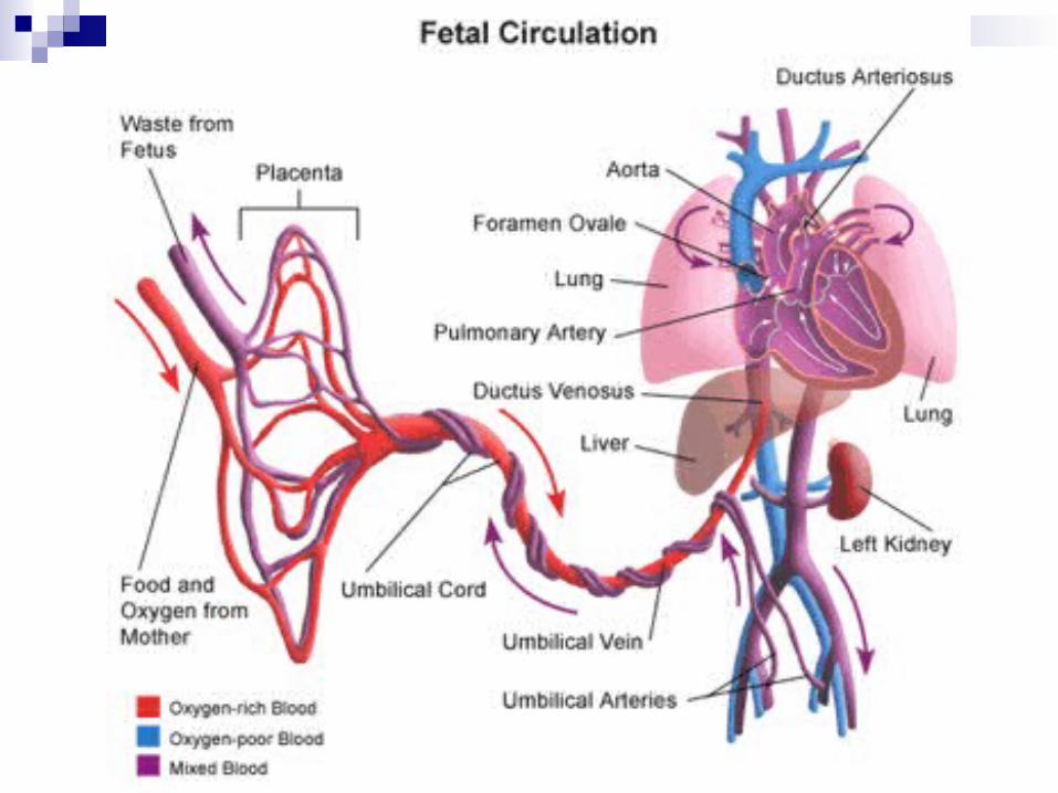
Fetal Circulation
Fetal Circulation: The circulatory system of the fetus. Fetus obtains oxygen & nutrients & elminates carbon
dioxide & other wastes through the umbilical cord & placenta.
Wastes travel from the intervillous spaces in the placenta to the uterine veins to be filtered out by the mother’s cardiovascular system.
Nutrients travel from the maternal blood vessels intervillous spaces of the placenta fetal capiullaries.
Fetal & maternal blood typically do not mix – all exchanges occur through capillary wall diffusion.