Embed Size (px)
Citation preview

The Clinical Pathology
of CHeRI: What Makes a
Healthy Fawn?Allison Cauvin, Nicole Stacy, Rebecca Shuman, Katherine Sayler

My role in CHeRI
Dr. Katherine Sayler’s MS student
Also under advisement of Drs.
Nicole Stacy and Sam Wisely
Clinical pathology nerd
Molecular techniques
Database Management
Talk Outline
Intro to clinical pathology
Overview of clinically normal findings of farmed neonate fawns
Conclusions
Future directions
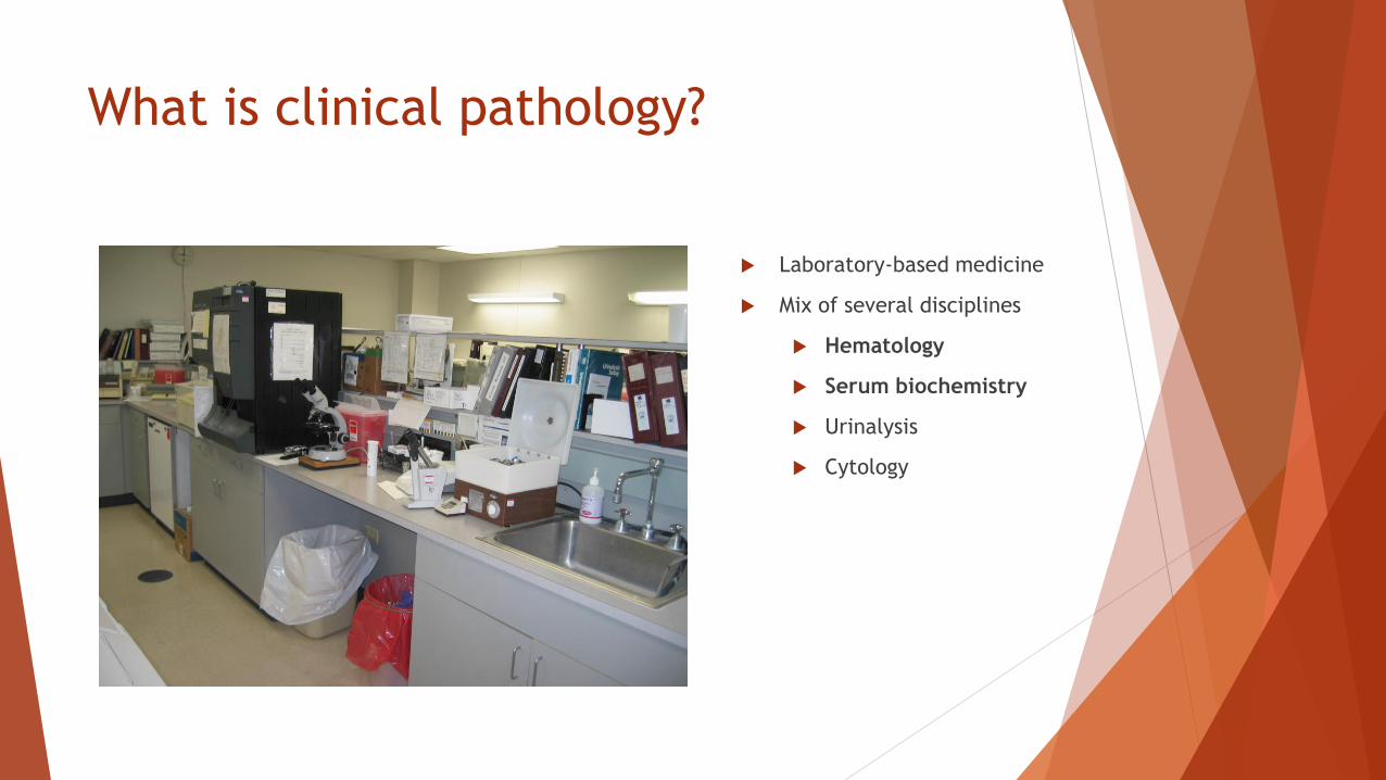
What is clinical pathology?
Laboratory-based medicine
Mix of several disciplines
Hematology
Serum biochemistry
Urinalysis
Cytology
Why do we need
clinical pathology?
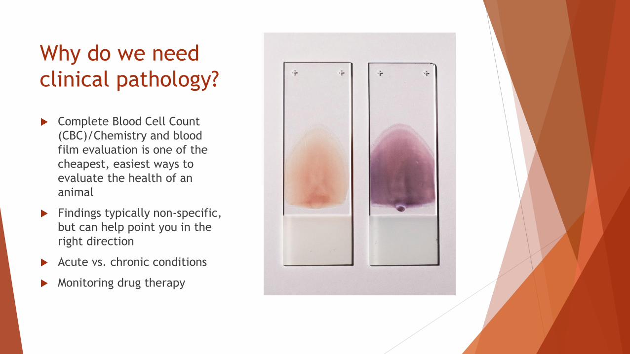
Complete Blood Cell Count
(CBC)/Chemistry and blood
film evaluation is one of the
cheapest, easiest ways to
evaluate the health of an
animal
Findings typically non-specific,
but can help point you in the
right direction
Acute vs. chronic conditions
Monitoring drug therapy
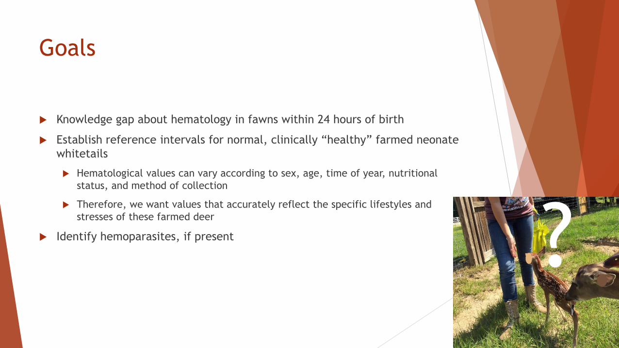
Goals
Knowledge gap about hematology in fawns within 24 hours of birth
Establish reference intervals for normal, clinically “healthy” farmed neonate
whitetails
Hematological values can vary according to sex, age, time of year, nutritional
status, and method of collection
Therefore, we want values that accurately reflect the specific lifestyles and
stresses of these farmed deer
Identify hemoparasites, if present ?
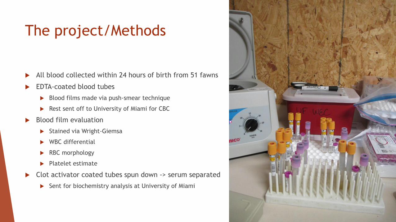
The project/Methods
All blood collected within 24 hours of birth from 51 fawns
EDTA-coated blood tubes
Blood films made via push-smear technique
Rest sent off to University of Miami for CBC
Blood film evaluation
Stained via Wright-Giemsa
WBC differential
RBC morphology
Platelet estimate
Clot activator coated tubes spun down -> serum separated
Sent for biochemistry analysis at University of Miami
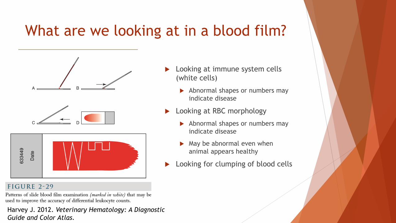
Harvey J. 2012. Veterinary Hematology: A Diagnostic
Guide and Color Atlas.
What are we looking at in a blood film?
Looking at immune system cells
(white cells)
Abnormal shapes or numbers may
indicate disease
Looking at RBC morphology
Abnormal shapes or numbers may
indicate disease
May be abnormal even when
animal appears healthy
Looking for clumping of blood cells
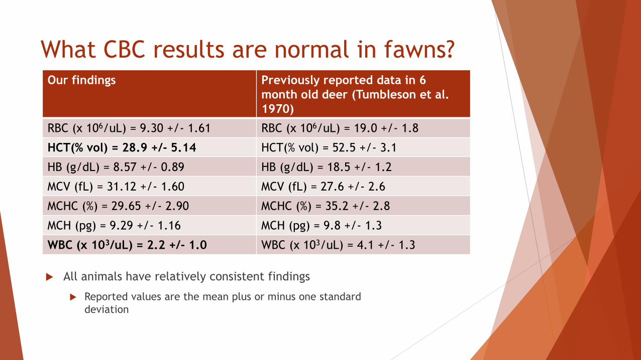
What CBC results are normal in fawns?Our findings Previously reported data in 6
month old deer (Tumbleson et al.
1970)
RBC (x 106/uL) = 9.30 +/- 1.61 RBC (x 106/uL) = 19.0 +/- 1.8
HCT(% vol) = 28.9 +/- 5.14 HCT(% vol) = 52.5 +/- 3.1
HB (g/dL) = 8.57 +/- 0.89 HB (g/dL) = 18.5 +/- 1.2
MCV (fL) = 31.12 +/- 1.60 MCV (fL) = 27.6 +/- 2.6
MCHC (%) = 29.65 +/- 2.90 MCHC (%) = 35.2 +/- 2.8
MCH (pg) = 9.29 +/- 1.16 MCH (pg) = 9.8 +/- 1.3
WBC (x 103/uL) = 2.2 +/- 1.0 WBC (x 103/uL) = 4.1 +/- 1.3
All animals have relatively consistent findings
Reported values are the mean plus or minus one standard
deviation
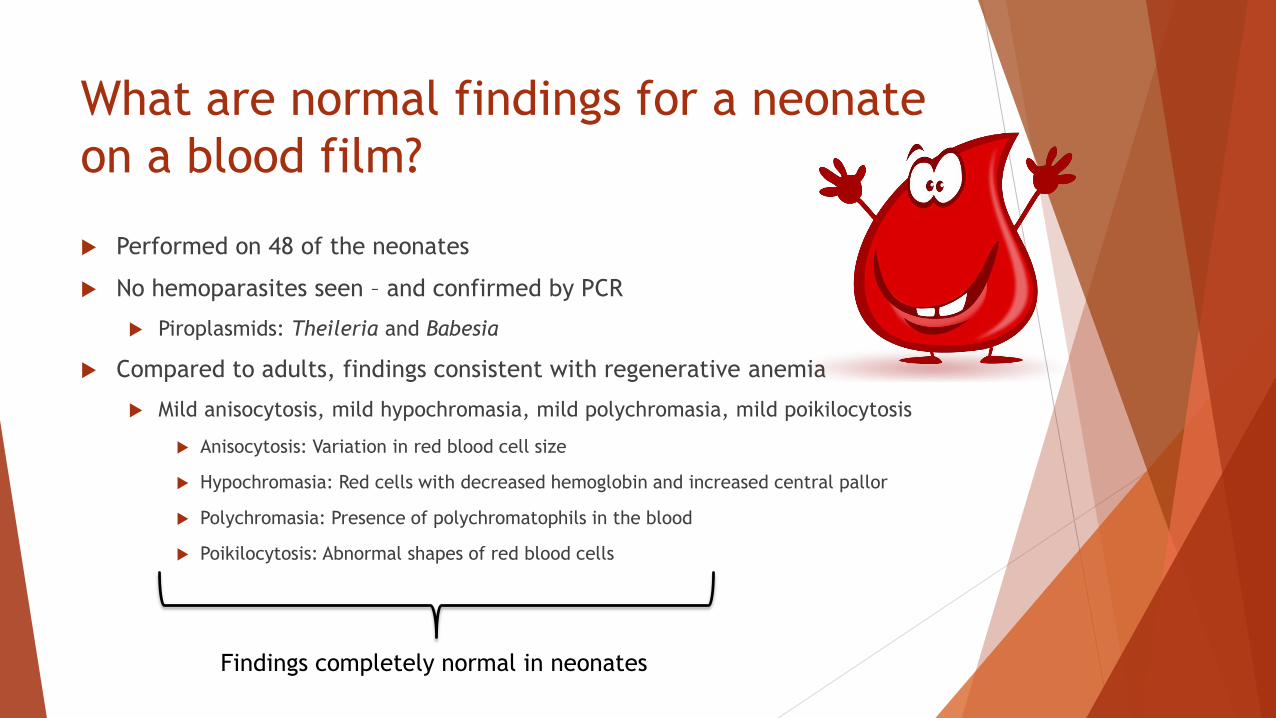
What are normal findings for a neonate
on a blood film?
Performed on 48 of the neonates
No hemoparasites seen – and confirmed by PCR
Piroplasmids: Theileria and Babesia
Compared to adults, findings consistent with regenerative anemia
Mild anisocytosis, mild hypochromasia, mild polychromasia, mild poikilocytosis
Anisocytosis: Variation in red blood cell size
Hypochromasia: Red cells with decreased hemoglobin and increased central pallor
Polychromasia: Presence of polychromatophils in the blood
Poikilocytosis: Abnormal shapes of red blood cells
Findings completely normal in neonates
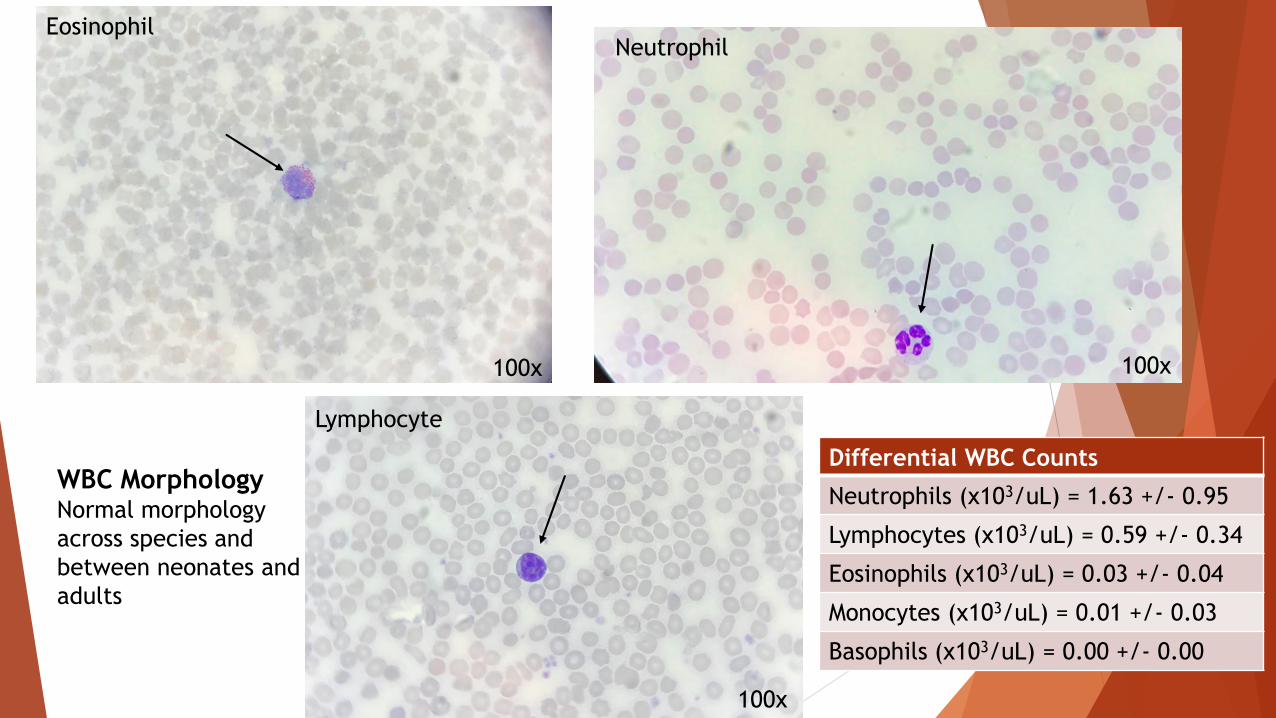
WBC MorphologyNormal morphology
across species and
between neonates and
adults
100x
100x100x
EosinophilNeutrophil
Lymphocyte
Differential WBC Counts
Neutrophils (x103/uL) = 1.63 +/- 0.95
Lymphocytes (x103/uL) = 0.59 +/- 0.34
Eosinophils (x103/uL) = 0.03 +/- 0.04
Monocytes (x103/uL) = 0.01 +/- 0.03
Basophils (x103/uL) = 0.00 +/- 0.00

100x
Echinocytes
Hypochromasia
Anisocytosis
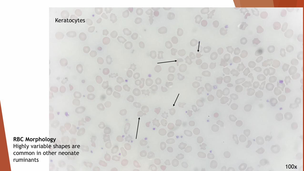
RBC Morphology
Highly variable shapes are
common in other neonate
ruminants
100x
Keratocytes
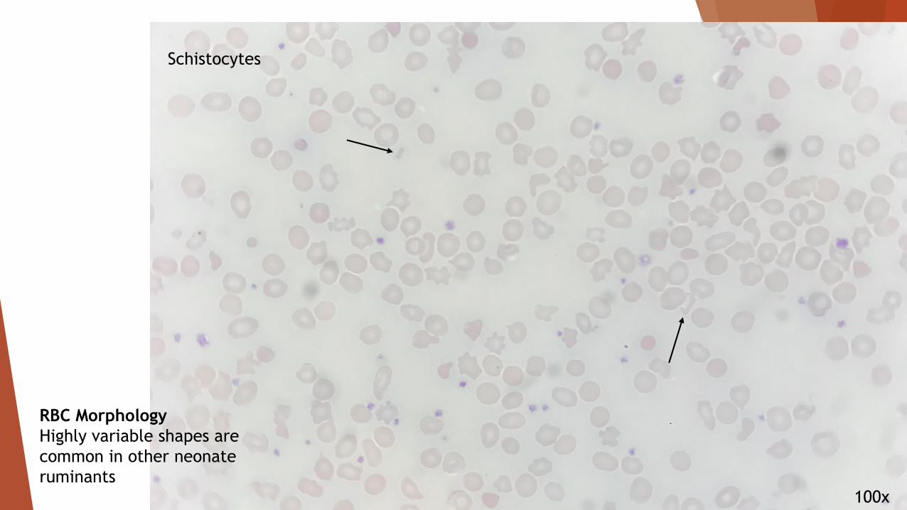
RBC Morphology
Highly variable shapes are
common in other neonate
ruminants
100x
Schistocytes
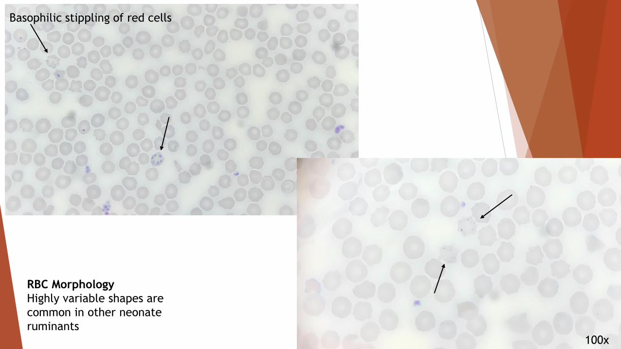
RBC Morphology
Highly variable shapes are
common in other neonate
ruminants
RBC Morphology
Highly variable shapes are
common in other neonate
ruminants
Basophilic stippling of red cells
100x

RBC Morphology
Highly variable shapes are
common in other neonate
ruminants
Drepanocytes
100x

What chemistry results are normal in
fawns?
Previously reported chemistry
reference values only reported for
adults
Neonate values seem to be similar
to adults
Why evaluate these?
When we understand what’s
normal, we can identify what is
abnormal
These analytes can indicate
diseases with certain organs
Our Chem Panel values
Glucose (mg/dL)= 97.76 +/- 20.59
BUN (mg/dL)= 17.37 +/- 5.05
Creatinine (mg/dL)= 1.13 +/- 0.36
BUN/Crea = 15.73 +/- 3.77
Calcium (mg/dL)= 9.66 +/- 0.53
Phosphorus (mg/dL)= 9.77 +/- 1.38
Total Protein (g/dL)= 5.72 +/- 1.13
ALT (U/L) = 40.65 +/- 9.82
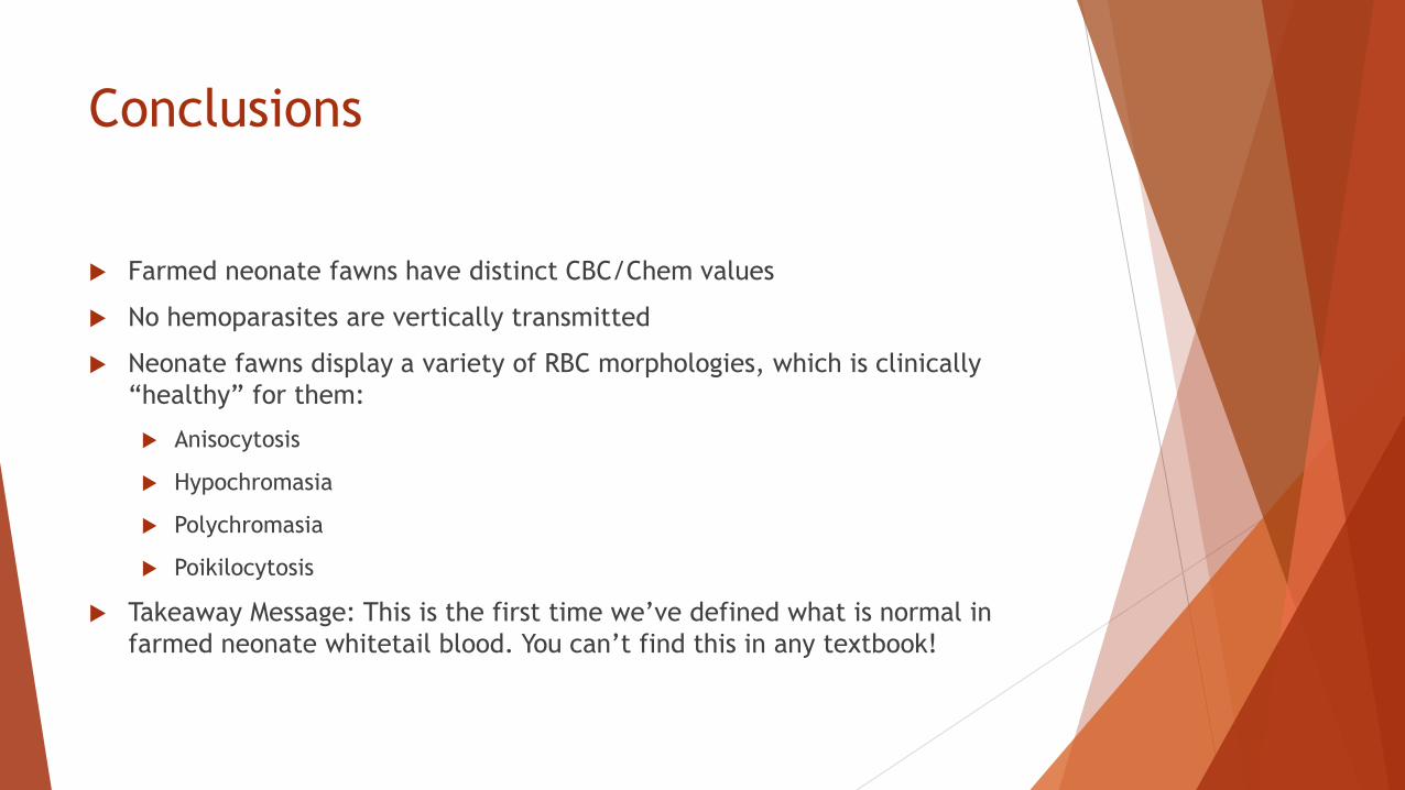
Conclusions
Farmed neonate fawns have distinct CBC/Chem values
No hemoparasites are vertically transmitted
Neonate fawns display a variety of RBC morphologies, which is clinically
“healthy” for them:
Anisocytosis
Hypochromasia
Polychromasia
Poikilocytosis
Takeaway Message: This is the first time we’ve defined what is normal in
farmed neonate whitetail blood. You can’t find this in any textbook!
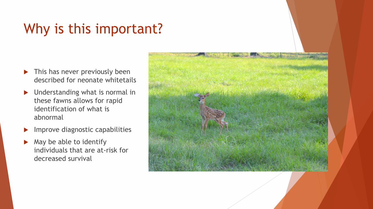
Why is this important?
This has never previously been
described for neonate whitetails
Understanding what is normal in
these fawns allows for rapid
identification of what is
abnormal
Improve diagnostic capabilities
May be able to identify
individuals that are at-risk for
decreased survival
Future Directions
Spend another fawning season to increase sample size
Collating the CBC/Chem data with maternal antibody levels to identify if
these serve as predictors for fawn survivorship
Multivariate regression analysis to identify risk factors for EHDV positivity
Develop additional diagnostic tests for passive transfer evaluation

Acknowledgements
Dr. Katherine Sayler
Dr. Nicole Stacy
Dr. Sam Wisely
Carisa Boyce
Shannon Moore
Katie Pothier
Rebecca Shuman
Dr. Jason Blackburn
The CHeRI Stakeholders
UF Veterinary Clinical Pathology
Lab
Starbucks