Embed Size (px)
Citation preview
Wheater et al. Journal of Translational Medicine 2013, 11:201http://www.translational-medicine.com/content/11/1/201
REVIEW Open Access
The clinical utility of bone marker measurementsin osteoporosisGillian Wheater1,2*, Mohsen Elshahaly2,3, Stephen P Tuck2,3, Harish K Datta2 and Jacob M van Laar2,3
Abstract
Osteoporosis is characterised by low bone mass and structural deterioration of bone tissue, resulting in increasedfragility and susceptibility to fracture. Osteoporotic fractures are a significant cause of morbidity and mortality.Direct medical costs from such fractures in the UK are currently estimated at over two billion pounds per year,resulting in a substantial healthcare burden that is expected to rise exponentially due to increasing life expectancy.Currently bone mineral density is the WHO standard for diagnosis of osteoporosis, but poor sensitivity means thatpotential fractures will be missed if it is used alone. During the past decade considerable progress has been madein the identification and characterisation of specific biomarkers to aid the management of metabolic bone disease.Technological developments have greatly enhanced assay performance producing reliable, rapid, non-invasive costeffective assays with improved sensitivity and specificity. We now have a greater understanding of the need toregulate pre-analytical sample collection to minimise the effects of biological variation. However, bone turnovermarkers (BTMs) still have limited clinical utility. It is not routinely recommended to use BTMs to select those at riskof fractures, but baseline measurements of resorption markers are useful before commencement of anti-resorptivetreatment and can be checked 3–6 months later to monitor response and adherence to treatment. Similarly,formation markers can be used to monitor bone forming agents. BTMs may also be useful when monitoringpatients during treatment holidays and aid in the decision as to when therapy should be recommenced. Recentrecommendations by the Bone Marker Standards Working Group propose to standardise research and include aspecific marker of bone resorption (CTX) and bone formation (P1NP) in all future studies. It is hoped that improvedresearch in turn will lead to optimised markers for the clinical management of osteoporosis and other bonediseases.
Keywords: Bone turnover markers, Bone formation, Bone resorption, Osteoporosis, Biological variability
IntroductionBone is a specialised connective tissue consisting primarilyof glycoproteins and proteoglycans. The fibres of bone aremostly composed of type-I collagen impregnated withmineral in the form of hydroxyapatite. The functional in-tegrity and strength of the skeleton is maintained by thishighly cross-linked structure. Several factors may be in-volved in determining bone quality, including bone densityand qualitative determinants of bone strength such as therate of bone turnover, the extent of trabecular connec-tivity, cortical and periosteal bone size and skeletal
* Correspondence: [email protected] of Biochemistry, The James Cook University Hospital,Middlesbrough TS4 3BW, UK2Institute of Cellular Medicine, Musculoskeletal Research Group, NewcastleUniversity, Newcastle upon Tyne NE1 7RU, UKFull list of author information is available at the end of the article
© 2013 Wheater et al.; licensee BioMed CentraCommons Attribution License (http://creativecreproduction in any medium, provided the or
morphometry [1]. Bone is metabolically active and is con-stantly being repaired and remodelled throughout an indi-vidual’s lifetime. Approximately twenty percent of bonetissue is replaced annually varying by site and type [2]. Re-modelling begins before birth and continues until death, itis a highly synchronised process contained within basicmulticellular units (Figure 1). Recent research has demon-strated the role of receptor activator of nuclear factorkappa B ligand/ receptor activator of nuclear factor kappaB/ osteoprotegerin (RANKL/RANK/OPG) in regulatingbone metabolism [3]. Parathyroid hormone (PTH), PTH-related peptide (PTH-rP), 1,25-dihydroxyvitamin D3, pros-taglandin E2, and interleukins among others regulate boneturnover through this system [4]. Additionally, bonemetabolism is now known to be at least partly regulatedby osteocytes, the fully differentiated osteoblasts present
l Ltd. This is an Open Access article distributed under the terms of the Creativeommons.org/licenses/by/2.0), which permits unrestricted use, distribution, andiginal work is properly cited.
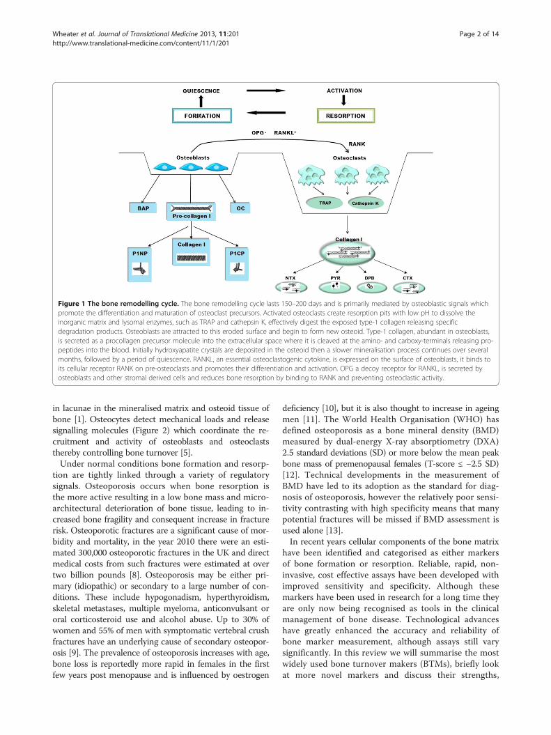
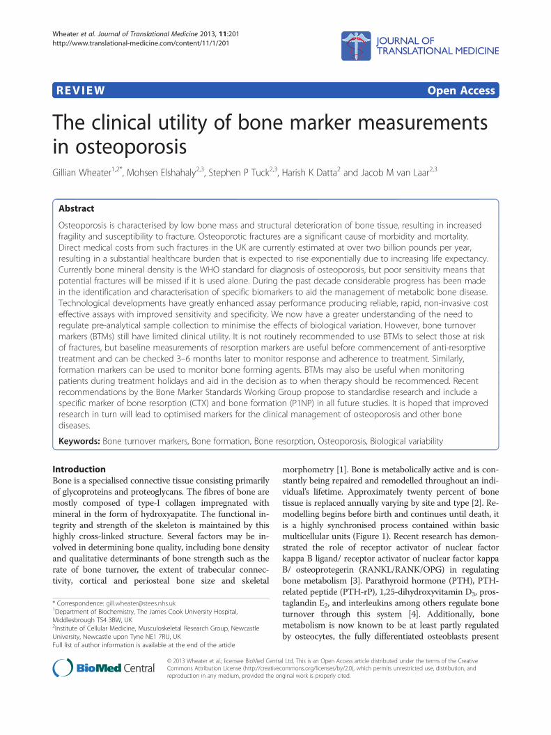
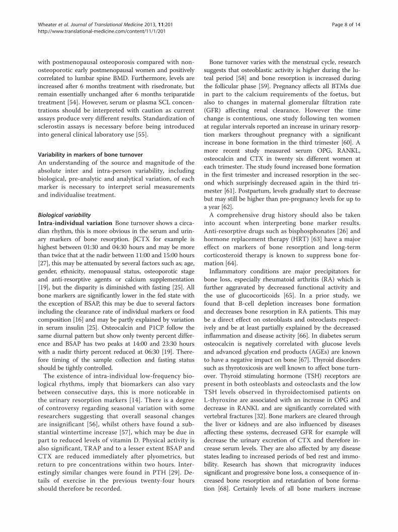
Figure 1 The bone remodelling cycle. The bone remodelling cycle lasts 150–200 days and is primarily mediated by osteoblastic signals whichpromote the differentiation and maturation of osteoclast precursors. Activated osteoclasts create resorption pits with low pH to dissolve theinorganic matrix and lysomal enzymes, such as TRAP and cathepsin K, effectively digest the exposed type-1 collagen releasing specificdegradation products. Osteoblasts are attracted to this eroded surface and begin to form new osteoid. Type-1 collagen, abundant in osteoblasts,is secreted as a procollagen precursor molecule into the extracellular space where it is cleaved at the amino- and carboxy-terminals releasing pro-peptides into the blood. Initially hydroxyapatite crystals are deposited in the osteoid then a slower mineralisation process continues over severalmonths, followed by a period of quiescence. RANKL, an essential osteoclastogenic cytokine, is expressed on the surface of osteoblasts, it binds toits cellular receptor RANK on pre-osteoclasts and promotes their differentiation and activation. OPG a decoy receptor for RANKL, is secreted byosteoblasts and other stromal derived cells and reduces bone resorption by binding to RANK and preventing osteoclastic activity.
Wheater et al. Journal of Translational Medicine 2013, 11:201 Page 2 of 14http://www.translational-medicine.com/content/11/1/201
in lacunae in the mineralised matrix and osteoid tissue ofbone [1]. Osteocytes detect mechanical loads and releasesignalling molecules (Figure 2) which coordinate the re-cruitment and activity of osteoblasts and osteoclaststhereby controlling bone turnover [5].Under normal conditions bone formation and resorp-
tion are tightly linked through a variety of regulatorysignals. Osteoporosis occurs when bone resorption isthe more active resulting in a low bone mass and micro-architectural deterioration of bone tissue, leading to in-creased bone fragility and consequent increase in fracturerisk. Osteoporotic fractures are a significant cause of mor-bidity and mortality, in the year 2010 there were an esti-mated 300,000 osteoporotic fractures in the UK and directmedical costs from such fractures were estimated at overtwo billion pounds [8]. Osteoporosis may be either pri-mary (idiopathic) or secondary to a large number of con-ditions. These include hypogonadism, hyperthyroidism,skeletal metastases, multiple myeloma, anticonvulsant ororal corticosteroid use and alcohol abuse. Up to 30% ofwomen and 55% of men with symptomatic vertebral crushfractures have an underlying cause of secondary osteopor-osis [9]. The prevalence of osteoporosis increases with age,bone loss is reportedly more rapid in females in the firstfew years post menopause and is influenced by oestrogen
deficiency [10], but it is also thought to increase in ageingmen [11]. The World Health Organisation (WHO) hasdefined osteoporosis as a bone mineral density (BMD)measured by dual-energy X-ray absorptiometry (DXA)2.5 standard deviations (SD) or more below the mean peakbone mass of premenopausal females (T-score ≤ −2.5 SD)[12]. Technical developments in the measurement ofBMD have led to its adoption as the standard for diag-nosis of osteoporosis, however the relatively poor sensi-tivity contrasting with high specificity means that manypotential fractures will be missed if BMD assessment isused alone [13].In recent years cellular components of the bone matrix
have been identified and categorised as either markersof bone formation or resorption. Reliable, rapid, non-invasive, cost effective assays have been developed withimproved sensitivity and specificity. Although thesemarkers have been used in research for a long time theyare only now being recognised as tools in the clinicalmanagement of bone disease. Technological advanceshave greatly enhanced the accuracy and reliability ofbone marker measurement, although assays still varysignificantly. In this review we will summarise the mostwidely used bone turnover makers (BTMs), briefly lookat more novel markers and discuss their strengths,
OPN
DMP
MEPE
OB
OCY
OBOB
WNT
WNTWNT
DKK-1
SCL
X XLRP5/6FZFZ FZ
LRP5/6LRP5/6
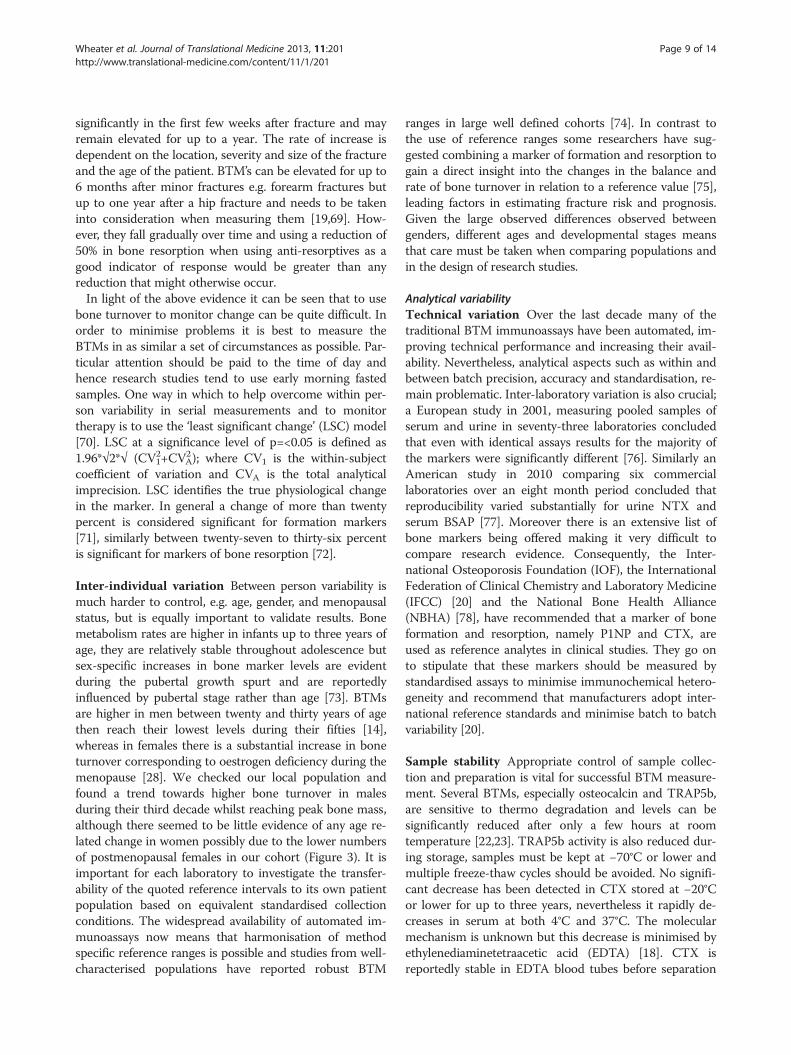
Figure 2 Mechanism of blockade of the Wnt signalling pathway by osteocytes. Osteocytes detect changes in bone morphology throughtheir sensitivity to mechanical forces, thereby regulating bone turnover through direct physical contact with osteoblasts. Osteocytes produceOPN, DMP, MEPE, SCL and DKK-1. The β-catenin-dependent canonical Wnt signalling pathway controls gene expression by stabilizing β-cateninin regulating a diverse array of biological processes. It is initiated by binding of appropriate Wnt ligands to the frizzled (Fz) and low-densitylipoprotein receptor-related proteins 5 and 6 (LRP-5/6) and can be antagonized by secreted proteins from SCL and the DKK family, that bindwith high affinity to LRP-5 or LRP-6, thereby directly prevent Wnt binding. Wnt proteins act on osteoblast precursor cells through this pathwayand promote their differentiation into mature osteoblasts. In addition, they can suppress bone resorption by up-regulating OPG and down-regulating RANKL expression in mature osteoblasts, leading to a net increase in bone mass [6]. Additionally research has targeted the complexregulation of osteocyte action by expression of PTH/PTHrP receptor’s (PPR’s). Osteocyte activation of PPR leads to down-regulation of Sost andincreased Wnt signalling stimulating bone formation, accompanied by up-regulation of RANKL expression and osteoclast number increasingresorption. In contrast the main effect of PPR deletion on osteocytes is reduced osteoclast and osteoblast numbers and decreased boneremodelling [7].
Wheater et al. Journal of Translational Medicine 2013, 11:201 Page 3 of 14http://www.translational-medicine.com/content/11/1/201
weaknesses and their clinical utility in the managementof osteoporosis.
Commonly used markers of bone turnoverBiomarkers of bone turnover can be measured in blood orurine and are used in selective combinations of formationand resorption markers that express the metabolic activityof osteoblasts or osteoclasts respectively, although in mostcircumstances the bone remodelling processes are coupledand tend to change in parallel. BTMs do not control skel-etal metabolism and are not disease specific; they reflectthe entire skeleton regardless of the underlying cause.Some markers represent both processes, e.g. osteocalcin(OC). Several of the available markers are non-specific, i.e.they are present in tissues other than bone and may there-fore be influenced by non-skeletal processes [14]. Resultsshould therefore always take into consideration the wholeclinical picture and an understanding of the nature andsource of each marker is essential for a comprehensive in-terpretation. The major advantages and disadvantages ofeach marker are included in Table 1.
Markers of bone formationMarkers of bone formation are either by-products ofactive osteoblasts expressed during the various phasesof their development or osteoblastic enzymes. Themost widely used markers of bone formation are mea-sured in serum or plasma and include: bone specificalkaline phosphatase (BSAP), osteocalcin and thecarboxy- and amino-terminal propeptides of type 1collagen (P1CP, P1NP). P1NP has several functionaladvantages and has been recommended by the BoneMarker Standards Working Group; it has low inter-individual variability [20] and is relatively stable inserum at room temperature [21]. P1NP is cleared byliver endothelial cells via a macrophage receptor spe-cies, the scavenger receptor, that recognises and endo-cytoses modified proteins [37]. P1NP is released as atrimeric structure, but is rapidly broken down to amonomeric form by thermal degradation [38]. Currentimmunoassays detect either the trimeric ‘intact’ mol-ecule (automated IDS iSYS assay) or can measure bothfractions and are thus called ‘total’ P1NP assays (auto-mated Roche Elecsys assay).
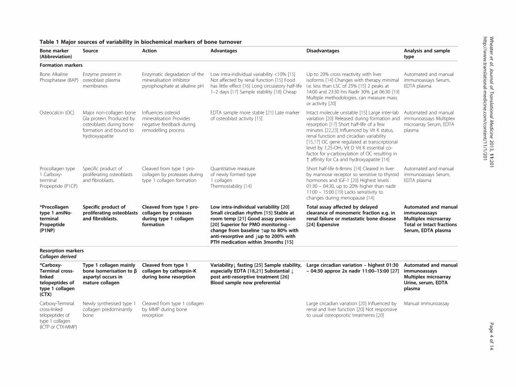
Table 1 Major sources of variability in biochemical markers of bone turnover
Bone marker(Abbreviation)
Source Action Advantages Disadvantages Analysis and sampletype
Formation markers
Bone AlkalinePhosphatase (BAP)
Enzyme present inosteoblast plasmamembranes
Enzymatic degradation of themineralisation inhibitorpyrophosphate at alkaline pH
Low intra-individual variability <10% [15]Not affected by renal function [15] Foodhas little effect [16] Long circulatory half-life1–2 days [17] Sample stability [18] Cheap
Up to 20% cross reactivity with liverisoforms [14] Changes with therapy minimali.e. less than LSC of 25% [15] 2 peaks at14:00 and 23:30 hrs Nadir 30% ↓at 06:30 [19]Multiple methodologies, can measure massor activity [20]
Automated and manualimmunoassays Serum,EDTA plasma
Osteocalcin (OC) Major non-collagen boneGla protein. Produced byosteoblasts during boneformation and bound tohydroxyapatite
Influences osteoidmineralisation Providesnegative feedback duringremodelling process
EDTA sample more stable [21] Late markerof osteoblast activity [15]
Intact molecule unstable [15] Large inter-labvariation [20] Released during formation andresorption [17] Short half-life of a fewminutes [22,23] Influenced by Vit K status,renal function and circadian variability[15,17] OC gene regulated at transcriptionallevel by 1,25-OH2 Vit D Vit K essential co-factor for γ-carboxylation of OC resulting in↑ affinity for Ca and hydroxyapatite [14]
Automated and manualimmunoassays Multiplexmicroarray Serum, EDTAplasma
Procollagen type1 Carboxy-terminalPropeptide (P1CP)
Specific product ofproliferating osteoblastsand fibroblasts.
Cleaved from type 1 pro-collagen by proteases duringtype 1 collagen formation
Quantitative measureof newly formed type1 collagenThermostability [14]
Short half-life 6-8mins [14] Cleared in liverby mannose receptor so sensitive to thyroidhormones and IGF-1 [20] Highest levels01:30 – 04:30, up to 20% higher than nadir11:00 – 15:00 [19] Lacks sensitivity tochanges during menopause [14]
Automated and manualimmunoassays Serum,EDTA plasma
*Procollagentype 1 amiNo-terminalPropeptide(P1NP)
Specific product ofproliferating osteoblastsand fibroblasts.
Cleaved from type 1 pro-collagen by proteasesduring type 1 collagenformation
Low intra-individual variability [20]Small circadian rhythm [15] Stable atroom temp [21] Good assay precision[20] Superior for PMO monitoring -change from baseline ↑up to 80% withanti-resorptive and ↓up to 200% withPTH medication within 3months [15]
Total assay affected by delayedclearance of monomeric fraction e.g. inrenal failure or metastatic bone disease[24] Expensive
Automated and manualimmunoassaysMultiplex microarrayTotal or Intact fractionsSerum, EDTA plasma
Resorption markersCollagen derived
*Carboxy-Terminal cross-linkedtelopeptides oftype 1 collagen(CTX)
Type 1 collagen mainlybone Isomerisation to βaspartyl occurs inmature collagen
Cleaved from type 1collagen by cathepsin-Kduring bone resorption
Variability↓ fasting [25] Sample stability,especially EDTA [18,21] Substantial ↓post anti-resorptive treatment [26]Blood sample now preferential
Large circadian variation – highest 01:30– 04:30 approx 2x nadir 11:00–15:00 [27]
Automated and manualimmunoassaysMultiplex microarrayUrine, serum, EDTAplasma
Carboxy-Terminalcross-linkedtelopeptides oftype 1 collagen(ICTP or CTX-MMP)
Newly synthesised type 1collagen predominantlybone
Cleaved from type 1 collagenby MMP during boneresorption
Large circadian variation [20] Influenced byrenal and liver function [20] Not responsiveto usual osteoporotic treatments [20]
Manual immunoassay
Wheater
etal.Journalof
TranslationalMedicine
2013,11:201Page
4of
14http://w
ww.translational-m
edicine.com/content/11/1/201
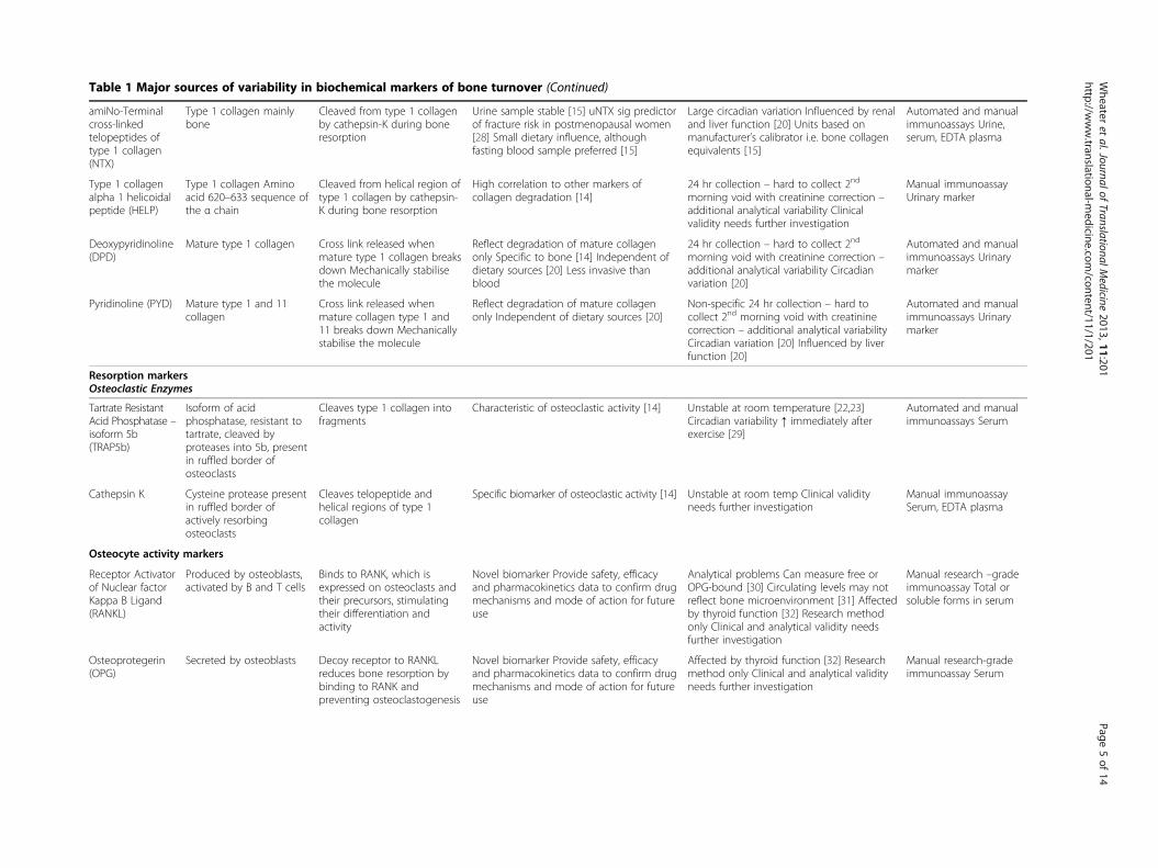
Table 1 Major sources of variability in biochemical markers of bone turnover (Continued)
amiNo-Terminalcross-linkedtelopeptides oftype 1 collagen(NTX)
Type 1 collagen mainlybone
Cleaved from type 1 collagenby cathepsin-K during boneresorption
Urine sample stable [15] uNTX sig predictorof fracture risk in postmenopausal women[28] Small dietary influence, althoughfasting blood sample preferred [15]
Large circadian variation Influenced by renaland liver function [20] Units based onmanufacturer’s calibrator i.e. bone collagenequivalents [15]
Automated and manualimmunoassays Urine,serum, EDTA plasma
Type 1 collagenalpha 1 helicoidalpeptide (HELP)
Type 1 collagen Aminoacid 620–633 sequence ofthe α chain
Cleaved from helical region oftype 1 collagen by cathepsin-K during bone resorption
High correlation to other markers ofcollagen degradation [14]
24 hr collection – hard to collect 2nd
morning void with creatinine correction –additional analytical variability Clinicalvalidity needs further investigation
Manual immunoassayUrinary marker
Deoxypyridinoline(DPD)
Mature type 1 collagen Cross link released whenmature type 1 collagen breaksdown Mechanically stabilisethe molecule
Reflect degradation of mature collagenonly Specific to bone [14] Independent ofdietary sources [20] Less invasive thanblood
24 hr collection – hard to collect 2nd
morning void with creatinine correction –additional analytical variability Circadianvariation [20]
Automated and manualimmunoassays Urinarymarker
Pyridinoline (PYD) Mature type 1 and 11collagen
Cross link released whenmature collagen type 1 and11 breaks down Mechanicallystabilise the molecule
Reflect degradation of mature collagenonly Independent of dietary sources [20]
Non-specific 24 hr collection – hard tocollect 2nd morning void with creatininecorrection – additional analytical variabilityCircadian variation [20] Influenced by liverfunction [20]
Automated and manualimmunoassays Urinarymarker
Resorption markersOsteoclastic Enzymes
Tartrate ResistantAcid Phosphatase –isoform 5b(TRAP5b)
Isoform of acidphosphatase, resistant totartrate, cleaved byproteases into 5b, presentin ruffled border ofosteoclasts
Cleaves type 1 collagen intofragments
Characteristic of osteoclastic activity [14] Unstable at room temperature [22,23]Circadian variability ↑ immediately afterexercise [29]
Automated and manualimmunoassays Serum
Cathepsin K Cysteine protease presentin ruffled border ofactively resorbingosteoclasts
Cleaves telopeptide andhelical regions of type 1collagen
Specific biomarker of osteoclastic activity [14] Unstable at room temp Clinical validityneeds further investigation
Manual immunoassaySerum, EDTA plasma
Osteocyte activity markers
Receptor Activatorof Nuclear factorKappa B Ligand(RANKL)
Produced by osteoblasts,activated by B and T cells
Binds to RANK, which isexpressed on osteoclasts andtheir precursors, stimulatingtheir differentiation andactivity
Novel biomarker Provide safety, efficacyand pharmacokinetics data to confirm drugmechanisms and mode of action for futureuse
Analytical problems Can measure free orOPG-bound [30] Circulating levels may notreflect bone microenvironment [31] Affectedby thyroid function [32] Research methodonly Clinical and analytical validity needsfurther investigation
Manual research –gradeimmunoassay Total orsoluble forms in serum
Osteoprotegerin(OPG)
Secreted by osteoblasts Decoy receptor to RANKLreduces bone resorption bybinding to RANK andpreventing osteoclastogenesis
Novel biomarker Provide safety, efficacyand pharmacokinetics data to confirm drugmechanisms and mode of action for futureuse
Affected by thyroid function [32] Researchmethod only Clinical and analytical validityneeds further investigation
Manual research-gradeimmunoassay Serum
Wheater
etal.Journalof
TranslationalMedicine
2013,11:201Page
5of
14http://w
ww.translational-m
edicine.com/content/11/1/201
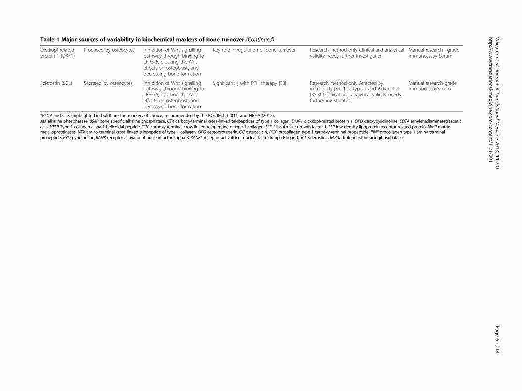
Table 1 Major sources of variability in biochemical markers of bone turnover (Continued)
Dickkopf-relatedprotein 1 (DKK1)
Produced by osteocytes Inhibition of Wnt signallingpathway through binding toLRP5/6, blocking the Wnteffects on osteoblasts anddecreasing bone formation
Key role in regulation of bone turnover Research method only Clinical and analyticalvalidity needs further investigation
Manual research –gradeimmunoassay Serum
Sclerostin (SCL) Secreted by osteocytes Inhibition of Wnt signallingpathway through binding toLRP5/6, blocking the Wnteffects on osteoblasts anddecreasing bone formation
Significant ↓ with PTH therapy [33] Research method only Affected byimmobility [34] ↑ in type 1 and 2 diabetes[35,36] Clinical and analytical validity needsfurther investigation
Manual research-gradeimmunoassaySerum
*P1NP and CTX (highlighted in bold) are the markers of choice, recommended by the IOF, IFCC (2011) and NBHA (2012).ALP alkaline phosphatase, BSAP bone specific alkaline phosphatase, CTX carboxy-terminal cross-linked telopeptides of type 1 collagen, DKK-1 dickkopf-related protein 1, DPD deoxypyridinoline, EDTA ethylenediaminetetraaceticacid, HELP Type 1 collagen alpha 1 helicoidal peptide, ICTP carboxy-terminal cross-linked telopeptide of type 1 collagen, IGF-1 insulin-like growth factor-1, LRP low-density lipoprotein receptor-related protein, MMP matrixmetalloproteinases, NTX amino-terminal cross-linked telopeptide of type 1 collagen, OPG osteoprotegerin, OC osteocalcin, PICP procollagen type 1 carboxy-terminal propeptide, PINP procollagen type 1 amino-terminalpropeptide, PYD pyridinoline, RANK receptor activator of nuclear factor kappa B, RANKL receptor activator of nuclear factor kappa B ligand, SCL sclerostin, TRAP tartrate resistant acid phosphatase.
Wheater
etal.Journalof
TranslationalMedicine
2013,11:201Page
6of
14http://w
ww.translational-m
edicine.com/content/11/1/201
Wheater et al. Journal of Translational Medicine 2013, 11:201 Page 7 of 14http://www.translational-medicine.com/content/11/1/201
Markers of bone resorptionThe majority of bone resorption markers are degradationproducts of bone collagen, the exception being tartrate-resistant acid phosphatase (TRAP5b). Earlier research intobone metabolism relied primarily on urinary markers suchas pyridinoline (PYD) and deoxypyridinoline (DPD), whichwere time-consuming and cumbersome and relied oncomplete twenty-four hour urine collections or secondmorning void/ creatinine ratios, increasing the imprecisionof the measurement. However, now that serum/ plasmamarkers are available these have become the preferredmeans of measuring resorption. Examples include carboxy-terminal and amino-terminal cross-linked telopeptide oftype 1 collagen (CTX and NTX respectively), of whichCTX is considered the marker of choice [20]. CTX is gen-erated by cathepsin K activity, the CTX epitope contains anaspartyl-glycine motif that is susceptible to spontaneousisomerisation and racemisation generating four isoforms[17]; the α-aspartic acid converts to the β-form as the boneages. Two automated immunoassays are available thattarget βCTX indicative of the breakdown of mature type 1collagen (IDS iSYS and Roche Elecsys). The major disad-vantage of CTX is its large circadian variation necessitat-ing a morning fasting sample for accurate interpretation[25]. The choice of marker in clinical practice needs to bemade on pragmatic grounds. Urine NTX may be thepreferred marker in the clinic setting as unlike plasmaCTX, it is not as sensitive to circadian changes and is notaffected by food intake, it also avoids the invasive vene-puncture associated with a blood sample and may be pre-ferred by patients [39]. However the various drugslicensed for the treatment of osteoporosis have a differingspectrum of effects on BTMs and not all markers respondby the same amount for a given degree of bone resorption.Amongst the bone resorption markers, plasma CTX tendsto change more than urine NTX which tends to changemore than TRAP5b [20].
Markers of osteoclastogenesisOsteoclast regulatory proteins are commonly measuredin research, but have yet to find a niche clinically. Thediscovery of the OPG/RANK/RANKL system has clari-fied a major component of the bone remodelling cycle.RANKL is expressed in vivo in either membrane-boundor soluble form (sRANKL) and is also present in serumas a free or OPG-bound molecule, as a consequence de-sign differences between immunoassays have created dif-ficulties in comparing research and interpreting clinicaldata [30]. Furthermore circulating levels may not reflectthe bone microenvironment [31]. Research into the rela-tionship between circulating levels of OPG and sRANKLto BMD in postmenopausal osteoporosis are controversial,some studies reporting an inverse relationship [40], whileothers have found no association [41]. Rigorous testing of
commercial assays and identification of the sources ofvariability are required before they can be adapted toroutine clinical practice.
Osteocyte markersOver the last decade research has focused mainly on therole of osteoclasts and osteoblasts in osteoporosis, morerecently however, osteocytes have been found to play akey role in the regulation of bone turnover. Osteocytesare fully differentiated osteoblasts and lie in lacunae inthe mineralized matrix and osteoid tissue of bone [42].Osteocytes are able to detect changes in bone morphology,particularly micro-fractures through their sensitivity tomechanical forces, acting like bone mechanoreceptors[43]. They regulate bone turnover both through directphysical contact with other bone cells and by producingvarious factors which affect bone formation and can bemeasured in blood such as, sclerostin (SCL), dickkopf-related protein 1 (DKK1), dentin matrix protein 1 (DMP1)and matrix extracellular phosphoglycoprotein (MEPE).DKK1 and SCL are secreted osteocyte markers acting as
inhibitors to the Wnt signalling pathway through bindingto low density lipoprotein receptor-related protein 5 and 6(LRP5/6) and hence blocking the Wnt effects on osteo-blasts decreasing bone formation (Figure 2) [44,45]. Invivo studies have shown that osteocyte depletion results inprofound loss of trabecular bone mass [46-48] and suggesta close interaction between osteocytes and other bonecells, highlighting their role in the regulation of both boneformation and resorption.Although widely used in research, their diagnostic im-
portance remains to be validated due in part to theiranalytical and biological variability. In healthy adults,SCL levels correlate positively with age, BMI, and bonemineral content and negatively with osteocalcin and cal-cium [49]. SCL is increased in type 2 diabetes. More-over, the transcriptional suppression of SCL productionby PTH might be impaired in type 1 and type 2 diabetes[35]. SCL levels are significantly lower in osteoporoticcompared to non-osteoporotic patients with type 2 dia-betes [36]. The Wnt signalling pathway has recentlybeen identified as central to the development of disuseosteoporosis [50]. Mechanical unloading in long-termimmobilized patients causes up regulation of SCL andtherefore inhibits bone formation via suppressed osteo-blast activity and survival [34]. Circulating SCL reflectsthe severity of bone loss and is a candidate biomarker ofosteoporosis severity in chronic spinal cord injury [51].Higher serum SCL levels are associated with a greaterrisk of hip fractures in older women. In addition, therisk of hip fracture is amplified when high SCL levelsare combined with lower BMD [52]. Serum SCL levelsare regulated by both estrogens and PTH in postmeno-pausal women [53]. Serum SCL is decreased in women
Wheater et al. Journal of Translational Medicine 2013, 11:201 Page 8 of 14http://www.translational-medicine.com/content/11/1/201
with postmenopausal osteoporosis compared with non-osteoporotic early postmenopausal women and positivelycorrelated to lumbar spine BMD. Furthermore, levels areincreased after 6 months treatment with risedronate, butremain essentially unchanged after 6 months teriparatidetreatment [54]. However, serum or plasma SCL concen-trations should be interpreted with caution as currentassays produce very different results. Standardization ofsclerostin assays is necessary before being introducedinto general clinical laboratory use [55].
Variability in markers of bone turnoverAn understanding of the source and magnitude of theabsolute inter and intra-person variability, includingbiological, pre-analytic and analytical variation, of eachmarker is necessary to interpret serial measurementsand individualise treatment.
Biological variabilityIntra-individual variation Bone turnover shows a circa-dian rhythm, this is more obvious in the serum and urin-ary markers of bone resorption. βCTX for example ishighest between 01:30 and 04:30 hours and may be morethan twice that at the nadir between 11:00 and 15:00 hours[27], this may be attenuated by several factors such as; age,gender, ethnicity, menopausal status, osteoporotic stageand anti-resorptive agents or calcium supplementation[19], but the disparity is diminished with fasting [25]. Allbone markers are significantly lower in the fed state withthe exception of BSAP, this may be due to several factorsincluding the clearance rate of individual markers or foodcomposition [16] and may be partly explained by variationin serum insulin [25]. Osteocalcin and P1CP follow thesame diurnal pattern but show only twenty percent differ-ence and BSAP has two peaks at 14:00 and 23:30 hourswith a nadir thirty percent reduced at 06:30 [19]. There-fore timing of the sample collection and fasting statusshould be tightly controlled.The existence of intra-individual low-frequency bio-
logical rhythms, imply that biomarkers can also varybetween consecutive days, this is more noticeable inthe urinary resorption markers [14]. There is a degreeof controversy regarding seasonal variation with someresearchers suggesting that overall seasonal changesare insignificant [56], whilst others have found a sub-stantial wintertime increase [57], which may be due inpart to reduced levels of vitamin D. Physical activity isalso significant, TRAP and to a lesser extent BSAP andCTX are reduced immediately after plyometrics, butreturn to pre concentrations within two hours. Inter-estingly similar changes were found in PTH [29]. De-tails of exercise in the previous twenty-four hoursshould therefore be recorded.
Bone turnover varies with the menstrual cycle, researchsuggests that osteoblastic activity is higher during the lu-teal period [58] and bone resorption is increased duringthe follicular phase [59]. Pregnancy affects all BTMs duein part to the calcium requirements of the foetus, butalso to changes in maternal glomerular filtration rate(GFR) affecting renal clearance. However the timechange is contentious, one study following ten womenat regular intervals reported an increase in urinary resorp-tion markers throughout pregnancy with a significantincrease in bone formation in the third trimester [60]. Amore recent study measured serum OPG, RANKL,osteocalcin and CTX in twenty six different women ateach trimester. The study found increased bone formationin the first trimester and increased resorption in the sec-ond which surprisingly decreased again in the third tri-mester [61]. Postpartum, levels gradually start to decreasebut may still be higher than pre-pregnancy levels for up toa year [62].A comprehensive drug history should also be taken
into account when interpreting bone marker results.Anti-resorptive drugs such as bisphosphonates [26] andhormone replacement therapy (HRT) [63] have a majoreffect on markers of bone resorption and long-termcorticosteroid therapy is known to suppress bone for-mation [64].Inflammatory conditions are major precipitators for
bone loss, especially rheumatoid arthritis (RA) which isfurther aggravated by decreased functional activity andthe use of glucocorticoids [65]. In a prior study, wefound that B-cell depletion increases bone formationand decreases bone resorption in RA patients. This maybe a direct effect on osteoblasts and osteoclasts respect-ively and be at least partially explained by the decreasedinflammation and disease activity [66]. In diabetes serumosteocalcin is negatively correlated with glucose levelsand advanced glycation end products (AGEs) are knownto have a negative impact on bone [67]. Thyroid disorderssuch as thyrotoxicosis are well known to affect bone turn-over. Thyroid stimulating hormone (TSH) receptors arepresent in both osteoblasts and osteoclasts and the lowTSH levels observed in thyroidectomised patients onL-thyroxine are associated with an increase in OPG anddecrease in RANKL and are significantly correlated withvertebral fractures [32]. Bone markers are cleared throughthe liver or kidneys and are also influenced by diseasesaffecting these systems, decreased GFR for example willdecrease the urinary excretion of CTX and therefore in-crease serum levels. They are also affected by any diseasestates leading to increased periods of bed rest and immo-bility. Research has shown that microgravity inducessignificant and progressive bone loss, a consequence of in-creased bone resorption and retardation of bone forma-tion [68]. Certainly levels of all bone markers increase
Wheater et al. Journal of Translational Medicine 2013, 11:201 Page 9 of 14http://www.translational-medicine.com/content/11/1/201
significantly in the first few weeks after fracture and mayremain elevated for up to a year. The rate of increase isdependent on the location, severity and size of the fractureand the age of the patient. BTM’s can be elevated for up to6 months after minor fractures e.g. forearm fractures butup to one year after a hip fracture and needs to be takeninto consideration when measuring them [19,69]. How-ever, they fall gradually over time and using a reduction of50% in bone resorption when using anti-resorptives as agood indicator of response would be greater than anyreduction that might otherwise occur.In light of the above evidence it can be seen that to use
bone turnover to monitor change can be quite difficult. Inorder to minimise problems it is best to measure theBTMs in as similar a set of circumstances as possible. Par-ticular attention should be paid to the time of day andhence research studies tend to use early morning fastedsamples. One way in which to help overcome within per-son variability in serial measurements and to monitortherapy is to use the ‘least significant change’ (LSC) model[70]. LSC at a significance level of p=<0.05 is defined as1.96*√2*√ (CV1
2+CVA2); where CV1 is the within-subject
coefficient of variation and CVA is the total analyticalimprecision. LSC identifies the true physiological changein the marker. In general a change of more than twentypercent is considered significant for formation markers[71], similarly between twenty-seven to thirty-six percentis significant for markers of bone resorption [72].
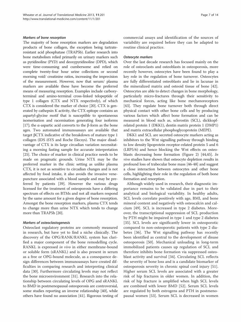
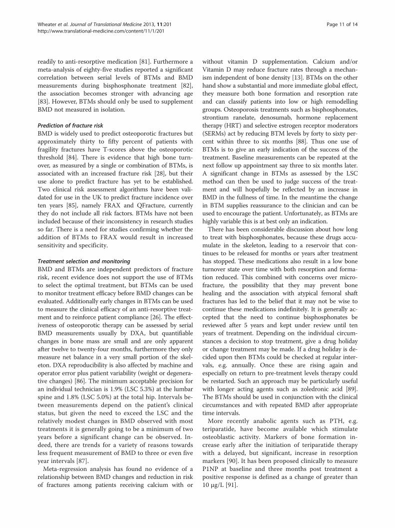
Inter-individual variation Between person variability ismuch harder to control, e.g. age, gender, and menopausalstatus, but is equally important to validate results. Bonemetabolism rates are higher in infants up to three years ofage, they are relatively stable throughout adolescence butsex-specific increases in bone marker levels are evidentduring the pubertal growth spurt and are reportedlyinfluenced by pubertal stage rather than age [73]. BTMsare higher in men between twenty and thirty years of agethen reach their lowest levels during their fifties [14],whereas in females there is a substantial increase in boneturnover corresponding to oestrogen deficiency during themenopause [28]. We checked our local population andfound a trend towards higher bone turnover in malesduring their third decade whilst reaching peak bone mass,although there seemed to be little evidence of any age re-lated change in women possibly due to the lower numbersof postmenopausal females in our cohort (Figure 3). It isimportant for each laboratory to investigate the transfer-ability of the quoted reference intervals to its own patientpopulation based on equivalent standardised collectionconditions. The widespread availability of automated im-munoassays now means that harmonisation of methodspecific reference ranges is possible and studies from well-characterised populations have reported robust BTM
ranges in large well defined cohorts [74]. In contrast tothe use of reference ranges some researchers have sug-gested combining a marker of formation and resorption togain a direct insight into the changes in the balance andrate of bone turnover in relation to a reference value [75],leading factors in estimating fracture risk and prognosis.Given the large observed differences observed betweengenders, different ages and developmental stages meansthat care must be taken when comparing populations andin the design of research studies.
Analytical variabilityTechnical variation Over the last decade many of thetraditional BTM immunoassays have been automated, im-proving technical performance and increasing their avail-ability. Nevertheless, analytical aspects such as within andbetween batch precision, accuracy and standardisation, re-main problematic. Inter-laboratory variation is also crucial;a European study in 2001, measuring pooled samples ofserum and urine in seventy-three laboratories concludedthat even with identical assays results for the majority ofthe markers were significantly different [76]. Similarly anAmerican study in 2010 comparing six commerciallaboratories over an eight month period concluded thatreproducibility varied substantially for urine NTX andserum BSAP [77]. Moreover there is an extensive list ofbone markers being offered making it very difficult tocompare research evidence. Consequently, the Inter-national Osteoporosis Foundation (IOF), the InternationalFederation of Clinical Chemistry and Laboratory Medicine(IFCC) [20] and the National Bone Health Alliance(NBHA) [78], have recommended that a marker of boneformation and resorption, namely P1NP and CTX, areused as reference analytes in clinical studies. They go onto stipulate that these markers should be measured bystandardised assays to minimise immunochemical hetero-geneity and recommend that manufacturers adopt inter-national reference standards and minimise batch to batchvariability [20].
Sample stability Appropriate control of sample collec-tion and preparation is vital for successful BTM measure-ment. Several BTMs, especially osteocalcin and TRAP5b,are sensitive to thermo degradation and levels can besignificantly reduced after only a few hours at roomtemperature [22,23]. TRAP5b activity is also reduced dur-ing storage, samples must be kept at −70°C or lower andmultiple freeze-thaw cycles should be avoided. No signifi-cant decrease has been detected in CTX stored at −20°Cor lower for up to three years, nevertheless it rapidly de-creases in serum at both 4°C and 37°C. The molecularmechanism is unknown but this decrease is minimised byethylenediaminetetraacetic acid (EDTA) [18]. CTX isreportedly stable in EDTA blood tubes before separation
0
100
200
300
400
500
600
700
800
0 5 10 15 20 25 30 35 40 45 50 55 60 65 70
βCT
X
co
nc
(n
g/L
)
Age (yrs)
FEMALE MALE
0
10
20
30
40
50
60
70
80
90
0 5 10 15 20 25 30 35 40 45 50 55 60 65 70
P1
NP
co
nc
(µ
g/ L
)
Age (yrs)
0
5
10
15
20
25
30
35
40
0 5 10 15 20 25 30 35 40 45 50 55 60 65 70
Os
t eo
ca
lci n
co
nc
(µ
g/L
)
Age (yrs)
βCTX: beta-isomerised carboxy-terminal telopeptide of type 1 collagen PINP: procollagen type 1 amino-terminal propeptide
Figure 3 Age related changes to bone turnover markers. Blood samples from seventy healthy volunteers (33 males aged 19 to 62 years and 37females aged 20 to 64 years), collected between 10–11 am, were analysed on the Elecsys 2010 (Roche, Lewes UK), for βCTX, P1NP and osteocalcin toverify the effect of age on bone turnover.
Wheater et al. Journal of Translational Medicine 2013, 11:201 Page 10 of 14http://www.translational-medicine.com/content/11/1/201
for up to forty-eight hours, likewise osteocalcin becomesstable for up to eight hours at room temperature [21].Consequently blood should be collected into EDTA tubesand separated as soon as possible, if samples are notanalysed immediately they should be stored at −20°C orlower. Both P1NP and BSAP were found to be stable inany sample type [21]. Notably current TRAP5b assaysare not affected by haemolysis, but erythrocytes areknown to contain proteases which degrade osteocalcin.Grossly haemolysed samples in general should alwaysbe avoided.
Clinical usefulness of bone turnover markers inosteoporosisBTMs are frequently used in clinical trials and providevaluable information on the efficacy of osteoporotictreatments, but their predictive value is limited by theirlarge biological variation and diagnostically they are lessoften used for individualized patient care. Other routinelaboratory investigations are frequently used to identifyor exclude secondary causes of osteoporosis such as
hyperparathyroidism, vitamin D status, thyrotoxicosisand hypogonadism [79].
DiagnosticCurrently the WHO recommends the use of BMD ofthe spine and proximal femur measured by DXA as thegold standard to diagnose osteoporosis and its severity[12]. Although BMD has methodological limitationsespecially in the elderly due in part to degenerativechanges in the lumbar spine [80], BTMs alone wouldnot be suitable to estimate bone loss.
Prediction of bone lossWomen generally lose about one to two percent of theirbone per year after the menopause, however thirty per-cent lose bone at a faster rate [28]. Longitudinal studiesof post-menopausal women have demonstrated twocharacteristic groups; high bone turnover and normal orlow bone turnover. Serial BTM measurements are effect-ive in identifying those women who lose bone most rap-idly, this is important because this group respond more
Wheater et al. Journal of Translational Medicine 2013, 11:201 Page 11 of 14http://www.translational-medicine.com/content/11/1/201
readily to anti-resorptive medication [81]. Furthermore ameta-analysis of eighty-five studies reported a significantcorrelation between serial levels of BTMs and BMDmeasurements during bisphosphonate treatment [82],the association becomes stronger with advancing age[83]. However, BTMs should only be used to supplementBMD not measured in isolation.
Prediction of fracture riskBMD is widely used to predict osteoporotic fractures butapproximately thirty to fifty percent of patients withfragility fractures have T-scores above the osteoporoticthreshold [84]. There is evidence that high bone turn-over, as measured by a single or combination of BTMs, isassociated with an increased fracture risk [28], but theiruse alone to predict fracture has yet to be established.Two clinical risk assessment algorithms have been vali-dated for use in the UK to predict fracture incidence overten years [85], namely FRAX and QFracture, currentlythey do not include all risk factors. BTMs have not beenincluded because of their inconsistency in research studiesso far. There is a need for studies confirming whether theaddition of BTMs to FRAX would result in increasedsensitivity and specificity.
Treatment selection and monitoringBMD and BTMs are independent predictors of fracturerisk, recent evidence does not support the use of BTMsto select the optimal treatment, but BTMs can be usedto monitor treatment efficacy before BMD changes can beevaluated. Additionally early changes in BTMs can be usedto measure the clinical efficacy of an anti-resorptive treat-ment and to reinforce patient compliance [26]. The effect-iveness of osteoporotic therapy can be assessed by serialBMD measurements usually by DXA, but quantifiablechanges in bone mass are small and are only apparentafter twelve to twenty-four months, furthermore they onlymeasure net balance in a very small portion of the skel-eton. DXA reproducibility is also affected by machine andoperator error plus patient variability (weight or degenera-tive changes) [86]. The minimum acceptable precision foran individual technician is 1.9% (LSC 5.3%) at the lumbarspine and 1.8% (LSC 5.0%) at the total hip. Intervals be-tween measurements depend on the patient’s clinicalstatus, but given the need to exceed the LSC and therelatively modest changes in BMD observed with mosttreatments it is generally going to be a minimum of twoyears before a significant change can be observed. In-deed, there are trends for a variety of reasons towardsless frequent measurement of BMD to three or even fiveyear intervals [87].Meta-regression analysis has found no evidence of a
relationship between BMD changes and reduction in riskof fractures among patients receiving calcium with or
without vitamin D supplementation. Calcium and/orVitamin D may reduce fracture rates through a mechan-ism independent of bone density [13]. BTMs on the otherhand show a substantial and more immediate global effect,they measure both bone formation and resorption rateand can classify patients into low or high remodellinggroups. Osteoporosis treatments such as bisphosphonates,strontium ranelate, denosumab, hormone replacementtherapy (HRT) and selective estrogen receptor moderators(SERMs) act by reducing BTM levels by forty to sixty per-cent within three to six months [88]. Thus one use ofBTMs is to give an early indication of the success of thetreatment. Baseline measurements can be repeated at thenext follow up appointment say three to six months later.A significant change in BTMs as assessed by the LSCmethod can then be used to judge success of the treat-ment and will hopefully be reflected by an increase inBMD in the fullness of time. In the meantime the changein BTM supplies reassurance to the clinician and can beused to encourage the patient. Unfortunately, as BTMs arehighly variable this is at best only an indication.There has been considerable discussion about how long
to treat with bisphosphonates, because these drugs accu-mulate in the skeleton, leading to a reservoir that con-tinues to be released for months or years after treatmenthas stopped. These medications also result in a low boneturnover state over time with both resorption and forma-tion reduced. This combined with concerns over micro-fracture, the possibility that they may prevent bonehealing and the association with atypical femoral shaftfractures has led to the belief that it may not be wise tocontinue these medications indefinitely. It is generally ac-cepted that the need to continue bisphosphonates bereviewed after 5 years and kept under review until tenyears of treatment. Depending on the individual circum-stances a decision to stop treatment, give a drug holidayor change treatment may be made. If a drug holiday is de-cided upon then BTMs could be checked at regular inter-vals, e.g. annually. Once these are rising again andespecially on return to pre-treatment levels therapy couldbe restarted. Such an approach may be particularly usefulwith longer acting agents such as zoledronic acid [89].The BTMs should be used in conjunction with the clinicalcircumstances and with repeated BMD after appropriatetime intervals.More recently anabolic agents such as PTH, e.g.
teriparatide, have become available which stimulateosteoblastic activity. Markers of bone formation in-crease early after the initiation of teriparatide therapywith a delayed, but significant, increase in resorptionmarkers [90]. It has been proposed clinically to measureP1NP at baseline and three months post treatment apositive response is defined as a change of greater than10 μg/L [91].
Wheater et al. Journal of Translational Medicine 2013, 11:201 Page 12 of 14http://www.translational-medicine.com/content/11/1/201
ConclusionsDuring the last decade significant advances have beenmade in the identification and characterisation ofspecific BTMs for use in clinical drug trials and to aid inthe therapeutic management of osteoporosis. Techno-logical developments have greatly enhanced assay per-formance producing reliable, rapid, non-invasive costeffective assays with improved sensitivity and specificity.We now have a greater understanding of the need toregulate pre-analytical sample collection to minimisethe effects of biological variation. The use of BTMsto select those at risk of fractures is not routinelyrecommended partly due to their degree of variability.However, baseline measurements of resorption markersare useful before commencement of anti-resorptive treat-ment e.g. bisphosphonates or denosumab and can bechecked 3–6 months later to check response and adher-ence to treatment. Similarly a formation marker such asP1NP can be used to monitor bone forming agents suchas PTH analogues. BTMs may also be useful when moni-toring patients during treatment holidays and aid in thedecision as to when therapy should be recommenced. Therecent recommendations by the Bone Marker StandardsWorking Group aim to standardise research and include amarker of bone resorption (CTX) and formation (P1NP)in all future studies. They anticipate that manufacturerswill calibrate their assays in future using an internationalreference standard to establish robust reference ranges. Itis hoped that improved research in turn will lead to opti-mise markers for the clinical management of osteoporosisand other bone diseases. The biochemical assessment, util-izing BSAP, is now the mainstay of the diagnosis andmanagement of metabolic bone disease in chronic kidneydisease.
AbbreviationsAGEs: Advanced glycation end products; ALP: Alkaline phosphatase;BSAP: Bone specific alkaline phosphatase; βCTX: Beta-isomerised carboxyterminal telopeptide of type I collagen; BMD: Bone mineral density;BTM: Bone turnover marker; CTX: Carboxy-terminal cross-linked telopeptidesof type 1 collagen; CVA: Total analytical imprecision; CV1: Within-subjectcoefficient of variation; DKK-1: Dickkopf-related protein 1; DMP1: Dentinmatrix protein 1; DPD: Deoxypyridinoline; DXA: Dual-energy X-rayabsorptiometry; EDTA: Ethylenediaminetetraacetic acid; Fz: Frizzled protein;GFR: Glomerular filtration rate; GH: Growth hormone; HELP: Type 1 collagenalpha 1 helicoidal peptide; HRT: Hormone replacement therapy;ICTP: Carboxy-terminal cross-linked telopeptide of type 1 collagen;IDS: Immuno-Diagnostic Systems Ltd; IFCC: International Federation ofClinical Chemistry and Laboratory Medicine; IGF1: Insulin-like growth factor 1;IOF: International Osteoporosis Foundation; LRP: Low-density lipoproteinreceptor-related protein; LSC: Least significant change; MEPE: Matrixextracellular phosphoglycoprotein; MMP: Matrix metalloproteinases;MoM: Multiple of the median; MoMF: Multiple of the median formationmarker; MoMR: Multiple of the median resorption marker; NBHA: NationalBone Health Alliance; NTX: Amino-terminal cross-linked telopeptide of type 1collagen; OB: osteoblast; OC: osteocalcin; OCY: osteocyte;OPG: osteoprotegerin; OPN: osteopontin; PICP: Procollagen type 1 carboxy-terminal propeptide; PINP: Procollagen type 1 amino-terminal propeptide;PPR: PTH/PTHrP receptor; PTH: Parathyroid hormone; PTHrP: Parathyroidhormone related peptide; PYD: pyridinoline; RA: Rheumatoid arthritis;
RANK: Receptor activator of nuclear factor kappa B; RANKL: Receptoractivator of nuclear factor kappa B ligand; sRANKL: Soluble receptor activatorof nuclear factor kappa B ligand; SCL: sclerostin; SD: Standard deviation;SERMs: Selective estrogen receptor moderators; Sost: SclerOSTeosis gene;TRAP: Tartrate resistant acid phosphatase; TSH: Thyroid stimulating hormone;UK: United Kingdom; WHO: World Health Organisation; Wnt: Wingless andIntegration-1.
Competing interestsJMvL has received a research grant, consultancy and speaker fees fromRoche. SPT has received speaker fees from Ely Lilly.
Author’ contributionsGW drafted the manuscript. ME revised the manuscript. SPT and HKD revisedand critically appraised the manuscript. JMvL revised, critically appraised andprovided overall supervision for the project. All authors read and approvedthe final manuscript.
AcknowledgementsThe authors wish to thank staff from the Biochemistry laboratory at theJames Cook University Hospital in Middlesbrough who made the referencerange study possible. In particular we acknowledge Cheryl Goodrum foranalysing the samples.
Author details1Department of Biochemistry, The James Cook University Hospital,Middlesbrough TS4 3BW, UK. 2Institute of Cellular Medicine, MusculoskeletalResearch Group, Newcastle University, Newcastle upon Tyne NE1 7RU, UK.3Department of Rheumatology, The James Cook University Hospital,Middlesbrough TS4 3BW, UK.
Received: 11 February 2013 Accepted: 21 August 2013Published: 29 August 2013
References1. Datta HK, Ng FW, Walker JA, Tuck SP, Varanasi SS: The cell biology of bone
metabolism-a review. J Clin Path 2008, 61:577–587.2. Carey JJ, Licata AA, Delaney MF: Biochemical markers of bone turnover.
Clin Rev Bone Miner Metab 2006, 4(3):197–212.3. Vega D, Maalouf NM, Sakhaee K: The role of receptor activator of nuclear
factor-κB (RANK)/RANK Ligand/Osteoprotegerin: clinical implications.J Clin Endocrinol Metab 2007, 92(12):4514–4521.
4. Leibbrandt A, Penninger JM: RANK/RANKL: regulators of immuneresponses and bone physiology. Ann N Y Acad Sci 2008, 1143:123–150.
5. Klein-Nulend J, Bakker AD, Bacabac RG, Vatsa A, Weinbaum S:Mechanosensation and transduction in osteocytes. Bone 2013,54(2):182–190.
6. Kobayashi Y, Maeda K, Takahashi N: Roles of Wnt signalling in boneformation and resorption. Jpn Dent Sci Rev 2008, 44:76–82.
7. Bellido T, Saini V, Pajevic PD: Effects of PTH on osteocyte function.Bone 2013, 54(2):250–257.
8. National osteoporosis society. http://www.nos.org.uk/page.aspx?pid=328.9. Tuck SP, Francis RM: Osteoporosis. Postgrad Med J 2002, 78:526–532.10. Bongartz TA, Scholmerich J, Straub RH: From Osteoporosis in
postmenopausal women. In Bone disease in rheumatology. Edited byMaricic M, Gluck OS. Arizona: Lippincott Williams and Wilkens;2005:155–156.
11. Bauer D, Garnero P, Harison SL, Cauley JA, Eastell R, Ensrud KE, Orwoll E:Biochemical markers on bone turnover, hip loss and fracture in oldermen: the MrOS study. J Bone Miner Res 2009, 24(12):2032–2038.
12. Kanis JA, McCloskey EV, Johansson H, Oden A, Melton LJ, Khaltaev N:A reference standard for the description of osteoporosis. Bone 2008,42:467–475.
13. Rabinda V, Bruyère O, Reginster JY: Relationship between bone mineraldensity changes and risk of fractures among patients receiving calciumwith or without vitamin D supplementation: a meta-regression.Osteoporos Int 2011, 22:893–901.
14. Seibel MJ: Biochemical markers of bone turnover: part 1: biochemistryand variability. Clin Biochem Rev 2005, 26:97–122.
15. Brown JP, Albert C, Nassar BA, Adachi JD, Cole D, Davison KS, Dooley KC,Don-Wauchope A, Douville P, Hanley DA, Jamal SA, Josse R, Kaiser S, Krahn
Wheater et al. Journal of Translational Medicine 2013, 11:201 Page 13 of 14http://www.translational-medicine.com/content/11/1/201
J, Krause R, Kremer R, Lepage R, Letendre E, Morin S, Ooi DS, PapaioaonnouA, Ste-Marie L-G: Bone turnover markers in the management ofosteoporosis. Clin Biochem 2009, 42:929–942.
16. Clowes JA, Hannon RA, Yap TS, Hoyle NR, Blumsohn A, Eastell R: Effect offeeding on bone turnover markers and its impact on biologicalvariability of measurements. Bone 2002, 30(6):886–890.
17. Swaminathan R: Biochemical markers of bone turnover. Clin Chim Acta2001, 313:95–105.
18. Qvist P, Munk M, Hoyle N, Christiansen C: Serum and plasma fragments ofC-telopeptides of type I collagen (CTX) are stable during storage at lowtemperatures for 3 years. Clin Chim Acta 2004, 350(1–2):167–173.
19. Hannon R, Eastell R: Preanalytical variability of biochemical markers ofbone turnover. Osteoporos Int 2000, 11(Suppl 6):S30–44.
20. Vasikaran S, Eastell R, Bruyère O, Foldes AJ, Garnero P, Griesmacher A,McClung M, Morris HA, Silverman S, Trenti T, Wahl DA, Cooper C, Kanis JA,for the IOF-IFCC Bone Marker Standards Working Group: Markers of boneturnover for the prediction of fracture risk and monitoring ofosteoporosis treatment: a need for international reference standards.Osteoporos Int 2011, 22:391–420.
21. Stokes FJ, Ivanov P, Bailey LM, Fraser WD: The effects of sampling proceduresand storage conditions on short-term stability of blood-based biochemicalmarkers of bone metabolism. Clin Chem 2011, 57(1):138–140.
22. Blumsohn A, Hannon RA, Eastell R: Apparent instability of osteocalcin inserum as measured with different commercially available immunoassays.Clin Chem 1995, 41:318–319.
23. Halleen JM, Alatalo SL, Suominen H, Cheng S, Janckila AJ, Väänänen HK:Tartrate-resistant acid phosphatase 5b: a novel serum marker of boneresorption. J Bone Miner Res 2000, 15(7):1337–1345.
24. Marin L, Koivula M-K, Jukkola-Vuorinen A, Leino A, Risteli J: Comparison oftotal and intact aminoterminal propeptide of type 1 procollagen assaysin patients with breast cancer with or without bone metastases. Ann ClinBiochem 2011, 48:447–451.
25. Bjarnason NH, Henriksen EEG, Alexandersen P, Christgau S, Henriksen DB,Christiansen C: Mechanism of circadian variation in bone resorption.Bone 2002, 30:307–313.
26. Bergmann P, Body JJ, Boonen S, Boutsen Y, Devogelaer JP, Goemaere S,Kaufman JM, Reginster JY, Gangji V, Members of the Advisory Board onBone Markers: Evidence-based guidelines for the use of biochemicalmarkers of bone turnover in the selection and monitoring ofbisphosphonate treatment in osteoporosis: a consensus document ofthe Belgian bone club. Int J Clin Pract 2009, 63(1):19–26.
27. Wichers M, Schmidt E, Bidlingmaier F, Klingmüller D: Diurnal rhythm ofcross laps in human serum. Clin Chem 1999, 45:1858–1860.
28. Garnero P, Sornay-Rendu E, Claustrat B, Delmas PD: Biochemical markers ofbone turnover, endogenous hormones and the risk of fractures inpostmenopausal women: the OFELY study. J Bone Miner Res 2000,15:1526–1536.
29. Rogers RS, Dawson AW, Wang Z, Thyfault JP, Hinton PS: Acute response ofplasma markers of bone turnover to a single bout of resistance trainingor plyometrics. J Appl Physiol 2011, 111:1353–1360.
30. Bowsher RR, Sailstad JM: Insights in the application of research-gradediagnostic kits for biomarker assessments in support of clinical drugdevelopment: Bioanalysis of circulating concentrations of solublereceptor activator of nuclear factor κB ligand. J Pharm Biomed Anal 2008,48(5):1282–1289.
31. Kearns AE, Khosla S, Kostenuik PJ: Receptor activator of nuclear factor κBligand and osteoprotegerin regulation of bone remodelling in healthand disease. Endocr Rev 2008, 29(2):155–192.
32. Nicholls JJ, Brassill MJ, Williams GR, Duncan Bassett JH: The skeletalconsequences of thyrotoxicosis. J Endocrinol 2012, 213(3):209–221.
33. Drake MT, Srinivasan B, Mödder UI, Peterson JM, McCready LK, Riggs BL,Dwyer D, Stolina M, Kostenuik P, Khosla S: Effects of parathyroid hormonetreatment on circulating sclerostin levels in postmenopausal women.J Clin Endocrinol Metab 2010, 95(11):5056–5062.
34. Gaudio A, Pennisi P, Bratengeier C, Torrisi V, Lindner B, Mangiafico RA,Pulvirenti I, Hawa G, Tringali G, Fiore CE: Increased sclerostin serum levelsassociated with bone formation and resorption markers in patients withimmobilization-induced bone loss. J Clin Endocrinol Metab 2010,95(5):2248–2253.
35. Gennari L, Merlotti D, Valenti R, Ceccarelli E, Ruvio M, Pietrini MG, CapodarcaC, Franci MB, Campagna MS, Calabrò A, Cataldo D, Stolakis K, Dotta F,
Nuti R: Circulating sclerostin levels and bone turnover in type 1 and type2 diabetes. J Clin Endocrinol Metab 2012, 97(5):1737–1744.
36. Garcia-Martin A, Rozas-Moreno P, Reyes-Garcia R, Morales-Santana S,Garcia-Fontana B, Garcia-salcedo JA, Muñoz-Torres M: Circulating levels ofsclerostin are increased in patients with type 2 diabetes mellitus.J Clin Endocrinol Metab 2012, 97(1):234–241.
37. Melkko J, Hellevik T, Risteli L, Risteli J, Smedsrød S: Clearance of NH2-terminal propeptides of types I and III Procollagen is a physiologicalfunction of the scavenger receptor in liver endothelial cells. J Exp Med1994, 179:405–412.
38. Brandt J, Krogh TN, Jensen CH, Frederiksen JK, Teisner B: Thermal instabilityof the trimeric structure of the N-terminal propeptide of humanProcollagen type I in relation to assay technology. Clin Chem 1999,45(1):47–53.
39. Baxter I, Rogers A, Eastell R, Peel N: Evaluation of urinary N-telopeptide oftype I collagen measurements in the management of osteoporosis inclinical practice. Osteoporos Int 2013, 24:941–947.
40. Jabbar S, Drury J, Fordham JN, Datta HK, Francis RM, Tuck SP:Osteoprotegerin, RANKL and bone turnover in postmenopausalosteoporosis. J Clin Pathol 2011, 64:354–357.
41. Liu JM, Zhao HY, Ning G, Zhao YJ, Chen Y, Zhang Z, Sun LH, Xu M-Y, ChenJL: Relationships between the changes of serum levels of OPG andRANKL with age, menopause, bone biochemical markers and bonemineral density in Chinese women aged 20–75. Calcif Tissue Int 2005,76(1):1–6.
42. Noble BS: The osteocyte lineage. Arch Biochem Biophys 2008,473(2):106–11.
43. Bonewald LF: Osteocytes: a proposed multifunctional bone cell.J Musculoskelet Neuronal Interact 2002, 2(3):239–41.
44. Zhang Y, Wang Y, Li X, Zhang J, Mao J, Li Z, Zhang J, Li L, Harris S, Wu D:The LRP5 high-bone-mass G171V mutation disrupts LRP5 interactionwith Mesd. Mol Cell Biol 2004, 24(11):4677–4684.
45. Li X, Zhang Y, Kang H, Liu W, Liu P, Zhang J, Harris SE, Wu D: Sclerostinbinds to LRP5/6 and antagonizes canonical Wnt signalling. J Biol Chem2005, 280:19883–19887.
46. You L, Temiyasathit S, Lee P, Kim CH, Tummala P, Yao W, Kingery W, MaloneAM, Kwon RY, Jacobs CR: Osteocytes as mechanosensors in the inhibitionof bone resorption due to mechanical loading. Bone 2008, 42(1):172–179.
47. Gross TS, King KA, Rabaia NA, Pathare P, Srinivasan S: Upregulation ofosteopontin by osteocytes deprived of mechanical loading or oxygen.J Bone Miner Res 2005, 20(2):250–256.
48. Noble BS, Peet N, Stevens HY, Brabbs A, Mosley JR, Reilly GC, Reave J, SkerryTM, Lanyon LE: Mechanical loading: biphasic osteocyte survival andtargeting of osteoclasts for bone destruction in rat cortical bone.Am J Physiol Cell Physiol 2003, 284:C934–C943.
49. Amrein K, Amrein S, Drexler C, Dimai HP, Dobnig H, Pfeifer K, Tomaschitz A,Pieber TR, Fahrleitner-Pammer A: Sclerostin and its association withphysical activity, age, gender, body composition and bone mineralcontent in healthy adults. J Clin Endocrinol Metab 2012, 97(1):148–154.
50. Lin C, Jiang X, Dai Z, Guo X, Weng T, Wang J, Li Y, Feng G, Gao X, He L:Sclerostin mediates bone response to mechanical unloading throughantagonizing Wnt/β-Catenin signalling. J Bone Miner Res 2009,24(10):1651–1661.
51. Morse LR, Sudhakar S, Lazzari AA, Tun C, Garshick E, Zafonte R, BattaglinoRA: Sclerostin: a candidate biomarker of SCI-induced osteoporosis.Osteoporos Int 2013, 24:961–968.
52. Arasu A, Cawthon PM, Lui L-Y, Do TP, Arora PS, Cauley JA, Ensrud KE,Cummings SR: Serum sclerostin and risk of hip fracture in olderCaucasian women. J Clin Endocrinol Metab 2012, 97(6):2027–2032.
53. Mirza FS, Padhi ID, Raisz LG, Lorenzo JA: Serum sclerostin levels negativelycorrelate with parathyroid hormone levels and free estrogen index inpostmenopausal women. J Clin Endocrinol Metab 2010, 95(4):1991–1997.
54. Polyzos SA, Anastasilakis AD, Bratengeier C, Woloszczuk W, PapatheodorouA, Terpos E: Serum sclerostin levels positively correlate with lumbarspinal bone mineral density in postmenopausal women – the six-montheffect of risedronate and teriparatide. Osteoporos Int 2012, 23:1171–1176.
55. McNulty M, Singh RJ, Li X, Bergstralh EJ, Kumar R: Determination of serumand plasma sclerostin concentrations by enzyme-linked immunoassays.J Clin Endocrinol Metab 2011, 96(7):E1159–E1162.
56. Blumsohn A, Naylor KE, Timm W, Eagleton AC, Hannon RA, Eastell R:Absence of marked seasonal change in bone turnover: a longitudinal
Wheater et al. Journal of Translational Medicine 2013, 11:201 Page 14 of 14http://www.translational-medicine.com/content/11/1/201
and multicentre cross-sectional study. J Bone Miner Res 2003,18:1274–1281.
57. Woitge HW, Scheidt-Nave C, Kissling C, Leidig-Bruckner G, Meyer K, GrauerA, Scharla SH, Ziegler R, Seibel MJ: Seasonal variation of biochemicalindices of bone turnover: results of a population-based study.J Clin Endocrinol Metab 1998, 83:68–75.
58. Nielsen HK, Brixen K, Bouillon R, Mosekilde L: Changes in biochemicalmarkers of osteoblastic activity during the menstrual cycle.J Clin Endocrinol Metab 1990, 70:1431–1437.
59. Chiu KM, Ju J, Mayes D, Bacchetti P, Weitz S, Arnaud CD: Changes in boneresorption during the menstrual cycle. J Bone Miner Res 1999,14(4):609–615.
60. Black AJ, Topping J, Durham B, Farquharson RG, Fraser WD: A detailedassessment of alterations in bone turnover, calcium homeostasis, andbone density in normal pregnancy. J Bone Miner Res 2000, 15(3):557–563.
61. Dorota D-K, Bogdan KG, Mieczyslaw G, Bozena L-G, Jan O: Theconcentrations of markers of bone turnover in normal pregnancy andpre-eclampsia. Hypertens Pregnancy 2012, 31:166–176.
62. Naylor KE, Iqbal P, Fledelius C, Fraser RB, Eastell R: The effect of pregnancyon bone density and turnover. J Bone Miner Res 2000, 15:129–137.
63. Hannon R, Blumsohn A, Naylor K, Eastell R: Response of biochemicalmarkers of bone turnover to hormone replacement therapy: impact ofbiological variability. J Bone Miner Res 1998, 13(7):1124–33.
64. van Staa TP, Leufkens HG, Cooper C: The epidemiology of corticosteroid-induced osteoporosis: a meta-analysis. Osteoporos Int 2002, 13:777–787.
65. Schett G: Osteoimmunology in rheumatic diseases. Arthritis Res Ther 2009,11(1):210.
66. Wheater G, Hogan VE, Teng YKO, Tekstra J, Tuck SP, Lafeber FP, HuizingaTWJ, Bijlsma JWJ, Francis RM, Datta HK, van Laar JM: Suppression of boneturnover by B-cell depletion in patients with rheumatoid arthritis.Osteoporos Int 2011, 12:3067–3072.
67. Yamaguchi T, Sugimoto T: Bone metabolism and fracture risk in type 2diabetes mellitus. Endocr J 2011, 58(8):613–624.
68. Inque M, Tanaka H, Moriwake T, Oka M, Sekiguchi C, Seino Y: Alteredbiochemical markers of bone turnover in humans during 120 days ofbed rest. Bone 2000, 26(3):281–286.
69. Veitch SW, Findlay SC, Hamer AJ, Blumsohn A, Eastell R, Ingle BM: Changesin bone mass and bone turnover following tibial shaft fracture.Osteoporos Int 2006, 17:364–372.
70. Vesper H, Cosman F, Endres DB, Garnero P, Hoyle NR, Kleerekoper MK,Mallinak NJS: Application of biochemical markers of bone turnover in theassessment and monitoring of bone diseases; approved guideline.NCCLS document 2004, 24(22):1–37.
71. Garnero P, Vergnaud P, Hoyle N: Evaluation of a fully automated serumassay for total N-terminal propeptide of type I collagen inpostmenopausal osteoporosis. Clin Chem 2008, 54(1):188–196.
72. Chubb SAP: Measurement of C-terminal telopeptide of type I collagen(CTX) in serum. Clin Biochem 2012, 45(12):928–935.
73. Mora S, Pitukcheewanont P, Kaufman FR, Nelson JC, Gilsanz V: Biochemicalmarkers of bone turnover and the volume and the density of bone inchildren at different stages of sexual development. J Bone Miner Res 1999,14:1664–1671.
74. Eastell R, Garnero P, Audebert C, Cahall DL: Reference intervals of boneturnover markers in healthy premenopausal women: results from across-sectional European study. Bone 2012, 50(5):1141–1147.
75. Bieglmayer C, Kudlacek S: The bone marker plot: an innovative method toassess bone turnover in women. Eur J Clin Invest 2009, 39:230–238.
76. Seibel MJ, Lang M, Geilenkeuser W-J: Interlaboratory variation ofbiochemical markers of bone turnover. Clin Chem 2001, 47(8):1443–1450.
77. Schafer AL, Vittinghoff E, Ramachandran R, Mahmoudi N, Bauer DC:Laboratory reproducibility of biochemical markers of bone turnover inclinical practice. Osteoporos Int 2010, 21:439–445.
78. Bauer D, Krege J, Lane N, Leary E, Libanati C, Miller P, Myers G, Silverman S,Vesper HW, Lee D, Payette M, Randall S: National bone health alliancebone turnover marker project: current practices and the need for USharmonization, standardization, and common reference ranges.Osteoporos Int 2012, 23:2425–2433.
79. Lee J, Vasikaran S: Current recommendations for laboratory testing anduse of bone turnover markers in management of osteoporosis. Ann LabMed 2012, 32:105–112.
80. Steiger P, Cummings SR, Black DM, Spencer NE, Genant HK: Age relateddecrements in bone mineral density in women over 65. J Bone Miner Res1992, 7:625–632.
81. Ross PD, Knowlton W: Rapid bone loss is associated with increased levelsof biochemical markers. J Bone Miner Res 1998, 13(2):297–302.
82. Crane M, Davis T, Kaldale R, Black C, Davies R, Devas V, Williams W: Relatingincreases in bone mineral density and fracture risk reduction with earlysuppression in biomarkers of bone turnover: a literature-basedmeta-analysis of bisphosphonates treatments. J Bone Miner Res 2005,20(Suppl 1):S95.
83. Delmas PD, Eastell R, Garnero P, Seibel MJ, Stepan J: The use ofbiochemical markers of bone turnover in osteoporosis. Committee ofScientific Advisors of the International Osteoporosis Foundation. Osteoporos Int2000, 11(Suppl 6):S2–S17.
84. Schuit SC, van der Klift M, Weel AE, de Laet CE, Burger H, Seeman E,Hofman A, Uitterlinden AG, van Leeuwen JP, Pols HA: Fracture incidenceand association with bone mineral density in elderly men and women:The rotterdam study. Bone 2004, 34:195–202.
85. Osteoporosis: assessing the risk of fragility fracture: NICE Clinical Guideline 146(August 2012). http://www.nice.org.uk/nicemedia/live/13857/60399/60399.pdf.
86. Blank RD, Malone DG, Christian RC, Vallarta-Ast NL, Krueger DC, Drezner MK,Binkley NC, Hansen KE: Patient variables impact lumbar spine dual energyx-ray absorptiometry precision. Osteoporos Int 2006, 17:768–774.
87. The international society for clinical densitometry official position 2007.http://www.iscd.org/official-positions/4th-iscd-position-development-conference-adult/.
88. Jordan N, Barry M, Murphy E: Comparative effects of antiresorptive agentson bone mineral density and bone turnover in postmenopausal women.Clin Interv Aging 2006, 1(4):377–387.
89. Watts NB, Diab DL: Long-term use of bispohosphonates in osteoporosis.J Clin Endocrinol Metab 2010, 95(4):1555–1565.
90. Finkelstein JS, Wyland JJ, Lee H, Neer RM: Effects of teriparatide,alendronate, or both in women with postmenopausal osteoporosis.J Clin Endocrinol Metab 2010, 95(4):1838–1845.
91. Meier C, Seibel MJ, Kraenzlin ME: From Biochemical markers of boneturnover – clinical aspects. In Contemporary Endocrinology: Osteoporosis:Pathophysiology and Clinical Management. Edited by Adler RA HumanaPress; 2010, 131–155.
doi:10.1186/1479-5876-11-201Cite this article as: Wheater et al.: The clinical utility of bone markermeasurements in osteoporosis. Journal of Translational Medicine2013 11:201.
Submit your next manuscript to BioMed Centraland take full advantage of:
• Convenient online submission
• Thorough peer review
• No space constraints or color figure charges
• Immediate publication on acceptance
• Inclusion in PubMed, CAS, Scopus and Google Scholar
• Research which is freely available for redistribution
Submit your manuscript at www.biomedcentral.com/submit