Embed Size (px)
Citation preview
1
DOTTORATO DI RICERCA IN BIOPATOLOGIA
THE COMBINED USE OF PRP AND ER,CR: YSGG LASER IN
THE TREATMENT OF OSTEONECROSIS OF THE JAWS.
IL COORDINATORE
Prof. Calogero Caruso
IL CANDIDATO IL TUTOR
Dott. Antonino Albanese Prof. Vito Rodolico
2
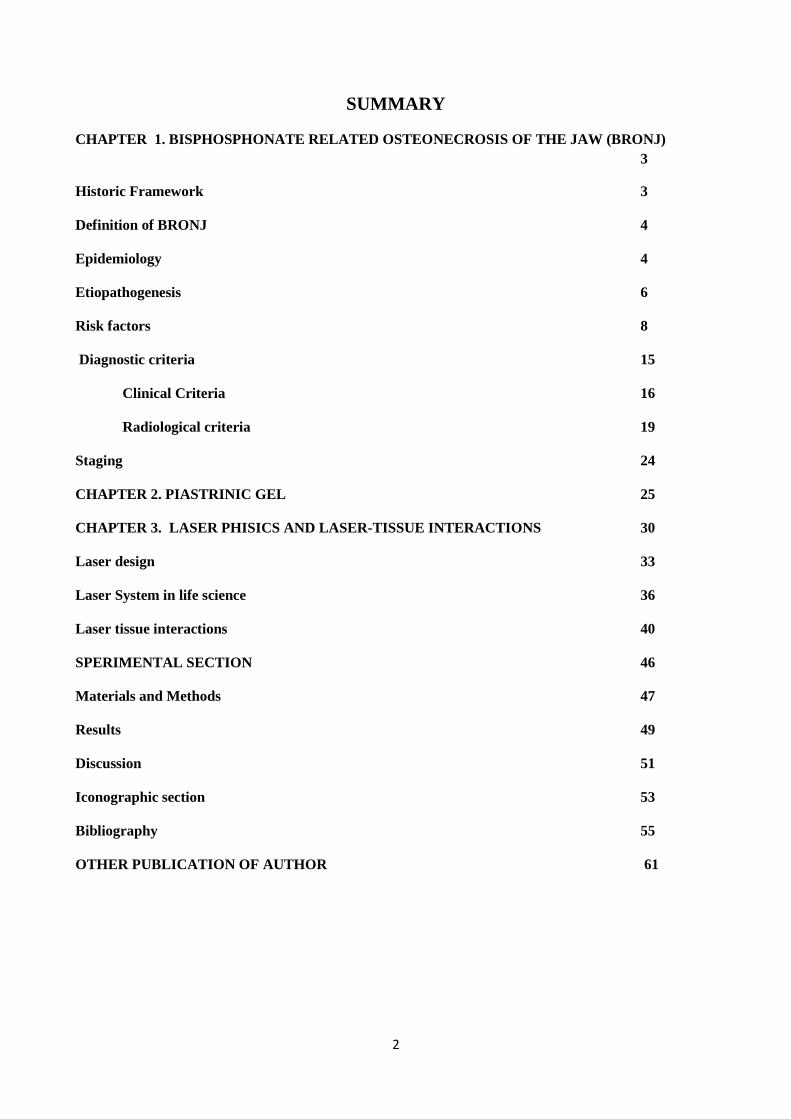
SUMMARY
CHAPTER 1. BISPHOSPHONATE RELATED OSTEONECROSIS OF THE JAW (BRONJ)
3
Historic Framework 3
Definition of BRONJ 4
Epidemiology 4
Etiopathogenesis 6
Risk factors 8
Diagnostic criteria 15
Clinical Criteria 16
Radiological criteria 19
Staging 24
CHAPTER 2. PIASTRINIC GEL 25
CHAPTER 3. LASER PHISICS AND LASER-TISSUE INTERACTIONS 30
Laser design 33
Laser System in life science 36
Laser tissue interactions 40
SPERIMENTAL SECTION 46
Materials and Methods 47
Results 49
Discussion 51
Iconographic section 53
Bibliography 55
OTHER PUBLICATION OF AUTHOR 61
3
HISTORIC FRAMEWORK.
Bisphosphonate Related Osteonecrosis of the Jaw (BRONJ) is one of the emerging diseases of this
century. It has clinical characteristics very similar to a clinical entity described as "Phosphorous Necrosis" or
"Phossy Jaw", and observed during the first half of the nineteenth century in workers of London matches
factories, who were in contact with the toxic fumes of yellow or white phosphorus. Lorinser was the first to
report in 1845 in Vienna, 22 cases of flogistic /infective events to the maxillary bones and oral tissues
among workers of Austrian factories (Hunter, 1935). In 1863 Simon’s publications he described in detail 61
cases of post-extractions jaw bone necrosis, characterized by the initial onset of an indolent ulcer and
subsequently by a persistent inflammation of soft tissues with a delayed healing. Simon was the first to name
as "sequestrum” the portions of porous and particularly light bone, very similar to the pumice stone and
separated from the surrounding tissue, observed in these patients (Simon, 1863). Some of the signs or
symptoms described by the authors were: persistent ulceration, gingival dehiscence, brownish-gray colored
bones exposures not recovering, gingival erythema and swelling, loss of the alveolar process, permanent
deformities and intense pain in the maxillo-facial district. The most affected anatomic region was the
mandible (60% of cases) and the onset was after about 5 years of exposure to risk factor (yellow/white
phosphorus). The most practiced therapeutic maneuver was the extraction of the "guilty" tooth, but it was
not a solution, on the contrary, it often aggravated the clinical condition of the patient, leading to exitus the
20% of patients affected mainly because of the infectious complications (Smith, 1865).
The diffusion of the BRONJ topic started when Marx described the first cases and the first series of cases
were published the following year by Marx and in 2006 by Ruggiero et al (Marx, 2003; Ruggiero, Fantasia
and Carlson, 2006).
Since then, in the world, more than 2400 cases of BRONJ have been documented (Filleul, Crompot
and Saussez, 2010) as case reports and retrospectives clinical series (Ruggiero, Gralow, Marx et al. , 2006).
Actually, the number of cases detected in the clinical practice seems to be much higher even than estimates
done on Italian territory (Ruggiero, 2009; Sottosanti, 2009; Vescovi, Campisi, Fusco and others. 2011).
In particular, the retrospective study of Ruggiero et al 2004 (Ruggiero and Mehrotra, 2004) was the
first to highlight the association between oral osteonecrosis and oral NBP in a sample of 63 patients, 7 of
which (about 10% of the sample) were in therapy with NBP for bone-related disorders with no cancer origin;
4
in addition, the authors not only revealed the risk factors for BRONJ but they also denounced the absence of
an adequate therapeutic protocol of management of patients , expressing for the first time the need to
formulate guidelines for the prevention and the management of these patients.
Hellstein and Donghue’s studies described the similarity between the BRONJ cases and the
phosphoric necrosis of maxillary bones in workers exposed to white phosphorus (Phossy jaw) at the end of
the nineteenth century and in the early XX century (Donoghue, 2005; Hellstein and Marek, 2005). This
historical fact leaded to the suspicion that the phenomenon of the osteonecrosis of the jaw was directly
related to the use of NBP (Ruggiero, Mehrotra, Rosenberg and Engroff, 2004; Marx, 2008): the molecule of
white /yellow phosphorus [P4O10], within our organism, reacts with the [H2O]and the [CO2] produced,
introduced and expelled continuously during the breathing, and with the common amino acids (e.g. lysine),
causing the formation of a product similar to a powerful amino-BP (e.g. pamidronate and alendronate).
DEFINITION OF BRONJ
The current definition of BRONJ, has been proposed by Bedogni et al. , they described osteonecrosis
associated with bisphosphonates (BRONJ) as an " adverse drug-related reaction, characterized by the
progressive destruction and necrosis of the jaw bone and/or the maxillary bone of subjects exposed to
treatment with amino-bisphosphonates, without a previous radiation treatment" (Bedogni, Fusco, Agrillo and
Campisi, 2012).
EPIDEMIOLOGY.
Some cases of BRONJ have been described in all categories of patients treated with NBP:
a. Hematological and cancer patients with skeletal injuries, treated monthly with intravenous injection
of NBP (i.e. pamidronate, zoledronic acid, ibandronate): this category encompass the largest number
of reported BRONJ cases. (Migliorati, Epstein, Abt and Berenson, 2011);
5
b. Cancer patients treated with zoledronic acid without skeletal injuries: currently they have been
documented only during experimental studies. (Mauri, Valachis, Polyzos, et al. , 2009; Coleman,
Burkinshaw, Winter et al. , 2010);
c. Cancer patients treated with oral bisphosphonates: sporadic cases has been reported.
d. Non-cancer patients treated with oral NBP (mainly for osteopenia/osteoporosis): today the number
of documented cases is much lower than the number of patients belonging to the a) group; they are
generally patients treated for a long time (usually > 2-3 years) and they possibly may have
concomitant local and/or systemic risk factors. (Mavrokokki, Cheng, Stein and Goss, 2007; Jeffcoat
and Watts, 2008; Fedele, Kumar, Davies et al. , 2009; Bedogni, Bettini, Totola, Saia and Nocini,
2010);
e. Non-cancer patients, with osteometabolic pathology, treated with intravenous NBP (i.e. ibandronate
3mg every 3 months or zoledronic acid 5mg every 12 months) but now there are no sufficient
follow-up data to determine the risk of BRONJ in these patients and with these therapeutic regimens,
even if, recently, sporadic cases has been reported. ( Otto, Sotlar, Ehrenfeld and Pautke, 2011).
Currently there are no accurate epidemiological data on BRONJ. The true incidence of the disease is still
unknown and available estimates of frequency in at-risk populations have a great variability; moreover,
definitive prospective data and the levels of evidence are missing in literature.
It is very difficult to determine the general incidence of BRONJ, also because of the many variables
contributing to its determinism (Ibrahim, Barbanti, Giorgio-Marrano et al. , 2008). As a matter of fact, it is
very difficult to compare studies because of their non-homogeneity of methods. (Koshla S, 2007).
According to the literature, the incidence in cancer patients range from 0.0 % to 27.5 % (Lyles KW, 2007;
Bianchi, Limonta, Frasunkiewicz, Biggioggero and Vai, 2008;). In non-cancer patients, the range of
incidence reported is between 0.0 - 4.3 % (Black, Schwartz, Ensrud et al. , 2006; Sedghizadeh, Stanley,
Caligiuri et al. , 2009).
There are a large number of data, even if not uniformed, regarding the frequency (as the percentage
of patients with BRONJ in a determined population of “ at risk” patients). These data are additionally
invalidate because in many cases only the numerator is well-defined ( reported BRONJ cases), contrary
to the denominator (total amount of patients treated with NBP). In addition, many studies of frequency do
6
not specify neither the type nor the duration of the treatments with NBP, the underlying disease, risk factors
and other important information; everything contributes to a lack of clarity about the BRONJ.
So, we can affirm that while epidemiological BRONJ data of patients with oncological pathologies
are even slowly emerging , is not the same for those relating to osteoporotic patients. There are few data
concerning the incidence of BRONJ (number of new cases/year in a given population) and there are no
estimates of prevalence (number of cases known and observed/year in a given population). Moreover, even if
the majority of the NBP is prescribed by internists, haematologists, urologists and gynecologists, the
majority of the literature concerning to the BRONJ is written on general dentistry magazines or maxillo-
facial surgery magazines and it is not a rarity when patients turn to a dentist or to a maxillo-facial surgeon;
besides, it is considerable that in gynecological and nephrologycal magazines it is possible to find only four
publications. Unfortunately, today, in these disciplines there is not much interest on BRONJ despite the
severity and the importance of the pathology.
Etiopathogenesis
Today we don’t know etiopatogenetics mechanisms through which NBP causes the BRONJ: Many
of these factors mentioned above and reported by international literature must be considered for a
multiphase-multifactorial interpretation.
Ischemia
First studies about osteonecrosis of maxillary bones described ischemia and the "avascular necrosis"
as a pathogenetic mechanism. But Hansen described a bleeding during the bone surgery, observing also the
presence of vessels in anatomical sections (Hansen, Kunkel, Weber and James Kirkpatrick, 2006): and then,
if the BRONJ was merely an avascular necrosis, this should not be localized only almost exclusively to the
maxillary bones, but, as in other conditions, also to other locations as the head of the femur, the hip and
knees.

7
The reduction of the bone turn-over.
The reduction of the bone turn-over, through the action of NBP, lead to the suppression of the bone
remodeling, preventing the repair of microfractures which occur continuously in the bone after the repeated
traumas, at the mastication level (Papapoulos, 2008). Today it has been assured that the reduction of the
bone remodelling and the failure of epithelization of post-extractives sites, also occurs in healthy patients
that would have a normal healing ( Abu-Id , Warnke, Gottschalk et al. , 2008). As a matter of fact, there are
conditions in which there is a chronic delay of bone remodelling, as in the hypoparathyroidism, even if
necrotic bone is not exposed. Osteopetrosis is an exception in which bone exposures have been described,
probably caused by the high density of the jawbone, and they prevent a correct formation of the vascular
network.
An interesting finding has been reported by Hansen, who highlighted how within BRONJ fractures,
compared with the necrotic fractures due to radiotherapy and in a control group, there were a number of
osteoclasts four times greater than in the case-control (Hansen, Kirkpatrick, Walter and Kunkel, 2006); this
finding and the bone scintigraphy findings showed how the bone turn-over was not deleted in BRONJ sites
and confirmed its regularity. (Chiandussi, Biasotto, Dore et al. , 2006): it has been supposed that this
paradoxical situation was due to the presence of a bacterial over infection stimulating the bone remodeling.
Direct toxicity of NBP on bone tissue.
NBP inhibits FPPSS (farnesyl-pyrophosphate synthetase), an enzyme of the mevalonate pathway
which leads to the cholesterol synthesis and to the production of carbon chains essentials for the proper
working of cells. In addition, it is important to consider the osteoclasts ability to internalise NBP during their
remodeling action, even if, in a high concentrations, like after an intravenous injection, effects can also
involve other cell types. NBP is mainly concentrated in maxillary bones and according to estimates the
monthly use of 4mg of zoledronic acid for 4 years, cause his average concentration in human skeleton of 70
nmol/ml, while in maxillary bones it can be even two or three times higher. It is important to consider also
that a concentration of zoledronic acid 1 nmol/ml is enough to cause cells toxicity (Coxon FP).
8
Infection
An infection is a dangerous condition because, together with the bacterial metabolism, causes the
bone resorption: as a matter of fact, many bacteria can stimulate bone resorption and inhibit his new
formation thanks to the presence, in the Gram-negative bacteria’s membrane, of lipopolysaccharide which, in
addition to have a direct toxic action, can stimulate prostaglandins encouraging resorption (Nair SP, 1996).
Bacteria can also act in other ways: for example, some proteins produced by Porphyromonas gingivalis
directly manage the production of RANKL and osteoprotegerin in periodontal ligament cells and of gingival
fibroblasts, stimulating consequently osteoclastogenesis (Belibasakis GN, 2007). Some Histological
evaluations showed the strong connection between the infection and the BRONJ (Marx, Cillo and Ulloa,
2007). A great variety of species are involved in the infective process but surely Actinomyces is the most
diffused.(Hansen, Kunkel, Weber and James Kirkpatrick, 2006;). Unfortunately, the isolation and
identification of this bacterium through conventional research methods is still a problem. (Hall, 2008).
RISK FACTORS
To make a distinction between categories of patients at high risk and low risk, and to define a
prevention protocols it is necessary to identify the risk factors of a disease with multifactorial etiopathology
as the BRONJ is. Unfortunately, there are no definitive findings about risk factors. (Assael, 2009). In fact,
the diversity and the low number of cohorts published about BRONJ (Bamias, Kastritis, Bamia et al. , 2005;
Vahtsevanos, Kyrgidis, Verrou et al. , 2009), the low number of case-control studies (Kyrgidis, Vahtsevanos,
Koloutsos et al. , 2008; Wessel, Dodson and Zavras, 2008), and the inadequate database (Khamaisi, Regev,
Yarom et al. , 2006; Jung, Hoffmann, Glaeske and Felsenberg, 2010) make impossible to draw conclusions.
In the majority of cases reported in the literature (60-85 %) BRONJ developed after a dental surgery,
generally an extraction or a dental implants insertion.
Marx and colleagues attributes the cases known as "spontaneous" to some trauma in mobile dentures
and in 39% of cases not associated to a dental surgery, bone exostoses were easily exposed to trauma. (Marx,
Sawatari, Fortin and Broumand, 2005).
9
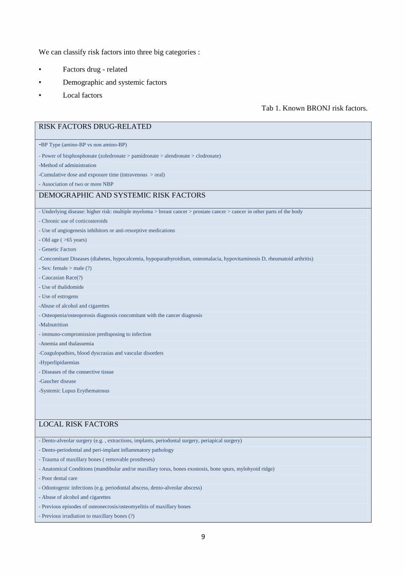
We can classify risk factors into three big categories :
• Factors drug - related
• Demographic and systemic factors
• Local factors
Tab 1. Known BRONJ risk factors.
RISK FACTORS DRUG-RELATED
-BP Type (amino-BP vs non amino-BP)
- Power of bisphosphonate (zoledronate > pamidronate > alendronate > clodronate)
-Method of administration
-Cumulative dose and exposure time (intravenous > oral)
- Association of two or more NBP
DEMOGRAPHIC AND SYSTEMIC RISK FACTORS
- Underlying disease: higher risk: multiple myeloma > breast cancer > prostate cancer > cancer in other parts of the body
- Chronic use of corticosteroids
- Use of angiogenesis inhibitors or anti-resorptive medications
- Old age ( >65 years)
- Genetic Factors
-Concomitant Diseases (diabetes, hypocalcemia, hypoparathyroidism, osteomalacia, hypovitaminosis D, rheumatoid arthritis)
- Sex: female > male (?)
- Caucasian Race(?)
- Use of thalidomide
- Use of estrogens
-Abuse of alcohol and cigarettes
- Osteopenia/osteoporosis diagnosis concomitant with the cancer diagnosis
-Malnutrition
- immuno-compromission predisposing to infection
-Anemia and thalassemia
-Coagulopathies, blood dyscrasias and vascular disorders
-Hyperlipidaemias
- Diseases of the connective tissue
-Gaucher disease
-Systemic Lupus Erythematosus
LOCAL RISK FACTORS
- Dento-alveolar surgery (e.g. , extractions, implants, periodontal surgery, periapical surgery)
- Dento-periodontal and peri-implant inflammatory pathology
- Trauma of maxillary bones ( removable prostheses)
- Anatomical Conditions (mandibular and/or maxillary torus, bones exostosis, bone spurs, mylohyoid ridge)
- Poor dental care
- Odontogenic infections (e.g. periodontal abscess, dento-alveolar abscess)
- Abuse of alcohol and cigarettes
- Previous episodes of osteonecrosis/osteomyelitis of maxillary bones
- Previous irradiation to maxillary bones (?)
10
Analysis of the possible drug-related risk factors:
1. BP type (amino-BP vs non amino-BP). Amino-bisphosphonates (NBP) have a great affinity for
bone and a power range from 10 to 1000 times greater than that of bisphosphonates non
containing amino groups. The risedronate and zoledronate are considered some of the most efficient
NBP (Nancollas, 2006), thanks to the presence, in the radical R2, of the heterocyclic ring of the
nitrogen atom; the ibandronate, which contains nitrogen fractions at the radical R2 level and it is
more powerful than alendronate and pamidronate.
2. Pharmacological efficiency. According to recent studies (Dimopoulos, Kastritis, Anagnostopoulos et
al. , 2006;) zoledronic acid combined with pamidronate can cause a risk of BRONJ 10 times higher
than the combined use of zoledronic acid and thalidomide (drug with antineoangiogenic properties)
or than pamidronate and ibandronate individually used, because of their molecular structure (amino-
BP) and because they are exclusively intravenously injected. However, in hematological and cancer
patients, zoledronic acid (the most used drug since 2002, now used in the majority of BRONJ cases)
seems to involve a risk of BRONJ statistically greater than pamidronate (Bamias, Kastritis, Bamia et
al. , 2005; Vahtsevanos, Kyrgidis, Verrou et al. , 2009; Fusco, Loidoris, Colella, Vescovi and
Campisi, 2010), even without randomized studies. Concerning non-cancer patients, currently there
are no findings to make a comparison between alendronate and risedronate (the two most largely
used drugs orally administered).
3. Method of administration (intravenous vs oral ). The risk seems to be much greater for NBP
intravenously injected: because it is mainly used in cancer patients, in total doses and for a very long
time (Woo, Hellstein and Kalmar, 2006; Migliorati, Woo, Hewson et al. , 2010).
4. Cumulative Dose and exposure time. NBP remains in bones for a long time, which is undoubtedly
related to the drug exposure time (necessary parameter to establish the cumulative dose of drug). It
is important to emphasize the difference between methods of administration (oral and intravenous)
because they affect extensively both the cumulative dose and the exposure time. It is evident how the
bioavailability of the drug depends on the method of administration, as a matter of fact, the
absorption of amino-BP at a gastrointestinal level, orally assumed and so extremely variable in
11
relation to meals, is reduced compared to the intravenously injection (bioavailability 1% NBP orally
assumed vs 50% NBP intravenously injected). (Marx, 2005; Dimopoulos, Kastritis,
Anagnostopoulos et al. , 2006;). Both for zoledronate and for pamidronate, findings indicate a
growing risk depending on the increase of the total dose of NBP intravenously injected to
hematological and cancer patients. (Bamias, Kastritis, Bamia et al. , 2005; Vahtsevanos, Kyrgidis,
Verrou et al. , 2009). Moreover, the majority of the BRONJ cases concerned patients under
treatment for a long time ( generally more than 2-3 years): the average time and the minimum time
for the BRONJ appearance were respectively 1.8 years / 10 months for zoledronate, and 2.8 years /
1.5 years for pamidronate(Palaska, Cartsos and Zavras, 2009). Another significant aspect, revealed
by the authors, is how the ONJ could arise both during the first 6 months of treatment with NBP, or
even over 60 months of therapy with these drugs. Finally, there is an hypothesis on a linear
relationship between the incidence of BRONJ and the duration of the therapy.
Among demographic and systemic risk factors considered as potentially causes of BRONJ there are :
1. Underlying disease. Patients with neoplastic disease are usually considered at higher risk, even if it
may depends on different types of treatment with NBP (Woo, Hellstein and Kalmar, 2006).
Among the hematological and cancer patients, those with multiple myeloma have a BRONJ
risk higher than those affected by metastatic breast or prostate cancer (Jung, Hoffmann, Glaeske and
Felsenberg, 2010; Migliorati, Woo, Hewson et al. , 2010); however, some authors disagree and
explain higher frequencies with an higher average exposure to NBP (Bamias, Kastritis, Bamia et al. ,
2005).
Wilkinson et al (Wilkinson, 2007) observed that patients under treatment with NBP
intravenously injected have an increased risk of osteomyelitis-BRONJ compared with patients non
treated with BP (5.48 % vs 0.3 %, during 6 years).
2. Steroid therapy. Corticosteroids inhibit the bone remodeling through the induction of osteoclasts
apoptosis and the inhibition of osteoclastogenesis (Weinstein, Jilka, Parfitt and Manolagas, 1998).
This, together with the action of NBP, determines an excessive reduction of the bone turn over with
a progressive accumulation of non-viable bone tissue and an inability to repair injuries caused by the

12
continuous exposure of oral cavity to microtrauma. In addition, corticosteroids are considered the
second most common cause of avascular osteonecrosis and this could explain a possible synergistic
reaction with NBP, even if, classically, bone necrosis induced by corticosteroids affects long bones
(necrosis of the femoral head) while, those induced by NBP affects short bones (i.e. jaws) (Zizic,
Marcoux, Hungerford, Dansereau and Stevens, 1985;). Moreover, with a reduction of the
inflammation, corticosteroids delay the healing of soft and hard tissues and this could explain the
bone exposure and the delayed alveolar healing.
Finally, prednisone increases NBP toxicity, facilitates and accelerates the bone exposure and make
the condition worse after it is developed.
3. Angiogenesis inhibition treatments and antiresorptive medications. In the most recent scientific
literature, an increase of BRONJ has been observed in patients treated with NBP and biological
antineoplastic drugs ( "target therapy” ), both associated and individually (Brunello, Saia, Bedogni,
Scaglione and Basso, 2009; Christodoulou, 2009). The use of new generation biological drugs, with
an inhibitory function on angiogenesis (e.g. , bevacizumab, sunitinib , sorafenib) associated with
NBP in patients with bone metastases from solid tumors seems to increase the incidence of BRONJ
(Christodoulou, Pervena, Klouvas et al. , 2009; Migliorati, Woo, Hewson et al. , 2010;), but there are
no sufficient findings on case reports, with the partial exception of bevacizumab (Guarneri, Miles,
Robert et al. , 2010).
In literature, other cases of osteonecrosis, although their rarity, (Kyrgidis and Toulis, 2010; Pichardo,
Kuypers, and van Merkesteyn, 2012) has been attributed to the use of denosumab, a humanized
monoclonal antibody indicated in the treatment of postmenopausal osteoporosis with an increased
risk of fractures, bone loss in rheumatoid arthritis (RA) and increased risk of fractures in hormonal
ablation therapy in prostate cancer patients; it complexes the RANK-L (RANK Ligand), primary
sign of the osteoclasts action for the bone resorption (Kyrgidis and Toulis, 2010;).
4. Age. Even if in some cases there are data showing a correlation between age and BRONJ risk
(Sarasquete, Garcia-Sanz , Marin et al. , 2008), the high number of cases in advanced age seems to
be probably related to the normal age distribution of patients with metastatic cancer. In fact, the

13
most affected patients are usually older than 40 years and NBP is prescribed for diseases mainly
affecting adults.
5. Genetic factors. Even if some variants of the gene CYP2C8 were predictives of BRONJ in patients
with myeloma (Sarasquete, Garcia-Sanz , Marin et al. , 2008), these findings were not confirmed
either in patients with prostate cancer (English, Baum, Adelberg et al. , 2010) nor in patients with
myeloma (Katz, Gong, Salmasinia et al. , 2011).
6. Concomitant Diseases: Some authors describe diabetes as a concomitant pathology, (Urade,
2009) but other authors disagree (Lazarovici, Mesilaty-Gross , Vered et al. , 2010; Katz,
Gong, Salmasinia et al. , 2011); hypocalcemia and hyperparathyroidism are still in doubt even after
the positive findings expressed in a single study (Ardine, Generali, Donadio et al., 2006);
osteomalacia and hypovitaminosis D are considered as predisposing factors in BRONJ development
(Bedogni, Saia, Bettini et al. , 2012;) in patients already treated with NBP, as already demonstrated
in an animal model (Hokugo, Christensen, Chung et al. , 2010).
Other factors (i.e.. , immunodepression, hypertension, peripheral vascular diseases, dyslipidaemias
hyperviscosity syndrome, malnutrition, Gaucher disease, anemia and talassemie, abuse of alcohol and
cigarettes, hypothyroidism, obesity, LES and other diseases of the connective tissue) has been theorized on
the basis of the etiopathogenetic hypothesis of an ischemic necrosis of BRONJ and / or because of his
similarity to femoral necrosis (Woo, Hellstein and Kalmar, 2006;), but today there are no findings
confirming or denying this.
Between local risk factors there are:
1. Dento-alveolar surgery. The relationship between surgical procedures in oral cavity (e.g. avulsion of
dental arch tooth or impacted tooth, endodontic surgery, periodontal and peri-implant surgery) and
the presence of BRONJ in patients under therapy with NBP is certainly the most studied, but there
are still no conclusive scientific evidences supporting it. The event "surgical dento-alveolar
procedure" has been reported as the most frequent factor temporally associated with the BRONJ,

14
with a risk of onset up to 44 times greater (Hoff, Toth, Altundag et al. , 2008) than patients under
no surgical procedure.
The frequency range of the surgical dento-alveolar procedure as a local risk factor of BRONJ varies
from 50% to 100 % (Marx, Sawatari, Fortin and Broumand, 2005; Marx, Cillo and Ulloa, 2007;
Yarom, Elad, Madrid and Migliorati, 2010).
2. Osteointegrated implantology. Today, the placement of dental implants is considered a surgical
procedure potentially at risk (Shabestari, Shayesteh, Khojasteh et al. , 2010; Yip, Borrell, Cho,
Francisco and Tarnow, 2012), just like the surgical dento-alveolar procedures, especially in patients
under therapy with NBP intravenously injected for cancer. The real risk of
BRONJ after implantology in cancer patients under therapy is unknown, but it is important to
consider that many cases of osteonecrosis published corrisponded with implant sites rehabilitated
before starting the therapy with NBP (Ruggiero, Mehrotra, Rosenberg and Engroff, 2004; Bedogni,
Blandamura, Lokmic et al. , 2008; Lazarovici, Yahalom, Taicher et al. , 2010). Over the risk of the
surgical procedure, it is necessary to consider that in the years after the positioning of the osteo-
integrated implant, it will be exposed to an increasing risk of BRONJ in case of peri-implantitis,
because of the presence of an inflammatory disease and the gradual increase of the drug
concentration in the peri-implant bone tissue. In fact, the weakening of bone immune defenses linked
to chronic therapy with NBP and the absence of a barrier effect to the bone-implant interface,
promote the risk of peri implantitis and the transfer of infection transported by implant structure to
the surrounding bone.
3. Dento-periodontal and peri-implant inflammatory disease. Pathological conditions such as dento-
periodontal and/or peri-implant inflammatory disease (i.e. periodontal chronic condition, odontogen
infections, endo-periodontal fracture, peri implantitis), caused by a poor oral care and a poor plaque
control in a subject in therapy with NBP, decidedly increase the risk of BRONJ development during
the treatment and represent a guaranteed factor of aggravation of the clinical case when BRONJ has
already been diagnosed (Ficarra, Beninati, Rubino et al. , 2005; Dodson, 2009).
4. Incongruous removable prostheses. Removable prosthetic devices are considered as risk factors for
BRONJ development because, if they are not well adapted to the mucosa, they can damage the
15
mechanical barrier facilitating the entry of microbes in underlying tissues. A significant correlation
between the use of removable prostheses and the development of BRONJ has been documented in a
population of subjects under treatment with high dose of intravenous NBP, suffering from metastatic
neoplasia (Kyrgidis, Vahtsevanos, Koloutsos et al. , 2008; Vahtsevanos, Kyrgidis, Verrou et al. ,
2009), but this finding has not been confirmed in other studies.
5. Anatomical conditions. The presence of anatomical irregularities (e.g. mandibular and/or palatal
torus, exostosis, mylohyoideus ridge particularly pronounced ) may be a risk factor of BRONJ
development, especially in patients with removable total prostheses positioned in the upper jaw and
with partial or total prostheses in the mandible (Ruggiero, Dodson, Assael et al. , American
Association of Oral and Maxillofacial Surgeons position paper on bisphosphonate-related
osteonecrosis of the jaws--2009 update, 2009).
Despite the established pathogenetic relation "surgical-BRONJ trauma ", several cases of spontaneous
onset have been reported in literature (cryptogenic BRONJ) (Merigo, Manfredi, Meleti, Corradi and
Vescovi, 2005;). However, in so-called " spontaneous forms " has been highlighted the presence of other
factors, such as periodontopathy in 84% of patients, dental caries in 28.6 %, dental abscesses in 13.4 %,
incompleted endodontic treatments in 10.9 %, exostosis (palatine and mandibular torus) in 9.2 % of the
cases. In 39.3 % of spontaneous or "idiopathic” BRONJ there were no evident dental pathologies. It is not
clear, in these forms not related to a surgical trauma, the role of other local unknown or general factors, in
the encouraging or in the progression of bone necrosis.
DIAGNOSTIC CRITERIA
The process to carry out diagnosis on BRONJ caused by NBP is not always simple and direct, both
for the clinical variability of BRONJ cases (especially if "early stage" ), and for the intrinsic difficulties of
the next and remote anamnestic data collection. As a matter of fact, the clinician has to connect information
received from the patient's medical history (next and remote pathological history) to those highlighted during
the objective examination of the oral cavity and the radiological evaluation. While the advanced fractures are
16
more easily classifiable, the clinician must pay attention especially to early fractures which, very often, are
asymptomatic for weeks, months and years, and they are not always detectable through instrumental
examinations. In fact, in a considerable number of patients under treatment with NBP and with a developed
BRONJ, there are, at least during the initial phase, signs and symptoms different from exposure bone.
(Yarom, Fedele, Lazarovici and Elad, 2010). Even if, still today, the exposure of necrotic bone is the most
assured indicator of BRONJ (Ruggiero, Dodson, Assael et al., American Association of Oral and
Maxillofacial Surgeons position paper on bisphosphonate-related osteonecrosis of the jaws--2009 update,
2009), there are other aspecific signs and symptoms which can and should lead us to a premature suspicion
of BRONJ, even with a verified dento-periodontal cause ( Mawardi, Treister, Richardson et al. , 2009).
The usefulness of biopsy is called into question, when it is done in correspondence of an exposed
necrotic bone in oral cavity, since the histological case does not seems to add anything to clinical case(Phal,
Myall, Assael and Weissman, 2007). According to the current guidelines, ultimately, the investigation
through biopsy must be used only in order to remove the suspicion of metastasis in mandibular / maxillary
site (Campisi, Di Fede, Musciotto et al. , 2007).
Clinical Criteria
One of the peculiarities of BRONJ is its almost exclusive location to the maxillary bones. The site-
specificity of the osteonecrotic fractures caused by NBP can be explained through a careful valuation of the
anatomo-physiological characteristics of the maxillary/mandibular district. The possible causes of this
phenomenon are yet unknown, but some reasons have been suggested:
Bone turnover of the jaw physiologically higher than the remaining skeleton in relation to the
presence of teeth in dental arch and to the forces created during mastication (Marx, 2003). Dixon et
al (Dixon, 1997) documented the level of bone remodeling in different parties of the body and
verified that the remodeling process in alveolar crest is 10 times faster than in tibia, 5 times than in
the mandible (at the mandibular canal level) and 3.5 times than the lower edge of the mandible . So
the uptake and the cumulative dose of NBP at the alveolar process level is relevant. This study
shows also how the alveolar process is physiologically subjected to a higher bone remodeling -
resorption (through osteoclast) than in any skeletal segment of an adult person. In subjects in therapy
17
with NBP, if the remodeling need continues, or if there is a trauma such as a tooth extraction, the
alveolar process cannot resist along with the formation of a new bone and could have the risk
of necrosis. Consequently, the overlying mucosa, deprived of its nutritional supply, ulcerates until
the bone exposure is manifested;
Terminal vascularization of the mandible (Bagan, Murillo, Jimenez et al. , 2005) which would favor
the accumulation of NBP at the maxillary district level, but may affect BRONJ pathogenesis because
of the absence/reduction of collateral circles (Urbaniak, 1997);
Presence of a thin periosteal mucus coating surrounding the bone to protect the underlying bone
tissue, easily exposed to trauma which contribute to the high bone turn-over and stimulate bone neo-
apposition phenomena through the production, by osteoblasts (OB) and osteoclast (OC), of cytokines
which will lead to the maturation, activation and recruitment of OC on bone surface (Marx, 2003);
Peculiar microflora/biofilm of the oral cavity (Eckert, Maurer, Meyer et al. , 2007): the exposure of
maxillary bone segments to the external environment may take place through the gingival sulcus,
facilitating the bone infection and the progression to osteomyelitis. Actinomyces, the bacterium
more frequently isolated in cases of BRONJ, showed (in vitro) its ability to induce the cells of the
periodontal ligament to synthesize cytokines and metalloproteins implicated in the damage of
periodontal ligament and in the destruction of the alveolar bone.
Characteristic dento-alveolar interface which, in case of a dento-periodontal disease(i.e. , periapical
injuries and abscesses, periodontopathy) or gold oral surgery, predisposes to the exposure of the
underlying bone tissue ( Baltensperger M, 2009).
In addition to the exposure bone, recognized as the major clinical sign and able to meet the diagnosis of
BRONJ in the presence of positive pharmacological anamnesis caused by NBP, and in light of the current
definition, it has been proposed also the use, for diagnostic purposes, of clinical minor signs and symptoms,
especially in cases where there is no exposure during BRONJ diagnosis (Fedele, Porter, D’Aiuto et al. ,
2010). All these signs are widely approved by the main international commissions of study about
BRONJ. Here is a list, in alphabetical order, of a series of clinical minor signs and symptoms which,
individually or associated, must create the suspicion of disease or its differential diagnosis.
18
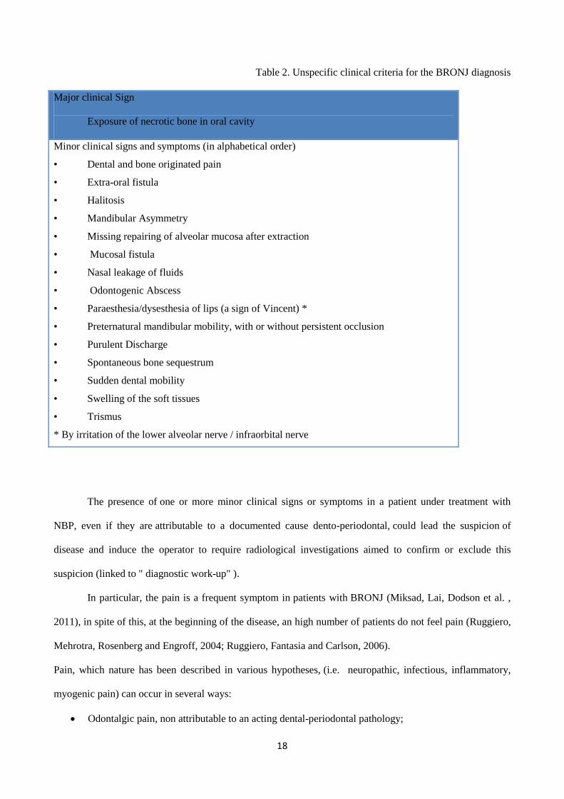
Table 2. Unspecific clinical criteria for the BRONJ diagnosis
Major clinical Sign
Exposure of necrotic bone in oral cavity
Minor clinical signs and symptoms (in alphabetical order)
• Dental and bone originated pain
• Extra-oral fistula
• Halitosis
• Mandibular Asymmetry
• Missing repairing of alveolar mucosa after extraction
• Mucosal fistula
• Nasal leakage of fluids
• Odontogenic Abscess
• Paraesthesia/dysesthesia of lips (a sign of Vincent) *
• Preternatural mandibular mobility, with or without persistent occlusion
• Purulent Discharge
• Spontaneous bone sequestrum
• Sudden dental mobility
• Swelling of the soft tissues
• Trismus
* By irritation of the lower alveolar nerve / infraorbital nerve
The presence of one or more minor clinical signs or symptoms in a patient under treatment with
NBP, even if they are attributable to a documented cause dento-periodontal, could lead the suspicion of
disease and induce the operator to require radiological investigations aimed to confirm or exclude this
suspicion (linked to " diagnostic work-up" ).
In particular, the pain is a frequent symptom in patients with BRONJ (Miksad, Lai, Dodson et al. ,
2011), in spite of this, at the beginning of the disease, an high number of patients do not feel pain (Ruggiero,
Mehrotra, Rosenberg and Engroff, 2004; Ruggiero, Fantasia and Carlson, 2006).
Pain, which nature has been described in various hypotheses, (i.e. neuropathic, infectious, inflammatory,
myogenic pain) can occur in several ways:
Odontalgic pain, non attributable to an acting dental-periodontal pathology;
19
Pressing "bone" pain, easily distinguishable because of his location (more frequent in mandibular
body);
Pain irradiated to the masticatory and cervical muscles, similar to the pain of temporo-mandibular
articulation pathology (myogenic pain);
Sinus pain;
Hyperesthesia or trigeminal pain (which selectively affects the branch placed on the BRONJ
manifestation site).
Pain is considered as a factor aggravating the prognosis of disease, because it is often poorly controlled
and in some cases it is refractory to medical therapy. It is often at high intensity, it occurs also during the
night, and can limit significantly life and relationships of the patient ; it often occurs in patients with
disseminated neoplasms, who already use opioids to manage metastatic skeletal pain, however without a
direct benefit on painful symptoms associated with BRONJ. The treatment of the pain symptom is a critical
point of the BRONJ therapy. So, it is important the evaluation of the pain, for example, through the VAS
(visual analogic scale) in order to check the progress of the disease and the effectiveness of treatments,
considering not exclusively the operator opinion but also the patient point of view (Bedogni, Saia and al,
2011; Miksad, Lai, Dodson et al. , 2011).
Clinically, osteonecrosis fractures may also occur as multifocal, simultaneous and bilateral fractures
(Bagan, Jiménez, Murillo et al. , 2006). The mandible is involved a little more often than the maxilla
(Bagan, Scully, Sabater and Jiménez, 2009); in 10% of cases fractures affect both jaws. In the mandible, in
particular, the BRONJ case occurs with a great frequency in rear areas located near the mylohyoideous ridge
(Treister, Richardson, Schlossman, Miller and Woo, 2008). It is important to give particular attention to the
hyperostosis areas : maxillary/mandibular torus and tuber maxillae (Marx, 2005).
Radiological criteria
The BRONJ radiological diagnosis, carried out with the main methods of inquiry known, is very
difficult because of the absence of peculiar signs of disease. Instrumental investigations can help the
clinician during the three BRONJ prevention levels (primary, secondary and tertiary):
20
In the research of local risk factors, both in patients still not under therapy with NBP, both in patients
already under therapy with NBP (in action or previous) but without developed osteonecrosis
fractures (primary prevention);
During phases of diagnostic evaluation of suspected BRONJ cases caused by NBP, and/or of sub-
clinical BRONJ cases caused by NBP (secondary prevention);
During the evaluation of the osteonecrosis fracture expansion and of the possible involvement of
adjacent tissues (tertiary prevention).
Since an optimal radiological examination for the BRONJ diagnosis does not exist yet, American
Society of Bone and Mineral Research affirm that although each radiological technique has limitations, the
combination of different radiological techniques could provide useful elements for the diagnosis (Khosla, S. ,
Burr, D. , Cauley, J. et al. , 2007). In the light of this, the radiologist has an important role in the
identification of fractures at BRONJ risk and in the future he could assume an increasingly important role.
In literature, a series of radiological signs of possible onset of the disease has been identified.
However it should be highlighted, that it has not been still possible to distinguish definitely the radiological
signs of BRONJ onset. The only sign reported in the literature as potentially specific of the BRONJ is
the thickening of the Schneider membrane of the maxillary sinus, visible to Computed Tomography (CT),
linked to an excessive osteogenesis (Bianchi, Scoletta, Cassione, Migliaretti and Mozzati, 2007). However,
this sign is simply an expression of a bone periosteal neo-apposition perpetuated in chronic flogistic process
of maxillary sinuses (Cho, Kim, Lee and Lee, 2007). Anyway, this is an advanced sign of
BRONJ, considering that the origin of the disease is almost always in dento-alveolar site and, secondarily,
affects the maxillary sinus.
In general, the Radiological surveys most used in the diagnosis of BRONJ are: orthopantogram
(OPT) (Mawardi, Treister, Richardson et al. , 2009), intraoral radiographsand the computed tomography
(CT) (Chiandussi, Biasotto, Dore et al. , 2006; Bianchi, Scoletta, Cassione, Migliaretti and Mozzati, 2007).
Orthopantogram (OPT) can be considered the first level instrumental survey in the diagnostic process of
BRONJ. With a simple OPT it is possible to highlight osteonecrosis sub-clinical signs (e.g. , enlargement of
the periodontal space, sclerosis of the lamina dura) or an alteration/inhomogeneity of the bone trabeculation

21
which probably could coincide with an initial involvement of the trabecular pattern in an Osteomyelitis
/osteonecrotic way.
Radiographic Signs found in BRONJ are:
Osteosclerosis areas associated with osteolysis areas;
Loss or thinning of the lamina dura;
Thinning of the bone cortex;
Presence of bone sequestra;
Sites of previous extractions not healed, detectable as periodontal sockets;
Pathological fractures.
In addition, the OPT can provide general information about the mandible and maxilla status, especially
useful during the identification of any bone sequestrum and osteolysis areas combined with areas of
osteosclerosis. However, the OPT cannot differentiate the nature of the osteolytic fractures from any bone
metastases, especially if they are isolated. Opt has a low contrast resolution and requires a significant mineral
loss (higher than 30-50 %) to identify an osteolytic fracture. We also know that OPT is a zonography, a
tomography with thick layer which could show a possible superposition on images of structures located on
different levels but in the same x-rayed layer. Images can be corrupted (e.g. artifacts of the patient
positioning, shadows of dragging) and the view of central areas is often poor. Consequently , the BRONJ
fractures may be frequently misunderstood by OPT and it is not possible to distinguish damaged tissue
and healthy bone tissue. In spite of this, today the use of the OPT is still largely approved as first approach
instrument, in case of suspicion of BRONJ.
With OPT , the persistence of the postextraction alveolus and the thickening of the lamina dura after
tooth extraction are considered possible early signs of BRONJ; but some studies indicate the persistence of
the alveolus postextraction, even a year after extraction in patients who showed no clinical signs of BRONJ
(Saia, Blandamura, Bettini et al. , 2010). According to the authors the missed restoration of alveolus is
linked to the local activation of NBP which inhibits the bone remodeling in the extraction site, maintaining
the structure for many years even if there are no clinical signs of disease. Therefore, the persistence at the
OPT of one or more post-extractives alveolus is not always considered as sign of disease and is not
necessarily premature.

22
When the comprehension of RX-OPT is difficult (in cases of suspected BRONJ caused by NBP), or
when it is necessary to know exactly the expansion of the osteonecrosis fracture and the possible
involvement of adjacent tissues (for example,in order to identify resection margins for a possible surgical
treatment), it is necessary to rely on level II imaging techniques.
With Computed Tomography (CT) it is possible to distinguish, with a good
approximation, between healthy and damaged bone tissue. CT, in particular in the spiral CT version, has a
better contrast resolution than the conventional radiography and supplies detailed information about number
and nature of any osteosclerotic and osteolytic fractures (Chiandussi, Biasotto, Dore et al. , 2006; Bianchi,
Scoletta, Cassione, Migliaretti and Mozzati, 2007); moreover, in many cases, it allows a precise examination
of both cortical bone and trabecular bone, providing useful information about the extension of the process. In
the future, the real limit of the CT could be revealed in the study of early bone alterations of BRONJ, where
its effectiveness has not yet been well documented.
The total-body bone Scintigraphy with CT99
is the most used functional test for the diagnosis of
skeletal localizations of solid tumors, and it is widely used to their monitoring. It can be useful, in the
evaluation of the tissue functionality, because in initial phases there is the possibility of an
an hypervascularity; while in terminal phases it is clearly revealable. Since the BROJ is much more frequent
in patients with metastatic skeletal disease and multiple myeloma, and because of the presence of
information about previous, or done nearby the revelation of osteonecrosis symptoms total-body,
scintigraphic examinations usually owned by patients, information deriving from scintigraphy may be a valid
support in the formulation of the suspicion of BRONJ (Bertoldo, Santini and Lo Cascio, 2007).
The total-body bone scintigraphy represents an useful diagnostic support, when already performed
for other reasons, but commonly this examination is not required for specific diagnostic purposes. In fact,
when a BRONJ is suspected in an at risk patient under therapy with NBP who has not yet made a total-body
scintigraphy, the II level test required will be certainly the CT examination.
23
The Magnetic Resonance Imaging (MRI) is indicated to study the extension of the osteonecrotic process
(Bedogni, Blandamura, Lokmic et al. , 2008;) and provides information on soft tissues and on bone
medullary compartment; it is also possible to observe the ischemia areas through a contrast medium. There
are still doubts about the investigation sensitivity, and in particular the edema is not considered within the
pathogenetic process of ONJ, many authors consider MRI as a choice survey, like CT, used to study the
preoperative refractory cases, since it allows the identification of the involvement level of bones and adjacent
soft tissues (Abscesses, phlegmons and mucocutaneous fistulas) (Bedogni, Blandamura, Lokmic et al. ,
2008; Garcia-Ferrer , Bagan, Martinez-Sanjuan et al. , 2008;).
Cone Beam Computed Tomography (cone beam CT): is a relatively new technique which requires a
radiation exposure of patient lower than CT, but higher than OPT, it is largely used because it supplies a
large number of information (Ludlow, Davies-Ludlow and Brooks, 2003) The survey has an high spatial
resolution and supplies detailed information about the bone density (e.g. thinness, integrity of cortical bone
and bone marrow).
24
STAGING
Stage 1 Focal BRONJ
Clinical signs and symptoms: bone exposure; sudden dental mobility; nonhealing postextraction socket;
mucosal fistula; swelling; abscess formation; trismus; gross mandibular deformity and⁄or
hypoesthesia⁄paraesthesia of the lips
CT findings: increased bone density limited to the alveolar bone region (trabecular thickening and⁄or focal
osteosclerosis ), with or without the following signs: markedly thickened and sclerotic lamina dura;
persisting alveolar socket; and ⁄ or cortical disruption
1a. Asymptomatic
1b. Symptomatic (pain and purulent discharge)
Stage 2 Diffuse BRONJ
Clinical signs and symptoms: same as Stage 1
CT findings: increased bone density extended to the basal bone (diffuse osteosclerosis), with or without the
following signs: prominence of the inferior alveolar nerve canal; periosteal reaction; sinusitis; sequestra
formation;and⁄or oro-antral fistula
2a. Asymptomatic
2b. Symptomatic (pain and purulent discharge)
Stage 3 Complicated BRONJ
Same as Stage 2, with one or more of the following clinical signs and symptoms: extra-oral fistula; displaced
mandibular stumps; nasal leakage of fluids
CT findings: osteosclerosis of adjacent bones (zygoma, hard palate); pathologic mandibular fracture; and⁄or
osteolysis extending to the sinus floor
25
PIASTRINIC GEL
After surgery, blood clots start the healing and regeneration of hard and soft tissues. Using platelet-
rich plasma, or PRP, is a way to accelerate and enhance the body’s natural wound-healing mechanisms. A
natural blood clot contains mainly red blood cells, approximately 5 percent platelets and less than 1 percent
white blood cells (University of Miami School of Medicine, 2002). Platelets are primarily involved in wound
healing through clot formation and the release of growth factors which start and support wound healing.
Using PRP involves a preoperative taking a patient blood sample, concentrating autologous platelets and
applying the resultant gel to the surgical site. This technique produces a blood clot that has nearly a reverse
ratio of red blood cells and platelets compared with a natural clot. . It has been showed how Surgical sites
enhanced with PRP healed at rates two to three times faster than normal surgical sites (Marx RE, Carlson ER
et al., 1998). Thus, PRP can be a great adjunct to many periodontal and oral surgical procedures such as
bone grafts, implants and maxillofacial reconstructions.
GROWTH FACTORS
Wound healing is a complex and growing science. Many cell types, growth factors and other proteins
interact with each other to bring about timely and efficient repair of wounds. Researchers continue studyng
various growth factors to determine the actual role and mechanism of each growth factor in healing process.
On vessel injury and exposure of subendothelial tissue to blood (either due to accident or surgical
manipulation), platelets begin to stick to exposed collagen proteins. Once platelets stick to collagen, they
release granules containing adenosine diphosphate, serotonin and thromboxane, all of which contribute to the
hemostatic mechanism and the clotting cascade. Additional platelets are drawn to the area and contribute to
the formation of a platelet plug. The resultant plug is strengthened by an insoluble protein fiber meshwork
known as fibrin which is formed as a result of the clotting cascade.
This platelet plug and the initiation of the clotting cascade once were thought to be the extent of a
platelet’s role in wound healing process. Now it is well known that platelets also actively extrude several
26
growth factors involved in initiating and sustaining wound repair. The two most important of these growth
factors are platelet-derived growth factor, or PDGF, and transforming growth factor-, or TGF-.
PDGF is chemotactic for polymorphonucleocytes, macrophages, fibroblasts and smooth muscle
cells. PDGF also stimulates the replication of stem cells important for fibroblasts and endothelial cells
(increasing budding of new capillaries), stimulates production of fibronectin a cell adhesion molecule used in
cellular proliferation and migration during the healing process, including osteoconduction and hyaluronic
acid and helps bring about wound contraction and remodeling.
TGF-stimulates the fibroblast chemotaxis and the production of collagen and fibronectin by cells,
while inhibiting collagen degradation by decreasing proteases and increasing protease inhibitors, all of which
favour fibrogenesis.( Townsend CM, Beauchamp RDet al., 2001; Robbins SL, Cotran RS et al., 1994)
It has been shown that the topical application of these growth factors to healing sites can accelerate
repair and wound maturation. A study conducted by Pierce and colleagues (Pierce GF, Tarpley JE et al.,
1992) examined the composition, quantity and rate of extracellular matrix deposition within growth factor–
treated rabbit ear excisional wounds. Full-thickness, excisional punch biopsies were performed on a rabbit
ear to bare cartilage. Individual growth factors were applied to wounds a single time, were followed and then
were compared with control wounds that had no growth factor added to them. Researchers found that PDGF
accelerated wound closure primarily through augmenting connective-tissue matrix deposition at the leading
edge of new granulation tissue. New collagen accumulation did not occur until later in the healing process.
Thus, this study indicated that PDGF primarily accelerates early wound closure primarily via enhanced
glycosaminoglycan, hyaluronic acid and fibronectin deposition. TGF-, on the other hand, stimulated new
collagen deposition and maturation into large bundles at the leading edge of the wound, creating a mature
fibroblastic wound directly and likely bypassing some of the acute inflammatory phase of wound repair
(Pierce GF, Tarpley JE et al., 1992). It also has been demonstrated that PDGF and TGF-specifically
stimulated significant new granulation tissue in vivo, thus suggesting their importance in the healing of full-
thickness dermal wounds. (Mustoe TA, Pierce GF et al. , 1991) Again, it was noted that each growth factor
has the capability to induce a unique response in the enhancement of healing, especially having inductive
effects on cells entering the wound.

27
PLATELET-RICH PLASMA
The study of these growth factors combined with the discovery of their extrusion by platelets has led to the
development of an autologous platelet gel—PRP—to be used in various surgical fields such as
otolaryngology, head and neck surgery, neurosurgery, general surgery, oral and maxillofacial surgery, and
periodontics. Whitman and colleagues8 have called PRP an “autologous alternative to fibrin glue.” Fibrin
glue obtained through blood bank donations has been used for years as a haemostatic agent and surgical
adhesive. The important difference in composition between PRP and fibrin glue is the presence of a high
concentration of platelets and native concentration of fibrinogen in PRP. The platelets in PRP, once activated
by the addition of thrombin, begin to release the growth factors PDGF and TGF-, as well as many others
that serve to accelerate the wound-healing process (Whitman DH, Berry et al., 1997). Thus, PRP can serve
both in haemostasis and adhesion of graft material, as well as contribute physiologically to more rapid
healing of the surgical site.
Numerous techniques have been put forth for the immediately preoperative development of
autologous PRP(Whitman DH, Berry et al., 1997; Oz MC, Jeevanandam V, Smith CR, et al. 1992). Most are
quite similar, but with some little variation. In a technique described by Whitman and colleagues (Whitman
DH, Berry et al., 1997), one unit of whole blood (approximately 450 milliliters) is drawn into a standard
collection bag containing a citrate-phosphate-dextrose anticoagulant. The blood first is centrifuged at 5,600
rotations per minute, or rpm, to separate the platelet-poor plasma from the erythrocytes, platelets and
leukocytes. The centrifuge speed then is slowed to 2,400 rpm to allow for further separation of the platelets
and leukocytes from the red blood cell pack. Removal of this red blood cell pack yields 30 mL of plasma
with the concentrated platelets. Platelet counts in this PRP often range from 500,000 to 1 million.
The resultant PRP is stored at room temperature until the surgical team is ready to use it. Once the
team is ready, the clinician prepares a mixture of 10,000 units of bovine thrombin in powder form and 10 mL
of 10 percent calcium chloride. Next, 7 mL of PRP and 2 mL of air are drawn into a 10-mL syringe. One mL
of the thrombin/calcium-chloride mixture then is aspirated into the syringe and gently rocked to allow the air
bubble to mix the components. Within five to 30 seconds, a gel is formed as the citrate is neutralized and the
thrombin activates polymerization of the fibrin and degranulation of the platelets. The gel then is injected
into the surgical field as required.
28
Marx and colleagues (Marx RE, Carlson ER et al., 1998) used a similar technique to prepare PRP
and discussed its use in the enhancement of bone-graft procedures. The purpose of their study was threefold.
The first purpose was to document that PRP does increase platelet concentration when placed into grafts, that
PRP contributes to the presence of PDGF and TGF-, and that cancellous bone marrow grafts do have
receptors for these growth factors. The second purpose was to determine the ability of PRP to increase the
rate of bone formation in a graft and to enhance the density of the bone as measured at six months. Finally,
the authors wanted to present a model of bone regeneration with grafting that illustrated the mechanism by
which PRP enhances the rate and amount of healing. The researchers selected from among human subjects
88 mandibular continuity defects of greater than 5 centimeters that arose from tumor extirpations without
radiotherapy that were to be treated with cancellous cellular bone marrow grafts. The defects were randomly
assigned to one of two groups; one group received PRP-enhanced grafts, and the other group received bone
grafts without PRP. Samples of PRP and venous blood were submitted at the time of surgery for study. The
bone grafts were evaluated radiographically at two, four and six months after surgery. At six months,
endosseous implants were placed using a technique that allowed for the removal of a 4-mm core bone
specimen for evaluation.
Monoclonal antibody studies revealed the presence and retention of both PDGF and TGF-in the
PRP preparation. A similar study of the bone harvested at the time of the implant placement confirmed the
presence of receptors to PDGF and TGF-, especially around blood vessels in a perivascular sheet. Platelet
counts of PRP and control blood measured 338 % more platelets in the PRP than in the control blood. In the
radiographic assessment, grafts with the PRP were assessed consistently at or up to twice their actual
maturity. An histomorphometric study of the bone samples taken at six months showed increased trabecular
bone density for PRP grafts compared with control grafts. All of these results together strongly suggest that
adding PRP to bone grafts accelerates the rate of bone formation and the degree of bone formation in bone
grafts at least during the first six months (Marx RE, Carlson ER et al., 1998). Whitman and colleagues
(Whitman DH, Berry et al., 1997) mentioned several oral surgery procedures in which PRP has been a
valuable adjunct; they include ablative surgical procedures, mandibular reconstruction and surgical repair of
alveolar cleft and associated oroantral or oronasal fistulas, as well as in procedures relating to the placement
of osseointegrated implants. In these procedures, the adhesive nature of PRP enables easier handling of graft
29
material, more predictable flap adaptation and haemostasis, and a more predictable seal than with flap
adaptation and hemostasis obtained through primary closure. There is also the resultant release of the
previously mentioned growth factors. Kassolis and colleagues (Kassolis JD, Rosen PS, Reynolds MA, 2000)
reported that the successful use of PRP-augmented demineralized freeze-dried bone allografts for alveolar
augmentation or sinus lift procedures before implant placement. In the field of periodontics, researchers are
studying PDGF for its use as an adjunct to regenerative therapy. Cho and colleagues (Cho MI, Lin WL,
Genco RJ, 1995) first attempted to identify the cell type and source that were most active in regenerative
therapy so they could select the most appropriate and functional growth factors to use for stimulating
periodontal regeneration. The early recruitment and rapid repopulation of progenitor cells from the
periodontal ligament were regarded as critical events for successful regeneration. Next, Cho and colleagues
(Cho MI, Lin WL, Genco RJ, 1995) studied the effects of PDGF, TGF-and other growth factors in vitro
and in vivo. They found that PDGF was the only growth factor that effectively stimulated periodontal
ligament fibroblast migration and proliferation without the added risk of the patient experiencing ankylosis
of the teeth. When used in clinical trials on beagles, PDGF-modulated guided tissue regeneration, or GTR,
therapy was shown to effectively aid in the regeneration of periodontal furcation defects. Park and colleagues
(Park JB, Matsuura M, Han KY, et al., 1995) conducted similar studies on beagles, comparing treatment of
Class III furcation defects with PDGF and GTR vs. with GTR alone. The study indicated a statistically
greater amount of bone and periodontal ligament in sites treated with PDGF and GTR together than in sites
treated with GTR alone. The newly formed bone filled 80 percent of the lesion at eight weeks and 87 percent
of the lesion at 11 weeks in the sites treated with PDGF and GTR, compared with 14 percent of the lesion at
eight weeks and 60 percent at 11 weeks in sites treated with GTR alone. Additionally, the sites treated with
PDGF and GTR seemed to have increased ratios of wanted tissue compared to unwanted tissue types filling
the wounds. In a recent review on PRP in dental and oral surgery, Albanese A. et al. ( Albanese A. et al.
2013) concluded that the combination of necrotic bone curettage and PRP application seem to be
encouraging for the treatment of refractory BRONJ, as it has proven successful outcomes with minimal
invasivity.
30
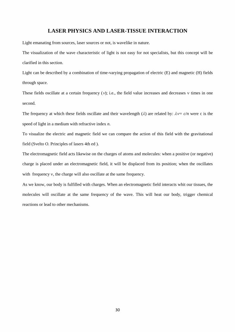
LASER PHYSICS AND LASER-TISSUE INTERACTION
Light emanating from sources, laser sources or not, is wavelike in nature.
The visualization of the wave characteristic of light is not easy for not specialists, but this concept will be
clarified in this section.
Light can be described by a combination of time-varying propagation of electric (E) and magnetic (H) fields
through space.
These fields oscillate at a certain frequency (); i.e., the field value increases and decreases times in one
second.
The frequency at which these fields oscillate and their wavelength () are related by: = c/n were c is the
speed of light in a medium with refractive index n.
To visualize the electric and magnetic field we can compare the action of this field with the gravitational
field (Svelto O. Principles of lasers 4th ed ).
The electromagnetic field acts likewise on the charges of atoms and molecules: when a positive (or negative)
charge is placed under an electromagnetic field, it will be displaced from its position; when the oscillates
with frequency , the charge will also oscillate at the same frequency.
As we know, our body is fulfilled with charges. When an electromagnetic field interacts whit our tissues, the
molecules will oscillate at the same frequency of the wave. This will heat our body, trigger chemical
reactions or lead to other mechanisms.

31
Tab 3 Parameter of laser light
In fig 1
We can visualize the explanation for the electromagnetic field in the space a; in time b. When a charge is
submitted to the influence of a wave its movement can also described by the electromagnetic wave observe c
and consider a positive charge at the origin; when the electric field of a wave, described by the arrow,
interacts whit the charge, its movement will follow the amplitude of electric field. Figure c shows the electric
field at a certain angle; if all photons have their electric field running in the same direction, the beam is
named polarized beam; if the photons oscillate at a non specific direction, the beam is not-polarized ad
represented in figure d. The different frequency of the electromagnetic field will determine the energy of the
32
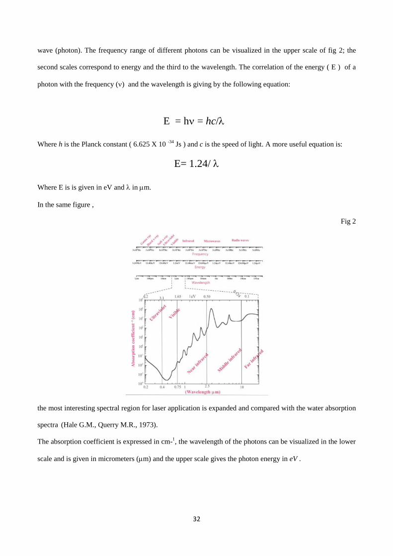
wave (photon). The frequency range of different photons can be visualized in the upper scale of fig 2; the
second scales correspond to energy and the third to the wavelength. The correlation of the energy ( E ) of a
photon with the frequency () and the wavelength is giving by the following equation:
E = h = hc/
Where h is the Planck constant ( 6.625 X 10 -34
Js ) and c is the speed of light. A more useful equation is:
E= 1.24/
Where E is is given in eV and in m.
In the same figure ,
Fig 2
the most interesting spectral region for laser application is expanded and compared with the water absorption
spectra (Hale G.M., Querry M.R., 1973).
The absorption coefficient is expressed in cm-1, the wavelength of the photons can be visualized in the lower
scale and is given in micrometers (m) and the upper scale gives the photon energy in eV .

33
LASER DESIGN
Some basic condition must be satisfied so that a laser functional system can be obtained. First of all here
must be a material named active medium, that allows population inversion. It is unlikely that this inversion
occurs in nature but it can occur also in some materials. The most probable behaviour for electron is to
remain at the ground energy level, whereas in an active medium excited electrons are located at higher
energy level for a long period of time allowing the stimulated emission. This phenomenon will be described
in the following paragraphs.
Fig 3
In figure 3 it is possible to visualize the description of the main instruments for a laser system. The
active medium will be excited by means of a pumping source, which can be another laser, an electric current
or a non-laser source. When population inversion occurs, stimulated emission will take place and avalanche
of photons will be emitted. These photons can resonate between the two mirrors; the output mirror is a
partially reflected mirror, this allows a photon emission (O’Shea D.C., 1977).

34
STIMULATED EMISSION
In nature all system tend to reach the lower energy state. Electrons try to occupy states with lower energy.
The phenomenon is represented in figure 4
Fig 4
if the lower state is totally occupied, the second lower state will start being occupied. But when the system
had more energy, like thermal energy, the electron can acquire this energy and transit to upper states. The
origin of stimulated emission can be visualized in fig. 5
Fig 5

35
by four boxes fig 5 a Represents the non-excited atom whit its electron in ground state. Stimulated
absorption of one photon will transit to an upper energy state fig 5b When a spontaneous emission occurs fig
5c a photon will be emitted and will stimulate the emission of another photon fig 5d. For the stimulated
emission to occur, it is necessary that the active medium presents population inversion. This situation is
described by the energy-state-transition diagram of fig 4 c: the most populated state is not the ground state,
but an upper state, named long-lived excited stated. With all the electrons in this upper state, stimulated
emission can occur more efficiently, thus predicting laser beam.
MONOCHROMATICITY
The monochromaticity of laser light originates from the different energies during the stimulated
emission. When a photon is emitted spontaneously, its energy is well-defined and the other photons
stimulated by the first one have the same energy. As a consequence, the laser beam output is composed of
well-established photon energy; i.e., a specific wavelength. If the source does not emit photons with the same
energy the emission spectra will be broad as in the case of a LED ( Light Emitting Diode) . or the sun.
DIRECTIONALITY
Laser emission occurs only in the direction toward which the system resonates; i.e. in the
longitudinal axis connecting by the first mirror to the two lateral faces of the laser medium and the second
mirror. Otherwise, photon emission can occur randomly at any direction in a lamb bulb, spreading out the
energy: consequently leading to a much lower irradiance than that of a laser beam.
CHOERENCE
Laser light has spatial and temporal coherence. This coherence means that all the photon waves that compose
a laser pulse are correlated in space and time. As described before, all the photons oscillates at the same
frequency ( monochromaticity ), and beyond this frequency oscillation of the laser photons starts at the same
time. In other words, when the amplitude of electric field of one photon is at its highest value all photons
have amplitude at this highest value too, which means temporal coherence.

36
Considering that in a certain position the amplitude of the electric field has a particular value. Spatial
coherence means that at a certain time, all the other wave photons will have the same amplitude value at
meters away from the first photon, and the next surrounding wave photons will also have the same
amplitude value. Therefore, all photons will be correlated with the first photon (Hale G.M., Querry M.R.,
1973).
LASER SYSTEMS IN LIFE SCIENCE
The main laser wavelengths applied in life science are listed in table 4.
Tab 4
The Excimer lasers , nitrogen, and higher harmonic neodymium lasers emanate at the ultraviolet
spectral region with the highest photon energy. The main interaction mechanism for this laser is
photoablation. There are laser systems with emission wavelengths in the visible spectral region and their
main interaction mechanism can be described as photochemical. Finally, the laser systems with emission in
the infrared spectral region exhibit an interaction mechanism dominated by thermal action. The photon
energy changes with its wavelength. For longer wavelengths, as in the case of lasers emitting in the infrared
spectral region, the photon energy is lower than the energy of a photon in the visible spectral region.

37
Likewise, the energy of a latter photon is lower than that a photon emitting in the ultraviolet spectral region.
The energy of photons in laser systems can be compared with the main interatomic bond energies
encountered in biological molecules tab 5
Tab 5
LASER BEAM OUTPUT
In a laser system (continuous or pulsed) it is necessary to describe the density of photons in space
and time.
In other words, it has to be said how many photons are presents in a determinate volume or instant of
time.
To facilitate calculation and explanations, we will use the area of an irradiated surface instead its
volume; i.e., the density of photons in a determined area instead of the number of photons in a determined
volume.
SPATIAL PROFILE
The most common spatial profile of a laser beam in clinical practice is the multimode or the
Gaussian beam profile.
The transversal profile of that kind of laser beam can be visualized in fig 6
38
Fig 6
The beam radius is defined at the position where the irradiance, or radiant exposure, falls down to e-2
( 14% of the maximum value).
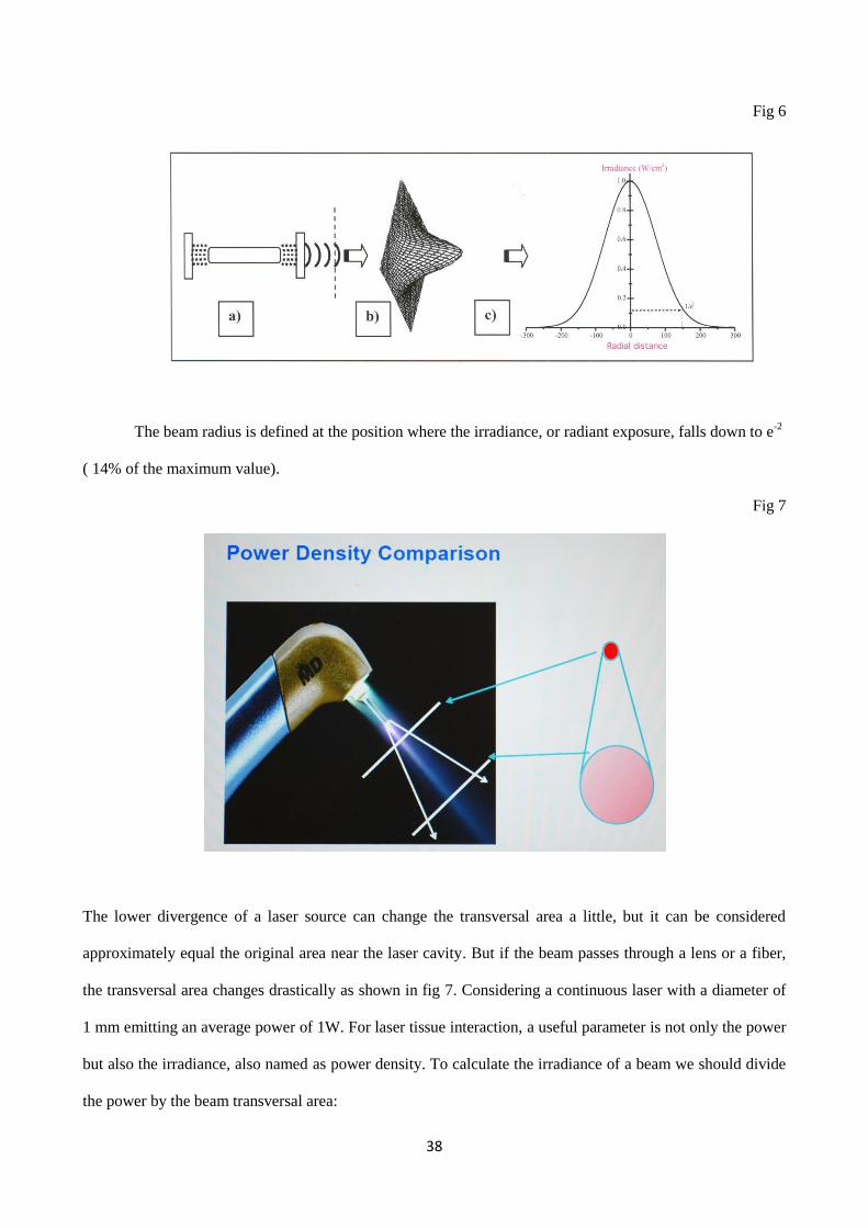
Fig 7
The lower divergence of a laser source can change the transversal area a little, but it can be considered
approximately equal the original area near the laser cavity. But if the beam passes through a lens or a fiber,
the transversal area changes drastically as shown in fig 7. Considering a continuous laser with a diameter of
1 mm emitting an average power of 1W. For laser tissue interaction, a useful parameter is not only the power
but also the irradiance, also named as power density. To calculate the irradiance of a beam we should divide
the power by the beam transversal area:
39
I= P/A = 1W/ (o.5mm)2 = 1.3 W/mm
2 = 130 W/ cm
2
TEMPORAL PROFILE
We have just discussed about the distribution of photons in space; now we talk about the distribution in the
time of a pulsed laser.
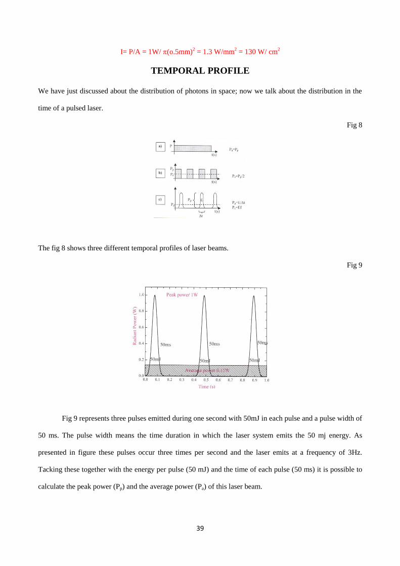
Fig 8
The fig 8 shows three different temporal profiles of laser beams.
Fig 9
Fig 9 represents three pulses emitted during one second with 50mJ in each pulse and a pulse width of
50 ms. The pulse width means the time duration in which the laser system emits the 50 mj energy. As
presented in figure these pulses occur three times per second and the laser emits at a frequency of 3Hz.
Tacking these together with the energy per pulse (50 mJ) and the time of each pulse (50 ms) it is possible to
calculate the peak power (Pp) and the average power (Pa) of this laser beam.
40
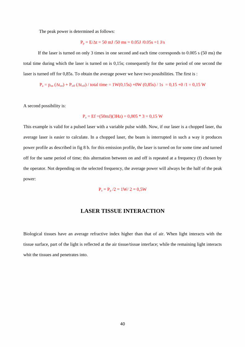
The peak power is determined as follows:
Pp = E/t = 50 mJ /50 ms = 0.05J /0.05s =1 J/s
If the laser is turned on only 3 times in one second and each time corresponds to 0.005 s (50 ms) the
total time during which the laser is turned on is 0,15s; consequently for the same period of one second the
laser is turned off for 0,85s. To obtain the average power we have two possibilities. The first is :
Pa = pon (ton) + Poff (toff) / total time = 1W(0,15s) +0W (0,85s) / 1s = 0,15 +0 /1 = 0,15 W
A second possibility is:
Pa = Ef =(50mJ)(3Hz) = 0,005 * 3 = 0,15 W
This example is valid for a pulsed laser with a variable pulse width. Now, if our laser is a chopped laser, tha
average laser is easier to calculate. In a chopped laser, the beam is interrupted in such a way it produces
power profile as described in fig 8 b. for this emission profile, the laser is turned on for some time and turned
off for the same period of time; this alternation between on and off is repeated at a frequency (f) chosen by
the operator. Not depending on the selected frequency, the average power will always be the half of the peak
power:
Pa = Pp /2 = 1W/ 2 = 0,5W
LASER TISSUE INTERACTION
Biological tissues have an average refractive index higher than that of air. When light interacts with the
tissue surface, part of the light is reflected at the air tissue/tissue interface; while the remaining light interacts
whit the tissues and penetrates into.
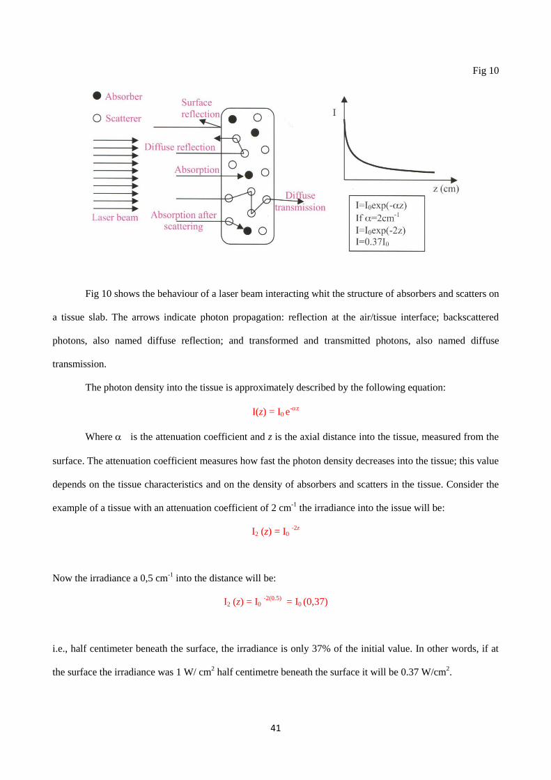
41
Fig 10
Fig 10 shows the behaviour of a laser beam interacting whit the structure of absorbers and scatters on
a tissue slab. The arrows indicate photon propagation: reflection at the air/tissue interface; backscattered
photons, also named diffuse reflection; and transformed and transmitted photons, also named diffuse
transmission.
The photon density into the tissue is approximately described by the following equation:
I(z) = I0 e-z
Where is the attenuation coefficient and z is the axial distance into the tissue, measured from the
surface. The attenuation coefficient measures how fast the photon density decreases into the tissue; this value
depends on the tissue characteristics and on the density of absorbers and scatters in the tissue. Consider the
example of a tissue with an attenuation coefficient of 2 cm-1
the irradiance into the issue will be:
I2 (z) = I0 -2z
Now the irradiance a 0,5 cm-1
into the distance will be:
I2 (z) = I0 -2(0.5)
= I0 (0,37)
i.e., half centimeter beneath the surface, the irradiance is only 37% of the initial value. In other words, if at
the surface the irradiance was 1 W/ cm2 half centimetre beneath the surface it will be 0.37 W/cm
2.

42
The attenuation coefficient change for different wavelength and tissues. The water is the main compound in
soft and hard tissue and will determine the attenuation. According to the water spectrum and ignoring some
scattering by the water itself, it is possible to determine how deep the wavelength travels into the water,
taking the absorption coefficient for different wavelengths( Niemz M.H. Laser –Tissue interaction. 3th
edition, 2003; 5 Tuchin V.V.: 2000 ).
INTERACTIONS MECHANISM
The consequence of interaction of a laser beam or non-laser source with a target tissue will be determined by
the beam irradiance.
Fig 11
The fig 11 describes the mechanism that predominates in laser-tissue interaction approximately (adapted
from Boulnois). The diagonal line represents the radiant exposure. It easy to see that two different
irradiances combined with two different interaction times can produce the same radiant exposure. In the
following sections we will discuss the main interaction mechanism taking place during laser irradiation in
medical practice: photochemical and photothermal interactions (Boulnois J.L. 1986).
PHOTOCHEMICAL INTERACTIONS
Approximately, chemical reactions involving photons can be classified as photochemical reactions.
The most popular example is photosynthesis; in our body other examples are the production of melanin and
of the light induced compound Vitamin D all this reactions involve photons. The main idea of the
photochemical treatment is to use a chromophore receptor acting as catalyst.
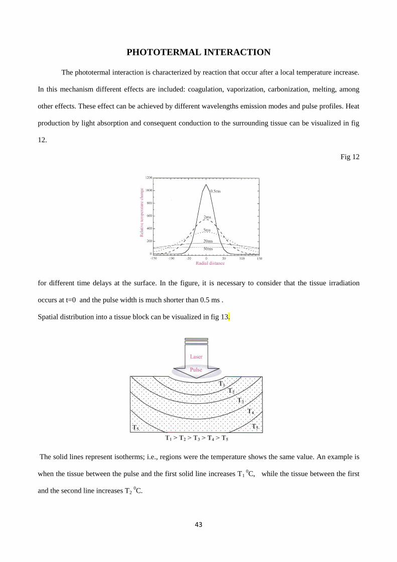
43
PHOTOTERMAL INTERACTION
The phototermal interaction is characterized by reaction that occur after a local temperature increase.
In this mechanism different effects are included: coagulation, vaporization, carbonization, melting, among
other effects. These effect can be achieved by different wavelengths emission modes and pulse profiles. Heat
production by light absorption and consequent conduction to the surrounding tissue can be visualized in fig
12.
Fig 12
for different time delays at the surface. In the figure, it is necessary to consider that the tissue irradiation
occurs at t=0 and the pulse width is much shorter than 0.5 ms .
Spatial distribution into a tissue block can be visualized in fig 13.
The solid lines represent isotherms; i.e., regions were the temperature shows the same value. An example is
when the tissue between the pulse and the first solid line increases T1 0
C, while the tissue between the first
and the second line increases T2 0C.
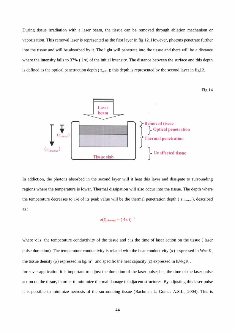
44
During tissue irradiation with a laser beam, the tissue can be removed through ablation mechanism or
vaporization. This removal laser is represented as the first layer in fig 12. However, photons penetrate further
into the tissue and will be absorbed by it. The light will penetrate into the tissue and there will be a distance
where the intensity falls to 37% ( 1/e) of the initial intensity. The distance between the surface and this depth
is defined as the optical penetraction depth ( zoptic ); this depth is represented by the second layer in fig12.
Fig 14
In addiction, the photons absorbed in the second layer will it heat this layer and dissipate to surrounding
regions where the temperature is lower. Thermal dissipation will also occur into the tissue. The depth where
the temperature decreases to 1/e of its peak value will be the thermal penetration depth ( z thermal), described
as :
z(t) thermal = ( 4 t) -1
where is the temperature conductivity of the tissue and t is the time of laser action on the tissue ( laser
pulse duraction). The temperature conductivity is related with the heat conductivity () espressed in W/mK,
the tissue density () espressed in kg/m3 and specific the heat capacity (c) expressed in kJ/kgK .
for sever application it is important to adjust the duraction of the laser pulse; i.e., the time of the laser pulse
action on the tissue, in order to minimize thermal damage to adjacent structures. By adjusting this laser pulse
it is possible to minimize necrosis of the surrounding tissue (Bachman L. Gomes A.S.L., 2004). This is

45
obtained by equating the optical penetration depth ( z optic) to the thermal penetration depth z thermal , hence :
z(t) optical = (4k thermal) -1
; and the interaction time (t) will be renamed as thermal relaxation time ( t thermal ).
The importance of the thermal relaxation time will be explained by the following consideration:
a) for a laser pulse duration < t thermal , heat does not even diffuse to the distance given by the optical
penetration depth ;
b) for < t thermal , it can diffuse to a higher value than the optical penetration depth and thermal damage of
the adjacent tissue is possible (Hayes J.R., Wolsbarsht M.L, Aerospace Medicine).
THERMAL EFFECT ON SOFT AND HARD TISSUES
The laser parameter together with the optical and thermal properties of the tissue will determine the
spatial and temporal distribution of the temperature inside the tissue and the maximum reached temperature
and the time during wich the tissue is submitted to the laser will lead to different biological effect. The
biological effect on soft and hard tissues are summarized in tab 6. (O’Shea D.C., 1977, Bachman L. Gomes
A.S.L., 2004).
46
The association of laser-assisted surgery with PRP for the treatment of bisphosphonate-related
osteonecrosis of the jaw in cancer patients. A preliminary study.
Introduction
Bisphosphonate-related osteonecrosis of the jaw (BRONJ) is a well-known potential complication of
bisphoshonate (BP) treatment, which occurs as a result of reduced bone resorption and bone turnover.
Several studies have identified numerous potential risk factors associated with the development of BRONJ.
The duration of BP therapy and the type of drug used determine the risk of this disease. Dentoalveolar
trauma is another risk factor for BRONJ. Correlation between the occurrence of BRONJ and other systemic
and local factors (denture use, poor oral hygiene, dental infection, diabetes, use of steroids) was identified.
BRONJ management is controversial: there is no definitive treatment in literature which leads to restitutio
ad integrum, but different approaches (local and systemic antibiotic therapy, sequestrectomy, surgical
resections, hyperbaric oxygen therapy, low parathormone doses and ozone therapy).
Recently, a few clinical studies using Er, Cr: YSGG laser-assisted surgery for the treatment of BRONJ
showed promising results. The rationale to use laser with low dosages is: i) the absence of thermal in the
cutting area and in the surrounding tissues during the removal of necrotic tissue, ii) the biostimulating effect
reported in literature on both bone and soft tissues, iii) the presence of an antibacterial effect.
Platelet-rich plasma (PRP) is a new approach to tissue regeneration in oral surgery. The rationale for the
employment of PRP in patients affected by BRONJ is based on the thesis that the presence of growth factors
(usually repressed by BPs) constitutes a substitute stimulation to bone healing, which is similar to
physiological healing. The growth factors in PRP might accelerate epithelial wound healing, decrease tissue
inflammation after surgery, improve the regeneration of bone and soft tissues, and promote tissue
vascularization. The additional advantages related to the use of this product are its biocompatibility and
safety, as an autologous product.
The aim of this study was to evaluate the efficacy, as clinical healing, of a combined treatment with the laser-
assisted surgery and PRP for the treatment of BRONJ in cancer patients.
47
Materials and Methods
Patients
Ten patients (n. 3 males, n. 7 females; age range: 69-89 years, mean age 76.2±5.8) with BRONJ were
identified for this study. The inclusion criteria were patients who had received i.v. BP therapy during the
treatment of their primary disease, exhibited exposed necrotic bone or radiological signs of bone necrosis
with and without pain in the maxillofacial region for more than 8 (10) weeks, and had not received previous
radiotherapy from the head and neck area. The control group consists of 22 patients treated only with
medical and surgical therapy performed with laser Er: Cr, without the application of platelet gel
In all the cases, the diagnosis of BRONJ was established after clinical examination (intra- and extra-oral
examination), and imaging tests.
Medical records on associated risk factors (history of diabetes), clinical features of BRONJ, therapeutic
approach (BP used, frequency and dosage of BP treatment, duration of BP therapy, history of chemotherapy,
steroid treatment) and clinical course of each case were collected. Demographics data (age, gender) of the
patients were also collected. All of the patients were also questioned about any dental surgery during the BP
therapy. The smoking habit and oral hygiene were registered.
Lesions were classified following SICMF-SIPMO clinical and radiological staging system of
BRONJ, as follows: Stage 1 (Focal BRONJ): Clinical signs and symptoms: bone exposure; sudden dental
mobility; nonhealing postextraction socket; mucosal fistula; swelling; abscess formation; trismus; gross
mandibular deformity and/or hypoesthesia/paraesthesia of the lips; CT findings: increased bone density
limited to the alveolar bone region (trabecular thickening and/or focal osteosclerosis ), with or without the
following signs: markedly thickened and sclerotic lamina dura; persisting alveolar socket; and/or cortical
disruption; 1a. Asymptomatic,1b. Symptomatic (pain and purulent discharge); Stage 2 (Diffuse BRONJ):
Clinical signs and symptoms: same as Stage 1; CT findings: increased bone density extended to the basal
bone (diffuse osteosclerosis), with or without the following signs: prominence of the inferior alveolar nerve
canal; periosteal reaction; sinusitis; sequestra formation; and/or oro-antral fistola; 2a. Asymptomatic, 2b.
Symptomatic (pain and purulent discharge); Stage 3 (Complicated BRONJ): Same as Stage 2, with one
or more of the following:
clinical signs and symptoms: extra-oral fistula; displaced mandibular stumps; nasal leakage of fluids; CT
findings: osteosclerosis of adjacent bones (zygoma, hard palate); pathologic mandibular fracture; and/or
osteolysis extending to the sinus floor.
Radiological examinations were visualized with computer tomography (CT) and panoramic radiographs.
Four of the ten patients had received a combined laser-assisted surgery and antibiotic therapy for BRONJ; n.
six of them had received only an antibiotic therapy medical treatment (Table 7) .
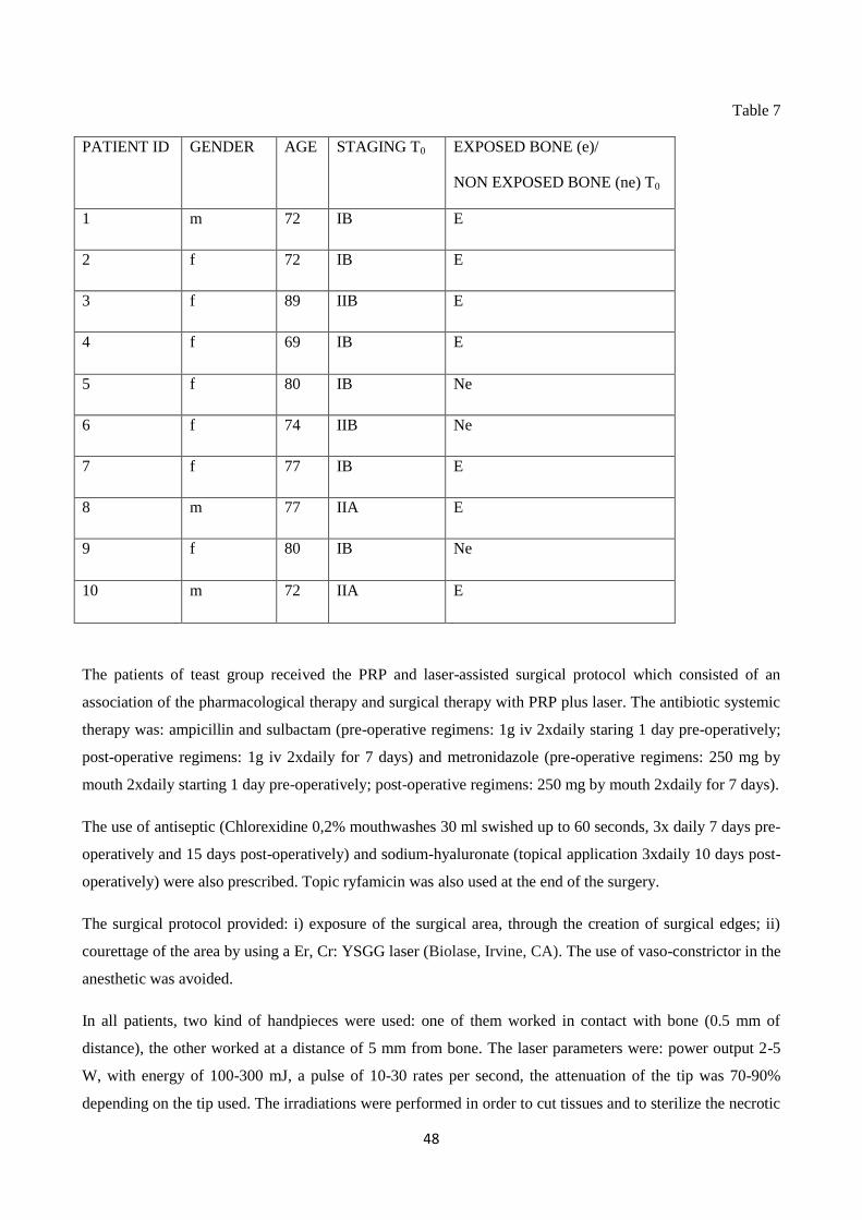
48
Table 7
PATIENT ID GENDER AGE STAGING T0 EXPOSED BONE (e)/
NON EXPOSED BONE (ne) T0
1 m 72 IB E
2 f 72 IB E
3 f 89 IIB E
4 f 69 IB E
5 f 80 IB Ne
6 f 74 IIB Ne
7 f 77 IB E
8 m 77 IIA E
9 f 80 IB Ne
10 m 72 IIA E
The patients of teast group received the PRP and laser-assisted surgical protocol which consisted of an
association of the pharmacological therapy and surgical therapy with PRP plus laser. The antibiotic systemic
therapy was: ampicillin and sulbactam (pre-operative regimens: 1g iv 2xdaily staring 1 day pre-operatively;
post-operative regimens: 1g iv 2xdaily for 7 days) and metronidazole (pre-operative regimens: 250 mg by
mouth 2xdaily starting 1 day pre-operatively; post-operative regimens: 250 mg by mouth 2xdaily for 7 days).
The use of antiseptic (Chlorexidine 0,2% mouthwashes 30 ml swished up to 60 seconds, 3x daily 7 days pre-
operatively and 15 days post-operatively) and sodium-hyaluronate (topical application 3xdaily 10 days post-
operatively) were also prescribed. Topic ryfamicin was also used at the end of the surgery.
The surgical protocol provided: i) exposure of the surgical area, through the creation of surgical edges; ii)
courettage of the area by using a Er, Cr: YSGG laser (Biolase, Irvine, CA). The use of vaso-constrictor in the
anesthetic was avoided.
In all patients, two kind of handpieces were used: one of them worked in contact with bone (0.5 mm of
distance), the other worked at a distance of 5 mm from bone. The laser parameters were: power output 2-5
W, with energy of 100-300 mJ, a pulse of 10-30 rates per second, the attenuation of the tip was 70-90%
depending on the tip used. The irradiations were performed in order to cut tissues and to sterilize the necrotic
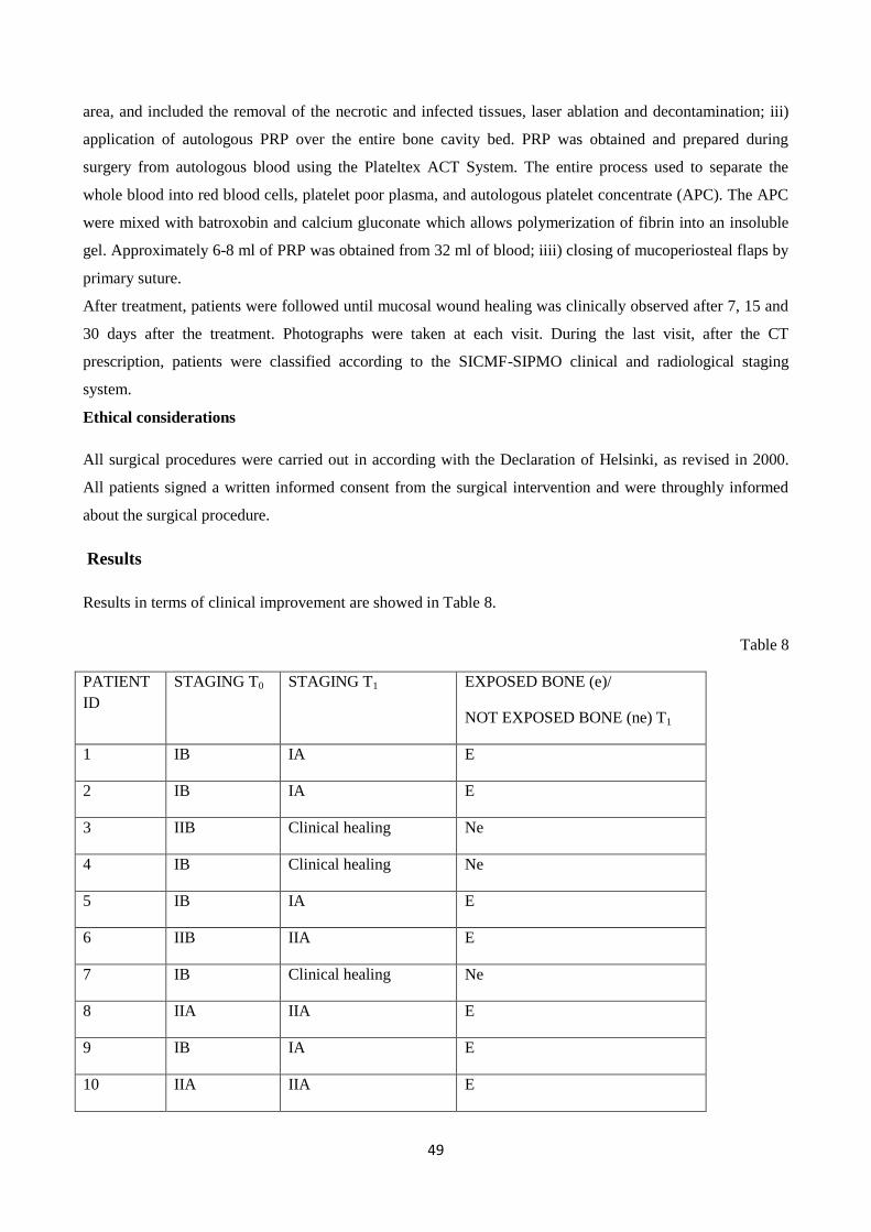
49
area, and included the removal of the necrotic and infected tissues, laser ablation and decontamination; iii)
application of autologous PRP over the entire bone cavity bed. PRP was obtained and prepared during
surgery from autologous blood using the Plateltex ACT System. The entire process used to separate the
whole blood into red blood cells, platelet poor plasma, and autologous platelet concentrate (APC). The APC
were mixed with batroxobin and calcium gluconate which allows polymerization of fibrin into an insoluble
gel. Approximately 6-8 ml of PRP was obtained from 32 ml of blood; iiii) closing of mucoperiosteal flaps by
primary suture.
After treatment, patients were followed until mucosal wound healing was clinically observed after 7, 15 and
30 days after the treatment. Photographs were taken at each visit. During the last visit, after the CT
prescription, patients were classified according to the SICMF-SIPMO clinical and radiological staging
system.
Ethical considerations
All surgical procedures were carried out in according with the Declaration of Helsinki, as revised in 2000.
All patients signed a written informed consent from the surgical intervention and were throughly informed
about the surgical procedure.
Results
Results in terms of clinical improvement are showed in Table 8.
Table 8
PATIENT
ID
STAGING T0 STAGING T1 EXPOSED BONE (e)/
NOT EXPOSED BONE (ne) T1
1 IB IA E
2 IB IA E
3 IIB Clinical healing Ne
4 IB Clinical healing Ne
5 IB IA E
6 IIB IIA E
7 IB Clinical healing Ne
8 IIA IIA E
9 IB IA E
10 IIA IIA E
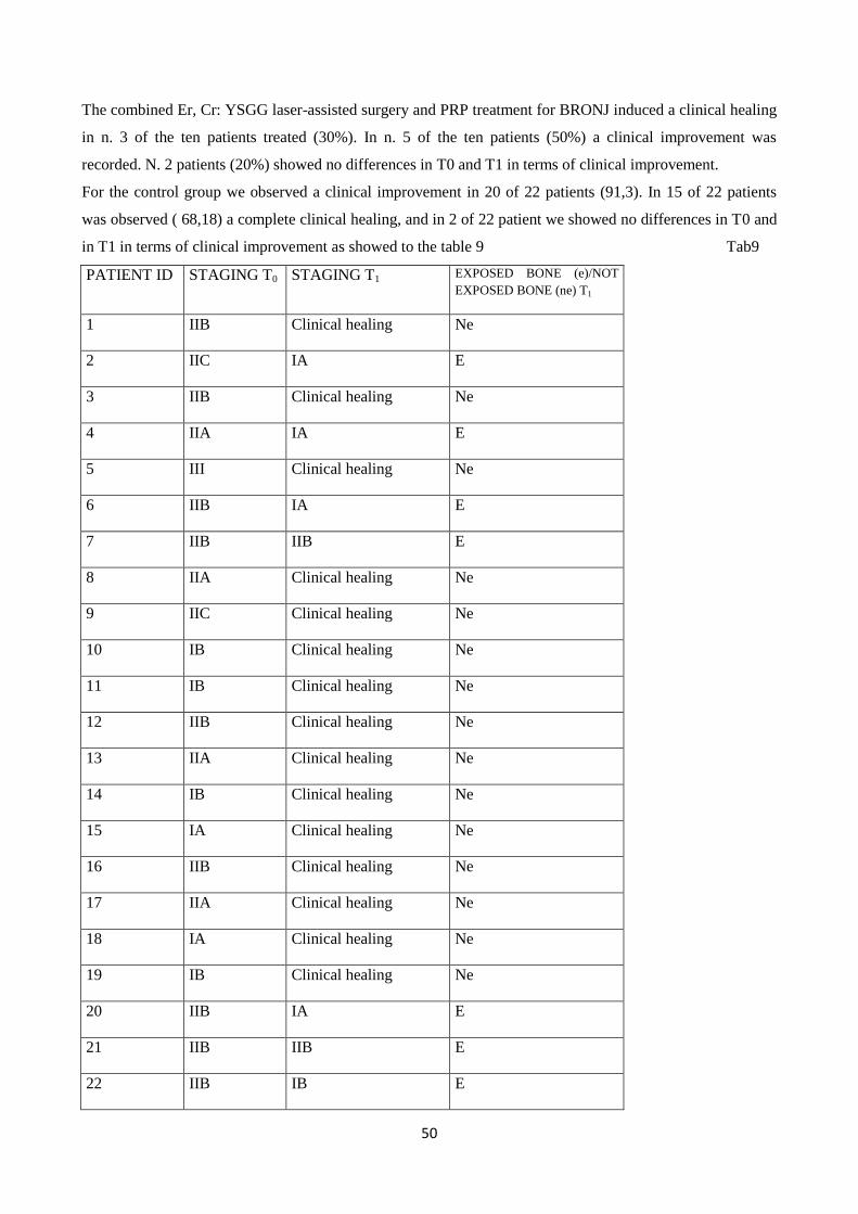
50
The combined Er, Cr: YSGG laser-assisted surgery and PRP treatment for BRONJ induced a clinical healing
in n. 3 of the ten patients treated (30%). In n. 5 of the ten patients (50%) a clinical improvement was
recorded. N. 2 patients (20%) showed no differences in T0 and T1 in terms of clinical improvement.
For the control group we observed a clinical improvement in 20 of 22 patients (91,3). In 15 of 22 patients
was observed ( 68,18) a complete clinical healing, and in 2 of 22 patient we showed no differences in T0 and
in T1 in terms of clinical improvement as showed to the table 9 Tab9
PATIENT ID STAGING T0 STAGING T1 EXPOSED BONE (e)/NOT
EXPOSED BONE (ne) T1
1 IIB Clinical healing Ne
2 IIC IA E
3 IIB Clinical healing Ne
4 IIA IA E
5 III Clinical healing Ne
6 IIB IA E
7 IIB IIB E
8 IIA Clinical healing Ne
9 IIC Clinical healing Ne
10 IB Clinical healing Ne
11 IB Clinical healing Ne
12 IIB Clinical healing Ne
13 IIA Clinical healing Ne
14 IB Clinical healing Ne
15 IA Clinical healing Ne
16 IIB Clinical healing Ne
17 IIA Clinical healing Ne
18 IA Clinical healing Ne
19 IB Clinical healing Ne
20 IIB IA E
21 IIB IIB E
22 IIB IB E

51
Discussion
BRONJ has emerged as an important secondary consequence of BPs use. BPs are used to significant clinical
benefit in both the treatment and prevention of conditions associated with pathology secondary to bone
resorption and turnover (i.e. osteoporosis, skeletal bony metastases from solid malignancies, hypercalcemia,
multiple myeloma, osteitis deformens). Accordingly, the use of BPs therapies has increased markedly,
particularly in the treatment of age related and post-menopausal osteoporosis, and breast cancer and other
solid malignancies where long-term survival is greatly increased. The use of bisphosphonate agents for
treatment and prophylaxis of bony complications associated with solid malignancies (predominantly breast
cancer) and multiple myeloma is a highly significant risk factor relative to the use of these agents for
osteoporosis.
In most cases, BRONJ occurs after a surgical procedure involving the jawbone. Spontaneous cases of
BRONJ have also been reported, with apparently no recent history of trauma of any form, to the jaws or
teeth.
Currently, the management of BRONJ remains controversial, and there is no definitive treatment in
literature. The objective of the treatment of BRONJ is to provide a higher level of quality of life to patients.
Minimally invasive surgical approach with Er, Cr: YSGG laser represents a good choice for BRONJ
treatment especially for antibacterial and biostimulant properties.
Moreover, the use of Er, Cr: YSGG laser has a great potential in the hard tissues surgery due to its high
absorption by both water and hydroxyapatite. In fact, it does not cause coagulation or carbonization and
provides a clean and precise cut with minimal injury to the adjacent hard and soft tissues.
The two types of treatment showed no significant differences when compared with the chi-square test
corrected according to Yates, both for what concerns the clinical improvements observed (p = 0.7731) than
for what pertains to clinical recovery (p = 0 , 1023). The PRP, however, seems greatly enhance the speed of
migration of the soft tissue over the osteonecrotic zone. There was, however, a new bone exposure in the
areas where it has not been completely removed necrotic tissue. This problem is because of the fact that it is
not possible to distinguish clinically, during surgery, the diseased area from the area not affected by necrosis.
Given the objective of a treatment minimally invasive, are necessary various interventions to achieve a bony
area not affected, for this reason, the use of PRP should be postponed until we know for sure that we have
cleared the entire portion of infected bone. At this point, this treatment may be very advantageous because,
reducing the healing time, reduces the possibility of re-infect the bone area affected equally reducing the
possibility of further necrosis. In consideration of the hypothesis described above, it is considered useful a
tool that can identify positively the limits of the treatment, in order to prevent large mutilation that might be
particularly harmful for the patient, also in view of the the precarious general clinical condition of the patient
himself. In any case, the method requires further investigation and further studies are necessary to test the
significance of the results with a larger-sized specimen.
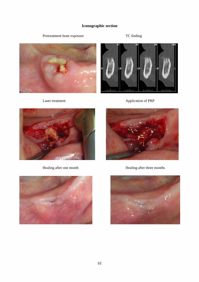
52
Iconographic section
Pretreatment bone exposure TC finding
Laser treatment Application of PRP
Healing after one month Healing after three months
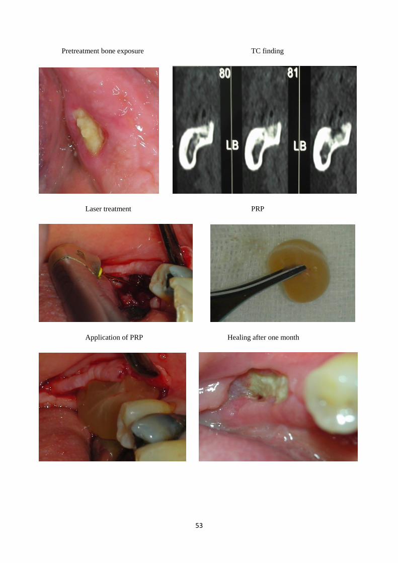
53
Pretreatment bone exposure TC finding
Laser treatment PRP
Application of PRP Healing after one month

54
Bibliography
Abu-Id, MH, Warnke, PH, Gottschalk, J, et al. (2008). "Bis-phossy jaws" - High and low risk factors for bisphosphonate-induced osteonecrosis of the jaw. J Craniomaxillofac Surg 36(2): 95-103.
Albanese A, Licata ME, Polizzi B, Campisi G "Platelet-rich plasma (PRP) in dental and oral surgery: from the wound healing to bone
regeneration" Immun Ageing. 2013 Jun 13;10(1):23. doi: 10.1186/1742-4933-10-23.
Bachman L. Gomes A.S.L., Zezell D.M. Bound energy of water in hard dental tissues. Spectroscopy Letters 2004; 37 (6): 565-579
Boulnois J.L. Photophysical processes in recent medical laser developments: a review. Laser in medical science 1986; 1;47-66.
Cho MI, Lin WL, Genco RJ. Platelet-derived growth factor modulated guided tissue regenerative therapy. J Periodontol 1995;66:522-30.
Hale G.M., Querry M.R. Optical constants of water in the 200-nm to 200-nm wavelength region. Applied Optics 1973 12(3): 555-562
Hayes J.R., Wolsbarsht M.L. Thermal model for retinal damage induced by pulsed lasers. Aerospace Medicine; 39:474-480.
Kassolis JD, Rosen PS, Reynolds MA. Alveolar ridge and sinus augmentation utilizing platelet-rich plasma in combination with freezedried bone allograft: case series. J Periodontol 2000;71:1654-61.
Marx RE, Carlson ER, Eichstaedt RM, Schimmele SR, Strauss JE, Georgeff KR. Platelet-rich plasma: growth factor enhancement for bone
grafts. Oral Surg Oral Med Oral Pathol Oral Radiol Endod 1998;85:638-46.
Mustoe TA, Pierce GF, Morishima C, Deuel TF. Growth factorinduced acceleration of tissue repair through direct and inductive activities in a
rabbit dermal ulcer model. J Clin Invest 1991;87:694-703.
Niemz M.H. Laser –Tissue interaction. 3th edition, Springler 2003
O’Shea D.C. Introduction to lasers and their applications. Addison-Wesley Pub.Co., 1977
Oz MC, Jeevanandam V, Smith CR, et al. Autologous fibrin glue from intraoperatively collected platelet-rich plasma. Ann Thorac Surg
1992;53:530-1.
Park JB, Matsuura M, Han KY, et al. Periodontal regeneration in class III furcation defects of beagle dogs using guided tissue regenerative
therapy with platelet-derived growth factor. J Periodontol 1995;66: 462-77.
Pierce GF, Tarpley JE, Yanagihara D, Mustoe TA, Fox GM, Thomason A. Platelet-derived growth factor (BB homodimer), transforming growth factor-beta 1, and basic fibroblast growth factor in dermal wound healing: neovessel and matrix formation and cessation of repair. Am J
Pathol 1992;140:1375-88.
Robbins SL, Cotran RS, Kumar V, eds. Robbins pathologic basis of disease. 5th ed. Philadelphia: Saunders; 1994:40-1.
Sonnleitner D, Huemer P, Sullivan DY. A simplified technique for producing platelet rich plasma and platelet concentrate for intraoral bone
grafting techniques: a technical note. J Oral Maxillofac Implants 2000;15:879-82.
Svelto O. Principles of lasers 4th ed. New York, Plenum Press1998
55
Townsend CM, Beauchamp RD, Evers BM, Mattox KL, eds. Sabiston textbook of surgery: The biological basis of modern surgical practice. 16th ed. Philadelphia: Saunders; 2001:1375.
Whitman DH, Berry RL, Green DM. Platelet gel: an autologous alternative to fibrin glue with applications in oral and maxillofacial surgery. J
Oral Maxillofac Surg 1997;55:1294-9.
Whitman DH, Berry RL. A technique for improving the handling of particulate cancellous bone and marrow grafts using platelet gel. J Oral
Maxillofac Surg 1998;56:1217-8.
Ardine, M, Generali, D, Donadio, M, et al. (2006). Could the long-term persistence of low serum calcium levels and high serum parathyroid hormone levels during bisphosphonate treatment predispose metastatic breast cancer patients to undergo osteonecrosis of the jaw? Ann Oncol
17(8): 1336-1337.
Assael, LA (2009). Oral bisphosphonates as a cause of bisphosphonate-related osteonecrosis of the jaws: clinical findings, assessment of risks, and preventive strategies. J Oral Maxillofac Surg 67(5 Suppl): 35-43.
Bagan, J, Scully, C, Sabater, V and Jimenez, Y (2009). Osteonecrosis of the jaws in patients treated with intravenous bisphosphonates
(BRONJ): A concise update. Oral Oncol 45(7): 551-554.
Bagan, JV, Jimenez, Y, Murillo, J, et al. (2006). Jaw osteonecrosis associated with bisphosphonates: multiple exposed areas and its relationship
to teeth extractions. Study of 20 cases. Oral Oncol 42(3): 327-329.
Baltensperger M, EG (2009). Osteomyelitis of the jaws. . Springer (Eds) 2009; chapter 22 5-36.
Bamias, A, Kastritis, E, Bamia, C, et al. (2005). Osteonecrosis of the jaw in cancer after treatment with bisphosphonates: incidence and risk
factors. J Clin Oncol 23(34): 8580-8587.
Bedogni, A, Bettini, G, Totola, A, Saia, G and Nocini, PF (2010). Oral bisphosphonate-associated osteonecrosis of the jaw after implant surgery: a case report and literature review. J Oral Maxillofac Surg 68(7): 1662-1666.
Bedogni, A, Blandamura, S, Lokmic, Z, et al. (2008). Bisphosphonate-associated jawbone osteonecrosis: a correlation between imaging
techniques and histopathology. Oral Surg Oral Med Oral Pathol Oral Radiol Endod 105(3): 358-364.
Bedogni, A, Fusco, V, Agrillo, A and Campisi, G (2012). Learning from experience. Proposal of a refined definition and staging system for
bisphosphonate-related osteonecrosis of the jaw (BRONJ). Oral Dis 18(6): 621-623.
Bedogni, A, Saia, G and al, e (2011). Long-term outcomes of surgical resection of the jaws in cancer patients with bisphosphonate-related osteonecrosis Oral Oncol 47(5): 420-424.
Belibasakis GN, BN, Hashim A, Johansson A, Aduse-Opoku J, Curtis MA, et al. (2007). Regulation of RANKL and OPG gene expression in
human gingival fibroblasts and periodontal ligament cells by Porphyromonas gingivalis: a putative role of the Arg-gingipains. . Microb Pathog.
Bertoldo, F, Santini, D and Lo Cascio, V (2007). Bisphosphonates and osteomyelitis of the jaw: a pathogenic puzzle. Nat Clin Pract Oncol
4(12): 711-721.
Bianchi, ML, Limonta, C, Frasunkiewicz, J, Biggioggero, M and Vai, S (2008). Comment on: Bisphosphonates and osteonecrosis of the jaw. Rheumatology (Oxford) 47(3): 383; author reply 383-384
Bianchi, SD, Scoletta, M, Cassione, FB, Migliaretti, G and Mozzati, M (2007). Computerized tomographic findings in bisphosphonate-
associated osteonecrosis of the jaw in patients with cancer. Oral Surg Oral Med Oral Pathol Oral Radiol Endod 104(2): 249-258.
56
Black, DM, Schwartz, AV, Ensrud, KE, et al. (2006). Effects of continuing or stopping alendronate after 5 years of treatment: the Fracture Intervention Trial Long-term Extension (FLEX): a randomized trial. Jama 296(24): 2927-2938.
Brunello, A, Saia, G, Bedogni, A, Scaglione, D and Basso, U (2009). Worsening of osteonecrosis of the jaw during treatment with sunitinib in a
patient with metastatic renal cell carcinoma. Bone 44(1): 173-175.
Campisi, G, Di Fede, O, Musciotto, A, et al. (2007). Bisphosphonate-related osteonecrosis of the jaw (BRONJ): run dental management designs
and issues in diagnosis. Ann Oncol 18 Suppl 6: vi168-172.
Chiandussi, S, Biasotto, M, Dore, F, et al. (2006). Clinical and diagnostic imaging of bisphosphonate-associated osteonecrosis of the jaws. Dentomaxillofac Radiol 35(4): 236-243.
Cho, SH, Kim, SY, Lee, KY and Lee, HC (2007). New bone formation in unilateral rhinosinusitis. Am J Rhinol 21(1): 37-39.
Christodoulou, C, Pervena, A, Klouvas, G, et al. (2009). Combination of bisphosphonates and antiangiogenic factors induces osteonecrosis of
the jaw more frequently than bisphosphonates alone. Oncology 76(3): 209-211.
Coleman, R, Burkinshaw, R, Winter, M, et al. (2010). Zoledronic acid. Expert Opin Drug Saf 10(1): 133-145.
Dimopoulos, MA, Kastritis, E, Anagnostopoulos, A, et al. (2006). Osteonecrosis of the jaw in patients with multiple myeloma treated with bisphosphonates: evidence of increased risk after treatment with zoledronic acid. Haematologica 91(7): 968-971.
Dixon, RB, Tricker,N.D.,Garetto,L.P. (1997). Bone turnover in elderly canine mandible and tibia (abstract 2579). J Dent Res 76: 336.
Dodson, TB (2009). Intravenous bisphosphonate therapy and bisphosphonate-related osteonecrosis of the jaws. J Oral Maxillofac Surg 67(5 Suppl): 44-52.
Donoghue, AM (2005). Bisphosphonates and osteonecrosis: analogy to phossy jaw. Med J Aust 183(3): 163-164.
Eckert, AW, Maurer, P, Meyer, L, et al. (2007). Bisphosphonate-related jaw necrosis - Severe complication in Maxillofacial surgery. Cancer Treat Rev 33(1): 58-63.
English, BC, Baum, CE, Adelberg, DE, et al. (2010). A SNP in CYP2C8 is not associated with the development of bisphosphonate-related
osteonecrosis of the jaw in men with castrate-resistant prostate cancer. Ther Clin Risk Manag 6: 579-583.
Fedele, S, Kumar, N, Davies, R, et al. (2009). Dental management of patients at risk of osteochemonecrosis of the jaws: a critical review. Oral
Dis 15(8): 527-537.
Fedele, S, Porter, SR, D'Aiuto, F, et al. (2010). Nonexposed variant of bisphosphonate-associated osteonecrosis of the jaw: a case series. Am J Med 123(11): 1060-1064.
Ficarra, G, Beninati, F, Rubino, I, et al. (2005). Osteonecrosis of the jaws in periodontal patients with a history of bisphosphonates treatment. J
Clin Periodontol 32(11): 1123-1128.
Filleul, O, Crompot, E and Saussez, S (2010). Bisphosphonate-induced osteonecrosis of the jaw: a review of 2,400 patient cases. J Cancer Res
Clin Oncol 136(8): 1117-1124.
Fusco, V, Loidoris, A, Colella, G, Vescovi, P and Campisi, G (2010). Osteonecrosis of the jaw (ONJ) risk in breast cancer patients after zoledronic acid treatment. Breast 19(5): 432-433; author reply 433-434.
57
Garcia-Ferrer, L, Bagan, JV, Martinez-Sanjuan, V, et al. (2008). MRI of mandibular osteonecrosis secondary to bisphosphonates. AJR Am J Roentgenol 190(4): 949-955.
Guarneri, V, Miles, D, Robert, N, et al. (2010). Bevacizumab and osteonecrosis of the jaw: incidence and association with bisphosphonate
therapy in three large prospective trials in advanced breast cancer. Breast Cancer Res Treat 122(1): 181-188.
Hall, V (2008). Actinomyces--gathering evidence of human colonization and infection. Anaerobe 14(1): 1-7.
Hansen, T, Kirkpatrick, CJ, Walter, C and Kunkel, M (2006). Increased numbers of osteoclasts expressing cysteine proteinase cathepsin K in
patients with infected osteoradionecrosis and bisphosphonate-associated osteonecrosis-a paradoxical observation? Virchows Arch 449(4): 448-454.
Hansen, T, Kunkel, M, Weber, A and James Kirkpatrick, C (2006). Osteonecrosis of the jaws in patients treated with bisphosphonates -
histomorphologic analysis in comparison with infected osteoradionecrosis. J Oral Pathol Med 35(3): 155-160.
Hellstein, JW and Marek, CL (2005). Bisphosphonate osteochemonecrosis (bis-phossy jaw): is this phossy jaw of the 21st century? J Oral
Maxillofac Surg 63(5): 682-689.
Hoff, AO, Toth, BB, Altundag, K, et al. (2008). Frequency and risk factors associated with osteonecrosis of the jaw in cancer patients treated with intravenous bisphosphonates. J Bone Miner Res 23(6): 826-836.
Hokugo, A, Christensen, R, Chung, EM, et al. (2010). Increased prevalence of bisphosphonate-related osteonecrosis of the jaw with vitamin D
deficiency in rats. J Bone Miner Res 25(6): 1337-1349.
Hunter, D (1935). Occupational diseases. Lecture II. Phosphorus, mercury, silver, manganese, metal fume fever, nickel carbonyl, infections,
anthrax, glanders, weils disease, ankylostomiasis, cysticercosis, deficiency diseases. . Lond Hosp Gaz 39: 25-50.
Ibrahim, T, Barbanti, F, Giorgio-Marrano, G, et al. (2008). Osteonecrosis of the jaw in patients with bone metastases treated with bisphosphonates: a retrospective study. Oncologist 13(3): 330-336.
Jeffcoat, M and Watts, NB (2008). Osteonecrosis of the jaw: balancing the benefits and risks of oral bisphosphonate treatment for osteoporosis.
Gen Dent 56(1): 96-102; quiz 103-104, 111-102.
Jung, TI, Hoffmann, F, Glaeske, G and Felsenberg, D (2010). Disease-specific risk for an osteonecrosis of the jaw under bisphosphonate
therapy. J Cancer Res Clin Oncol 136(3): 363-370.
Katz, J, Gong, Y, Salmasinia, D, et al. (2011). Genetic polymorphisms and other risk factors associated with bisphosphonate induced osteonecrosis of the jaw. Int J Oral Maxillofac Surg 40(6): 605-611.
Khamaisi, M, Regev, E, Yarom, N, et al. (2006). Possible Association Between Diabetes and Bisphosphonate-Related Jaw Osteonecrosis. J Clin
Endocrinol Metab.
Khosla, S, Burr, D, Cauley, J, et al. (2007). Bisphosphonate-Associated Osteonecrosis of the Jaw: Report of a Task Force of the American
Society for Bone and Mineral Research. J. Bone Miner. Res. 22: 1479-1491.
Kyrgidis, A and Toulis, KA (2010). Denosumab-related osteonecrosis of the jaws. Osteoporos Int 22(1): 369-370.
Kyrgidis, A, Vahtsevanos, K, Koloutsos, G, et al. (2008). Bisphosphonate-Related Osteonecrosis of the Jaws: A Case-Control Study of Risk
Factors in Breast Cancer Patients. J Clin Oncol.
58
Lazarovici, TS, Mesilaty-Gross, S, Vered, I, et al. (2010). Serologic bone markers for predicting development of osteonecrosis of the jaw in patients receiving bisphosphonates. J Oral Maxillofac Surg 68(9): 2241-2247.
Lazarovici, TS, Yahalom, R, Taicher, S, et al. (2010). Bisphosphonate-related osteonecrosis of the jaw associated with dental implants. J Oral
Maxillofac Surg 68(4): 790-796.
Ludlow, JB, Davies-Ludlow, LE and Brooks, SL (2003). Dosimetry of two extraoral direct digital imaging devices: NewTom cone beam CT
and Orthophos Plus DS panoramic unit. Dentomaxillofac Radiol 32(4): 229-234.
Lyles KW, C-EC, Magaziner JS, Adachi JD, Pieper CF, Mautalen C, et al. (2007). Zoledronic acid and clinical fractures and mortality after hip fracture. N Engl J Med.
Marx, RE (2003). Pamidronate (Aredia) and zoledronate (Zometa) induced avascular necrosis of the jaws: a growing epidemic. J Oral
Maxillofac Surg 61(9): 1115-1117.
Marx, RE (2008). Uncovering the cause of "phossy jaw" Circa 1858 to 1906: oral and maxillofacial surgery closed case files-case closed. J Oral
Maxillofac Surg 66(11): 2356-2363.
Marx, RE, Cillo, JE, Jr. and Ulloa, JJ (2007). Oral bisphosphonate-induced osteonecrosis: risk factors, prediction of risk using serum CTX testing, prevention, and treatment. J Oral Maxillofac Surg 65(12): 2397-2410.
Marx, RE, et al. (2005). Bisphosphonateinduced exposed bone (osteonecrosis/osteopetrosis) of the jaws: risk factors, recognition, prevention,
and treatment. . J Oral Maxillofac Surg. 63: 1567-1575.
Marx, RE, Sawatari, Y, Fortin, M and Broumand, V (2005). Bisphosphonate-induced exposed bone (osteonecrosis/osteopetrosis) of the jaws:
risk factors, recognition, prevention, and treatment. J Oral Maxillofac Surg 63(11): 1567-1575
Mauri, D, Valachis, A, Polyzos, IP, et al. (2009). Osteonecrosis of the jaw and use of bisphosphonates in adjuvant breast cancer treatment: a meta-analysis. Breast Cancer Res Treat 116(3): 433-439.
Mavrokokki, T, Cheng, A, Stein, B and Goss, A (2007). Nature and frequency of bisphosphonate-associated osteonecrosis of the jaws in
Australia. J Oral Maxillofac Surg 65(3): 415-423.
Mawardi, H, Treister, N, Richardson, P, et al. (2009). Sinus tracts--an early sign of bisphosphonate-associated osteonecrosis of the jaws? J Oral
Maxillofac Surg 67(3): 593-601.
Merigo, E, Manfredi, M, Meleti, M, Corradi, D and Vescovi, P (2005). Jaw bone necrosis without previous dental extractions associated with the use of bisphosphonates (pamidronate and zoledronate): a four-case report. J Oral Pathol Med 34(10): 613-617.
Migliorati, CA, Epstein, JB, Abt, E and Berenson, JR (2011). Osteonecrosis of the jaw and bisphosphonates in cancer: a narrative review. Nat
Rev Endocrinol 7(1): 34-42.
Migliorati, CA, Woo, SB, Hewson, I, et al. (2010). A systematic review of bisphosphonate osteonecrosis (BON) in cancer. Support Care Cancer
18(8): 1099-1106.
Miksad, RA, Lai, KC, Dodson, TB, et al. (2011). Quality of life implications of bisphosphonate-associated osteonecrosis of the jaw. Oncologist 16(1): 121-132.
Nair SP, MS, Wilson M, Reddi K, White P, Henderson B. (1996). Bacterially induced bone destruction: mechanisms and misconceptions. Infect
Immun

59
Otto, S, Sotlar, K, Ehrenfeld, M and Pautke, C (2011). Osteonecrosis of the jaw as a possible rare side effect of annual bisphosphonate administration for osteoporosis: A case report. J Med Case Rep 5: 477.
Palaska, PK, Cartsos, V and Zavras, AI (2009). Bisphosphonates and time to osteonecrosis development. Oncologist 14(11): 1154-1166.
Papapoulos, SE (2008). Bisphosphonates: how do they work? Best Pract Res Clin Endocrinol Metab 22(5): 831-847.
Phal, PM, Myall, RW, Assael, LA and Weissman, JL (2007). Imaging findings of bisphosphonate-associated osteonecrosis of the jaws. AJNR
Am J Neuroradiol 28(6): 1139-1145.
Pichardo, SE, Kuypers, SC and van Merkesteyn, JP (2012). Denosumab osteonecrosis of the mandible: A new entity?A case report. J Craniomaxillofac Surg.
Ruggiero, S, Gralow, J, Marx, RE, et al. (2006). Practical guidelines for the prevention, diagnosis, and treatment of osteonecrosis of the jaw in
patients with cancer. J Oncol Pract 2(1): 7-14.
Ruggiero, SL (2009). Bisphosphonate-related osteonecrosis of the jaw (BRONJ): initial discovery and subsequent development. J Oral
Maxillofac Surg 67(5 Suppl): 13-18.
Ruggiero, SL and Mehrotra, B (2004). Ten years of alendronate treatment for osteoporosis in postmenopausal women. N Engl J Med 351(2): 190-192; author reply 190-192.
Ruggiero, SL, Dodson, TB, Assael, LA, et al. (2009). American Association of Oral and Maxillofacial Surgeons position paper on
bisphosphonate-related osteonecrosis of the jaws--2009 update. J Oral Maxillofac Surg 67(5 Suppl): 2-12.
Ruggiero, SL, Fantasia, J and Carlson, E (2006). Bisphosphonate-related osteonecrosis of the jaw: background and guidelines for diagnosis,
staging and management. Oral Surg Oral Med Oral Pathol Oral Radiol Endod 102(4): 433-441.
Ruggiero, SL, Mehrotra, B, Rosenberg, TJ and Engroff, SL (2004). Osteonecrosis of the jaws associated with the use of bisphosphonates: a review of 63 cases. J Oral Maxillofac Surg 62(5): 527-534.
Saia, G, Blandamura, S, Bettini, G, et al. (2010). Occurrence of bisphosphonate-related osteonecrosis of the jaw after surgical tooth extraction. J
Oral Maxillofac Surg 68(4): 797-804
Sarasquete, ME, Garcia-Sanz, R, Marin, L, et al. (2008). Bisphosphonate-related osteonecrosis of the jaw is associated with polymorphisms of
the cytochrome P450 CYP2C8 in multiple myeloma: a genome-wide single nucleotide polymorphism analysis. Blood.
Sedghizadeh, PP, Stanley, K, Caligiuri, M, et al. (2009). Oral bisphosphonate use and the prevalence of osteonecrosis of the jaw: an institutional inquiry. J Am Dent Assoc 140(1): 61-66.
Shabestari, GO, Shayesteh, YS, Khojasteh, A, et al. (2010). Implant placement in patients with oral bisphosphonate therapy: a case series. Clin
Implant Dent Relat Res 12(3): 175-180.
Simon, J (1863). Fifo Report of the Medical Officer of the Privy Council. HMSU London.
Smith, J (1865). Case report. St Bart Hosp Rep 1: 101.
60
Sottosanti, L (2009). LE SEGNALAZIONI ALL’AIFA. I dati della Rete Nazionale di Farmacovigilanza (RNF) sui Bifosfonati e ONJ. http://www.reteoncologica.it/images/stories/ONJ/Osteonecrosi_mascellari_2009/02-04%20Sottosanti%20Laura.pdf Congresso Alessandria, 23
giugno 2009.
Treister, NS, Richardson, P, Schlossman, R, Miller, K and Woo, SB (2008). Painful tongue ulcerations in patients with bisphosphonate-associated osteonecrosis of the jaws. Oral Surg Oral Med Oral Pathol Oral Radiol Endod 105(6): e1-4.
Tuchin V.V. Tissue optics: Light scattering method and instruments for medical diagnosis. SPIE-International Society for Optical Engine 2000
University of Miami School of Medicine, Division of Oral and Maxillofacial Surgery. PRP Central. Available at: “www.plateletrich.net”. Accessed Aug. 27, 2002.
Urade, M (2009). [New development in bisphosphonate treatment. Bisphosphonate therapy and osteonecrosis of the jaws]. Clin Calcium 19(1):
100-108.
Urbaniak, JR, Jones,J.P. (1997). Osteonecrosis: etiology, diagnosis and treatment. Am Academy Orthopedic Surg 23: 57-63.
Vahtsevanos, K, Kyrgidis, A, Verrou, E, et al. (2009). Longitudinal cohort study of risk factors in cancer patients of bisphosphonate-related
osteonecrosis of the jaw. J Clin Oncol 27(32): 5356-5362.
Vescovi, P, Campisi, G, Fusco, V, et al. (2011). Surgery-triggered and non surgery-triggered Bisphosphonate-related Osteonecrosis of the Jaws
(BRONJ): A retrospective analysis of 567 cases in an Italian multicenter study. Oral Oncol 47(3): 191-194.
Weinstein, RS, Jilka, RL, Parfitt, AM and Manolagas, SC (1998). Inhibition of osteoblastogenesis and promotion of apoptosis of osteoblasts and osteocytes by glucocorticoids. Potential mechanisms of their deleterious effects on bone. J Clin Invest 102(2): 274-282.
Wilkinson, GS, Kuo, YF, Freeman, JL and Goodwin, JS (2007). Intravenous bisphosphonate therapy and inflammatory conditions or surgery of
the jaw: a population-based analysis. J Natl Cancer Inst 99(13): 1016-1024.
Woo, SB, Hellstein, JW and Kalmar, JR (2006). Narrative [corrected] review: bisphosphonates and osteonecrosis of the jaws. Ann Intern Med
144(10): 753-761.
Yarom, N, Elad, S, Madrid, C and Migliorati, CA (2010). Osteonecrosis of the jaws induced by drugs other than bisphosphonates - a call to update terminology in light of new data. Oral Oncol 46(1): e1.
Yarom, N, Fedele, S, Lazarovici, TS and Elad, S (2010). Is exposure of the jawbone mandatory for establishing the diagnosis of
bisphosphonate-related osteonecrosis of the jaw? J Oral Maxillofac Surg 68(3): 705.
Yip, JK, Borrell, LN, Cho, SC, Francisco, H and Tarnow, DP (2012). Association between oral bisphosphonate use and dental implant failure
among middle-aged women. J Clin Periodontol 39(4): 408-414.
61
Publications produced by the author during the PhD
Laser-assisted surgery in oral medicine: treatment of fibrous epulis with diode 915 nm.
Polizzi B, Albanese A, Giannatempo G, Colella G, Campisi G.
Ann Stomatol (Roma). 2013 Oct 24;4(Suppl 2):37. No abstract available.
Paraneoplastic acanthosis nigricans maligna.
Mascitti M, Santarelli A, Albanese A, Campisi G, Muzio LL.
Ann Stomatol (Roma). 2013 Oct 24;4(Suppl 2):29. No abstract available.
Minimally invasive approach to eliminate pyogenic granuloma using Er, Cr: YSGG laser.
Licata ME, Albanese A, Giannatempo G, Dioguardi M, Campisi G.
Ann Stomatol (Roma). 2013 Oct 24;4(Suppl 2):25. No abstract available.
Treatment of post-surgical scars of cheek's mucosa with Er, Cr: YSGG laser.
Albanese A, Licata ME, Giannatempo G, Ciavarella D, Campisi G.
Ann Stomatol (Roma). 2013 Oct 24;4(Suppl 2):5. No abstract available.
Drug-induced gingival hyperplasia, treatment with diode laser.
D'ErricoB, Albanese A.
Ann Stomatol (Roma). 2013 Oct 24;4(Suppl 2):14.
Effectiveness of a new method of disinfecting the root canal, using Er, Cr:YSGG laser to kill Enterococcus
faecalis in an infected tooth model.
Licata ME, Albanese A, Campisi G, Geraci DM, Russo R, Gallina G.
Lasers Med Sci. 2013 Aug 6. [Epub ahead of print]
Platelet-rich plasma (PRP) in dental and oral surgery: from the wound healing to bone regeneration.
Albanese A, Licata ME, Polizzi B, Campisi G.
Immun Ageing. 2013 Jun 13;10(1):23. doi: 10.1186/1742-4933-10-23.
Demographic and behavioural profiles of patients with common oral mucosal lesions by a homogeneity
analysis.
Matranga D, Di Fede O, Paderni C, Albanese A, Pizzo G, Magro R, Compilato D, Campisi G.
Oral Dis. 2012 May;18(4):396-401. doi: 10.1111/j.1601-0825.2011.01888.x. Epub 2012 Jan 3.