Embed Size (px)
Citation preview

The comparison of tensile strength among different surfaces of implant custom abutments
A THESIS
SUBMITTED TO THE FACULTY OF UNIVERSITY OF MINNESOTA
BY
Sae-Eun Schlottke, D.D.S.
IN PARTIAL FULFILLMENT OF THE REQUIREMENTS FOR THE DEGREE OF
MASTER OF ARTS OR SCIENCE
Heather Joan Conrad, D.M.D. Faculty Advisor
May, 2016

© Sae-Eun Schlottke 2016
ACKNOWLEDGEMENTS
I would like to thank the following:
My program director and research advisor, Dr. Heather Conrad, for her tireless support
and guidance throughout this research and my residency. Her dedication to education is
what made this project possible.
Mike Weston, a laboratory machinist at School of Dentistry, for his creativity and
insights on fabricating the apparatus utilized in this thesis.
Dr. Alex Fok and Mr. Young Huh for their time and patience in showing me how to use
the equipment in the Minnesota Dental Research Center for Biomaterials and
Biomechanics.
Lei Zhang, for her time and effort analyzing the data presented in this thesis.
i

DEDICATION
To my husband, Duane Schlottke, who has been loving me and supporting me in every
step of my education. All the countless sacrifices he made in order for me to pursue my
personal dream will not be forgotten.
To my children, Sung-Jin, Lydia, and Daniel Schlottke, who are a tremendous joy in
my heart. My greatest pride is to be their mother.
To my mother, Kyung-Hae Ryu, and my parents-in-law, Duane Schlottke and Marilyn
Schlottke for being a part of this journey by reaching out to my family whenever we
needed them. I am very thankful for their support and prayers.
To my brothers, Kyung-Bae Park and Seung-Bae Park, who have always been there for
me throughout my life, supporting and encouraging in every way.
I am so grateful to have them in my life. Thank you.
ii

ABSTRACT
Purpose This in-vitro study was designed from a clinical case and investigated how mechanical and chemical changes on implant abutment surfaces would result in different tensile strengths between computer-aided design/computer-aided manufactured lithium disilicate crowns and implant abutments. Material & Methods A clinical case master cast of a maxillary right central incisor single implant restoration was utilized to fabricate five different abutment types: titanium smooth surface (Ts), titanium with retentive grooves (Tr), titanium with titanium nitride coating without grooves (Gs), titanium with titanium nitride coating with retentive feature (Gr), and zirconia(Z). A total of 50 lithium disilicate crowns were fabricated and equally divided into five groups. The maximum tensile strength of each combination was measured using a universal testing machine until the interface failed. Results The rank of mean retention value was found from highest to lowest, titanium with titanium nitride coating with grooves (Gr), titanium with titanium nitride coating without grooves (Gs), titanium with retentive grooves (Tr), titanium without retentive grooves (Ts), and zirconia (Z). One-way ANOVA analysis indicated the retention value of the Gr has statistically significant difference compared to all other groups (p <0.05). Gs and Gr both significantly improved retention compared to Z group (p value <0.05). No statistically significant differences were found between other pairs of groups in terms of retentiveness. Conclusion The retentive grooves or titanium nitride coating on titanium alloy abutments alone did not significantly increase retention, but when they were used together, there was substantial improvement in retention. Titanium with titanium nitride coating in conjunction with retentive grooves can significantly improve the surface retention compared to a zirconia abutment. This data can be used by clinicians in clinical decision-making when additional retention is desired in the esthetically challenged regions such as the anterior maxilla.
iii

TABLE OF CONTENTS
Acknowledgement i
Dedication ii
Abstract iii
Table of Contents iv
List of Tables vi
List of Figures vii
Chapter 1: Introduction 1
Chapter 2: Literature Review 3
Specific Aim 18
Statement of the Problem 18
Null Hypothesis (H0) 19
Alternative Hypothesis (H1) 19
Chapter 3: Material and Methods 20
Chapter 4: Results 28
Chapter 5: Discussion 32
Chapter 6: Summary & Conclusion 37
References 38
Appendix 50
iv
LIST OF TABLES
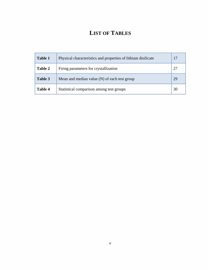
Table 1 Physical characteristics and properties of lithium disilicate 17
Table 2 Firing parameters for crystallization 27
Table 3 Mean and median value (N) of each test group 29
Table 4 Statistical comparison among test groups 30
v

LIST OF FIGURES
Figure 1 TiN coating under the dark field light microscopy 13
Figure 2 Summary of zirconia surface treatments 15
Figure 3 Custom abutment fabrication 20
Figure 4a Abutment type: Titanium with no groove 21
Figure 4b Abutment type: Titanium with retentive grooves 21
Figure 4c Abutment type: Titanium gold hue 21
Figure 4d Abutment type: Titanium gold hue with retentive grooves 21
Figure 4e Abutment type: Zirconia 21
Figure 5 Maxillary right central incisor implant crown wax-up 22
Figure 6 Maxillary right central incisor implant crown modified wax-up 22
Figure 7 Scanning wax-up model 23
Figure 8 NobelProcera Scanner 24
Figure 9 Vita Vacuumat 40 24
Figure 10 Premier Implant Cement 25
Figure 11a Cementation procedures: Abutment attached to the analog 25
Figure 11b Cementation procedures: Cement application 25
Figure 11c Cementation procedures: Excess cement removal 25
Figure 11d Cementation procedures: Cementation pressure 25
vi

Figure 12a Testing equipment: Universal testing machine, MTS 858 Mini Bionix II 26
Figure 12b Testing equipment: Pull-up test apparatus 26
Figure 12c Testing equipment: Close-up attachment 26
Figure 13 Mean and median value (N) of each test group 29
vii

CHAPTER 1: INTRODUCTION
Successful implant restorative therapy depends not only on the osseointegrated implant
but also the integrity of the prosthesis that delivers function to the oral cavity.1 When a
patient is missing a tooth, a tooth-supported fixed dental prosthesis (FDP) or an implant-
supported single crown can be fabricated to restore esthetics and function. The latter has
shown favorable success rates compared to the FDP option.2
Cement-retained implant-supported prostheses, when compared to their screw-retained
counterparts, are more advantageous in regards to esthetics and occlusion. This is
because the screw access channel is not incorporated into the crown. It allows the crown
to have a continuous coverage and structural integrity without the need to add a
restorative material to fill the screw access channel.3 Cement-retained restorations are,
however, more challenging to retrieve in case of abutment screw loosening or fracture,
abutment fractures, or necessary surgical re-intervention.4
To overcome the difficulty of the retrieval process, temporary cements, such as zinc-
oxide eugenol and non-eugenol zinc-oxide, have been suggested for cementation of
implant restorations.5 This, in turn, may result in more frequent crown decementation that
can lead to more chairside time for the restorative dentist.4 In order to minimize this
complication, several studies have recommended the use of glass-ionomer, zinc
phosphate, and resin composite luting agents. These cements have shown to enhance the
1

cement failure loads of the prostheses on titanium abutments in comparison to provisional
luting agents.6-8
Various custom abutment types and designs are available to clinicians for implant fixed
restorations. With high esthetic demands in the maxillary anterior region, gold-shaded
titanium nitride-coated abutments and zirconia abutments have been popular materials of
choice.9,10
The aim of this study was to investigate the effects of different abutment materials and
surface designs on the tensile bond strength of resin-based temporary cement with a
lithium disilicate crown. The results from this study will allow clinicians to select the
most optimal material and design when considering esthetics and function for each
clinical case.
2

CHAPTER 2: LITERATURE REVIEW
1. FIXED DENTAL PROSTHESIS VS. IMPLANT-SUPPORTED FIXED PROSTHESES
As dental implants become a more established treatment option, increasing number of
patients with partial edentulism are opting for implant-supported fixed prostheses (ISFP)
as an alternative to traditional fixed dental prostheses (FDP).2 Studies show comparable
survival rates between FDP and ISFP in restoring a missing tooth.11
The most frequent complications for FDP are biological complications, such as loss of
abutment tooth vitality and caries, followed by technical complications, such as loss of
retention caused by cement fracture.12 With ISFD, technical complications are more
common than biological complications. Technical complications for ISFD include
abutment screw loosening and fracture of veneering material, while biological
complications include peri-mucositis and peri-implantitis. The incidence of technical
complications of ISFD is significantly higher than that of FDP.13
3

2. SCREW-RETAINED VS. CEMENT-RETAINED RESTORATIONS
There are two different techniques used to provide restorations for implant-supported
prostheses: screw-retained and cement-retained prostheses. The advantages and
disadvantages of each method have been discussed in the literature in terms of esthetics,
occlusion, porcelain fracture resistance, passive fit, and other considerations.4,14-16
Screw-retained prostheses provide a great advantage over cement-retained restorations in
terms of their retrievability. Screw access channel provides ease of retrieval of the
implant superstructure in the event of abutment screw loosening, and surgical
reintervention.17 In order to provide the same benefits, implant provisional cements were
suggested for ease of cement-retained crown retrieval.4,5,18
Inadequate retention under function and water solubility were encountered when non-
eugenol zinc oxide or zinc oxide eugenol cements were used. Mehl19 studied
retrievability of cement-retained crowns with five cement types and concluded that zinc
phosphate and glass ionomer cement are suitable for “semi-permanent cementation”.
Additionally, the pull-out test by Sheets20 with various cements including resin-based
provisional cement provides data ranking the mean value of load required to break the
bond between the abutment and the casting. Resin-based provisional cement commonly
used for implant restoration had an average value of 131.6N (+/-31.8) in this study.
4

Another study showed that resin adhesive provisional cement had the strongest retentive
values, while non-eugenol zinc-oxide exhibited the lowest retentive values. The author
suggested that non-eugenol zinc-oxide could be the most appropriate cement when
retrievability and removal of the provisionally cemented superstructure is anticipated.21
Despite the main advantage of retrievability for screw-retained restorations, cement-
retained restorations have several distinctive advantages. An example of this can be seen
in the maxillary anterior region. Although prosthetically-driven implant surgery has been
heavily emphasized for the optimal function and longevity of implant superstructure, the
challenges of implant placement with ideal angulation still remains in the anterior
maxilla.22 The presence of anatomical undercuts in the apical portion of the anterior
maxilla restricts the ideal positioning of the implant, which may affect the screw access
channel by making it appear on the facial surface instead of lingual to the incisal edge.23
To manage concerns regarding the visibility of the screw access channel, cement-retained
implant crowns on custom abutments have been recommended in the esthetic zone,
making the restoration more esthetically pleasing. In addition, patients may prefer
cement-retained implant restoration in the mandibular arch as the color difference of the
composite resin restoration in the screw access channel compared to the surrounding
porcelain may be unacceptable esthetically.
Occlusal stability is another topic of interest in comparison between screw-retained and
cement-retained implant restorations. When the screw access channel is sealed with
5

composite resin, ideal occlusion may be compromised particularly if the opposing
restoration has porcelain occlusion. Ekfeldt24 documented that the contacts established on
the composite resin in the screw access channel opposing a porcelain restoration is not
stable when it comes to long term prognosis due to wear. Considering this, cement-
retained implant restorations may provide more predictable occlusal stability.
Torrado25 concluded that, regardless of where the screw access opening is on the occlusal
surface, screw-retained restorations demonstrated significantly lower porcelain fracture
resistance compared to cement-retained restorations. Hebel3 pointed out that esthetics
and occlusion should not be arbitrarily sacrificed in return for retrievability of implant-
supported restorations since the survival rate of implants has significantly improved, and
proper handling of cement-retained restorations may provide retrievability.
In regards to passive fit, Taylor26 stated that cement-retained implant superstructures have
the potential of being completely passive compared to screw-retained restorations;
however, pure passivity of a restoration is extremely rare due to errors accumulating in
each step of prosthesis fabrication.
A non-passively fitting prosthesis can negatively influence the effect of load transfer to
the prosthesis-implant-bone system. This can lead to additional bone loss and the
migration of microorganisms in the gap between the implant and the abutment. Guichet27
studied the relationship of marginal discrepancy and passive fit of screw-retained and
6

cement-retained implant fixed dental prosthesis designs. He concluded that cement-
retained FDP had significantly less stress concentrations around the implants related to its
passive fit. This was explained by noting that the cement layer compensated for the errors
accumulated during prosthesis fabrication. It is also suggested that the intervening cement
layer acts as a shock absorber and enhances the transfer of load from prosthesis through
the implant to the bone.28 Additionally, prosthetic complications such as loosening or
fracture of abutment screws and implant fracture can occur due to non-passively fitting
framework.
Although cement-retained restorations have many benefits, a critical concern and
drawback is the possibility of residual cement left in the sulcus after cementation.29
Residual excess cement may cause bone loss around implants and potentially lead to
implant loss.30 The signs of peri-implantitis caused by excess cement include swelling,
bleeding on probing, deep pocket depth, and radiographic loss of peri-implant bone.
Some of these signs of inflammation may not appear for years after the restoration has
been cemented. In order to reduce residual excess cement on the implant surface, it is
recommended to use individually designed abutments by the use of casting or computer-
aided design/computer-aided manufacturing (CAD/CAM) technology, which brings the
restoration margin closer to the free gingival margin and eases the excess cement removal
process.31
7

Another way to reduce the possibility of leaving residual cement on the implant or
abutment surface is to create a practice abutment or an abutment analog. It can be done
by taking an impression of the abutment, which then can be poured with pattern resin
material.32 The restoration is filled with cement and placed onto the abutment analog.
After excess cement is removed, the restoration is then seated onto the definitive
abutment. Lastly, a screw access channel can be incorporated within the crown during the
fabrication process. This allows for extraoral cementation of the crown on the abutment
and for the excess cement to escape through the screw access channel when the
restoration is cemented.33 A disadvantage of this technique is that the screw access
channel will need to be sealed with a restorative material which may be an esthetic
concern and may increase the risk of porcelain fracture as seen in screw-retained implant
restorations.4
8

3. FACTORS CONTRIBUTING TO RETENTION OF IMPLANT RESTORATION
The factors that affect the retention of restorations on implants are similar to those factors
that affect the retention of restorations on natural teeth. Implant manufacturers machine
their implant abutments with 6 degrees of taper, which is documented to be ideal.34
Considering the fact that most clinicians prepare teeth with taper between 15 and 25
degrees, cement-retained restorations can have significantly improved retention
compared to full cusp coverage restorations on natural teeth, assuming all other factors
are constant.35
A rough axial wall surface improves the retention of the restoration, and can be produced
by either a diamond bur or airborne particle abrasion.36 Most studies involving air
particle abrasion on titanium surfaces 37-39 use 50 μm aluminum oxide particles at a
pressure of 2 bars for 10 seconds with a distance of 10 mm between the specimen and the
sandblast gun tip. The sandblasted surface increases the retention, but the degree of
increase depends on the type of cement.1 Although more retention can be achieved with a
roughened surface, it is not necessary to modify the surface if ideal taper and sufficient
axial wall heights are present.4
Retention and resistance forms can be improved by modifying the abutment surface area
and axial wall height.40 Compared to stock abutments, the surface area and axial wall
height of implant custom abutments can be created ideally.41 A stock abutment used in a
molar site may have a compromised retention form due to the discrepancy in size
9

between the implant and the partially edentulous space. With the emergence of computer
aided design of custom abutments, it became possible to increase the abutment surface
area to resemble the natural tooth morphology.7
Definitive cements are becoming more popular for implant-supported restorations since
the survival rate of dental implants is increasing and the prosthetic component connection
is becoming more stable, although complications and concerns still remain with
loosening and/or fracture of abutment screws.42 These cements should only be used when
retrieval of prosthesis and surgical intervention for peri-implantitis is not anticipated.5
10

4. IMPLANT ABUTMENT MATERIAL
4.1. TITANIUM ABUTMENT
Commercially pure titanium and titanium alloys have been used in dentistry for more
than two decades because of their superior biocompatibility when compared to other
metals used for dental prostheses.43 The most commonly used titanium alloy used is
titanium with 6% aluminum and 4% vanadium (Ti-6Al-4V), which can be designed and
machined using CAD/CAM technology.44 Although its metallic color showing through
soft tissue can compromise esthetics, its desirable mechanical and physical properties
compared to ceramic abutments have made it a popular material of choice.45
4.2. TITANIUM ABUTMENT WITH ANODIZED SURFACE
Titanium oxide is a thin film of approximately 20 nm that allows light to reflect off the
underlying titanium alloy which produces a gray hue.46,47 This thin layer causes light
interference and can give off different wavelengths of the visible light spectrum. The
desired colors such as yellow and pink can mask the color of titanium alloy and be useful
for maxillary anterior restorations.48
The anodization process involves titanium alloy being connected to a positive electric
probe, which is then submerged into an electrolytic solution. When a voltage is applied to
the electrolyte, electrons are deposited onto the titanium by a self-limiting process.49 The
thickness of a post-anodized TiO2 layer is close to that of the visible light wavelength and
can produce optical interference patterns.50
11
This anodized surface does not alter the surface chemistry but maintains the surface
biocompatibility.51 This inexpensive, simple, rapid technique can improve esthetics of the
restoration and soft tissue for an implant restoration.52
4.3. TITANIUM ABUTMENT WITH TITANIUM NITRIDE COATING
Titanium Nitride (TiN) is an extremely hard ceramic that can be prepared by direct
reaction of titanium or titanium hydrogen powder with nitrogen at 1200 ºC. It has general
properties such as great hardness (2000 kg/mm2), high decomposition temperature (2949
ºC), and superconductivity.53 Additionally, TiN coating enhances other materials such as
titanium alloy with its excellent biological properties, hardness, resistance to corrosion,
and decorative yellowish color.9,54 This gold-hue color was adapted to the implant
abutment industry to obtain esthetically pleasing results under the soft tissue in the
esthetic zone.10
There are multiple techniques to achieve a TiN coating on titanium alloy (TiAl6V4),
which include nitrogen ion implantation, physical vapor deposition, and plasma ion
nitriding.55 Galetz56 concluded in his study that physical vapor deposition (PVD) of TiN,
which is the most common technique to prepare TiN coating on titanium alloy, is shown
to increase roughness due to multiple small pits and pinholes found on the surface of TiN
as it shows in the image of dark field light microscopy (Figure 1). These defects are
typically generated during the growth of the coating in the PVD process.
12

Figure 1. TiN coating under the dark field light microscopy
4.4. ZIRCONIA ABUTMENT
Zirconia is a crystalline dioxide of zirconium, which can be organized in three different
patterns: monoclinic, cubic, and tetragonal. It has mechanical properties similar to those
of stainless steel with a compression resistance of about 2000 MPa.57 Mixing ZrO2 with
other metallic oxides such as MgO, CaO, or Y2O3 can result in great molecular stability.
Yttrium-stabilized zirconia, also known as tetragonal zirconia polycrystal, is currently the
most studied combination. It has better mechanical properties than other combinations
although it is more difficult to sinter.58
One of the great properties of zirconia is transformation toughening, which happens when
a force on the zirconia surface induces volumetric changes in the crystalline structure.
13
During this process, the transformation from tetragonal to monoclinic seals the expansion
of cracks.59 There are some concerns, however, regarding zirconia material. Exposure to
moisture for an extended period of time, such as in an oral environment, can cause
changes in the physical properties of zirconia. This phenomenon where zirconia is subject
to aqueous and low temperature degradation is called “zirconia aging”. Surface grinding
can also introduce microcracks on the surface, thus reducing toughness.60
Zirconia abutments are available as prefabricated and customized abutments. The custom
abutment is favored over a prefabricated abutment because of the ability to control
emergence profile and to locate the margin for residual cement removal.61 Although
zirconia abutments have earlier reports of high success rates, particularly in the anterior
region where lower forces are applied, 62-64 some concerns remain regarding the long-
term performance of zirconia abutments. These concerns include the fracture tendency of
zirconia abutments with the narrow diameter implants65 and fritting between the zirconia
abutment and titanium implant that may lead to misfit of the component, damage to
implant surface, promotion of micromotion, and mechanical failure.66
In order to maximize the esthetic results, all-ceramic restorations are preferred as a
material of choice for zirconia abutments.67 The surface of zirconia abutments can be
treated to improve bonding with all-ceramic restorations and provide better longevity of
the restorations. Bonding of resin cement with zirconia is shown to reduce microleakage
and increases retention.68,69 Because zirconia is not etchable, additional surface treatment
14

may be necessary to achieve stronger adhesion using resin cement.70,71 Tzanakakis 72
summarized micromechanical and chemical bonding techniques available for zirconia
surfaces as seen in Figure 2.
Figure 2. Summary of zirconia surface treatments
15
5. ALL-CERAMIC CROWN
Ceramic materials deliver excellent esthetics and function in the anterior and posterior
regions of the oral cavity. The two most common ceramic materials are zirconia and
lithium disilicate. Zirconia has greater mechanical strength, but less light translucency
than lithium disilicate. Both materials have been reported to have complications of
cracking, chipping, and fracturing veneer porcelain material.73
Lithium disilicate glass-ceramic was introduced into the market in 1998 as IPS Empress 2
(Ivoclar Vivadent), but due to its higher failure rate as a FDP framework, this material
was discontinued.74 The newer version of lithium disilicate, which is a pressable and
machinable monolithic material, has been marketed since 2005 as IPS e.max. Through a
different firing process, e.max was made with improvements to its translucency and
flexural strength (360 Mpa) when compared to IPS Empress 2.75 This material soon
became popular among practitioners who desire a metal-free restoration with good
strength and esthetics.76
A systematic review on lithium disilicate performance indicates that it shows excellent
survival rates in the short- term, but the evidence for medium-term survival rates is
limited. The study also indicates that the majority of failures occurred in the posterior
region and that lithium disilicate FDPs are discouraged in this area.77 The physical
properties of lithium disilicate are listed in the table 1.78
16

Table 1. Physical characteristics and properties of lithium disilicate
Physical properties LS2 complete cystallized state
Biaxial strength 360 ± 60Mpa
Vickers hardness 5,800 ± 200 Mpa
Modulus of elasticity 95 ± 5 GPa
Density 2.5 ± 0.1 g/cm3
17

SPECIFIC AIM
To measure the tensile strength between an implant custom abutment of different surface
materials and designs of lithium disilicate glass ceramic crowns when the crowns are
cemented with resin-based temporary cement.
STATEMENT OF THE PROBLEM
Various types of materials and designs of an implant abutment is available to assist
esthetics and function of implant therapy in the maxillary anterior region. There were no
studies found that compared these various types of custom abutments in terms of their
retentiveness when resin-based temporary implant cement is used.
18

NULL HYPOTHESIS (H0)
The difference in surface material and the presence of retentive features on the implant
custom abutment of this specific manufacturer will not influence the retentiveness of a
lithium disilicate all-ceramic crown when cemented with resin-based implant temporary
cement.
ALTERNATE HYPOTHESIS (H1)
The difference in surface material and the presence of retentive feature on the implant
custom abutment of this specific manufacturer will influence the retentiveness of a
lithium disilicate all-ceramic crown when cemented with resin-based implant temporary
cement.
19

CHAPTER 3: METHOD AND MATERIALS
The design of a custom abutment (ATLANTIS abutment; Dentsply implants) made for a
4.0mm by 13mm maxillary right central incisor implant (OsseoSpeed TX; DENTSPLY
implants) from a clinical case (Figure 3) was digitally duplicated with five different types
of abutments by the manufacturer: titanium smooth surface (Ts), titanium with retentive
feature (Tr), titanium gold-hue without retentive feature (Gs), titanium gold-hue with
retentive feature (Gr), and zirconia (Z) (Figure 4).
Figure 3. Custom abutment fabrication
20

Figure 4. Different abutment types: a) Titanium with no groove b) Titanium with
retentive grooves c) Titanium gold hue d) Titanium gold hue with retentive grooves e)
Zirconia
Total convergence taper of the abutment is 6 degrees and the height from the most apical
facial margin to the top of the abutment is 4mm. A wax-up model was created on a
zirconia abutment with pod-like extensions on the mesial and distal sides for the pull-out
test machine to hold on to (Figure 5,6).
21

Figure 5. Maxillary right central incisor implant crown wax-up
Figure 6. Maxillary right central incisor implant crown modified wax-Up
22

The zirconia abutment and the wax model were both scanned with an optical scanner
(NobelProcera; Nobel Biocare, Zurich, Switzerland) (Figure 7,8). The default die space
of the scanner is 70 μm. A total of 50 CAD/CAM lithium disilicate crowns (IPS e.max,
Ivocalar Vivadant, Somerset, NJ) were milled. The shape of each crown and its fit on the
master abutment of each group were examined at the stage of blue-colored intermediated
crystalline phase. Each crown went through the final firing cycle according to the
manufacturer’s recommendation on the duration and temperature of the cycle (Table 2)
(Figure 9).
Figure 7. Scanning wax-up model
23

Figure 8. NobelProcera Scanner Figure 9. Vita Vacuumat 40
The crowns were divided equally into five groups (n =10). With 20 Ncm torque value,
each master abutment was connected on to the implant analog that was embedded in the
clear orthodontic acrylic resin bath (Caulk Orthodontic Resin; Dentsply, Milford, DE).
An inch-long Teflon tape was cut and placed into the screw access channel to protect the
screw hex. A resin-based implant temporary cement (Premier Implant Cement; Premier,
Plymouth Meeting, PA) was dispensed on to a mixing pad between two lines that were 7
mm apart (Figure 10). The distance was determined to standardize the quantity of the
cement. The cement was then loaded into the internal surface of each, and the excess
cement was removed with a micro brush. By monitoring the instant feedback from the
axial load vs. the time graph shown on the MTS 858 Mini Bionix monitor, consistent
finger pressure of 10 Ncm was achieved until complete setting was reached. According to
the manufacturer’s instruction, the working time of the temporary cement is 2 minutes,
and the setting time is 4 minutes from dispensing. (Figure 11)
24

Figure 10. Premier Implant Cement
Figure 11. Cementation procedures a) Abutment attached to the analog b) Cement application c) Excess cement removal d) Cementation pressure
25

The pull-out apparatus was assembled and the crosshead speed was set at 5 mm/min in a
universal testing machine (MTS 858 Mini Bionix II) (Figure 12). The axial load and axial
stroke were recorded against time, and the data of the maximum axial load was collected.
Once the maximum axial load was reached, the crown was separated from the abutment.
The abutment was then cleaned with 0.12% chlorhexidine (Peridex Oral Rinse; 3M, St.
Paul, MN) and a piece of 2 x 2 clinical gauze (Avant Gauze®; Medline, Mundelein, IL).
The specimens were dried and visually inspected to ensure complete removal of the
luting agent. The process was repeated with all five groups.
Figure 12. Testing machine a) Universal testing machine, MTS 858 Mini Bionix II b) Pull-up test apparatus c) Close-up attachment
26

Table 2. Firing parameters crystallization
Firing Temperature (ºC)
Holding Time (min)
Vacuum1 11(ºC) 12(ºC)
Vacuum2 21(ºC) 22(ºC)
Long-term cooling [°C]
Cooling rate [°C/min]
P300
840
7:00
550
770
770
850
700
20
P500
P700
For the statistical analysis, a spreadsheet in an XLS file was created with the types and
designs of custom abutments with the maximum cement failure loads. For group
comparison, one-way analysis of variance (ANOVA) was used to analyze the data. An
overall p-value was generated to find out if there were at least two groups significantly
different from each other (p <0.001). To find out which groups were different, pairwise
comparisons were performed with Tukey-Kramer method for multiple comparison
adjustment. A p-value of <0.05 was considered significant.
Stand-by Temperature (ºC)
Closing Time (min)
Heating Rate [ºC/min]
Firing Temperature (ºC)
Holding Time (min)
Heating Rate [ºC/min]
P300
403
6:00 90 820 0:10 30 P500
P700
27

CHAPTER 4: RESULTS
The null hypothesis stated that the difference in surface material and the presence of
retentive features on the implant custom abutment of this specific manufacturer will not
influence the retentiveness of a lithium disilicate all-ceramic crown when cemented with
resin-based implant temporary cement. The primary analysis involved five test groups
with a total of 50 all-ceramic crowns from which cement failure loads were collected.
The mean, median, minimum and maximum tensile forces required to pull the all-ceramic
crowns from different types and designs of custom abutments are presented in Table 3
and Figure 13.
Each master abutment had a similar interface failure mode that involved adhesive failure
followed by cohesive failure. All of the resin-based provisional cement was found on the
intaglio surface of the first all-ceramic crown tested in each group, but in the second test
and beyond, the cement was found on both the abutment surfaces and the intaglio
surfaces of the all-ceramic crowns.
Overall p-value of one-way ANOVA statistic was calculated (p < 0.001) and concluded
that there were at least two groups that are statistically significant. Pairwise comparisons
were performed with Tukey-Kramer method for multiple comparison adjustment.
28

Table 3. Mean and median value (N) of each test group
Test Groups
Category
N Median Mean (SD) (Min, Max)
Ts 10 94.58 98.13 (13.91) (83.09, 118.5)
Tr 10 96.94 98.36 (6.13) (92.25, 109.9)
Gs 10 102.5 103.2 (5.86) (96.61, 113.6)
Gr 10 123.9 124.4 (17.88) (92.49, 152.7)
Z 10 80.58 84.77 (13.80) (69.03, 109.6)
Figure 13. Mean and median value (N) of each test group
29

The abutment of titanium gold-hue with retentive grooves (Gr) showed the highest
cement failure load followed by titanium gold-hue smooth surface (Gs), titanium with
retentive grooves (Tr), titanium with smooth surface (Ts), and zirconia (Z). There was a
statistically significant increase in retentiveness from Gr compared to all other groups.
(Table 4)
Table 4. Statistical comparison among test groups
The retentive grooves on a titanium abutment did not increase the tensile strength at a
statistically signicant level. Conversely, retentive grooves on titanium gold-hue abutment
did considerably increase the te nsile strength (p < 0.0036). The titanium nitride coating
on titanium abutment showed a slight elevation in the mean value of tensile strength, but
did not have a substantial effect when compared to the control group. The zirconia
GROUP 1 GROUP 2 Estimate Standard Error Adjusted P-value
Gs Ts 5.043 5.5694 0.8933
Gs Tr 4.81 5.5694 0.9086
Gs Gr -21.267 5.5694 0.0036 *
Gs Z 18.4 5.5694 0.0153 *
Ts T -0.233 5.5694 1
Ts Gr -26.31 5.5694 0.0002 *
Ts Z 13.357 5.5694 0.1345
Tr Gr -26.077 5.5694 0.0002 *
Tr Z 13.59 5.5694 0.1234
Gr Z 39.667 5.5694 <.0001 *
30

abutment showed the lowest value of cement failure load among all test groups but
demonstrated statistical significance only w ith Gr group.
31

CHAPTER 5: DISCUSSION
The data from this study partially support the acceptance of the null hypothesis; that there
is no difference among Ts, Tr, and Gs groups. With Gr group in comparison to the rest of
the test groups (Ts, Tr, Gs, Z), however, the null hypothesis was rejected. (p < 0.0002,
0.0002, 0.0036, <.0001). In addition, Z group showed statistically significant difference
compared to Gs group. (p < 0.0153)
Cement-retained implant restorations are commonly used in restorative dentistry
especially when it is necessary to hide a screw access channel. In the anterior maxilla, the
implant placement may not be ideal for screw-retained implant restorations, so a cement-
retained restoration may be an excellent alternative to provide the best possible esthetic
results.
One of the most common technical complications with implant restorations are abutment
screw-loosening,79-81 When that complication arises, either the screw needs to be
retightened or replaced with a new one. Another complication that may occur throughout
the life of the implant restoration is peri-implantitis.12,82 When there is an active infection
around the implant, additional treatment is indicated.83 During the treatment, the
restoration may have to be detached from the implant for better prognosis of the
treatment. Other complications include fracture of abutment screws or abutment hex,
32

veneering layer fracture of restoration that requires laboratory repair or fabrication of a
new restoration, and implant loss.84
The screw channel is easily accessible with a screw-retained implant restoration but can
be more challenging to accurately locate in a cement-retained restoration. Provisional
cement has been proposed for cementing implant restorations to simplify access to the
abutment screw. With the benefit of retrievability of provisional cement, there comes a
disadvantage of frequent decementation and increased chairside time for the restorative
dentist. Akca85 concluded in his study that provisional cements have low uniaxial
resistance forces when used with implant-supported crowns, and it may necessitate
frequent recementation of implant-supported crowns. The application of permanent
cement or selecting an abutment that provides improved retention may be necessary for
abutments with decreased axial wall surfaces due to smaller implant sizes. Dramatically
corrected angle of the implant at the abutment level may also lead to significant loss of
abutment wall surface area, which compromises retention of the restoration.86
This investigation was to compare the retentiveness of different abutment surface designs
and materials on which lithium disilicate restorations were cemented with resin-based
implant provisional cement.
No studies were found that reported on the tensile strength of a cement-retained all-
ceramic restoration when cemented with implant provisional cement on either titanium or
33

zirconia abutments. The data from the five test groups resulted in slightly lower mean
value of tensile strength than that of Sheets’20 study, which used the same type of cement
but a cast metal coping as a restoration: the mean uniaxial forces noted in the study was
131.6N (+/-31.8). Considering the smooth intaglio surface of the monolithic lithium
disilicate all-ceramic crown, it is reasonable to conclude that the data of this study is
comparable with that of Sheets’.
The failure mode observed in this study suggests that the finished surfaces of the
abutments are relatively smooth. The first pull-out test resulted in adhesive failure with
most of the cement being inside the lithium disilicate crowns. Successive tests showed
cohesive failures with the cement being on both the abutments and the intaglio surfaces
of the crowns. This implies that the finished surface was removed with the cement layer
during the first pull-out test. This surface may have been created by the manufacturer to
give polished surface characteristics.
The retentive grooves on this particular manufacturer’s titanium abutment may increase
the surface area between the cement layer and the abutment, and also provide small
undercuts for mechanical retention. The data did not show statistically significant
increase of retention from this feature.
The thin coating of titanium nitride on a solid titanium grade 5 alloy (Ti-6Al-4V)
provides warm gold-shaded color. This aids to overcome esthetic challenges in thin soft
34

tissue when using an all-ceramic crown. Galetz56 explained about PVD of titanium nitride
on titanium alloy surfaces and how it creates small pits and pinholes during the growth of
the PVD process. Although there is a slight increase in the mean value of maximum
tensile strength, statistically significant improvement was not observed from this study.
When the retentive grooves and titanium nitride were combined, however, the uniaxial
force of the cement layer break increased significantly. This can be explained by the
synergistic effects of both retention grooves and the microscopic mechanical retention
added from the PVD process.
It is worth noting that the Gr group had significantly better retention compared to Z
group. Gold-hue titanium abutment and zirconia are the material of choice in the esthetic
zone of the anterior maxilla when anticipating superior esthetic results. The results from
this investigation suggest the use of Gr abutments in place of Z abutments when
additional retention is indicated.
Limitations to the current study include small sample size and lack of simulating oral
environment. Some in vitro studies stored cemented restorations in 100% humidity at
37°C for 24 hours, or other studies use artificial saliva and stored them at room
temperature for 24 hours before a pull-out test was performed.21,85,87 This study did not
include this extra step to mimic the oral environment, and it could have affected the data
35
accuracy in terms of numerical value. The relative retentiveness, however, of the data is
still valid because all the test groups were tested under the same condition.
Another source of error could be introduced from non-standardized manufacturer’s
calibration. Because this study heavily relied on the accuracy of virtual technology in
fabricating abutments and crowns, if there were inaccuracies in duplicating abutments
with different types and designs, or in milling lithium disilicate crowns with multiple
machines, it could have possibly caused errors in this study.
The future study is needed to reevaluate the synergistic effects of titanium nitride coating
and mechanical grooves on titanium abutments. The surface treatment to increase
retention on zirconia abutments can be investigated to provide additional data for clinical
decision-making. In order to add more clinically relevant information, the specimen
could undergo a certain number of cycles in a chewing machine, or artificial mouth,
before a pull-out test is executed.
36

CHAPTER 6: SUMMARY AND CONCLUSION
This study was done to determine whether different surface materials and designs of
implant custom abutments affected the retentiveness of all-ceramic crowns when
cemented with a resin-based provisional cement. The null hypothesis was partially
rejected. Based on the limitations of this study, the following conclusions may be drawn:
1. The retentive grooves in this specific manufacturer’s custom abutment do not
significantly affect retention on a titanium alloy abutment (Ti-6Al-4V).
2. The retentive grooves in this specific manufacturer’s custom abutment increase
retention if used on a titanium-nitride-coated titanium abutment.
3. Titanium nitride coating on titanium alloy abutment does not significantly increase
retention.
4. When titanium nitride coating and the retentive grooves are used together, there is
a synergistic effect that increases retention of lithium disilicate crowns.
5. Zirconia abutment showed the lowest mean value of tensile strength although the
difference was only statistically significant with Gs and Gr.
6. When esthetics is a priority in the maxillary anterior region, a titanium abutment
with titanium nitride coating (Gs) or titanium nitride coating with retentive
grooves (Gr) can be used to improve retention of lithium disilicate restorations.
37

REFERENCES
1. Cano-Batalla J, Soliva-Garriga J, Campillo-Funollet M, Munoz-Viveros CA, Giner-
Tarrida L. Influence of abutment height and surface roughness on in vitro retention of
three luting agents. Int J Oral Maxillofac Implants 2012;27:36-41.
2. Muddugangadhar BC, Amarnath GS, Sonika R, Chheda PS, Garg A. Meta-analysis of
failure and survival rate of implant-supported single crowns, fixed partial denture, and
implant tooth-supported prostheses. J Int Oral Health 2015;7:11-17.
3. Hebel KS, Gajjar RC. Cement-retained versus screw-retained implant restorations:
Achieving optimal occlusion and esthetics in implant dentistry. J Prosthet Dent
1997;77:28-35.
4. Michalakis KX, Hirayama H, Garefis PD. Cement-retained versus screw-retained
implant restorations: A critical review. Int J Oral Maxillofac Implants 2003;18:719-728.
5. Breeding LC, Dixon DL, Bogacki MT, Tietge JD. Use of luting agents with an implant
system: Part I. J Prosthet Dent 1992;68:737-741.
6. Kent DK, Koka S, Froeschle ML. Retention of cemented implant-supported
restorations. J Prosthodont 1997;6:193-196.
38

7. Covey DA, Kent DK, St Germain HA,Jr, Koka S. Effects of abutment size and luting
cement type on the uniaxial retention force of implant-supported crowns. J Prosthet Dent
2000;83:344-348.
8. Squier RS, Agar JR, Duncan JP, Taylor TD. Retentiveness of dental cements used with
metallic implant components. Int J Oral Maxillofac Implants 2001;16:793-798.
9. Mezger PR, Creugers NH. Titanium nitride coatings in clinical dentistry. J Dent
1992;20:342-344.
10. Watkin A, Kerstein RB. Improving darkened anterior peri-implant tissue color with
zirconia custom implant abutments. Compend Contin Educ Dent 2008;29:238-40, 242.
11. Pjetursson BE, Tan K, Lang NP, Bragger U, Egger M, Zwahlen M. A systematic
review of the survival and complication rates of fixed partial dentures (FPDs) after an
observation period of at least 5 years. Clin Oral Implants Res 2004;15:667-676.
12. Bragger U, Aeschlimann S, Burgin W, Hammerle CH, Lang NP. Biological and
technical complications and failures with fixed partial dentures (FPD) on implants and
teeth after four to five years of function. Clin Oral Implants Res 2001;12:26-34.
13. Bragger U, Karoussis I, Persson R, Pjetursson B, Salvi G, Lang N. Technical and
biological complications/failures with single crowns and fixed partial dentures on
implants: A 10-year prospective cohort study. Clin Oral Implants Res 2005;16:326-334.
39

14. Johansson LA, Ekfeldt A. Implant-supported fixed partial prostheses: A retrospective
study. Int J Prosthodont 2003;16:172-176.
15. Karl M, Graef F, Taylor TD, Heckmann SM. In vitro effect of load cycling on metal-
ceramic cement- and screw-retained implant restorations. J Prosthet Dent 2007;97:137-
140.
16. Taylor TD, Agar JR. Twenty years of progress in implant prosthodontics. J Prosthet
Dent 2002;88:89-95.
17. Chiche GJ, Pinault A. Considerations for fabrication of implant-supported posterior
restorations. Int J Prosthodont c;4:37-44.
18. Ramp MH, Dixon DL, Ramp LC, Breeding LC, Barber LL. Tensile bond strengths of
provisional luting agents used with an implant system. J Prosthet Dent 1999;81:510-514.
19. Mehl C, Harder S, Wolfart M, Kern M, Wolfart S. Retrievability of implant-retained
crowns following cementation. Clin Oral Implants Res 2008;19:1304-1311.
20. Sheets JL, Wilcox C, Wilwerding T. Cement selection for cement-retained crown
technique with dental implants. J Prosthodont 2008;17:92-96.
21. Michalakis KX, Pissiotis AL, Hirayama H. Cement failure loads of 4 provisional
luting agents used for the cementation of implant-supported fixed partial dentures. Int J
Oral Maxillofac Implants 2000;15:545-549.
40

22. Garber DA, Belser UC. Restoration-driven implant placement with restoration-
generated site development. Compend Contin Educ Dent 1995;16:796, 798-802, 804.
23. Chee WW, Torbati A, Albouy JP. Retrievable cemented implant restorations. J
Prosthodont 1998;7:120-125.
24. Ekfeldt A, Oilo G. Occlusal contact wear of prosthodontic materials. an in vivo study.
Acta Odontol Scand 1988;46:159-169.
25. Torrado E, Ercoli C, Al Mardini M, Graser GN, Tallents RH, Cordaro L. A
comparison of the porcelain fracture resistance of screw-retained and cement-retained
implant-supported metal-ceramic crowns. J Prosthet Dent 2004;91:532-537.
26. Taylor TD, Agar JR, Vogiatzi T. Implant prosthodontics: Current perspective and
future directions. Int J Oral Maxillofac Implants 2000;15:66-75.
27. Guichet DL, Caputo AA, Choi H, Sorensen JA. Passivity of fit and marginal opening
in screw- or cement-retained implant fixed partial denture designs. Int J Oral Maxillofac
Implants 2000;15:239-246.
28. Bidez MW, Misch CE. Force transfer in implant dentistry: Basic concepts and
principles. J Oral Implantol 1992;18:264-274.
29. Pauletto N, Lahiffe BJ, Walton JN. Complications associated with excess cement
around crowns on osseointegrated implants: A clinical report. Int J Oral Maxillofac
Implants 1999;14:865-868.
41

30. Wilson TG,Jr. The positive relationship between excess cement and peri-implant
disease: A prospective clinical endoscopic study. J Periodontol 2009;80:1388-1392.
31. Santosa RE, Martin W, Morton D. Effects of a cementing technique in addition to
luting agent on the uniaxial retention force of a single-tooth implant-supported
restoration: An in vitro study. Int J Oral Maxillofac Implants 2010;25:1145-1152.
32. Dumbrigue HB, Abanomi AA, Cheng LL. Techniques to minimize excess luting
agent in cement-retained implant restorations. J Prosthet Dent 2002;87:112-114.
33. Sharifi MN, Pang IC, Chai J. Alternative restorative techniques of the CeraOne
single-tooth abutment: A technical note. Int J Oral Maxillofac Implants 1994;9:235-238.
34. Wilson AH,Jr, Chan DC. The relationship between preparation convergence and
retention of extracoronal retainers. J Prosthodont 1994;3:74-78.
35. JORGENSEN KD. The relationship between retention and convergence angle in
cemented veneer crowns. Acta Odontol Scand 1955;13:35-40.
36. Felton DA, Kanoy BE, White JT. The effect of surface roughness of crown
preparations on retention of cemented castings. J Prosthet Dent 1987;58:292-296.
37. Ebert A, Hedderich J, Kern M. Retention of zirconia ceramic copings bonded to
titanium abutments. Int J Oral Maxillofac Implants 2007;22:921-927.
42

38. Kim Y, Yamashita J, Shotwell JL, Chong KH, Wang HL. The comparison of
provisional luting agents and abutment surface roughness on the retention of provisional
implant-supported crowns. J Prosthet Dent 2006;95:450-455.
39. Reddy SV, Reddy MS, Reddy CR, Pithani P, R SK, Kulkarni G. The influence of
implant abutment surface roughness and the type of cement on retention of implant
supported crowns. J Clin Diagn Res 2015;9:ZC05-7.
40. Kaufman EG, Coelho AB, Colin L. Factors influencing the retention of cemented
gold castings. J prosthet Dent 1961;9:49-54.
41. Reid PE, Burke TM. Customized implant abutments: Technical notes. Implant Dent
1994;3:243-246.
42. Laney WR, Jemt T, Harris D, Henry PJ, Krogh PH, Polizzi G, et al. Osseointegrated
implants for single-tooth replacement: Progress report from a multicenter prospective
study after 3 years. Int J Oral Maxillofac Implants 1994;9:49-54.
43. Anusvice K, Phillips RW, Shen C, Rawls HR. Phillips' science of dental materials. St.
Louis, Mo: Elsevier/Saunders; 2003.
44. Sghaireen MG. Fracture resistance and mode of failure of ceramic versus titanium
implant abutments and single implant-supported restorations. Clin Implant Dent Relat
Res 2015;17:554-561.
43

45. Leutert CR, Stawarczyk B, Truninger TC, Hammerle CH, Sailer I. Bending moments
and types of failure of zirconia and titanium abutments with internal implant-abutment
connections: A laboratory study. Int J Oral Maxillofac Implants 2012;27:505-512.
46. Wang RR, Fenton A. Titanium for prosthodontic applications: A review of the
literature. Quintessence Int 1996;27:401-408.
47. Nakajima H, Okabe T. Titanium in dentistry: Development and research in the U.S.A.
Dent Mater J 1996;15:77-90.
48. Stavenga DG. Thin film and multilayer optics cause structural colors of many insects
and birds. Materials Today 2014;1S:109-121.
49. Karim MA. Electro-optical displays. London: Taylor and Francis; 1992.
50. Kern P, Schwalier P, Michler J. Electrolytic deposition of titania films as interference
coatings on biomedical implants: Microstructure, chemistry and nano-mechanical
properties. Thin Solid Films 2006;494:279-286.
51. Jaeggi C, Kern P, Michler J, Zehnder T, Siegenthaler H. Anodic thin films of titanium
used as masks for surface micro patterning of biomedical devices. Surf Coat Technol
2005;200:1931-1919.
52. Wadhwani CP, O'Brien R, Kattadiyil MT, Chung KH. Laboratory technique for
coloring titanium abutments to improve esthetics. J Prosthet Dent 2016;115:409-411.
44

53. Toth LE. Transition metal carbides and nitrides. New York, USA: Academic Press;
1971.
54. Wisbey A, Gregson PJ, Tuke M. Application of PVD TiN coating to co-cr-mo based
surgical implants. Biomaterials 1987;8:477-480.
55. Maurer AM, Brown SA, Payer JH, Merritt K, Kawalec JS. Reduction of fretting
corrosion of ti-6Al-4V by various surface treatments. J Orthop Res 1993;11:865-873.
56. Galetz MC, Fleischmann EW, Konrad CH, Schuetz A, Glatzel U. Abrasion resistance
of oxidized zirconium in comparison with CoCrMo and titanium nitride coatings for
artificial knee joints. J Biomed Mater Res B Appl Biomater 2010;93:244-251.
57. Piconi C, Maccauro G. Zirconia as a ceramic biomaterial. Biomaterials 1999;20:1-25.
58. Gupta TK, Bechtold JH, Kuznickie RC, Cadoff LH, Rossing B. Stabilization of
tetragonal phase in polycrystalline zirconia. Journal of Materials Science 1978;13:1464-
1470.
59. Garvie RC, Nicholson PS. Structure and thermodynamically properties of partially
stabilized zirconia in the CaO-ZrO2 system. Journal of American Ceramic Society
1972;55:152-157.
60. Swab JJ. Low temperature degradation of Y-TZP materials. Journal of Materials
Science 1991;26:6706-6714.
45

61. Bidra AS, Rungruanganunt P. Clinical outcomes of implant abutments in the anterior
region: A systematic review. J Esthet Restor Dent 2013;25:159-176.
62. Zembic A, Bosch A, Jung RE, Hammerle CH, Sailer I. Five-year results of a
randomized controlled clinical trial comparing zirconia and titanium abutments
supporting single-implant crowns in canine and posterior regions. Clin Oral Implants Res
2013;24:384-390.
63. Glauser R, Sailer I, Wohlwend A, Studer S, Schibli M, Scharer P. Experimental
zirconia abutments for implant-supported single-tooth restorations in esthetically
demanding regions: 4-year results of a prospective clinical study. Int J Prosthodont
2004;17:285-290.
64. Canullo L. Clinical outcome study of customized zirconia abutments for single-
implant restorations. Int J Prosthodont 2007;20:489-493.
65. Shabanpour R, Mousavi N, Ghodsi S, Alikhasi M. Comparative evaluation of fracture
resistance and mode of failure of zirconia and titanium abutments with different
diameters. J Contemp Dent Pract 2015;16:613-618.
66. Stimmelmayr M, Edelhoff D, Guth JF, Erdelt K, Happe A, Beuer F. Wear at the
titanium-titanium and the titanium-zirconia implant-abutment interface: A comparative in
vitro study. Dent Mater 2012;28:1215-1220.
46

67. Ekfeldt A, Furst B, Carlsson GE. Zirconia abutments for single-tooth implant
restorations: A retrospective and clinical follow-up study. Clin Oral Implants Res
2011;22:1308-1314.
68. Manicone PF, Rossi Iommetti P, Raffaelli L. An overview of zirconia ceramics: Basic
properties and clinical applications. J Dent 2007;35:819-826.
69. Ozcan M, Nijhuis H, Valandro LF. Effect of various surface conditioning methods on
the adhesion of dual-cure resin cement with MDP functional monomer to zirconia after
thermal aging. Dent Mater J 2008;27:99-104.
70. Ernst CP, Aksoy E, Stender E, Willershausen B. Influence of different luting concepts
on long term retentive strength of zirconia crowns. Am J Dent 2009;22:122-128.
71. Qeblawi DM, Munoz CA, Brewer JD, Monaco EA,Jr. The effect of zirconia surface
treatment on flexural strength and shear bond strength to a resin cement. J Prosthet Dent
2010;103:210-220.
72. Tzanakakis EG, Tzoutzas IG, Koidis PT. Is there a potential for durable adhesion to
zirconia restorations? A systematic review. J Prosthet Dent 2016;115:9-19.
73. Chu SJ. Current clinical strategies with lithium-disilicate restorations. Compend
Contin Educ Dent 2012;33:64, 66-7.
74. Helvey G. Ceramics. Compend Contin Educ Dent 2010;31:309-311.
47

75. Conrad HJ, Seong WJ, Pesun IJ. Current ceramic materials and systems with clinical
recommendations: A systematic review. J Prosthet Dent 2007;98:389-404.
76. Christensen GJ. The all-ceramic restoration dilemma: Where are we? J Am Dent
Assoc 2011;142:668-671.
77. Pieger S, Salman A, Bidra AS. Clinical outcomes of lithium disilicate single crowns
and partial fixed dental prostheses: A systematic review. J Prosthet Dent 2014;112:22-30.
78. Joda T, Burki A, Bethge S, Bragger U, Zysset P. Stiffness, strength, and failure
modes of implant-supported monolithic lithium disilicate crowns: Influence of titanium
and zirconia abutments. Int J Oral Maxillofac Implants 2015;30:1272-1279.
79. Jemt T, Laney WR, Harris D, Henry PJ, Krogh PH,Jr, Polizzi G, et al.
Osseointegrated implants for single tooth replacement: A 1-year report from a
multicenter prospective study. Int J Oral Maxillofac Implants 1991;6:29-36.
80. Henry PJ, Laney WR, Jemt T, Harris D, Krogh PH, Polizzi G, et al. Osseointegrated
implants for single-tooth replacement: A prospective 5-year multicenter study. Int J Oral
Maxillofac Implants 1996;11:450-455.
81. Ekfeldt A, Carlsson GE, Borjesson G. Clinical evaluation of single-tooth restorations
supported by osseointegrated implants: A retrospective study. Int J Oral Maxillofac
Implants 1994;9:179-183.
48
82. Behneke A, Behneke N, d'Hoedt B. The longitudinal clinical effectiveness of ITI
solid-screw implants in partially edentulous patients: A 5-year follow-up report. Int J
Oral Maxillofac Implants 2000;15:633-645.
83. Leonhardt A, Dahlen G, Renvert S. Five-year clinical, microbiological, and
radiological outcome following treatment of peri-implantitis in man. J Periodontol
2003;74:1415-1422.
84. Lekholm U, Grondahl K, Jemt T. Outcome of oral implant treatment in partially
edentulous jaws followed 20 years in clinical function. Clin Implant Dent Relat Res
2006;8:178-186.
85. Akca K, Iplikcioglu H, Cehreli MC. Comparison of uniaxial resistance forces of
cements used with implant-supported crowns. Int J Oral Maxillofac Implants
2002;17:536-542.
86. Emms M, Tredwin CJ, Setchell DJ, Moles DR. The effects of abutment wall height,
platform size, and screw access channel filling method on resistance to dislodgement of
cement-retained, implant-supported restorations. J Prosthodont 2007;16:3-9.
87. Pan YH, Ramp LC, Lin CK, Liu PR. Comparison of 7 luting protocols and their
effect on the retention and marginal leakage of a cement-retained dental implant
restoration. Int J Oral Maxillofac Implants 2006;21:587-592.
49

APPENDIX
Table 4. Raw data
Ts Tr Gs Gr Z 1 83.09 92.25 96.61 92.49 69.03 2 83.23 92.32 96.85 107.75 71.28 3 85.16 93.27 97.69 110.44 73.09 4 89.63 93.99 100.37 121.35 76.77 5 92.65 95.86 102.28 122.02 78.65 6 96.51 98.02 102.72 125.71 82.5 7 102.72 100.32 102.89 130.75 88.6 8 111.62 101.04 108.41 138.8 98.71 9 118.12 106.58 110.27 142.32 99.46 10 118.53 109.94 113.6 152.73 109.6
Sum 981.26 983.59 1031.69 1244.36 847.69 Avg 98.13 98.36 103.17 124.44 84.77
Table 5. Statistical analysis raw data
Group 1 Group 2 Estimate Standard Error
Raw P-value
Adjusted P-value
Gs Ts 5.043 5.5694 0.37 0.8933 Gs Tr 4.81 5.5694 0.3924 0.9086 Gs Gr -21.267 5.5694 0.0004 0.0036 Gs Z 18.4 5.5694 0.0019 0.0153 Ts Tr -0.233 5.5694 0.9668 1 Ts Gr -26.31 5.5694 <.0001 0.0002 Ts Z 13.357 5.5694 0.0207 0.1345 Tr Gr -26.077 5.5694 <.0001 0.0002 Tr Z 13.59 5.5694 0.0187 0.1234 Gr Z 39.667 5.5694 <.0001 <.0001
50