Embed Size (px)
Citation preview
Molecular Cell
Review
The CRISPR System: Small RNA-GuidedDefense in Bacteria and Archaea
Fedor V. Karginov1,2,* and Gregory J. Hannon1,2,*1Watson School of Biological Sciences2Howard Hughes Medical InstituteCold Spring Harbor Laboratory, 1 Bungtown Road, Cold Spring Harbor, NY 11724, USA*Correspondence: [email protected] (F.V.K.), [email protected] (G.J.H.)DOI 10.1016/j.molcel.2009.12.033
All cellular systems evolve ways to combat predators and genomic parasites. In bacteria and archaea,numerous resistance mechanisms have developed against phage. Our understanding of this defensiverepertoire has recently been expanded to include the CRISPR system of clustered, regularly interspacedshort palindromic repeats. In this remarkable pathway, short sequence tags from invading genetic elementsare actively incorporated into the host’s CRISPR locus to be transcribed and processed into a set of smallRNAs that guide the destruction of foreign genetic material. Here we review the inner workings of this adapt-able and heritable immune system and draw comparisons to small RNA-guided defense mechanisms ineukaryotic cells.
IntroductionThe battle between predator and prey is perhaps the second-
oldest conflict on earth, and phage may represent one of the
planet’s oldest predators. For bacteria and archaea, phage are
a formidable force, being responsible for 4%–50% of their
destruction (Breitbart and Rohwer, 2005; Rohwer and Thurber,
2009). The predatory challenge is substantial and dynamic;
phage outnumber their prey by 10-fold and benefit from signifi-
cantly greater genome variability and faster rates of mutation
(Hatfull, 2008; Hendrix, 2003).
This diverse and rapidly evolving challenge has prompted the
development of multiple layers of resistance mechanisms in
bacteria. As a first line of defense, bacteria can disrupt phage
adsorption to the cell surface by eliminating or masking the corre-
sponding receptors (Forde and Fitzgerald, 1999). Injection of
phage DNA can also be blocked in some cases. Once within the
bacterial cells, phage DNA is subject to the well-studied restric-
tion/modification systems that degrade foreign DNA. These rely
on differences in methylation status to accomplish self-nonself
recognition and block the activity of sequence-specific nucleases
toward endogenous DNA, while targeting the invaders (Bickle and
Kruger, 1993). Finally, abortive infection systems interfere with
variousaspects of phage replication and packaging, while leading
to death of the host (Chopin et al., 2005). The importance of
evading these defenses for the phage is demonstrated by specific
adaptations that they have evolved in response (Chibani-Chen-
noufi et al., 2004; Forde and Fitzgerald, 1999).
The past several years have brought an understanding of an
additional bacterial and archaeal defense against exogenous
nucleic acids. The CRISPR (clustered regularly interspaced short
palindromic repeats) system is a highly adaptive and heritable
resistance mechanism that incorporates sequences derived
from the foreign element into a small-RNA-based repertoire.
These small RNAs program an enzymatic complex to recognize
and destroy the invader. Conceptually, many aspects of the
CRISPR system are similar to adaptive mechanisms of small
RNA-based defense that protect animal germ cells from mobile
genetic elements (Aravin et al., 2007). In this review, we describe
the recent, substantial progress toward understanding the
CRISPR system and draw parallels to mobile element defense
mechanisms in animals. We would also like to point the reader
to excellent existing reviews on this topic (Sorek et al., 2008;
van der Oost et al., 2009).
Anatomy of a CRISPR LocusThe CRISPR story began with the discovery of a peculiar short
repeat in the E. coli genome by Ishino and coworkers in the
1980s (Ishino et al., 1987; Nakata et al., 1989). Subsequently,
similar repeats were noted in a number of bacteria and archaea
(Bult et al., 1996; Groenen et al., 1993; Hermans et al., 1991; Hoe
et al., 1999; Kawarabayasi et al., 1998, 1999; Klenk et al., 1997;
Masepohl et al., 1996; Mojica et al., 1995; Nelson et al., 1999;
Sensen et al., 1998; She et al., 1998, 2001; Smith et al., 1997).
Mojica and Jansen and their colleagues unified these observa-
tions, coined the CRISPR acronym, and characterized the
CRISPR locus (Jansen et al., 2002; Mojica et al., 2000). In
prokaryotes, genes that impact similar biological processes
often travel through evolution as physically linked units (Galperin
and Koonin, 2000; Overbeek et al., 1999). Accordingly, the inves-
tigators also characterized the protein-coding genes that were
often adjacent to the repeat cluster (CRISPR-associated genes
or cas genes). We now know that these genes form elemental
components of the CRISPR defense pathway. Our under-
standing of CRISPR loci and cas genes was further refined and
expanded as more genomic sequence information became
available (Bolotin et al., 2005; Godde and Bickerton, 2006;
Grissa et al., 2007; Haft et al., 2005; Kunin et al., 2007; Lillestol
et al., 2006; Makarova et al., 2002, 2006; Pourcel et al., 2005).
A wealth of this information on CRISPRs and cas genes is now
accessible in the form of online databases and tools (Grissa
et al., 2007, 2008; Oberle et al., 1991). Overall, CRISPR loci
have been found in about 40% of bacterial and in most archaeal
Molecular Cell 37, January 15, 2010 ª2010 Elsevier Inc. 7
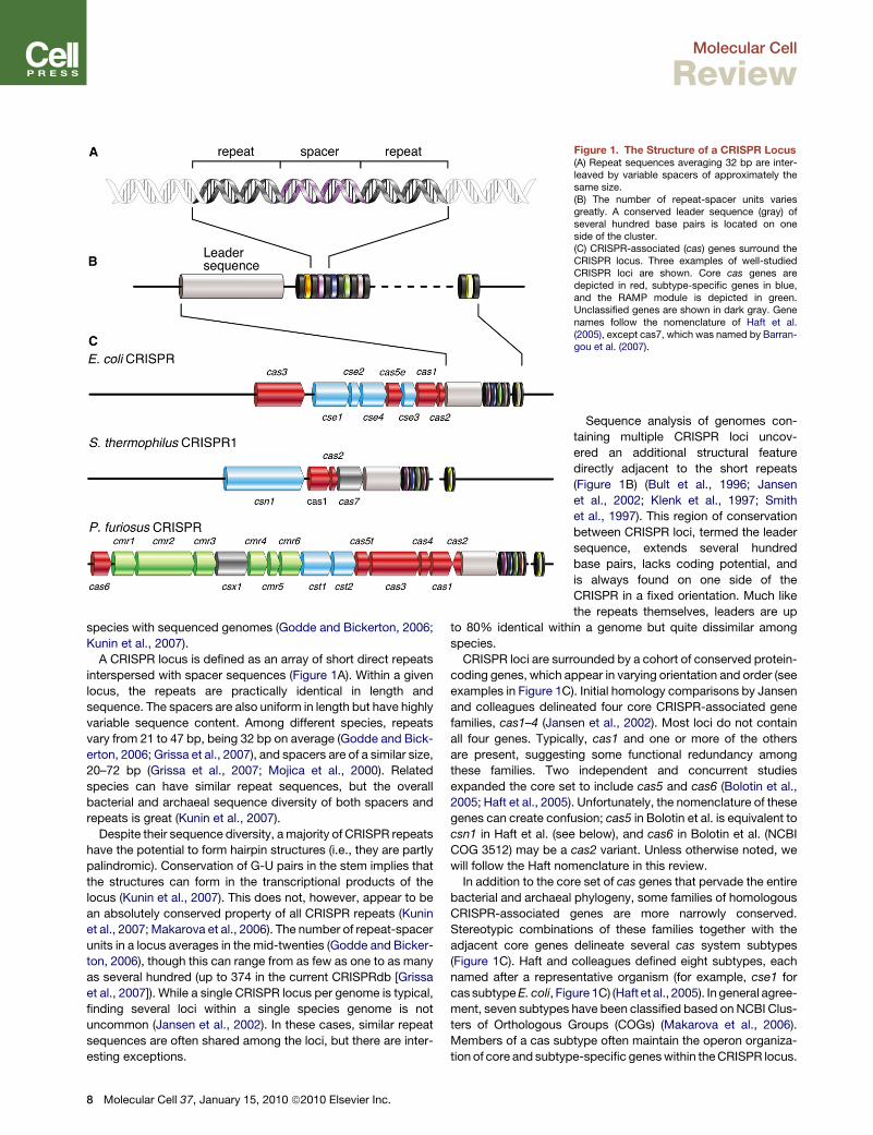
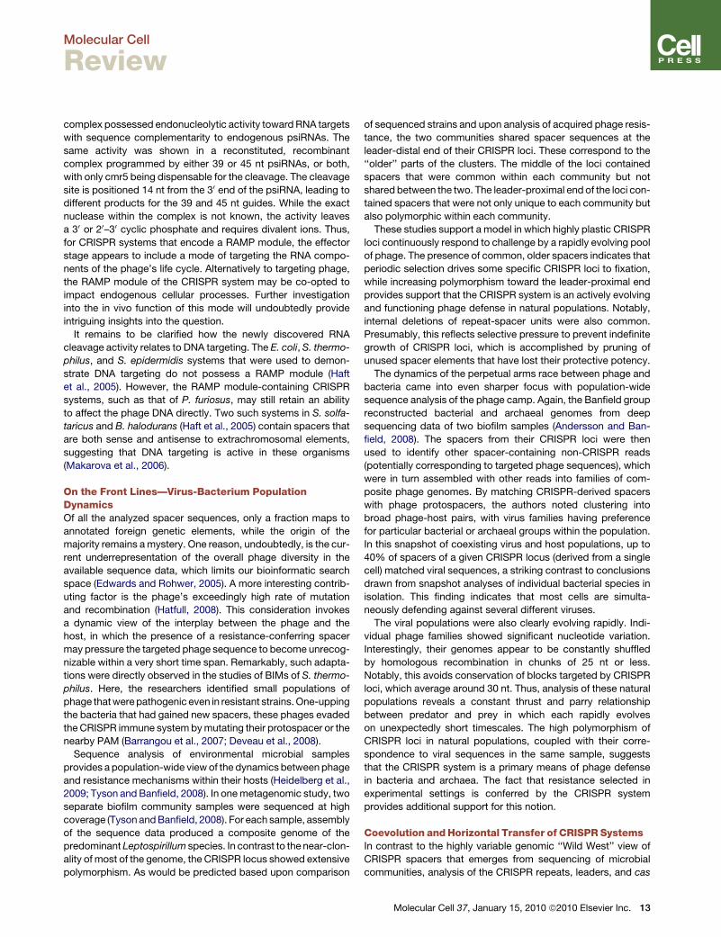
Figure 1. The Structure of a CRISPR Locus(A) Repeat sequences averaging 32 bp are inter-leaved by variable spacers of approximately thesame size.(B) The number of repeat-spacer units variesgreatly. A conserved leader sequence (gray) ofseveral hundred base pairs is located on oneside of the cluster.(C) CRISPR-associated (cas) genes surround theCRISPR locus. Three examples of well-studiedCRISPR loci are shown. Core cas genes aredepicted in red, subtype-specific genes in blue,and the RAMP module is depicted in green.Unclassified genes are shown in dark gray. Genenames follow the nomenclature of Haft et al.(2005), except cas7, which was named by Barran-gou et al. (2007).
Molecular Cell
Review
species with sequenced genomes (Godde and Bickerton, 2006;
Kunin et al., 2007).
A CRISPR locus is defined as an array of short direct repeats
interspersed with spacer sequences (Figure 1A). Within a given
locus, the repeats are practically identical in length and
sequence. The spacers are also uniform in length but have highly
variable sequence content. Among different species, repeats
vary from 21 to 47 bp, being 32 bp on average (Godde and Bick-
erton, 2006; Grissa et al., 2007), and spacers are of a similar size,
20–72 bp (Grissa et al., 2007; Mojica et al., 2000). Related
species can have similar repeat sequences, but the overall
bacterial and archaeal sequence diversity of both spacers and
repeats is great (Kunin et al., 2007).
Despite their sequence diversity, a majority of CRISPR repeats
have the potential to form hairpin structures (i.e., they are partly
palindromic). Conservation of G-U pairs in the stem implies that
the structures can form in the transcriptional products of the
locus (Kunin et al., 2007). This does not, however, appear to be
an absolutely conserved property of all CRISPR repeats (Kunin
et al., 2007; Makarova et al., 2006). The number of repeat-spacer
units in a locus averages in the mid-twenties (Godde and Bicker-
ton, 2006), though this can range from as few as one to as many
as several hundred (up to 374 in the current CRISPRdb [Grissa
et al., 2007]). While a single CRISPR locus per genome is typical,
finding several loci within a single species genome is not
uncommon (Jansen et al., 2002). In these cases, similar repeat
sequences are often shared among the loci, but there are inter-
esting exceptions.
8 Molecular Cell 37, January 15, 2010 ª2010 Elsevier Inc.
Sequence analysis of genomes con-
taining multiple CRISPR loci uncov-
ered an additional structural feature
directly adjacent to the short repeats
(Figure 1B) (Bult et al., 1996; Jansen
et al., 2002; Klenk et al., 1997; Smith
et al., 1997). This region of conservation
between CRISPR loci, termed the leader
sequence, extends several hundred
base pairs, lacks coding potential, and
is always found on one side of the
CRISPR in a fixed orientation. Much like
the repeats themselves, leaders are up
to 80% identical within a genome but quite dissimilar among
species.
CRISPR loci are surrounded by a cohort of conserved protein-
coding genes, which appear in varying orientation and order (see
examples in Figure 1C). Initial homology comparisons by Jansen
and colleagues delineated four core CRISPR-associated gene
families, cas1–4 (Jansen et al., 2002). Most loci do not contain
all four genes. Typically, cas1 and one or more of the others
are present, suggesting some functional redundancy among
these families. Two independent and concurrent studies
expanded the core set to include cas5 and cas6 (Bolotin et al.,
2005; Haft et al., 2005). Unfortunately, the nomenclature of these
genes can create confusion; cas5 in Bolotin et al. is equivalent to
csn1 in Haft et al. (see below), and cas6 in Bolotin et al. (NCBI
COG 3512) may be a cas2 variant. Unless otherwise noted, we
will follow the Haft nomenclature in this review.
In addition to the core set of cas genes that pervade the entire
bacterial and archaeal phylogeny, some families of homologous
CRISPR-associated genes are more narrowly conserved.
Stereotypic combinations of these families together with the
adjacent core genes delineate several cas system subtypes
(Figure 1C). Haft and colleagues defined eight subtypes, each
named after a representative organism (for example, cse1 for
cas subtype E. coli, Figure 1C) (Haft et al., 2005). In general agree-
ment, seven subtypes have been classified based on NCBI Clus-
ters of Orthologous Groups (COGs) (Makarova et al., 2006).
Members of a cas subtype often maintain the operon organiza-
tion of core and subtype-specific genes within the CRISPR locus.
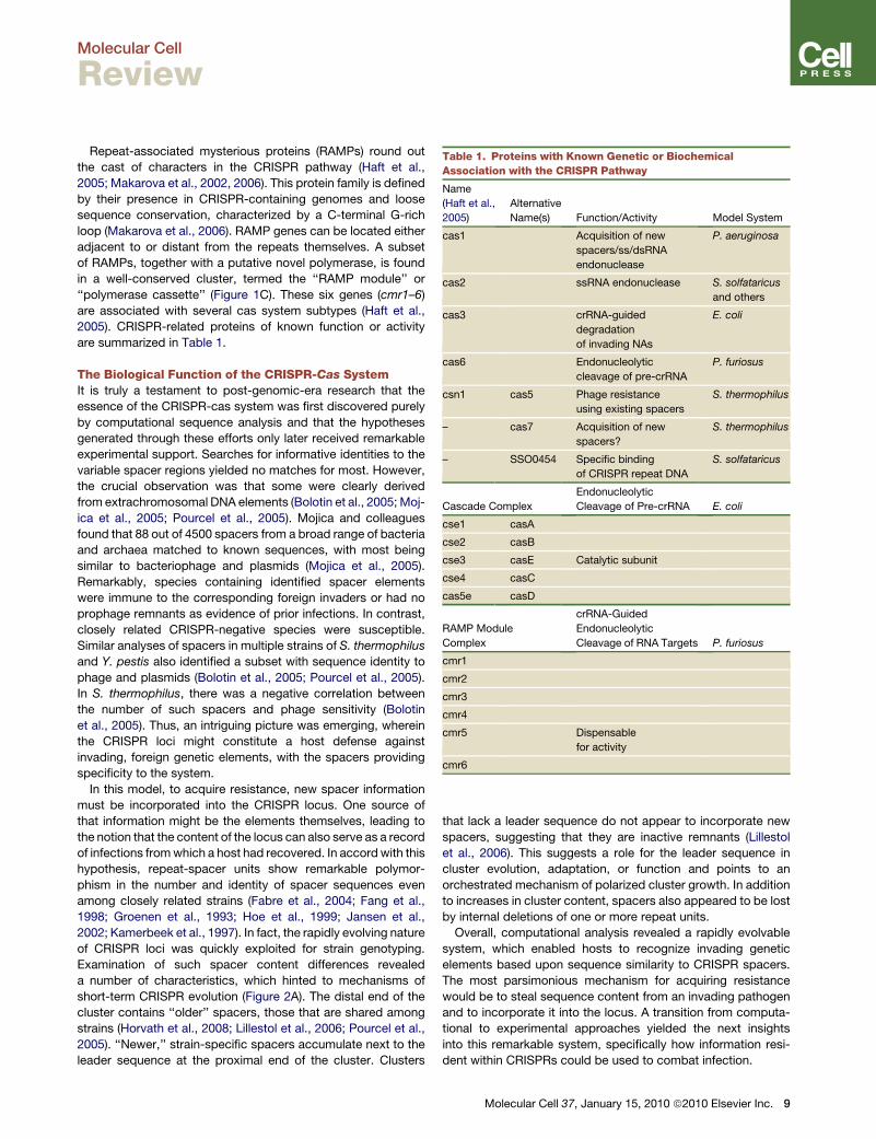
Table 1. Proteins with Known Genetic or Biochemical
Association with the CRISPR Pathway
Name
(Haft et al.,
2005)
Alternative
Name(s) Function/Activity Model System
cas1 Acquisition of new
spacers/ss/dsRNA
endonuclease
P. aeruginosa
cas2 ssRNA endonuclease S. solfataricus
and others
cas3 crRNA-guided
degradation
of invading NAs
E. coli
cas6 Endonucleolytic
cleavage of pre-crRNA
P. furiosus
csn1 cas5 Phage resistance
using existing spacers
S. thermophilus
– cas7 Acquisition of new
spacers?
S. thermophilus
– SSO0454 Specific binding
of CRISPR repeat DNA
S. solfataricus
Cascade Complex
Endonucleolytic
Cleavage of Pre-crRNA E. coli
cse1 casA
cse2 casB
cse3 casE Catalytic subunit
cse4 casC
cas5e casD
RAMP Module
Complex
crRNA-Guided
Endonucleolytic
Cleavage of RNA Targets P. furiosus
cmr1
cmr2
cmr3
cmr4
cmr5 Dispensable
for activity
cmr6
Molecular Cell
Review
Repeat-associated mysterious proteins (RAMPs) round out
the cast of characters in the CRISPR pathway (Haft et al.,
2005; Makarova et al., 2002, 2006). This protein family is defined
by their presence in CRISPR-containing genomes and loose
sequence conservation, characterized by a C-terminal G-rich
loop (Makarova et al., 2006). RAMP genes can be located either
adjacent to or distant from the repeats themselves. A subset
of RAMPs, together with a putative novel polymerase, is found
in a well-conserved cluster, termed the ‘‘RAMP module’’ or
‘‘polymerase cassette’’ (Figure 1C). These six genes (cmr1–6)
are associated with several cas system subtypes (Haft et al.,
2005). CRISPR-related proteins of known function or activity
are summarized in Table 1.
The Biological Function of the CRISPR-Cas SystemIt is truly a testament to post-genomic-era research that the
essence of the CRISPR-cas system was first discovered purely
by computational sequence analysis and that the hypotheses
generated through these efforts only later received remarkable
experimental support. Searches for informative identities to the
variable spacer regions yielded no matches for most. However,
the crucial observation was that some were clearly derived
from extrachromosomal DNA elements (Bolotin et al., 2005; Moj-
ica et al., 2005; Pourcel et al., 2005). Mojica and colleagues
found that 88 out of 4500 spacers from a broad range of bacteria
and archaea matched to known sequences, with most being
similar to bacteriophage and plasmids (Mojica et al., 2005).
Remarkably, species containing identified spacer elements
were immune to the corresponding foreign invaders or had no
prophage remnants as evidence of prior infections. In contrast,
closely related CRISPR-negative species were susceptible.
Similar analyses of spacers in multiple strains of S. thermophilus
and Y. pestis also identified a subset with sequence identity to
phage and plasmids (Bolotin et al., 2005; Pourcel et al., 2005).
In S. thermophilus, there was a negative correlation between
the number of such spacers and phage sensitivity (Bolotin
et al., 2005). Thus, an intriguing picture was emerging, wherein
the CRISPR loci might constitute a host defense against
invading, foreign genetic elements, with the spacers providing
specificity to the system.
In this model, to acquire resistance, new spacer information
must be incorporated into the CRISPR locus. One source of
that information might be the elements themselves, leading to
the notion that the content of the locus can also serve as a record
of infections from which a host had recovered. In accord with this
hypothesis, repeat-spacer units show remarkable polymor-
phism in the number and identity of spacer sequences even
among closely related strains (Fabre et al., 2004; Fang et al.,
1998; Groenen et al., 1993; Hoe et al., 1999; Jansen et al.,
2002; Kamerbeek et al., 1997). In fact, the rapidly evolving nature
of CRISPR loci was quickly exploited for strain genotyping.
Examination of such spacer content differences revealed
a number of characteristics, which hinted to mechanisms of
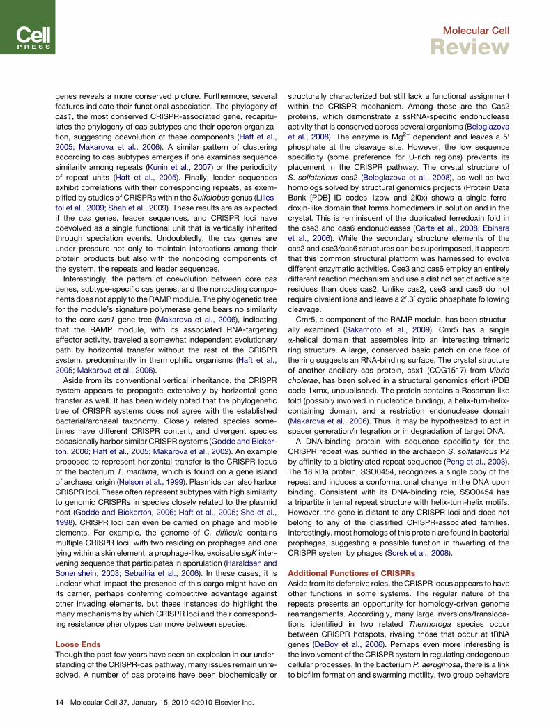
short-term CRISPR evolution (Figure 2A). The distal end of the
cluster contains ‘‘older’’ spacers, those that are shared among
strains (Horvath et al., 2008; Lillestol et al., 2006; Pourcel et al.,
2005). ‘‘Newer,’’ strain-specific spacers accumulate next to the
leader sequence at the proximal end of the cluster. Clusters
that lack a leader sequence do not appear to incorporate new
spacers, suggesting that they are inactive remnants (Lillestol
et al., 2006). This suggests a role for the leader sequence in
cluster evolution, adaptation, or function and points to an
orchestrated mechanism of polarized cluster growth. In addition
to increases in cluster content, spacers also appeared to be lost
by internal deletions of one or more repeat units.
Overall, computational analysis revealed a rapidly evolvable
system, which enabled hosts to recognize invading genetic
elements based upon sequence similarity to CRISPR spacers.
The most parsimonious mechanism for acquiring resistance
would be to steal sequence content from an invading pathogen
and to incorporate it into the locus. A transition from computa-
tional to experimental approaches yielded the next insights
into this remarkable system, specifically how information resi-
dent within CRISPRs could be used to combat infection.
Molecular Cell 37, January 15, 2010 ª2010 Elsevier Inc. 9
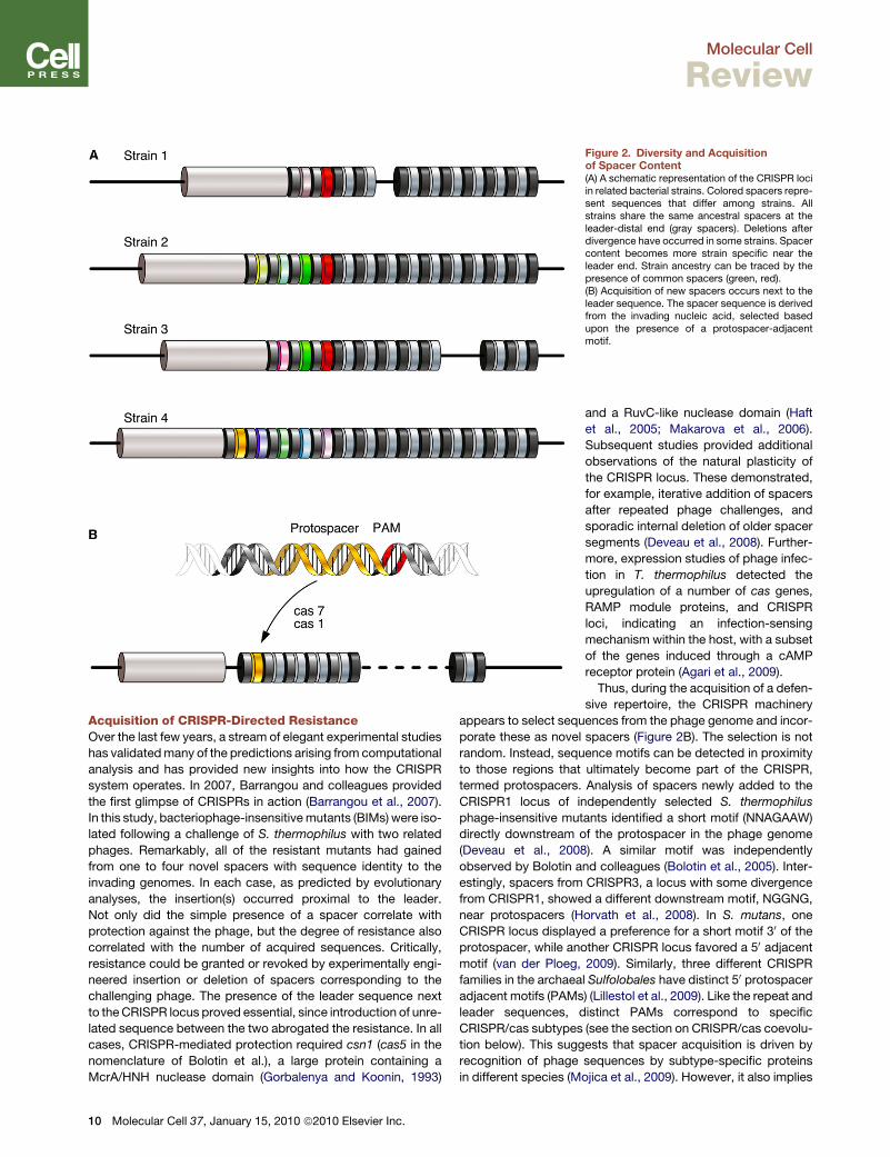
Figure 2. Diversity and Acquisitionof Spacer Content(A) A schematic representation of the CRISPR lociin related bacterial strains. Colored spacers repre-sent sequences that differ among strains. Allstrains share the same ancestral spacers at theleader-distal end (gray spacers). Deletions afterdivergence have occurred in some strains. Spacercontent becomes more strain specific near theleader end. Strain ancestry can be traced by thepresence of common spacers (green, red).(B) Acquisition of new spacers occurs next to theleader sequence. The spacer sequence is derivedfrom the invading nucleic acid, selected basedupon the presence of a protospacer-adjacentmotif.
Molecular Cell
Review
Acquisition of CRISPR-Directed ResistanceOver the last few years, a stream of elegant experimental studies
has validated many of the predictions arising from computational
analysis and has provided new insights into how the CRISPR
system operates. In 2007, Barrangou and colleagues provided
the first glimpse of CRISPRs in action (Barrangou et al., 2007).
In this study, bacteriophage-insensitive mutants (BIMs) were iso-
lated following a challenge of S. thermophilus with two related
phages. Remarkably, all of the resistant mutants had gained
from one to four novel spacers with sequence identity to the
invading genomes. In each case, as predicted by evolutionary
analyses, the insertion(s) occurred proximal to the leader.
Not only did the simple presence of a spacer correlate with
protection against the phage, but the degree of resistance also
correlated with the number of acquired sequences. Critically,
resistance could be granted or revoked by experimentally engi-
neered insertion or deletion of spacers corresponding to the
challenging phage. The presence of the leader sequence next
to the CRISPR locus proved essential, since introduction of unre-
lated sequence between the two abrogated the resistance. In all
cases, CRISPR-mediated protection required csn1 (cas5 in the
nomenclature of Bolotin et al.), a large protein containing a
McrA/HNH nuclease domain (Gorbalenya and Koonin, 1993)
10 Molecular Cell 37, January 15, 2010 ª2010 Elsevier Inc.
and a RuvC-like nuclease domain (Haft
et al., 2005; Makarova et al., 2006).
Subsequent studies provided additional
observations of the natural plasticity of
the CRISPR locus. These demonstrated,
for example, iterative addition of spacers
after repeated phage challenges, and
sporadic internal deletion of older spacer
segments (Deveau et al., 2008). Further-
more, expression studies of phage infec-
tion in T. thermophilus detected the
upregulation of a number of cas genes,
RAMP module proteins, and CRISPR
loci, indicating an infection-sensing
mechanism within the host, with a subset
of the genes induced through a cAMP
receptor protein (Agari et al., 2009).
Thus, during the acquisition of a defen-
sive repertoire, the CRISPR machinery
appears to select sequences from the phage genome and incor-
porate these as novel spacers (Figure 2B). The selection is not
random. Instead, sequence motifs can be detected in proximity
to those regions that ultimately become part of the CRISPR,
termed protospacers. Analysis of spacers newly added to the
CRISPR1 locus of independently selected S. thermophilus
phage-insensitive mutants identified a short motif (NNAGAAW)
directly downstream of the protospacer in the phage genome
(Deveau et al., 2008). A similar motif was independently
observed by Bolotin and colleagues (Bolotin et al., 2005). Inter-
estingly, spacers from CRISPR3, a locus with some divergence
from CRISPR1, showed a different downstream motif, NGGNG,
near protospacers (Horvath et al., 2008). In S. mutans, one
CRISPR locus displayed a preference for a short motif 30 of the
protospacer, while another CRISPR locus favored a 50 adjacent
motif (van der Ploeg, 2009). Similarly, three different CRISPR
families in the archaeal Sulfolobales have distinct 50 protospacer
adjacent motifs (PAMs) (Lillestol et al., 2009). Like the repeat and
leader sequences, distinct PAMs correspond to specific
CRISPR/cas subtypes (see the section on CRISPR/cas coevolu-
tion below). This suggests that spacer acquisition is driven by
recognition of phage sequences by subtype-specific proteins
in different species (Mojica et al., 2009). However, it also implies

Figure 3. Processing of CRISPR Contentinto crRNAsThe locus is transcribed from the leader sequence,and the RNA is cleaved within the repeat by cse3or cas6. An additional processing step yields themature crRNA/psiRNA consisting of an 8 nt repeattag and the spacer sequence.
Molecular Cell
Review
that when an individual species harbors multiple CRISPR loci
from different subtypes, these represent distinct and compart-
mentalized resistance systems. In addition to its suggested
role in spacer selection, the PAM also appears to be important
at the effector stage of defense, since phage can evade resis-
tance to a particular spacer by mutating this nearby motif
(Deveau et al., 2008).
Presently, the precise mechanisms by which information is
transferred from phage or plasmids into CRISPR loci are
obscure. The original experimental study in S. thermophilus
showed a requirement for cas7 to generate BIMs, but not to
mount a response using existing spacers (Barrangou et al.,
2007). Although cas7 (str0660) resides in the CRISPR1 locus, it
has homology limited to the Streptococcus clade and lacks
any classification by comparative sequence analyses (Haft
et al., 2005; Makarova et al., 2006). Notably, its place in the
mostly syntenic CRISPR3 locus of the same genome is occupied
by csn2 (Horvath et al., 2008); however, there is no similarity
between the two genes.
Another likely participant in resistance acquisition is the hall-
mark CRISPR gene, cas1. Cas1 is dispensable for the employ-
ment of existing CRISPR spacers in the effector phase of
defense (see below) and has been proposed to act in making
new spacers, either cleaving foreign DNA or facilitating integra-
tion of new sequences into the CRISPR locus. Sequence anal-
ysis predicted cas1 to be a novel nuclease/integrase (Makarova
et al., 2002). Recently, Wiedenheft and colleagues validated
these predictions, demonstrating a ss/dsDNA endonuclease
activity for P. aeruginosa cas1 (Wiedenheft et al., 2009). The
DNase activity was not sequence or methylation specific and
yielded final products of �80 bp. Interestingly, dsDNA cleavage
required Mn2+ or Mg2+, while ssDNA degradation was only sup-
ported by Mn2+. Moreover, the authors determined the cas1
crystal structure, which revealed a novel a-helical fold and
a single metal ion site. Another study of cas1 from S. solfataricus
Molecular Cell 3
revealed ss/dsDNA and ss/dsRNA bind-
ing and annealing activities but could
not detect any nuclease activity, possibly
due to the somewhat unusual Mn2+
dependence (Han et al., 2009).
As a whole, the aforementioned studies
strongly support a model in which incor-
poration of sequences from invading
nucleic acids into CRISPR loci allows
acquisition of resistance based upon
sequence similarity. Thus far, only the
barest hints have emerged to how this is
accomplished at a mechanistic level.
For example, the factors responsible for
motif recognition and protospacer selection remain unknown,
as do the proteins that ensure proper spacer length.
The Mechanics of CRISPR-Mediated DefenseA major step forward in understanding the effector phase of the
pathway came with the discovery of processed RNAs from the
locus, termed CRISPR RNAs (crRNAs) or prokaryotic silencing
RNAs (psiRNAs) (Figure 3). Transcription of the CRISPR repeats
initiates in or near the leader sequence and generates a long
pre-crRNA precursor that can span the entire locus (Lillestol
et al., 2006, 2009). The pre-crRNA is then endonucleolytically
processed into fragments corresponding to the interval between
repeats, producing mature products and a laddering pattern of
intermediates (Brouns et al., 2008; Carte et al., 2008; Tang
et al., 2002, 2005).
Irregular patterns of transcripts have also been detected from
the opposite strand in S. acidocaldarius (Lillestol et al., 2006;
Lillestol et al., 2009). However, no evidence of antisense products
was seen in S. epidermidis (Marraffini and Sontheimer, 2008),
P. furiosus (Hale et al., 2008), or E. coli (Brouns et al., 2008).
Further studies will be needed to resolve this discrepancy and
to determine the relevance of these products to phage defense.
In E. coli, a complex termed Cascade produces 57 nt units
from the multimeric precursor transcript by cleavage within the
repeat sequence (Brouns et al., 2008). Cascade is comprised
of cse1, cse2, cse4, cas5e, and cse3 (also known as CasA–
CasE). Within the complex, Cse3/casE (cas subtype E. coli 3)
is necessary and sufficient to define the 50 end of the product.
At least two nucleotides are removed from the 30 end of cse3
products by unknown mechanisms. Remarkably, the ortholo-
gous sequence-specific cleavage activity in P. furiosus is carried
out by a different protein, cas6 (Carte et al., 2008). This protein
has no homolog within the E. coli cas operon subtype that
includes cse3 (Haft et al., 2005). The product of cse3 or cas6 is
an RNA consisting of an 8 nt repeat sequence ‘‘tag’’ followed
7, January 15, 2010 ª2010 Elsevier Inc. 11
Molecular Cell
Review
by the spacer sequence, followed by the next partial repeat
(Figure 3).
In P. furiosus, an additional processing step was characterized
that produces two discrete species of mature psiRNA, 38–45 nt
and 43–46 nt, depending on the spacer length (Hale et al., 2008).
This final step is presumed to be exonucleolytic. The resulting
RNAs maintain the 50 repeat tag but lose the downstream
repeat-derived sequence (Figure 3) (Carte et al., 2008; Hale
et al., 2008). In E. coli, potentially similar, shorter species can
also be seen on northern blots in addition to the prominent
57-mers (see Figure 2; Brouns et al., 2008), but these have not
been discussed in the literature. In S. acidocaldarius, CRISPR-
derived small RNAs appear as products from 35 to 52 nt,
presumably generated by endonucleolytic cleavage of long
precursors (Lillestol et al., 2006). Thus, at present, the maturation
to a 35–46 nt RNA appears to be a conserved processing
feature. An examination of the ribonucleoprotein complexes
(RNPs) that are assembled on the RNAs revealed that the
precursor and mature crRNAs are found in distinct RNPs,
providing the first details of the processing/assembly pathway
(Hale et al., 2008).
The structures of T. thermophilus cse3 and P. furiosus cas6
explain their common endonucleolytic function. These proteins
display similar architectures, despite their lack of sequence
homology (Carte et al., 2008; Ebihara et al., 2006; van der Oost
et al., 2009). Both enzymes are composed of a duplicated ferre-
doxin fold, a common domain topology that also underlies the
well-known RNA-recognition motif (RRM) domain. However,
the conserved sequence signatures of RRMs are absent in
cse3 and cas6. The two proteins contain a spatially conserved
active site with an essential histidine residue and a G-rich loop
(van der Oost et al., 2009). The crystal structure of T. thermophi-
lus cse2/casB, another component of the Cascade complex,
reveals a novel a-helical fold with a conserved basic patch that
may be involved in binding RNA (Agari et al., 2008).
Accumulating evidence supports a model in which processed
crRNAs serve as sequence-specific guides during the effector
stage of resistance against invading elements. This was demon-
strated by reconstitution of a functioning CRISPR system in
E. coli BL21(DE3), which lacks endogenous cas genes (Brouns
et al., 2008). These cells were engineered to express the
Cascade complex, as well as Cas3 and a modified CRISPR locus
in which spacer sequences targeting phage lambda had been
incorporated. This was sufficient to create de novo resistance
to the phage and allowed an exploration of the properties of
cas proteins that were important for mounting an effective
response. The catalytic activity of the cse3 nuclease within
Cascade proved essential, indicating a crucial role for crRNAs
in the overall defense pathway. Cas3 is not required for the
generation of crRNAs, as described above, but was necessary
for phage resistance in this system. This fact, along with
a consideration of the domain structure of cas3, has led to the
proposal that it might catalyze crRNA-guided destruction of
foreign nucleic acids. Cas3 has an HD nuclease domain fused
to a DExD/H helicase module (Makarova et al., 2002). The two
domains also exist as separate proteins in the CRISPR loci of
some species, indicating some flexibility in this arrangement
(Makarova et al., 2002). Interestingly, one such stand-alone HD
12 Molecular Cell 37, January 15, 2010 ª2010 Elsevier Inc.
domain in S. solfataricus was demonstrated to possess nucleo-
lytic activity, being able to use either dsDNA or dsRNA as a
substrate (Han and Krauss, 2009). Like cas1, the only remaining
gene in the E. coli CRISPR locus, cas2, was not required for
the effector phase, implicating this gene in some other aspect
of the response.
Obvious analogies to eukaryotic RNAi-related pathways
provoked an initial model in which a crRNA-guided complex
would target mRNAs derived from the invader (Makarova et al.,
2006). However, multiple lines of evidence point to the direct
recognition of foreign DNA by the core CRISPR machinery. To
date, sequence analyses have only detected spacers from
phage with DNA genomes (Mojica et al., 2009; Wiedenheft
et al., 2009). However, any conclusions based upon this obser-
vation must be tempered by the relative scarcity of RNA phage
sequences. Detailed analyses in S. thermophilus (Bolotin et al.,
2005), and more broadly in bacteria and archaea (Makarova
et al., 2006; Shah et al., 2009), show that spacers encode
crRNAs corresponding to both the coding and template strands
of the phage, without a preference for any particular region.
Similar conclusions can be reached by examination of spacers
arising in experimentally induced phage-resistant mutants of
S. thermophilus (Barrangou et al., 2007). Here, some bias toward
the coding strand was observed, but this may be explained by
the higher occurrence of the PAM on that strand (Deveau
et al., 2008). Direct support for DNA rather than mRNA targeting
comes from E. coli, where the use of engineered spacers
demonstrated that effective crRNAs could be produced from
either the coding or template strand of lambda phage (Brouns
et al., 2008).
Additional support for DNA targeting comes from a recent
study of CRISPR activity in a clinical isolate of S. epidermidis,
RP62a (Marraffini and Sontheimer, 2008). Here, the CRISPR
locus contains a spacer against the nickase gene of staphylo-
coccal conjugative plasmids. Since nickase activity is required
for conjugation only in donor cells, targeting of its mRNA would
ablate RP62a’s function as a donor but not as a recipient. The
spacer negated both donor and recipient activity, as predicted
by a DNA-targeting model. Insertion of the protospacer into
a nonconjugative plasmid prevented that plasmid from being
transformed into RP62a, demonstrating that resistance was
not linked to the mode of plasmid entry. The DNA-targeting
model was supported by the observation that the targeted region
was equally effective in either orientation within the plasmid. As
an additional test of the model, Marraffini and Sontheimer
cleverly designed a variant of the plasmid in which the nickase
protospacer was interrupted by a self-splicing intron. This split
the targeted sequence in the plasmid but reformed it in the
encoded mRNA. This construct was capable of conjugation
into RP62a, indicating that the CRISPR defense was circum-
vented when the DNA target was disrupted, but the mRNA target
remained as a potential substrate.
The above evidence notwithstanding, very recent results
demonstrate a capacity for RNA targeting in CRISPR systems
containing the RAMP module. In P. furiosus (Figure 1C), Hale
and colleagues identified the six RAMP module proteins in
a RNP containing the mature 39 and 45 nt psiRNAs with the
shared 8 nt 50 repeat tag (Hale et al., 2009). Remarkably, the
Molecular Cell
Review
complex possessed endonucleolytic activity toward RNA targets
with sequence complementarity to endogenous psiRNAs. The
same activity was shown in a reconstituted, recombinant
complex programmed by either 39 or 45 nt psiRNAs, or both,
with only cmr5 being dispensable for the cleavage. The cleavage
site is positioned 14 nt from the 30 end of the psiRNA, leading to
different products for the 39 and 45 nt guides. While the exact
nuclease within the complex is not known, the activity leaves
a 30 or 20–30 cyclic phosphate and requires divalent ions. Thus,
for CRISPR systems that encode a RAMP module, the effector
stage appears to include a mode of targeting the RNA compo-
nents of the phage’s life cycle. Alternatively to targeting phage,
the RAMP module of the CRISPR system may be co-opted to
impact endogenous cellular processes. Further investigation
into the in vivo function of this mode will undoubtedly provide
intriguing insights into the question.
It remains to be clarified how the newly discovered RNA
cleavage activity relates to DNA targeting. The E. coli, S. thermo-
philus, and S. epidermidis systems that were used to demon-
strate DNA targeting do not possess a RAMP module (Haft
et al., 2005). However, the RAMP module-containing CRISPR
systems, such as that of P. furiosus, may still retain an ability
to affect the phage DNA directly. Two such systems in S. solfa-
taricus and B. halodurans (Haft et al., 2005) contain spacers that
are both sense and antisense to extrachromosomal elements,
suggesting that DNA targeting is active in these organisms
(Makarova et al., 2006).
On the Front Lines—Virus-Bacterium PopulationDynamicsOf all the analyzed spacer sequences, only a fraction maps to
annotated foreign genetic elements, while the origin of the
majority remains a mystery. One reason, undoubtedly, is the cur-
rent underrepresentation of the overall phage diversity in the
available sequence data, which limits our bioinformatic search
space (Edwards and Rohwer, 2005). A more interesting contrib-
uting factor is the phage’s exceedingly high rate of mutation
and recombination (Hatfull, 2008). This consideration invokes
a dynamic view of the interplay between the phage and the
host, in which the presence of a resistance-conferring spacer
may pressure the targeted phage sequence to become unrecog-
nizable within a very short time span. Remarkably, such adapta-
tions were directly observed in the studies of BIMs of S. thermo-
philus. Here, the researchers identified small populations of
phage that were pathogenic even in resistant strains. One-upping
the bacteria that had gained new spacers, these phages evaded
the CRISPR immune system by mutating their protospacer or the
nearby PAM (Barrangou et al., 2007; Deveau et al., 2008).
Sequence analysis of environmental microbial samples
provides a population-wide view of the dynamics between phage
and resistance mechanisms within their hosts (Heidelberg et al.,
2009; Tyson and Banfield, 2008). In one metagenomic study, two
separate biofilm community samples were sequenced at high
coverage (Tyson and Banfield, 2008). For each sample, assembly
of the sequence data produced a composite genome of the
predominant Leptospirillum species. In contrast to the near-clon-
ality of most of the genome, the CRISPR locus showed extensive
polymorphism. As would be predicted based upon comparison
of sequenced strains and upon analysis of acquired phage resis-
tance, the two communities shared spacer sequences at the
leader-distal end of their CRISPR loci. These correspond to the
‘‘older’’ parts of the clusters. The middle of the loci contained
spacers that were common within each community but not
shared between the two. The leader-proximal end of the loci con-
tained spacers that were not only unique to each community but
also polymorphic within each community.
These studies support a model in which highly plastic CRISPR
loci continuously respond to challenge by a rapidly evolving pool
of phage. The presence of common, older spacers indicates that
periodic selection drives some specific CRISPR loci to fixation,
while increasing polymorphism toward the leader-proximal end
provides support that the CRISPR system is an actively evolving
and functioning phage defense in natural populations. Notably,
internal deletions of repeat-spacer units were also common.
Presumably, this reflects selective pressure to prevent indefinite
growth of CRISPR loci, which is accomplished by pruning of
unused spacer elements that have lost their protective potency.
The dynamics of the perpetual arms race between phage and
bacteria came into even sharper focus with population-wide
sequence analysis of the phage camp. Again, the Banfield group
reconstructed bacterial and archaeal genomes from deep
sequencing data of two biofilm samples (Andersson and Ban-
field, 2008). The spacers from their CRISPR loci were then
used to identify other spacer-containing non-CRISPR reads
(potentially corresponding to targeted phage sequences), which
were in turn assembled with other reads into families of com-
posite phage genomes. By matching CRISPR-derived spacers
with phage protospacers, the authors noted clustering into
broad phage-host pairs, with virus families having preference
for particular bacterial or archaeal groups within the population.
In this snapshot of coexisting virus and host populations, up to
40% of spacers of a given CRISPR locus (derived from a single
cell) matched viral sequences, a striking contrast to conclusions
drawn from snapshot analyses of individual bacterial species in
isolation. This finding indicates that most cells are simulta-
neously defending against several different viruses.
The viral populations were also clearly evolving rapidly. Indi-
vidual phage families showed significant nucleotide variation.
Interestingly, their genomes appear to be constantly shuffled
by homologous recombination in chunks of 25 nt or less.
Notably, this avoids conservation of blocks targeted by CRISPR
loci, which average around 30 nt. Thus, analysis of these natural
populations reveals a constant thrust and parry relationship
between predator and prey in which each rapidly evolves
on unexpectedly short timescales. The high polymorphism of
CRISPR loci in natural populations, coupled with their corre-
spondence to viral sequences in the same sample, suggests
that the CRISPR system is a primary means of phage defense
in bacteria and archaea. The fact that resistance selected in
experimental settings is conferred by the CRISPR system
provides additional support for this notion.
Coevolution and Horizontal Transfer of CRISPR SystemsIn contrast to the highly variable genomic ‘‘Wild West’’ view of
CRISPR spacers that emerges from sequencing of microbial
communities, analysis of the CRISPR repeats, leaders, and cas
Molecular Cell 37, January 15, 2010 ª2010 Elsevier Inc. 13
Molecular Cell
Review
genes reveals a more conserved picture. Furthermore, several
features indicate their functional association. The phylogeny of
cas1, the most conserved CRISPR-associated gene, recapitu-
lates the phylogeny of cas subtypes and their operon organiza-
tion, suggesting coevolution of these components (Haft et al.,
2005; Makarova et al., 2006). A similar pattern of clustering
according to cas subtypes emerges if one examines sequence
similarity among repeats (Kunin et al., 2007) or the periodicity
of repeat units (Haft et al., 2005). Finally, leader sequences
exhibit correlations with their corresponding repeats, as exem-
plified by studies of CRISPRs within the Sulfolobus genus (Lilles-
tol et al., 2009; Shah et al., 2009). These results are as expected
if the cas genes, leader sequences, and CRISPR loci have
coevolved as a single functional unit that is vertically inherited
through speciation events. Undoubtedly, the cas genes are
under pressure not only to maintain interactions among their
protein products but also with the noncoding components of
the system, the repeats and leader sequences.
Interestingly, the pattern of coevolution between core cas
genes, subtype-specific cas genes, and the noncoding compo-
nents does not apply to the RAMP module. The phylogenetic tree
for the module’s signature polymerase gene bears no similarity
to the core cas1 gene tree (Makarova et al., 2006), indicating
that the RAMP module, with its associated RNA-targeting
effector activity, traveled a somewhat independent evolutionary
path by horizontal transfer without the rest of the CRISPR
system, predominantly in thermophilic organisms (Haft et al.,
2005; Makarova et al., 2006).
Aside from its conventional vertical inheritance, the CRISPR
system appears to propagate extensively by horizontal gene
transfer as well. It has been widely noted that the phylogenetic
tree of CRISPR systems does not agree with the established
bacterial/archaeal taxonomy. Closely related species some-
times have different CRISPR content, and divergent species
occasionally harbor similar CRISPR systems (Godde and Bicker-
ton, 2006; Haft et al., 2005; Makarova et al., 2002). An example
proposed to represent horizontal transfer is the CRISPR locus
of the bacterium T. maritima, which is found on a gene island
of archaeal origin (Nelson et al., 1999). Plasmids can also harbor
CRISPR loci. These often represent subtypes with high similarity
to genomic CRISPRs in species closely related to the plasmid
host (Godde and Bickerton, 2006; Haft et al., 2005; She et al.,
1998). CRISPR loci can even be carried on phage and mobile
elements. For example, the genome of C. difficule contains
multiple CRISPR loci, with two residing on prophages and one
lying within a skin element, a prophage-like, excisable sigK inter-
vening sequence that participates in sporulation (Haraldsen and
Sonenshein, 2003; Sebaihia et al., 2006). In these cases, it is
unclear what impact the presence of this cargo might have on
its carrier, perhaps conferring competitive advantage against
other invading elements, but these instances do highlight the
many mechanisms by which CRISPR loci and their correspond-
ing resistance phenotypes can move between species.
Loose EndsThough the past few years have seen an explosion in our under-
standing of the CRISPR-cas pathway, many issues remain unre-
solved. A number of cas proteins have been biochemically or
14 Molecular Cell 37, January 15, 2010 ª2010 Elsevier Inc.
structurally characterized but still lack a functional assignment
within the CRISPR mechanism. Among these are the Cas2
proteins, which demonstrate a ssRNA-specific endonuclease
activity that is conserved across several organisms (Beloglazova
et al., 2008). The enzyme is Mg2+ dependent and leaves a 50
phosphate at the cleavage site. However, the low sequence
specificity (some preference for U-rich regions) prevents its
placement in the CRISPR pathway. The crystal structure of
S. solfataricus cas2 (Beloglazova et al., 2008), as well as two
homologs solved by structural genomics projects (Protein Data
Bank [PDB] ID codes 1zpw and 2i0x) shows a single ferre-
doxin-like domain that forms homodimers in solution and in the
crystal. This is reminiscent of the duplicated ferredoxin fold in
the cse3 and cas6 endonucleases (Carte et al., 2008; Ebihara
et al., 2006). While the secondary structure elements of the
cas2 and cse3/cas6 structures can be superimposed, it appears
that this common structural platform was harnessed to evolve
different enzymatic activities. Cse3 and cas6 employ an entirely
different reaction mechanism and use a distinct set of active site
residues than does cas2. Unlike cas2, cse3 and cas6 do not
require divalent ions and leave a 20,30 cyclic phosphate following
cleavage.
Cmr5, a component of the RAMP module, has been structur-
ally examined (Sakamoto et al., 2009). Cmr5 has a single
a-helical domain that assembles into an interesting trimeric
ring structure. A large, conserved basic patch on one face of
the ring suggests an RNA-binding surface. The crystal structure
of another ancillary cas protein, csx1 (COG1517) from Vibrio
cholerae, has been solved in a structural genomics effort (PDB
code 1xmx, unpublished). The protein contains a Rossman-like
fold (possibly involved in nucleotide binding), a helix-turn-helix-
containing domain, and a restriction endonuclease domain
(Makarova et al., 2006). Thus, it may be hypothesized to act in
spacer generation/integration or in degradation of target DNA.
A DNA-binding protein with sequence specificity for the
CRISPR repeat was purified in the archaeon S. solfataricus P2
by affinity to a biotinylated repeat sequence (Peng et al., 2003).
The 18 kDa protein, SSO0454, recognizes a single copy of the
repeat and induces a conformational change in the DNA upon
binding. Consistent with its DNA-binding role, SSO0454 has
a tripartite internal repeat structure with helix-turn-helix motifs.
However, the gene is distant to any CRISPR loci and does not
belong to any of the classified CRISPR-associated families.
Interestingly, most homologs of this protein are found in bacterial
prophages, suggesting a possible function in thwarting of the
CRISPR system by phages (Sorek et al., 2008).
Additional Functions of CRISPRsAside from its defensive roles, the CRISPR locus appears to have
other functions in some systems. The regular nature of the
repeats presents an opportunity for homology-driven genome
rearrangements. Accordingly, many large inversions/transloca-
tions identified in two related Thermotoga species occur
between CRISPR hotspots, rivaling those that occur at tRNA
genes (DeBoy et al., 2006). Perhaps even more interesting is
the involvement of the CRISPR system in regulating endogenous
cellular processes. In the bacterium P. aeruginosa, there is a link
to biofilm formation and swarming motility, two group behaviors
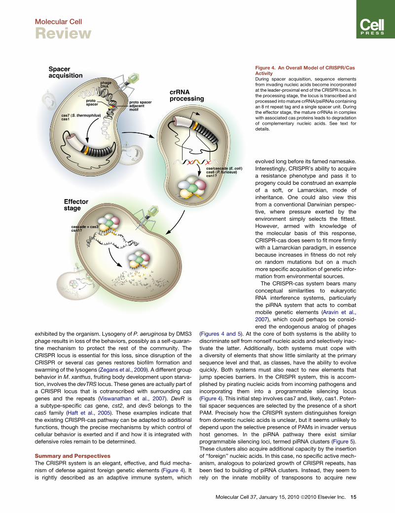
Figure 4. An Overall Model of CRISPR/CasActivityDuring spacer acquisition, sequence elementsfrom invading nucleic acids become incorporatedat the leader-proximal end of the CRISPR locus. Inthe processing stage, the locus is transcribed andprocessed into mature crRNA/psiRNAs containingan 8 nt repeat tag and a single spacer unit. Duringthe effector stage, the mature crRNAs in complexwith associated cas proteins leads to degradationof complementary nucleic acids. See text fordetails.
Molecular Cell
Review
exhibited by the organism. Lysogeny of P. aeruginosa by DMS3
phage results in loss of the behaviors, possibly as a self-quaran-
tine mechanism to protect the rest of the community. The
CRISPR locus is essential for this loss, since disruption of the
CRISPR or several cas genes restores biofilm formation and
swarming of the lysogens (Zegans et al., 2009). A different group
behavior in M. xanthus, fruiting body development upon starva-
tion, involves the devTRS locus. These genes are actually part of
a CRISPR locus that is cotranscribed with surrounding cas
genes and the repeats (Viswanathan et al., 2007). DevR is
a subtype-specific cas gene, cst2, and devS belongs to the
cas5 family (Haft et al., 2005). These examples indicate that
the existing CRISPR-cas pathway can be adapted to additional
functions, though the precise mechanisms by which control of
cellular behavior is exerted and if and how it is integrated with
defensive roles remain to be determined.
Summary and PerspectivesThe CRISPR system is an elegant, effective, and fluid mecha-
nism of defense against foreign genetic elements (Figure 4). It
is rightly described as an adaptive immune system, which
Molecular Cell 3
evolved long before its famed namesake.
Interestingly, CRISPR’s ability to acquire
a resistance phenotype and pass it to
progeny could be construed an example
of a soft, or Lamarckian, mode of
inheritance. One could also view this
from a conventional Darwinian perspec-
tive, where pressure exerted by the
environment simply selects the fittest.
However, armed with knowledge of
the molecular basis of this response,
CRISPR-cas does seem to fit more firmly
with a Lamarckian paradigm, in essence
because increases in fitness do not rely
on random mutations but on a much
more specific acquisition of genetic infor-
mation from environmental sources.
The CRISPR-cas system bears many
conceptual similarities to eukaryotic
RNA interference systems, particularly
the piRNA system that acts to combat
mobile genetic elements (Aravin et al.,
2007), which could perhaps be consid-
ered the endogenous analog of phages
(Figures 4 and 5). At the core of both systems is the ability to
discriminate self from nonself nucleic acids and selectively inac-
tivate the latter. Additionally, both systems must cope with
a diversity of elements that show little similarity at the primary
sequence level and that, as classes, have the ability to evolve
quickly. Both systems must also react to new elements that
jump species barriers. In the CRISPR system, this is accom-
plished by pirating nucleic acids from incoming pathogens and
incorporating them into a programmable silencing locus
(Figure 4). This initial step involves cas7 and, likely, cas1. Poten-
tial spacer sequences are selected by the presence of a short
PAM. Precisely how the CRISPR system distinguishes foreign
from domestic nucleic acids is unclear, but it seems unlikely to
depend upon the selective presence of PAMs in invader versus
host genomes. In the piRNA pathway there exist similar
programmable silencing loci, termed piRNA clusters (Figure 5).
These clusters also acquire additional capacity by the insertion
of ‘‘foreign’’ nucleic acids. In this case, no specific active mech-
anism, analogous to polarized growth of CRISPR repeats, has
been tied to building of piRNA clusters. Instead, they seem to
rely on the innate mobility of transposons to acquire new
7, January 15, 2010 ª2010 Elsevier Inc. 15
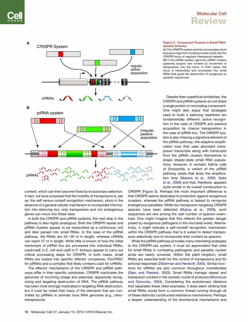
Figure 5. Conserved Themes in Small RNA-Guided Defense(A) The CRISPR system actively incorporates shortsequence tags from invading nucleic acids into theCRISPR locus of regularly interspaced repeats.(B) In the piRNA system, genomic piRNA clusterspassively acquire new content by movement oftransposons into the locus. In both cases, thelocus is transcribed and processed into smallRNAs that guide the destruction of exogenous orparasitic sequences.
Molecular Cell
Review
content, which can then become fixed by evolutionary selection.
In fact, we have proposed that the mobility of transposons is, per
se, the self versus nonself recognition mechanism, since in the
absence of a general cellular mechanism to incorporate informa-
tion into silencing loci, only transposons and not endogenous
genes can move into these sites.
In both the CRISPR and piRNA systems, the next step in the
pathway is also highly analogous. Both the CRISPR repeat and
piRNA clusters appear to be transcribed as a continuous unit
and later parsed into small RNAs. In the case of the piRNA
pathway, the RNAs are 24-–30 nt in length, whereas crRNAs
can reach 57 nt in length. While little is known of how the initial
transcripts of piRNA loci are processed into individual RNAs,
cse3/casE in E. coli and cas6 in P. furiosus appear to carry out
critical processing steps for CRISPR. In both cases, small
RNAs are loaded into specific effector complexes, Piwi/RISC
for piRNAs and a complex that likely contains cas3 for crRNAs.
The effector mechanisms of the CRISPR and piRNA path-
ways differ in their specific substrates. CRISPR inactivates the
genomes of incoming phage and plasmids, apparently recog-
nizing and targeting destruction of DNA. The piRNA pathway
has been most strongly implicated in targeting RNA destruction,
but it must be noted that many of the elements that are con-
trolled by piRNAs in animals have RNA genomes (e.g., retro-
transposons).
16 Molecular Cell 37, January 15, 2010 ª2010 Elsevier Inc.
Despite their superficial similarities, the
CRISPR and piRNA systems do not share
a single protein or noncoding component.
One might also argue that strategies
used to build a silencing repertoire are
fundamentally different: active recogni-
tion in the case of CRISPR and passive
acquisition by chance transposition in
the case of piRNA loci. The CRISPR sys-
tem is also missing a signature element of
the piRNA pathway, the adaptive amplifi-
cation loop that uses abundant trans-
poson transcripts along with transcripts
from the piRNA clusters themselves to
shape steady-state small RNA popula-
tions. However, in somatic follicle cells
of Drosophila, a variant of the piRNA
pathway exists that lacks the ampifica-
tion loop (Malone et al., 2009; Saito
et al., 2009) and that, therefore, appears
quite similar in its overall construction to
CRISPR (Figure 5). Perhaps the most important difference is
that CRISPR seems dedicated to protection against exogenous
invaders, whereas the piRNA pathway is tasked to recognize
endogenous parasites. While two transposon-targeting CRISPR
spacers have been detected (Mojica et al., 2009), such
sequences are rare among the vast number of spacers exam-
ined. One might imagine that this reflects the greater danger
posed by exogenous pathogens in the microbial world. Alterna-
tively, it might indicate a self-nonself recognition mechanism
within the CRISPR pathway that is ill suited to detect transpo-
sons selectively and to incorporate their content as spacers.
While the piRNA pathway provides many interesting analogies
to the CRISPR-cas system, it must be appreciated that roles
for small RNAs in combating pathogenic and parasitic nucleic
acids are nearly universal. Within the plant kingdom, small
RNAs are essential both for the control of transposons and for
antiviral responses (Zilberman and Henikoff, 2005). Similar func-
tions for siRNAs are also common throughout invertebrates
(Sijen and Plasterk, 2003). Small RNAs manage repeat and
transposon content in the somatic nuclei of protozoa (Mochizuki
and Gorovsky, 2004). Considering the evolutionary distance
that separates these cited examples, it does seem striking that
small RNAs would form a common thread running through all
of these distinctly constructed resistance mechanisms. Perhaps
a deeper understanding of the biochemical mechanisms and
Molecular Cell
Review
evolution of small RNA pathways throughout the kingdoms of
life may provide clues to the reasons underlying this curious
convergence.
ACKNOWLEDGMENTS
The authors would like to thank P. Rajesh Kumar for help with structural align-ments and Xavier Roca for comments on the manuscript. We are greatlyindebted to James Duffy for assistance with figures. F.V.K. is supported bya postdoctoral fellowship from the American Cancer Society, PF-07-058-01-GMC. This work was supported by grants from the National Institutes of Healthand by a kind gift from Kathryn W. Davis. G.J.H. is a professor of the HowardHughes Medical Institute.
REFERENCES
Agari, Y., Yokoyama, S., Kuramitsu, S., and Shinkai, A. (2008). X-ray crystalstructure of a CRISPR-associated protein, Cse2, from Thermus thermophilusHB8. Proteins 73, 1063–1067.
Agari, Y., Sakamoto, K., Tamakoshi, M., Oshima, T., Kuramitsu, S., andShinkai, A. (2009). Transcription profile of Thermus thermophilus CRISPRsystems after phage infection. J. Mol. Biol. Published online November 3,2009. 10.1016/j.jmb.2009.10.057.
Andersson, A.F., and Banfield, J.F. (2008). Virus population dynamics andacquired virus resistance in natural microbial communities. Science 320,1047–1050.
Aravin, A.A., Hannon, G.J., and Brennecke, J. (2007). The Piwi-piRNA pathwayprovides an adaptive defense in the transposon arms race. Science 318,761–764.
Barrangou, R., Fremaux, C., Deveau, H., Richards, M., Boyaval, P., Moineau,S., Romero, D.A., and Horvath, P. (2007). CRISPR provides acquired resis-tance against viruses in prokaryotes. Science 315, 1709–1712.
Beloglazova, N., Brown, G., Zimmerman, M.D., Proudfoot, M., Makarova,K.S., Kudritska, M., Kochinyan, S., Wang, S., Chruszcz, M., Minor, W., et al.(2008). A novel family of sequence-specific endoribonucleases associatedwith the clustered regularly interspaced short palindromic repeats. J. Biol.Chem. 283, 20361–20371.
Bickle, T.A., and Kruger, D.H. (1993). Biology of DNA restriction. Microbiol.Rev. 57, 434–450.
Bolotin, A., Quinquis, B., Sorokin, A., and Ehrlich, S.D. (2005). Clusteredregularly interspaced short palindrome repeats (CRISPRs) have spacers ofextrachromosomal origin. Microbiology 151, 2551–2561.
Breitbart, M., and Rohwer, F. (2005). Here a virus, there a virus, everywhere thesame virus? Trends Microbiol. 13, 278–284.
Brouns, S.J., Jore, M.M., Lundgren, M., Westra, E.R., Slijkhuis, R.J., Snijders,A.P., Dickman, M.J., Makarova, K.S., Koonin, E.V., and van der Oost, J. (2008).Small CRISPR RNAs guide antiviral defense in prokaryotes. Science 321,960–964.
Bult, C.J., White, O., Olsen, G.J., Zhou, L., Fleischmann, R.D., Sutton, G.G.,Blake, J.A., FitzGerald, L.M., Clayton, R.A., Gocayne, J.D., et al. (1996).Complete genome sequence of the methanogenic archaeon, Methanococcusjannaschii. Science 273, 1058–1073.
Carte, J., Wang, R., Li, H., Terns, R.M., and Terns, M.P. (2008). Cas6 is anendoribonuclease that generates guide RNAs for invader defense in prokary-otes. Genes Dev. 22, 3489–3496.
Chibani-Chennoufi, S., Bruttin, A., Dillmann, M.L., and Brussow, H. (2004).Phage-host interaction: an ecological perspective. J. Bacteriol. 186,3677–3686.
Chopin, M.C., Chopin, A., and Bidnenko, E. (2005). Phage abortive infection inlactococci: variations on a theme. Curr. Opin. Microbiol. 8, 473–479.
DeBoy, R.T., Mongodin, E.F., Emerson, J.B., and Nelson, K.E. (2006). Chro-mosome evolution in the Thermotogales: large-scale inversions and straindiversification of CRISPR sequences. J. Bacteriol. 188, 2364–2374.
Deveau, H., Barrangou, R., Garneau, J.E., Labonte, J., Fremaux, C., Boyaval,P., Romero, D.A., Horvath, P., and Moineau, S. (2008). Phage response toCRISPR-encoded resistance in Streptococcus thermophilus. J. Bacteriol.190, 1390–1400.
Ebihara, A., Yao, M., Masui, R., Tanaka, I., Yokoyama, S., and Kuramitsu, S.(2006). Crystal structure of hypothetical protein TTHB192 from Thermusthermophilus HB8 reveals a new protein family with an RNA recognitionmotif-like domain. Protein Sci. 15, 1494–1499.
Edwards, R.A., and Rohwer, F. (2005). Viral metagenomics. Nat. Rev. Micro-biol. 3, 504–510.
Fabre, M., Koeck, J.L., Le Fleche, P., Simon, F., Herve, V., Vergnaud, G., andPourcel, C. (2004). High genetic diversity revealed by variable-number tandemrepeat genotyping and analysis of hsp65 gene polymorphism in a large collec-tion of ‘‘Mycobacterium canettii’’ strains indicates that the M. tuberculosiscomplex is a recently emerged clone of ‘‘M. canettii’’. J. Clin. Microbiol. 42,3248–3255.
Fang, Z., Morrison, N., Watt, B., Doig, C., and Forbes, K.J. (1998). IS6110transposition and evolutionary scenario of the direct repeat locus in a groupof closely related Mycobacterium tuberculosis strains. J. Bacteriol. 180,2102–2109.
Forde, A., and Fitzgerald, G.F. (1999). Bacteriophage defence systems in lacticacid bacteria. Antonie Van Leeuwenhoek 76, 89–113.
Galperin, M.Y., and Koonin, E.V. (2000). Who’s your neighbor? New computa-tional approaches for functional genomics. Nat. Biotechnol. 18, 609–613.
Godde, J.S., and Bickerton, A. (2006). The repetitive DNA elements calledCRISPRs and their associated genes: evidence of horizontal transfer amongprokaryotes. J. Mol. Evol. 62, 718–729.
Gorbalenya, A.E., and Koonin, E.V. (1993). Helicases: amino acid sequencecomparisons and structure-function relationships. Curr. Opin. Struct. Biol. 3,419–429.
Grissa, I., Vergnaud, G., and Pourcel, C. (2007). The CRISPRdb database andtools to display CRISPRs and to generate dictionaries of spacers and repeats.BMC Bioinformatics 8, 172.
Grissa, I., Vergnaud, G., and Pourcel, C. (2008). CRISPRcompar: a website tocompare clustered regularly interspaced short palindromic repeats. NucleicAcids Res. 36, W145–W148.
Groenen, P.M., Bunschoten, A.E., van Soolingen, D., and van Embden, J.D.(1993). Nature of DNA polymorphism in the direct repeat cluster of Mycobac-terium tuberculosis; application for strain differentiation by a novel typingmethod. Mol. Microbiol. 10, 1057–1065.
Haft, D.H., Selengut, J., Mongodin, E.F., and Nelson, K.E. (2005). A guild of 45CRISPR-associated (Cas) protein families and multiple CRISPR/Cas subtypesexist in prokaryotic genomes. PLoS Comput. Biol. 1, e60.
Hale, C., Kleppe, K., Terns, R.M., and Terns, M.P. (2008). Prokaryotic silencing(psi)RNAs in Pyrococcus furiosus. RNA 14, 2572–2579.
Hale, C.R., Zhao, P., Olson, S., Duff, M.O., Graveley, B.R., Wells, L., Terns,R.M., and Terns, M.P. (2009). RNA-guided RNA cleavage by a CRISPRRNA-Cas protein complex. Cell 139, 945–956.
Han, D., and Krauss, G. (2009). Characterization of the endonucleaseSSO2001 from Sulfolobus solfataricus P2. FEBS Lett. 583, 771–776.
Han, D., Lehmann, K., and Krauss, G. (2009). SSO1450—a CAS1 protein fromSulfolobus solfataricus P2 with high affinity for RNA and DNA. FEBS Lett. 583,1928–1932.
Haraldsen, J.D., and Sonenshein, A.L. (2003). Efficient sporulation inClostridium difficile requires disruption of the sigmaK gene. Mol. Microbiol.48, 811–821.
Hatfull, G.F. (2008). Bacteriophage genomics. Curr. Opin. Microbiol. 11,447–453.
Heidelberg, J.F., Nelson, W.C., Schoenfeld, T., and Bhaya, D. (2009). Germwarfare in a microbial mat community: CRISPRs provide insights into theco-evolution of host and viral genomes. PLoS ONE 4, e4169. 10.1371/journal.pone.0004169.
Molecular Cell 37, January 15, 2010 ª2010 Elsevier Inc. 17
Molecular Cell
Review
Hendrix, R.W. (2003). Bacteriophage genomics. Curr. Opin. Microbiol. 6,506–511.
Hermans, P.W., van Soolingen, D., Bik, E.M., de Haas, P.E., Dale, J.W., andvan Embden, J.D. (1991). Insertion element IS987 from Mycobacterium bovisBCG is located in a hot-spot integration region for insertion elements in Myco-bacterium tuberculosis complex strains. Infect. Immun. 59, 2695–2705.
Hoe, N., Nakashima, K., Grigsby, D., Pan, X., Dou, S.J., Naidich, S., Garcia, M.,Kahn, E., Bergmire-Sweat, D., and Musser, J.M. (1999). Rapid moleculargenetic subtyping of serotype M1 group A Streptococcus strains. Emerg.Infect. Dis. 5, 254–263.
Horvath, P., Romero, D.A., Coute-Monvoisin, A.C., Richards, M., Deveau, H.,Moineau, S., Boyaval, P., Fremaux, C., and Barrangou, R. (2008). Diversity,activity, and evolution of CRISPR loci in Streptococcus thermophilus.J. Bacteriol. 190, 1401–1412.
Ishino, Y., Shinagawa, H., Makino, K., Amemura, M., and Nakata, A. (1987).Nucleotide sequence of the iap gene, responsible for alkaline phosphataseisozyme conversion in Escherichia coli, and identification of the gene product.J. Bacteriol. 169, 5429–5433.
Jansen, R., van Embden, J.D., Gaastra, W., and Schouls, L.M. (2002). Identi-fication of a novel family of sequence repeats among prokaryotes. OMICS 6,23–33.
Kamerbeek, J., Schouls, L., Kolk, A., van Agterveld, M., van Soolingen, D.,Kuijper, S., Bunschoten, A., Molhuizen, H., Shaw, R., Goyal, M., and vanEmbden, J. (1997). Simultaneous detection and strain differentiation of Myco-bacterium tuberculosis for diagnosis and epidemiology. J. Clin. Microbiol. 35,907–914.
Kawarabayasi, Y., Sawada, M., Horikawa, H., Haikawa, Y., Hino, Y., Yama-moto, S., Sekine, M., Baba, S., Kosugi, H., Hosoyama, A., et al. (1998).Complete sequence and gene organization of the genome of a hyper-thermo-philic archaebacterium, Pyrococcus horikoshii OT3. DNA Res. 5, 55–76.
Kawarabayasi, Y., Hino, Y., Horikawa, H., Yamazaki, S., Haikawa, Y., Jin-no,K., Takahashi, M., Sekine, M., Baba, S., Ankai, A., et al. (1999). Completegenome sequence of an aerobic hyper-thermophilic crenarchaeon, Aeropy-rum pernix K1. DNA Res. 6, 83–101, 145–152.
Klenk, H.P., Clayton, R.A., Tomb, J.F., White, O., Nelson, K.E., Ketchum, K.A.,Dodson, R.J., Gwinn, M., Hickey, E.K., Peterson, J.D., et al. (1997). Thecomplete genome sequence of the hyperthermophilic, sulphate-reducingarchaeon Archaeoglobus fulgidus. Nature 390, 364–370.
Kunin, V., Sorek, R., and Hugenholtz, P. (2007). Evolutionary conservation ofsequence and secondary structures in CRISPR repeats. Genome Biol. 8, R61.
Lillestol, R.K., Redder, P., Garrett, R.A., and Brugger, K. (2006). A putative viraldefence mechanism in archaeal cells. Archaea 2, 59–72.
Lillestol, R.K., Shah, S.A., Brugger, K., Redder, P., Phan, H., Christiansen, J.,and Garrett, R.A. (2009). CRISPR families of the crenarchaeal genus Sulfolo-bus: bidirectional transcription and dynamic properties. Mol. Microbiol. 72,259–272.
Makarova, K.S., Aravind, L., Grishin, N.V., Rogozin, I.B., and Koonin, E.V.(2002). A DNA repair system specific for thermophilic Archaea and bacteriapredicted by genomic context analysis. Nucleic Acids Res. 30, 482–496.
Makarova, K.S., Grishin, N.V., Shabalina, S.A., Wolf, Y.I., and Koonin, E.V.(2006). A putative RNA-interference-based immune system in prokaryotes:computational analysis of the predicted enzymatic machinery, functional anal-ogies with eukaryotic RNAi, and hypothetical mechanisms of action. Biol.Direct 1, 7.
Malone, C.D., Brennecke, J., Dus, M., Stark, A., McCombie, W.R., Sachida-nandam, R., and Hannon, G.J. (2009). Specialized piRNA pathways act ingermline and somatic tissues of the Drosophila ovary. Cell 137, 522–535.
Marraffini, L.A., and Sontheimer, E.J. (2008). CRISPR interference limitshorizontal gene transfer in staphylococci by targeting DNA. Science 322,1843–1845.
Masepohl, B., Gorlitz, K., and Bohme, H. (1996). Long tandemly repeatedrepetitive (LTRR) sequences in the filamentous cyanobacterium Anabaenasp. PCC 7120. Biochim. Biophys. Acta 1307, 26–30.
18 Molecular Cell 37, January 15, 2010 ª2010 Elsevier Inc.
Mochizuki, K., and Gorovsky, M.A. (2004). Small RNAs in genome rearrange-ment in Tetrahymena. Curr. Opin. Genet. Dev. 14, 181–187.
Mojica, F.J., Ferrer, C., Juez, G., and Rodriguez-Valera, F. (1995). Longstretches of short tandem repeats are present in the largest replicons of theArchaea Haloferax mediterranei and Haloferax volcanii and could be involvedin replicon partitioning. Mol. Microbiol. 17, 85–93.
Mojica, F.J., Diez-Villasenor, C., Soria, E., and Juez, G. (2000). Biologicalsignificance of a family of regularly spaced repeats in the genomes of Archaea,Bacteria and mitochondria. Mol. Microbiol. 36, 244–246.
Mojica, F.J., Diez-Villasenor, C., Garcia-Martinez, J., and Soria, E. (2005).Intervening sequences of regularly spaced prokaryotic repeats derive fromforeign genetic elements. J. Mol. Evol. 60, 174–182.
Mojica, F.J., Diez-Villasenor, C., Garcia-Martinez, J., and Almendros, C.(2009). Short motif sequences determine the targets of the prokaryoticCRISPR defence system. Microbiology 155, 733–740.
Nakata, A., Amemura, M., and Makino, K. (1989). Unusual nucleotide arrange-ment with repeated sequences in the Escherichia coli K-12 chromosome.J. Bacteriol. 171, 3553–3556.
Nelson, K.E., Clayton, R.A., Gill, S.R., Gwinn, M.L., Dodson, R.J., Haft, D.H.,Hickey, E.K., Peterson, J.D., Nelson, W.C., Ketchum, K.A., et al. (1999).Evidence for lateral gene transfer between Archaea and bacteria from genomesequence of Thermotoga maritima. Nature 399, 323–329.
Oberle, I., Rousseau, F., Heitz, D., Kretz, C., Devys, D., Hanauer, A., Boue, J.,Bertheas, M.F., and Mandel, J.L. (1991). Instability of a 550-base pair DNAsegment and abnormal methylation in fragile X syndrome. Science 252,1097–1102.
Overbeek, R., Fonstein, M., D’Souza, M., Pusch, G.D., and Maltsev, N. (1999).The use of gene clusters to infer functional coupling. Proc. Natl. Acad. Sci. USA96, 2896–2901.
Peng, X., Brugger, K., Shen, B., Chen, L., She, Q., and Garrett, R.A. (2003).Genus-specific protein binding to the large clusters of DNA repeats (shortregularly spaced repeats) present in Sulfolobus genomes. J. Bacteriol. 185,2410–2417.
Pourcel, C., Salvignol, G., and Vergnaud, G. (2005). CRISPR elements inYersinia pestis acquire new repeats by preferential uptake of bacteriophageDNA, and provide additional tools for evolutionary studies. Microbiology151, 653–663.
Rohwer, F., and Thurber, R.V. (2009). Viruses manipulate the marine environ-ment. Nature 459, 207–212.
Saito, K., Inagaki, S., Mituyama, T., Kawamura, Y., Ono, Y., Sakota, E., Kotani,H., Asai, K., Siomi, H., and Siomi, M.C. (2009). A regulatory circuit for piwi bythe large Maf gene traffic jam in Drosophila. Nature 461, 1296–1299.
Sakamoto, K., Agari, Y., Agari, K., Yokoyama, S., Kuramitsu, S., and Shinkai,A. (2009). X-ray crystal structure of a CRISPR-associated RAMP superfamilyprotein, Cmr5, from Thermus thermophilus HB8. Proteins 75, 528–532.
Sebaihia, M., Wren, B.W., Mullany, P., Fairweather, N.F., Minton, N., Stabler,R., Thomson, N.R., Roberts, A.P., Cerdeno-Tarraga, A.M., Wang, H., et al.(2006). The multidrug-resistant human pathogen Clostridium difficile hasa highly mobile, mosaic genome. Nat. Genet. 38, 779–786.
Sensen, C.W., Charlebois, R.L., Chow, C., Clausen, I.G., Curtis, B., Doolittle,W.F., Duguet, M., Erauso, G., Gaasterland, T., Garrett, R.A., et al. (1998).Completing the sequence of the Sulfolobus solfataricus P2 genome. Extrem-ophiles 2, 305–312.
Shah, S.A., Hansen, N.R., and Garrett, R.A. (2009). Distribution of CRISPRspacer matches in viruses and plasmids of crenarchaeal acidothermophilesand implications for their inhibitory mechanism. Biochem. Soc. Trans. 37,23–28.
She, Q., Phan, H., Garrett, R.A., Albers, S.V., Stedman, K.M., and Zillig, W.(1998). Genetic profile of pNOB8 from Sulfolobus: the first conjugative plasmidfrom an archaeon. Extremophiles 2, 417–425.
She, Q., Singh, R.K., Confalonieri, F., Zivanovic, Y., Allard, G., Awayez, M.J.,Chan-Weiher, C.C., Clausen, I.G., Curtis, B.A., De Moors, A., et al. (2001).

Molecular Cell
Review
The complete genome of the crenarchaeon Sulfolobus solfataricus P2. Proc.Natl. Acad. Sci. USA 98, 7835–7840.
Sijen, T., and Plasterk, R.H. (2003). Transposon silencing in the Caenorhabditiselegans germ line by natural RNAi. Nature 426, 310–314.
Smith, D.R., Doucette-Stamm, L.A., Deloughery, C., Lee, H., Dubois, J.,Aldredge, T., Bashirzadeh, R., Blakely, D., Cook, R., Gilbert, K., et al. (1997).Complete genome sequence of Methanobacterium thermoautotrophicumdeltaH: functional analysis and comparative genomics. J. Bacteriol. 179,7135–7155.
Sorek, R., Kunin, V., and Hugenholtz, P. (2008). CRISPR—a widespreadsystem that provides acquired resistance against phages in bacteria andarchaea. Nat. Rev. Microbiol. 6, 181–186.
Tang, T.H., Bachellerie, J.P., Rozhdestvensky, T., Bortolin, M.L., Huber, H.,Drungowski, M., Elge, T., Brosius, J., and Huttenhofer, A. (2002). Identificationof 86 candidates for small non-messenger RNAs from the archaeon Archaeo-globus fulgidus. Proc. Natl. Acad. Sci. USA 99, 7536–7541.
Tang, T.H., Polacek, N., Zywicki, M., Huber, H., Brugger, K., Garrett, R., Bach-ellerie, J.P., and Huttenhofer, A. (2005). Identification of novel non-codingRNAs as potential antisense regulators in the archaeon Sulfolobus solfatari-cus. Mol. Microbiol. 55, 469–481.
Tyson, G.W., and Banfield, J.F. (2008). Rapidly evolving CRISPRs implicatedin acquired resistance of microorganisms to viruses. Environ. Microbiol. 10,200–207.
van der Oost, J., Jore, M.M., Westra, E.R., Lundgren, M., and Brouns, S.J.(2009). CRISPR-based adaptive and heritable immunity in prokaryotes. TrendsBiochem. Sci. 34, 401–407.
van der Ploeg, J.R. (2009). Analysis of CRISPR in Streptococcus mutanssuggests frequent occurrence of acquired immunity against infection byM102-like bacteriophages. Microbiology 155, 1966–1976.
Viswanathan, P., Murphy, K., Julien, B., Garza, A.G., and Kroos, L. (2007).Regulation of dev, an operon that includes genes essential for Myxococcusxanthus development and CRISPR-associated genes and repeats. J. Bacter-iol. 189, 3738–3750.
Wiedenheft, B., Zhou, K., Jinek, M., Coyle, S.M., Ma, W., and Doudna, J.A.(2009). Structural basis for DNase activity of a conserved protein implicatedin CRISPR-mediated genome defense. Structure 17, 904–912.
Zegans, M.E., Wagner, J.C., Cady, K.C., Murphy, D.M., Hammond, J.H., andO’Toole, G.A. (2009). Interaction between bacteriophage DMS3 and hostCRISPR region inhibits group behaviors of Pseudomonas aeruginosa.J. Bacteriol. 191, 210–219.
Zilberman, D., and Henikoff, S. (2005). Epigenetic inheritance in Arabidopsis:selective silence. Curr. Opin. Genet. Dev. 15, 557–562.
Molecular Cell 37, January 15, 2010 ª2010 Elsevier Inc. 19


![Selective targeting of the oncogenic KRAS G12S mutant allele by CRISPR/Cas9 … · 83 CRISPR/Cas9 system to control tumor growth [23, 24]. In addition, the CRISPR-Cas13a system was](https://img.pdfslide.net/doc/110x75/5ec9dccaf4c826280677c020/selective-targeting-of-the-oncogenic-kras-g12s-mutant-allele-by-crisprcas9-83-crisprcas9.jpg)