Embed Size (px)
Citation preview
The curious case of NG2 cells: transienttrend or game changer?Jean-Marie Mangin1 and Vittorio GalloCenter for Neuroscience Research, Children’s National Medical Center, Washington, DC 20010, U.S.A.
Cite this article as: Mangin J-M and Gallo V (2011) The curious case of NG2 cells: transient trend or game changer? ASN NEURO3(1):art:e00052.doi:10.1042/AN20110001
ABSTRACT
It has been 10 years since the seminal work of DwightBergles and collaborators demonstrated that NG2 (nerve/glial antigen 2)-expressing oligodendrocyte progenitor cells(NG2 cells) receive functional glutamatergic synapses fromneurons (Bergles et al., 2000), contradicting the old dogmathat only neurons possess the complex and specializedmolecular machinery necessary to receive synapses. Whilethis surprising discovery may have been initially shunnedas a novelty item of undefined functional significance, thestudy of neuron-to-NG2 cell neurotransmission has sincebecome a very active and exciting field of research. Manylaboratories have now confirmed and extended the initialdiscovery, showing for example that NG2 cells can alsoreceive inhibitory GABAergic synapses (Lin and Bergles,2004) or that neuron-to-NG2 cell synaptic transmission is arather ubiquitous phenomenon that has been observed in allbrain areas explored so far, including white matter tracts(Kukley et al., 2007; Ziskin et al., 2007; Etxeberria et al.,2010). Thus, while still being in its infancy, this field ofresearch has already brought many surprising and interest-ing discoveries, and has become part of a continuouslygrowing effort in neuroscience to re-evaluate the longunderestimated role of glial cells in brain function (Barres,2008). However, this area of research is now reaching animportant milestone and its long-term significance will bedefined by its ability to uncover the still elusive function ofNG2 cells and their synapses in the brain, rather than by itssensational but transient successes at upsetting the oldorder established by neuronal physiology. To participate inthe effort to facilitate such a transition, here we propose acritical review of the latest findings in the field of NG2 cellphysiology – discussing how they inform us on the possiblefunction(s) of NG2 cells in the brain – and we present somepersonal views on new directions the field could benefit fromin order to achieve lasting significance.
Key words: a-amino-3-hydroxy-5-methylisoxazole-4-pro-pionic acid receptor (AMPAR), nerve/glial antigen 2 (NG2)cells, neuron, oligodendrocyte progenitor cell (OPC),postsynaptic density (PSD).
INTRODUCTION
The NG2 (nerve/glial antigen 2) is a chondroitin sulfate pro-
teoglycan predominantly expressed in the brain by a
subpopulation of glial cells called NG2 cells. These cells are
classically described as OPCs (oligodendrocyte progenitor
cells), since they usually co-express OPC markers such as
PDGFRa (platelet-derived growth factor receptor a) and O4
(Nishiyama et al., 1996; Reynolds and Hardy, 1997), and give
rise to most – if not all – oligodendrocytes in the brain
(Dimou et al., 2008; Rivers et al., 2008; Zhu et al., 2008; Kang
et al., 2010). The only other cells known to express NG2 in the
brain are the pericytes lining blood vessels (Ozerdem et al.,
2001). While pericytes are not usually considered as NG2 cells
per se, they may be confused with them, especially in the
absence of co-staining for other OPC markers. In the last
decade, NG2 cells have generated a lot of interest among
neuroscientists, because they display a combination of
features unexpected in OPCs. These include: (i) an almost
uniform distribution in both grey and white matter areas; (ii)
a complex stellate morphology; (iii) a tendency to intimately
associate with neuronal cell bodies and dendrites (Dawson
et al., 2003; Butt et al., 2005); (iv) the ability to keep
proliferating in the adult brain (Dawson et al., 2003; Aguirre
et al., 2004; Ligon et al., 2006); and (v) a latent ability – in the
context of brain injury or pathology – to give rise to
astrocytes and neurons that may be recruited to areas of
lesion (Belachew et al., 2003; Aguirre et al., 2007; Tamura
et al., 2007; Rivers et al., 2008; Zhu et al., 2008). However, the
most striking of these features – and the main focus of this
review – is their ability to receive synapses from neurons.
1 To whom correspondence should be addressed (email [email protected]).Abbreviations: AMPAR, a-amino-3-hydroxy-5-methylisoxazole-4-propionic acid receptor; CNP-GFP, C-type natriuretic peptide-green fluorescent protein; CNS, centralnervous system; EGFP, enhanced green fluorescent protein; EPSC, excitatory postsynaptic current; GABA, c-aminobutyric acid; GABAAR, GABA type A receptor; LTP, long-termpotentiation; MBP, maltose-binding protein; NG2, nerve/glial antigen 2; NMDAR, N-methyl-D-aspartate receptor; OL, oligodendrocyte lineage; OPC, oligodendrocyteprogenitor cell; PDGFRa, platelet-derived growth factor receptor a; PSD, postsynaptic density; SCP, Schwann cell progenitor.E 2011 The Author(s) This is an Open Access article distributed under the terms of the Creative Commons Attribution Non-Commercial Licence (http://creativecommons.org/licenses/by-nc/2.5/) which permits unrestricted non-commercial use, distribution and reproduction in any medium, provided the original work isproperly cited.
REVIEWASN NEURO 3(1):art:e00052.doi:10.1042/AN20110001
asnneuro.org / Volume 3 (1) / art:e00052 37
Neuron-NG2 cell glutamatergic synapses have been origin-
ally described in NG2 cells recorded in hippocampal slices from
both young postnatal mice and adult mice (Bergles et al.,
2000). Since then, these synapses have been shown to be a
near-universal feature of NG2 cells in all grey and white matter
areas explored so far, including brainstem (Muller et al., 2009),
cerebellar cortex (Lin et al., 2005), dentate gyrus (Mangin et al.,
2008), cortex (Chittajallu et al., 2004; Kukley et al., 2008; Ge
et al., 2009), corpus callosum (Kukley et al., 2007; Ziskin et al.,
2007; De Biase et al., 2010a; Etxeberria et al., 2010) and
cerebellar white matter (Karadottir et al., 2008; De Biase et al.,
2010a). Additionally, NG2 cells are also able to receive
GABAergic synapses (GABA is c-aminobutyric acid) in hip-
pocampus (Lin and Bergles, 2004), dentate gyrus (Mangin et al.,
2008) and cortex (Ge et al., 2009; Tanaka et al., 2009). However,
in spite of their universality, the functions of these synapses
still remain completely unknown.
HOW FAR ARE WE FROM DEFINING THEFUNCTIONS OF NEURON-NG2 SYNAPSES?
For a long time, a major reason why the function of neuron-
NG2 synapses has been so hard to define has been the lack of
tools that would allow selective modulation of synaptic
transmission in NG2 cells. However, this roadblock is now
mostly cleared by the recent generation of several transgenic
lines allowing specific deletion or overexpression of selected
genes in NG2 cells. These lines were generated by using
different gene promoters actively expressed in NG2 cells, and
include the NG2-Cre, NG2-CreErt2, PDGFRa-CreErt2, CNP-Cre
and PLP-Cre lines (Lappe-Siefke et al., 2003; Delaunay et al.,
2008; Rivers et al., 2008; Zhu et al., 2008; De Biase et al., 2010a;
Kang et al., 2010). However, in order to properly use these
genetic tools, a more detailed knowledge of the identity of the
postsynaptic proteins mediating synaptic currents in NG2 cells
is necessary. For example, while it is known that glutamatergic
EPSCs (excitatory postsynaptic currents) in NG2 cells are
mediated by AMPAR (a-amino-3-hydroxy-5-methylisoxazole-
4-propionic acid receptor), it appears that NG2 cells can
express all of the four known subunits that can compose these
receptors (a1–a4) (De Biase et al., 2010a). This implies that,
because of possible compensatory effects between subunits, a
quadruple conditional knockout of all these subunits might
have to be generated in order to completely eliminate EPSCs in
NG2 cells – a rather daunting task at best.
A better approach maybe to target critical postsynaptic
anchoring proteins, such as PSD-95 (postsynaptic density 95)
or other equivalent proteins playing similar functional roles
(Feng and Zhang, 2009). However, information on the basic
composition of the PSD in NG2 cells is still lacking. These
challenges also apply to GABAergic synapses between
neurons and NG2 cells, as the GABAAR (GABA type A
receptor) subtypes expressed by NG2 cells at postsynaptic site
are still undefined. Postsynaptic GABAARs in neurons are
pentamers made of 2 a-subunits, 2 b-subunits and 1 c-
subunit, and based on the large number of GABAAR subunits
identified so far (6 for a, 3 for b and 3 for c) (Jacob et al.,
2008), a conditional multiple knockout approach may be
necessary. However, since removing all subunits from either
the a or the b category is sufficient to render all postsynaptic
GABAAR non-functional in a cell, this approach may remain
relatively easy to achieve, especially since it is unlikely that
NG2 cells co-express all possible subunit combinations. Also,
as for glutamatergic synapses, a more feasible approach
maybe to target critical postsynaptic anchoring proteins, such
as gephyrin (Fritschy et al., 2008). But once again, we are
lacking crucial information about the structure of the
GABAergic PSD in NG2 cells in order to be able to successfully
apply a conditional knockout strategy to understand the
physiological role of these synapses.
Based on the issues described above, we believe that some
groundwork will have to be done in order to characterize the
composition of PSDs in NG2 cells, before we can apply
genetic approaches to understand the function of synapses in
these cells. However, this goal maybe reached earlier than we
think, based on a recent report demonstrating removal of
most if not all functional NMDARs (N-methyl-D-aspartate
receptors) in NG2 cells after a single conditional knockout for
the NR1 subunit utilizing a PDGFRa-CreErt2 mouse (De Biase
et al., 2010b). Even though this successful cell-specific
genetic/functional ablation takes advantage of the fact that
NR1 alone is necessary to the function of all NMDAR
subtypes, these findings demonstrate that the conditional
ablation of a subtype of postsynaptic receptor can be
successfully achieved in NG2 cells.
HOW ARE SYNAPTIC CURRENTS TRANSDUCEDIN NG2 CELLS?
Another important area of inquiry that could help us
understand the function of neuron-NG2 cell synapses relates
to the transduction of synaptic currents in NG2 cells
themselves.
Do NG2 cells fire action potentials in response tosynaptic stimulation?The recent report of a subpopulation of NG2 cells firing
action potentials in the rat cerebellum (Karadottir et al.,
2008) would suggest that a subpopulation of NG2 cells (and
glia) could display functional properties more similar to
neurons than previously thought – i.e. translating synaptic
inputs into action potentials. However, the existence of such
a subpopulation of ‘spiking’ NG2 cells remains uncertain,
since a recent report by the same group found that, in
contrast with what was found in rats, only few NG2 cells are
J-M Mangin and V Gallo
38 E 2011 The Author(s) This is an Open Access article distributed under the terms of the Creative Commons Attribution Non-Commercial Licence (http://creativecommons.org/licenses/by-nc/2.5/)which permits unrestricted non-commercial use, distribution and reproduction in any medium, provided the original work is properly cited.
able to produce regenerative spikes in mice (Clarke et al.,
2010). This finding is consistent with reports by other
laboratories (Chittajallu et al., 2004; De Biase et al., 2010a).
Furthermore, since almost all NG2 cells are contacted by
synapses, the presence of synaptic inputs cannot be
correlated with their ability to fire action potentials.
Therefore, it is currently unclear whether the ability of few
NG2 cells to potentially generate real action potentials is
relevant to the quest for the specific physiological function
of synapses in NG2 cells, or is rather an attempt to use
concepts ‘imported’ from neuronal physiology and apply
them unaltered to glial physiology. In conclusion, based on
current data, action potentials are unlikely to represent the
way most NG2 cells translate synaptic inputs, particularly in
the absence of any classical output for these action
potentials, such as the ability to release neurotransmitters
in response to depolarization.
Neuron-to-NG2 synapses and calcium entryA current hypothesis regarding how synaptic currents are
transduced in NG2 cells involves calcium entry in response to
glutamate as well as GABA. This question has already been
extensively discussed in recent reviews (Gallo et al., 2008;
Bergles et al., 2010) and we will only summarize the main
points here. In the case of glutamatergic synapses, it has been
shown that postsynaptic AMPAR expressed by NG2 cells are
often permeable to calcium (Bergles et al., 2000; Lin et al.,
2005; Ge et al., 2006; Mangin et al., 2008). Therefore, synaptic
activation of these Ca2+ permeable AMPARs would directly
result in a local increase of calcium, proportional to the
amplitude of the synaptic current. Another mode of calcium
entry involves the hypothetical ability of both glutamatergic
and GABAergic synapses to depolarize NG2 cells to a
membrane potential that is sufficient to activate voltage-
dependent calcium channels (Berger et al., 1992).
Since NG2 cells frequently express voltage-dependent
sodium channels, it has also been proposed that the depo-
larization induced by the synaptic release of glutamate or
GABA could be sufficient to activate these sodium channels
and to induce an increase in intracellular sodium large enough
to allow calcium entry via the Na+/Ca2+-exchanger (Blaustein
and Lederer, 1999; Tong et al., 2009). However, except for
glutamatergic synapses between climbing fibres and cerebellar
NG2 cells, the degree of depolarization induced by individual
neuron-NG2 cell synapses is usually small (1–5 mV).
Considering that NG2 cells also exhibit a strongly hyperpolar-
ized membrane resting potential (280 to 2100 mV), the
synchronized activation of many synapses would then be
necessary to activate either voltage-dependent calcium
channels or sodium channels. Such synchronization may be
hard to achieve, if one also takes into account the limited
number of functional synapses per NG2 cell reported in most
studies. Indeed, while there is evidence that the exogenous
application of GABA and glutamate can induce an intracellular
calcium increase in NG2 cells (Ge et al., 2006), there is still
surprisingly no evidence that physiological/endogenous syn-
aptic release of one of these neurotransmitters is able to cause
a similar effect. One of the reasons why such synaptic-induced
calcium increase has not yet been observed in NG2 cells may
relate to the fact that Ca2+-transients do not arise in the soma,
but instead are restricted to the cell processes, i.e. where
neuron-NG2 cell synapses are frequently observed (Bergles
et al., 2000; Kukley et al., 2007; Ziskin et al., 2007). More
importantly, even if physiologically induced Ca2+ transients are
demonstrated in NG2 cells, intracellular calcium is a ubiquitous
second messenger, involved in a variety of distinct and
complex cellular processes. Therefore, limited information on
the function(s) of synaptic transmission in NG2 cells can be
obtained solely based on the demonstration of Ca2+ transients
elicited by synaptic activation of glutamate or GABA receptors
in these cells.
In fact, we believe that, in order to understand the specific
function of neuron-NG2 cell synapses, we should understand
what make these cells a unique player in brain physiology –
distinct from neurons and astrocytes – rather than focusing
on the similarities between NG2 cells and other neural cell
types. In other words, in order to understand the role of
synaptic inputs in NG2 cell physiology, we first need to know
more about what NG2 cells are and what specific tasks they
accomplish in the brain that would necessitate such an
anatomical and functional attribute.
NG2 CELL: A PROGENITOR CELL OR A FOURTHMAJOR GLIAL CELL TYPE?
NG2 cells have long been classified as a progenitor cell
type, i.e. a cell displaying a rather short-lived phenotype, with
the main function of giving rise to mature and fully
differentiated neural cells. Both in the developing and adult
brain, NG2 cells exhibit the three main features associated
with a progenitor state, including the ability to: (i) actively
proliferate; (ii) migrate long distances; and (iii) generate other
cell types. Importantly, it has now been shown that NG2 cells
can be contacted by neuron-NG2 synapses, while they are
engaged in any of these progenitor-related functions.
There are currently few doubts remaining over the fact
that NG2-expressing cells are the main and probably sole
source of oligodendrocytes in the brain (Dimou et al., 2008;
Rivers et al., 2008; Zhu et al., 2008; Kang et al., 2010),
and several studies have hinted of a limited ability of these
cells to give rise to both astrocytes and neurons (Bela-
chew et al., 2003; Aguirre et al., 2007; Tamura et al., 2007;
Rivers et al., 2008; Zhu et al., 2008). A recent study by our
laboratory has also provided the first evidence that neuron-
NG2 synapses are actively formed and regulated during
spontaneous remyelination occurring after demyelination
of the corpus callosum, suggesting that they may play a role
in the early steps of the myelination/remyelination process
The curious case of NG2 cells
E 2011 The Author(s) This is an Open Access article distributed under the terms of the Creative Commons Attribution Non-Commercial Licence (http://creativecommons.org/licenses/by-nc/2.5/)which permits unrestricted non-commerical use, distribution and reproduction in any medium, provided the original work is properly cited.
39
(Etxeberria et al., 2010). The hypothesis of an early function
of neuron-NG2 synapses during the myelination process is
supported by other studies showing that NG2 cells lose their
synapses as they differentiate into myelinating oligodendro-
cytes (De Biase et al., 2010a; Kukley et al., 2010). However, it
remains to be defined whether the loss of synapses is causal
or consequential to this differentiation process. Nonetheless,
since electrical activity is known to influence myelination
(Demerens et al., 1996), it is tempting to hypothesize that
neuron-NG2 neurotransmission conveys information on
axonal electrical activity to these progenitors and regulates
NG2 cell lineage progression to fully mature myelinating
oligodendrocytes.
It has also been clearly shown that NG2 cells represent
the main proliferating cell population in the postnatal and
adult brain, both in grey and white matter areas (Nishi-
yama et al., 2002; Dawson et al., 2003; Aguirre et al., 2004;
Ligon et al., 2006). Moreover, three independent laboratories
have now demonstrated that NG2 cells remain connected to
neurons by functional glutamatergic and GABAergic
synapses, while dividing (Kukley et al., 2008; Ge et al.,
2009; Tanaka et al., 2009). Since a previous study has shown
that glutamate inhibits OPC proliferation in cell culture and
that an undefined endogenous source of glutamate inhibits
NG2 cell proliferation in developing cerebellum (Yuan et al.,
1998), it is likely that glutamatergic synapses inhibit NG2 cell
proliferation in an activity-dependent manner.
Finally, NG2 cells are able to migrate over long distance,
both in the postnatal brain (Levison and Goldman, 1993; Zhu
et al., 2008) as well as in the adult brain (Etxeberria et al.,
2010). There is evidence that NG2 cells can be contacted
by glutamatergic synapses as they migrate short distances
after division (Kukley et al., 2008), and that they may be
able to do the same as they migrate longer distances to
remyelinate axons in a demyelinated lesion (Etxeberria
et al., 2010). As it has been shown that glutamate can
promote OPC migration via an av integrin/myelin proteolipid
protein complex (Gudz et al., 2006), it is conceivable that
these synapses could regulate OPC migration in an activity-
dependent manner.
However, several other findings on NG2 cells do not fit
with the pure oligodendrocyte progenitor hypothesis. First,
both during development and in the adult, NG2 cells can
be found at an almost uniform density in most grey and
white matter areas (Butt et al., 2005). While there is a
significant level of myelination in grey matter areas, one
would expect a higher accumulation of OPCs in the most
heavily myelinated fibre tracts. Moreover, at least half of
these cells do not appear to be normally involved in
generating other cell types (Rivers et al., 2008; Psachoulia
et al., 2009). In fact, most NG2 cells exhibit a complex stellate
morphology with many fine processes, more or less uniformly
distributed around the soma territory in a 50 mm radius
(Butt et al., 2005). These processes are often associated with
neuronal soma and dendrites, two neuronal compartments
lacking myelination (Wigley and Butt, 2009).
Therefore, while having all the hallmarks of a progenitor
cell type, it has been proposed that NG2 cells represent a
fourth glial cell type in the developing and adult brain, whose
function(s) would not be directly related to their ability to
generate oligodendrocytes. As a consequence, this ambiguous
identity of NG2 cells has led some authors to propose the
existence of at least two different classes of NG2 cells (Horner
et al., 2002; Bakiri et al., 2009; Lytle et al., 2009): (i) a
progenitor NG2 cell type exhibiting a simple bipolar
morphology, a high rate of proliferation and giving rise to
oligodendrocytes, and (ii) an adult NG2 glial cell type
exhibiting a complex stellate morphology, a low proliferation
rate and an inability to form oligodendrocytes. However,
there is still limited evidence in favour of such a clear
dichotomy. Moreover, the apparently diverse features of NG2
cells may not be due to the existence of several distinct
subpopulations – each with a single function – but rather
reflect the fact that NG2 cells may have multiple functions,
which we still do not entirely understand in the context of
overall brain development and physiology. Therefore, the next
and last section of this review will be a speculative attempt at
integrating what is currently known about NG2 cell
physiology into a unifying framework.
EXPLORING NEW TERRITORIES: A POSSIBLEFUNCTION OF NG2 CELLS IN THEDEVELOPMENT OF NEURAL NETWORKS
Evolutionary considerations on NG2 cellsThe only physiological role of NG2 cells identified so far is their
ability to generate oligodendrocytes. In fact, there is now
strong experimental evidence that NG2 cells are the main and
possibly the only source of oligodendrocytes in the brain
(Dimou et al., 2008; Rivers et al., 2008; Zhu et al., 2008; Kang
et al., 2010). However, as we mentioned above, it is also
strongly suspected that NG2 cells may have other functions in
the normal physiology of the brain, although we still lack
specific hypotheses of what these functions could be and
whether they might be related to their established role in
generating myelinating oligodendrocytes.
A potentially fruitful way to approach this question is to
take an evolutionary viewpoint. Indeed, myelin and myelinat-
ing cells are a relatively recent innovation, mostly found in
vertebrate nerves (Hartline and Colman, 2007). This implies
that precursors of myelin-forming cells, such as oligoden-
drocytes and Schwann cells, should also be present in
unmyelinated ancestor organisms with a function distinct
from that of generating myelin, but probably close enough to
lead easily into this innovation in myelinated nervous
systems. Moreover, it can be hypothesized that the
acquisition of a myelinating function has not completely
eliminated other roles of these ancestral proto-myelinating
J-M Mangin and V Gallo
40 E 2011 The Author(s) This is an Open Access article distributed under the terms of the Creative Commons Attribution Non-Commercial Licence (http://creativecommons.org/licenses/by-nc/2.5/)which permits unrestricted non-commercial use, distribution and reproduction in any medium, provided the original work is properly cited.
cells in vertebrates. These other functions could therefore still
be found in the large population of non-myelinating NG2
cells present in the mammalian CNS (central nervous system).
In support of this hypothesis, it has been shown that in many
non-myelinated organisms, such as crabs, lobsters and
Drosophila, non-myelinating glial cells are indeed frequently
found to closely interact with axons and to ensheath nerves
without myelinating them (Hartline and Colman, 2007;
Banerjee and Bhat, 2008). This anatomical relationship is
reminiscent of non-myelinating Schwann cells and NG2 cells
and their relationship with axons. It would be of great
interest to determine whether glial cells in Drosophila or in
other non-myelinated organisms can also receive synapses
from neurons and whether OPCs in non-mammalian organ-
isms displaying myelination – such as zebrafish – can receive
synapses. This information would greatly help us to determine
if the existence of neuron–glial cell synapses are part of the
myelination process per se or have a more ancestral function
in neuron–glia interactions.
Studies on axon–glia interactions in model organisms
lacking myelin, such a Drosophila melanogaster, could offer
important insights into some of the non-myelinating functions
of NG2 cells in the mammalian brain. For example, subpopula-
tions of glial cells which intimately interact with axons during
development are known to influence axon pathfinding by
expressing diffusible and membrane-bound guidance cues,
including netrins and slits in Drosophila (Oland and Tolbert,
2003; Parker and Auld, 2006). Interestingly, many of these
guidance cues are also known to influence axon pathfinding in
mammals and, while their exact cellular origin remains to be
defined, it is known that cells belonging to the OL (oligo-
dendrocyte lineage) can express netrins (Manitt et al., 2001;
Low et al., 2008), as well as other proteins involved in axon
pathfinding such as semaphorin4D (Moreau-Fauvarque et al.,
2003) and ephrinB3 (Benson et al., 2005). In Drosophila, glial
cells are also involved in the defasciculation of bundled group
of axons by migrating and physically separating different
tracts of axons that project to different targets (Klambt et al.,
1991). This process may be related to the tendency of NG2
cells to align and delimit separate groups of axons in mam-
malian white matter tracts well before myelination occurs
(Figure 2D and later).
It is also thought that during development, an organism
goes through a sequence of steps that roughly reflect their
relative order of appearance in its evolutionary history.
Therefore, one could speculate ancestral non-myelinating
functions of NG2 cells to be active before the myelination
process starts, i.e. during embryonic and early postnatal
development.
Role of NG2 cells during the formation andregeneration of neuronal networksBecause neuron-NG2 cell synapses are formed when axons
are still unmyelinated (Kukley et al., 2007; Ziskin et al., 2007)
and are lost as NG2 cells begin to differentiate into
oligodendrocytes (Etxeberria et al., 2010; De Biase et al.,
2010a; Kukley et al., 2010), it is very likely that the function
of these synapses is related to the role of NG2 cells during
early development. Unfortunately, while NG2 cells are clearly
present during embryonic and early postnatal development,
we still lack information of their role during this period. NG2
cells are generated 2 or 3 weeks before the onset of
myelination, suggesting that they may be involved in early
phases of postnatal development (Richardson et al., 2006;
Kessaris et al., 2006). It has been shown that virally mediated
ablation of OL cells during postnatal development of the
cerebellar cortex disrupts its layering and alters Purkinje cell
dendrite arborization and axon fasciculation (Mathis et al.,
2003), demonstrating that OL cells can influence normal
development of the nervous system. Moreover, during the
development of the peripheral nervous system in mammals, it
is known that disrupting the normal interactions between
motor axon and SCPs (Schwann cell progenitors) leads to
abnormal motor axon growth, pathfinding, fasciculation and
muscle innervations (Riethmacher et al., 1997; Morris et al.,
1999; Woldeyesus et al., 1999). Since NG2 cells and SCP
share many common features, among them the ability to
give rise to myelinating cells, it is plausible that NG2 cells
may also exert an early influence on axon growth, path-
finding and synapse formation during the development of
the CNS. While a role of NG2 cells during earlier phase of
development has not yet been demonstrated, it may explain
why these cells express a variety of molecules known to
inhibit axon outgrowth, such as NG2 itself, Nogo and
potentially other MAIs (myelin-associated inhibitors) of axon
growth (Filbin, 2003).
It is known that NG2 is associated with postsynaptic AMPARs
via the PSD protein GRIP (glutamate receptor-interacting
protein; Stegmuller et al., 2003), suggesting not only that the
NG2 proteoglycan may be enriched at neuron-NG2 cell synapse
but also that the postsynaptic expression of NG2 and AMPARs
may perhaps be co-regulated. This last possibility is particularly
interesting because it is known that LTP (long-term potentia-
tion) can be induced at neuron-to-NG2 cell synapses between
Schaffer collateral and NG2 cells in the CA1 area (Ge et al.,
2006) and that the associated increase in EPSC amplitude
results from an increase in the postsynaptic expression/
insertion of AMPARs. If AMPARs and NG2 are associated and
co-inserted at the synapse, LTP in NG2 cells would increase
local expression of NG2 at single neuron-to-NG2 cell synapses.
Therefore, synaptic activity could regulate NG2 expression at
the level of a single synapse, a phenomenon that could then
inhibit the ability of the axon forming the synapse to grow
collaterals and form additional synapses on neighbouring
neurons. Importantly, it has recently been shown that Nogo
and Omgp have a negative influence on LTP between Schaffer
collateral and CA1 pyramidal neurons (Raiker et al., 2010).
Therefore, it would be of great interest to determine whether
synaptic activity between Schaffer collateral and NG2 cells can
influence the expression of inhibitory factors such as NG2,
Omgp or Nogo, and whether this phenomenon allows neurons
The curious case of NG2 cells
E 2011 The Author(s) This is an Open Access article distributed under the terms of the Creative Commons Attribution Non-Commercial Licence (http://creativecommons.org/licenses/by-nc/2.5/)which permits unrestricted non-commerical use, distribution and reproduction in any medium, provided the original work is properly cited.
41
to regulate the inhibitory activity of NG2 cells on their axonal
and synaptic growth in an activity-dependent manner.
Until now, most of the work published on inhibitory factors
expressed by NG2 cells has been focusing on their negative
influence on the regeneration of axons after injury in the
adult nervous system. NG2 cells undergo proliferation in
response to a variety of insults, including trauma, infection
and demyelination (Levine, 1994; Keirstead et al., 1998;
Redwine and Armstrong, 1998; Levine and Reynolds, 1999;
Wu et al., 2000; McTigue et al., 2001; Mason et al., 2001;
Watanabe et al., 2002; Aguirre et al., 2007). After a stab
wound or a spinal contusion, NG2 cells participate in the glial
scar surrounding the lesion and inhibit the regeneration of
axons from unlesioned parts of the brain into or through the
lesion. While this inhibitory effect is mostly seen as
detrimental – in terms of regeneration and functional
recovery of injured brain regions – it might have beneficial
effects outweighing disadvantages. For example, such an
inhibitory action would help contain extension of the damage
from the lesion to healthy parts of the brain. Indeed, mature
functional networks depend on the specificity of their
connection and architecture, a specificity that can only be
acquired after a long and complex developmental process
(Harel and Strittmater, 2006). Therefore, by preventing
haphazard growth of axons and the formation of aberrant
connections from and towards the damaged area (Harel and
Strittmater, 2006), NG2 cells and other cells forming the glial
scar would limit further destabilization of the remaining
neural networks. Since it is known that in adult, axons can
still form new synapses with NG2 cells after a demyelinating
lesion (Etxeberria et al., 2010), it would be of great interest to
determine whether in other types of injuries, regenerating
axons can also from synapses onto the NG2 cells forming the
glial scar.
Interestingly, a role of NG2 in delimiting injured brain
regions in a pathological context may also give us clues on
their normal physiological role during development and in
the adult brain. In other words, both in normal and
pathological conditions, NG2 cells may participate to isolate
distinct brain areas from each other and regulate their
connectivity.
Neuron–NG2 cell interactions: a two-waydynamic systemIn order to understand the possible role that synaptic
transmission plays in NG2 cells, it is important to remember
that fast synaptic transmission via ionotropic receptors is only
one of many ways for glial cells to detect and be influenced
by neuronal activity. Therefore, its unique features should be
taken into account when we consider the possible functions
of neuron–NG2 cell synaptic transmission. Indeed, as
mentioned in previous reviews (Gallo et al., 2008; Bergles
et al., 2010), synaptic neurotransmission probably provides
the best spatio-temporal resolution among all cell–cell
signalling systems, allowing a cell to discriminate the activity
of individual axons and synapses on a millisecond scale.
Many authors, including us (Gallo et al., 2008), have
hypothesized that neuron–NG2 glutamatergic synapses may
regulate NG2 cell proliferation, migration and differentiation.
Indeed, glutamate is known to inhibit NG2 cell proliferation,
increase their migration speed and inhibit their ability to
differentiate into oligodendrocytes (Gallo et al., 1996; Yuan et al.,
1998; Gudz et al., 2006). Importantly, the effects of glutamate
on NG2 cell proliferation, migration and differentiation are
concentration dependent. Therefore, since synchronization
of presynaptic neurons results in the simultaneous release of
glutamate via multiple neuron–NG2 synapses and larger
glutamatergic currents (Mangin et al., 2008), one would predict
that the more synchronized the axons contacting a given NG2
cell are, the more efficiently they will be able to decrease its rate
of proliferation, and to increase its speed of migration, while
inhibiting its ability to differentiate into an oligodendrocyte.
Since NG2 cells are already quite uniformly distributed in
the brain (Zhu et al., 2008) when the first functional neuron–
NG2 cell glutamatergic synapses are detected around P3
(Mangin et al., 2008; De Biase et al., 2010a), one could gain
insights by modelling what the influence of localized and
synchronized synaptic releases of glutamate on an initially
uniformly distributed group of NG2 cells would be. Figure 1
illustrates such a situation, where NG2 cells (in red) are initially
uniformly distributed in an area of grey or white matter before
a group of axons/synapses begin to release glutamate in a
synchronized fashion (in blue). Since glutamate released from
these axons will locally inhibit the proliferation rate of NG2
cells in the area where activity is synchronized, the density of
NG2 cells should increase at a faster rate outside of the
synaptically synchronized area than inside. In terms of cell
migration, the migration speed of NG2 cells should specifically
increase in the area of synchronized activity. If one simply
assumes that glutamate increases NG2 cell mobility in an
undirected manner, NG2 cells in the synchronized/high-
glutamate area should keep moving randomly until they reach
by chance the non-synchronized/low-glutamate surrounding
tissue where their migration speed will then be reduced. This
would not only lead to a decrease in NG2 cell density inside the
synchronized/high glutamate area but also to a local accumu-
lation of NG2 cells at the interface between synchronized/
high-glutamate and non-synchronized/low-glutamate areas.
As the density of NG2 cells increases in non-synchronized/low-
glutamate areas, their proliferation rate would progressively
drop because of a density-dependent inhibitory feedback
(Zhang and Miller, 1996), therefore limiting the maximum
density of NG2 cells that can be attained in a given area.
Ultimately, NG2 cells outside of the synchronized/high-
glutamate area would be more likely to differentiate into
oligodendrocytes. It is interesting to note that, in such a
context, the effects of glutamate on cell proliferation and
migration would synergize and contribute to a decrease in the
density of NG2 cells in regions where axons tend to be
synchronized in their activity.
J-M Mangin and V Gallo
42 E 2011 The Author(s) This is an Open Access article distributed under the terms of the Creative Commons Attribution Non-Commercial Licence (http://creativecommons.org/licenses/by-nc/2.5/)which permits unrestricted non-commercial use, distribution and reproduction in any medium, provided the original work is properly cited.
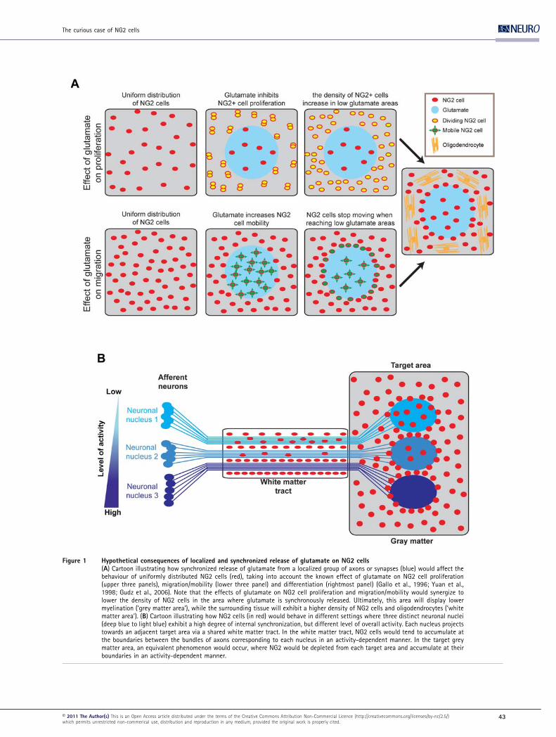
Figure 1 Hypothetical consequences of localized and synchronized release of glutamate on NG2 cells(A) Cartoon illustrating how synchronized release of glutamate from a localized group of axons or synapses (blue) would affect thebehaviour of uniformly distributed NG2 cells (red), taking into account the known effect of glutamate on NG2 cell proliferation(upper three panels), migration/mobility (lower three panel) and differentiation (rightmost panel) (Gallo et al., 1996; Yuan et al.,1998; Gudz et al., 2006). Note that the effects of glutamate on NG2 cell proliferation and migration/mobility would synergize tolower the density of NG2 cells in the area where glutamate is synchronously released. Ultimately, this area will display lowermyelination (‘grey matter area’), while the surrounding tissue will exhibit a higher density of NG2 cells and oligodendrocytes (‘whitematter area’). (B) Cartoon illustrating how NG2 cells (in red) would behave in different settings where three distinct neuronal nuclei(deep blue to light blue) exhibit a high degree of internal synchronization, but different level of overall activity. Each nucleus projectstowards an adjacent target area via a shared white matter tract. In the white matter tract, NG2 cells would tend to accumulate atthe boundaries between the bundles of axons corresponding to each nucleus in an activity-dependent manner. In the target greymatter area, an equivalent phenomenon would occur, where NG2 would be depleted from each target area and accumulate at theirboundaries in an activity-dependent manner.
The curious case of NG2 cells
E 2011 The Author(s) This is an Open Access article distributed under the terms of the Creative Commons Attribution Non-Commercial Licence (http://creativecommons.org/licenses/by-nc/2.5/)which permits unrestricted non-commerical use, distribution and reproduction in any medium, provided the original work is properly cited.
43
Of course, in the brain such a simplified situation is unlikely
to occur and NG2 cells are more likely to encounter a series of
adjacent microenvironments, each exhibiting a high level of
internal synchronization, and corresponding for example to
distinct neuronal nuclei, cortical columns and layers. A good
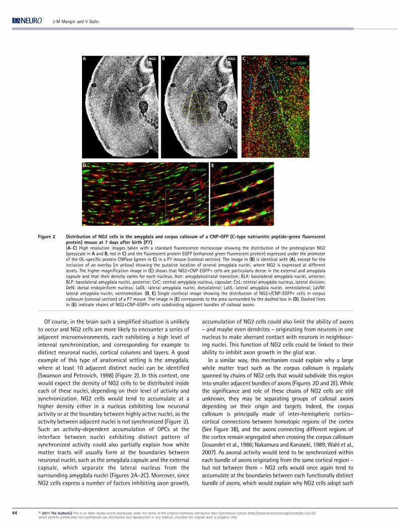
example of this type of anatomical setting is the amygdala,
where at least 10 adjacent distinct nuclei can be identified
(Swanson and Petrovich, 1998) (Figure 2). In this context, one
would expect the density of NG2 cells to be distributed inside
each of these nuclei, depending on their level of activity and
synchronization. NG2 cells would tend to accumulate at a
higher density either in a nucleus exhibiting low neuronal
activity or at the boundary between highly active nuclei, as the
activity between adjacent nuclei is not synchronized (Figure 2).
Such an activity-dependent accumulation of OPCs at the
interface between nuclei exhibiting distinct pattern of
synchronized activity could also partially explain how white
matter tracts will usually form at the boundaries between
neuronal nuclei, such as the amygdala capsule and the external
capsule, which separate the lateral nucleus from the
surrounding amygdala nuclei (Figures 2A–2C). Moreover, since
NG2 cells express a number of factors inhibiting axon growth,
accumulation of NG2 cells could also limit the ability of axons
– and maybe even dendrites – originating from neurons in one
nucleus to make aberrant contact with neurons in neighbour-
ing nuclei. This function of NG2 cells could be linked to their
ability to inhibit axon growth in the glial scar.
In a similar way, this mechanism could explain why a large
white matter tract such as the corpus callosum is regularly
spanned by chains of NG2 cells that would subdivide this region
into smaller adjacent bundles of axons (Figures 2D and 2E). While
the significance and role of these chains of NG2 cells are still
unknown, they may be separating groups of callosal axons
depending on their origin and targets. Indeed, the corpus
callosum is principally made of inter-hemispheric cortico–
cortical connections between homotopic regions of the cortex
(See Figure 3B), and the axons connecting different regions of
the cortex remain segregated when crossing the corpus callosum
(Jouandet et al., 1986; Nakamura and Kanaseki, 1989; Wahl et al.,
2007). As axonal activity would tend to be synchronized within
each bundle of axons originating from the same cortical region –
but not between them – NG2 cells would once again tend to
accumulate at the boundaries between each functionally distinct
bundle of axons, which would explain why NG2 cells adopt such
Figure 2 Distribution of NG2 cells in the amygdala and corpus callosum of a CNP-GFP (C-type natriuretic peptide-green fluorescentprotein) mouse at 7 days after birth (P7)(A–C) High resolution images taken with a standard fluorescence microscope showing the distribution of the proteoglycan NG2(greyscale in A and B, red in C) and the fluorescent protein EGFP (enhanced green fluorescent protein) expressed under the promoterof the OL-specific protein CNPase (green in C) in a P7 mouse (coronal section). The image in (B) is identical with (A), except for theinclusion of an overlay (in yellow) showing the putative location of several amygdala nuclei, where NG2 is expressed at differentlevels. The higher magnification image in (C) shows that NG2+CNP-EGFP+ cells are particularly dense in the external and amygdalacapsule and that their density varies for each nucleus. Astr: amygdalostriatal transition; BLA: basolateral amygdala nuclei, anterior;BLP: basolateral amygdala nuclei, posterior; CeC: central amygdala nucleus, capsular; CeL: central amygdala nucleus, lateral division;DeN: dorsal endopiriform nucleus; LaDL: lateral amygdala nuclei, dorsolateral; LaVL: lateral amygdala nuclei, ventrolateral; LaVM:lateral amygdala nuclei, ventromedian. (D, E) Single confocal image showing the distribution of NG2+/CNP-EGFP+ cells in corpuscallosum (coronal section) of a P7 mouse. The image in (E) corresponds to the area surrounded by the dashed box in (D). Dashed linesin (E) indicate chains of NG2+CNP-EGFP+ cells subdividing adjacent bundles of callosal axons
J-M Mangin and V Gallo
44 E 2011 The Author(s) This is an Open Access article distributed under the terms of the Creative Commons Attribution Non-Commercial Licence (http://creativecommons.org/licenses/by-nc/2.5/)which permits unrestricted non-commercial use, distribution and reproduction in any medium, provided the original work is properly cited.
a peculiar distribution pattern in corpus callosum. What could
then be the function of such patterned distribution of NG2 cells?
One possibility is that NG2 cells could actually regulate
axon growth and drive electrically synchronized axons
originating from the same cortical area to form tight
independent fascicles, while growing towards the contra-
lateral hemisphere. For example, callosal axons originating
from the somatosensory cortex are known to grow during the
first postnatal days in mice, crossing the midline around P3
and reaching their target in the contralateral hemisphere
around P6 (Wang et al., 2007). Importantly, it is known that,
during this developmental period, callosal NG2 cells already
receive functional glutamatergic synapses (De Biase et al.,
2010a) and that the speed of growth of callosal axon is
reduced when their electrical activity is reduced (Wang et al.,
2007). Therefore, callosal axon growth would be globally
decreased by inhibitory factors expressed by NG2 cells, which
would push axons to organize into tighter electrically
synchronized fascicles. This synchronized activity would
locally reduce NG2 cell density and create NG2 cell-free
territories suitable for axonal growth. Such a role in axon
fasciculation would be consistent with the hypothesis that
NG2 cells may also have functions similar to those of non-
myelinating glial cells in Drosophila (Klambt et al., 1991).
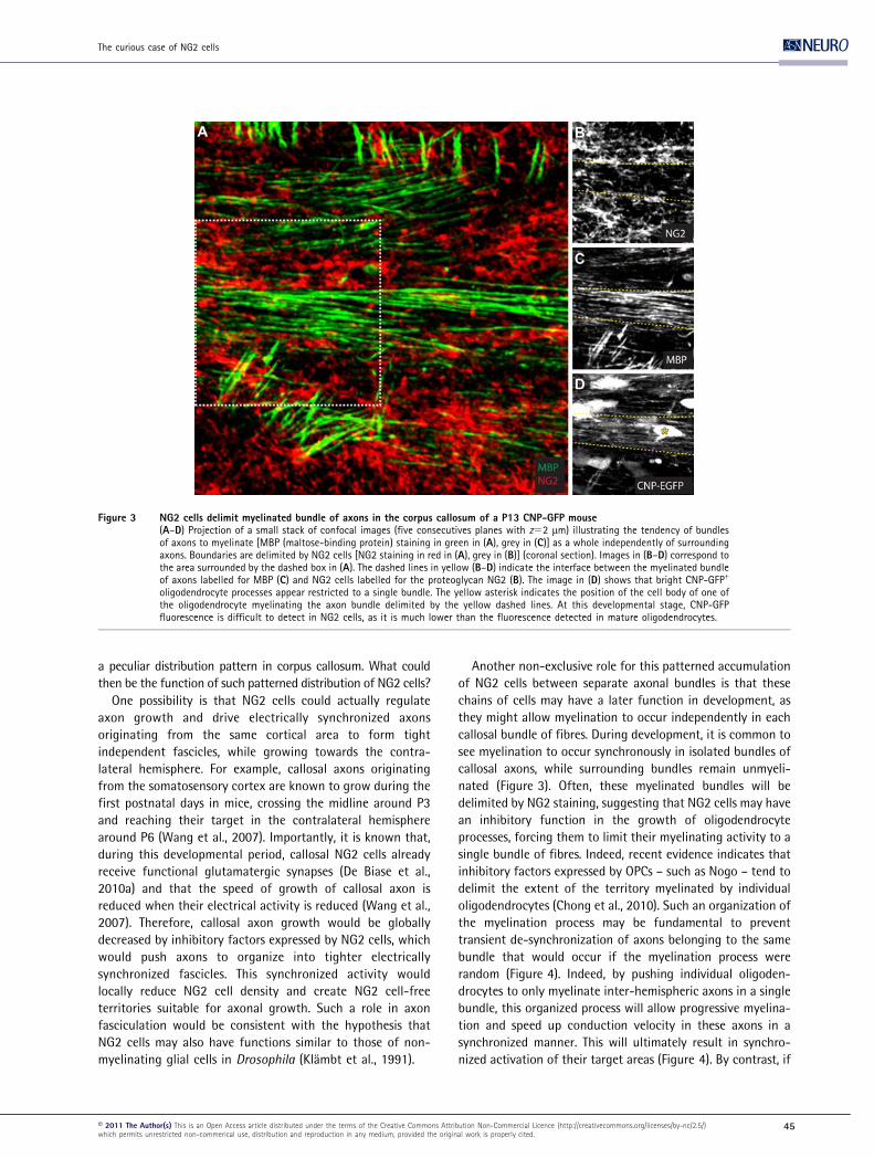
Another non-exclusive role for this patterned accumulation
of NG2 cells between separate axonal bundles is that these
chains of cells may have a later function in development, as
they might allow myelination to occur independently in each
callosal bundle of fibres. During development, it is common to
see myelination to occur synchronously in isolated bundles of
callosal axons, while surrounding bundles remain unmyeli-
nated (Figure 3). Often, these myelinated bundles will be
delimited by NG2 staining, suggesting that NG2 cells may have
an inhibitory function in the growth of oligodendrocyte
processes, forcing them to limit their myelinating activity to a
single bundle of fibres. Indeed, recent evidence indicates that
inhibitory factors expressed by OPCs – such as Nogo – tend to
delimit the extent of the territory myelinated by individual
oligodendrocytes (Chong et al., 2010). Such an organization of
the myelination process may be fundamental to prevent
transient de-synchronization of axons belonging to the same
bundle that would occur if the myelination process were
random (Figure 4). Indeed, by pushing individual oligoden-
drocytes to only myelinate inter-hemispheric axons in a single
bundle, this organized process will allow progressive myelina-
tion and speed up conduction velocity in these axons in a
synchronized manner. This will ultimately result in synchro-
nized activation of their target areas (Figure 4). By contrast, if
Figure 3 NG2 cells delimit myelinated bundle of axons in the corpus callosum of a P13 CNP-GFP mouse(A–D) Projection of a small stack of confocal images (five consecutives planes with z52 mm) illustrating the tendency of bundlesof axons to myelinate [MBP (maltose-binding protein) staining in green in (A), grey in (C)] as a whole independently of surroundingaxons. Boundaries are delimited by NG2 cells [NG2 staining in red in (A), grey in (B)] (coronal section). Images in (B–D) correspond tothe area surrounded by the dashed box in (A). The dashed lines in yellow (B–D) indicate the interface between the myelinated bundleof axons labelled for MBP (C) and NG2 cells labelled for the proteoglycan NG2 (B). The image in (D) shows that bright CNP-GFP+
oligodendrocyte processes appear restricted to a single bundle. The yellow asterisk indicates the position of the cell body of one ofthe oligodendrocyte myelinating the axon bundle delimited by the yellow dashed lines. At this developmental stage, CNP-GFPfluorescence is difficult to detect in NG2 cells, as it is much lower than the fluorescence detected in mature oligodendrocytes.
The curious case of NG2 cells
E 2011 The Author(s) This is an Open Access article distributed under the terms of the Creative Commons Attribution Non-Commercial Licence (http://creativecommons.org/licenses/by-nc/2.5/)which permits unrestricted non-commerical use, distribution and reproduction in any medium, provided the original work is properly cited.
45
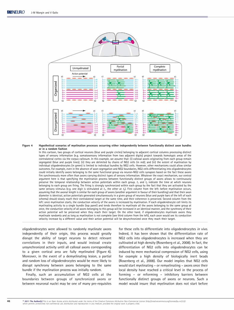
oligodendrocytes were allowed to randomly myelinate axons
independently of their origin, this process would greatly
disrupt the ability of target neurons to detect relevant
correlations in their inputs, and would instead create
unsynchronized activity until all callosal axons corresponding
to a given cortical area are fully myelinated (Figure 4).
Moreover, in the event of a demyelinating lesion, a partial
and random loss of oligodendrocytes would be more likely to
disrupt synchrony between axons belonging to the same
bundle if the myelination process was initially random.
Finally, such an accumulation of NG2 cells at the
boundaries between groups of synchronized axons or
between neuronal nuclei may be one of many pre-requisites
for these cells to differentiate into oligodendrocytes in vivo.
Indeed, it has been shown that the differentiation rate of
NG2 cells into oligodendrocytes is increased when they are
cultivated at high density (Rosenberg et al., 2008). In fact, the
differentiation of NG2 cells into oligodendrocytes can be
induced by mere mechanical compression of NG2 cells, using
for example a high density of biologically inert beads
(Rosenberg et al., 2008). Our model implies that NG2 cells
would start myelinating – or remyelinating – axons once their
local density have reached a critical level in the process of
forming – or reforming – inhibitory barriers between
functionally distinct groups of axons or neurons. Such a
model would insure that myelination does not start before
Figure 4 Hypothetical scenarios of myelination processes occurring either independently between functionally distinct axon bundlesor in a random fashionIn this cartoon, two groups of cortical neurons (blue and purple circles) belonging to adjacent cortical columns processing distincttypes of sensory information (e.g. somatosensory information from two adjacent digits) project towards homotypic areas of thecontralateral cortex via the corpus callosum. In this example, we assume that: (i) callosal axons originating from each group remainsegregated (blue and purple lines); (ii) they are delimited by chains of NG2 cells (in red); and (iii) the extent of myelination byindividual oligodendrocytes (in green) is limited to individual bundles by NG2 cells. However, other mechanisms could allow similaroutcomes. For example, even in the absence of axon segregation and NG2 boundaries, NG2 cells differentiating into oligodendrocytescould initially identify axons belonging to the same functional group via neuron-NG2 cells synapses based on the fact these axonsfire synchronously more often than axons carrying distinct types of sensory information. Whatever the exact mechanism, our centralargument here is that specifying the myelination process between functionally distinct groups of axons allows to continuouslypreserve the temporal relationship between action potentials within each group. t1 and t2 indicate the time at which neuronsbelonging to each group are firing. The firing is strongly synchronized within each group by the fact that they are activated by thesame sensory stimulus (e.g. one digit is stimulated at t1, the other at t2). First column from the left: before myelination occurs,assuming that the axonal length is similar for each group of axons (another argument in favour of their bundling) and that their axondiameter is identical, action potentials generated simultaneously in a given group of neurons (blue and purple bars of the left of eachschema) should slowly reach their contralateral target at the same time, and their coherence is preserved. Second column from theleft: once myelination starts, the conduction velocity of the axons is increased by myelination. If each oligodendrocyte cell limits itsmyelinating activity to a single bundle (top panel) and tends therefore to myelinate all the axons belonging to the same group atonce, the conduction velocity of all axons belonging to this group will be increased in an identical manner, and the synchrony of theiraction potential will be preserved when they reach their target. On the other hand, if oligodendrocytes myelinate axons theymyelinate randomly and as long as myelination is not complete (see third column from the left), each axon would see its conductionvelocity increase by a different value and their action potential will be desynchronized once they reach their target.
J-M Mangin and V Gallo
46 E 2011 The Author(s) This is an Open Access article distributed under the terms of the Creative Commons Attribution Non-Commercial Licence (http://creativecommons.org/licenses/by-nc/2.5/)which permits unrestricted non-commercial use, distribution and reproduction in any medium, provided the original work is properly cited.
axon bundles and neuron groups are organized and
partitioned into distinct functional groups, thereby insuring
orderly myelination.
CONCLUSIONS
In this review article, we attempted to gather disparate facts
known about NG2 cells and neuron-to-NG2 cell synapses into
one possible unifying framework. While it is possible that much
of the specific and oversimplified hypotheses provided here will
be disproven by the harsh reality of biological complexity, we
hope that some of the general ideas proposed may at least offer
new ways to think about NG2 cells and their function in the
brain. More particularly, we believe that our understanding of
these cells will greatly benefit from: (i) studying their role before
myelination and during early development; (ii) looking at the
function of possible homologous cell types in non-myelinated
organisms; (iii) examining how synchronized/non-synchronized
activity in neurons influence their proliferation, mobility and
differentiation; (iv) taking into account the importance of
isochronicity/coherence in signal conduction and the potentially
disruptive influence of a random myelination process; and (v)
conceptualizing neuron–OPC interactions as a complex recip-
rocal system emerging from the dynamic equilibrium between
cooperative and competitive influences. Because NG2 cells have
a fundamental role in myelin pathologies such as multiple
sclerosis and axon regeneration after brain injuries, an
integrated understanding of their diverse roles during develop-
ment will greatly contribute to our ability to develop
comprehensive and original strategies to treat such pathologies.
REFERENCES
Aguirre A, Chittajallu R, Belachew S, Gallo V (2004) NG2-expressing cells inthe subventricular zone are type C-like cells and contribute tointerneuron generation in the postnatal hippocampus. J Cell Biol 165:575–589.
Aguirre A, Dupree JL, Mangin JM, Gallo V (2007) A functional role for EGFRsignaling in myelination and remyelination. Nature Neurosci 10, 990–1002.
Bakiri Y, Attwell D, Karadottir R (2009) Electrical signalling properties ofoligodendrocyte precursor cells. Neuron Glia Biol 5:3–11.
Banerjee S, Bhat MA (2008) Glial ensheathment of peripheral axons inDrosophila. J Neurosci Res 86:1189–1198.
Barres BA (2008) The mystery and magic of glia: a perspective on their rolesin health and disease. Neuron 60:430–440.
Belachew S, Chittajallu R, Aguirre AA, Yuan X, Kirby M, Anderson S, Gallo V(2003) Postnatal NG2 proteoglycan-expressing progenitor cells areintrinsically multipotent and generate functional neurons. J Cell Biol161:169–186.
Benson MD, Romero MI, Lush ME, Lu QR, Henkemeyer M, Parada LF (2005)Ephrin-B3 is a myelin-based inhibitor of neurite outgrowth. Proc NatlAcad Sci USA 102:10694–10699.
Berger T, Schnitzer J, Orkand PM, Kettenmann H (1992) Sodium and calciumcurrents in glial cells of the mouse corpus callosum slice. Eur J Neurosci4:1271–1284.
Bergles DE, Roberts JD, Somogyi P, Jahr CE (2000) Glutamatergic synapses onoligodendrocyte precursor cells in the hippocampus. Nature 405:187–191.
Bergles DE, Jabs R, Steinhauser C (2010) Neuron–glia synapses in the brain.Brain Res Rev 63:130–137.
Blaustein MP, Lederer WJ (1999) Sodium/calcium exchange: its physiologicalimplications. Physiol Rev 79:763–854.
Butt AM, Hamilton N, Hubbard P, Pugh M, Ibrahim M (2005) Synantocytes:the fifth element. J Anat 207:695–706.
Chittajallu R, Aguirre A, Gallo V (2004) NG2-positive cells in the mouse whiteand grey matter display distinct physiological properties. J Physiol561:109–122.
Chong SYC, Rosenberg SS, Shen YA, Hahn AT, Zheng B, Zhang LI, Mcgee AW,Lu QR, Chan JR (2010) Nogo-A establishes spatial segregation and extentof myelination during development. SfN Meeting 2010, San Diego, Poster539.18/C31.
Clarke LN, Hamilton NB, Young KM, Kessaris N, Richardson WD, Attwell D(2010) The distribution and excitability of two types of oligodendrocyteprecursor cell in white and grey matter. SfN Meeting 2010 San Diego,Poster 853.21/H14.
Dawson MR, Polito A, Levine JM, Reynolds R (2003) NG2-expressing glialprogenitor cells: an abundant and widespread population of cycling cellsin the adult rat CNS. Mol Cell Neurosci 24:476–488.
De Biase LM, Nishiyama A, Bergles DE (2010a) Excitability and synapticcommunication within the oligodendrocyte lineage. J Neurosci 30:3600–3611.
De Biase LM, Kang SH, Potter E, Fukaya M, Srivastava I, Rowitch D, MishinaM, Bergles DE (2010b) NMDA receptor function in the oligodendrocytelineage. In SfN Meeting 2010, San Diego, Poster 853.18/H11.
Delaunay D, Heydon K, Cumano A, Schwab MH, Thomas JL, Suter U, Nave KA,Zalc B, Spassky N (2008) Early neuronal and glial fate restriction ofembryonic neural stem cells. J Neurosci 28:2551–2562.
Demerens C, Stankoff B, Logak M, Anglade P, Allinquant B, Couraud F, Zalc B,Lubetzki C (1996) Induction of myelination in the central nervous systemby electrical activity. Proc Natl Acad Sci USA 93:9887–9892.
Dimou L, Simon C, Kirchhoff F, Takebayashi H, Gotz M (2008) Progeny ofOlig2-expressing progenitors in the gray and white matter of the adultmouse cerebral cortex. J Neurosci 28:10434–10442.
Etxeberria A, Mangin JM, Aguirre A, Gallo V (2010) Adult-born SVZprogenitors receive transient synapses during remyelination in corpuscallosum. Nat Neurosci 13:287–289.
Feng W, Zhang M (2009) Organization and dynamics of PDZ-domain-relatedsupramodules in the postsynaptic density. Nat Rev Neurosci 10:87–99.
Filbin MT (2003) Myelin-associated inhibitors of axonal regeneration in theadult mammalian CNS. Nat Rev Neurosci 4:703–713.
Fritschy JM, Harvey RJ, Schwarz G (2008) Gephyrin: where do we stand,where do we go? Trends Neurosci 31:257–264.
Gallo V, Zhou JM, McBain CJ, Wright P, Knutson PL, Armstrong RC (1996)Oligodendrocyte progenitor cell proliferation and lineage progression areregulated by glutamate receptor-mediated K+ channel block. J Neurosci16:2659–2670.
Gallo V, Mangin JM, Kukley M, Dietrich D (2008) Synapses on NG2-expressingprogenitors in the brain: multiple functions? J Physiol 586:3767-3781.
Ge WP, Yang XJ, Zhang Z, Wang HK, Shen W, Deng QD, Duan S (2006) Long-term potentiation of neuron–glia synapses mediated by Ca2+-permeableAMPA receptors. Science 312:1533–1537.
Ge WP, Zhou W, Luo Q, Jan LY, Jan YN (2009) Dividing glial cells maintaindifferentiated properties including complex morphology and functionalsynapses. Proc Natl Acad Sci USA 106:328–333.
Gudz TI, Komuro H, Macklin WB (2006) Glutamate stimulates oligodendrocyteprogenitor migration mediated via an alphav integrin/myelin proteolipidprotein complex. J Neurosci 26:2458–2466.
Harel NY, Strittmatter SM (2006) Can regenerating axons recapitulatedevelopmental guidance during recovery from spinal cord injury? Nat RevNeurosci 7:603–616.
Hartline DK, Colman DR (2007) Rapid conduction and the evolution of giantaxons and myelinated fibers. Curr Biol 17:R29–R35.
Horner PJ, Thallmair M, Gage FH (2002) Defining the NG2-expressing cell ofthe adult CNS. J Neurocytol 31:469–480.
Jacob TC, Moss SJ, Jurd R (2008) GABA(A) receptor trafficking and its rolein the dynamic modulation of neuronal inhibition. Nat Rev Neurosci9:331–343.
Jouandet ML, Lachat JJ, Garey LJ (1986) Topographic distribution of callosalneurons and terminals in the cerebral cortex of the cat. Anat Embryol(Berl) 173:323–342.
The curious case of NG2 cells
E 2011 The Author(s) This is an Open Access article distributed under the terms of the Creative Commons Attribution Non-Commercial Licence (http://creativecommons.org/licenses/by-nc/2.5/)which permits unrestricted non-commerical use, distribution and reproduction in any medium, provided the original work is properly cited.
47
Kang SH, Fukaya M, Yang JK, Rothstein JD, Bergles DE (2010) NG2+ CNS glialprogenitors remain committed to the oligodendrocyte lineage inpostnatal life and following neurodegeneration. Neuron 68:668–681.
Karadottir R, Hamilton NB, Bakiri Y, Attwell D (2008) Spiking and nonspikingclasses of oligodendrocyte precursor glia in CNS white matter. NatNeurosci 11:450–456.
Keirstead HS, Levine JM, Blakemore WF (1998) Response of the oligoden-drocyte progenitor cell population (defined by NG2 labelling) todemyelination of the adult spinal cord. Glia 22:161–170.
Klambt C, Jacobs JR, Goodman CS (1991) The midline of the Drosophilacentral nervous system: a model for the genetic analysis of cell fate, cellmigration, and growth cone guidance. Cell 9:331–343.
Kessaris N, Fogarty M, Iannarelli P, Grist M, Wegner M, Richardson WD (2006)Competing waves of oligodendrocytes in the forebrain and postnatalelimination of an embryonic lineage. Nat Neurosci 9:173–179.
Kukley M, Capetillo-Zarate E, Dietrich D (2007) Vesicular glutamate releasefrom axons in white matter. Nat Neurosci 10:311–320.
Kukley M, Kiladze M, Tognatta R, Hans M, Swandulla D, Schramm J, Dietrich D(2008) Glial cells are born with synapses. FASEB J 22:2957–2969.
Kukley M, Nishiyama A, Dietrich D (2010) The fate of synaptic input to NG2glial cells: neurons specifically downregulate transmitter release ontodifferentiating oligodendroglial cells. J Neurosci 30:8320–8331.
Lappe-Siefke C, Goebbels S, Gravel M, Nicksch E, Lee J, Braun PE, Griffiths IR,Nave KA (2003) Disruption of Cnp1 uncouples oligodendroglial functionsin axonal support and myelination. Nat Genet 33:366–374.
Levine JM (1994) Increased expression of the NG2 chondroitin-sulfateproteoglycan after brain injury. J Neurosci 4:4716–4730.
Levine JM, Reynolds R (1999) Activation and proliferation of endogenousoligodendrocyte precursor cells during ethidium bromide-induceddemyelination. Exp Neurol 160:333–347.
Levison SW, Goldman JE (1993) Both oligodendrocytes and astrocytesdevelop from progenitors in the subventricular zone of the rat forebrain.Neuron 10:201–212.
Ligon KL, Kesari S, Kitada M, Sun T, Arnett HA, Alberta JA, Anderson DJ, StilesCD, Rowitch DH (2006) Development of NG2 neural progenitor cellsrequires Olig gene function. Proc Natl Acad Sci USA 103:7853–7858.
Lin SC, Bergles DE (2004) Synaptic signaling between GABAergic interneuronsand oligodendrocyte precursor cells in the hippocampus. Nat Neurosci7:24–32.
Lin SC, Huck JH, Roberts JD, Macklin WB, Somogyi P, Bergles DE (2005)Climbing fiber innervation of NG2-expressing glia in the mammaliancerebellum. Neuron 46:773–785.
Low K, Culbertson M, Bradke F, Tessier-Lavigne M, Tuszynski MH (2008)Netrin-1 is a novel myelin-associated inhibitor to axon growth.J Neurosci 28:1099–1108.
Lytle JM, Chittajallu R, Wrathall JR, Gallo V (2009) NG2 cell response in theCNP-EGFP mouse after contusive spinal cord injury. Glia 57:270–285.
Mangin JM, Kunze A, Chittajallu R, Gallo V (2008) Satellite NG2+ progenitorcells share common glutamatergic inputs with associated interneurons inthe mouse dentate gyrus. J Neurosci 28:7610–7623.
Manitt C, Colicos MA, Thompson KM, Rousselle E, Peterson AC, Kennedy TE(2001) Widespread expression of netrin-1 by neurons and oligoden-drocytes in the adult mammalian spinal cord. J Neurosci 21:3911–3922.
Mason JL, Suzuki K, Chaplin DD, Matsushima GK (2001) Interleukin-1betapromotes repair of the CNS. J Neurosci 2001 21:7046–7052.
Mathis C, Collin L, Borrelli E (2003) Oligodendrocyte ablation impairscerebellum development. Development 130:4709–4718.
McTigue DM, Wei P, Stokes BT (2001) Proliferation of NG2-positive cells andaltered oligodendrocyte numbers in the contused rat spinal cord.J Neurosci 21:3392–3400.
Moreau-Fauvarque C, Kumanogoh A, Camand E, Jaillard C, Barbin G, Boquet I,Love C, Jones EY, Kikutani H, Lubetzki C, Dusart I, Chedotal A (2003) Thetransmembrane semaphorin Sema4D/CD100, an inhibitor of axonalgrowth, is expressed on oligodendrocytes and upregulated after CNSlesion. J Neurosci 23:9229–9239.
Morris JK, Lin W, Hauser C, Marchuk Y, Getman D, Lee KF (1999) Rescue ofthe cardiac defect in ErbB2 mutant mice reveals essential roles of ErbB2in peripheral nervous system development. Neuron 23:273–283.
Muller J, Reyes-Haro D, Pivneva T, Nolte C, Schaette R, Lubke J, KettenmannH (2009) The principal neurons of the medial nucleus of the trapezoidbody and NG2(+) glial cells receive coordinated excitatory synaptic input.J Gen Physiol 134:115–127.
Nakamura H, Kanaseki T (1989) Topography of the corpus callosum in the cat.Brain Res 485:171–175.
Nishiyama A, Lin XH, Giese N, Heldin CH, Stallcup WB (1996) Co-localizationof NG2 proteoglycan and PDGF alpha-receptor on O2A progenitor cells inthe developing rat brain. J Neurosci Res 43:299–314.
Nishiyama A, Watyanabe M, Yang Z, Bu J (2002) Identity, distribution, anddevelopment of polydendrocytes: NG2-expressing glial cells. J Neurocytol31:437–455.
Oland LA, Tolbert LP (2003) Key interactions between neurons and glial cellsduring neural development in insects. Annu Rev Entomol 48:89–110.
Ozerdem U, Grako KA, Dahlin-Huppe K, Monosov E, Stallcup WB (2001) NG2proteoglycan is expressed exclusively by mural cells during vascularmorphogenesis. Dev Dyn 222:218–227.
Parker RJ, Auld VJ (2006) Roles of glia in the Drosophila nervous system.Semin Cell Dev Biol 17:66–77.
Psachoulia K, Jamen F, Young KM, Richardson WD (2009) Cell cycle dynamicsof NG2 cells in the postnatal and ageing brain. Neuron Glia Biol 5:57–67.
Raiker SJ, Lee H, Baldwin KT, Duan Y, Shrager P, Giger RJ (2010)Oligodendrocyte-myelin glycoprotein and Nogo negatively regulateactivity-dependent synaptic plasticity. J Neurosci 30:12432–12445.
Redwine JM, Armstrong RC (1998) In vivo proliferation of oligodendrocyteprogenitors expressing PDGFalphaR during early remyelination.J Neurobiol 37:413–428.
Reynolds R, Hardy R (1997) Oligodendroglial progenitors labeled with the O4antibody persist in the adult rat cerebral cortex in vivo. J Neurosci Res47:455–470.
Richardson WD, Kessaris N, Pringle N (2006) Oligodendrocyte wars. Nat RevNeurosci 7:11–18.
Riethmacher D, Sonnenberg-Riethmacher E, Brinkmann V, Yamaai T, LewinGR, Birchmeier C (1997) Severe neuropathies in mice with targetedmutations in the ErbB3 receptor. Nature 389:725–730.
Rivers LE, Young KM, Rizzi M, Jamen F, Psachoulia K, Wade A, Kessaris N,Richardson WD (2008) PDGFRA/NG2 glia generate myelinating oligoden-drocytes and piriform projection neurons in adult mice. Nat Neurosci11:1392–1401.
Rosenberg SS, Kelland EE, Tokar E, De la Torre AR, Chan JR (2008) Thegeometric and spatial constraints of the microenvironment induceoligodendrocyte differentiation. Proc Natl Acad Sci USA 105:14662–14667.
Stegmuller J, Werner H, Nave KA, Trotter J (2003) The proteoglycan NG2 iscomplexed with alpha-amino-3-hydroxy-5-methyl-4-isoxazolepropionicacid (AMPA) receptors by the PDZ glutamate receptor interaction protein(GRIP) in glial progenitor cells.Implications for glial-neuronal signaling.J Biol Chem 278:3590–3598.
Swanson LW, Petrovich GD (1998) What is the amygdala? Trends Neurosci21:323–331.
Tamura Y, Kataoka Y, Cui Y, Takamori Y, Watanabe Y, Yamada H (2007) Multi-directional differentiation of doublecortin- and NG2-immunopositive progen-itor cells in the adult rat neocortex in vivo. Eur J Neurosci 25:3489–3498.
Tanaka Y, Tozuka Y, Takata T, Shimazu N, Matsumura N, Ohta A, Hisatsune T(2009) Excitatory GABAergic activation of cortical dividing glial cells.Cereb Cortex 19:2181–2195.
Tong XP, Li XY, Zhou B, Shen W, Zhang ZJ, Xu TL, Duan S (2009) Ca(2+)signaling evoked by activation of Na(+) channels and Na(+)/Ca(2+)exchangers is required for GABA-induced NG2 cell migration. J Cell Biol186:113–128.
Wahl M, Lauterbach-Soon B, Hattingen E, Jung P, Singer O, Volz S, Klein JC,Steinmetz H, Ziemann U (2007) Human motor corpus callosum:topography, somatotopy, and link between microstructure and function.J Neurosci 27:12132–12138.
Wang CL, Zhang L, Zhou Y, Zhou J, Yang XJ, Duan SM, Xiong ZQ, Ding YQ(2007) Activity-dependent development of callosal projections in thesomatosensory cortex. J Neurosci 27:11334–11342.
Watanabe M, Toyama Y, Nishiyama A (2002) Differentiation of proliferatedNG2-positive glial progenitor cells in a remyelinating lesion. J NeurosciRes 69:826–836.
Wigley R, Butt AM (2009) Integration of NG2-glia (synantocytes) into theneuroglial network. Neuron Glia Biol 5:21–28.
Woldeyesus MT, Britsch S, Riethmacher D, Xu L, Sonnenberg-Riethmacher E,Abou-Rebyeh F, Harvey R, Caroni P, Birchmeier C (1999) Peripheralnervous system defects in ErbB2 mutants following genetic rescue ofheart development. Genes Dev 13:2538–2548.
Wu Q, Miller RH, Ransohoff RM, Robinson S, Bu J, Nishiyama A (2000)Elevated levels of the chemokine GRO-1 correlate with elevatedoligodendrocyte progenitor proliferation in the jimpy mutant. JNeurosci 20:2609–2617.
J-M Mangin and V Gallo
48 E 2011 The Author(s) This is an Open Access article distributed under the terms of the Creative Commons Attribution Non-Commercial Licence (http://creativecommons.org/licenses/by-nc/2.5/)which permits unrestricted non-commercial use, distribution and reproduction in any medium, provided the original work is properly cited.
Yuan X, Eisen AM, McBain CJ, Gallo V (1998) A role for glutamate and itsreceptors in the regulation of oligodendrocyte development in cerebellartissue slices. Development 125:2901–2914.
Zhang H, Miller RH (1996) Density-dependent feedback inhibition ofoligodendrocyte precursor expansion. J Neurosci 16:6886–6895.
Zhu X, Bergles DE, Nishiyama A (2008) NG2 cells generate botholigodendrocytes and gray matter astrocytes. Development 135:145–157.
Ziskin JL, Nishiyama A, Rubio M, Fukaya M, Bergles DE (2007) Vesicularrelease of glutamate from unmyelinated axons in white matter. NatNeurosci 10:321–330.
Received 27 December 2010/accepted 3 February 2011
Published as Immediate Publication 3 February 2011, doi 10.1042/AN20110001
The curious case of NG2 cells
E 2011 The Author(s) This is an Open Access article distributed under the terms of the Creative Commons Attribution Non-Commercial Licence (http://creativecommons.org/licenses/by-nc/2.5/)which permits unrestricted non-commerical use, distribution and reproduction in any medium, provided the original work is properly cited.
49