Embed Size (px)
Citation preview
Faraday DiscussionsCite this: Faraday Discuss., 2016, 188, 369
PAPER
Ope
n A
cces
s A
rtic
le. P
ublis
hed
on 0
2 D
ecem
ber
2015
. Dow
nloa
ded
on 2
/2/2
022
7:31
:01
AM
. T
his
artic
le is
lice
nsed
und
er a
Cre
ativ
e C
omm
ons
Attr
ibut
ion-
Non
Com
mer
cial
3.0
Unp
orte
d L
icen
ce.
View Article OnlineView Journal | View Issue
The curious case of zeolite–clay/binderinteractions and their consequences forcatalyst preparation†
Gareth T. Whiting,‡* Abhishek Dutta Chowdhury,‡ Ramon Oord,Pasi Paalanen and Bert M. Weckhuysen*
Received 30th November 2015, Accepted 2nd December 2015
DOI: 10.1039/c5fd00200a
Zeolite-based catalyst bodies are commonly employed in a range of important industrial
processes. Depending on the binder and shaping method chosen, vast differences in the
reactivity, selectivity and stability are obtained. Here, three highly complementary micro-
spectroscopic techniques were employed to study zeolite ZSM-5–binder interactions in
SiO2-, Al2O3-, SiO2 : Al2O3- (2 : 1 mix) and kaolinite-bound catalyst pellets. We establish
how their preparation influences the zeolite–clay/binder interactions. Using thiophene
as an acid-catalyzed staining reaction, light absorbing oligomers produced in each
sample were followed. To our surprise, kaolinite decreased the overall reactivity of the
sample due to the phase change of the binder, creating a hard impenetrable outer
layer. Aluminum migration to the zeolite was observed when Al2O3 was selected as
a binder, creating additional Brønsted acid sites, which favored the formation of ring-
opened thiophene oligomers compared to the larger oligomer species produced when
SiO2 was used as a binder. In the latter case, the interaction of the Si–OH groups in the
binder with thiophene was revealed to have a large impact in creating such large
oligomer species. Furthermore, the combination of a SiO2 : Al2O3 mix as a binder
enhanced the reactivity, possibly due to the creation of additional Brønsted acid sites
between the two binder components during pellet preparation. It is evident that,
independent of the shaping method, the intimate contact between the zeolite and
binder heavily impacts the reactivity and product selectivity, with the type of binder
playing a vital role.
1. Introduction
The use of catalyst materials in the petrochemical and bulk chemical synthesisindustries is vast, and plays a vital role in supplying many of our everyday
Inorganic Chemistry and Catalysis Group, Debye Institute for Nanomaterials Science, Utrecht University,
Universiteitsweg 99, 3584 CG Utrecht, The Netherlands. E-mail: [email protected]; [email protected]
† Electronic supplementary information (ESI) available. See DOI: 10.1039/c5fd00200a
‡ Authors contributed equally to this work.
This journal is © The Royal Society of Chemistry 2016 Faraday Discuss., 2016, 188, 369–386 | 369
Faraday Discussions PaperO
pen
Acc
ess
Art
icle
. Pub
lishe
d on
02
Dec
embe
r 20
15. D
ownl
oade
d on
2/2
/202
2 7:
31:0
1 A
M.
Thi
s ar
ticle
is li
cens
ed u
nder
a C
reat
ive
Com
mon
s A
ttrib
utio
n-N
onC
omm
erci
al 3
.0 U
npor
ted
Lic
ence
.View Article Online
needs.1–3 In industrial processes such as hydrocarbon and/or aromatic isomeri-zation- and alkylation-type reactions, zeolites provide the main active phase of thecatalyst materials.4,5 However, in order for the zeolite to function at its optimumlevel, it is oen dispersed non-homogeneously in a binder or matrix, i.e. SiO2,Al2O3 and/or clay, and shaped to the desired needs. Such catalysts are commonlyreferred to as ‘catalyst bodies’ or ‘technical bodies’, and take the form of extru-dates, pellets or granules.6 Binder/matrix components offer numerous advantagesto the zeolite (as highlighted in a perspective article by Hargreaves et al.7), buttheir overriding role is to increase the mechanical strength in order to resistattrition loss within industrial reactor conditions. However, our preconceivedideas that the zeolite's physicochemical properties and catalytic performance willbe fully maintained during/aer the preparation of catalyst bodies (i.e. shapingwith binders, heat treatment, etc.) would be naive, with the binder known tointeract with the zeolite and also known to play a vital role in the catalyst'sperformance.4,7–9
Until recently, limited academic research has been reported on such industrialcatalysts, but a spate in the development of advanced characterization techniquesable to probe the multi-dimensional parameters of these materials with highspatiotemporal resolution (and also non-invasively) has provided signicantinsights into the structure–performance relationships of these closely guardedmaterials.10–20 Particularly in the eld of Fluid Catalytic Cracking (FCC) particlesand zeolite-based binder-bound extrudates and pellets for e.g. methanol-to-hydrocarbon (MTH) catalysis, combinations of chemical imaging and structuralimaging techniques, together with standard physicochemical characterization ofthe materials, have enhanced our knowledge into how these complex materialsfunction.17,18,21–23 For instance, staining probe molecule reactions (i.e. acid-cata-lyzed uorostyrene oligomerization), coupled with visible micro-spectroscopy,confocal microscopy and X-ray absorption micro-spectroscopy, have shown thatthe distribution of the zeolite, the presence of metal poisons, and the type/amount of binder present, all signicantly inuence the catalytic performanceand, in particular, the catalyst’s ability to resist deactivation.12,16,20,24,25 Overall, theinformation gained on binder effects from such advanced characterizationstudies of industrially relevant catalyst materials is key to improving the catalystbody design, which could play a vital role in the future of these catalysts. This isparticularly prominent, given the ever stricter and challenging societal, environ-mental and economical demands enforced on catalytic industrial reactionprocesses.
The use of inexpensive inorganic binders, such as SiO2, Al2O3 and clay, forzeolite-based catalyst bodies is common in industrial processes, such as xyleneisomerization and benzene alkylation (among others), but several reports notethat the zeolite–binder interactions can indeed inuence the reactivity, selectivityand stability of the overall catalyst material. When employing clay (i.e. bentonite,kaolin and attapulgite) as a binder with zeolites, high-temperatures are used inthe pelletization process in order to eliminate the activity of the clay and destroythe surface area, whilst creating meso- and/or macro-porosity.26 However, it hasbeen reported that a substantial decrease in the inter- and intra-crystalline acidityof the H-zeolite occurs during pelletization due to the neutralization of acid siteswithmobile alkaline or alkaline-earth cations from the clay binder.27 Clay has alsobeen shown to decrease weak acidity and increase high strength acid site
370 | Faraday Discuss., 2016, 188, 369–386 This journal is © The Royal Society of Chemistry 2016

Paper Faraday DiscussionsO
pen
Acc
ess
Art
icle
. Pub
lishe
d on
02
Dec
embe
r 20
15. D
ownl
oade
d on
2/2
/202
2 7:
31:0
1 A
M.
Thi
s ar
ticle
is li
cens
ed u
nder
a C
reat
ive
Com
mon
s A
ttrib
utio
n-N
onC
omm
erci
al 3
.0 U
npor
ted
Lic
ence
.View Article Online
density,28 as well as inuence product selectivity29 and improve coke stability (bytrapping coke precursors in the kaolinite binder).30 Al2O3 as a binder for zeolite-based catalyst bodies has also been known to inuence product selectivity andoverall activity due to the migration of aluminum from the binder to the zeoliteframework, creating additional Brønsted acid sites.17,31,32 It is also used as a heatsink and a trap for metal poisons in petroleum feedstocks, hence retaining zeoliteacidity.33 A similar trend occurs for SiO2-bound zeolite materials, whereby a moreunexpected solid-state ion exchange takes place, with Michels et al. and Lee et al.reporting a loss of Brønsted acidity due to dealumination from the framework ofthe zeolite.34,35 Gelin et al. also witnessed the opposite trend, with Si species fromthe SiO2–Al2O3 matrix migrating to the zeolite framework, healing defects andpreserving crystallinity.36 It is clear that, although some binders are oenconsidered as inert, the intimate contact between the zeolite and binder incatalyst bodies can produce remarkable consequences or advantages and theirinteractions therefore require a greater degree of attention, if improvements inthe catalyst design are to be made.
In our previous work, we have focused on SiO2- and Al2O3-bound zeolite-basedcatalysts in the form of extrudates.17 However, in this work, we set out tosynthesize four ZSM-5–binder-bound pellets, using either SiO2, Al2O3, SiO2 : Al2O3
(mix 2 : 1) or kaolinite as a binder, and study whether any zeolite–binder inter-actions have taken place during their physical mixing, and how these parametersinuence the overall catalytic properties of the catalysts prepared. To do this,a staining probe reaction, namely thiophene oligomerization, was employedalong with several highly complementary non-invasive in situmicro-spectroscopictechniques: UV-Visible, confocal uorescence and IR micro-spectroscopy. Usingthese three techniques, we are able to follow variations in product selectivity aswell as in the location and distribution of the light absorbing oligomers formed ineach sample and correlate these to any zeolite–clay/binder effects.
2. Experimental2.1. Materials
Four batches of SiO2- (purchased from Aldrich, Davisil™), Al2O3- (Catapal®),SiO2/Al2O3- (2 : 1 mix of Davisil™ : Catapal®) and kaolinite- (purchased fromSigma Aldrich) bound ZSM-5 samples were prepared by a pelletization procedure:NH4–ZSM-5 powder (purchased from Zeolyst CBV 8014) with a SiO2 : Al2O3 ratio of80 was thoroughly mixed with the relevant binder at a ZSM-5 : binder wt% ratio of30 : 70 (to ensure noticeable binder effects) before pressing into pellets. Eachpellet underwent heat treatment under a N2 atmosphere (to form H–ZSM-5) from30 �C to 600 �C (at 15 �C min�1) and it was then held at this temperature for 3 h,before cooling to 30 �C. Sample notations are as follows: ZSM-5–a: where ‘a’corresponds to either: Si (SiO2); Al (Al2O3); SiAl (SiO2–Al2O3 mix, 2 : 1); Kao(kaolinite).
2.2. Ar physisorption
Ar physisorption on all samples was performed with an automated gas sorptionsystemMicromeritics TriStar 3000. Prior to the measurements at 77 K, all sampleswere dried at 300 �C for 20 h. The surface areas were calculated using the BET
This journal is © The Royal Society of Chemistry 2016 Faraday Discuss., 2016, 188, 369–386 | 371

Faraday Discussions PaperO
pen
Acc
ess
Art
icle
. Pub
lishe
d on
02
Dec
embe
r 20
15. D
ownl
oade
d on
2/2
/202
2 7:
31:0
1 A
M.
Thi
s ar
ticle
is li
cens
ed u
nder
a C
reat
ive
Com
mon
s A
ttrib
utio
n-N
onC
omm
erci
al 3
.0 U
npor
ted
Lic
ence
.View Article Online
model, whilst the external surface area, micropore surface area and microporevolume were determined using the t-plot method (Table 1 and Fig. 1).
2.3. X-ray powder diffraction
All samples were measured with a Bruker D8 Discover diffractometer equippedwith a 1-dimensional LynxEye XE energy dispersive detector, in transmissioncapillary geometry with a Mo (Ka1 0.709 A) source. The sample powders wereloaded into 980 mm inner diameter quartz capillaries (1000 mm outer diameter) sothat the sample bed length was about z20 mm. The measurement parametersused were 2q ¼ 2.5–24� with a step size of 0.008 and an exposure time of 3.5 s perstep. The Mo source was run with a power of 2500 W (50 kV � 50 mA) (Fig. 2).
2.3.1. Rietveld analysis. Rietveld analysis on the measured samples wasperformed using the TOPAS 5 soware package. Fitting was done with structuresfrom the ICCD PDF-4+ 2015 database. Individual peaks were used to t theamorphous peaks (2qz 10–11) and the primary beam (2q¼ 2.0–2.5), and the restof the background was corrected with a soware provided polynomial. Sampledisplacement and absorption path length error in the capillary geometry werecorrected with soware provided functions (Table 2).
2.4. NH3-TPD
The acidity of the samples wasmeasured by temperature-programmed desorption(TPD) with ammonia as the probe molecule using Micrometrics AutoChem 2920apparatus. Prior to the adsorption of ammonia, 100 mg of the sample placed ina quartz tube reactor was preheated at 550 �C under 50mLmin�1 of He ow for 15min (rate 10 �Cmin�1). Adsorption of ammonia (10 vol% in He) was performed ina pulse wise manner at 100 �C. In order to remove the physisorbed ammonia, thesample was ushed with a He stream (25 mL min�1) at 100 �C for 1 h. Theammonia desorption was carried out under a He ow in the temperature range100–700 �C at a heating rate of 5 �Cmin�1. The amount of adsorbed ammonia wasmonitored with a thermal conductivity detector (TCD) (Fig. 3 and Table 2).
Table 1 Textural properties of the ZSM-5–clay/binder-bound pellets determined by Arphysisorption
SampleBET surface area(m2 g�1)
t-Plotmicropore area(m2 g�1)
Porevolume(cm3 g�1)
t-Plot microporevolume(m2 g�1)
ZSM-5 371 306 0.18 0.12ZSM-5–Si 252 (296)a 90 (110)a 0.46 (0.80)a 0.03 (0.04)a
ZSM-5–Al 269 (316) 93 (97) 0.30 (0.33) 0.03 (0.04)ZSM-5–SiAl 272 106 0.35 0.04 (0.04)ZSM-5–Kao 176 (113) 150 (97) 0.09 (0.06) 0.06 (0.04)SiO2 264 26 1.06 0Al2O3 293 33 0.39 0Kaolinite 3 7 0.01 0
a Predicted values calculated from the pellet composition and the experimental values of thepure binder and pure zeolite components.
372 | Faraday Discuss., 2016, 188, 369–386 This journal is © The Royal Society of Chemistry 2016

Fig. 1 Pore volume distributions, determined by the Barrett–Joyner–Halenda adsorptionmethod with Ar, for each selected binder (dashed lines) and its corresponding zeolite–clay/binder pellets (solid lines): ZSM-5 (black diamonds); ZSM-5–Si (red triangles); ZSM-5–Al (green circles); ZSM-5–SiAl (blue squares); ZSM-5–Kao (orange crosses).
Paper Faraday DiscussionsO
pen
Acc
ess
Art
icle
. Pub
lishe
d on
02
Dec
embe
r 20
15. D
ownl
oade
d on
2/2
/202
2 7:
31:0
1 A
M.
Thi
s ar
ticle
is li
cens
ed u
nder
a C
reat
ive
Com
mon
s A
ttrib
utio
n-N
onC
omm
erci
al 3
.0 U
npor
ted
Lic
ence
.View Article Online
2.5. UV-Vis absorption micro-spectroscopy
The UV-Vis micro-spectroscopy measurements were performed with a CRAIC 20/30 PV™ UV-Vis-NIR micro-spectrophotometer using a 15� objective. A 75 Wxenon lamp was used for illumination. The pellets were placed on the heatingstage of an in situ Linkam cell (THMS600) equipped with a temperature controller(Linkam TMS94), where they were impregnated with 8 mL of thiophene. Duringeach experiment, aer complete adsorption of 8 mL of thiophene under
Fig. 2 Powder X-ray diffraction (XRD) patterns of the following ZSM-5-bound pellets(from top to bottom): ZSM-5 (black); ZSM-5–Si (red); ZSM-5–Al (green); ZSM-5–SiAl(blue); and ZSM-5–Kao (orange). The inset corresponds to the main zeolite diffractionpeaks, noting no noticeable shifts in 2q between the samples under study.
This journal is © The Royal Society of Chemistry 2016 Faraday Discuss., 2016, 188, 369–386 | 373
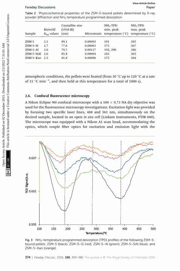
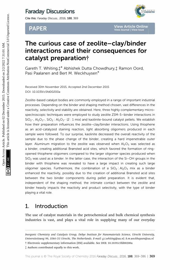
Table 2 Physicochemical properties of the ZSM-5-bound pellets determined by X-raypowder diffraction and NH3 temperature programmed desorption
SampleRietveldRwp values
Crystallite size(LVol-IB)(nm) Microstrain
NH3-TPDmin. peaktemperature (�C)
NH3-TPDmax. peaktemperature (�C)
ZSM-5 3.1 89.1 0.00093 191 385ZSM-5–Si 2.7 77.6 0.00063 175 367ZSM-5–Al 3.0 79.7 0.00117 192, 290 386ZSM-5–SiAl 2.6 85.8 0.00094 183 365ZSM-5–Kao 2.5 81.8 0.00096 175 384
Faraday Discussions PaperO
pen
Acc
ess
Art
icle
. Pub
lishe
d on
02
Dec
embe
r 20
15. D
ownl
oade
d on
2/2
/202
2 7:
31:0
1 A
M.
Thi
s ar
ticle
is li
cens
ed u
nder
a C
reat
ive
Com
mon
s A
ttrib
utio
n-N
onC
omm
erci
al 3
.0 U
npor
ted
Lic
ence
.View Article Online
atmospheric conditions, the pellets were heated (from 30 �C up to 120 �C at a rateof 15 �C min�1, and then held at this temperature for a total of 1000 s).
2.6. Confocal uorescence microscopy
A Nikon Eclipse 90i confocal microscope with a 100 � 0.73 NA dry objective wasused for the uorescence microscopy investigations. Excitation light was providedby focusing two specic laser lines, 488 and 561 nm, simultaneously on thedesired sample, located in an open in situ cell (Linkam Instruments, FTIR 600).The microscope was equipped with a Nikon A1 scan head, accommodating theoptics, which couple ber optics for excitation and emission light with the
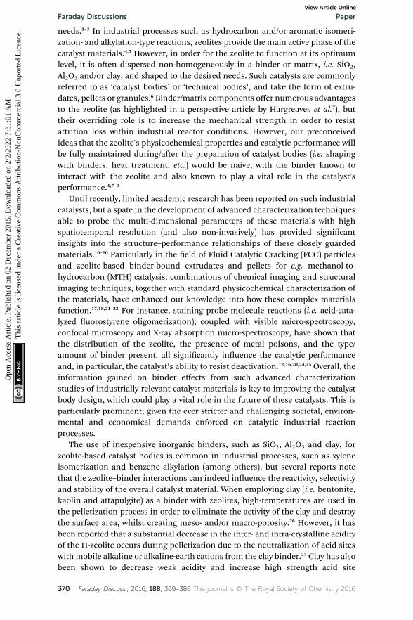
Fig. 3 NH3-temperature programmed desorption (TPD) profiles of the following ZSM-5-bound pellets: ZSM-5 (black); ZSM-5–Si (red); ZSM-5–Al (green); ZSM-5–SiAl (blue); andZSM-5–Kao (orange).
374 | Faraday Discuss., 2016, 188, 369–386 This journal is © The Royal Society of Chemistry 2016

Paper Faraday DiscussionsO
pen
Acc
ess
Art
icle
. Pub
lishe
d on
02
Dec
embe
r 20
15. D
ownl
oade
d on
2/2
/202
2 7:
31:0
1 A
M.
Thi
s ar
ticle
is li
cens
ed u
nder
a C
reat
ive
Com
mon
s A
ttrib
utio
n-N
onC
omm
erci
al 3
.0 U
npor
ted
Lic
ence
.View Article Online
microscope. A spectral analyzer in the Nikon A1 system was equipped with 32photomultiplier tubes (PMTs) set to collect emission light in the region of 480–730 nm, with a resolution of 6 nm. 3D confocal uorescence microscopy imageswere recorded at a similar focal depth in each sample using an identical laserpower. Prior to microscopic analysis, to each sample, 8 mL of the desired probemolecule was impregnated at 30 �C, before ramping the temperature (at 15 �Cmin�1) to 120 �C, holding at this temperature for 15 min, and cooling to 30 �C.
2.7. Infrared micro-spectroscopy
In situ infrared micro-spectroscopy experiments were performed using a PerkinElmer Spotlight 400 FT-IR microscope with an aperture size of 100 � 100 mm.Each zeolite–clay/binder-bound pellet was placed under the microscope andspectra were obtained in reectance mode before, during and aer thiopheneoligomerization (from 30 �C to 120 �C at 15 �C min�1).
3. Results and discussion3.1. UV-Vis absorption micro-spectroscopy
In order to probe possible zeolite–clay/binder interactions created during thecatalyst preparation procedure (i.e. pelletization and/or heat treatment), a suitablereaction was chosen. The oligomerization of thiophene has previously beenutilized by our research group for such purposes on zeolite–binder-bound catalystextrudates, and offers an excellent means to gain insight into characteristics suchas how the nature and/or number of Brønsted acid sites within the samplesinuence the catalytic reactivity and intermediate/product selectivity.17,18 Aparticular highlight of the chosen reaction is that the majority of oligomer speciesproduced on the acid sites within the samples are light absorbing (Scheme 1).Therefore, UV-Vis absorption spectroscopy allows us to obtain, in situ, theformation of absorption bands and hence the nature and relative amount ofoligomer species formed, as a function of reaction time. Scheme 1 providesa general overview of the possible light absorbing species produced (measuredusing Time-Dependent Density Functional Theory (TD-DFT)), either during anoligomerization reaction pathway, or during a ring-opening pathway (leading tothe formation of thiol-like species), with the pathway chosen determined by thestrength and number of Brønsted acid sites of the zeolite.
Given the complex nature of the overall UV-Vis absorption spectra of all thesamples (Fig. S1 and S2†), absorption spectra of each sample were compared atvarious reaction times during the oligomerization reaction (Fig. 4). Fig. 4(a)displays the absorption spectra for pellets of ZSM-5, ZSM-5–Si, ZSM-5–Al, ZSM-5–SiAl and ZSM-5–Kao aer 10 s of reaction time. Even at this early stage of thereaction, visible differences between each catalyst's absorption spectrum areapparent. The absorption band formed at 275 nm can be assigned to a-protonatedthiophene monomers (Scheme 1B) on zeolite Brønsted acid sites.37 The band at310 nm is attributed to a protonated dimeric carbocation, formed by the inter-action of two thiophene molecules linked together in the zeolite pores (C).37
The more intense absorption bands at 410, 500, and 550 nm are assigned tomolecules formed via the oligomerization route, namely: a protonated trimericcarbocation species, i.e. C12H13S3
+; a protonated trimeric carbocation species, i.e.
This journal is © The Royal Society of Chemistry 2016 Faraday Discuss., 2016, 188, 369–386 | 375

Scheme 1 Proposed reaction pathway of thiophene oligomerization on zeolite Brønstedacid sites. Thiophene monomer (A) undergoes protonation on the Brønsted acid sites ofH–ZSM-5 to form the protonated monomer (B). Two different reaction pathways arepossible at this stage: opening of the thiophene ring and subsequent reaction witha second monomer to form a thiol-like carbocation (F1/F1-T/F2/F2-T), or, alternatively,dimerization (to C) can occur followed by further oligomerization (to D) or opening of thethiophene ring, forming a thiol-like carbocation (F1/F1-T/F2/F2-T). The trimeric carbo-cationic species (D) can undergo further oligomerization to form more extended/conju-gated species (E1/E2). [Reproduced from ref. 17].
Faraday Discussions PaperO
pen
Acc
ess
Art
icle
. Pub
lishe
d on
02
Dec
embe
r 20
15. D
ownl
oade
d on
2/2
/202
2 7:
31:0
1 A
M.
Thi
s ar
ticle
is li
cens
ed u
nder
a C
reat
ive
Com
mon
s A
ttrib
utio
n-N
onC
omm
erci
al 3
.0 U
npor
ted
Lic
ence
.View Article Online
C12H9S3+; and a tetrameric carbocation species (E1), respectively.37 Finally, the
band at 445 nm can be assigned to a thiol-like species (F2) formed via a ring-opening reaction pathway.
Taking each sample into consideration, a feature that stands out in the spectraat 10 s of reaction time (Fig. 4(a)) is the surprising lack of intense absorptionbands for the kaolinite-bound sample, ZSM-5–Kao. This suggests a clear inactivitywhen ZSM-5 crystals are bound to clay, compared with ZSM-5 bound to anamorphous binder. As NH3-TPD showed (Fig. 3), the sample bound to kaolinitedisplayed the lowest amount of weak and strong Brønsted acid sites, which couldexplain the low thiophene oligomerization reactivity, but this is not likelyconsidering the similar prole for ZSM-5–Si (which shows a far higher reactivity,and even has slightly weaker acid sites due to a slight shi to lower temperaturesin the NH3 desorption peaks). X-ray diffraction of the ZSM-5–Kao material (Fig. 2)showed a clear phase change from kaolinite (Fig. S3†) to a combination ofmuscovite and orthoclase upon preparation of the ZSM-5–Kao pellet. This featureis due to the loss of hydroxyl groups from kaolinite upon heat treatment of thepellet, which propagates the phase change of the clay,38 and could play a vitalrole in the inactivity of the sample. Further evidence to support this claim isobserved in Fig. S2(a),† where the heat-treated ZSM-5 powder pelletized with
376 | Faraday Discuss., 2016, 188, 369–386 This journal is © The Royal Society of Chemistry 2016
Fig. 4 UV-Vis absorption spectra of ZSM-5 (black line), ZSM-5–Si (red), ZSM-5–Al (green),ZSM-5–SiAl (blue), and ZSM-5–Kao (orange) pellets reacted with thiophene after: (a) 10 s;(b) 50 s; (c) 100 s; (d) 200 s; (e) 400 s; and (f) 800 s of reaction time.
Paper Faraday DiscussionsO
pen
Acc
ess
Art
icle
. Pub
lishe
d on
02
Dec
embe
r 20
15. D
ownl
oade
d on
2/2
/202
2 7:
31:0
1 A
M.
Thi
s ar
ticle
is li
cens
ed u
nder
a C
reat
ive
Com
mon
s A
ttrib
utio
n-N
onC
omm
erci
al 3
.0 U
npor
ted
Lic
ence
.View Article Online
non-heat-treated kaolinite reveals a far higher reactivity towards thiophene olig-omerization (due to the unchanged phase of kaolinite). Looking towards theMohs hardness properties of the newly developed binder in ZSM-5–Kao, anincrease from 2.5 (kaolinite) to 6 (orthoclase) gives an indication that thiopheneimpregnation into the pellet could be hindered. Ar physisorption results providefurther support that the phase change of the material is detrimental to thetextural properties of the catalyst, with limited porosity available in ZSM-5–Kao,and even a striking elevation in the expected micropore area and microporevolume values (Table 1). Given the temperature of the heat treatment of the pelletswas 600 �C, it is highly likely that upon the loss of hydroxyl groups and H2O,partial steaming of the zeolite takes place, exposing a greater micropore area. Alsoof importance is to take into account solid-state interfacial reactions between theclay material and the zeolite, leading to a decrease in the number of Brønsted acidsites.
This journal is © The Royal Society of Chemistry 2016 Faraday Discuss., 2016, 188, 369–386 | 377
Faraday Discussions PaperO
pen
Acc
ess
Art
icle
. Pub
lishe
d on
02
Dec
embe
r 20
15. D
ownl
oade
d on
2/2
/202
2 7:
31:0
1 A
M.
Thi
s ar
ticle
is li
cens
ed u
nder
a C
reat
ive
Com
mon
s A
ttrib
utio
n-N
onC
omm
erci
al 3
.0 U
npor
ted
Lic
ence
.View Article Online
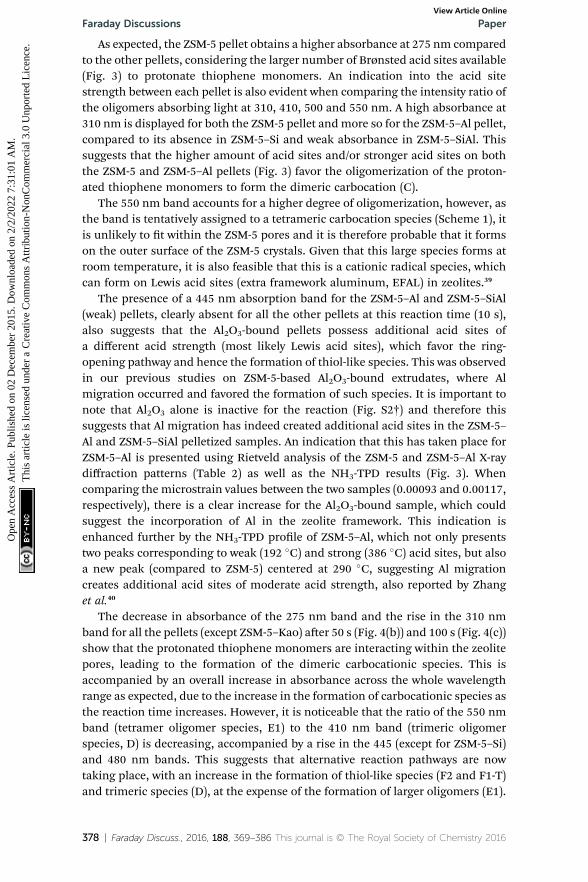
As expected, the ZSM-5 pellet obtains a higher absorbance at 275 nm comparedto the other pellets, considering the larger number of Brønsted acid sites available(Fig. 3) to protonate thiophene monomers. An indication into the acid sitestrength between each pellet is also evident when comparing the intensity ratio ofthe oligomers absorbing light at 310, 410, 500 and 550 nm. A high absorbance at310 nm is displayed for both the ZSM-5 pellet andmore so for the ZSM-5–Al pellet,compared to its absence in ZSM-5–Si and weak absorbance in ZSM-5–SiAl. Thissuggests that the higher amount of acid sites and/or stronger acid sites on boththe ZSM-5 and ZSM-5–Al pellets (Fig. 3) favor the oligomerization of the proton-ated thiophene monomers to form the dimeric carbocation (C).
The 550 nm band accounts for a higher degree of oligomerization, however, asthe band is tentatively assigned to a tetrameric carbocation species (Scheme 1), itis unlikely to t within the ZSM-5 pores and it is therefore probable that it formson the outer surface of the ZSM-5 crystals. Given that this large species forms atroom temperature, it is also feasible that this is a cationic radical species, whichcan form on Lewis acid sites (extra framework aluminum, EFAL) in zeolites.39
The presence of a 445 nm absorption band for the ZSM-5–Al and ZSM-5–SiAl(weak) pellets, clearly absent for all the other pellets at this reaction time (10 s),also suggests that the Al2O3-bound pellets possess additional acid sites ofa different acid strength (most likely Lewis acid sites), which favor the ring-opening pathway and hence the formation of thiol-like species. This was observedin our previous studies on ZSM-5-based Al2O3-bound extrudates, where Almigration occurred and favored the formation of such species. It is important tonote that Al2O3 alone is inactive for the reaction (Fig. S2†) and therefore thissuggests that Al migration has indeed created additional acid sites in the ZSM-5–Al and ZSM-5–SiAl pelletized samples. An indication that this has taken place forZSM-5–Al is presented using Rietveld analysis of the ZSM-5 and ZSM-5–Al X-raydiffraction patterns (Table 2) as well as the NH3-TPD results (Fig. 3). Whencomparing the microstrain values between the two samples (0.00093 and 0.00117,respectively), there is a clear increase for the Al2O3-bound sample, which couldsuggest the incorporation of Al in the zeolite framework. This indication isenhanced further by the NH3-TPD prole of ZSM-5–Al, which not only presentstwo peaks corresponding to weak (192 �C) and strong (386 �C) acid sites, but alsoa new peak (compared to ZSM-5) centered at 290 �C, suggesting Al migrationcreates additional acid sites of moderate acid strength, also reported by Zhanget al.40
The decrease in absorbance of the 275 nm band and the rise in the 310 nmband for all the pellets (except ZSM-5–Kao) aer 50 s (Fig. 4(b)) and 100 s (Fig. 4(c))show that the protonated thiophene monomers are interacting within the zeolitepores, leading to the formation of the dimeric carbocationic species. This isaccompanied by an overall increase in absorbance across the whole wavelengthrange as expected, due to the increase in the formation of carbocationic species asthe reaction time increases. However, it is noticeable that the ratio of the 550 nmband (tetramer oligomer species, E1) to the 410 nm band (trimeric oligomerspecies, D) is decreasing, accompanied by a rise in the 445 (except for ZSM-5–Si)and 480 nm bands. This suggests that alternative reaction pathways are nowtaking place, with an increase in the formation of thiol-like species (F2 and F1-T)and trimeric species (D), at the expense of the formation of larger oligomers (E1).
378 | Faraday Discuss., 2016, 188, 369–386 This journal is © The Royal Society of Chemistry 2016

Paper Faraday DiscussionsO
pen
Acc
ess
Art
icle
. Pub
lishe
d on
02
Dec
embe
r 20
15. D
ownl
oade
d on
2/2
/202
2 7:
31:0
1 A
M.
Thi
s ar
ticle
is li
cens
ed u
nder
a C
reat
ive
Com
mon
s A
ttrib
utio
n-N
onC
omm
erci
al 3
.0 U
npor
ted
Lic
ence
.View Article Online
However, the lower ratio of the 550 nm absorbance band can be explained inFig. 4(d) at a reaction time of 200 s, whereby an increase in a broad band >550 nmis now visible, suggesting that these species are oligomerizing to even largerspecies over time. Again, the increase in the absorbance of the 445, 460 and 480nm bands shows that ring-opening is now one of the main reaction pathways forall the catalysts (except ZSM-5–Kao), possibly due to the lack of strong acid sites atthis point of the reaction, which are involved in forming larger oligomer species.It is also important to note the much higher overall absorbance of the ZSM-5–SiAlpellet compared with ZSM-5, ZSM-5–Si and ZSM-5–Al (which are all relativelysimilar), suggesting a binder effect in this sample. A possible explanation couldbe the interaction between the two components of the binder (i.e. Al migration toSiO2, or Si migration to Al2O3), creating additional acid sites to perform thethiophene oligomerization reaction. This is again backed by the NH3-TPD proleof ZSM-5–SiAl (Fig. 3), which shows a far higher ratio of stronger acid sites toweaker sites compared to ZSM-5 and a greater relative amount of both weak andstrong acid sites compared to ZSM-5–Si. However, further studies are needed todevelop a better understanding of how this mixed binder promotes such a highproduct formation.
Similar reactivity and selectivity trends continue towards the termination ofthe reaction at 400 s (Fig. 4(e)) and 800 s (Fig. 4(f)), with a decrease in theabsorbance of both the 275 and 310 nm bands and a continued rise in bands >400nm, particularly towards the formation of thiol-like species and large oligomers.The appearance of both 580 and 620 nm bands and the broadness of the UV-Visspectrum background (particularly in the case of ZSM-5–Si) suggests the forma-tion of complex larger molecules and/or coke in each pellet. Regarding ZSM-5–Si,it is important to note at this stage, that a SiO2 pellet produces a very broad bandin the >580 nm region (Fig. S2(c)†), which explains the high absorbance in theZSM-5–Si spectra. The reason for SiO2 reacting with thiophene is, at this stage, notclear, as SiO2 is not expected to provide acid sites with enough strength to convertthiophene monomers.
Confocal uorescence microscopy corresponds well with UV-Vis micro-spec-troscopy and is an excellent method to provide visible insight into the oligomerspecies formed and how they are distributed over each ZSM-5–clay/binder-boundpellet. Fig. 5 presents the 3D confocal uorescence microscopy volumes of eachindividual sample excited with both 488 and 561 nm lasers simultaneously, aerthe completion of the thiophene oligomerization reaction. These two laser linewavelengths were chosen to gain an understanding into the relative amount andnature of ‘smaller’ to ‘larger’ oligomer species formed. As expected for the ZSM-5pellet (Fig. 5(a)), a homogeneous distribution of uorescent oligomers are ob-tained, with an orange uorescence indicating the formation both <3-thiophenering oligomers/thiol-like species and >3-thiophene ring oligomers, with a higherrelative amount of the latter (Fig. S4†).
ZSM-5–Al (Fig. 5(c)) and ZSM-5–SiAl (Fig. 5(d)) show similar results to that ofZSM-5, with a more heterogeneous distribution of ZSM-5 crystals/agglomeratesdue to the binder distribution. However, ZSM-5–Si reveals a higher uorescenceintensity than that for all the other pellets, showing its higher ratio of largeroligomers/coke species (Fig. S4†), as evidenced by UV-Vis absorption (Fig. 4(f)),and this is again explained by the unexpected reactivity of SiO2 (Fig. S2(c)†). Thelack of reactivity for the ZSM-5–Kao pellet leads to an almost blank image, with
This journal is © The Royal Society of Chemistry 2016 Faraday Discuss., 2016, 188, 369–386 | 379

Fig. 5 3D confocal fluorescence microscopy images of the following ZSM-5–binder-bound pellets after reaction of thiophene at 120 �C: (a) ZSM-5; (b) ZSM-5–Si; (c) ZSM-5–Al; (d) ZSM-5–SiAl; and (e) ZSM-5–Kao. The XYZ axis values correspond to: 127 � 127 �27 mm.
Faraday Discussions PaperO
pen
Acc
ess
Art
icle
. Pub
lishe
d on
02
Dec
embe
r 20
15. D
ownl
oade
d on
2/2
/202
2 7:
31:0
1 A
M.
Thi
s ar
ticle
is li
cens
ed u
nder
a C
reat
ive
Com
mon
s A
ttrib
utio
n-N
onC
omm
erci
al 3
.0 U
npor
ted
Lic
ence
.View Article Online
only a few visible uorescent ZSM-5 crystals, conrming that the binder hasa detrimental effect to the active zeolite phase by blocking access to the activezeolite beneath the hard outer layer.
In order to develop a greater understanding of the chemical species formed ineach ZSM-5–binder-bound pellet upon thiophene oligomerization, in situ FT-IRmicro-spectroscopy was performed. Given the high concentration of moisturepresent in the ZSM-5 pellet (Fig. S5†), it was difficult to draw detailed conclusionsfrom the whole range of FT-IR spectra before, during and aer thiophene oligo-merization. Therefore, we will focus on the results of the other pellets (which haveonly 30 wt% ZSM-5 present).
Fig. 6(a) and (b) present the FT-IR spectra of ZSM-5–Si before, during and aerthiophene oligomerization, allowing a correlation to those timescales in the UV-
380 | Faraday Discuss., 2016, 188, 369–386 This journal is © The Royal Society of Chemistry 2016
Paper Faraday DiscussionsO
pen
Acc
ess
Art
icle
. Pub
lishe
d on
02
Dec
embe
r 20
15. D
ownl
oade
d on
2/2
/202
2 7:
31:0
1 A
M.
Thi
s ar
ticle
is li
cens
ed u
nder
a C
reat
ive
Com
mon
s A
ttrib
utio
n-N
onC
omm
erci
al 3
.0 U
npor
ted
Lic
ence
.View Article Online
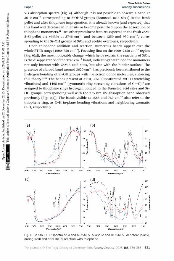
Vis absorption spectra (Fig. 4). Although it is not possible to observe a band at3610 cm�1 corresponding to SiOHAl groups (Brønsted acid sites) in the freshpellet and aer thiophene impregnation, it is already known (and expected) thatthis band will decrease in intensity or become perturbed upon the adsorption ofthiophene monomers.41 Two other prominent features expected in the fresh ZSM-5–Si pellet are visible at 3746 cm�1 and between 1250 and 950 cm�1, corre-sponding to the Si–OH groups of SiO2 and zeolite overtones, respectively.
Upon thiophene addition and reaction, numerous bands appear over thewhole FT-IR range (4000–750 cm�1). Focusing rst on the 4000–2250 cm�1 region(Fig. 6(a)), the most noticeable change, which helps explain the reactivity of SiO2,is the disappearance of the 3746 cm�1 band, indicating that thiophenemonomersnot only interact with ZSM-5 acid sites, but also with the binder surface. Thepresence of a broad band around 3620 cm�1 has previously been attributed to thehydrogen bonding of Si–OH groups with p-electron donor molecules, enforcingthis theory.41,42 The bands present at 3110, 3076 (unsaturated ]C–H stretchingvibrations) and 1408 cm�1 (symmetric ring stretching vibrations of C]C)43 areassigned to thiophene rings hydrogen bonded to the Brønsted acid sites and Si–OH groups, corresponding well with the 275 nm UV absorption band observedpreviously (Fig. 4(a)). The bands visible at 1588 and 760 cm�1 also refer to thethiophene ring, as C–H in-plane bending vibrations and neighboring aromaticC–H, respectively.
Fig. 6 In situ FT-IR spectra of (a and b) ZSM-5–Si and (c and d) ZSM-5–Al before (black),during (red) and after (blue) reaction with thiophene.
This journal is © The Royal Society of Chemistry 2016 Faraday Discuss., 2016, 188, 369–386 | 381
Faraday Discussions PaperO
pen
Acc
ess
Art
icle
. Pub
lishe
d on
02
Dec
embe
r 20
15. D
ownl
oade
d on
2/2
/202
2 7:
31:0
1 A
M.
Thi
s ar
ticle
is li
cens
ed u
nder
a C
reat
ive
Com
mon
s A
ttrib
utio
n-N
onC
omm
erci
al 3
.0 U
npor
ted
Lic
ence
.View Article Online
However, there are not only bands due to intact/adsorbed thiophene present,but also additional bands even at a low reaction temperature, suggesting severalspecies are formed. The bands observed at 2970, 2932 and 2880 cm�1 areattributed to CH2 groups in proximity to a double bond, i.e. asymmetric *CH2–
(CH]CH2) stretching vibrations, or a neighboring S atom (CH2–S–).41 Therefore,these are attributed to thiol-like species with further bands at 1806–1770 and 930cm�1 also indicating their formation (C]C–H stretch and bending, respectively).However, Geobaldo et al. propose an alternative theory, suggesting that the bandspresent at 1800–1600 cm�1 and 2980–2800 cm�1 correspond with carbocationicoligomer species, also reported in our UV-Vis absorption spectra (Fig. 4).37 Bothproposals correspond well with our results in this work, however, we can prove thepresence of thiol-like species given that perturbed S–H bands are present at 2714,2584 and 2374 cm�1.41
Towards the termination of the reaction, it is clear that no bands corre-sponding to adsorbed thiophene monomers are present (i.e. 3110 and 3076cm�1). This is accompanied by the re-appearance of the Si–OH groups (3746cm�1), suggesting full conversion of thiophene to its respective intermediate/products, recognized by bands still present at 2970, 2932, 930 cm�1. Anothernoticeable feature in the region of 1250–900 cm�1 is the shi towards lowerwavenumbers for the zeolite overtone bands upon thiophene reaction. Given thehigher ratio of larger oligomers/coke species formed in ZSM-5–Si compared to inthe other samples (also displayed by the higher orange uorescence intensity), itcould be that such large species formed (inside the pores) are distorting thezeolite framework, but further studies with in situ XRD (Rietveld analysis) areneeded to conrm this nding.
The in situ FT-IR spectra of ZSM-5–Al (Fig. 6(c) and (d)) show similar featuresbut also distinct differences compared to the ZSM-5–Si spectra. The decrease inintensity of the 3746 cm�1 band of fresh ZSM-5–Al is expected considering thesignicant decrease in Si–OH groups compared to ZSM-5–Si, but it is alsoapparent that bands present upon thiophene impregnation are much lower inintensity in the 4000–2250 cm�1 region. This could be due to the presence of H2Oin the sample (as with ZSM-5), but yet it is still possible to distinguish vitaladditional bands. The presence of bands at 3110, 3076, 2970, 2930, 2710 and 2950cm�1 all agree with the proposed formation of both oligomer and thiol-likespecies. However, additional bands at 1610 and 1380 cm�1, as well as the largeincrease and broadening of the 1410 cm�1 band, conrm perturbed vibration ofthiophene molecules, which are strongly interacting with EFAL species.41 Thisagain enforces our previous proposal that Al migration has occurred from theAl2O3 binder to ZSM-5, creating additional acid sites that are now interacting withthiophene. As with ZSM-5–Si, the re-appearance of the 3746 cm�1 band andabsence of the 3110 and 3076 cm�1 bands towards the termination of the reactionagain suggest the full conversion of thiophene monomers.
As in the case of ZSM-5–Si, very distinct and clear bands are formed with ZSM-5–SiAl upon its impregnation with thiophene (Fig. 7(a) and (b)). The increase inintensity of all relevant bands related to thiophene and intermediate/productspecies could relate to the high absorbance obtained in the UV-Vis spectra (Fig. 4).This indicates that a far greater absorption of thiophene is present in this samplecompared with the other pellets. Other than the main bands relating to oligomerand thiol-like species, similar bands to ZSM-5–Al are also present (1620 cm�1),
382 | Faraday Discuss., 2016, 188, 369–386 This journal is © The Royal Society of Chemistry 2016

Fig. 7 In situ FT-IR spectra of (a and b) ZSM-5–SiAl and (c and d) ZSM-5–Kao, before(black), during (red) and after (blue) reaction with thiophene.
Paper Faraday DiscussionsO
pen
Acc
ess
Art
icle
. Pub
lishe
d on
02
Dec
embe
r 20
15. D
ownl
oade
d on
2/2
/202
2 7:
31:0
1 A
M.
Thi
s ar
ticle
is li
cens
ed u
nder
a C
reat
ive
Com
mon
s A
ttrib
utio
n-N
onC
omm
erci
al 3
.0 U
npor
ted
Lic
ence
.View Article Online
again suggesting the interaction of thiophene with EFAL species created from Almigration in ZSM-5–SiAl.
Finally, the in situ FT-IR spectra of ZSM-5–Kao conrm the limited reactivity ofthe catalyst pellet. Taking the bands present in the region of 4000–3800 cm�1 asan ‘internal standard’ between samples (relating to the adsorption of thiophene),it is apparent that a far lower concentration of thiophene is adsorbed on ZSM-5–Kao, conrming the earlier proposal that the phase change upon pelletization/heat treatment creates a hard layer hindering the access of thiophene to thezeolite active phase. Nevertheless, the vibrational bands present around 2968,2928 and 2872 cm�1 suggest the formation of oligomers and thiol-like species,possibly due to some accessible zeolite on the surface, but the absence ofobserved bands in the 2800–750 cm�1 region do not allow further interpretation.
4. Conclusions
In relation to our previous studies on ZSM-5–binder extrudates, four ZSM-5–binder-bound pellets were prepared using SiO2, Al2O3, SiO2 : Al2O3 (2 : 1 mix) andkaolinite as binder materials, in order to determine how zeolite–clay/binderinteractions upon physical mixing inuence the catalytic performance. Usingcomplementary micro-spectroscopic techniques, coupled with thiophene oligo-merization as a staining probe reaction, it is evident that, independent of the
This journal is © The Royal Society of Chemistry 2016 Faraday Discuss., 2016, 188, 369–386 | 383
Faraday Discussions PaperO
pen
Acc
ess
Art
icle
. Pub
lishe
d on
02
Dec
embe
r 20
15. D
ownl
oade
d on
2/2
/202
2 7:
31:0
1 A
M.
Thi
s ar
ticle
is li
cens
ed u
nder
a C
reat
ive
Com
mon
s A
ttrib
utio
n-N
onC
omm
erci
al 3
.0 U
npor
ted
Lic
ence
.View Article Online
shaping method (i.e. pellet or extrudate), the intimate contact between the zeoliteand clay/binder in catalyst bodies heavily impacts their reactivity and productselectivity, with the type of binder playing a vital role. Kaolinite was found to limitthe reactivity of ZSM-5 within the pellet due to its phase change upon heattreatment, creating a hard impenetrable outer layer, which hindered thiopheneaccessibility to the ZSM-5 crystals. Aluminummigration, previously witnessed forZSM-5–binder-bound extrudates, also occurs in the pellet formation of ZSM-5–Al2O3-bound samples, suggesting that it is difficult to limit this process occurringwhen these components are in close proximity. Given the higher selectivitytowards the ring opening of thiophene and its oligomers, compared with ZSM-5and ZSM-5–Si, it should be taken into account that aluminummigration can alterproduct selectivity when chosen as a binder. The high reactivity towards largeroligomer species for ZSM-5–Si, clearly visible with confocal uorescencemicroscopy, was due to the interaction of the SiO2 binder with thiophene(observed using IR spectroscopy), which, given the lower acid site strength andnumber compared to ZSM-5 and ZSM-5–Al, was unexpected. Finally, the combi-nation of a SiO2 : Al2O3 mixed binder enhanced the reactivity of ZSM-5, whichcould be due to the interaction of the two binder components creating additionalacid sites, but further studies are needed to resolve this nding. Evidently, thecombination of UV-Vis, confocal uorescence and IR micro-spectroscopy withstaining probe reactions is highly complementary, providing new insights intozeolite-based catalyst bodies and proving that the choice of binder is crucial to thecatalytic performance, whether it be detrimental or advantageous.
Acknowledgements
The authors would like to thank Lennart Webber (Utrecht University, UU) for theAr physisorption analysis, and Joris Goetze (UU) for fruitful discussions. Thiswork was nancially supported by a NWO personal ‘Veni’ grant awarded to G. T.W. and a Top Grant of NWO to B. M. W.
References
1 A. Corma, Chem. Rev., 1995, 95, 559–614.2 M. J. Climent, A. Corma and S. Iborra, Chem. Rev., 2011, 111, 1072–1133.3 A. Corma, M. J. Dıaz-Cabanas, J. Martınez-Triguero, F. Rey and J. Rius, Nature,2002, 418, 514–517.
4 A. de Lucas, J. L. Valverde, P. Sanchez, F. Dorado and M. J. Ramos, Ind. Eng.Chem. Res., 2004, 43, 8217–8225.
5 C. Perego and P. Ingallina, Catal. Today, 2002, 73, 3–22.6 S. Mitchell, N.-L. Michels and J. Perez-Ramırez, Chem. Soc. Rev., 2013, 42, 6094–6112.
7 J. S. J. Hargreaves and A. L. Munnoch, Catal. Sci. Technol., 2013, 3, 1165.8 P. Sanchez, F. Dorado, A. Funez, V. Jimenez, M. J. Ramos and J. L. Valverde, J.Mol. Catal. A: Chem., 2007, 273, 109–113.
9 E. T. C. Vogt, G. T. Whiting, A. Dutta Chowdhury and B. M. Weckhuysen, Adv.Catal., 2015, 58, 143–314.
10 F. Meirer, D. T. Morris, S. Kalirai, Y. Liu, J. C. Andrews and B. M. Weckhuysen,J. Am. Chem. Soc., 2015, 137, 102–105.
384 | Faraday Discuss., 2016, 188, 369–386 This journal is © The Royal Society of Chemistry 2016
Paper Faraday DiscussionsO
pen
Acc
ess
Art
icle
. Pub
lishe
d on
02
Dec
embe
r 20
15. D
ownl
oade
d on
2/2
/202
2 7:
31:0
1 A
M.
Thi
s ar
ticle
is li
cens
ed u
nder
a C
reat
ive
Com
mon
s A
ttrib
utio
n-N
onC
omm
erci
al 3
.0 U
npor
ted
Lic
ence
.View Article Online
11 Z. Ristanovic, J. P. Hofmann, G. de Cremer, A. V. Kubarev, M. Rohnke,F. Meirer, J. Hoens, M. B. J. Roeffaers and B. M. Weckhuysen, J. Am. Chem.Soc., 2015, 137, 6559–6568.
12 F. Meirer, S. Kalirai, D. Morris, S. Soparawalla, Y. Liu, G. Mesu, J. C. Andrewsand B. M. Weckhuysen, Sci. Adv., 2015, 1, 1400199.
13 F. Meirer, S. Kalirai, J. Nelson Weker, Y. Liu, J. C. Andrews andB. M. Weckhuysen, Chem. Commun., 2015, 51, 8097–8100.
14 S. Kalirai, U. Boesenberg, G. Falkenberg, F. Meirer and B. M. Weckhuysen,ChemCatChem, 2015, 7, 3674–3682.
15 J. Ruiz-Martınez, A. M. Beale, U. Deka, M. G. O'Brien, P. D. Quinn,J. F. W. Mosselmans and B. M. Weckhuysen, Angew. Chem., Int. Ed., 2013,52, 5983–5987.
16 Z. Ristanovic, M. M. Kerssens, A. V. Kubarev, F. C. Hendriks, P. Dedecker,J. Hoens, M. B. J. Roeffaers and B. M. Weckhuysen, Angew. Chem., Int. Ed.,2015, 54, 1836–1840.
17 G. T. Whiting, F. Meirer, M. M. Mertens, A.-J. Bons, B. M. Weiss, P. A. Stevens,E. de Smit and B. M. Weckhuysen, ChemCatChem, 2015, 7, 1312–1321.
18 G. T. Whiting, F. Meirer, D. Valencia, M. M. Mertens, A.-J. Bons, B. M. Weiss,P. A. Stevens, E. de Smit and B. Weckhuysen, Phys. Chem. Chem. Phys., 2014,16, 21531–21542.
19 S. Mitchell, N.-L. Michels, K. Kunze and J. Perez-Ramırez, Nat. Chem., 2012, 4,825–831.
20 I. L. C. Buurmans, J. Ruiz-Martınez, W. V. Knowles, D. van der Beek,J. A. Bergwerff, E. T. C. Vogt and B. M. Weckhuysen, Nat. Chem., 2011, 3,862–867.
21 E. T. C. Vogt and B. M. Weckhuysen, Chem. Soc. Rev., 2015, 44, 7342–7370.22 I. L. C. Buurmans, F. Soulimani, J. Ruiz-Martınez, H. E. van der Bij and
B. M. Weckhuysen, Microporous Mesoporous Mater., 2013, 166, 86–92.23 I. L. C. Buurmans and B. M. Weckhuysen, Nat. Chem., 2012, 4, 873–886.24 M. Ibanez, M. Gamero, J. Ruiz-Martınez, B. M. Weckhuysen, A. T. Aguayo,
J. Bilbao and P. Castano, Catal. Sci. Technol., 2016, 6, 296–306.25 P. Castano, J. Ruiz-Martınez, E. Epelde, A. G. Gayubo and B. M. Weckhuysen,
ChemCatChem, 2013, 5, 2827–2831.26 R. V. Jasra, B. Tyagi, Y. M. Badheka, V. N. Choudary and T. S. G. Bhat, Ind. Eng.
Chem. Res., 2003, 42, 3263–3272.27 V. R. Choudhary, P. Devadas, A. K. Kinage andM. Guisnet, Appl. Catal., A, 1997,
162, 223–233.28 P. Canizares, A. Duran, F. Dorado and M. Carmona, Appl. Clay Sci., 2000, 16,
273–287.29 F. Dorado, R. Romero and P. Canizares, Appl. Catal., A, 2002, 236, 235–243.30 J. M. Fougerit, N. S. Gnep and M. Guisnet, Microporous Mesoporous Mater.,
1999, 29, 79–89.31 M. W. Kasture, P. S. Niphadkar, V. V. Bokade and P. N. Joshi, Catal. Commun.,
2007, 8, 1003–1008.32 G. de la Puente, E. Falabella Sousa-Aguiar, A. Figueiredo Costa and U. Sedran,
Appl. Catal., A, 2003, 242, 381–391.33 F. V. Pinto, A. S. Escobar, B. G. de Oliveira, Y. L. Lam, H. S. Cerqueira, B. Louis,
J. P. Tessonnier, D. S. Su and M. M. Pereira, Appl. Catal., A, 2010, 388, 15–21.34 N.-L. Michels, S. Mitchell and J. Perez-Ramırez, ACS Catal., 2014, 4, 2409–2417.
This journal is © The Royal Society of Chemistry 2016 Faraday Discuss., 2016, 188, 369–386 | 385

Faraday Discussions PaperO
pen
Acc
ess
Art
icle
. Pub
lishe
d on
02
Dec
embe
r 20
15. D
ownl
oade
d on
2/2
/202
2 7:
31:0
1 A
M.
Thi
s ar
ticle
is li
cens
ed u
nder
a C
reat
ive
Com
mon
s A
ttrib
utio
n-N
onC
omm
erci
al 3
.0 U
npor
ted
Lic
ence
.View Article Online
35 K.-Y. Lee, H.-K. Lee and S.-K. Ihm, Top. Catal., 2010, 53, 247–253.36 P. Gelin and T. Des Courieres, Appl. Catal., 1991, 72, 179–192.37 F. Geobaldo, T. Palomino, S. Bordiga, C. Fisica and V. Pietro Giuria, Phys.
Chem. Chem. Phys., 1999, 1, 561–569.38 M. Alkan, Ç. Hopa, Z. Yilmaz and H. Guler, Microporous Mesoporous Mater.,
2005, 86, 176–184.39 X. Liu, K.-K. Iu, J. K. Thomas, H. He and J. Klinowski, J. Am. Chem. Soc., 1994,
116, 11811–11818.40 Y. Zhang, Y. Zhou, A. Qiu, Y. Wang, Y. Xu and P.Wu, Ind. Eng. Chem. Res., 2006,
45, 2213–2219.41 C. L. Garcia and J. A. Lercher, J. Phys. Chem., 1992, 96, 2669–2675.42 H. Knozinger, The Hydrogen Bond, ed. P. Schuster, G. Zundel and C. Sandorfy,
North Holland, Amsterdam, 1976, vol. 3, p. 1275.43 Z. Wu, C. Li, Z. Wei, P. Ying and Q. Xin, J. Phys. Chem. B, 2002, 106, 979–987.
386 | Faraday Discuss., 2016, 188, 369–386 This journal is © The Royal Society of Chemistry 2016