Embed Size (px)
Citation preview
REVIEW Open Access
The current status and future directions ofmyxoma virus, a master in immune evasionBart Spiesschaert1*, Grant McFadden2, Katleen Hermans3, Hans Nauwynck1 and Gerlinde R Van de Walle4
Abstract
Myxoma virus (MYXV) gained importance throughout the twentieth century because of the use of the highlyvirulent Standard Laboratory Strain (SLS) by the Australian government in the attempt to control the feralAustralian population of Oryctolagus cuniculus (European rabbit) and the subsequent illegal release of MYXV inEurope. In the European rabbit, MYXV causes a disease with an exceedingly high mortality rate, namedmyxomatosis, which is passively transmitted by biting arthropod vectors. MYXV still has a great impact onEuropean rabbit populations around the world. In contrast, only a single cutaneous lesion, restricted to the point ofinoculation, is seen in its natural long-term host, the South-American Sylvilagus brasiliensis and the North-AmericanS. Bachmani. Apart from being detrimental for European rabbits, however, MYXV has also become of interest inhuman medicine in the last two decades for two reasons. Firstly, due to the strong immune suppressing effects ofcertain MYXV proteins, several secreted virus-encoded immunomodulators (e.g. Serp-1) are being developed totreat systemic inflammatory syndromes such as cardiovascular disease in humans. Secondly, due to the inherentability of MYXV to infect a broad spectrum of human cancer cells, the live virus is also being developed as anoncolytic virotherapeutic to treat human cancer. In this review, an update will be given on the current status ofMYXV in rabbits as well as its potential in human medicine in the twenty-first century.
Table of contents1. The virus2. History3. Pathogenesis and disease symptoms4. Immunomodulatory proteins of MYXV
4.1. MYXV proteins with anti-apoptotic functions4.1.1. Inhibition of pro-apoptotic molecules4.1.2. Inhibition by protein-protein interactionsby ankyrin repeat viral proteins4.1.3. Inhibition of apoptosis by enhancing thedegradation of cellular proteins4.1.4. Inhibition of apoptosis by blocking hostProtein Kinase R (PKR)
4.2. MYXV proteins interfering with leukocytechemotaxis4.3. MYXV serpins that inhibit cellular pro-inflam-matory or pro-apoptotic proteases
4.4. MYXV proteins that interfere with leukocyteactivation4.5. MYXV proteins with sequence similarity to HIVproteins4.6. MYXV proteins with unknown immune function
5. Vaccination strategies against myxomatosis
5.1. Current MYXV vaccines5.2. Vaccination campaigns to protect European rab-bits in the wild
6. Applications of myxoma virus for human medicine
6.1. MYXV proteins as therapeutics for allograft vas-culopathy and atherosclerosis6.2. Applications for MYXV as a live oncolytic virusto treat cancer
7. Discussion and Conclusions8. List of AbbreviationsReferencesAuthor Details
* Correspondence: [email protected] of Virology, Parasitology and Immunology, Faculty of VeterinaryMedicine, Ghent University, Salisburylaan 133, B-9820 Merelbeke, BelgiumFull list of author information is available at the end of the article
Spiesschaert et al. Veterinary Research 2011, 42:76http://www.veterinaryresearch.org/content/42/1/76 VETERINARY RESEARCH
© 2011 Spiesschaert et al; licensee BioMed Central Ltd. This is an Open Access article distributed under the terms of the CreativeCommons Attribution License (http://creativecommons.org/licenses/by/2.0), which permits unrestricted use, distribution, andreproduction in any medium, provided the original work is properly cited.

Authors' contributionsCompeting interestsAcknowledgements
1. The virusMyxoma virus (MYXV) is a member of the genus Lepori-poxvirus, subfamily Chordopoxvirinae, belonging to thefamily of Poxviridae. MYXV contains a large linear doublestranded deoxyribonucleic acid (DNA) genome consistingof 159 unique viral genes, with diploid terminal invertedrepeats (TIRs). Each TIR contains twelve viral genes, nineof which encoding diverse immunomodulating proteins,giving a total of 171 genes [1]. The viral genome is encap-sidated within a brick shaped virion, and the completereplication cycle of MYXV takes place exclusively in thecytoplasm of the infected cells from where it expresses aspectrum of host-interactive immunomodulatory proteins[2]. The virus infects only rabbits and European BrownHares (only occasionally with clinical signs) in the wild,and is nonpathogenic in any tested hosts apart from lago-morphs, but MYXV can replicate in cultured cells frommany species, including most human cancer cells whichare particularly permissive for MYXV [3,4].
2. HistoryThe disease caused by MYXV was seen for the first timein laboratory rabbits in 1896 by Giuseppe Sanarelli inMontevideo, Uruguay [4] Sanarelli was invited by theUruguayan government to found a new Institute ofHygiene in Montevideo that required the production ofantibodies, for which European rabbits were necessary.Following their importation from Europe, these rabbitswere housed in an outdoor facility that was open to bit-ing arthropod vectors, such as mosquitoes. Shortlythereafter, the European rabbits were struck down by ahighly contagious and lethal disease, which was charac-terized by multiple lesions and tumors of the skin andconjunctiva. Sanarelli named this new rabbit diseasemyxomatosis and thought it might be caused by aninfectious agent, which was confirmed later on by isola-tion of the causative poxvirus, called MYXV [4].The European rabbit evolved in Europe and Eastern
Asia, and prior to human intervention was not found inthe rest of the world, including Australasia and theAmericas. During the colonization of new territories, theEuropean rabbit was often taken along as a source ofmeat or for sport hunting, and this is what occurredduring the colonization of Australia in 1860. Between1860 and 1930, a growing population of feral Europeanrabbits derived from a small number of initial breedingpairs spread over the entire continent and their numbersrose up to an estimated three billion [5]. Since it wasestimated that ten rabbits consumed as much vegetationas a single domesticated sheep, the elimination of
rabbits was seen as a very necessary step to economic-ally raise the food and wool production of Australia.Rewards were issued to attract new proposals for redu-cing the size of the feral rabbit population and one ofthe people who responded was Louis Pasteur, whorecommended using Pasteurella cultures that are patho-genic to rabbits. But due to the well-justified skepticismof the Australian government, he was not allowed torelease this microbial agent into the wild [4,6].Since the original MYXV had a mortality rate of 99.5%
in the European rabbit, it was first suggested as a possi-ble means of controlling the Australian feral rabbitpopulation in 1936. Shortly thereafter, the Australiangovernment approved several field release experiments[4,5]. Before the Second World War, two such field testswere conducted, the first of which took place on anisland off the South Australian coast and the second inthe dry inland outback region. Both experiments showedalmost no prolonged spread of the test virus. After theSecond World War, MYXV was again tested in a thirdregion in Southern Australia with more seasonal rainfall[6]. Since it was originally thought that MYXV wasmostly spread rabbit-to-rabbit by direct contact, thefield-tests were conducted mostly in the dry interior ofAustralia during autumn, winter and spring but withoutmuch success in inducing widespread disease in the rab-bit population [5]. However, shortly after these fieldtests when the rainy season ensued, numerous deadMYXV-infected rabbits were found alongside rivers andstreams, infected via the seasonally expanded popula-tions of Culex annulirostris, a mechanical vector forMYXV. However, it rapidly became clear that almostany biting or sucking arthropod could serve as a vectorfor the virus, which enabled MYXV to spread over largeareas [5,6]. An estimated 400 million rabbits succumbedin as little as one year, and within less than a decade,the feral rabbit population was reduced by 95% [4,6]. Incontrast to South America where the virus co-existedwith Sylvilagus sp. hosts in a relatively nonpathogenicrelationship, there was no natural host reservoir speciesfor MYXV present in Australia. Therefore, MYXV wasconstrained to sustain itself in the same host animalsthat it was killing with such extreme efficiency. This ledto a strong selection for less virulent MYXV strains thatkilled their European rabbit hosts with less efficiencythan the originally introduced high pathogenicity strainof MYXV, enabling more extensive replication andspread of the virus by increasing the survival time of itshost [4]. At the same time, the selection pressureexerted by this extremely virulent pathogen caused thehost rabbit population to select for the genetically resis-tant strains that were less susceptible to the lethal dis-ease manifestations of myxomatosis. The co-evolutionary selection pressure reduced the overall
Spiesschaert et al. Veterinary Research 2011, 42:76http://www.veterinaryresearch.org/content/42/1/76
Page 2 of 18

mortality rate of MYXV in the field rabbits to less than30% within seven years after introducing MYXV in Aus-tralia. After the introduction of MYXV in Great Britainand France, a similar course of co-evolution betweenMYXV and its rabbit host was seen [7-9] and at present,MYXV is sustained as a chronic enzootic in rabbitpopulations in America, Southern Europe, New Zealandand Australia [10] (Figure 1).
3. Pathogenesis and disease symptomsThe pathogenesis in immunologically naïve Europeanrabbits after infection with MYXV largely depends onthe MYXV strain and the rabbit breed or strain. Asidefrom naturally occurring MYXV strains, frequently usedstrains in laboratories are the standard laboratory strain(SLS), the Lausanne strain and the Uriarra (Ur) strain.The SLS and the Lausanne strain, which are both viru-lent MYXV strains originally isolated in South America,were released in Australia and Europe, respectively[4,7,11]. The Ur strain was isolated from more resistantfield rabbits in Australia in 1953, three years after thefirst release of SLS. Since the Ur strain had alreadyundergone three years of selection to less virulence, it isoften used as a model for attenuated MYXV strains[12]. In this review, the pathogenesis of an SLS infectionin laboratory rabbits will be described extensively as thecanonical example of myxomatosis, whereas the otherpathogenesis patterns will be discussed briefly in com-parison to this standard model.Laboratory rabbits can be infected with MYXV
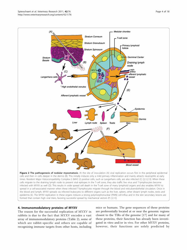
through direct contact with an infected individual, by anarthropod vector, or simply in the laboratory by directneedle inoculation (Figure 2). Through the infection ofdendritic cells (DCs) at the primary infection site, andsubsequent circulation via the lymphatic and vascular
system, MYXV then spreads throughout the body intonumerous secondary organs. New myxoma lesions("myxomes”) are formed at multiple secondary sites onthe skin where arthropod vectors can acquire highenough titers on their biting organs in order to success-fully infect new hosts once they have been serially bittenby the vector (Figure 2) [2,12,13].This course of disease induces a number of diagnostic
clinical symptoms that have been well documented forboth the SLS and the Lausanne strains in laboratoryrabbits, which are representative for the rabbit popula-tion used in either the meat industry or as pets. Essen-tially all infected immunologically naive laboratoryrabbits will develop clinical myxomatosis characterizedby a pink color and edema at the point of inoculation,followed by progressive conjunctivitis and serous/muco-purulent secretion from eyes and nose [7,11]. In addi-tion, the infection is accompanied by swelling of theanogenital region and supervening bacterial infections inthe respiratory tract as the immune system of theinfected host progressively collapses. Animals willusually die 10-14 days post infection (dpi) [14]. WildAustralian rabbits from more resistant populationsinfected with the SLS strain or laboratory rabbitsinfected with the Ur strain will display a similar diseasecourse, however usually with a reduced degree of sever-ity, including a delayed onset of clinical symptoms, anda lower overall mortality (Table 1).Apart from the classical nodular form of myxomatosis
discussed above, a second less frequent form of myxo-matosis exists. This atypical myxomatosis is believed tospread by direct contact, rather then by biting arthro-pods [15]. The clinical signs of this second form, namedamyxomatous myxomatosis, are predominantly respira-tory while skin lesions are few and small [16].
Figure 1 MYXV worldwide distribution. The global distribution of MYXV during the period 01/07/2009-31/12/2009 (copied from [152).
Spiesschaert et al. Veterinary Research 2011, 42:76http://www.veterinaryresearch.org/content/42/1/76
Page 3 of 18
4. Immunomodulatory proteins of MYXVThe reason for the successful replication of MYXV inrabbits is due to the fact that MYXV encodes a vastarray of immunomodulatory proteins (Table 2), some ofwhich are rabbit-specific and others are capable ofrecognizing immune targets from other hosts, including
mice or humans. The gene sequences of these proteinsare preferentially located at or near the genomic regionsclosest to the TIRs of the genome [17] and for many ofthese proteins, their function has already been investi-gated in vitro and/or in vivo. For other MYXV proteins,however, their functions are solely predicted by
Figure 2 The pathogenesis of nodular myxomatosis. At the site of inoculation (A) viral replication occurs first in the peripheral epidermalcells and then in cells deeper in the dermis (B). This initially induces only a mild primary inflammation and mainly attracts neutrophils at earlytimes. Resident Major Histocompatibility Complex-2 (MHC-2) positive cells, such as Langerhans cells, are also infected (C) [2,12,13]. When thesecells migrate to the draining lymph node to present viral epitopes in the T-cell zone, they also traffic live virus and T-lymphocytes becomeinfected with MYXV as well (D). This results in wide spread cell death in the T-cell zone of many lymphoid organs and also enables MYXV tospread in a cell-associated manner when these infected T-lymphocytes migrate through the blood and reticuloendothelial circulation. Once inthe blood and lymph, MYXV spreads via infected leukocytes to different organs such as the liver, spleen, other distant lymph nodes, testis andepidermis (E). The MYXV replication in these organs induces a strong polymorphonuclear (PMN) cell-influx and in the skin secondary lesions areformed that contain high viral titers, favoring successful spread by mechanical vectors (F) [2,12].
Spiesschaert et al. Veterinary Research 2011, 42:76http://www.veterinaryresearch.org/content/42/1/76
Page 4 of 18

comparing them to analogous related genes from eukar-yotic cells or other viruses, such as the vaccinia virus(VACV) [18]. In the present review, the functions ofthese immunomodulatory proteins will be only brieflydiscussed but for a more elaborate description of themost important examples, we would like to refer toother recent reviews [19-22]. Moreover, aside from thisvariety of immunomodulatory proteins, MYXV alsoencodes genes with important functions in diseasepathogenenis, e.g. M127L encodes the DNA repairenzyme photolyase which can promote MYXV survival
in the environment [23]. As a description of these pro-teins is beyond the scope of this review, we would alsolike to refer for these proteins to other reviews [1,23,24].
4.1. MYXV proteins with anti-apoptotic functions4.1.1. Inhibition of pro-apoptotic moleculesMulticellular organisms use programmed cell death, alsodesignated apoptosis, as an innate defense mechanismto eliminate cells that pose a threat for the rest of theorganism, such as virus-infected cells [25,26]. Sinceapoptosis can have a negative effect on viral replication
Table 1 Comparison of symptoms in wild and domestic rabbits infected with different MYXV strains inducing nodularmyxomatosis (adapted from [7,11])
Daypi
Laboratory rabbits Wild rabbits
SLS Lausanne Uriarra (Ur) SLS Uriarra (Ur)
1
2 Pink & edematousinoculation site
Pink/edematous inoculation site Pink inoculation site
3 Pink/edematousinoculation site
4 Conjunctivitis Conjunctivitis Pinkinoculationsite
5 Secondary skin lesionsAnogenital edema
ConjunctivitisAnogenital edema
6 Secondary skin lesions &anogenital edema
Serous eye discharge Conjunctivitis &Anogenital edema
7 Respiratory distress Mucopurulent eye discharge & purple/black inoculation site
Mildconjunctivitis
8 Necrotic inoculation site; Edematouseyelids; Respiratory distress
Secondary skin lesions
9 Secondary skinlesions
10 Severe respiratory distressSecondary lesions acrossthe skin #
Serous secretion from inoculation site Respiratory distress Respiratory distress Mildanogenitaledema;Secondary skinlesions
11 Severe dyspnoea; Secondary lesionsacross the skin #
12 Lesion regression
13
Daypi
Laboratory rabbits Wild rabbits
14
15 Improved breathing;Decreased anogenital edema
Lesionregression
16
17 Lesion regression
18
19
20 Still recovering Virtually recovered#: euthanasia of the experimentally infected animals due to too severe clinical symptoms; pi: post inoculation
Spiesschaert et al. Veterinary Research 2011, 42:76http://www.veterinaryresearch.org/content/42/1/76
Page 5 of 18

and spread, many viruses, including MYXV, have devel-oped multiple strategies to inhibit this process. Onepoint of interference is the direct inhibition of key pro-apoptotic molecules.The MYXV protein M-T2 (M002R/L) is a secreted
tumor necrosis factor receptor (TNFR) homolog thatinhibits apoptosis by binding and subsequently inhibit-ing rabbit TNF via a conserved ligand-binding domainat the N-terminus of the viral protein [27-29]. A highlyconserved viral preligand assembly domain (vPLAD) onthe M-T2 protein is responsible for inhibiting intracellu-lar apoptosis, making it the first described viral immu-nomodulatory protein for which two separate domainswith distinct immune inhibiting functions are known[29,30].M11L is a viral protein that localizes to the outer
mitochondrial membrane through close association withthe mitochondrial peripheral benzodiazepine receptor(PBR) component of the permeabilitytransition (PT)pore. There, M11L prevents the release of mitochondrial
cytochrome c induced by staurosporine or protopor-phyrin IX (PPIX), which are ligands of PBR [31,32].Furthermore, M11L is also able to inhibit apoptosis bybinding with B-cell lymphoma 2 (BCL2)-antagonist/killer (Bak), which, following pro-apoptotic stimuli, isalso able to release cytochrome c and subsequentlyinduce cell death [26,33,34]. Another function for M11Lis the ability to interact with activated Bcl-2 associated× protein (Bax), which it inhibits when it is translocatedfrom the cytoplasm to the outer mitochondrial mem-brane during viral infection [35].Although not yet demonstrated experimentally,
M146R is also a potential inhibitor of the pro-apoptoticBcl-2 family, in the sense that the MYXV protein is clo-sely related to the VACV protein N1L, which has beenshown to bind and inhibit Bcl-2 proteins [36,37].Recently, it was shown that N1L also retains a role ininfluencing the Toll-like receptor (TLR) signaling path-way [38]. Also, and even more recently, it was foundthat the VACV ortholog N1L of ectromelia virus
Table 2 Immunomodulatory proteins of MYXV
ORF Protein name Function references
M002R/L M-T2 TNFR homolog; inhibits TNF/TNFR; prevents apoptosis of lymphocytes intracellularly [27-30]
M11L Binds with Bak and/or Bax; inhibits apoptosis [33,34]
M146R Postulated to bind Bcl-2 proteins and inhibit TLR-mediated innate responses [1,38]
M005R/L M-T5 Enhances degradation of CDK inhibitors; prevents checkpoint-induced apoptosis [42-44]
M150R MNF ANK-repeat protein; inhibits NF-�B [45,48]
M148R ANK-repeat virulence factor, but no specific function defined to date [49]
M149R ANK-repeat virulence factor, but no specific function defined to date [49]
M153R MV-LAP Downregulates cell surface CD4, Fas-CD95 and MHC-1 [13,50,51]
M143 Ubiquitin ligase with a possible anti-apoptotic function [1]
M004R/L M-T4 Possible interference with BAP31 in the ER; apoptosis inhibitor [13,52,53]
M156R PKR pseudosubstrate and inhibitor [19,57]
M029L Predicted inhibitor of PKR via dsRNA binding [1]
M062R Host range factor essential for replication in rabbit or human cancer cells; binds SAMD9 [1,61,65]
M063R Host range factor, essential for replication in rabbit cells [1,61,62]
M064R Related to M062 and M064 host range factors but function not yet determined [1,61]
M001R/L M-T1 CC-chemokine binding protein [66,67,153,154]
M104L Postulated inhibitor of chemokine receptor signaling [1]
M007R/L M-T7 Secreted inhibitor of IFN-g and chemokines; anti-inflammatory activities [69]
M008R/L Serp-1 Secreted serpin that inhibits diverse host serine proteases; anti-inflammatory activities [70,155-157]
M151R Serp-2 Intracellular serpin that inhibits ICE and granzyme B [71,73,158]
M152R Serp-3 Serpin and virulence factor, but no specific host target yet described [74]
M141R vCD200 Cell surface virulence factor that inhibits CD200R+ leukocytes (also called vOX-2) [1,76]
M121R/M122R Predicted NK regulatory receptor homologs [1]
M128L vCD47 Cell surface virulence factor and CD47 homolog [1,80]
M13L Pyrin-containing protein that inhibits the inflammasomes and NF-�B signaling [81,83]
M129R Predicted HIV gp120 homolog, but function still unknown [60]
M130R Predicted HIV Tat homolog; virulence factor [60,86]
M010L MGF Secreted virulence factor, related to TGFalpha and EGF [89]
M135R B19R (VACV) homolog, virulence factor, but no specific function or ligand known [60,90]
M144R C3L/B5R (VACV) homolog, no confirmed function yet [60]
Spiesschaert et al. Veterinary Research 2011, 42:76http://www.veterinaryresearch.org/content/42/1/76
Page 6 of 18

(ECTV), the causative agent of mousepox, interfereswith host T cell function in vivo, independently of TLRsignaling. This was also illustrated by the impairment ofin vivo ECTV spread when N1L was deleted [39]. Thisstudy provided different results for the virulence factorN1L than those previously predicted in vitro, emphasiz-ing again the importance of studying virulence factors ina natural virus-host system in vivo.4.1.2. Inhibition by protein-protein interactions by ankyrinrepeat viral proteinsSeveral MYXV proteins contain ankyrin (ANK) repeatsand a “Pox protein Repeats of Ankyrin C-terminal”(PRANC) domain (an F-box-like domain), which enablethe binding with unique cellular protein targets [40,41].Through these protein-protein interactions MYXVANK-repeat proteins act as substrate adaptors that canrecruit cellular proteins for ubiquitination and degrada-tion via the 26S proteasome. This is the case for M-T5(M005R/L), which binds via an adaptor protein calledSkp1 with the cellular protein cullin-1 in vitro [42]. Cul-lin-1 is an E3 ubiquitin ligase, which normally binds tocyclin-dependent kinase (CDK) inhibitors, enzymes thatare otherwise degraded in a cell cycle fashion by protea-somes. The binding of M-T5 with Skp1/cullin-1 aug-ments the proteosomal degradation of CDK inhibitors,like p27-Kip, thereby stimulating the progression of thecell cycle past the G0/G1 checkpoint that could other-wise trigger an apoptosis response when the cell cyclestalls at this checkpoint [42,43]. In vivo experimentshave shown that M-T5 is necessary for MYXV to spreadsecondary immune organs in the infected rabbit host[44], suggesting that M-T5 permits virus spread viainfected lymphocytes that traffic to multiple distalimmune organs [20].The myxoma nuclear factor (MNF) M150R is crucial
for productive viral infection in vivo and is predicted tointerfere with apoptosis by inhibiting Nucleus Factor-�B(NF-�B). This prediction is based on the analogy withANK-repeats and the nuclear location of M150R whencompared to cellular NF-�B inhibitors (I�Bs) [45]. NF-�B is an important transcription factor that is activatedby pro-inflammatory stimuli, subsequently translocatingfrom the cytoplasm to the nucleus where it upregulateshost genes critical for immunity, inflammation andapoptosis [46]. I�Bs inhibit NF-�B by interfering withthe translocation of the active transcription factor dimerto the nucleus and hence, a similar action is predictedfor M150R [45,47]. More recently, an alternate possibi-lity of inhibition has been proposed, where M150Rdirects the ubiquitin ligase complex to new substratetargets, such as NF-�B, for ubiquitination and subse-quently degradation [48]. Both M148R and M149R arealso members of the MYXV ANK-repeat family andalthough the virulence of infection with M148R and
M149R deletion mutants was reduced in in vivo experi-ments, no specific function has yet been determined forthese proteins to date [49].4.1.3. Inhibition of apoptosis by enhancing the degradationof cellular proteinsApoptosis can also be inhibited by downregulatingimmune-receptors located on the cell surface, therebydecreasing the chance of recognition by apoptosis-indu-cing immune cells such as cytotoxic T-lymphocytes [50].M153R, also known as Myxoma Virus-Leukemia-Asso-ciated Protein (MV-LAP), is a ubiquitin ligase whichachieves the downregulation of a number of immune-receptors at the cell surface, such as major histocompat-ibility complex 1 (MHC-1), cluster of differentiation(CD)-4 and Fas (CD95), by augmented endocytosis or bydecreased recycling to the surface [13,50,51]. For Fas, thisdownregulation was also accomplished by degradation[50]. The MYXV protein M143 is also a ubiquitin ligase,but it remains to be determined whether this viral pro-tein displays a similar anti-apoptotic role as MV-LAP [1].M-T4 (M004R/L) is an anti-apoptotic intracellular
virulence factor that has been suggested to functionwithin the endoplasmic reticulum (ER) of infected cellswhere it binds to calreticulin [13,52,53]. Its anti-apopto-tic action, necessary for in vivo spreading throughoutthe infected rabbit host, is still not understood but itmight be explained by a possible interference with thefunction of BAP31, an ER-localized host proteininvolved in (i) the egress of MHC-1 and other proteins,(ii) inhibiting apoptosis and (iii) downregulating MHC-1expression respectively [13,54].4.1.4. Inhibition of apoptosis by blocking host ProteinKinase R (PKR)Protein kinase R (PKR) is upregulated by interferon(IFN) and is catalytically activated by double strandedRNA (dsRNA), which can be produced during replica-tion of many classes of viruses, including poxviruses.The activation of PKR causes many downstream anti-viral effects, including apoptosis of the cell, therebyimpeding viral spread, and thus many viruses haveevolved anti-PKR countermeasures [55]. For example,the VACV K3L protein binds PKR, hereby inhibiting thephosphorylation of the eukaryotic initiation factor 2alpha (eIF2a), an important regulator of translationinitiation [56]. Interestingly, the gene sequence of theprotein M156R of MYXV shows extensive sequencesimilarities with VACV K3L and a similar function ofM156R was confirmed since M156R protein was shownto be an efficient substrate for PKR in vitro [57]. Indeed,MYXV shows a partial resistance against type-1 IFN inhuman cells in vitro [58], indicating that MYXV is cap-able of interfering with IFN signaling in virus-infectedcells, even though it is relatively inhibited when theanti-viral state is pre-induced prior to virus infection
Spiesschaert et al. Veterinary Research 2011, 42:76http://www.veterinaryresearch.org/content/42/1/76
Page 7 of 18

[59]. It has been shown that MYXV can partially inhibittype-1 IFN signaling by blocking the interferon-inducedactivation of the Janus kinase Tyk2 in MYXV-infectedhuman cells. The responsible MYXV protein however, isyet to be determined [58]. A conserved region ofM029L, located in its C-terminal domain sequence,shows strong resemblance with the dsRNA bindingregion of the VACV gene E3L, which is responsibledecreasing the activation of PKR [1,56,60].In addition, M062R, M063R and M064R are all ortho-
logs of the VACV host range protein C7L, although onlyM062R shows similar host range capabilities as C7L asdetermined by gene swap experiments in the context of aVACV infection [1,61]. M063R shows sequence similaritywith its C-terminal half a with a glutamate-rich domainof the cellular death-domain associated protein, Daxx,leading to the hypothesis that M063R can inhibit Fas-associated cell death through interfering with the func-tion of Daxx [1,61,62] However, this model remainsunproven and it is still not completely understood underwhich circumstances Daxx functions as either a pro-apoptotic or an anti-apoptotic protein [63,64]. Alterna-tively, it is hypothesized that M063R functions as a hostrange gene like M062R, but that it exerts this functionthrough a different pathway [62,65]. Recent evidence sug-gests that M062R, but not C7L, binds to a specific hostanti-viral protein called “sterile alpha motif domain con-taining protein 9” (SAMD9) implicated in human inflam-matory disorders, suggesting that even these closelyrelated viral host range factors may block cellular self-defense pathways by different mechanisms [65].
4.2. MYXV proteins interfering with leukocyte chemotaxisThe coordinated recruitment of leukocytes at the site ofinfection is an important aspect of the early immuneresponse against viruses [66]. Therefore, it is not sur-prising that poxviruses such as MYXV have also devel-oped multiple ways to interfere with this process. M-T1(M001R/L) is a secreted viral protein that is unnecessaryfor productive viral replication but it can bind and inhi-bit chemokines of the CC-subfamily in vitro, therebyinhibiting the directional migration and activation ofthese cells in vivo [66,67]. In contrast, M-T1 was notable to bind or inhibit chemokines of the CXC subfam-ily, at least as assessed by chemical crosslinking in vitro,such as CXCL8 (ie IL-8, in humans), and this couldexplain why neutrophils are the dominant early respon-der leukocytes at sites of infection in experimentallyMYXV-infected laboratory rabbits [12,67].M104L is a small hydrophobic MYXV protein which
shares amino acid identity of 42% over a 40-residueregion (containing a receptor domain implicated in che-mokine receptor signaling) with a subset of the trans-membrane domains of the IL-8 receptor analogue
ORF74 of Ateline herpesvirus 3, which is believed toinhibit the signaling of a yet to be determined chemo-kine receptor via heterodimerization, hereby preventingthe formation of functional receptor dimers [1]. Anotherviral MYXV protein, M-T7 (M007R/L), which is anabundantly secreted 37 kDa glycoprotein from MYXV-infected cells, binds rabbit interferon-g (IFN-g), herebyinhibiting interferon-g (IFN-g) through competitivebinding [68]. In addition, it was shown that M-T7 pro-tein is able to bind chemokines of the CXC, CC and Csubfamilies in vitro in a pan-species specific fashion,thereby blocking the chemokine-mediated recruiting ofleukocytes from many species, including rabbits, miceand humans [69].
4.3. MYXV serpins that inhibit cellular pro-inflammatoryor pro-apoptotic proteasesSerine protease inhibitors (serpins) are the largest familyof cellular protease inhibitors and MYXV encodes threemembers, each with its own specific functions [1]. Serp-1 (M008.1R/L) is a secreted MYXV serpin that is able tobind and inhibit the functions of several human cellularproteases, such as plasmin, urokinase, plasminogeneactivator and C1s of the complement cascade [70].However, more research is needed to clarify the interac-tions of Serp-1 with similar cellular proteases in the rab-bit and the implications of such interactions on thecourse of disease. Serp-2 (M151R), on the other hand, isan intracellular cysteine proteinase that decreases thesecretion of bioactive IL-1b by inhibiting the IL-1b-con-verting enzyme (ICE) in vitro [71]. Serp-2 is also a weakinhibitor of granzyme B, which plays an important roleduring T cell-induced apoptosis [72,73]. Serp-3 (M152R)is the third MYXV protein with a canonical predictedserpin structure, although it has several apparentdomain deletions compared to the gene sequences ofother serpins [74]. When this Serp-3 gene is deletedfrom MYXV, no secondary skin lesions are observed inrabbits in vivo, despite the fact that no decrease in viralreplication nor an increase of apoptosis can be detectedin cultured cells [74].
4.4. MYXV proteins that interfere with leukocyteactivationMYXV encodes M141R, a structural homologue ofCD200, which is a cellular membrane-bound immunecell surface protein that is present on a broad range ofcells [1,75]. Cellular CD200 functions as a ligand for areceptor (CD200R) which is only present on myeloidcells and is thought to function by transmitting inhibi-tory signals [75]. It is believed that M141R (also calledvCD200 or vOX-2) sends inhibitory signals to CD200R+
DCs or macrophages, hereby decreasing the capacity ofantigen presenting cells to activate lymphocytes [76,77].
Spiesschaert et al. Veterinary Research 2011, 42:76http://www.veterinaryresearch.org/content/42/1/76
Page 8 of 18

This theory is supported by some in vivo data, where anincreased activation of circulating lymphocytes andmacrophages was observed when rabbits were infectedwith a recombinant MYXV mutant lacking M141R [76].Two other predicted MYXV proteins, M121R and
M122R, have been proposed to bind with MHC-1 onthe cell surface, since they have a similar genesequence as two natural ligands of MHC-I, namely thenatural killer cell group 2 (NKG2) in humans and thelymphocyte 49 (LY-49) receptors in mice [1]. M128L(vCD47) is another MYXV immunomodulator, encod-ing a membrane-associated protein that resembles thecellular immune CD47, also known as the integrinassociated protein (IAP) [1]. CD47 functions as a sti-mulatory cell surface ligand for the signal regulatoryprotein a (SIRPa) on the surface of myeloid cells [78].CD47, together with SIRPa, form a cell-cell communi-cation system mediating immune cellular functionssuch as adhesion, mobility, activation and phagocytosisof leukocytes [78,79]. The current hypothesis is thatM128L competes with CD47 for binding to SIRPa,hereby disrupting the leukocyte cell functionsdescribed above [80].Pro-inflammatory caspases also play an important role
in the immune responses by regulating the activationand secretion of various pro-inflammatory cytokines likeIL-1b and IL-18 [81]. These caspases are activated by amultiprotein complex named the inflammasome [82].M13L is a pyrin domain (PYD)-containing MYXV pro-tein that binds with a pyrin-containing component(called ASC) of the inflammasome, thereby inhibitingthe activation of IL-1b and IL-18 [81]. In addition,M13L also directly inhibits cellular NF-�B signaling bybinding NF-�B1/p105 protein, which regulates thesecretion of pro-inflammatory cytokines such as TNF,IL-6 and monocyte chemotactic protein (MCP)-1,thereby giving M13L a dual immune-subversive role byinhibiting both the inflammasome and NF-�B signaling[83]. Finally, M154R encodes a protein presenting 50%identity with M2L, a vaccinia virus gene that was alsoshown to inhibit induction of NF-�B activation [45,84].
4.5. MYXV proteins with sequence similarity to HIVproteinsMost genes of MYXV have orthologous family mem-bers in other poxviruses, with the exception of M129Rand M130R. These two MYXV proteins exhibit a par-tial sequence similarity with key proteins from anotherhighly immunomodulatory virus, namely humanimmunodeficiency virus (HIV). The MYXV proteinM129R partly resembles the V3 loop of the HIV glyco-protein 120 (gp120) (critical for HIV binding withCCR5), but lacks a transmembrane domain and a sig-nal sequence. The gp120 protein is involved in HIV
entry through binding with CD4 and CCR5 or CXCR4.These receptors normally fulfill important roles in T-lymphocyte effector functions [85]. It has beenhypothesized that M129R may fulfill a similar role asgp120, namely mediating viral entry, but due to thelack of a transmembrane domain, it is not clear yet ifM129R is expressed at all on the viral envelope ofMYXV [60]. The MYXV protein M130R is expressedas a late protein and exhibits some sequence similaritywith the glutamine rich region of the HIV proteintransactivator (Tat) [60,86]. Tat is partially releasedfrom HIV-infected cells, although other analyses alsorevealed a nuclear location for this protein [87]. Manydifferent functions have been accredited to Tat, includ-ing RNA binding, inhibition of T-cell proliferation,induction of apoptosis in T-cells and neurons, inhibi-tion of phagocytosis, decrease of apoptosis in infectedcells, interfering with NK-cells, etc. Because of thelarge variety in functions of Tat, the MYXV proteinM130R needs to be tested for all these functions toelucidate if M130R actually shares any functional rela-tionship with HIV Tat [60,88].
4.6. MYXV proteins with unknown immune functionFinally, there are also still many MYXV proteins forwhich no function or sequence similarity with otherobvious immunomodulatory counterpart proteins havebeen found to date. The myxoma growth factor (MGF),also designated M010L, is a secreted virulence factorwith similarity to transforming growth factor-alpha andepidermal growth factor (including six conservedcysteine residues involved in Epidermal growth factor(EGF) receptor binding), which is necessary for a suc-cessful infection in vivo, although the underlyingmechanism is still to be determined [89]. Because of astrong sequence similarity with the VACV protein B19Rfrom strain Copenhagen (called B18R in the VACVstrain Western Reserve), it was originally thought thatM135R was a viral scavenger of type-1 IFN, therebyinhibiting this ligand family [60,90-92]. However, thishypothesis has not been validated since all binding andinhibition assays conducted to date, designed to demon-strate that M135R can indeed interact with type-1 IFN,were negative [90]. Nevertheless, M135R remains animportant virulence factor for myxomatosis, so moreresearch on the exact function of this protein iswarranted.Finally, the MYXV protein M144R shows similarity to
the VACV proteins C3L (31% by means of blast search)and B5R (37% by means of alignment analysis) [1,60].These latter proteins have a complement-binding andstructural function respectively, making it tempting tospeculate that M144R also possesses these functions,although this still needs to be confirmed [60].
Spiesschaert et al. Veterinary Research 2011, 42:76http://www.veterinaryresearch.org/content/42/1/76
Page 9 of 18

5. Vaccination strategies against myxomatosis5.1. Current MYXV vaccinesPrevention of myxomatosis is a matter of great impor-tance for industrial/domestic rabbit farms for laboratoryand pet rabbits because of its high prevalence and mor-tality rate [10]. This prevention can be partly achievedthrough controlling vectors such as mosquitoes andfleas. However, such sanitary measures are rarely suffi-cient and therefore, an effective vaccination strategy isdesired for successful protection against MYXV [93].Since a robust cellular immunity is necessary for protec-tion against MYXV, inactivated vaccines have generallyproven unsuccessful [93]. Plasmid-based subunit vac-cines were also unable to protect rabbits from disease,even though both antigen-specific cell-mediated andhumoral immune responses were induced [94]. There-fore, only live vaccines, which can be divided into twogroups, are commercially available to date. The firstgroup of vaccines consists of heterologous vaccinesbased on the closely related but nonpathogenic lepori-poxvirus Shope fibroma virus (SFV), due to the closeantigenic resemblance of this virus with MYXV [93].SFV originates from the Eastern cottontail rabbit (Sylvi-lagus floridanus) and provides considerable cross-pro-tection against MYXV without causing disease inEuropean rabbits older than two weeks [2,10]. Possiblereasons for this attenuated phenotype of SFV might liein the partial or complete deletion of several SFV genesfound in MYXV, such as those encoding predicted (egM150R, M139R) or confirmed (eg Serp-1, M135R) viru-lence factors [95]. Current commercial SFV vaccines arebased on the original Shope OA strain, Boerlage strainor closely related strains [93]. The main disadvantage ofthese vaccines is that they only induce a limited amountof protective immunity that lasts no longer than threemonths [96,97]. Contradictory findings have beenreported as to whether SFV-based vaccines can comple-tely prevent clinical signs of myxomatosis upon infection[98,99]. A possible explanation in this regard could bedifferences in the adjuvant used or the dosage of vaccinevirus used [98]. In addition to adjuvant and dosage ofthe vaccine virus used, also the vaccination schemes(route, number of injections) could influence the level ofprotection [99] The second group of vaccines consists ofhomologous attenuated live vaccines, such as the SG33,Borghi or BTK/RB/84 strains of MYXV [93,100]. SG33is a homologous vaccine strain, derived from the Lau-sanne strain of MYXV by serial passages on RK13 rabbitkidney and chicken embryo cells at 33°C [101], duringwhich genomic deletions were introduced [15]. Specifi-cally, a large deletion near the right end of the genomewas seen during preliminary analysis [102]. This waslater confirmed when the SG33 genome was analyzed
[15,45]. These deletions include the loss of importantimmunomodulatory genes, such as Serp-2, M148R,M149R, M150R, M152R, M153R, M154R and M-T1[15,45]. In addition, a mutation of the M143 gene wasreported in the SG33 strain [15]. The recent analysis ofthe entire SG33 genome also gave new information con-cerning the origin of this vaccine strain. It was estab-lished that its present sequence composition is theresult of field recombination between a wild-type Lau-sanne strain and a Californian MSD-derived vaccinestrain [45]. The Borghi strain is a homologous vaccinestrain, derived from the Californian strain MSD ofMYXV by serial passages in RK13 cells. The attenuationof the Borghi strain could be due to truncation of Serp-2, M-T4 and/or a mutation in the M121 gene [15].These homologous MYXV-based vaccines induce astronger protective immune response than any of thecurrent SFV vaccines and they induce disease protectionat least four months after vaccination [93]. The maindisadvantage of these vaccines, however, is a reportedimmunodepression in young rabbits after vaccination.Still, no problems were reported after vaccination withSG33 of young Angora rabbits, which is a breed knownto be particularly sensitive to the myxomatosis virusunder field conditions [103]. Other side effectsreported include lesions of the skin, edema and rash atthe injection point, as well as secondary myxomalesions after vaccination with SG33 [103]. Recently,vaccination with an Uriarra strain deficient for M063Rwas also evaluated giving long-term results similar tothose of heterologous live vaccines [104]. More potentvaccine candidates have been constructed throughdeletion of one or more virulence genes (e.g. M-T7 orM007L/R, M010L and M011L) in the naturally attenu-ated MYXV Ur strain [7,12,105]. Vaccination withthese vaccines, however, is accompanied by mild clini-cal symptoms in adult rabbits, thereby making themstill too virulent for widespread use as a vaccine. Whenall three deletions were created simultaneously in thesame vaccine, no symptoms were observed in test rab-bits but the long-term protection against wild-typeMYXV was also limited [7,12,105]. By inserting a VP60(rabbit hemorrhagic disease (RHD) capsid componentthat induces a protective antibody response againstRHD [106]) construct in the SG33 strain, anotherpotent vaccine strain was created which protected rab-bits against both MYXV and RHD [107]. Despite show-ing promising preliminary results, this recombinantvaccine is still not commercially available [93].In general, the currently recommended vaccination
scheme against myxomatosis consists of a primary vacci-nation with SFV at three or four weeks of age and abooster vaccination with a homologous MYXV-based
Spiesschaert et al. Veterinary Research 2011, 42:76http://www.veterinaryresearch.org/content/42/1/76
Page 10 of 18
vaccine three weeks to two months later, depending ifthe rabbits are exposed to a high or low infection risk.The rabbits then need to be re-vaccinated every four tosix months, depending on the infection risk [93].
5.2. Vaccination campaigns to protect European rabbits inthe wildThe European rabbit is also an important part of Eur-ope’s ecosystems and it is an essential source of foodfor at least 29 predatory species [108]. MYXV, togetherwith RHD and hunting, is an important reason for thepoor condition of some of these indigenous prey popu-lations, such as the Iberian lynx and many raptors[109]. Management strategies such as captivity breed-ing, restocking, translocations, predator control orhabitat management, aimed at raising the number ofrabbits for hunting and conservation purposes, havebeen attempted but they have had very little impact onthe overall rabbit population numbers, especially inMediterranean ecosystems [110]. Therefore, manage-ment strategies should include a vaccination program,with adequate procedures [97,110]. However, all of thecurrently available commercial vaccines are not suita-ble for vaccination in the wild since they have to beadministered individually [93,111]. Therefore, attemptshave been made to develop transmissible vaccines thatare able to serially spread throughout the rabbit popu-lations in the wild. For this reason, twenty MYXV fieldstrains were isolated from the wild and were evaluatedfor their virulence and horizontal transmissibility [112].One of these field strains, isolate 6918, showed appro-priate characteristics for safety and immunization,which also remained the case after inserting a VP60construct for inducing additional immunization againstRHD [112-114]. The attenuated phenotype of isolate6918, in comparison to the Lausanne MYXV strainfrom which it originated, is possibly caused by frame-shift mutations that severely disrupted at least four dif-ferent viral genes (M009L, M036L, M135R, M148R)[115]. The disruption of M135R and M148R could beimportant since they are known virulence factors[49,90,115]. The use of such a vaccine, with a
successful horizontal transmission capacity, couldeventually lead to the protection of a sufficient portionof the rabbit population after capturing, directly vacci-nating and releasing a small number of rabbits [112].Therefore, a field test was authorized on a small islandnear mainland Europe with a dense rabbit population.After inoculating one fourth of the population, a lim-ited transmission was seen with a seroconversion in50% of the non-inoculated rabbits [116]. Although pro-mising, the relatively low transmissibility might be anobstacle for immunization in less densely populatedareas, so more field tests are needed to fully investigatethe potential of this vaccine [117].Apart from the vaccination itself, the time of vaccina-
tion has to be taken into account also in order to maxi-mize the vaccination campaign’s effectiveness in areaswhere myxomatosis is already enzootic. The time of vac-cination would be most efficient before the start of theyearly epidemics, since the herd immunity will then beat its lowest and numerous non-immune kits will havebeen born [117]. Research has shown that there is atwo-month window between the birth of susceptibleanimals and the appearance of a myxomatosis epidemic,which would be the best time for introducing a newMYXV strain in an already endemic area [118]. This,however, could be difficult to implement since the tim-ing of the epidemics varies yearly [117].
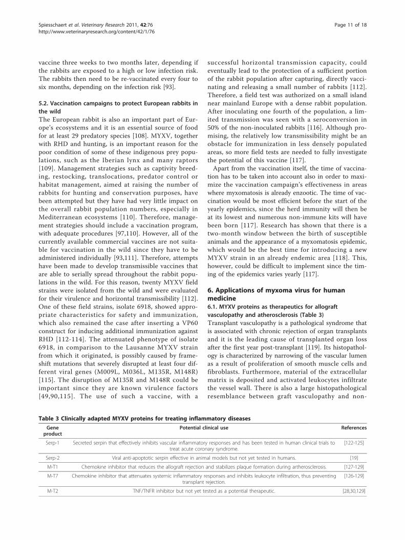
6. Applications of myxoma virus for humanmedicine6.1. MYXV proteins as therapeutics for allograftvasculopathy and atherosclerosis (Table 3)Transplant vasculopathy is a pathological syndrome thatis associated with chronic rejection of organ transplantsand it is the leading cause of transplanted organ lossafter the first year post-transplant [119]. Its histopathol-ogy is characterized by narrowing of the vascular lumenas a result of proliferation of smooth muscle cells andfibroblasts. Furthermore, material of the extracellularmatrix is deposited and activated leukocytes infiltratethe vessel wall. There is also a large histopathologicalresemblance between graft vasculopathy and non-
Table 3 Clinically adapted MYXV proteins for treating inflammatory diseases
Geneproduct
Potential clinical use References
Serp-1 Secreted serpin that effectively inhibits vascular inflammatory responses and has been tested in human clinical trials totreat acute coronary syndrome.
[122-125]
Serp-2 Viral anti-apoptotic serpin effective in animal models but not yet tested in humans. [19]
M-T1 Chemokine inhibitor that reduces the allograft rejection and stabilizes plaque formation during artherosclerosis. [127-129]
M-T7 Chemokine inhibitor that attenuates systemic inflammatory responses and inhibits leukocyte infiltration, thus preventingtransplant rejection.
[126-129]
M-T2 TNF/TNFR inhibitor but not yet tested as a potential therapeutic. [28,30,129]
Spiesschaert et al. Veterinary Research 2011, 42:76http://www.veterinaryresearch.org/content/42/1/76
Page 11 of 18

transplant forms of atherosclerosis, due to overlappingpathogenetic pathways [120]. Atherosclerosis can becaused by many factors, including hyperlipidemia, dia-betes and hypertension [121]. Numerous proteases areimportant factors for the development of atherosclerosis,since they help regulate atherosclerosis-related vascularremodeling. An important group of these proteases arethe serine proteases since they not only mediate matrixbreakdown, but also activate and release diverse pro-inflammatory cytokines and growth factors, thus activat-ing a wide spectrum of leukocyte receptors and promot-ing increased leukocyte migration into damaged tissues[122]. The ability of purified recombinant Serp-1 proteinto inhibit several of these serine protease in a species-independent manner raises the possibility that this viralprotein could attenuate the vascular inflammatoryresponses in a wide variety of cardiovascular diseases,such as atherosclerosis, transplantation rejection andinjury vasculopathy [122,123]. Indeed, there are poten-tially numerous applications in human medicine, forexample treating other diseases driven by systemicinflammation. Recently, successful human Phase II clini-cal trials have been reported using purified Serp-1 pro-tein to treat patients with acute coronary syndromes[124,125].The MYXV protein Serp-2, on the other hand, is an
intracellular regulator that selectively binds and inhibitsmediators of T-cell killing, enabling a generalized reduc-tion of arterial inflammation through a granzyme B/per-forin dependent pathway. This may prolong certainspecific T-cell functions, allowing subsets of T-cells toprovide anti-inflammatory actions or alternatively it mayinitiate apoptotic responses in disease-causing macro-phages and/or smooth muscle cells [122]. Serp-2 proteinhas not yet been tested in clinical trials [19].Another important feature of vascular inflammation is
the attraction of leukocytes by chemokines to specificsites in the vasculature and their migration through sur-rounding tissues [121]. The M-T1 and M-T7 secretedproteins of MYXV, which inhibit in a species-indepen-dent fashion a variety of diverse chemokines, block thechemotaxis of leukocytes into these sites of inflamma-tion, subsequently reducing the allograft rejection in amurine aortic allograft model [126-129]. In the case ofimmune-based joint diseases like rheumatoid arthritis,high quantities of pro-inflammatory cytokines, such asTNF, are found in the synovial fluids and the amount ofTNF elevation closely correlates with disease severity.Subsequently, TNF is considered an important target fortreatment [129]. The MYXV protein M-T2 has high affi-nity for rabbit TNF, and also can bind and inhibithuman TNF-receptors, but more research is still neededto determine if this MYXV protein would have thera-peutic potential [28-30,129].
6.2. Applications for MYXV as a live oncolytic virus totreat cancerMYXV is also considered a promising oncolytic virusthat is essentially harmless to non-lagomorphs, includingmice and humans, but nevertheless is able to selectivelyinfect and kill many diverse human tumor cells. Thistranslational application of MYXV is based on thegenetic alterations in intracellular signaling pathwayscommonly found in cancer cells, which allows the trans-formed cells to continue dividing without being con-trolled by normal cellular checkpoints [20,130-136]. Onthe other hand, these cellular checkpoints also form animportant part of the innate self-protection mechanismsagainst viral infection, and therefore, most transformedcells tend to be more susceptible to many viral infec-tions [134].The absence of such viral protection mechanisms in
cancer cells makes the selective replication possible forat least some oncolytic viruses that normally cannotpropagate successfully in human tissues, such as MYXV[134]. More specifically for MYXV, malignant cancercells in general lose the ability to induce the TNF/IFN-bsynergistic anti-viral state, a feature that largely deter-mines the host range of MYXV in primary human cells[130]. In addition, the host range of MYXV in humancancer cells is also influenced by oncogenic activationand tumor suppressor dysfunction [137]. Indeed, MYXVhas been shown to be capable of infecting a wide varietyof human cancer cell lines in vitro and it has been pro-ven to be a safe and potent oncolytic virus in vivo[132,138,139]. However, in order to be suitable for anti-tumoral therapy in the clinic, MYXV also needs tocause regression of metastatic tumors in immune com-petent patients [132]. When administered systemically,MYXV was able to decrease murine B16 melanomalung metastasis in an immune competent mouse model.This regression of tumors was significantly enhanced bythe chemotherapeutic rapamycin [140], an effect whichwas later on shown to be achieved by reconfiguring theinternal cell signaling environment of many cancer cellsto one which is more optimal for productive virus repli-cation [134]. A more elaborate description of MYXV asan oncolytic virus has recently been reviewed by Liu etal. [19], and the particular exploitation of MYXV topurge cancer cells from human bone marrow trans-plants is described by Rahman et al. [141].
7. Discussion and conclusionsResearch on MYXV has been conducted for many yearsand the interest in this virus is basically two-fold. On theone hand, MYXV is an important pathogen for the Eur-opean rabbit population and due to economical reasonsand ecological consequences, MYXV merits attention forboth population control strategies in places (like
Spiesschaert et al. Veterinary Research 2011, 42:76http://www.veterinaryresearch.org/content/42/1/76
Page 12 of 18

Australia) where feral rabbit populations have becomedamaging, as well as vaccination strategies for situations(like commercial rabbit breading operations, or for nativerabbits in Europe) where myxomatosis is a detrimentaldisease. On the other hand, MYXV is also gaining moreinterest from a therapeutic point of view now that it hasbeen shown to have great potential for treatment of cer-tain inflammatory-based diseases and cancer in humans.Thus, it is of utmost importance to continue to acquiredetailed knowledge on the behavior and pathogenesis ofthis rabbit-specific viral pathogen in both permissive (ierabbit) and nonpermissive (especially human) hosts.It has become clear over the years that developing
effective vaccines and efficient vaccination strategies forrabbits are not as straightforward as originally thought.The major problems encountered are balancing theneed to induce a potent acquired immune responsewithout being accompanied by disease symptoms. Dele-tions of MYXV virulence genes, but maintaining virusreplication fitness, is probably the best approach for alonger lasting immunity, as has been shown by Adamset al. [105]. However, improved potency in terms of thedesired immune response was often accompanied by theappearance of some clinical symptoms in vaccinatedrabbits, indicating that the level of protection that canbe achieved to date with the available vaccines on themarket might remain the best approach since these vac-cines are of no risk for the rabbit itself.Another possibility for creating a more potent immune
reaction against MYXV could be insertion of importantimmunogenic genes of MYXV into heterologous vaccineplatforms. Such approach has already been reported forcanine influenza, equine influenza virus, West Nile virusand bovine viral diarrhea virus, where virulence genes ofthese viruses were inserted into an equine herpesvirustype 1 (EHV1) vaccine [142-145]. The rationale behindthe insertion of MYXV immunogenic genes into anequine pathogen is based on the fact that EHV1 growsextremely well in rabbit cells and hence, rabbit kidney(RK13) are the most commonly used in vitro cell line togrow EHV1 [146,147]. Still, despite this efficient growthin vitro, it needs to be determined whether EHV1 is cap-able of inducing robust immune responses in rabbits invivo. Preliminary experiments, however, have shown thatrabbits are susceptible for EHV1 infection (Van de Walleet al., unpublished results), indicating that a recombinantEHV1 vector expressing MYXV proteins could prove anovel strategy to efficiently protect rabbits againstMYXV. In addition, another possible viral vector vehiclefor immunization against MYXV could be exploited withnonpathogenic variants of Vesicular Stomatitis Virus(VSV) since it has shown excellent replication kinetics inthe rabbit during experiments for vaccine development[148-150].
Numerous discoveries have been made concerning theimmunomodulatory capabilities of MYXV since thesequencing of the full MYXV genome, although thefunctions of many candidate proteins remain undeter-mined to date. For some of the latter class of proteins, aputative function had been sometimes ascribed based onsequence similarity with other immunomodulatory viralor cellular proteins, but these conjectures have notalways been validated, such as the cases of M063 [1,62]and M135 [90]. This indicates that one needs to be judi-cious when predicting how a viral protein functionssolely based on sequence or structural similarities.Rather, a true biological role can only be assigned afterthorough in vitro and in vivo experimentations.In terms of clinical applications, several of the MYXV
immunomodulatory proteins have become promising can-didates for treatment of inflammation-based disorders. Todate, the first of these virus-derived drugs (Serp-1) hasbehaved particularly well in humans, with essentially noclinical adverse side effects, in Phase I and II clinical trials.MYXV as an oncolytic virus candidate also shows consid-erable potential, both against murine cancers in immuno-competent mouse hosts, as well as human cancersxenografted into immunodeficient mice. MYXV appearsto be capable of substantially counteracting malignanciesderived from many tissues, especially in combination withrapamycin, and the live virus has exhibited essentially nodetectable side effects in all murine hosts tested, evenmice that are extensively immunocompromised. In thefuture, insertion of selective anti-cancer therapeutic genes[131] and/or deletion of MYXV virulence genes [151]could further augment its safety against all vertebratehosts (including rabbits) and therefore improve its clinicalsuitability. For example, the Interleukin-15 (IL-15) genehas been inserted into the MYXV genome and this recom-binant virus remained capable of replicating in vitro to adegree resembling wild MYXV [131]. Very promisingresults, concerning safety improvement, have also beenobtained by deleting the host range genes M063 or M135since either deletion showed significant attenuation in itsnatural rabbit host while retaining and even improving theviral oncolytic capabilities compared to wild MYXV [151].The exploitation of MYXV constructs that are nonpatho-genic in all known vertebrate hosts, but still potently onco-lytic against human cancers, will hopefully make MYXVan important factor in improving the outcome of treat-ment for numerous cancers in the future [19].
List of abbreviations(ANK): ankyrin; (BCL2): B-cell lymphoma 2; (Bak): (BCL2)-antagonist/killer; (Bax):Bcl-2 associated × protein; (CD): cluster of differentiation; (CDK): cyclin-dependent kinase; (DCs): dendritic cells; (DNA): double strandeddeoxyribonucleic acid; (dsRNA): double stranded RNA; (ECTV): ectromeliavirus; (ER): endoplasmic reticulum; (EGF): epidermal growth factor; (eIF2α):eukaryotic initiation factor 2 alpha; (gp120): glycoprotein 120; (ICE): IL-1β-
Spiesschaert et al. Veterinary Research 2011, 42:76http://www.veterinaryresearch.org/content/42/1/76
Page 13 of 18

converting enzyme; (IAP): integrin associated protein; (IFN): interferon; (IFN-γ):interferon-γ; (IL-15): Interleukin-15; (Ur): Lausanne strain and the Uriarra; (LY-49): lymphocyte 49; (MHC-1): major histocompatibility complex 1; (MCP)-1:monocyte chemotactic protein; (MGF): myxoma growth factor; (MNF):myxoma nuclear factor; (MYXV): Myxoma virus; (MV-LAP): Myxoma Virus-Leukemia-Associated Protein; (NKG2): natural killer cell group 2; (IκBs): NF-κBinhibitors; (NF-κB): Nucleus Factor-Κb; (PBR): peripheral benzodiazepinereceptor; (PT): permeabilitytransition; (PRANC): Pox protein Repeats ofAnkyrin C-terminal; (PKR): Protein Kinase R; (PPIX): protoporphyrin IX; (PYD):pyrin domain; (RHD): rabbit hemorrhagic disease; (RK): rabbit kidney;(serpins): Serine protease inhibitors; (SFV): Shope fibroma virus; (SIRPα): signalregulatory protein α; (SLS): Standard Laboratory Strain; (SAMD9): sterile alphamotif domain containing protein 9; (TIRs): terminal inverted repeats; (TLR):Toll-like receptor; (Tat): transactivator; (TNFR): tumor necrosis factor receptor;(VACV): vaccinia virus; (VSV): Vesicular Stomatitis Virus; (vPLAD): viral preligandassembly domain.
AcknowledgementsGM’s lab is supported by grants from NIAID (R01 AI080607-01A1), NCI (R01CA138541-01 and R21 CA149869) and the Bankhead-Coley Foundation (1BT02).GVdW is a postdoctoral fellow of the FWO Flanders. We would also like tothank Jia Liu and Masmudur Rahman for proof-reading the manuscript.
Author details1Department of Virology, Parasitology and Immunology, Faculty of VeterinaryMedicine, Ghent University, Salisburylaan 133, B-9820 Merelbeke, Belgium.2Department of Molecular Genetics and Microbiology, University of FloridaCollege of Medicine, 1600 SW Archer Rd, P.O. Box 100266, Gainesville, FL32610, USA. 3Department of Pathology, Bacteriology and Poultry diseases,Faculty of Veterinary Medicine, Ghent University, Salisburylaan 133, B-9820Merelbeke, Belgium. 4Department of Comparative Physiology and Biometrics,Faculty of Veterinary Medicine, Ghent University, Salisburylaan 133, B-9820Merelbeke, Belgium.
Authors’ contributionsBS performed an extensive literature study on the subject, analyzed theacquired information and wrote the manuscript; GM and KH both revisedthe manuscript critically for important intellectual content concerning theirown area of expertise; HN was involved in the initial drafting of themanuscript; GVdW participated in its design and coordination, criticallyrevised the manuscript and helped to draft the manuscript. All authors readand approved the final manuscript.
Competing interestsGM is a co-founder of Viron Therapeutics, which is developing viral proteinsas therapeutics. The other authors declare that they have no competinginterests.
Received: 25 February 2011 Accepted: 9 June 2011Published: 9 June 2011
References1. Cameron C, Hota-Mitchell S, Chen L, Barrett J, Cao JX, Macaulay C, Willer D,
Evans D, McFadden G: The complete DNA sequence of myxoma virus.Virology 1999, 264:298-318.
2. Kerr P, McFadden G: Immune responses to myxoma virus. Viral Immunol2002, 15:229-246.
3. Saari SA, Rudback E, Niskanen M, Syrjala P, Nylund M, Anttila M: Contagiousmucocutaneous dermatitis of the mountain hare (Lepus timidus):pathology and cause. J Wildl Dis 2005, 41:775-782.
4. Fenner F, Ratcliffe F: Myxomatosis University Press, Cambridge Eng; 1965.5. Fenner F: Deliberate introduction of the European rabbit, Oryctolagus
cuniculus, into Australia. Rev Sci Tech 2010, 29:103-111.6. Burnet FM: Myxomatosis as a method of biological control against the
Australian rabbit. Am J Public Health Nations Health 1952, 42:1522-1526.7. Best SM, Kerr PJ: Coevolution of host and virus: the pathogenesis of
virulent and attenuated strains of myxoma virus in resistant andsusceptible European rabbits. Virology 2000, 267:36-48.
8. Ross J, Tittensor AM: The establishment and spread of myxomatosis andits effect on rabbit populations. Philos Trans R Soc Lond B Biol Sci 1986,314:599-606.
9. Bartrip PW: Myxomatosis in 1950s Britain. 20 Century Br Hist 2008,19:83-105.
10. Fenner F, Fantini B: Biological Control of vertebrate pests: the history ofmyxomatosis - an experiment in evolution CABI Publishing, Oxford; 1999.
11. Jeklova E, Leva L, Matiasovic J, Kovarcik K, Kudlackova H, Nevorankova Z,Psikal I, Faldyna M: Characterisation of immunosuppression in rabbitsafter infection with myxoma virus. Vet Microbiol 2008, 129:117-130.
12. Best SM, Collins SV, Kerr PJ: Coevolution of host and virus: cellularlocalization of virus in myxoma virus infection of resistant andsusceptible European rabbits. Virology 2000, 277:76-91.
13. Zuniga MC: Lessons in detente or know thy host: theimmunomodulatory gene products of myxoma virus. J Biosci 2003,28:273-285.
14. Robinson AJ, Muller WJ, Braid AL, Kerr PJ: The effect of buprenorphine onthe course of disease in laboratory rabbits infected with myxoma virus.Lab Anim 1999, 33:252-257.
15. Cavadini P, Botti G, Barbieri I, Lavazza A, Capucci L: Molecularcharacterization of SG33 and Borghi vaccines used against myxomatosis.Vaccine 2010, 28:5414-5420.
16. Marlier D, Cassart D, Boucraut-Baralon C, Coignoul F, Vindevogel H:Experimental infection of specific pathogen-free New Zealand Whiterabbits with five strains of amyxomatous myxoma virus. J Comp Pathol1999, 121:369-384.
17. Nash P, Barrett J, Cao JX, Hota-Mitchell S, Lalani AS, Everett H, Xu XM,Robichaud J, Hnatiuk S, Ainslie C, Seet BT, McFadden G:Immunomodulation by viruses: the myxoma virus story. Immunol Rev1999, 168:103-120.
18. Goebel SJ, Johnson GP, Perkus ME, Davis SW, Winslow JP, Paoletti E: Thecomplete DNA sequence of vaccinia virus. Virology 1990, 179:247-266,517-563.
19. Liu J, Wennier S, McFadden G: The immunoregulatory properties ofoncolytic myxoma virus and their implications in therapeutics. MicrobesInfect 2010, 12:1144-1152.
20. Stanford MM, Werden SJ, McFadden G: Myxoma virus in the Europeanrabbit: interactions between the virus and its susceptible host. Vet Res2007, 38:299-318.
21. Seet BT, Johnston JB, Brunetti CR, Barrett JW, Everett H, Cameron C,Sypula J, Nazarian SH, Lucas A, McFadden G: Poxviruses and immuneevasion. Annu Rev Immunol 2003, 21:377-423.
22. Johnston JB, Wang G, Barrett JW, Nazarian SH, Colwill K, Moran M,McFadden G: Myxoma virus M-T5 protects infected cells from the stressof cell cycle arrest through its interaction with host cell cullin-1. J Virol2005, 79:10750-10763.
23. Bennett CJ, Webb M, Willer DO, Evans DH: Genetic and phylogeneticcharacterization of the type II cyclobutane pyrimidine dimer photolyasesencoded by Leporipoxviruses. Virology 2003, 315:10-19.
24. Webb MAM: An investigation of factors affecting poxvirus genetic stabilityNational Library of Canada, Ottawa, Canada; 2004.
25. Hengartner MO: The biochemistry of apoptosis. Nature 2000, 407:770-776.26. Taylor JM, Barry M: Near death experiences: poxvirus regulation of
apoptotic death. Virology 2006, 344:139-150.27. Schreiber M, McFadden G: The myxoma virus TNF-receptor homologue
(T2) inhibits tumor necrosis factor-alpha in a species-specific fashion.Virology 1994, 204:692-705.
28. Schreiber M, Rajarathnam K, McFadden G: Myxoma virus T2 protein,a tumor necrosis factor (TNF) receptor homolog, is secreted as amonomer and dimer that each bind rabbit TNFalpha, but thedimer is a more potent TNF inhibitor. J Biol Chem 1996,271:13333-13341.
29. Sedger LM, Osvath SR, Xu XM, Li G, Chan FK, Barrett JW, McFadden G:Poxvirus tumor necrosis factor receptor (TNFR)-like T2 proteins contain aconserved preligand assembly domain that inhibits cellular TNFR1-induced cell death. J Virol 2006, 80:9300-9309.
30. Schreiber M, Sedger L, McFadden G: Distinct domains of M-T2, themyxoma virus tumor necrosis factor (TNF) receptor homolog, mediateextracellular TNF binding and intracellular apoptosis inhibition. J Virol1997, 71:2171-2181.
31. Castedo M, Perfettini JL, Kroemer G: Mitochondrial apoptosis and theperipheral benzodiazepine receptor: a novel target for viral andpharmacological manipulation. J Exp Med 2002, 196:1121-1125.
Spiesschaert et al. Veterinary Research 2011, 42:76http://www.veterinaryresearch.org/content/42/1/76
Page 14 of 18
32. Everett H, Barry M, Sun X, Lee SF, Frantz C, Berthiaume LG, McFadden G,Bleackley RC: The myxoma poxvirus protein, M11L, prevents apoptosisby direct interaction with the mitochondrial permeability transitionpore. J Exp Med 2002, 196:1127-1139.
33. Everett H, Barry M, Lee SF, Sun X, Graham K, Stone J, Bleackley RC,McFadden G: M11L: a novel mitochondria-localized protein of myxomavirus that blocks apoptosis of infected leukocytes. J Exp Med 2000,191:1487-1498.
34. Wang G, Barrett JW, Nazarian SH, Everett H, Gao X, Bleackley C, Colwill K,Moran MF, McFadden G: Myxoma virus M11L prevents apoptosis throughconstitutive interaction with Bak. J Virol 2004, 78:7097-7111.
35. Su J, Wang G, Barrett JW, Irvine TS, Gao X, McFadden G: Myxoma virusM11L blocks apoptosis through inhibition of conformational activationof Bax at the mitochondria. J Virol 2006, 80:1140-1151.
36. Aoyagi M, Zhai D, Jin C, Aleshin AE, Stec B, Reed JC, Liddington RC:Vaccinia virus N1L protein resembles a B cell lymphoma-2 (Bcl-2) familyprotein. Protein Sci 2007, 16:118-124.
37. Cooray S, Bahar MW, Abrescia NG, McVey CE, Bartlett NW, Chen RA,Stuart DI, Grimes JM, Smith GL: Functional and structural studies of thevaccinia virus virulence factor N1 reveal a Bcl-2-like anti-apoptoticprotein. J Gen Virol 2007, 88:1656-1666.
38. Gonzalez JM, Esteban M: A poxvirus Bcl-2-like gene family involved inregulation of host immune response: sequence similarity andevolutionary history. Virol J 2010, 7:59.
39. Gratz MS, Suezer Y, Kremer M, Volz A, Majzoub M, Hanschmann KM,Kalinke U, Schwantes A, Sutter G: N1L is an ectromelia virus virulencefactor and essential for in vivo spread upon respiratory infection. J Virol2011, 85:3557-3569.
40. Mercer AA, Fleming SB, Ueda N: F-box-like domains are present in mostpoxvirus ankyrin repeat proteins. Virus Genes 2005, 31:127-133.
41. Sedgwick SG, Smerdon SJ: The ankyrin repeat: a diversity of interactionson a common structural framework. Trends Biochem Sci 1999, 24:311-316.
42. Werden SJ, Lanchbury J, Shattuck D, Neff C, Dufford M, McFadden G: Themyxoma virus m-t5 ankyrin repeat host range protein is a novel adaptorthat coordinately links the cellular signaling pathways mediated by Aktand Skp1 in virus-infected cells. J Virol 2009, 83:12068-12083.
43. Johnston JB, Wang G, Barrett JW, Nazarian SH, Colwill K, Moran M,McFadden G: Myxoma virus M-T5 protects infected cells from the stressof cell cycle arrest through its interaction with host cell cullin-1. J Virol2005, 79:10750-10763.
44. Mossman K, Lee SF, Barry M, Boshkov L, McFadden G: Disruption of M-T5,a novel myxoma virus gene member of poxvirus host rangesuperfamily, results in dramatic attenuation of myxomatosis in infectedEuropean rabbits. J Virol 1996, 70:4394-4410.
45. Camus-Bouclainville C, Fiette L, Bouchiha S, Pignolet B, Counor D, Filipe C,Gelfi J, Messud-Petit F: A virulence factor of myxoma virus colocalizeswith NF-kappaB in the nucleus and interferes with inflammation. J Virol2004, 78:2510-2516.
46. Chen ZJ: Ubiquitin signalling in the NF-kappaB pathway. Nat Cell Biol2005, 7:758-765.
47. Baeuerle PA: IkappaB-NF-kappaB structures: at the interface ofinflammation control. Cell 1998, 95:729-731.
48. Blanie S, Gelfi J, Bertagnoli S, Camus-Bouclainville C: MNF, an ankyrinrepeat protein of myxoma virus, is part of a native cellular SCF complexduring viral infection. Virol J 2010, 7:56.
49. Blanie S, Mortier J, Delverdier M, Bertagnoli S, Camus-Bouclainville C: M148Rand M149R are two virulence factors for myxoma virus pathogenesis inthe European rabbit. Vet Res 2009, 40:11.
50. Guerin JL, Gelfi J, Boullier S, Delverdier M, Bellanger FA, Bertagnoli S,Drexler I, Sutter G, Messud-Petit F: Myxoma virus leukemia-associatedprotein is responsible for major histocompatibility complex class I andFas-CD95 down-regulation and defines scrapins, a new group of surfacecellular receptor abductor proteins. J Virol 2002, 76:2912-2923.
51. Mansouri M, Bartee E, Gouveia K, Hovey Nerenberg BT, Barrett J, Thomas L,Thomas G, McFadden G, Fruh K: The PHD/LAP-domain protein M153R ofmyxomavirus is a ubiquitin ligase that induces the rapid internalizationand lysosomal destruction of CD4. J Virol 2003, 77:1427-1440.
52. Barry M, Hnatiuk S, Mossman K, Lee SF, Boshkov L, McFadden G: Themyxoma virus M-T4 gene encodes a novel RDEL-containing protein thatis retained within the endoplasmic reticulum and is important for theproductive infection of lymphocytes. Virology 1997, 239:360-377.
53. Hnatiuk S, Barry M, Zeng W, Liu L, Lucas A, Percy D, McFadden G: Role ofthe C-terminal RDEL motif of the myxoma virus M-T4 protein in termsof apoptosis regulation and viral pathogenesis. Virology 1999,263:290-306.
54. Zuniga MC: A pox on thee! Manipulation of the host immune system bymyxoma virus and implications for viral-host co-adaptation. Virus Res2002, 88:17-33.
55. Sadler AJ, Williams BR: Interferon-inducible antiviral effectors. Nat RevImmunol 2008, 8:559-568.
56. Langland JO, Jacobs BL: The role of the PKR-inhibitory genes, E3L andK3L, in determining vaccinia virus host range. Virology 2002, 299:133-141.
57. Ramelot TA, Cort JR, Yee AA, Liu F, Goshe MB, Edwards AM, Smith RD,Arrowsmith CH, Dever TE, Kennedy MA: Myxoma virusimmunomodulatory protein M156R is a structural mimic of eukaryotictranslation initiation factor eIF2alpha. J Mol Biol 2002, 322:943-954.
58. Wang F, Barrett JW, Shao Q, Gao X, Dekaban GA, McFadden G: Myxomavirus selectively disrupts type I interferon signaling in primary humanfibroblasts by blocking the activation of the Janus kinase Tyk2. Virology2009, 387:136-146.
59. Bartee E, Mohamed MR, Lopez MC, Baker HV, McFadden G: The addition oftumor necrosis factor plus beta interferon induces a novel synergisticantiviral state against poxviruses in primary human fibroblasts. J Virol2009, 83:498-511.
60. Barrett JW, Cao JX, Hota-Mitchell S, McFadden G: Immunomodulatoryproteins of myxoma virus. Semin Immunol 2001, 13:73-84.
61. Meng X, Chao J, Xiang Y: Identification from diverse mammalianpoxviruses of host-range regulatory genes functioning equivalently tovaccinia virus C7L. Virology 2008, 372:372-383.
62. Barrett JW, Shun Chang C, Wang G, Werden SJ, Shao Z, Barrett C, Gao X,Belsito TA, Villenevue D, McFadden G: Myxoma virus M063R is a hostrange gene essential for virus replication in rabbit cells. Virology 2007,361:123-132.
63. Curtin JF, Cotter TG: Live and let die: regulatory mechanisms in Fas-mediated apoptosis. Cell Signal 2003, 15:983-992.
64. Salomoni P, Khelifi AF: Daxx: death or survival protein? Trends Cell Biol2006, 16:97-104.
65. Liu J, Wennier S, Zhang L, McFadden G: M062 is a host range factoressential for myxoma virus pathogenesis and functions as an antagonistof host SAMD9 in human cells. J Virol 2011, 85:3270-3282.
66. Lalani AS, Masters J, Graham K, Liu L, Lucas A, McFadden G: Role of themyxoma virus soluble CC-chemokine inhibitor glycoprotein, M-T1,during myxoma virus pathogenesis. Virology 1999, 256:233-245.
67. Lalani AS, Ness TL, Singh R, Harrison JK, Seet BT, Kelvin DJ, McFadden G,Moyer RW: Functional comparisons among members of the poxvirus T1/35 kDa family of soluble CC-chemokine inhibitor glycoproteins. Virology1998, 250:173-184.
68. Upton C, Mossman K, McFadden G: Encoding of a homolog of the IFN-gamma receptor by myxoma virus. Science 1992, 258:1369-1372.
69. Boomker JM, Luttikhuizen DT, Veninga H, de Leij LF, The TH, de Haan A,van Luyn MJ, Harmsen MC: The modulation of angiogenesis in theforeign body response by the poxviral protein M-T7. Biomaterials 2005,26:4874-4881.
70. Macen JL, Upton C, Nation N, McFadden G: SERP1, a serine proteinaseinhibitor encoded by myxoma virus, is a secreted glycoprotein thatinterferes with inflammation. Virology 1993, 195:348-363.
71. Petit F, Bertagnoli S, Gelfi J, Fassy F, Boucraut-Baralon C, Milon A:Characterization of a myxoma virus-encoded serpin-like protein withactivity against interleukin-1 beta-converting enzyme. J Virol 1996,70:5860-5866.
72. Chavez-Galan L, Arenas-Del Angel MC, Zenteno E, Chavez R, Lascurain R:Cell death mechanisms induced by cytotoxic lymphocytes. Cell MolImmunol 2009, 6:15-25.
73. Turner PC, Sancho MC, Thoennes SR, Caputo A, Bleackley RC, Moyer RW:Myxoma virus Serp2 is a weak inhibitor of granzyme B and interleukin-1beta-converting enzyme in vitro and unlike CrmA cannot blockapoptosis in cowpox virus-infected cells. J Virol 1999, 73:6394-6404.
74. Guerin JL, Gelfi J, Camus C, Delverdier M, Whisstock JC, Amardeihl MF, Py R,Bertagnoli S, Messud-Petit F: Characterization and functional analysis ofSerp3: a novel myxoma virus-encoded serpin involved in virulence. JGen Virol 2001, 82:1407-1417.
Spiesschaert et al. Veterinary Research 2011, 42:76http://www.veterinaryresearch.org/content/42/1/76
Page 15 of 18

75. Barclay AN, Wright GJ, Brooke G, Brown MH: CD200 and membraneprotein interactions in the control of myeloid cells. Trends Immunol 2002,23:285-290.
76. Cameron CM, Barrett JW, Liu L, Lucas AR, McFadden G: Myxoma virusM141R expresses a viral CD200 (vOX-2) that is responsible for down-regulation of macrophage and T-cell activation in vivo. J Virol 2005,79:6052-6067.
77. Zhang L, Stanford M, Liu J, Barrett C, Jiang L, Barclay AN, McFadden G:Inhibition of macrophage activation by the myxoma virus M141 protein(vCD200). J Virol 2009, 83:9602-9607.
78. van Beek EM, Cochrane F, Barclay AN, van den Berg TK: Signal regulatoryproteins in the immune system. J Immunol 2005, 175:7781-7787.
79. Matozaki T, Murata Y, Okazawa H, Ohnishi H: Functions and molecularmechanisms of the CD47-SIRPalpha signalling pathway. Trends Cell Biol2009, 19:72-80.
80. Cameron CM, Barrett JW, Mann M, Lucas A, McFadden G: Myxoma virusM128L is expressed as a cell surface CD47-like virulence factor thatcontributes to the downregulation of macrophage activation in vivo.Virology 2005, 337:55-67.
81. Johnston JB, Barrett JW, Nazarian SH, Goodwin M, Ricciuto D, Wang G,McFadden G: A poxvirus-encoded pyrin domain protein interacts withASC-1 to inhibit host inflammatory and apoptotic responses to infection.Immunity 2005, 23:587-598.
82. Petrilli V, Papin S, Tschopp J: The inflammasome. Curr Biol 2005, 15:R581.83. Rahman MM, Mohamed MR, Kim M, Smallwood S, McFadden G: Co-
regulation of NF-kappaB and inflammasome-mediated inflammatoryresponses by myxoma virus pyrin domain-containing protein M013. PLoSPathog 2009, 5:e1000635.
84. Gedey R, Jin XL, Hinthong O, Shisler JL: Poxviral regulation of the host NF-kappaB response: the vaccinia virus M2L protein inhibits induction ofNF-kappaB activation via an ERK2 pathway in virus-infected humanembryonic kidney cells. J Virol 2006, 80:8676-8685.
85. Qin S, Rottman JB, Myers P, Kassam N, Weinblatt M, Loetscher M, Koch AE,Moser B, Mackay CR: The chemokine receptors CXCR3 and CCR5 marksubsets of T cells associated with certain inflammatory reactions. J ClinInvest 1998, 101:746-754.
86. Barrett JW, Werden SJ, Wang F, McKillop WM, Jimenez J, Villeneuve D,McFadden G, Dekaban GA: Myxoma virus M130R is a novel virulencefactor required for lethal myxomatosis in rabbits. Virus Res 2009,144:258-265.
87. Rubartelli A, Poggi A, Sitia R, Zocchi MR: HIV-I Tat: a polypeptide for allseasons. Immunol Today 1998, 19:543-545.
88. Zocchi MR, Rubartelli A, Morgavi P, Poggi A: HIV-1 Tat inhibits humannatural killer cell function by blocking L-type calcium channels. JImmunol 1998, 161:2938-2943.
89. Opgenorth A, Graham K, Nation N, Strayer D, McFadden G: Deletionanalysis of two tandemly arranged virulence genes in myxoma virus,M11L and myxoma growth factor. J Virol 1992, 66:4720-4731.
90. Barrett JW, Sypula J, Wang F, Alston LR, Shao Z, Gao X, Irvine TS,McFadden G: M135R is a novel cell surface virulence factor of myxomavirus. J Virol 2007, 81:106-114.
91. Colamonici OR, Domanski P, Sweitzer SM, Larner A, Buller RM: Vacciniavirus B18R gene encodes a type I interferon-binding protein that blocksinterferon alpha transmembrane signaling. J Biol Chem 1995,270:15974-15978.
92. Symons JA, Alcami A, Smith GL: Vaccinia virus encodes a soluble type Iinterferon receptor of novel structure and broad species specificity. Cell1995, 81:551-560.
93. Marlier D: Vaccination strategies against myxomavirus infections: are wereally doing the best? Tijdschr Diergeneeskd 2010, 135:194-198.
94. Adams MM, van Leeuwen BH, Kerr PJ: Limitations of plasmid vaccines tocomplex viruses: selected myxoma virus antigens as DNA vaccines werenot protective. Vaccine 2004, 23:198-204.
95. Willer DO, McFadden G, Evans DH: The complete genome sequence ofshope (rabbit) fibroma virus. Virology 1999, 264:319-343.
96. Fenner F, Woodroofe GM: Protection of laboratory rabbits againstmyxomatosis by vaccination with fibroma virus. Aust J Exp Biol Med Sci1954, 32:653-668.
97. Jerabek J: Applicability of shope fibroma virus replicated in cell culturesfor immunoprophylaxis of rabbit myxomatosis. Acta Vet Brno 1980,49:259-267.
98. Alfonso M, Pagés-Manté A: Serological response to myxomatosisvaccination by different inoculation systems on farm rabbits. WorldRabbit Sci 2003, 11:145-156.
99. Marlier D, Mainil J, Boucraut-Baralon C, Linden A, Vindevogel H: Theefficacy of two vaccination schemes against experimental infection witha virulent amyxomatous or a virulent nodular myxoma virus strain. JComp Pathol 2000, 122:115-122.
100. Lavazza A, Graziani M, Tranquillo VM, Botti G, Palotta C, Cerioli M, Capucci L:Serological evaluation of the immunity induced in commercial rabbitsby vaccination for myxomatosis and RHD. Proc. 8th World RabbitCongress, Puebla; 2004, 569-575.
101. Saurat P, Gilbert Y, Ganiere JP: Study on Myxomatous Virus ModifiedStrain. Rev Med Vet 1978, 129:415-451 (in French).
102. Petit F, Boucraut-Baralon C, Py R, Bertagnoli S: Analysis of myxoma virusgenome using pulsed-field gel electrophoresis. Vet Microbiol 1996,50:27-32.
103. Lemière S, Alaphilippe A, Boucher S, Bertagnoli S: Field study of safety andantibody production further to a combined myxomatosis and viralhaemorrhagic disease (VHD) vaccination in dwarf rabbits by intradermalroute. World Rabbit Sci 2003, 11:41-47.
104. Adams MM, van Leeuwen BH, McFadden G, Kerr PJ: Construction andtesting of a novel host-range defective myxoma virus vaccine with theM063 gene inactivated that is non-permissive for replication in rabbitcells. Vet Res 2008, 39:60.
105. Adams MM, van Leeuwen BH, Kerr PJ: Construction and evaluation of liveattenuated myxoma virus vaccines with targeted virulence genedeletions. Vaccine 2008, 26:5843-5854.
106. Parra F, Prieto M: Purification and characterization of a calicivirus as thecausative agent of a lethal hemorrhagic disease in rabbits. J Virol 1990,64:4013-4015.
107. Bertagnoli S, Gelfi J, Le Gall G, Boilletot E, Vautherot JF, Rasschaert D,Laurent S, Petit F, Boucraut-Baralon C, Milon A: Protection againstmyxomatosis and rabbit viral hemorrhagic disease with recombinantmyxoma viruses expressing rabbit hemorrhagic disease virus capsidprotein. J Virol 1996, 70:5061-5066.
108. Ferrer M, Negro J: The near extinction of two large European predators:super specialists pay a price. Conserv Biol 2004, 18:344-349.
109. Gortazar C, Ferroglio E, Hofle U, Frolich K, Vicente J: Diseases sharedbetween wildlife and livestock: a European perspective. Eur J Wildlife Res2007, 53:241-256.
110. Garcia-Bocanegra I, Astorga RJ, Napp S, Casal J, Huerta B, Borge C, Arenas A:Myxomatosis in wild rabbit: design of control programs inMediterranean ecosystems. Prev Vet Med 2010, 93:42-50.
111. Ferreira C, Ramirez E, Castro F, Ferreras P, Alves PC, Redpath S, Villafuerte R:Field experimental vaccination campaigns against myxomatosis andtheir effectiveness in the wild. Vaccine 2009, 27:6998-7002.
112. Barcena J, Pages-Mante A, March R, Morales M, Ramirez MA, Sanchez-Vizcaino JM, Torres JM: Isolation of an attenuated myxoma virus fieldstrain that can confer protection against myxomatosis on contacts ofvaccinates. Arch Virol 2000, 145:759-771.
113. Angulo E, Bárcena J: Towards a unique and transmissible vaccine againstmyxomatosis and rabbit haemorrhagic disease for rabbit populations.Wildl Res 2007, 34:567-577.
114. Torres JM, Ramirez MA, Morales M, Barcena J, Vazquez B, Espuna E, Pages-Mante A, Sanchez-Vizcaino JM: Safety evaluation of a recombinantmyxoma-RHDV virus inducing horizontal transmissible protectionagainst myxomatosis and rabbit haemorrhagic disease. Vaccine 2000,19:174-182.
115. Morales M, Ramirez MA, Cano MJ, Parraga M, Castilla J, Perez-Ordoyo LI,Torres JM, Barcena J: Genome comparison of a nonpathogenic myxomavirus field strain with its ancestor, the virulent Lausanne strain. J Virol2009, 83:2397-2403.
116. Torres JM, Sanchez C, Ramirez MA, Morales M, Barcena J, Ferrer J, Espuna E,Pages-Mante A, Sanchez-Vizcaino JM: First field trial of a transmissiblerecombinant vaccine against myxomatosis and rabbit hemorrhagicdisease. Vaccine 2001, 19:4536-4543.
117. Guitton JS, Devillard S, Guenezan M, Fouchet D, Pontier D, Marchandeau S:Vaccination of free-living juvenile wild rabbits (Oryctolagus cuniculus)against myxomatosis improved their survival. Prev Vet Med 2008, 84:1-10.
118. Merchant JC, Kerr PJ, Simms NG, Hood GM, Pech RP, Robinson AJ:Monitoring the spread of myxoma virus in rabbit Oryctolagus cuniculus
Spiesschaert et al. Veterinary Research 2011, 42:76http://www.veterinaryresearch.org/content/42/1/76
Page 16 of 18

populations on the southern tablelands of New South Wales, Australia.III. Release, persistence and rate of spread of an identifiable strain ofmyxoma virus. Epidemiol Infect 2003, 130:135-147.
119. Munuswamy-Ramanujam G, Dai E, Liu L, Shnabel M, Sun YM, Bartee M,Lomas DA, Lucas AR: Neuroserpin, a thrombolytic serine proteaseinhibitor (serpin), blocks transplant vasculopathy with associatedmodification of T-helper cell subsets. Thromb Haemost 2010, 103:545-555.
120. Chen J, Myllarniemi M, Akyurek LM, Hayry P, Marsden PA, Paul LC:Identification of differentially expressed genes in rat aortic allograftvasculopathy. Am J Pathol 1996, 149:597-611.
121. Richardson J, Viswanathan K, Lucas A: Serpins, the vasculature, and viraltherapeutics. Front Biosci 2006, 11:1042-1056.
122. Bot I, van Berkel TJ, Biessen EA: Viral serine protease inhibitors as anti-atherosclerotic therapy. Curr Opin Investig Drugs 2007, 8:729-735.
123. Dai E, Guan H, Liu L, Little S, McFadden G, Vaziri S, Cao H, Ivanova IA,Bocksch L, Lucas A: Serp-1, a viral anti-inflammatory serpin, regulatescellular serine proteinase and serpin responses to vascular injury. J BiolChem 2003, 278:18563-18572.
124. Lucas A, Liu L, Dai E, Bot I, Viswanathan K, Munuswamy-Ramunujam G,Davids JA, Bartee MY, Richardson J, Christov A, Wang H, Macaulay C,Poznansky M, Zhong R, Miller L, Biessen E, Richardson M, Sullivan C,Moyer R, Hatton M, Lomas DA, McFadden G: The serpin saga;development of a new class of virus derived anti-inflammatory proteinimmunotherapeutics. Adv Exp Med Biol 2009, 666:132-156.
125. Tardif JC, L’Allier PL, Gregoire J, Ibrahim R, McFadden G, Kostuk W,Knudtson M, Labinaz M, Waksman R, Pepine CJ, Macaulay C, Guertin MC,Lucas A: A randomized controlled, phase 2 trial of the viral serpin Serp-1in patients with acute coronary syndromes undergoing percutaneouscoronary intervention. Circ Cardiovasc Interv 2010, 3:543-548.
126. Bedard EL, Kim P, Jiang J, Parry N, Liu L, Wang H, Garcia B, Li X,McFadden G, Lucas A, Zhong R: Chemokine-binding viral protein M-T7prevents chronic rejection in rat renal allografts. Transplantation 2003,76:249-252.
127. Dai E, Liu LY, Wang H, McIvor D, Sun YM, Macaulay C, King E, Munuswamy-Ramanujam G, Bartee MY, Williams J, Davids J, Charo I, McFadden G,Esko JD, Lucas AR: Inhibition of chemokine-glycosaminoglycaninteractions in donor tissue reduces mouse allograft vasculopathy andtransplant rejection. PLoS One 2010, 5:e10510.
128. Liu L, Dai E, Miller L, Seet B, Lalani A, Macauley C, Li X, Virgin HW, Bunce C,Turner P, Moyer R, McFadden G, Lucas A: Viral chemokine-bindingproteins inhibit inflammatory responses and aortic allograft transplantvasculopathy in rat models. Transplantation 2004, 77:1652-1660.
129. Munuswamy-Ramanujam G, Khan KA, Lucas AR: Viral anti-inflammatoryreagents: the potential for treatment of arthritic and vasculitic disorders.Endocr Metab Immune Disord Drug Targets 2006, 6:331-343.
130. Bartee E, McFadden G: Human cancer cells have specifically lost theability to induce the synergistic state caused by tumor necrosis factorplus interferon-beta. Cytokine 2009, 47:199-205.
131. Liu J, Wennier S, Reinhard M, Roy E, Macneill A, McFadden G: Myxomavirus expressing IL-15 fails to cause lethal myxomatosis in Europeanrabbits. J Virol 2009, 83:5933-5938.
132. Lun X, Yang W, Alain T, Shi ZQ, Muzik H, Barrett JW, McFadden G, Bell J,Hamilton MG, Senger DL, Forsyth PA: Myxoma virus is a novel oncolyticvirus with significant antitumor activity against experimental humangliomas. Cancer Res 2005, 65:9982-9990.
133. Stanford MM, Barrett JW, Nazarian SH, Werden S, McFadden G: Oncolyticvirotherapy synergism with signaling inhibitors: Rapamycin increasesmyxoma virus tropism for human tumor cells. J Virol 2007,81:1251-1260.
134. Stanford MM, McFadden G: Myxoma virus and oncolytic virotherapy: anew biologic weapon in the war against cancer. Expert Opin Biol Ther2007, 7:1415-1425.
135. Wang F, Ma Y, Barrett JW, Gao X, Loh J, Barton E, Virgin HW, McFadden G:Disruption of Erk-dependent type I interferon induction breaks themyxoma virus species barrier. Nat Immunol 2004, 5:1266-1274.
136. Woo Y, Kelly KJ, Stanford MM, Galanis C, Shin Chun Y, Fong Y, McFadden G:Myxoma virus is oncolytic for human pancreatic adenocarcinoma cells.Ann Surg Oncol 2008, 15:2329-2335.
137. Kim M, Williamson CT, Prudhomme J, Bebb DG, Riabowol K, Lee PW, Lees-Miller SP, Mori Y, Rahman MM, McFadden G, Johnston RN: The viraltropism of two distinct oncolytic viruses, reovirus and myxoma virus, is
modulated by cellular tumor suppressor gene status. Oncogene 2010,29:3990-3996.
138. Kim M, Madlambayan GJ, Rahman MM, Smallwood SE, Meacham AM,Hosaka K, Scott EW, Cogle CR, McFadden G: Myxoma virus targets primaryhuman leukemic stem and progenitor cells while sparing normalhematopoietic stem and progenitor cells. Leukemia 2009, 23:2313-2317.
139. Lun XQ, Zhou H, Alain T, Sun B, Wang L, Barrett JW, Stanford MM,McFadden G, Bell J, Senger DL, Forsyth PA: Targeting humanmedulloblastoma: oncolytic virotherapy with myxoma virus is enhancedby rapamycin. Cancer Res 2007, 67:8818-8827.
140. Stanford MM, Shaban M, Barrett JW, Werden SJ, Gilbert PA, Bondy-Denomy J, Mackenzie L, Graham KC, Chambers AF, McFadden G: Myxomavirus oncolysis of primary and metastatic B16F10 mouse tumors in vivo.Mol Ther 2008, 16:52-59.
141. Rahman MM, Madlambayan GJ, Cogle CR, McFadden G: Oncolytic viralpurging of leukemic hematopoietic stem and progenitor cells withMyxoma virus. Cytokine Growth Factor Rev 2010, 21:169-175.
142. Rosas C, Van de Walle GR, Metzger SM, Hoelzer K, Dubovi EJ, Kim SG,Parrish CR, Osterrieder N: Evaluation of a vectored equine herpesvirustype 1 (EHV-1) vaccine expressing H3 haemagglutinin in the protectionof dogs against canine influenza. Vaccine 2008, 26:2335-2343.
143. Rosas CT, Tischer BK, Perkins GA, Wagner B, Goodman LB, Osterrieder N:Live-attenuated recombinant equine herpesvirus type 1 (EHV-1) inducesa neutralizing antibody response against West Nile virus (WNV). Virus Res2007, 125:69-78.
144. Rosas CT, Konig P, Beer M, Dubovi EJ, Tischer BK, Osterrieder N: Evaluationof the vaccine potential of an equine herpesvirus type 1 vectorexpressing bovine viral diarrhea virus structural proteins. J Gen Virol2007, 88:748-757.
145. Van de Walle GR, May MA, Peters ST, Metzger SM, Rosas CT, Osterrieder N:A vectored equine herpesvirus type 1 (EHV-1) vaccine elicits protectiveimmune responses against EHV-1 and H3N8 equine influenza virus.Vaccine 2010, 28:1048-1055.
146. Ruitenberg KM, Walker C, Wellington JE, Love DN, Whalley JM: DNA-mediated immunization with glycoprotein D of equine herpesvirus 1(EHV-1) in a murine model of EHV-1 respiratory infection. Vaccine 1999,17:237-244.
147. Wellington JE, Lawrence GL, Love DN, Whalley JM: Expression andcharacterization of equine herpesvirus 1 glycoprotein D in mammaliancell lines. Arch Virol 1996, 141:1785-1793.
148. Reuter JD, Vivas-Gonzalez BE, Gomez D, Wilson JH, Brandsma JL,Greenstone HL, Rose JK, Roberts A: Intranasal vaccination with arecombinant vesicular stomatitis virus expressing cottontail rabbitpapillomavirus L1 protein provides complete protection againstpapillomavirus-induced disease. J Virol 2002, 76:8900-8909.
149. Roberts A, Reuter JD, Wilson JH, Baldwin S, Rose JK: Complete protectionfrom papillomavirus challenge after a single vaccination with a vesicularstomatitis virus vector expressing high levels of L1 protein. J Virol 2004,78:3196-3199.
150. Brandsma JL, Shylankevich M, Su Y, Roberts A, Rose JK, Zelterman D,Buonocore L: Vesicular stomatitis virus-based therapeutic vaccinationtargeted to the E1, E2, E6, and E7 proteins of cottontail rabbitpapillomavirus. J Virol 2007, 81:5749-5758.
151. Barrett JW, Alston LR, Wang F, Stanford MM, Gilbert PA, Gao X, Jimenez J,Villeneuve D, Forsyth P, McFadden G: Identification of host range mutantsof myxoma virus with altered oncolytic potential in human glioma cells.J Neurovirol 2007, 13:549-560.
152. OIE, Disease distribution maps of myxomatosis. [http://www.oie.int/wahis/public.php?page=disease_status_map&disease_type=Terrestrial&disease_id=90&disease_category_terrestrial=-1&empty=999999&disease_category_aquatic=-1&disease_serotype=0&sta_method=semesterly&selected_start_year=2009&selected_report_period=2&selected_start_month=1&page=disease_status_map&date_submit=OK].
153. Graham KA, Lalani AS, Macen JL, Ness TL, Barry M, Liu LY, Lucas A, Clark-Lewis I, Moyer RW, McFadden G: The T1/35kDa family of poxvirus-secreted proteins bind chemokines and modulate leukocyte influx intovirus-infected tissues. Virology 1997, 229:12-24.
154. Lalani AS, McFadden G: Secreted poxvirus chemokine binding proteins. JLeukoc Biol 1997, 62:570-576.
155. Dai E, Guan H, Liu L, Little S, McFadden G, Vaziri S, Cao H, Ivanova IA,Bocksch L, Lucas A: Serp-1, a viral anti-inflammatory serpin, regulates
Spiesschaert et al. Veterinary Research 2011, 42:76http://www.veterinaryresearch.org/content/42/1/76
Page 17 of 18

cellular serine proteinase and serpin responses to vascular injury. J BiolChem 2003, 278:18563-18572.
156. Lomas DA, Evans DL, Upton C, McFadden G, Carrell RW: Inhibition ofplasmin, urokinase, tissue plasminogen activator, and C1S by a myxomavirus serine proteinase inhibitor. J Biol Chem 1993, 268:516-521.
157. Upton C, Macen JL, Wishart DS, McFadden G: Myxoma virus andmalignant rabbit fibroma virus encode a serpin-like protein importantfor virus virulence. Virology 1990, 179:618-631.
158. Messud-Petit F, Gelfi J, Delverdier M, Amardeilh MF, Py R, Sutter G,Bertagnoli S: Serp2, an inhibitor of the interleukin-1beta-convertingenzyme, is critical in the pathobiology of myxoma virus. J Virol 1998,72:7830-7839.
doi:10.1186/1297-9716-42-76Cite this article as: Spiesschaert et al.: The current status and futuredirections of myxoma virus, a master in immune evasion. VeterinaryResearch 2011 42:76.
Submit your next manuscript to BioMed Centraland take full advantage of:
• Convenient online submission
• Thorough peer review
• No space constraints or color figure charges
• Immediate publication on acceptance
• Inclusion in PubMed, CAS, Scopus and Google Scholar
• Research which is freely available for redistribution
Submit your manuscript at www.biomedcentral.com/submit
Spiesschaert et al. Veterinary Research 2011, 42:76http://www.veterinaryresearch.org/content/42/1/76
Page 18 of 18