Embed Size (px)
Citation preview
5
The Development of Magnetic Drug Delivery and Disposition
Michał Piotr Marszałł Department of Medicinal Chemistry, Collegium Medicum in Bydgoszcz,
Nicolaus Copernicus University Poland
1. Introduction
The process of drug delivery and disposition in the modern scientific aspect is very complex. Advances in many fields are converging to make the commercialisation of advanced drug delivery concepts possible. It integrates many disciplines, including biotechnology, medicine and pharmacology. Innovative devices should protect labile active ingredients, precisely control drug release kinetics and minimise the release of the drug to non-target sites. Rapid advances in these areas led to the revolutionary change in discovery of new methods of drug delivery and disposition.
In the last two decades, interest in nanometre-size particles such dendrimers, liposomes, micelles and polymeric particles has increased significantly. As a new drug delivery system, nanoparticles resolved many drug pharmacokinetics problems related with precisely control drug release, poor stability and toxicity of active ingredient. Crucially, the nano-sized particles have very important advantage in intravenous application (Ilium et al., 1982). Generally, their circulation time is limited by the smallest blood capillaries in human vascular system (Table 1) (Arruebo et al., 2007; Yoo et al., 2011). Therefore, the particles size smaller than 1000 nm are usually objects of interest as a new potential carriers for efficient drug delivery (Figure 1). Also, the other parameters including shape and surface activity influence on their distribution and elimination from vascular system.
In the last years, a new drug targeting employing magnetic particles as a drug carriers is a
promising tool for the more effective and safe chemotherapy. Initially, the micro- and nano-
sized magnetic particles (MMPs; MNPs) provided an original modern technology for
bioseparations, especially for ligand “fishing”, protein, enzyme, DNA, RNA and cell
isolation or purification (Corchero & Villaverde, 2009). Because of the superparamagnetic
properties, magnetic particles can be used to simply isolate any target and can be linked
with diverse manual and automated applications (Marszałł et al., 2008, 2011a).
The stability of captured protein/enzyme/drug depends on the morphology of the magnetic microspheres. There are many materials available with magnetic properties. However, the iron oxide (Fe3O4) or meghemite (γ-Fe2O3) are most commonly employed for in vivo applications. The materials, such as cobalt, chromium, nickel and other iron-based metal oxides, such as CoFe2O4, NiFe2O4, MnFe2O4 should be limited for biomedical
www.intechopen.com

The Delivery of Nanoparticles
96
applications due to their high toxicity and long-term changes in enzyme kinetics. The ferrite nanoparticles have large surface area to volume ratio and can adsorb plasma proteins and other biomolecules as well as agglomerate in vivo (Douziech-Eyrolles et al., 2007). Additionally, the magnetic nanoparticles without coating are rapidly cleared by macrophages in the reticulo-endothelial system. Because of the high physicochemical activity of the surface of magnetic particles and risk of corrosion process they are often encapsulated or coated with protective material: carbohydrates, lipids, gold, proteins and synthetic polymers.
Classes of blood capillaries Permeability (size)
Location
Tight-junction capillaries < 1nm Blood-brain barrier (BBB) Continuous capillaries ~ 6 nm Muscle, lung, skin Fenestrated capillaries ~ 5-60 nm Kidney, intestine and some
endocrine and exocrine glands Sinusoid capillaries ~ 100-1000 nm Liver, spleen, bone narrow
Table 1. Different classes of blood capillaries in human vascular system (Arruebo et al., 2007).
-drug
Dendrimers Micelle Liposomes 3-5 nm 5-10 nm 100-150 nm drug encapsulated or attached
Fig. 1. The most popular nano-sized drug delivery systems for in vitro and in vivo applications.
With the development of improved syntheses and techniques for determining the particle-
size distribution the new name of MNPs was defined as a superparamagnetic iron oxide
naoparticles (SPIONs). The main principles of SPIONs compared to ferromagnetic particles
is lack of permanent magnetic dipole. The promising perspectives for biomedical
application of SPIONs is that the moment of each particles fluctuates between different
directions at the rate given by the temperature and the applied magnetic field (Woodward et
al., 2007). This phenomenon allows for the accumulation of local chemotherapeutic agent in
vivo with the use of external magnetic field which can be controlled by magnetisation
expressed as:
1M c L( ) c [coth( ) ( / )]
www.intechopen.com
The Development of Magnetic Drug Delivery and Disposition
97
where c is particle concentration, μ is the magnetic moment of the particle and L( ) is the
Langevin function in which =μH/kT, where H is the applied magnetic field, k is the
Boltzman constant and T is the temperature in Kelvin. μ is the magnetic moment of the particle which is related to the particle volume (v) by μ = vMs where Ms is the saturation magnetization.
SPIONs have interesting properties such as high field irreversibility, high saturation field and they do not show magnetic interaction after the external magnetic field is removed (Mahmoudi et al., 2011). The advantage of SPIONs compared to ferromagnetic particles is that the standard particles tend to agglomerate due to their permanent magnetic dipoles. Thus, the suspension of SPIONs produce very stable ferrofluids which in vivo application are still investigated.
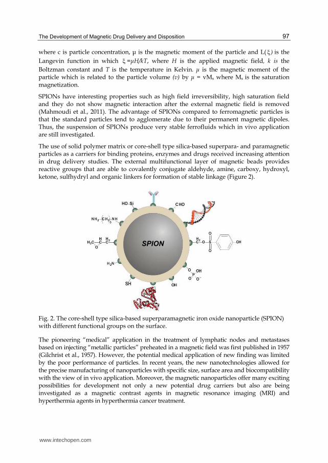
The use of solid polymer matrix or core-shell type silica-based superpara- and paramagnetic particles as a carriers for binding proteins, enzymes and drugs received increasing attention in drug delivery studies. The external multifunctional layer of magnetic beads provides reactive groups that are able to covalently conjugate aldehyde, amine, carboxy, hydroxyl, ketone, sulfhydryl and organic linkers for formation of stable linkage (Figure 2).
Fig. 2. The core-shell type silica-based superparamagnetic iron oxide nanoparticle (SPION) with different functional groups on the surface.
The pioneering “medical” application in the treatment of lymphatic nodes and metastases based on injecting “metallic particles” preheated in a magnetic field was first published in 1957 (Gilchrist et al., 1957). However, the potential medical application of new finding was limited by the poor performance of particles. In recent years, the new nanotechnologies allowed for the precise manufacturing of nanoparticles with specific size, surface area and biocompatibility with the view of in vivo application. Moreover, the magnetic nanoparticles offer many exciting possibilities for development not only a new potential drug carriers but also are being investigated as a magnetic contrast agents in magnetic resonance imaging (MRI) and hyperthermia agents in hyperthermia cancer treatment.
SPION
www.intechopen.com
The Delivery of Nanoparticles
98
2. Magnetic drug delivery system in cancer therapy
2.1 Ionic binding of the drug to the magnetic nanoparticles
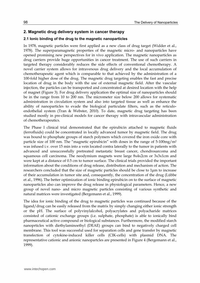
In 1978, magnetic particles were first applied as a new class of drug target (Widder et al., 1978). The superparamagnetic properties of the magnetic micro- and nanoparticles have opened promising new perspectives for in vivo application. The magnetic nanoparticles as drug carriers provide huge opportunities in cancer treatment. The use of such carriers in targeted therapy considerably reduces the side effects of conventional chemotherapy. A novel carrier system allows for intravenous drug delivery and the local accumulation of chemotherapeutic agent which is comparable to that achieved by the administration of a 100-fold higher dose of the drug. The magnetic drug targeting enables the fast and precise location of drug in the body with the use of external magnetic field. After the vascular injection, the particles can be transported and concentrated at desired location with the help of magnet (Figure 3). For drug delivery application the optimal size of nanoparticles should be in the range from 10 to 200 nm. The micrometer size below 200 allows for systematic administration in circulation system and also into targeted tissue as well as enhance the ability of nanoparticles to evade the biological particulate filters, such as the reticulo-endothelial system (Tran & Webster, 2010). To date, magnetic drug targeting has been studied mostly in pre-clinical models for cancer therapy with intravascular administration of chemotherapeutics.
The Phase I clinical trial demonstrated that the epirubicin attached to magnetic fluids (ferrofluids) could be concentrated in locally advanced tumor by magnetic field. The drug was bound to phosphate groups of starch polymers which covered the iron oxide core with
particle size of 100 nm. The “magnetic epirubicin” with doses in the range of 5-100mg/m2 was infused i.v. over 15 min into a vein located contra laterally to the tumor in patients with advanced and unsuccessfully pretreated metastatic breast cancer, chondrosarcoma and squamous cell carcinoma. The neodymium magnets were large 8x4x2cm or 3x3x1cm and
were kept at a distance of 0.5 cm to tumor surface. The clinical trials provided the important information about the conditions of drug release, distribution and mechanism of action. The researchers concluded that the size of magnetic particles should be close to 1μm to increase of their accumulation in tumor site and, consequently, the concentration of the drug (Lübbe
et al., 1996). The better optimization of ionic binding epirubicin on to the surface of magnetic nanoparticles also can improve the drug release in physiological parameters. Hence, a new group of novel nano- and micro magnetic particles consisting of various synthetic and
natural matrices were investigated (Bergemann et al., 1999).
The idea for ionic binding of the drug to magnetic particles was continued because of the
ligand/drug can be easily released from the matrix by simply changing either ionic strength
or the pH. The surface of polyvinylalcohol, polyacrylates and polyacharide matrices
consisted of cationic exchange groups (i.e. sulphate, phosphate) is able to ionically bind
pharmaceutical active compound or biological substances. Furthermore, the modified starch
nanoprticles with diethylaminoethyl (DEAE) groups can bind to negatively charged cell
membrane. This tool was successful used for separation cells and gene transfer by magnetic
transfection of cytokine-induced killer cells (CIK-cells) with plasmid DNA. The
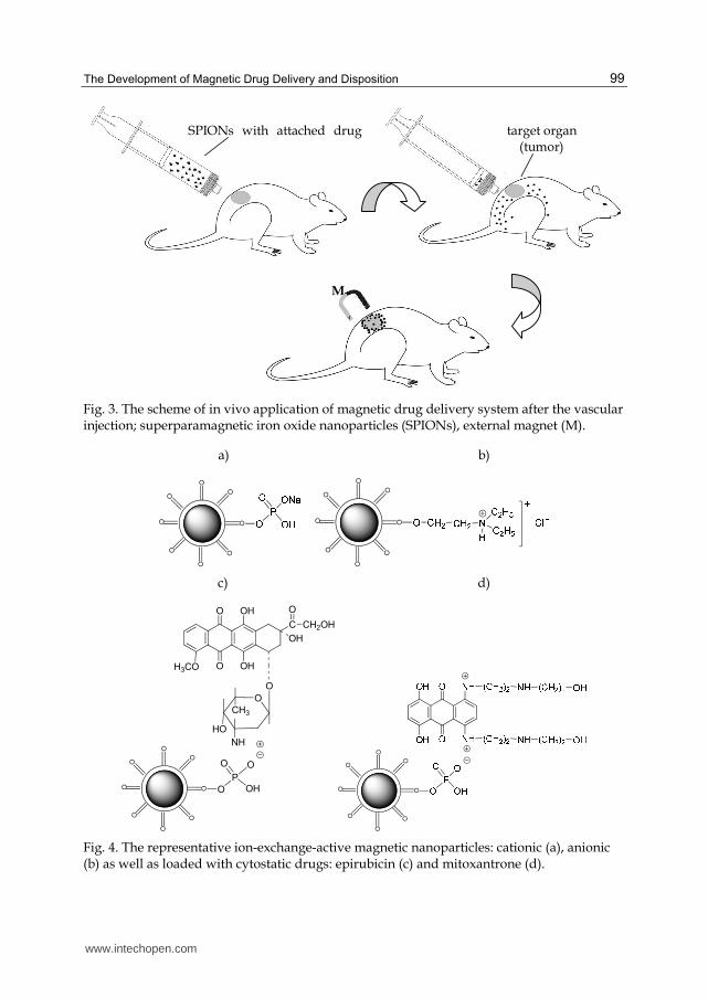
representative cationic and anionic nanopartcles are presented in Figure 4 (Bergemann et al.,
1999).
www.intechopen.com
The Development of Magnetic Drug Delivery and Disposition
99
Fig. 3. The scheme of in vivo application of magnetic drug delivery system after the vascular injection; superparamagnetic iron oxide nanoparticles (SPIONs), external magnet (M).
a) b)
c) d)
P
O O
OHO
O
H3CO O
OH
OH
C
O
CH2OH
OH
O
CH3
HO
NH
O
Fig. 4. The representative ion-exchange-active magnetic nanoparticles: cationic (a), anionic (b) as well as loaded with cytostatic drugs: epirubicin (c) and mitoxantrone (d).
www.intechopen.com
The Delivery of Nanoparticles
100
The magnetic dug targeting approach offers a new opportunity to treat malignant tumors
locoregionally. The treatment of squamous cell carcinoma in rabbits with nanoparticles
covered with modified starch to which the mitoxantrone was ionically bound, caused
complete and permanent remission of the cancer compared with control group (Alexiou et
al., 2000). The advantage of ionically bound mitoxantrone is that the anticancer agent is able
to desorb from the magnetic caries after the 30 min (half-time). Determination of time of
desorption is very important because the ferrofluids have to be transferred to the tumor
region by the magnetic field. Next, the drug must dissociate to act within the tumor.
Generally, the total release of drug from the magnetic carriers is recommended at less than
1 h. The 100 nm particles size and strong magnetic field (1.7 Tesla) are optimal for efficient
treatment of smaller animals such as mouse or rat. However, the appropriate magnetic field
strength and particle size for treatment of deep body cavities and human cancer has to be
optimised. The extensive biodistribution study with Iod123 – labeled ferrofluids
demonstrated that magnetic flux density is an important factor in magnetic drug targeting
(Alexiou et al., 2005).
2.2 Covalent binding of the drug to the magnetic nanoparticles
On the contrary to ionic binding, the covalent binding of the drug to magnetic particles
prevents unwanted drug release in a physiological environment. Hence, the new strategy is
based on the covalent coupling of antibodies, nucleic acids, proteins and active compounds
on to the surface of functional magnetic particles coated with polymer or silica. The most
popular surface groups such as amine, carboxy and aldehyde allow for a covalent conjugate
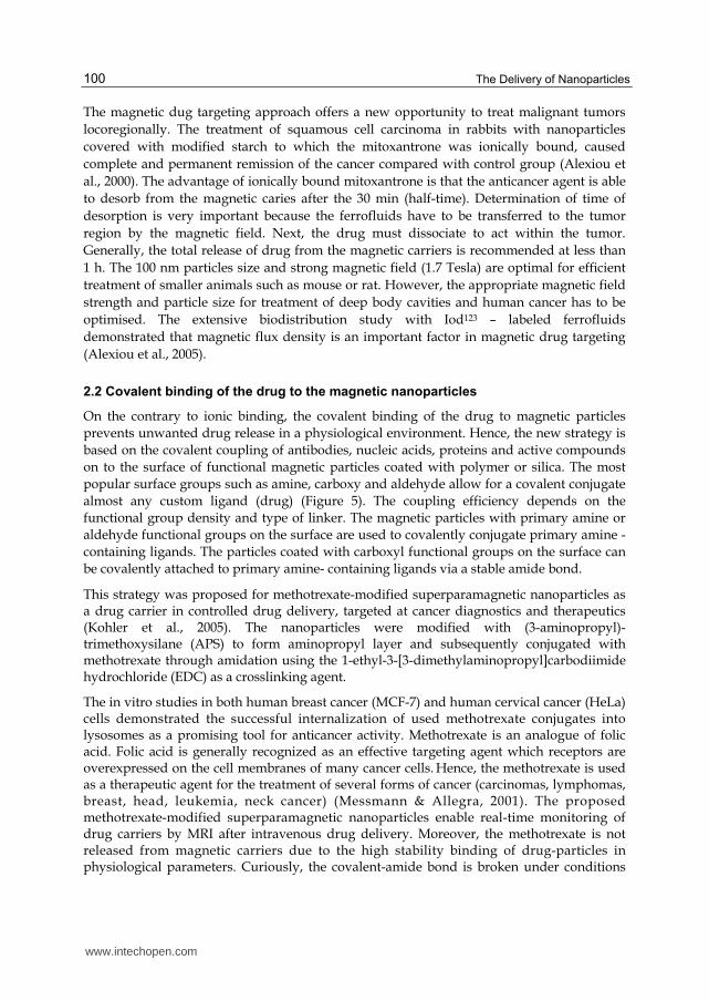
almost any custom ligand (drug) (Figure 5). The coupling efficiency depends on the
functional group density and type of linker. The magnetic particles with primary amine or
aldehyde functional groups on the surface are used to covalently conjugate primary amine -
containing ligands. The particles coated with carboxyl functional groups on the surface can
be covalently attached to primary amine- containing ligands via a stable amide bond.
This strategy was proposed for methotrexate-modified superparamagnetic nanoparticles as a drug carrier in controlled drug delivery, targeted at cancer diagnostics and therapeutics (Kohler et al., 2005). The nanoparticles were modified with (3-aminopropyl)-trimethoxysilane (APS) to form aminopropyl layer and subsequently conjugated with methotrexate through amidation using the 1-ethyl-3-[3-dimethylaminopropyl]carbodiimide hydrochloride (EDC) as a crosslinking agent.
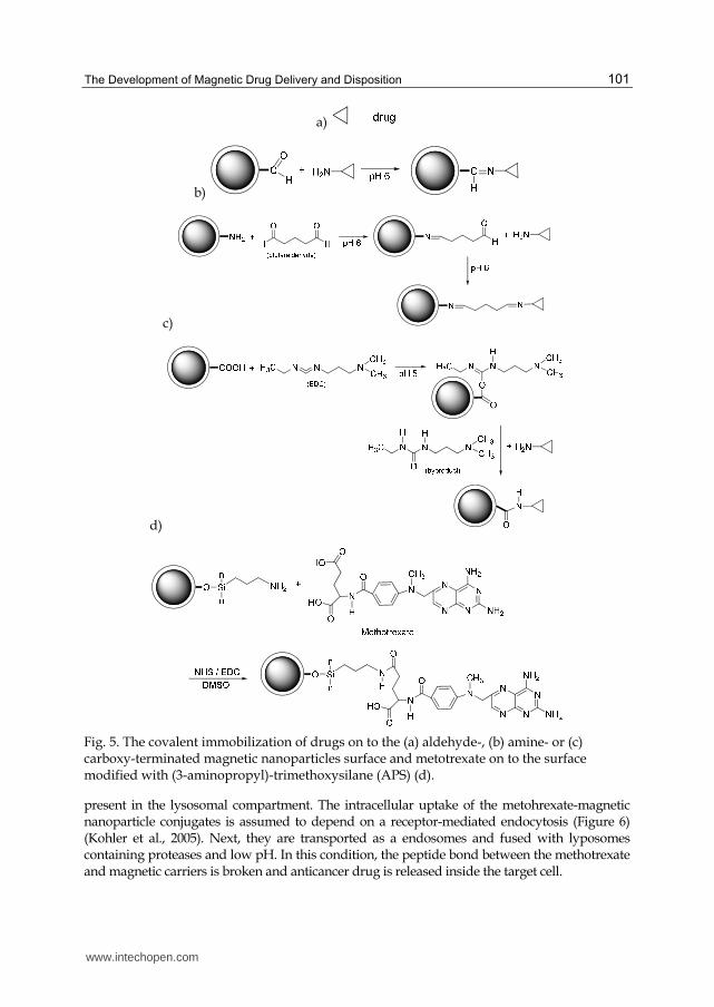
The in vitro studies in both human breast cancer (MCF-7) and human cervical cancer (HeLa) cells demonstrated the successful internalization of used methotrexate conjugates into lysosomes as a promising tool for anticancer activity. Methotrexate is an analogue of folic acid. Folic acid is generally recognized as an effective targeting agent which receptors are overexpressed on the cell membranes of many cancer cells. Hence, the methotrexate is used as a therapeutic agent for the treatment of several forms of cancer (carcinomas, lymphomas, breast, head, leukemia, neck cancer) (Messmann & Allegra, 2001). The proposed methotrexate-modified superparamagnetic nanoparticles enable real-time monitoring of drug carriers by MRI after intravenous drug delivery. Moreover, the methotrexate is not released from magnetic carriers due to the high stability binding of drug-particles in physiological parameters. Curiously, the covalent-amide bond is broken under conditions
www.intechopen.com
The Development of Magnetic Drug Delivery and Disposition
101
a)
b)
c)
d)
Fig. 5. The covalent immobilization of drugs on to the (a) aldehyde-, (b) amine- or (c) carboxy-terminated magnetic nanoparticles surface and metotrexate on to the surface modified with (3-aminopropyl)-trimethoxysilane (APS) (d).
present in the lysosomal compartment. The intracellular uptake of the metohrexate-magnetic nanoparticle conjugates is assumed to depend on a receptor-mediated endocytosis (Figure 6) (Kohler et al., 2005). Next, they are transported as a endosomes and fused with lyposomes containing proteases and low pH. In this condition, the peptide bond between the methotrexate and magnetic carriers is broken and anticancer drug is released inside the target cell.
www.intechopen.com
The Delivery of Nanoparticles
102
Fig. 6. The intracellular model of the uptake of methotrexate (MTX) – modified nanoparticles into breast cancer cells (Kohler et al., 2005). (Adapted with permission from @ 2005 American Chemical Society).
2.3 Magnetic nanoparticles for tumor imaging and therapy
Magnetic resonance imaging (MRI) is widely used as a screening non-invasive method of the human body. It is also used to monitor cell migration to targets tissue in cell-based therapy. The fact that the magnetic nanoparticles with labeled cells or as a drug targeting can be visualized using MRI, they are a new alternatives and noninvasive imaging techniques for monitoring of cell or drug migration to target tissue. For in vitro application, their advanced development in cell manipulation/therapy, biomolecule separation, selection and purification was found (Gijs, 2004). The ability to produce a distortion in magnetic field monitored by MRI, allowed for the increasing application of magnetic beads in vivo. The different strategies of the use of anionic magnetic nanoparticles (AMNPs), ultrasmall paramagnetic iron oxides (USPIOs) and superparamagnetic iron oxide nanoparticles (SPIONs) have been demonstrated as a contrast agents to identify magnetically labelled cells during MRI monitoring of cellular therapies (Wilhelm & Gazeau, 2008; Modo et al., 2005). The demonstrated studies confirm the efficacy of labelling with magnetic particles for a wide variety of mammalian cells, including non- and phagocytic cells, different species, cell size, types and culture properties. The MRI contrast is a result of different signal intensities of tissue, produced in response to applied radio frequency pulses (Gijs, 2004). Labelled-specific magnetic particles provide a suitable source of contrast and convenient tool for the non-invasive study of biological processes, such a tumor imaging and therapy with the use of MRI.
The use of magnetic particles can significantly improve hyperthermia cancer treatment (Marszałł, 2011b). This therapy involves raising the temperature of the target tissue to 43-46˚C. In this conditions its sensitivity to chemo- and radiotherapy increases and may additionally stimulate activities of the host immune system (Ang et al., 2007). The problem
www.intechopen.com
The Development of Magnetic Drug Delivery and Disposition
103
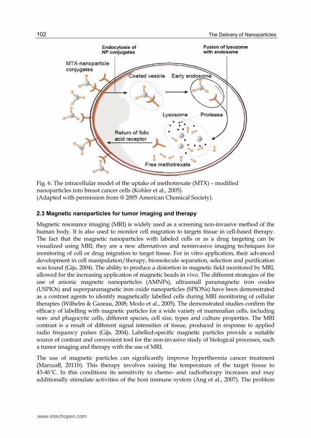
with hypethermia therapy is the heating the large area of tissue or body in general, not only the tumor region. The healthy tissues adsorb microwave, laser and ultrasound energy which can cause burns and blisters (Phillips & Johnson, 2005). Magnetic hyperthermia is one of the anti-cancer approches based on the introduction of ferro- or superparamagnetic particles into the tumor tissue. The main advantage of magnetic particle hyperthermia is that the particles can heat previously localized target issue by external magnetic field. Next, under an applied magnetic field, energy is converted to thermal energy in tumor region which destroys cancer tissues (Figure 7) (Cole et al., 2011).
Fig. 7. Scheme of magnetic hyperthermia treatment of affected tissue; (a) accumulation of magnetic nanoparticles (MNPs) by the magnet at the tumor site; (b) exposition of tumor cells to an altering current (AC) magnetic field (Cole et al., 2011). (Adapted with permission from @ 2011 Elsevier).
www.intechopen.com
The Delivery of Nanoparticles
104
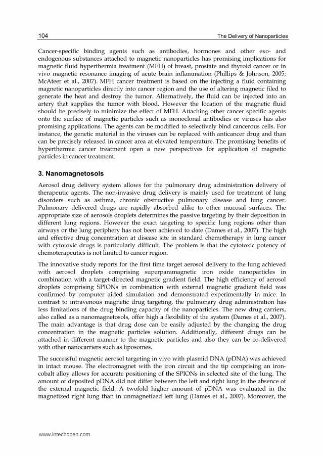
Cancer-specific binding agents such as antibodies, hormones and other exo- and endogenous substances attached to magnetic nanoparticles has promising implications for magnetic fluid hyperthermia treatment (MFH) of breast, prostate and thyroid cancer or in vivo magnetic resonance imaging of acute brain inflammation (Phillips & Johnson, 2005; McAteer et al., 2007). MFH cancer treatment is based on the injecting a fluid containing magnetic nanoparticles directly into cancer region and the use of altering magnetic filed to generate the heat and destroy the tumor. Alternatively, the fluid can be injected into an artery that supplies the tumor with blood. However the location of the magnetic fluid should be precisely to minimize the effect of MFH. Attaching other cancer specific agents onto the surface of magnetic particles such as monoclonal antibodies or viruses has also promising applications. The agents can be modified to selectively bind cancerous cells. For instance, the genetic material in the viruses can be replaced with anticancer drug and than can be precisely released in cancer area at elevated temperature. The promising benefits of hyperthermia cancer treatment open a new perspectives for application of magnetic particles in cancer treatment.
3. Nanomagnetosols
Aerosol drug delivery system allows for the pulmonary drug administration delivery of therapeutic agents. The non-invasive drug delivery is mainly used for treatment of lung disorders such as asthma, chronic obstructive pulmonary disease and lung cancer. Pulmonary delivered drugs are rapidly absorbed alike to other mucosal surfaces. The appropriate size of aerosols droplets determines the passive targeting by their deposition in different lung regions. However the exact targeting to specific lung regions other than airways or the lung periphery has not been achieved to date (Dames et al., 2007). The high and effective drug concentration at disease site in standard chemotherapy in lung cancer with cytotoxic drugs is particularly difficult. The problem is that the cytotoxic potency of chemoterapeutics is not limited to cancer region.
The innovative study reports for the first time target aerosol delivery to the lung achieved with aerosol droplets comprising superparamagnetic iron oxide nanoparticles in combination with a target-directed magnetic gradient field. The high efficiency of aerosol droplets comprising SPIONs in combination with external magnetic gradient field was confirmed by computer aided simulation and demonstrated experimentally in mice. In contrast to intravenous magnetic drug targeting, the pulmonary drug administration has less limitations of the drug binding capacity of the nanoparticles. The new drug carriers, also called as a nanomagnetosols, offer high a flexibility of the system (Dames et al., 2007). The main advantage is that drug dose can be easily adjusted by the changing the drug concentration in the magnetic particles solution. Additionally, different drugs can be attached in different manner to the magnetic particles and also they can be co-delivered with other nanocarriers such as liposomes.
The successful magnetic aerosol targeting in vivo with plasmid DNA (pDNA) was achieved in intact mouse. The electromagnet with the iron circuit and the tip comprising an iron-cobalt alloy allows for accurate positioning of the SPIONs in selected site of the lung. The amount of deposited pDNA did not differ between the left and right lung in the absence of the external magnetic field. A twofold higher amount of pDNA was evaluated in the magnetized right lung than in unmagnetized left lung (Dames et al., 2007). Moreover, the
www.intechopen.com
The Development of Magnetic Drug Delivery and Disposition
105
authors conclude that the nanomagnetosol droplets with SPIONs are responsible for their transport, deposition the accumulation of the attached ligand/drug in affected tissue and not the single SPION as was supposed previously.
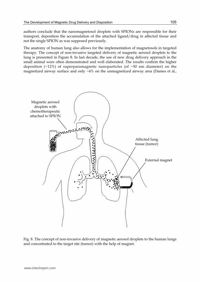
The anatomy of human lung also allows for the implementation of magnetosols in targeted therapy. The concept of non-invasive targeted delivery of magnetic aerosol droplets to the lung is presented in Figure 8. In last decade, the use of new drug delivery approach in the small animal were often demonstrated and well elaborated. The results confirm the higher deposition (~12%) of superparamagnetic nanoparticles (of ~50 nm diameter) on the magnetized airway surface and only ~4% on the unmagnetized airway area (Dames et al.,
Fig. 8. The concept of non-invasive delivery of magnetic aerosol droplets to the human lungs and concentrated to the target site (tumor) with the help of magnet.
External magnet
Magnetic aerosol droplets with
chemotherapeutic attached to SPION
Affected lung tissue (tumor)
www.intechopen.com
The Delivery of Nanoparticles
106
2007). Crucially, the non-deposited aerosol droplets can be transferred to further lung zones
or exhaled. Regardless of the advantages for aerosol droplets with magnetic particles, the
objective risk assessment studies have to be performed. Many of studies confirmed that the
metal oxide particles are very toxic, especially in the pulmonary system (Machado et al.,
2010). Nowadays, the in vitro studies are focused on the characterization and cytotoxic
assessment of aerosol particulates, especially the nanoparticles based on heavy alloys W-Fe-
Ni and W-Fe-Co. The exposure to Co, Ni, W and Fe can cause pulmonary fibrosis asthma,
oedema and pneumonia among other effects.
The cytotoxic assessment studies of aerosol particulates are usually performed with the use
of model for lung tissue – human epithelial cells (A549). The use of different filter exposure
cytotoxicity assays allow to assess the inflammatory and related respiaratory health effects.
Generally, the inhaled ~1μm particulates are moved by cilia of the bronchial epithelial cells
towards the upper respiratory tract. The nanoparticles at size of <100nm can accumulate,
aggregate and even cause oxidative stress and inflammation. So far it is proved that the
most important parameters that can affect cytotoxicity are chemical composition, size and
time of deposition of magnetic nanoparticles. Because of a lack of detailed limitations
regarding the in vivo use of nanomagnetosols, it is necessary to continue the risk assessment
studies and establish the well-defined regulations.
4. Magnetic drug delivery system in antimicrobial and antiviral therapy
The magnetic nanoparticles are also promising carries for antibacterial agents such copper
and silver and can provide an alternative treatment for bacterial infections. As known, silver
is distinguished by extraordinary inhibitory and bactericidal properties for a broad
spectrum of bacterial strains. Antibacterial coatings based on hydrogen bonded multilayer
containing in situ synthesized silver magnetic nanoparticles can be delivered to a specific
region to localize a high concentration of antibacterial silver while maintaining a low
concentration in general (Lee et al., 2005). The silver multilayer assembled on spherical
support such as magnetic micro- or nanospheres were localized and focused by external
magnetic field and showed excellent antibacterial properties against the Gram-positive
strain Staphylococcus epidermidis and the Gram-negative strain Escherichia coli. It is expected
that the modification of silver particles multilayer by the increase of surface-volume ratios
can improve the antibacterial treatment (Tran & Webster, 2010).
The other studies demonstrates that the magnetic formulation of 3’-azido-3’-
deoxythymidine-5’-triphosphate (AZTTP) is a new potential drug carries for targeted
delivery across the blood brain barrier (Saiyed et al., 2009, 2010). Nucleoside and nucleotide
analog reverse transcriptase inhibitors (NRTIs) are important part of the antiretroviral
therapy (ART). Their inefficient cellular phosphorylation cause a limitation for treatment of
human immunodeficiency virus (HIV). Main assumption of the in vitro study is that active
NRTIs can directly bind to magnetic nanoparticles by ionic interaction and can inhibit HIV-1
replication. The development of magnetic AZTTP liposomal nanoformulation (150 nm)
allows to cross blood brain barrier model by direct transport or via monocyte-mediated
transport by external magnetic field. Hence, the magnetic carriers give a new opportunity to
for treatment of neurological disorders.
www.intechopen.com

The Development of Magnetic Drug Delivery and Disposition
107
5. Conclusion
The micro- and nanoparticle technology is highly novel and offers many possibilities for
future development of new drug delivery systems. The innovative methods for drug
targeting and delivery based on the micro- and nano-sized magnetic support provide a
numerous advantages compared to conventional drug delivery systems. Presently, only
molecular targeting ligands coupled to magnetic particles are successfully used and
commercial available as contrast agents in MRI. Although the magnetic particles possess
unique and useful properties for biomedical and pharmaceutical applications, they also
carry potential health risks. The small size and high surface area to volume ratio of magnetic
nanoparticles may have important implications due to higher biological activity per given
mass compared to larger particulate forms (Helland et al., 2008). That may cause their
remarkable activity and toxicity effect. Therefore, the material used for surface coating of the
magnetic particles for in vivo application must not only be nontoxic but also biocompatible.
So far there are no criteria and tests for evaluation of toxicity level, parameters of release as
well as uptake magnetically targeted drugs. Regarding the in vivo application, it will have
to be proved, that the new drug carriers possess not only useful properties but also are quit
safe for environment and human health. Hence, the Food and Drug Administration issued a
report to consider developing guidance for regulation of nanotechnology products and their
adaptation from science to biomedical application.
6. Acknowledgment
Thanks are due to Mr. Tomasz Siódmiak, Mr. Wiktor Sroka and Ms. Beata Kochanek for technical assistance in preparing this manuscript.
7. References
Alexiou, Ch.; Arnold, W.; Klein, R.J. et al. (2000). Locoregional Cancer Treatment with
Magnetic Drug Targeting. Cancer Research, Vol.60, (December 2000), pp. 6641-6648,
ISSN 1538-7445.
Alexiou, Ch.; Jurgons, R.; Schmid, R.; Hilpert, A.; Bergemann, Ch.; Parak, F.; Iro, H. (2005).
In vitro and in vivo investigations of targeted chemotherapy with magnetic
nanoparticles. Journal of Magnetism and Magnetic Materials, Vol.293, (March 2005),
pp. 389-393, ISSN 0304-8853
Ang, K.L; Venkatraman, S; Ramanujan, R.V. (2007). Magnetic PNIPA Hydrogels for
Hyperthermia Applications in Cancer Therapy. Materials Science and Engineering C.
Vol.27, No.3 (April 2007), pp. 347–351, ISSN 0928-4931
Arruebo, M.; Fernández-Pacheco, R.; Ibarra, M. R. & Santamaría. J. (2007). Magnetic
nanoparticles for drug delivery. Nanotoday, Vol.2, No.3, (June 2007), pp. 22-32, ISSN
1748-0132
Bergemann, C.; Müller-Schulte, D.; Oster, J.; Brassard, L.; Lübbe, A.S. (1999). Magnetic ion-
exchange nano- and microparticles for medical, biochemical and molecular
biological applications. Journal of Magnetism and Magnetic Materials, Vol.194, (April
1999), pp.45-52, ISSN 0304-8853
www.intechopen.com
The Delivery of Nanoparticles
108
Cole, A.J., Yang, V.C., David, A.E. (2011). Cancer theranostics: the rise of targeted magnetic
nanoparticles. Trends in Biotechnology, Vol.29, No.7, (July 2011) pp. 323-332, ISSN
0167-7799
Corchero, J.L. & Villaverde, A. (2009). Biomedical application of distally controlled magnetic
nanoparticles. Trends in Biotechnology, Vol.27, No.8, (June 2009) pp. 468-476, ISSN
0167-7799
Dames, P.; Gleich, B.; Flemmer, A. et al. (2007). Targeted Delivery of Magnetic Aerosol
Droplets to the Lung. Nature Nanotechnology. Vol.2, No.8 (July 2007), pp. 495–499,
ISSN 1748-3395
Douziech-Eyrolles, L.; Marchais, H.; Hervé, K.; Munnier, E. & Soucé, M. (2007). Nanovectors
for anticancer agents based on superparamagnetic iron oxide nanoparticles.
International Journal of Nanomedicine, Vol.2, No.4, pp.541-550, ISSN 1178-2013
Gijs, M.A.M. (2004).Magnetic Beads Handling on-chip: New Opportunities for Analytical
Applications. Macrofluid Nanofluid Vol.1, No.1 (November 2004), pp. 22-40, ISNN
1613-4982
Gilchrist, R.D.; Medal, R.; Shorey, W.D.; Hanselman, R.C.; Parrott, J.C. & Taylor, C.B. (1957).
Selective inductive heating of lymph nodes. Annals of Surgery. Vol.146, No.4,
(October 1957), pp. 596-606, ISSN 1528-1140
Helland, A.; Scheringer, M.; Siegrist, M.; Kastenholz, H.G.; Wiek, A.; Scholz, R.W. (2008).
Risk Assessment of Engineered Nanomaterials: A Survey of Industrial Approaches.
Environmental Science & Technology, Vol.42, No.2 (January 2008), pp. 640-646, ISSN
1520-5851
Ilium, L.; Davis, S.; Wilson, C.; Thomas, N.; Frier, M. & Hardy, J. (1982). Blood clearance and
organ deposition of intravenously administered colloidal particles. The effects of
particle size, nature and shape. International Journal of Pharmaceutics, Vol.12, No 2–3,
(October 1992) pp. 135–146, ISSN 0378-5173
Kohler, N.; Sun, C.; Wang, J.; Zhang, M. (2005). Methotrexate-modified superparamagnetic
nanoparticles and their intracellular uptake into human cancer cells. Langmuir,
Vol.21, No.19 (June 2005), pp. 8858–8864, ISSN 1520-5827
Lee, D.; Cohen, R.E.; Rubner, M.F. (2005). Antibacterial Properties of Ag Nanoparticle
Loaded Multilayers and Formation of Magnetically Directed Antibacterial
Microparticles, Langmuir Vol.21, No.21 (October 2005), pp. 9651-9659, ISSN 1520-
5827
Lübbe, A.S.; Bergemann, Ch.; Riess, H. et al. (1996). Preclinical experiences with magnetic
drug targeting: tolerance and efficacy. Cancer Research, Vol.56, (October 1996), pp.
4686-4693, ISNN 1538-7445
Mahmoudi, M.; Sant, S.; Wang, B.; Laurent, S. & Sen, T. (2011). Superparamagnetic iron
oxide nanoparticles (SPIONs): Development, surfach modification and applications
in chemotherapy. Advanced Drug Delivery Reviews, Vol.63, pp. 24–46, ISSN 0169-
409X
Machado, B.I.; Murr, L.E.; Suro, R.M.; Gaytan, S.M.; Ramirez, D.A.; Garza, K.M.; Schuster,
B.E. (2010). Characterization and Cytotoxic Assessment of Ballistic Aerosol
Particulates for Tungsten Alloy Penetrators into Steel Target Plates. International
www.intechopen.com
The Development of Magnetic Drug Delivery and Disposition
109
Journal of Environmental Research and Public Health. Vol.7, No.9 (September 2010), pp.
3313-3331, ISSN 1660-4601
Marszałł, M.P.; Moaddel, R.; Kole, S.; Gandhari, M.; Bernier, M. & Wainer, I.W. (2008).
Ligand and protein fishing with heat shock protein 90 coated magnetic beads.
Analytical Chemistry,Vol.80, No.19, (October 2008), pp. 7571-7575, ISSN 0003-
2700
Marszałł, M.P.; Buciński, A.; Kruszewski, S.; Ziomkowska B. (2011a). A New Approach to
Determine Camptothecin and Its Analogues Affinity to Human Serum Albumin.
Journal of Pharmaceutical Science,, Vol.100, No.3, (March 2011) pp. 1142-1146, ISSN
1520-6017
Marszałł, M.P. (2011b). Application of Magnetic Particles in Pharmaceutical Sciences.
Pharmaceutical Research, Vol.28, No.3 (March 2005), pp. 480–483, ISSN 1573-904X
McAteer, M.A.; Sibson, N.R.; von zur Muhlen, C.; Schneider, J.E.; Lowe, A.S.; Warrick, N.;
Channon, K.M.; Anthony, D.C.; Choudhury R.P. (2007). In vivo magnetic resonance
imaging of acute brain inflammation using microparticles of iron oxide. Nature
Medicine.Vol.13, No.10 (October 2007), pp. 1253–1258, ISSN 1078-8956
Messmann, R.& Allegra, C. (2001) Antifolates. In: Cancer Chemotherapy& Biotherapy, Chabner,
B., Longo, D. (3 ed.), ISSN 0036-8075, 139-184, Eds.; Lippincott Williams & Wilkins,
Philadelphia
Modo, M; Hoehn, M; Bulte, J.W. (2005). Cellular MR Imaging. Molecular Imaging. Vol.4, No.3
(August 2005), pp. 143–164, ISSN 1535-3508Phillips, J. & Johnson, D.T. (2005).
Magnetic Fluid Hyperthermia: A Topical Review, The Journal of Science and Health at
The University of Alabama.Vol.3, (August 2005), pp. 14–18
Saiyed, Z.M.; Gandhi, N.H.; Nair, M.P.N. (2009). AZT 5’-triphosphate nanoformulation
suppresses HIV-1 replication in peripheral blood mononuclearcells. Journal of
Neurovirology. Vol.15, No.4 (July 2009), pp. 343-347, ISSN 1355-0284
Saiyed, Z.M.; Gandhi, N.H.; Nair, M.P.N. (2010). Magnetic nanoformulation of
azidothymidine 5’-triphosphate for targeted delivery across the blood–brain
barrier. International Journal of Nanomedicine Vol.5 (March 2010), pp. 157-166, ISSN
1178-2013
Tran, N. & Webster, T.J. (2010). Magnetic nanoparticles: biomedical applications and
challenges. Journal of Materials Chemistry, Vol.20, No.40 2010, pp. 8760–8767,
ISNN 0959-9428
Widder, K.J; Senyei, A.E. & Scarpelli, D.G. (1978). Magnetic microspheres: a model system of
site specific drug delivery in vivo. Proceedings of the Society for Experimental Biology
and Medicine, Vol.158, No.2 (June 1978) pp. 141–146, ISSN 0037-9727
Wilhelm, C.; Gazeau, F. (2008) Universal Cell Labelling with Anionic Magnetic
Nanoparticles. Biomaterials, Vol.29, No.22 (August 2008), pp. 3161–3174, ISSN 0142-
9612
Woodward, R.C.; Heeris, J.; Pierre, T.G.St.; Saunders, M.; Gilbert, E.P.; Rutnakornipituk, M.;
Zhang. Q.; Riffle J.S. (2007). A comparison of methods for the measurement of the
particle-size distribution of magnetic nanoparticles. Journal of Applied
Crystallography, Vol.40, (January 2007) ISSN 0021-8898
www.intechopen.com
The Delivery of Nanoparticles
110
Yoo, J-W.; Doshi, N.& Mitragotri, S. (2011) Adaptive micro and nanoparticles: Temporal
control over carrier properties to facilitate drug delivery, Advanced Drug Delivery
Reviews, in press, doi:10.1016/j.addr.2011.05.004, ISNN 0169-409X
www.intechopen.com
The Delivery of NanoparticlesEdited by Dr. Abbass A. Hashim
ISBN 978-953-51-0615-9Hard cover, 540 pagesPublisher InTechPublished online 16, May, 2012Published in print edition May, 2012
InTech EuropeUniversity Campus STeP Ri Slavka Krautzeka 83/A 51000 Rijeka, Croatia Phone: +385 (51) 770 447 Fax: +385 (51) 686 166www.intechopen.com
InTech ChinaUnit 405, Office Block, Hotel Equatorial Shanghai No.65, Yan An Road (West), Shanghai, 200040, China
Phone: +86-21-62489820 Fax: +86-21-62489821
Nanoparticle is a general challenge for today's technology and the near future observations of science.Nanoparticles cover mostly all types of sciences and manufacturing technologies. The properties of thisparticle are flying over today scientific barriers and have passed the limitations of conventional sciences. Thisis the reason why nanoparticles have been evaluated for the use in many fields. InTech publisher and thecontributing authors of this book in nanoparticles are all overconfident to invite all scientists to read this newbook. The book's potential was held until it was approached by the art of exploring the most advancedresearch in the field of nano-scale particles, preparation techniques and the way of reaching their destination.25 reputable chapters were framed in this book and there were alienated into four altered sections; ToxicNanoparticles, Drug Nanoparticles, Biological Activities and Nano-Technology.
How to referenceIn order to correctly reference this scholarly work, feel free to copy and paste the following:
Michal Piotr Marszall (2012). The Development of Magnetic Drug Delivery and Disposition, The Delivery ofNanoparticles, Dr. Abbass A. Hashim (Ed.), ISBN: 978-953-51-0615-9, InTech, Available from:http://www.intechopen.com/books/the-delivery-of-nanoparticles/the-development-of-magnetic-drug-delivery-and-disposition