Embed Size (px)
Citation preview

DEVELO
PMENT
3763DEVELOPMENT AND DISEASE RESEARCH ARTICLE
INTRODUCTIONA crucial feature in embryonic development is the assembly ofindependently formed organs into complex systems that conductsubstances such as food, air and waste into and out of the embryo.The organs that comprise the upper (kidney and ureter) and lower(bladder and urethra) urinary tract form independently, connectingat mid-gestation to form an outflow tract that conducts urine fromthe kidneys to the bladder for storage and excretion. The kidneys,ureters and Wolffian ducts, paired epithelial tubes that form mostof the male genital tract, are largely derived from intermediatemesoderm, a strip of tissue lying between the lateral plate and theparaxial mesoderm. Wolffian ducts open into the cloaca, whichdifferentiates into the urogenital sinus, the primordium of thebladder and urethra. The ureteric bud, which will give rise to therenal collecting duct system and extra-renal ureter, forms as acaudal sprout from the Wolffian duct that invades the metanephricblastema and undergoes successive rounds of branchingmorphogenesis in response to signals from the metanephricmesenchyme. The portion of the ureteric bud lying outside thekidney differentiates into the ureters, which are muscular tubes thatmediate myogenic peristalsis, propelling urine from the renal pelvisto the bladder.
The upper and lower urinary tract compartments join when theureters undergo transposition, moving from their primary insertionsite in the Wolffian ducts to the urogenital sinus epithelium, where
they make final connections in a triangular structure, known as thetrigone, situated between the bladder and urethra (Fig. 1). Ourprevious studies suggest that formation of these final connectionsinvolves apoptosis, which enables the ureters to disconnect from theWolffian ducts, and fusion, in which the ureter orifice inserts into theurogenital sinus epithelium at the level of the trigone (Batourina etal., 2005). Precise connections between ureters and the trigone arecrucial for function of the valve mechanism that prevents back flowof urine from the bladder to the ureters, a major cause of reflux andobstruction, which can damage the kidney and cause severe healthproblems including end-stage renal disease.
Despite its central importance in urinary tract function, the originand role of the trigone in the anti-reflux mechanism remainscontroversial. Analysis of human and animal specimens has led to thesuggestion that the trigone is structurally distinct from the bladder andurethra, differentiating from the common nephric duct and ureter(Hutch, 1972; Tanagho, 1981; Weiss, 1988; Wesson, 1925). Otherstudies suggest that the bladder muscle (detrusor) might also be partof the trigone structure (Meyer, 1946). Hence, a number of questionsremain: what is the derivation of the trigone, how is the anti-refluxmechanism established, and how do positional abnormalities of theureteric bud translate into reflux and obstruction? To begin to addressthese questions, we used mouse models to study the structure of thetrigone and to determine which lineages contribute to its formation.We find, unexpectedly, that the trigone derives largely from bladdermuscle and that ureteral fibers are an important contributor to trigonestructure. A number of studies also suggest that the ureteral pathwaythrough the bladder is formed by a sheath of ureteral muscle(Waldeyer, 1892) (reviewed by Hutch, 1972). We find, paradoxically,that the ureteral pathway is present in the bladder wall and formsindependently of the ureter. These studies elucidate importantmechanisms controlling urinary tract assembly that are also importantfor formation of the ureteral valve that is crucial for preventing refluxand preserving renal function.
The development of the bladder trigone, the center of theanti-reflux mechanismRenata Viana1, Ekatherina Batourina1, Hongying Huang2, Gregory R. Dressler3, Akio Kobayashi4, Richard R. Behringer4, Ellen Shapiro2, Terry Hensle1, Sarah Lambert1 and Cathy Mendelsohn1,*
The urinary tract is an outflow system that conducts urine from the kidneys to the bladder via the ureters that propel urine to thebladder via peristalsis. Once in the bladder, the ureteral valve, a mechanism that is not well understood, prevents backflow ofurine to the kidney that can cause severe damage and induce end-stage renal disease. The upper and lower urinary tractcompartments form independently, connecting at mid-gestation when the ureters move from their primary insertion site in theWolffian ducts to the trigone, a muscular structure comprising the bladder floor just above the urethra. Precise connectionsbetween the ureters and the trigone are crucial for proper function of the ureteral valve mechanism; however, the developmentalevents underlying these connections and trigone formation are not well understood. According to established models, the trigonedevelops independently of the bladder, from the ureters, Wolffian ducts or a combination of both; however, these models havenot been tested experimentally. Using the Cre-lox recombination system in lineage studies in mice, we find, unexpectedly, that thetrigone is formed mostly from bladder smooth muscle with a more minor contribution from the ureter, and that trigoneformation depends at least in part on intercalation of ureteral and bladder muscle. These studies suggest that urinary tractdevelopment occurs differently than previously thought, providing new insights into the mechanisms underlying normal andabnormal development.
KEY WORDS: Bladder, Reflux, Trigone, Ureter, Urinary tract formation, Mouse, Human
Development 134, 3763-3769 (2007) doi:10.1242/dev.011270
1Columbia University, Department of Urology, 650 West 168th Street, New York, NY10032, USA. 2Department of Urology, New York University School of Medicine NewYork, NY, USA. 3Department of Pathology, University of Michigan, MSRB1, BSRB2049, 109 Zina Pitcher Dr, Ann Arbor, MI 481093, USA. 4Department of MolecularGenetics, University of Texas M. D. Anderson Cancer Center, Houston, TX 77030,USA.
*Author for correspondence (e-mail: [email protected])
Accepted 27 July 2007
Development ePress online publication date 19 September 2007http://dev.biologists.org/lookup/doi/10.1242/dev.011270Access the most recent version at First posted online on 19 September 2007 as 10.1242/dev.011270
DEVELO
PMENT
3764
MATERIALS AND METHODSImmunostainingFor cryosections (10 �m), tissue was fixed in 4% paraformaldehyde (PFA)for 1-3 hours at 4°C and embedded in OCT compound. For vibratomesections (100-150 �m), tissue was fixed overnight in 4% PFA, washed inPBS and then embedded in 3% agarose. Sections were then permeabilizedwith 0.3% hydrogen peroxide in cold methanol for 20 minutes, washed inPBS/0.1% Triton X-100 for 30 minutes then processed for immunostaining.For double staining with uroplakin and smooth muscle alpha actin, sampleswere incubated in blocking solution (2% horse serum in washing buffer)then primary uroplakin antibody, a marker of urothelial terminaldifferentiation (Wu et al., 1994). UP3 antibody (clone #744) was a gift of DrT. T. Sun (New York University, NY) was applied overnight at 4°C. Afterwashing, the secondary antibody (donkey anti-rabbit IgG) was applied for 2hours at room temperature. After washing and reblocking, the tissue wasincubated in (ASMA)FITC- or Cy3-conjugated antibodies (Sigma)overnight at 4°C then washed and mounted.
Human tissuesWith approval from the New York University Institutional Board ofResearch Associates, lower urinary tracts were removed from four humanfetuses ranging in gestational age from 19 to 22 weeks. Informed consentwas obtained by the consulting obstetrician. The gestational ages wereestimated from date of last menstrual period as well as from sonographicmeasurements of crown rump and foot length. Specimens were formalin-fixed, paraffin-embedded and serially sectioned at 4 �m.
Immunohistochemistry for smooth muscle actinRepresentative tissue sections were deparaffinized and rehydrated.Endogenous peroxidase activity was blocked with 3% hydrogen peroxidefor 5 minutes. Antigen retrieval was performed by incubating paraffinsections with antigen unmasking solution (Vector Labs #H-3300) andmicrowave treatment (900 W) for 20 minutes, followed by blocking with10% normal goat serum. Mouse monoclonal antibody (M0851, Dako,Carpinteria, CA) was used to detect the human smooth muscle actin. Afterovernight incubation at 4°C with anti-smooth muscle actin, a biotinylatedgoat anti-mouse secondary antibody was applied. Slides were then treatedwith avidin-biotinylated peroxidase complex and developed in a solutioncontaining 3,3�-diaminobenzidine (DAB). All sections werecounterstained with Hematoxylin, dehydrated, mounted and observed bylight microscopy
X-Gal histochemistryTo reveal lacZ expression, vibratome or cryostat sections were fixed incold 2% PFA in PBS for 5 minutes at 4°C, washed in PBS, and stained inX-Gal solution for 2-5 hours at 37°C (5 mM potassium ferricyanide, 5mM potassium ferrocyanide, 2 mM magnesium chloride in PBS and 1.2mg/ml X-Gal in dimethyl sulfate). After staining, samples were washed2-3 times with PBS, post-fixed with 4% PFA and stored at 4°C in 80%glycerol.
Animals and genotypingFor timed matings, males and females were placed in a cage together at16.00-17.00 h, and the morning when the vaginal plug was visualized wastaken to be E0.5. Hoxb7-Gfp mice (Srinivas et al., 1999) were a kind giftfrom Dr Frank Costantini (Columbia University, New York, NY).Genotyping was with PCR using primers: 5�-AGCGCGATCACATGGTC -CTG-3� and 5�-ACGATCCTGAGACTTCCACACT-3�. Pax2 mutant micewere genotyped using the following three primers: Pax2F, 5�-CCCAC -CGTCCCTTCCTTTTCTCCTCA-3�; Pax2R, 5�-GAAAGGCCAGTGTG -GCCTCTAGGGTG-3�; and PGK, 5�-AGACTGCCTTGGGAAAAGCGC-3�. Sm22-Cre mice (Holtwick et al., 2002) were obtained from the JacksonLaboratory and genotyped by PCR using: 5�-CAGACACCGA AGCT -ACTCTCCTTCC-3� and 5�-CGCATAACCAGTGAA ACAG CATTGC-3�.Rosa26 lacZ mice (Soriano, 1999) were also obtained from the JacksonLaboratory and genotyped using: 5�-AAAGTCG CTCTGAGTTGTTAT-3�,5�-GCGAAGAGTTTGTCCTCAACC-3� and 5�-GGAGCGGGAGAAA -TGG ATATG-3�. Rarb2-Cre mice were genotyped as described (Kobayashiet al., 2005).
RESULTSIn newborn mouse urogenital tracts, the bladder is encircled by athick layer of muscle called the detrusor and the ureters enter thetrigone at the base of the bladder between the bladder and urethra(Fig. 1A,C,D). The trigone can be visualized in dissectedurogenital tracts as a smooth triangular shaped region bounded bythe ureters laterally, terminating at the bladder neck where theurethra begins (Fig. 1D,E). The surface of the urethra and ureters,like the bladder, is covered by the urothelium, a specializedtransitional epithelium that prevents leakage and damage (Fig.1D, the urothelium is red). The intramural ureters pass through thebladder muscle and submucosa and open into the trigone at itslateral edges (Fig. 1E). Higher magnification reveals the eyelet-shaped ureter orifice opening into the urothelium (Fig. 1F). Unlikethe bladder, which is covered by folds, the trigone is generallysmooth, which has led to the suggestion that its origin might bedistinct from the bladder.
Development of the trigoneThe trigone has been defined in a number of ways; here, we willconsider the trigone to be the muscular triangle bounded laterallyby the ureter orifices extending posteriorly to the urethra (Fig.1C). The unique features of the trigone including its appearanceand physiological properties have led to the idea that the trigoneoriginates from non-urogenital sinus tissue, in particular from thecommon nephric duct that is the caudal-most segment of Wolffianduct. However, our previous studies suggest that this is not thecase because the common nephric duct undergoes apoptosisduring ureter transposition, hence the trigone is likely to form ina different manner than previously thought. Other studies suggestthat the trigone is formed in large part from ureteral fibers that fanout laterally forming an inter-ureteric ridge and posteriorlyforming Bell’s muscle (Fig. 1C). To begin to address this questionwe first established which muscles are present in the trigone byanalyzing its formation in mouse urogenital tracts at differentdevelopmental and post-natal stages. At E15, analysis forexpression of smooth muscle alpha actin revealed extensivesmooth muscle differentiation (green) in the bladder, urethra andin the extra-vesicular ureters (the portion of ureter outside thebladder), but there was little if any detectable smooth musclelining the intramural ureter (the portion of the ureter within thebladder) in the trigonal region (Fig. 2A).
Analysis of urogenital tracts at P0 revealed a thick smooth musclecoat surrounding the extra-vesicular ureter and a few longitudinalfibers surrounding the intramural ureter extending through thedetrusor and submucosa (Fig. 2B,E,F). Analysis at adult stagesrevealed additional smooth muscle lining the intramural ureter. Thetrigone appeared at this stage to be a hybrid between the bladder andurethra. Its surface was smooth and free of folds like the urethra wascovered by a thick muscularis submucosa, similar to that in thebladder (Fig. 2C,D,G,H). The ureteral wall outside the bladder isthick, containing at least three layers of circular and longitudinalmuscle (Fig. 2E). However, as reported by other groups (Yucel andBaskin, 2004), only a small subset of longitudinal ureteral fibersextend into the intramural region, where they appear to intercalatewith the bladder muscle and terminate in the submucosa, below theurothelium (Fig. 2F,G). These findings suggest that two majormuscle types are present in the trigone: the bladder muscle (detrusor)and the muscle associated with the intramural ureter. Extensiveanalysis of whole-mount urogenital tracts, cryosections andvibratome sections did not reveal additional muscle groups reportedto be part of the trigone, including an intra-ureteric bar which is said
RESEARCH ARTICLE Development 134 (20)

DEVELO
PMENT
to extend laterally between the two ureter orifices, and Bell’s musclewhich is said to extend caudally from the ureter orifices to thetrigone apex (Tanagho et al., 1968).
The trigone is evolutionarily conservedThe failure to identify structures in the mouse thought to beassociated with the trigone suggests that either the trigone is formeddifferently than previously thought, or that there are substantialdifferences in the structure of the mouse and human trigone. Toaddress this question, we compared the trigone in human and mouse.Sections through the trigone of a 22-week human fetus stained forsmooth muscle alpha actin revealed the ureter passing through thebladder muscle and into the submucosa (Fig. 3A). The morphologyof the bladder muscle, which is organized in bundles, was seen to bedistinct from the thin longitudinal smooth muscle fibers that surroundthe ureter (Fig. 3A,C). Analysis of the mouse trigone at similar stagesrevealed few, if any, differences. The ureter is ensheathed in a thin
layer of longitudinal smooth muscle one or two cell layers thick,surrounded by and distinct from the bladder muscle (Fig. 3B). Cross-sections through the ureter as it passes through the bladder revealedextensive similarity across species. The intramural ureter in thehuman trigone is surrounded by a thin layer of longitudinal fibers thatare most likely ureteral smooth muscle, similar to that in the sectionthrough the mouse trigone at a comparable level (Fig. 3C,D). Theobservation that the mouse trigone displays similar morphology andmuscle arrangement to that in human suggests that the trigonedevelops in a similar manner in both species, and is likely to beformed primarily from the ureter and bladder muscle.
Lineage analysis reveals the origin of trigonalmuscleUreteral muscle is thought to make a major contribution to thetrigone (Roshani et al., 1996; Tanagho et al., 1968; Woodburne,1964). However, given the complexity of the trigonal region it is not
3765RESEARCH ARTICLEDevelopment of the bladder trigone
Fig. 1. The trigone is the site of the anti-refluxmechanism. (A). Schematic of the trigone at the bladderbase and its connections with the ureters showing theintramural ureter segment that is normally compressed toprevent back-flow of urine to the ureters and kidneys. (B) Schematic showing compression of the intramuralureter. (C) A detailed representation of the trigone, whichis thought to be composed of ureteral fibers that enter thebladder via Waldeyer’s sheath, fan out across the base toform the inter-ureteric ridge and extend down toward theapex to form Bell’s muscle. (D) A vibratome section froman adult mouse stained for uroplakin (red) to reveal theurothelium, and for smooth muscle alpha actin (green) toreveal smooth muscle. (E) Opened bladder showing thetrigone in an adult Hoxb7-Gfp mouse. The ureter orifices(yellow) are located at the base of the trigone. (F) Highmagnification of the ureter orifice, showing its eyeletshape at the point it opens into the urothelium (red,uroplakin). Magnification: �100 in D,E; �200 in F.
Fig. 2. Development of the trigone. (A) Brightfield/darkfield composite showing a frontal section through an E15 embryo stained for uroplakin(red) to reveal the urothelium, and smooth muscle alpha actin (green) to reveal smooth muscle. Note the absence of muscle surrounding theintramural ureter compared with the extra-mural ureter, which already has a thick smooth coat. (B) The trigone in a newborn mouse showing theintramural ureter crossing the bladder muscle and submucosa. Note the longitudinal muscle fibers surrounding the intramural ureter. (C) Thetrigone in an adult mouse. (D) The bladder of a newborn mouse showing the deep folds of the lining, and the muscularis mucosa and smoothmuscle layers below. (E) Higher magnification of the ureteral tunnel shown in B. (F) High-magnification image of the intramural ureter showing thelongitudinal muscle fibers (green). (G) Higher magnification of the region in C showing the position in the trigone where the ureter joins. Note thelongitudinal fibers that intercalate with the bladder muscle (yellow arrows). (H) The urethra in a newborn mouse showing the thick muscle coat(green) and smooth urothelial surface (red). Magnification: �50 in A-C; �100 in D,E,G,H; �200 in F.

DEVELO
PMENT
3766
possible to determine whether this is the case by visual inspection.To address this question, we performed lineage studies permanentlylabeling smooth muscle progenitors in the ureter using the Cre-loxrecombination system. We then followed the fate of ureteralmesenchymal cells at late stages of development to determinewhether their descendents populate the trigone. We crossed Rarb2-Cre mice (Kobayashi et al., 2005), which express the Crerecombinase in mesonephric mesenchyme surrounding the nephricduct, in mesenchymal cell types within the kidney and in ureteralmesenchyme (Kobayashi et al., 2005), with Rosa26 lacZ reporter(R26RlacZ) mice (Soriano, 1999). lacZ expression is permanentlyactivated in cells expressing both the Rosa26 reporter and the Rarb2-Cre transgene and in their descendents, enabling us to determine thecontribution of ureteral muscle to the trigone.
Analysis of Rarb2-Cre;R26RlacZ embryos at E14 revealed lacZexpression in mesenchymal cells around the ureters, but not insmooth muscle progenitors in the bladder and trigone (Fig. 4A,B).At birth, lacZ expression persisted in smooth muscle cells in theextra-vesicular ureter coat in both circular and longitudinal fibers,which were most likely descendents of the labeled mesenchymalcells observed at E14, but not in the bladder or urethra (Fig. 4C). Inthe trigonal region, careful analysis revealed lacZ activity in thelongitudinal fibers surrounding the ureter that extended into thebladder muscle and submucosa (Fig. 4D,E). Despite the large
amount of muscle in this region, we did not observe ureteral fibersextending further into the trigone, which have been postulated togenerate the inter-ureteric bar, nor into the posterior trigoneextending toward the urethra, which have been postulated to formMercier’s bar (Fig. 4C,D). Comparison of the distribution of musclein the mouse and human trigone at this stage revealed few, if any,differences (Fig. 4E,F), suggesting that the failure to identify a moreextensive contribution from ureteral fibers is not due to interspeciesdifferences. These findings suggest that the trigone is formedpredominantly from bladder muscle, with a contribution fromureteral fibers that is much more limited than previously thought.
The trigone is formed predominantly frombladder muscleHistological studies suggest that two muscle groups reside in thetrigonal region: the detrusor muscle of the bladder and longitudinalureteral fibers. To assess the contribution of bladder muscle to the
RESEARCH ARTICLE Development 134 (20)
Fig. 3. Comparison of the trigone in humans and mice. (A) Asection through the human trigone at the level of the intramural ureterstained for smooth muscle alpha actin (brown). Black arrows point tothe intramural muscle fibers. (B) A section through a newborn mouseshowing the trigone stained for smooth muscle alpha actin (green) andthe urothelium stained for uroplakin (red). The yellow arrows point tothe longitudinal ureteral muscle fibers that encircle the intramuralureter. (C) Section through a human trigone showing the intramuralpath of the ureter and its surrounding thin layer of fibers (black arrows).(D). Section through the mouse trigone at birth showing the path ofthe intramural ureter, stained for uroplakin (red) to reveal theurothelium and smooth muscle alpha actin (green). The yellow arrowspoint to the longitudinal muscle fibers associated with the intramuralureter. Magnification: �20.
Fig. 4. Ureteral fibers contribute to the trigone. (A) Sagittal sectionthrough a Rarb2-Cre;R26RlacZ embryo at E14 showing lacZ-expressingmesenchymal cells surrounding the ureter (yellow arrowheads in allpanels). Note the absence of lacZ-expressing cells in the bladder, trigoneand urethra. (B) Higher magnification of a region of A. (C) Whole-mountof a newborn Rarb2-Cre;R26RlacZ urogenital tract showing lacZ-expressing smooth muscle cells lining the extra-mural and intramuralureter. (D) A section through the trigone showing lacZ-expressing cellssurrounding the intramural ureter. (E) Smooth muscle uroplakin stainingof a section serial to D, showing that the lacZ activity in D corresponds tosmooth muscle. (F). Section through a human fetus at the same level asE, showing the ureteral muscle embedded in bladder muscle in thetrigone. wd; Wolffian duct. Magnification: �100 in A; �200 in B-F.
DEVELO
PMENT
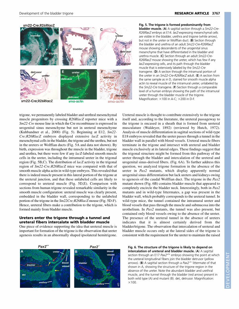
trigone, we permanently labeled bladder and urethral mesenchymalmuscle progenitors by crossing R26RlacZ reporter mice with aSm22-Cre mouse line in which the Cre recombinase is expressed inurogenital sinus mesenchyme but not in ureteral mesenchyme(Kuhbandner et al., 2000) (Fig. 5). Beginning at E12, Sm22-Cre;R26RlacZ embryos displayed extensive lacZ activity inmesenchymal cells in the bladder, the trigone and the urethra, but notin the ureters or Wolffian ducts (Fig. 5A and data not shown). Bybirth, expression was throughout the muscle in the bladder, trigoneand urethra, but there were few if any lacZ-labeled smooth musclecells in the ureter, including the intramural ureter in the trigonalregion (Fig. 5B,C). The distribution of lacZ activity in the trigonalregion of Sm22-Cre;R26RlacZ mice was compared with that ofsmooth muscle alpha actin in wild-type embryos. This revealed thatthere is indeed muscle present in this lateral portion of the trigone atthe ureteral junction, and that these unlabeled cells are likely tocorrespond to ureteral muscle (Fig. 5D,E). Comparison withsections from human trigone revealed remarkable similarity in thesmooth muscle configuration: ureteral muscle was clearly present,embedded in the bladder wall, corresponding to the unlabeledportion of the trigone in the Sm22Cre;R26RlacZ mouse (Fig. 5D-F).Hence, ureteral fibers make a contribution to the trigone, which isformed mainly from bladder muscle.
Ureters enter the trigone through a tunnel andureteral fibers intercalate with bladder muscleOne piece of evidence supporting the idea that ureteral muscle isimportant for formation of the trigone is the observation that ureteragenesis results in an abnormally shaped ipsolateral hemitrigone.
Ureteral muscle is thought to contribute extensively to the trigoneitself and, according to the literature, the ureteral passageway tothe trigone is encased in a sheath that is formed from ureteralmusculature (Waldeyer, 1892) (reviewed by Hutch, 1972).Analysis of muscle differentiation in sagittal sections of wild-typeE18 embryos revealed that the ureter passes through a tunnel in thebladder wall in parallel with blood vessels. Ureteral muscle fibersterminate in the trigone and intersect with ureteral and bladdermuscle exclusively at its lateral edges. These findings suggest thatthe trigonal structure might be formed from this pathway of theureter through the bladder and intercalation of the ureteral andurogenital sinus-derived fibers. (Fig. 6A). To further address thisquestion, we analyzed trigone formation in the absence of theureter in Pax2 mutants, which display apparently normalurogenital sinus differentiation but lack ureters and kidneys owingto agenesis of the caudal Wolffian duct. The trigone in the Pax2mutant shown (Fig. 6B) contains bladder muscle that appeared tocompletely encircle the bladder neck. Interestingly, both in Pax2mutants and in wild-type littermates, a gap was present in thebladder wall, which probably corresponds to the ureteral tunnel. Inwild-type mice, the tunnel contained the intramural ureter andblood vessels that pass through the muscle and submucosa into theurothelium. In Pax2 mutants, the tunnel was also present, butcontained only blood vessels owing to the absence of the ureter.The presence of the ureteral tunnel in the absence of uretersindicates that it is almost certainly derived from thebladder/trigone. The observation that intercalation of ureteral andbladder muscle occurs only at the lateral sides of the trigone isconsistent with the requirement for the ureter to maintain the raised
3767RESEARCH ARTICLEDevelopment of the bladder trigone
Fig. 5. The trigone is formed predominantly frombladder muscle. (A) A sagittal section through a Sm22-Cre-R26RlacZ embryo at E14. lacZ-expressing mesenchymal cellsare visible in the bladder, urethra and trigone (white arrow),but not in the ureter or Wolffian duct. (B) Section throughthe bladder and urethra of an adult Sm22-Cre-R26RlacZmouse showing descendents of the urogenital sinusmesenchyme that have differentiated in the bladder andurethra muscle. (C) Section through an adult Sm22-Cre-R26RlacZ mouse showing the ureter, which has few if anylacZ-expressing cells, and its path through the bladdermuscle that is extensively labeled by the Sm22-Cretransgene. (D) A section through the intramural portion ofthe ureter in an Sm22-Cre-R26RlacZ adult. (E) A section fromthe same sample as in D, stained for smooth muscle alphaactin to reveal muscle of the intramural ureter, unlabeled bythe Sm22-Cre transgene. (F) Section through a comparablelevel of a human embryo showing the path of the intramuralureter through the bladder muscle of the trigone.Magnification: �100 in A-C; �200 in D-F.
Fig. 6. The structure of the trigone is likely to depend onintercalation of ureteral and bladder muscle. (A) A sagittalsection through an E17 Pax2+/+ embryo showing the point at whichthe ureteral longitudinal fibers join the bladder detrusor (yellowarrows). (B) A sagittal section through a Pax2–/– littermate of thatshown in A, showing the structure of the trigone region in theabsence of the ureter. Note the abundant bladder and urethralmuscle, and the tunnel through the bladder (red arrow) present inboth wild type (A) and mutant (B). det, detrusor. Magnification:�100.

DEVELO
PMENT
3768
triangular structure normally associated with the trigone,explaining why the absence of the ipsolateral ureter results indeformation of the trigone.
DISCUSSIONRethinking urogenital tract formationAccording to the literature, the structure of the trigone is complex,derived predominantly from ureteral muscle that stretches across thebase to form the ureteral ridge, and also toward the trigone base toform Bell’s muscle (Fig. 7). The ureters are said to penetrate thebladder via a tunnel (Waldeyer’s sheath or space) derived from theureter (Brooks, 2002; Tanagho et al., 1968; Wesson, 1925). Thecommon nephric duct, which is the most caudal Wolffian ductsegment, is thought to contribute to the trigone as it differentiatesand expands during ureter transposition, repositioning the ureterorifices in the bladder neck. However, it is unclear which portion ofthe trigone this tissue would form, as the common nephric duct is anepithelial tube, an extension of the Wolffian duct, whereas thetrigonal muscle is likely to be derived from mesenchyme, as areother muscular tissues in the embryo. Our previous findings and thecurrent lineage study suggest an alternate model of urinary tractformation. We have established that the common nephric duct doesnot contribute to any part of the bladder, trigone or urethra, butinstead undergoes apoptosis during ureter transposition (Batourinaet al., 2005). Here, using Cre-lox recombination, we followed thefate of ureteral and bladder muscle progenitors and find that thetrigone is formed predominantly from bladder muscle, with acontribution from ureteral longitudinal fibers at the lateral edges thatis much more limited than previously thought (Fig. 7B). Theintercalation of ureteral and bladder muscle is likely to be crucial fortrigone formation and for maintaining the ureteral anti-refluxmechanism. These studies also suggest that muscles such asMercier’s bar and Bell’s muscle, which have been considered to beformed from the ureter, are in fact derived from the bladder (Fig. 7),as suggested by others (Woodburne, 1964). The observation that thetrigone is formed from the same primordial tissue as the rest of thebladder (the urogenital sinus) is consistent with studiesdemonstrating that the urothelial covering of the trigone isindistinguishable from that of the bladder, but is distinct from thatof the ureter (Liang et al., 2005).
Distinct patterning along the urinary outflowtractRecent studies indicate that most, if not all, of the mesenchymalmuscle progenitors lining the ureter and urogenital sinus derive fromthe tail bud or cloacal mesoderm (Brenner-Anantharam et al., 2007;Haraguchi et al., 2007). However, the morphology of these tissues
is diverse. Ureters are ensheathed by 3-4 layers of muscle thatmediate myogenic peristalsis. The bladder is surrounded by a thicklayer of smooth muscle, a muscularis mucosa and a surfacecomposed of deep folds that enable contraction and expansion. Thetrigone is smooth and has a distinctive shape probably generated byinteraction between bladder and ureteral muscle fibers at its lateraledges. Its cellular morphology is likely to depend not on itsembryological origin, as has been suggested, but on spatiallyexpressed signaling molecules, including Hox genes, Bmp4, Tbx18and Shh, that are crucial for patterning other urinary tract tissues(Airik et al., 2006; Brenner-Anantharam et al., 2007; Haraguchi etal., 2007; Raatikainen-Ahokas et al., 2000; Scott et al., 2005; Yu etal., 2002). Future studies will determine the role of these pathwaysin normal trigone development and whether mutations in these genesalso lead to trigone abnormalities.
Application of this new model to human diseaseThe pathway taken by the ureter through the bladder muscle andsubmucosa is thought to be important for function of the anti-refluxmechanism, which normally prevents back-flow of urine to theureters and kidney by compressing the intramural ureter against thesmooth muscle bladder wall. The ability to effectively compress thisterminal ureteral segment is thought to depend on several factors,including sufficient length of the intramural segment, its pathwaythrough the bladder and insertion of the ureter orifice at astereotypical position in the trigone (King et al., 1974; Stephens etal., 1996) and innervation that regulates opening of the ureteralorifice (reviewed by Radmayr, 2005).
A shortening of the intramural segment, or ureter orifices joiningthe trigone abnormally, can be caused by sprouting of the uretericbud from the Wolffian duct from a location more cranial or caudalthan normal (Mackie and Stephens, 1975; Pope et al., 1999;Stephens, 1983) as seen in several mouse models (Basson et al.,2005; Batourina et al., 2005; Grieshammer et al., 2004; Kume et al.,2000; Lu et al., 2007; Miyazaki et al., 2000; Yu et al., 2004), or byabnormalities in ureter transposition, at the time when the ureternormally separates from the Wolffian duct (Batourina et al., 2005).Intrinsic ureteral abnormalities, such as a failure in muscledifferentiation, can also result in reflux owing to faulty urinetransport or peristalsis (Airik et al., 2006; Chang et al., 2004; Yu etal., 2002).
The trigone is the site at which surgery is performed to correctreflux, whereby the refluxing ureter is detached from its originalinsertion site and reinserted in the trigone in such a way that thelength of the intramural segment is increased and has improvedmuscular backing. The observations from our studies that trigoneformation and, by default, ureteral valve function, depend on
RESEARCH ARTICLE Development 134 (20)
Fig. 7. Models of trigone formation. (A) Old model oftrigone formation, showing the trigone to be continuouswith the ureters (green), formed in large part from ureteralfibers that fan out across the surface generating the inter-ureteric ridge and Bell’s muscle. Note that the trigone hasbeen considered to form independently of the bladder. (B) Current model of trigone formation, showing a smallcontribution from ureteral fibers (green) and the bulk ofthe structure derived from bladder muscle and the spacearound the ureter that functions as a tunnel.

DEVELO
PMENT
intercalation of ureteral fibers with bladder muscle, suggest that inaddition to increasing the length of the intramural ureter,reimplantation of ureters might also inadvertently help establishbetter connections with underlying bladder muscle and the trigone.This will further our understanding of the anti-reflux mechanism thatis paramount for renal function.
We thank Christopher Choi for technical assistance; Nancy Heim (ColumbiaUniversity) for artwork; and Dr T. T. Sun (NY University) for the kind gift ofuroplakin antibody. This work was supported by grants from NIH: DK061459to C.M. and HD30284 to R.R.B.
ReferencesAirik, R., Bussen, M., Singh, M. K., Petry, M. and Kispert, A. (2006). Tbx18
regulates the development of the ureteral mesenchyme. J. Clin. Invest. 116,663-674.
Basson, M. A., Akbulut, S., Watson-Johnson, J., Simon, R., Carroll, T. J.,Shakya, R., Gross, I., Martin, G. R., Lufkin, T., McMahon, A. P. et al. (2005).Sprouty1 is a critical regulator of GDNF/RET-mediated kidney induction. Dev. Cell8, 229-239.
Batourina, E., Tsai, S., Lambert, S., Sprenkle, P., Viana, R., Dutta, S., Hensle,T., Wang, F., Niederreither, K., McMahon, A. P. et al. (2005). Apoptosisinduced by vitamin A signaling is crucial for connecting the ureters to thebladder. Nat. Genet. 37, 1082-1089.
Brenner-Anantharam, A., Cebrian, C., Guillaume, R., Hurtado, R., Sun, T. T.and Herzlinger, D. (2007). Tailbud-derived mesenchyme promotes urinary tractsegmentation via BMP4 signaling. Development 134, 1967-1975.
Brooks, J. D. (2002). Anatomy of the lower urinary tract and male genitalia. InCampbell’s Urology. Vol. I (ed. P. C. Walsh, A. B. Retik, E. D. Vaughan and A. J.Wein), pp. 89-128. Philadelphia: W. B. Saunders.
Chang, C. P., McDill, B. W., Neilson, J. R., Joist, H. E., Epstein, J. A., Crabtree,G. R. and Chen, F. (2004). Calcineurin is required in urinary tract mesenchymefor the development of the pyeloureteral peristaltic machinery. J. Clin. Invest.113, 1051-1058.
Grieshammer, U., Le, M., Plump, A. S., Wang, F., Tessier-Lavigne, M. andMartin, G. R. (2004). SLIT2-mediated ROBO2 signaling restricts kidney inductionto a single site. Dev. Cell 6, 709-717.
Haraguchi, R., Motoyama, J., Sasaki, H., Satoh, Y., Miyagawa, S., Nakagata,N., Moon, A. and Yamada, G. (2007). Molecular analysis of coordinatedbladder and urogenital organ formation by Hedgehog signaling. Development134, 525-533.
Holtwick, R., Gotthardt, M., Skryabin, B., Steinmetz, M., Potthast, R.,Zetsche, B., Hammer, R. E., Herz, J. and Kuhn, M. (2002). Smooth muscle-selective deletion of guanylyl cyclase-A prevents the acute but not chroniceffects of ANP on blood pressure. Proc. Natl. Acad. Sci. USA 99, 7142-7147.
Hutch, J. A. (1972). Anatomy and physiology of the bladder, trigone and urethra.London, New York: Butterworths Appleton-Century-Crofts.
King, L. R., Kazmi, S. O. and Belman, A. B. (1974). Natural history ofvesicoureteral reflux. Outcome of a trial of nonoperative therapy. Urol. Clin.North Am. 1, 441-455.
Kobayashi, A., Kwan, K. M., Carroll, T. J., McMahon, A. P., Mendelsohn, C. L.and Behringer, R. R. (2005). Distinct and sequential tissue-specific activities ofthe LIM-class homeobox gene Lim1 for tubular morphogenesis during kidneydevelopment. Development 132, 2809-2823.
Kong, X. T., Deng, F. M., Hu, P., Liang, F. X., Zhou, G., Auerbach, A. B.,Genieser, N., Nelson, P. K., Robbins, E. S., Shapiro, E. et al. (2004). Roles ofuroplakins in plaque formation, umbrella cell enlargement, and urinary tractdiseases. J. Cell Biol. 167, 1195-1204.
Kuhbandner, S., Brummer, S., Metzger, D., Chambon, P., Hofmann, F. andFeil, R. (2000). Temporally controlled somatic mutagenesis in smooth muscle.Genesis 28, 15-22.
Kume, T., Deng, K. and Hogan, B. L. (2000). Murine forkhead/winged helixgenes Foxc1 (Mf1) and Foxc2 (Mfh1) are required for the early organogenesis ofthe kidney and urinary tract. Development 127, 1387-1395.
Liang, F. X., Bosland, M. C., Huang, H., Romih, R., Baptiste, S., Deng, F. M.,Wu, X. R., Shapiro, E. and Sun, T. T. (2005). Cellular basis of urothelialsquamous metaplasia: roles of lineage heterogeneity and cell replacement. J.Cell Biol. 171, 835-844.
Lu, W., van Eerde, A. M., Fan, X., Quintero-Rivera, F., Kulkarni, S., Ferguson,H., Kim, H. G., Fan, Y., Xi, Q., Li, Q. G. et al. (2007). Disruption of ROBO2 isassociated with urinary tract anomalies and confers risk of vesicoureteral reflux.Am. J. Hum. Genet. 80, 616-632.
Mackie, G. G. and Stephens, F. D. (1975). Duplex kidneys: a correlation of renaldysplasia with position of the ureteral orifice. J. Urol. 114, 274-280.
Meyer, R. (1946). Normal and abnormal development of the ureter in the humanembryo – a mechanistic consideration. Anat. Rec. 68, 355-371.
Miyazaki, Y., Oshima, K., Fogo, A., Hogan, B. L. and Ichikawa, I. (2000). Bonemorphogenetic protein 4 regulates the budding site and elongation of themouse ureter. J. Clin. Invest. 105, 863-873.
Pope, J. C., IV, Brock, J. W., III, Adams, M. C., Stephens, F. D. and Ichikawa, I.(1999). How they begin and how they end: classic and new theories for thedevelopment and deterioration of congenital anomalies of the kidney andurinary tract, CAKUT. J. Am. Soc. Nephrol. 10, 2018-2028.
Raatikainen-Ahokas, A., Hytonen, M., Tenhunen, A., Sainio, K. and Sariola,H. (2000). BMP-4 affects the differentiation of metanephric mesenchyme andreveals an early anterior-posterior axis of the embryonic kidney. Dev. Dyn. 217,146-158.
Radmayr, C., Fritsch, H., Schwentner, C., Lnacek, A., Deibl, M., Bartsch, G.and Oswald, J. (2005). Fetal equipment of the vesico-ureteric junction, andimmunohistochemistry of the ends of refluxing ureters. J. Pediatr. Urol. 1, 53-59.
Roshani, H., Dabhoiwala, N. F., Verbeek, F. J. and Lamers, W. H. (1996).Functional anatomy of the human ureterovesical junction. Anat. Rec. 245, 645-651.
Scott, V., Morgan, E. A. and Stadler, H. S. (2005). Genitourinary functions ofHoxa13 and Hoxd13. J. Biochem. 137, 671-676.
Soriano, P. (1999). Generalized lacZ expression with the ROSA26 Cre reporterstrain. Nat. Genet. 21, 70-71.
Srinivas, S., Goldberg, M. R., Watanabe, T., D’Agati, V., al-Awqati, Q. andCostantini, F. (1999). Expression of green fluorescent protein in the ureteric budof transgenic mice: a new tool for the analysis of ureteric bud morphogenesis.Dev. Genet. 24, 241-251.
Stephens, F. D. (1983). Congenital Malformations of the Urinary Tract. New York:Praeger.
Stephens, F. D., Smith, E. D. and Hutson, J. M. (1996). Congenital Anomalies ofthe Urinary and Genital Tracts. Oxford: Isis Medical Media.
Tanagho, E. A. (1981). Development of the ureter. In The Ureter (ed. H. Bergman),pp. 1-12. New York: Springer-Verlag.
Tanagho, E. A., Smith, D. R. and Meyers, F. H. (1968). The trigone: anatomicaland physiological considerations. 2. In relation to the bladder neck. J. Urol. 100,633-639.
Waldeyer, W. (1892). Ueber die sogenannte Ureter-scheide. Anat. Anz. 6, 259-260.
Weiss, J. P. (1988). Embryogenesis of ureteral anomalies: a unifying theory. Aust.N. Z. J. Surg. 58, 631-638.
Wesson, M. B. (1925). Anatomical, embryological and physiological studies of thetrigone and bladder neck. J. Urol. 4, 280-306.
Woodburne, R. T. (1964). Anatomy of the ureterovesical junction. J. Urol. 92,431-435.
Wu, X. R., Lin, J. H., Walz, T., Haner, M., Yu, J., Aebi, U. and Sun, T. T. (1994).Mammalian uroplakins. A group of highly conserved urothelial differentiation-related membrane proteins. J. Biol. Chem. 269, 13716-13724.
Yu, J., Carroll, T. J. and McMahon, A. P. (2002). Sonic hedgehog regulatesproliferation and differentiation of mesenchymal cells in the mouse metanephrickidney. Development 129, 5301-5312.
Yu, O. H., Murawski, I. J., Myburgh, D. B. and Gupta, I. R. (2004).Overexpression of RET leads to vesicoureteric reflux in mice. Am. J. Physiol. RenalPhysiol. 287, F1123-F1130.
Yucel, S. and Baskin, L. S. (2004). An anatomical description of the male andfemale urethral sphincter complex. J. Urol. 171, 1890-1897.
3769RESEARCH ARTICLEDevelopment of the bladder trigone