Embed Size (px)
Citation preview

CASE REPORT
The diagnosis and conservativetreatment of a complex type 3 densinvaginatus using cone beamcomputed tomography (CBCT) and3D plastic models
A. Kfir1, Y. Telishevsky-Strauss1, A. Leitner2 & Z. Metzger1
1Department of Endodontology, Tel Aviv University, Tel Aviv; and 2Panorama, Naharya, Israel
Abstract
Kfir A, Telishevsky-Strauss Y, Leitner A, Metzger Z. The diagnosis and conservative treatment
of a complex type 3 dens invaginatus using cone beam computed tomography (CBCT) and 3D plastic
models. International Endodontic Journal, 46, 275–288, 2013.
Aim To investigate the use of 3D plastic models, printed from cone beam computed
tomography (CBCT) data, for accurate diagnosis and conservative treatment of a com-
plex case of dens invaginatus.
Summary A chronic apical abscess with a draining sinus tract was diagnosed during the
treatment planning stage of orthodontic therapy. Radiographic examination revealed a
large radiolucent area associated with an invaginated right maxillary central incisor, which
was found to contain a vital pulp. The affected tooth was strategic in the dental arch. Con-
ventional periapical radiographs provided only partial information about the invagination
and its relationship with the main root canal and with the periapical tissues. A limited-
volume CBCT scan of the maxilla did not show evidence of communication between the
infected invagination and the pulp in the main root canal, which could explain the pulp vital-
ity. A novel method was adopted to allow for instrumentation, disinfection and filling of the
invagination, without compromising the vitality of the pulp in the complex root canal sys-
tem. The CBCT data were used to produce precise 3D plastic models of the tooth. These
models facilitated the treatment planning process and the trial of treatment approaches.
This approach allowed the vitality of the pulp to be maintained in the complex root canal
space of the main root canal whilst enabling the healing of the periapical tissues.
Key learning points
• Even when extensive periapical pathosis is associated with a tooth with type III
dens invaginatus, pulp sensibility tests should be performed.
• CBCT is a diagnostic tool that may allow for the management of such teeth with
complex anatomy.
Correspondence: Anda Kfir, Department of Endodontology, School of Dental Medicine,
Tel Aviv University, Ramat Aviv, Tel Aviv 62646, Israel (Fax: 972 3 6409250;
e-mail: [email protected]).
© 2012 International Endodontic Journal. Published by Blackwell Publishing Ltd International Endodontic Journal, 46, 275–288, 2013
doi:10.1111/iej.12013
275

• 3D printed plastic models may be a valuable aid in the process of assessing and
planning effective treatment modalities and practicing them ex vivo before actually
performing the clinical procedure.
• Unconventional technological approaches may be required for detailed treatment
planning of complex cases of dens invaginatus.
Keywords: cone beam computed tomography, dens invaginatus, 3 dimensional
model, stereolithography.
Received 14 February 2012; accepted 24 August 2012
Introduction
Dens invaginatus is a dental developmental abnormality resulting from the invagination
of the enamel organ into the dental papilla prior to the mineralization phase (Shafer
et al. 1983, Hulsmann 1997, Reddy et al. 2008). The cavity that forms may serve as an
external route of communication with the pulp or even with the periapical tissues
through the foramen caecum. The precise aetiology of dens invaginatus is controversial.
A number of theories have been proposed regarding the pathogenesis of dens invagina-
tus, including uncontrolled growth of a portion of the enamel epithelium (Kronfeld
1934), tooth bud infection during tooth development (Fischer 1936), pressure from the
adjacent developing tooth germ (Atkinson 1943), trauma (Gustafson & Sundberg 1950)
and genetic components (Grahnen et al. 1959, Hosey & Bedy 1996, Dassule et al.
2000). Nevertheless, the exact aetiology remains uncertain.
Dens invaginatus is a relatively common condition with a reported incidence ranging
from 0.3% to 10% of all teeth (Atkinson 1943, Boyne 1952, Hamasha & Al-Omari
2004). The wide ranges of prevalence quoted in the literature result most probably from
variations in the study designs as well as the use of different diagnostic methods and
study populations. The permanent maxillary lateral incisors are the most frequently
involved teeth (Shafer et al. 1983, Hamasha & Al-Omari 2004), with the maxillary cen-
tral incisors as the second most common area of involvement (Yeh et al. 1999). Multi-
ple dens invaginatus involving all four maxillary incisors has been reported (Cronklin
1978). Backman & Wahlin (2001) reported a diagnosis of dens invaginatus in combina-
tion with other dental malformations.
The clinical appearance of the crown in dens invaginatus varies considerably; the
morphology may be normal or it may display unusual forms, such as a peg shape, bar-
rel shape or talon cusps (Ridell et al. 2001, Reddy et al. 2008). The first clinical sign of
an invaginated tooth might be a deep foramen caecum lined by hypomineralized brittle
enamel where caries can rapidly develop, enabling microorganisms from the oral cavity
to directly penetrate into the pulp, causing pulp necrosis and the development of apical
periodontitis (Jung 2004).
The most clinically relevant and widely used classification system for dens invagina-
tus was proposed by Oehlers (1957): Type I – an enamel-lined minor form, not extend-
ing beyond the cemento–enamel junction. Type II – an enamel-lined form that invades
the root but remains confined as a blind sac. The invagination may or may not commu-
nicate with the dental pulp. Type III – an invagination that penetrates through the root
and communicates directly with the periodontal ligament laterally (Type IIIa) or at the
apical foramen (Type IIIb). In such cases, there may be no immediate communication
with the pulp. In this type, infection within the invagination can cause an inflammatory
response in the periodontal or periapical tissues (Alani & Bishop 2008).
CASE
REPORT
© 2012 International Endodontic Journal. Published by Blackwell Publishing LtdInternational Endodontic Journal 46, 275–288, 2013276

On the basis of the diagnosis and treatment plan, different treatment modalities are
available, ranging from prophylactic treatment of the deep foramen caecum (Jung
2004), conservative restorative treatment (Hulsmann 1997), nonsurgical root canal treat-
ment (Rotstein et al. 1987, Szajkis & Kaufman 1993), endodontic surgery (Soares et al.
2007, Vier-Pelisser et al. 2012), intentional replantation and extraction (Hata & Toda
1987, Sousa & Bramante 1998, Tsurumachi et al. 2002a,b, De Martin et al. 2005).
Radiography has an important role in the diagnosis and assessment of the irregular
morphology of the root canal system, but conventional planar radiography only provides
a two-dimensional representation of the complex anatomy (Patel 2010, Durack & Patel
2011, Vier-Pelisser et al. 2012). The limited two-dimensional representation might not
yield sufficient information for the clinician to diagnose the true anatomy of the dens
invaginatus, thus hindering the effective management of the case.
Cone beam computed tomography (CBCT) provides three-dimensional (3D) undistort-
ed images of the maxillofacial skeleton, including the teeth and their surrounding tis-
sues, and this technique has demonstrated efficacy in a large number of endodontic
applications, including but not limited to complex dental anatomy (Patel et al. 2009,
Al-Rawy et al. 2010, Patel 2010, Durack & Patel 2011). Although the effective radiation
dose used in CBCT is higher than that of conventional radiographic techniques, it is sub-
stantially lower compared to conventional CT (Arai et al. 2001, Ngan et al. 2003, Ludlow
et al. 2006, Ludlow et al. 2006, Patel & Dawood 2007).
The scanned volume derived from the CBCT software may allow generation of
images in three planes that can be continuously scrolled through, thus allowing a three-
dimensional understanding of the structure involved. They can also be converted into
additional types of files. One of the new types of files that can be derived from the
CBCT scans is the stereo litography (STL) file. An STL file is a format used by stereoli-
thography software to generate information needed to produce 3D plastic models using
stereolithography machines or printers. Recently, this file format has been used for pro-
totyping and computer-aided manufacturing in other medical fields (Peltola et al. 2008).
The aim of this clinical article is to report on the use of a 3D plastic model printed
from CBCT digital imaging and communications in medicine (DICOM) data for the diag-
nosis and conservative treatment of a complex case of dens invaginatus.
Case report
A 15-year-old female was referred to the endodontic department at Goldschleger
School of Dental Medicine at Tel Aviv University for treatment of a diffuse periradicular
radiolucency around teeth 11 and 12 (Fig. 1). The patient was about to begin orthodon-
tic treatment, and the orthodontic team hoped to maintain the affected tooth, as it was
of strategic importance to the patient’s maxillary dental arch. Her medical history was
unremarkable, and there was no history of dental trauma. The patient reported that the
tooth had never been symptomatic.
Clinical examination revealed permanent dentition with teeth 13 and 23, which were
in ectopic eruption, and a sinus tract on the labial mucosa near tooth 12 (Fig. 1a), which
was traced to tooth 11 (Fig. 1b). Radiographic examination revealed that tooth 11 had a
dens invaginatus morphology and had an associated apical radiolucency extending from
the mesial aspect of tooth 11 to the distal aspect of tooth 12. The crown of tooth 11
was slightly wider than tooth 21, both mesio-distally and more so bucco-lingually
(Fig. 1). A prominent cingulum and a small indication of a foramen caecum were pres-
ent on the palatal side of the tooth. There were no signs of caries or existing restora-
tions, no discoloration of the tooth and no abnormal mobility. Periodontal probing was
within normal limits, and despite the extensive radiolucency, the pulps of teeth 11 and
CASE
REPORT
© 2012 International Endodontic Journal. Published by Blackwell Publishing Ltd International Endodontic Journal 46, 275–288, 2013 277

12 were vital and responded positively to thermal and electric pulp sensitivity testing.
The periapical radiograph of tooth 11 presented signs suggesting a class III invagination
that appeared to have its own ‘apical foramen’ (Fig. 1b). Because the morphology of
the invagination, the morphology of the pulp canal space and the relationship between
the two were not entirely clear from the diagnostic periapical radiograph, it was decided
that a limited-volume CBCT scan of the maxilla may be helpful for understanding the
internal anatomy of the tooth and determining the most appropriate treatment protocol.
The patient and her mother received information on the expected benefits and on the
potential risks of the CBCT scan. After obtaining written informed consent, a CBCT
(iCAT; Imaging Sciences International, Hatfield, PA, USA) of the maxilla was performed
with the following exposure parameters: 120 kV, 3.0 mA and 20 s with field of view of
6 cm. The cross-sectional images revealed a deep invagination surrounded by a thick
(a)
(b)
Figure 1 Clinical and radiographic presentation of the case. (a) Sinus tract at the area of tooth 12,
traced with a gutta-percha cone. Teeth 11 and 12 were both found to be vital. (b) Periapical radio-
graph presenting a large radiolucent lesion; a sinus tract traced with a gutta-percha cone. Note the
dens invaginatus structure of tooth 11, with a large ballooning space connected to the lesion
through a large apical opening. The yellow arrows indicate the pulp space, which was compressed
by the invagination.
CASE
REPORT
© 2012 International Endodontic Journal. Published by Blackwell Publishing LtdInternational Endodontic Journal 46, 275–288, 2013278
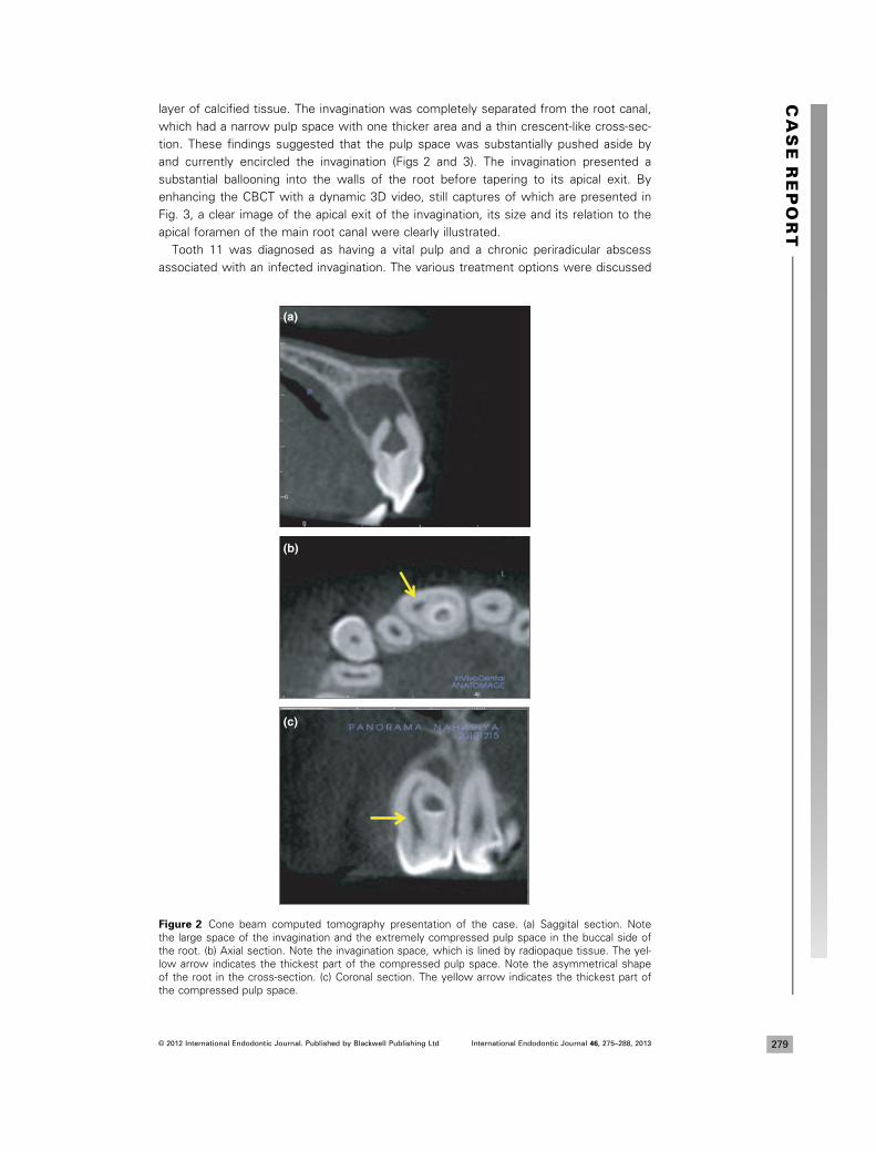
layer of calcified tissue. The invagination was completely separated from the root canal,
which had a narrow pulp space with one thicker area and a thin crescent-like cross-sec-
tion. These findings suggested that the pulp space was substantially pushed aside by
and currently encircled the invagination (Figs 2 and 3). The invagination presented a
substantial ballooning into the walls of the root before tapering to its apical exit. By
enhancing the CBCT with a dynamic 3D video, still captures of which are presented in
Fig. 3, a clear image of the apical exit of the invagination, its size and its relation to the
apical foramen of the main root canal were clearly illustrated.
Tooth 11 was diagnosed as having a vital pulp and a chronic periradicular abscess
associated with an infected invagination. The various treatment options were discussed
(a)
(c)
(b)
Figure 2 Cone beam computed tomography presentation of the case. (a) Saggital section. Note
the large space of the invagination and the extremely compressed pulp space in the buccal side of
the root. (b) Axial section. Note the invagination space, which is lined by radiopaque tissue. The yel-
low arrow indicates the thickest part of the compressed pulp space. Note the asymmetrical shape
of the root in the cross-section. (c) Coronal section. The yellow arrow indicates the thickest part of
the compressed pulp space.
CASE
REPORT
© 2012 International Endodontic Journal. Published by Blackwell Publishing Ltd International Endodontic Journal 46, 275–288, 2013 279

(a)
(b)
(c)
Figure 3 Still captures from a 3D video. (a) Apical view of tooth 11. Note the crater-like apical
opening of the invagination (white arrow) and a small depression next to it (red arrow) (b) a more
coronal view/section reveals that the small depression seen in ‘a’ was actually the apical foramen
leading to the pulp space. Separation of these two opening explains the vitality of the pulp.
(c) A more coronal view/section gives a view of the coronal ‘dome’ of the invagination and the
compressed pulp space. A wide part of the pulp space is seen on the bucco-distal side of the tooth
whilst narrower parts are seen on the buccal and mesio-buccal sides. Compare also to Fig. 1b and
to Fig. 2b,c.
CASE
REPORT
© 2012 International Endodontic Journal. Published by Blackwell Publishing LtdInternational Endodontic Journal 46, 275–288, 2013280

with the patient and her mother, including conservative treatment as well as a surgical
approach and it was decided to treat the infected invagination alone, attempting to
leave the vital pulp untreated.
As the morphology of the present dens invaginatus was complex and the pulp was
vital, and to avoid unexpected difficulties whilst accessing the invagination and its treat-
ment, a novel therapeutic approach was adopted.
STL files were derived from the CBCT scan data. The scan data were first derived as
DICOM3 multiple format which was converted to STL files using Mimics Z software
(3DSystems, Rock Hill, SC, USA). The STL files were then subjected to modulation with
Geomagic software (Geomagic, Morrisville, NC, USA) and used with a stereolithography
3D printer (Eden 260 VTM, Objet Eden 260 V, Objet Inc. Billerica, MA, USA) to generate
precise 3D VeroClearTM transparent and FullCureTM opaque plastic 3D models of tooth
11 that included the detailed internal anatomy. The 3D models (prototypes) were
printed using a photopolymerized acrylic resin (either VeroClear 810 TM or FullCure 720TM Polyjet resin) that was injected and cured one layer at a time to produce each model
at a resolution of 4 9 42 9 16 l (Erez Rapid Prototyping, Holon Institute of Technology,
Holon, Israel). The transparent 3D models (Fig. 4a) were used to study the anatomy of
the tooth and enabled the optimal therapeutic approach to be chosen for this complex
invagination case. Furthermore, the opaque model (Fig. 4b) was used to practice the
techniques required by the different treatment alternatives until sufficient skill was
(a) (b)
(c) (d)
Figure 4 Planning and training for treatment. (a) Lingual view of the transparent VeroClear plastic
model. The granular-looking area is the nonsmooth inner surface of the large invagination space.
(b) Buccal view of the opaque FullCure model. (c) A modified Compules Syringe (Dentsply Maille-
fer). The elongation tip of the compule was inserted to a given depth through the access channel
of the FullCure model (the tube is not visible in the picture). Note the screw (yellow arrow) that
calibrates and limits the amount of mineral trioxide aggregate (MTA) that is delivered into the invag-
ination cavity (Fig. 5a). (d) Drilling guide in position. The acrylic drilling guide was prepared on the
transparent VeroClear plastic model to indicate the optimal penetration point and drilling direction
that will allow to access the invagination space without passing through or touching the pulp space.
The guide was then transferred to tooth 11 and secured in position using some glass–ionomer
cement. Drilling was done along the path dictated by the drilling guide (dotted line).
CASE
REPORT
© 2012 International Endodontic Journal. Published by Blackwell Publishing Ltd International Endodontic Journal 46, 275–288, 2013 281

achieved with the filling technique before clinically implementing the procedures
(Figs 4c and 5a,b).
Endodontic treatment was carried out over three visits under rubber dam isolation
and local anaesthesia with the aid of an operating microscope. The transparent model
described earlier revealed that the point of penetration and the direction of drilling to
create the access channel would be critical if the pulp was to be preserved. Therefore,
an external acrylic drilling-guide device was prepared, which was attached to the tooth
during the preparations of access to the invagination cavity (Fig. 4d), thus enabling com-
plete control over the drilling point, direction and depth. At the first visit, the invagina-
tion was identified, accessed using a F06RF bur (Strauss & Co., Naharia, Netanya,
Israel) using the above-mentioned drilling guide and negotiated with stainless steel
hand files (Dentsply Maillefer, Ballaigues, Switzerland). Based on the CBCT data in
conjunction with a periapical radiograph, the working length of the invagination was
(a) (b)
(c) (d)
Figure 5 Planning and training for the clinical treatment and its results. (a) The transparent Vero-
Clear plastic model. The area selected for penetration is marked with a red arrow. The direction
and depth of the access channel (yellow arrow) were planned on this transparent model and exe-
cuted with the assistance of an external drilling guide that was also used clinically (Fig. 4d). The
access channel and cavity were filled with yellow paste (Ledermix) so that they could be seen in
the model. (b) The opaque FullCure plastic model was used for training of the operator in the accu-
rate placement of the MTA. The operator was considered ready to perform the clinical procedure
only after radiographic images such as the one shown in B could be reproducibly generated. Note
that the FullCure plastic is almost radiolucent. (c) Radiograph taken at the second visit: the sinus
tract persisted. The balloon-like radiolucent cavity is not clearly seen due to the radiopacity of the
Ledermix that was used as interappointment dressing. (d) Twelve months follow-up. Periapical
healing is almost complete. Both teeth 11 and 12 were found to be vital.
CASE
REPORT
© 2012 International Endodontic Journal. Published by Blackwell Publishing LtdInternational Endodontic Journal 46, 275–288, 2013282

established, and its coronal part, up to the ballooned area, was instrumented. The
invagination was repeatedly irrigated with copious amounts of 5.25% sodium hypochlo-
rite, which was repeatedly agitated with pre-curved K-files that scrapped the walls,
and pre-measured thick gutta-percha master cones that were used to agitate the
irrigant. These were done in an attempt to debride and disinfect the ballooned part of
the invagination. The invagination was then dried and dressed with Ledermix paste
(Haupt Pharma GmbH, Wolfratshausen, Germany) and sealed with a sterile cotton
pellet and FUJI IX (GC Corporation, Tokyo, Japan).
Two weeks later, at the second appointment, the patient was asymptomatic. The
pulps of teeth 11 and 12 were vital, but the sinus tract was still there (Fig. 5c). The
invagination was accessed and again irrigated with large volumes of 5.25% sodium
hypochlorite and similar use of bent files, then dressed again with Ledermix paste. At
the third appointment, the sinus tract disappeared, whilst the vitality of the pulps in
both incisors was maintained.
The planned procedure included filling the invagination with MTA Angelus (Industria
de produtos odontologicos Ltd., Londrina, Brazil). To ensure control over the most diffi-
cult part of the planned procedure, namely ensuring optimal sealing of the irregular and
complex shape of the invagination without pushing the material through the apical
opening of the invagination, the placement was first practiced extensively using the 3D
plastic model (Fig. 5b). The opaque model was used for this purpose so that the opera-
tor could not see the progress of the MTA, mimicking the clinical conditions. The exact
amount as well as the proper viscosity of the MTA was tested and calibrated. Due to
the unique morphology of the invagination space, MTA guns were too small and inef-
fective for the treatment of this condition; amalgam carriers could not be inserted to
the required point. A special device consisting of a modified Compules Syringe (Dents-
ply Maillefer) was assembled. An AccuDose® compule (Centrix, Shelton, CT, USA) was
adapted with an elongation tip (which is originally designed to be used with Rely X
Unichem Aplicap, 3M ESPE, St Paul, MN, USA) that allowed access to the desired
depth. The Compules Syringe was adapted with a screw (Fig. 4c) that allowed for cali-
bration and limiting of the amount of injected MTA. This was performed so that it
would allow optimal filling of the invagination space without passing the material into
the periradicular tissues. Radiographs were used to test the result on the model
(Fig. 5b). After the operator had become familiar and fluent with the technique using
the model, the MTA filling procedure was performed on the patient. The apical part of
the invagination was filled with MTA, including part of the ballooning area. The result
was checked radiographically, and the MTA was allowed to set for 15 min. Only then
the main part of the invagination was condensed with MTA (Fig. 5d). The access cavity
was then restored with a GRADIA composite resin, (GC Corporation). The patient was
recalled 6 month later and was asymptomatic. Teeth 11 and 12 responded normally to
thermal and electrical sensitivity testing, and a substantial reduction was observed in
the size of the periradicular radiolucency. At the next follow-up, at 12 months, the radio-
graph showed almost complete healing (Fig. 5d) and both teeth responded positively to
thermal and electric sensitivity testing.
Discussion
Root canal treatment in teeth with dens invaginatus associated with apical pathosis
often involves complicated procedures that require precise diagnosis, appropriate treat-
ment planning and adequate implementation (De Martin et al. 2005, Patel 2010). Con-
ventional two-dimensional radiographs have inherent limitations, and their diagnostic
yield is further limited by geometric distortion (Grondahl & Huumonen 2004) and by the
CASE
REPORT
283© 2012 International Endodontic Journal. Published by Blackwell Publishing Ltd International Endodontic Journal 46, 275–288, 2013

lack of information about the third dimension (Patel et al. 2009). CBCT has been shown
to be useful for assessing the complex anatomy of teeth (Cotton et al. 2007, Patel &
Dawood 2007, Patel et al. 2009, Patel 2010).
Before referring a patient to CBCT, a thorough consideration of risk versus benefit
should be performed, especially in a young patient as in the present case. Also the size
of FOV (field of view) should be carefully considered. In the present case, 3D imaging
was considered essential for treatment planning, but this could have also been
achieved with a small FOV. Nevertheless, as the patient had also to go through ortho-
dontic treatment and the orthodontist decided that proper orthodontic assessment of
the relation between the roots of the maxillary teeth, in her case, required a larger field
of view, it was decided to get a CBCT scan of the entire maxilla, with a 6 cm FOV, to
avoid subsequent additional repeated exposure to radiation.
The CBCT scan that was performed in the present case provided a three-dimensional
representation of the invaginated tooth and allowed for a true understanding of the nat-
ure of the invagination and its relationship to the main canal of the tooth. The CBCT
scan was also particularly useful for demonstrating how the invagination compressed
the pulp space of the main canal at different levels, resulting in an irregular main canal
with a cross-section resembling a thin crescent encircling the invagination, with no
communication between the invagination and the pulp space.
The positive response of tooth 11 to vitality testing indicated that the pulp in the
main root canal was still viable and presented a normal response to stimuli. Given the
complex anatomy of this pulp space, it was considered essential to avoid, as far as pos-
sible, perforation of the main root canal or devitalization of the pulp whilst accessing
the invagination (Patel 2010). This notion was based on many reports that indicate that
pulp spaces with such complex anatomy cannot be expected to be effectively debrided
and thus cannot be expected to be adequately obturated (De-Deus et al. 2010, 2011,
Metzger et al. 2010, Solomonov et al. 2012).
An alternative treatment plan involving apical surgery was also initially considered and
was rejected or at least postponed until the result of the conservative approach could
be evaluated. It was estimated that surgical treatment was not the best choice as it
would likely lead to devitalization of tooth 12 and would be more traumatic than the
conservative approach (Patel 2010, Vier-Pelisser et al. 2012).
After adopting the conservative approach, two issues were to be decided: (i) what
will be the best path of penetration? (ii) How should the invagination space be cleaned
and disinfected?
The printing of a transparent 3D plastic model of this tooth, with its complex invagi-
nation, provided an effective means for defining the correct point, direction and depth
for the planned access channel, which was created with the assistance of the drilling
guide whilst avoiding the viable pulp space (Fig. 4d). The use of this model allowed us
to eliminate the trial and error that otherwise would have been required to find access
to the invagination. Furthermore, it allowed practice of the treatment modalities before
performing the actual procedure on the patient, thus avoiding unexpected complications
during implementation. The opaque model (Fig. 4b) was useful in training the operator
in placement of the MTA (Fig. 5b). Nevertheless transparent model could also have
been used for this purpose by masking it, thus saving 3D printing expenses.
It should be kept in mind that the accuracy of such a procedure, in which CBCT
DICOM data are transferred to the STL format may not perfect (Platzer et al. 2012).
This accuracy may be affected by the type and extent of filtration applied to generate a
printable model. In the case at hand, the accuracy was most probably good enough clin-
ically as the drilling guide that was prepared on the printed plastic model had an optimal
fit on tooth 11.
CASE
REPORT
© 2012 International Endodontic Journal. Published by Blackwell Publishing LtdInternational Endodontic Journal 46, 275–288, 2013284

Cleaning of the invagination space was performed with copious amounts of sodium
hypochlorite, combined with the use of pre-curved hand files that were used to
mechanically scrap the internal walls of the invagination space. The use of ultrasonics
for this stage was considered and avoided as (i) it may not be effective enough in this
kind of balloon-like cavity and (ii) pulp vitality could potentially be risked by local heat
generation. Calcium hydroxide was first considered for inter-appointment dressing, but
was avoided as it may be extremely difficult to completely remove from such a com-
plex space (Tasdemir et al. 2011), thus potentially compromising effective obturation of
the invagination space. Ledermix was used as an alternative, as it is easier to remove
by irrigation (Rodig et al. 2011). The potential staining of an anterior tooth by Ledermix
was considered, thus the medicament was carefully removed from the access channel.
Nevertheless, as the Ledermix was placed in the invagination channel not in the pulp
chamber (which remained vital), penetration of dentinal tubules that may cause such
staining was very unlikely (Kim et al. 2000, Thomson et al. 2012).
Thermoplasticized gutta-percha with sealer is often used when irregular canal spaces
are to be filled. In the present case, there was a large apical opening which may have
made it difficult to control the apical flow of the softened gutta-percha. MTA was
selected as it allowed better apical control (Mohammadi 2011). Furthermore, it was
used in two stages, allowing the first layer to set before applying the second one. The
MTA Angelus, which has a relatively short setting time, was thus preferred for such
procedure (Moore et al. 2011).
Cone beam computed tomography involves increased radiation exposure compared
to periapical radiography, and therefore, its use should be justified by an analysis and
optimization of the risk/benefit ratio for a given patient. In the case reported here, CBCT
allowed the operator to make data-based decisions that were essential for treatment
planning. Furthermore, the CBCT data could further be enhanced by dynamic evaluation
of the full volume of the scan, using the iCat software to allow continuous scrolling in
the three planes, thus allowing for in-depth understanding of the intricate anatomical
features of this complex case. It allowed also generation of a 3D video, still captures of
which are presented in Fig. 3, which also enhanced the understanding of the unique
structure of the tooth involved and its surrounding structures. Last but not least, it
allowed for stereolithographic printing of 3D plastic models, which provided substantial
added value. The endodontic treatment in this case could most probably not have been
planned as accurately or performed as safely and successfully without the aid of the
CBCT scan.
The process of generation of 3D models from the CBCT data and printing them may
sound complicated and costly, thus a little extravagant. Nevertheless, it allowed a con-
servative treatment plan whilst the alternative treatment plan of apical surgery would
have been more traumatic, especially when likely devitalization of tooth 12 is consid-
ered. Recruiting the assistance of professionals from other disciplines (3D modelling
and printing) proved to be valuable both in clinical and in educational terms. 3D model-
ling and printing is commonly used in advanced mechanical engineering and the cost in
the current case did not exceed 130$ for transfer of the CBCT data into STL files and
100$ for printing the model.
Conclusion
Nonsurgical conservative treatment of a case with complex invagination proved to be
successful in promoting the healing of periradicular pathosis, whilst maintaining the via-
bility of the pulp tissue in the main complex canal. Such an option should be considered
and attempted as a first choice, irrespective of the size of the periradicular lesion. CBCT
CASE
REPORT
© 2012 International Endodontic Journal. Published by Blackwell Publishing Ltd International Endodontic Journal 46, 275–288, 2013 285

is an indispensable tool in the diagnosis and management of invaginated root canals.
In complex cases, it may also allow for stereolithographic printing of 3D models to
facilitate treatment planning and education.
Acknowledgements
The creative assistance of Eng. Gilad Zelniker and of Mr. Erez Sherman from Erez Rapid
Prototyping, Holon Institute of Technology, in printing the 3D plastic models that were
used in this study was instrumental to its success and are greatly appreciated.
Disclaimer
Whilst this article has been subjected to Editorial review, the opinions expressed,
unless specifically indicated, are those of the author. The views expressed do not
necessarily represent best practice, or the views of the IEJ Editorial Board, or of its
affiliated Specialist Societies.
References
Alani A, Bishop K (2008) Dens invaginatus. Part 1: classification, prevalence and aetiology. Interna-
tional Endodontic Journal 41, 1123–36.
Al-Rawy B, Hassan B, Vandenberg B, Jacobs R (2010) Accuracy assessment of three-dimensional
surface reconstructions of teeth from cone-beam computed tomography scans. Journal of Oral
Rehabilitation 37, 352–8.
Arai Y, Honda K, Iwai K, Shinoda K (2001) Practical model ‘3DX’ of limited cone-beam X-ray CT for
dental use. International Congress Series 1230, 713–8.
Atkinson SP (1943) The permanent maxillary lateral incisor. American Journal of Orthodontics 29,
685–98.
Backman B, Wahlin YB (2001) Variations in number and morphology of permanent teeth in 7-year--
old Swedish children. International Journal of Paediatric Dentistry 11, 11–7.
Boyne PJ (1952) Dens in dente: report of three cases. The Journal of the American Dental Associa-
tion 45, 209–10.
Cotton TP, Geisler TM, Holden DT, Schwartz SA, Schindler WG (2007) Endodontic applications of
cone-beam volumetric tomography. Journal of Endodontics 9, 1211–32.
Cronklin WW (1978) Bilateral dens invaginatus in the mandibular incisor region. Oral Surgery, Oral
Medicine, and Oral Pathology 45, 905–8.
Dassule HR, Lewis P, Bei M, Maas R, McMahon AP (2000) Sonic hedgehong regulates growth and
morphogenesis of the tooth. Development 127, 4775–85.
De Martin AS, de Silveira Bueno CE, Sandhes Cunha R, Aranha de Aranjo R, Fernandes de Magalh-
aes Silveira C (2005) Endodontic treatment of dens invaginatus with a periradicular lesion: case
report. Australian Endodontic Journal 31, 123–5.
De-Deus G, Barino B, Zamolyi RQ et al. (2010) Suboptimal debridement quality produced by
the single-file F2 ProTaper technique in oval-shaped canals. Journal of Endodontics 36, 1897–
900.
De-Deus G, Souza EM, Barino B et al. (2011) The self-adjusting file optimizes debridement quality
in oval-shaped root canals. Journal of Endodontics 37, 701–5.
Durack C, Patel S (2011) The use of cone beam computed tomography in the management of dens
invaginatus affecting a strategic tooth in a patient affected by hypodontia: a case report. Interna-
tional Endodontic Journal 44, 474–83.
Fischer CH (1936) Zur frage des dens in dente. Deutsche Zahn-, Mundund Kieferheilkunde 3,
621–34.
Grahnen H, Lindahl B, Omnel K (1959) Dens invaginatus. I. A clinical, roentgenological and genetical
study of permanent upper lateral incisor. Odontologisk Revy 10, 115–37.
CASE
REPORT
© 2012 International Endodontic Journal. Published by Blackwell Publishing LtdInternational Endodontic Journal 46, 275–288, 2013286

Grondahl H-G, Huumonen S (2004) Radiographic manifestations of periapical inflammatory lesions.
Endodontic Topics 8, 55–67.
Gustafson G, Sundberg S (1950) Dens in dente. British Dental Journal 88, 83–8, 111–22, 144–6.
Hamasha AA, Al-Omari QD (2004) Prevalence of dens invaginatus in Jordanian adults. International
Endodontic Journal 37, 307–10.
Hata G, Toda T (1987) Treatment of dens invaginatus by endodontic therapy, apicocurettage, and
retrofilling. Journal of Endodontics 13, 469–72.
Hosey MT, Bedy R (1996) Multiple dens invaginatus in two brothers. Endodontics and Dental
Traumatology 12, 44–7.
Hulsmann M (1997) Dens invaginatus: aetiology, classification, prevalence, diagnosis, and treatment
considerations. International Endodontic Journal 30, 79–90.
Jung M (2004) Endodomtic treatment of dens invaginatus type lll with three root canals and open
apical foramen. International Endodontic Journal 37, 205–13.
Kim ST, Abbott PV, McGinley P (2000) The effects of Ledermix paste on discoloration of mature
teeth. International Endodontic Journal 33, 227–32.
Kronfeld R (1934) Dens in dente. Journal of Dental Research 14, 49–66.
Ludlow JB, Davies-Ludlow LE, Brooks SL, Howerton WB (2006) Dosimetry of 3 CBCT devices for
oral and maxillofacial radiology: CB Mercuray, NewTom 3 G and i-CAT. Dentomaxillofacial Radiol-
ogy 35, 219–26.
Metzger Z, Zary R, Cohen R, Teperovich E, Paque F (2010) The quality of root canal preparation
and root canal obturation in canals treated with rotary versus Self Adjusting Files: a three-dimen-
sional micro-computed tomographic study. Journal of Endodontics 36, 1569–73.
Mohammadi Z (2011) Strategies to manage permanent non-vital teeth with open apices: a clinical
update. International Dental Journal 61, 25–30.
Moore A, Howley MF, O’Connell AC (2011) Treatment of open apex teeth using two types of
white mineral trioxide aggregate after initial dressing with calcium hydroxide in children. Dental
Traumatology 27, 166–73.
Ngan DCS, Kharbanda OP, Geenty JP, Darendeliler MA (2003) Comparison of radiation levels
from computed tomography and conventional dental radiographs. Australian Dental Journal 19,
67–75.
Oehlers FA (1957) Dens invaginatus. l. Variations of the invagination process and associated
anterior crown forms. Oral Surgery, Oral Medicine and Oral Pathology 10, 1204–18.
Patel S (2010) The use of cone beam computed tomography in the conservative management of
dens invaginatus: a case report. International Endodontic Journal 43, 707–13.
Patel S, Dawood A (2007) The use of cone beam computed tomography in the management of
external cervical resorption lesions. International Endodontic Journal 40, 818–30.
Patel S, Dawood A, Whaites E, Pitt Ford T (2009) New dimensions in endodontic imaging: part 1.
Conventional and alternative radiographic systems. International Endodontic Journal 42, 447–62.
Peltola SM, Melchels FP, Grijpma DW, Kellomaki M (2008) A review of rapid prototyping techni-
ques for tissue engineering purposes. Annals of Medicine 40, 268–80.
Platzer S, Bertha G, Heschl A, Wegscheider WA, Lorenzoni M (2012) Three-dimensional accuracy
of guided implant placement: indirect assessment of clinical outcomes. Clinical Implant Dentistry
and Related Research. Article first published online : 15 DEC 2011, DOI: 10.1111/j.1708-
8208.2011.00406.x
Reddy YP, Karpagavinayagam K, Subbarao CV (2008) Management of dens invaginatus diagnosed
by spiral computed tomography: a case report. Journal of Endodontics 34, 1138–42.
Ridell K, Mejara I, Matsson L (2001) Dens invaginatus: a retrospective study of prophylactic invagi-
nation treatment. International Journal of Paediatric Dentistry 11, 92–7.
Rodig T, Hirschleb M, Zapf A, Hulsmann M (2011) Comparison of ultrasonic irrigation and RinsEndo
for the removal of calcium hydroxide and Ledermix paste from root canals. International
Endodontic Journal 44, 1155–61.
Rotstein I, Stabholz A, Heling I, Friedman S (1987) Clinical consideration in treatment of dens
invaginatus. Endodontics & Dental Traumatology 3, 249–54.
Shafer WG, Hine MK, Levy B (1983) A Textbook of Pathology, 4th edn. Philadelphia, PA: WB
Saunders Co.
CASE
REPORT
© 2012 International Endodontic Journal. Published by Blackwell Publishing Ltd International Endodontic Journal 46, 275–288, 2013 287

Soares J, Santos S, Silveira F, Nunes E (2007) Calcium hydroxide barrier over the apical root-end of
a type lll dens invaginatus after endodontic and surgical treatment. International Endodontic
Journal 40, 146–55.
Solomonov M, Paque F, Fan B, Eilat Y, Berman LH (2012) The challenge of C-shaped canal
systems: a comparative study of the Self-Adjusting File and ProTaper. Journal of Endodontics 38,
209–14.
Sousa SMG, Bramante CM (1998) Dens invaginatus: treatment choices. Endodontics and dental
Traumatology 14, 152–8.
Szajkis S, Kaufman AY (1993) Root infolding treatment: a conservative approach in endodontics.
Journal of Endodontics 19, 576–8.
Tasdemir T, Celik D, Er K, Yildirim T, Ceyhanli KT, Yesilyurt C (2011) Efficacy of several techniques
for the removal of calcium hydroxide medicament from root canals. International Endodontic
Journal 44, 505–9.
Thomson AD, Athanassiadis B, Kahler B, Walsh L (2012) Tooth discoloration: staining effects of
various sealers and medicaments. Australian Endodontic Journal 38, 2–9.
Tsurumachi T, Hayashi M, Takeichi O (2002a) Non-Surgical root canal treatment of dens invaginatus
type 2 in a maxillary lateral incisor. International Endodontic Journal 35, 68–72.
Tsurumachi T, Hayashi M, Takeichi O (2002b) Non-Surgical root canal treatment of dens invaginatus
type 2 in a maxillary lateral incisor. International Endodontic Journal 35, 310–4.
Vier-Pelisser FV, Pelisser A, Recuero LC, So MVR, Borda MG, Figuerido AP (2012) Use of cone
beam computed tomography in the diagnosis planning and follow up of a type lll dens invagina-
tus case. International Endodontic Journal 45, 198–208.
Yeh SC, Lin YT, Lu SY (1999) Dens invaginatus in the maxillary lateral incisor: treatment of 3 cases.
Oral Surgery, Oral Medicine, Oral Pathology, Oral Radiology, and Endodontology 87, 628–31.
CASE
REPORT
© 2012 International Endodontic Journal. Published by Blackwell Publishing LtdInternational Endodontic Journal 46, 275–288, 2013288


![Successful management of a type II Dens invaginatus with an … · 2018-11-28 · of permanent tooth germs, taurodontism, supernumerary tooth, and dentinogenesis imperfecta [3]. Clinically,](https://img.pdfslide.net/doc/110x75/5e9529423a2cec077d2f9125/successful-management-of-a-type-ii-dens-invaginatus-with-an-2018-11-28-of-permanent.jpg)