Embed Size (px)
Citation preview

Journal of Cancer Therapy, 2016, 7, 889-900 http://www.scirp.org/journal/jct
ISSN Online: 2151-1942 ISSN Print: 2151-1934
DOI: 10.4236/jct.2016.712086 November 9, 2016
The Diagnostic Value of Serum TgAb in the Tg-Negative and TgAb-Positive DTC Patients after Successful Ablation
Qun Fan1, Xinhui Su1, Anren Kuang2*
1Department of Nuclear Medicine, Zhongshan Hospital, Xiamen University, Xiamen, China 2Department of Nuclear Medicine, West China Hospital, Sichuan University, Chengdu, China
Abstract Objective: The aim of this retrospective study was to evaluate diagnostic accuracy of serum thyroglobulin antibody (TgAb) in thyroglobulin (Tg)-negative and TgAb- positive (Tg−TgAb+) patients with differentiated thyroid carcinoma (DTC). Method: We studied 341 patients with histologically confirmed DTC who had undergone remnant ablation and showed Tg−TgAb+ assessed by electrochemiluminescence im-munoassay (ECLIA). The cases were divided into two groups, including recurrence group 119 cases and no evidence of disease (NED) group 222 cases. Receiver operat-ing characteristic (ROC) curve analysis was carried out. The symmetric measures of the two diagnostic methods (the golden standard and the diagnostic standard as se-rum TgAb level alone) were analyzed using McNemar test and measure of agreement Kappa. Results: Serum TgAb level (1381.292 ± 1017.221) IU/ml of patients with re-current group was significantly higher than that (125.559 ± 314.047) IU/ml of NED group (P = 0.000 < 0.001). The area under the ROC curve was 0.962 and its asymp-totic 95% confidence interval (CI) was (0.942, 0.982) that was high statistical signi-ficance. The cut-off value of TgAb was determined and interpreted at 246.695 IU/ml with sensitivity (92.40%) and specificity (92.30%). McNemar test showed that the diagnostic results of the two methods were not significant difference (P = 0.230 > 0.05). Measure of agreement Kappa was 0.841, P = 0.000 < 0.001 that showed the agreement of the two diagnostic methods was significant. Conclusion: Serum TgAb is a useful tumor marker for recurrence in Tg-negative and TgAb-positive DTC pa-tients who underwent thyroidectomy and remnant ablation. The cut-off value of TgAb is 246.695 IU/ml, that is to say, serum TgAb level upon 246.695 IU/ml may be associated with the persistence or recurrence of DTC.
How to cite this paper: Fan, Q., Su, X.H. and Kuang, A.R. (2016) The Diagnostic Value of Serum TgAb in the Tg-Negative and TgAb-Positive DTC Patients after Suc-cessful Ablation. Journal of Cancer Therapy, 7, 889-900. http://dx.doi.org/10.4236/jct.2016.712086 Received: September 19, 2016 Accepted: November 6, 2016 Published: November 9, 2016 Copyright © 2016 by authors and Scientific Research Publishing Inc. This work is licensed under the Creative Commons Attribution International License (CC BY 4.0). http://creativecommons.org/licenses/by/4.0/
Open Access

Q. Fan et al.
890
Keywords DTC, TgAb, Cut-Off Value
1. Introduction
Serum thyroglobulin (Tg) level and 131I whole-body scanning (WBS) are the leading recognized sensitive and specific tools for the detection of recurrence or metastases during follow-up of thyroidectomized patients with differentiated thyroid carcinoma (DTC) who underwent remnant ablation. These have been widely accepted manage-ment protocols [1] [2]. However, the presence of serum anti-Tg antibody (TgAb) may interfere with Tg measurement made by immunometric assay (IMA) or radioimmu-noassay (RIA) methods and cause false results [3] [4] [5] [6]. In the presence of TgAb, IMA methods consistently underestimate serum Tg levels, while RIA methods generally overestimate serum TG levels [5] [6] [7]. Previous studies have showed the incidence of TgAb-positive (TgAb+) in DTC patients ranges from 10% to 30% [8] [9] [10] [11] [12] and about 20% - 30% have been proven recurrence or metastasis among the TgAb+ DTC patients. TgAb currently compromises the use of serum Tg as a tumor marker in this kind of DTC patients. Also, TgAb has been studied extensively in order to evaluate its place in clinical practice [8]-[13]. Serum TgAb levels can be used as a marker for the studied patients that have been shown to be cost-effective in a few studies [8]-[17], but its possible diagnostic benefit remains controversial. And, it has not been confirmed the diagnostic significance of elevated serum TgAb levels with TG-undetectable and TgAb- detectable (Tg−TgAb+) DTC patients.
Therefore, the aims of the present study were to evaluate diagnostic accuracy of se-rum TgAb in Tg−TgAb+ DTC patients who had previously received thyroidectomy and radioiodine ablation therapy and ascertain the cut-off value of TgAb to differentiate between the pathologic and disease-free patients.
2. Materials and Methods 2.1. Patients
We performed a retrospective review on medical records of1236 patients with histologi-cally confirmed DTC who were admitted to the Nuclear Medicine Department of Zhong-shan Hospital Xiamen University from January 2001 to December 2014. All patients un-derwent total or subtotal thyroidectomy, followed by radioiodine remnant ablation com-pletely and thyroid hormone suppression of thyroid-stimulating hormone (TSH).
According to the collecting and excluding standards, 341 patients entered the study finally. The patients were divided into the recurrence group (n = 119) and no evidence of disease (NED) group (n = 222) on the basis of the golden standard.
2.2. Definitions
A serum TG level of below 1 µg/L with suppressed TSH levels or 2 µg/L with stimulated

Q. Fan et al.
891
TSH levels was considered undetectable (Tg−). We defined TgAb-detectable (TgAb+) as upon 10 IU/ml. Recurrence was defined as either a regional recurrence and/or distant spread.
The DTC patients with Tg−TgAb+ and successful ablation of thyroid remnant en- rolled the study. The DTC patients with TG-detectable, TgAb-undetectable (Tg+TgAb−) or thyroid remnant in thyroid bed were excluded the study. The gold standard for the study was either the diagnosis of recurrence established by biopsy and/or the diagnosis of the routine clinical examinations (chest x-rays, CT scanning, neck high-resolution echography and radioiodine whole body scan).
Sensitivity (Sen) and specificity (Spe) were defined as true positive and true negative, respectively. Positive likelihood ratio (+LR) was defined as the ratio of the true positive and false positive (Sen/1 – Spe). Youden Index (YI) = Sen + Spe – 1. Pre-test probability is defined as the probability that a patient suffers from recurrence or metastases by means of his/her medical history and physical sign. Post-test probability is defined as the probability of recurrent disease in a patient with a certain test result of TgAb. Pre- test odds = pre-test probability/(1 – pre-test probability). Post-test odds = pre-test odds × (+LR). Post-test probability = post-test odds/(1 + post-test odds).
2.3. Serum Tg and TgAb Assays
Serum Tg level was determined by means of electrochemiluminescence immunoassay (ECLIA) method using a commercially available kit (Roche Diagnostics GmbH), which has a measuring range of 0.100 - 1000 µg/L. The intra- and interassay coefficient of va-riability (CV) were 1.8% and 3.2%, respectively.
Serum TgAb level was measured by means of ECLIA method (Roche Diagnostics GmbH) with measurable range of 10 - 4000 IU/ml. The intra- and interassay CV were 5.2% and 7.3%, respectively.
2.4. Statistical Analysis
Data were expressed as mean ± SD. Statistical analysis was done using Indepen-dent-samples t test and the χ2 test. A P-value of less than 0.05 was considered to indi-cate a statistically significant difference.
The analysis of diagnostic value was performed by receiver operating characteristic (ROC) curve at the time of evaluation. The nonparametric method could be used to es-timate the area under ROC curve. The area under the ROC curve (AUC) with 95% con-fidence interval (CI) was calculated to express the overall diagnostic accuracy of the test. The AUC, P value, and cutoff point were obtained from the curve. The cut-off val-ue was defined as the threshold value of the maximum Youden Index (YI). Corres-ponding to Spe, Sen, PV and LR were calculated for the cut-off value in contingency tables.
The symmetric measures of the two diagnostic methods (the golden standard and the diagnostic standard as serum TgAb level alone)were analyzed using McNemar test and measure of agreement Kappa. And, the analysis of positive likelihood ratio (+LR) with
Q. Fan et al.
892
different threshold values were carried out in the study. Statistical analyses were performed with SPSS for Windows Software package (Re-
lease 17.0, SPSS Inc., US).
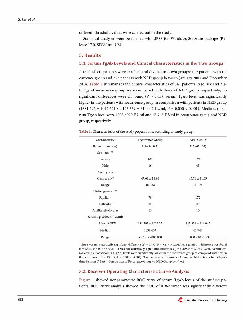
3. Results 3.1. Serum TgAb Levels and Clinical Characteristics in the Two Groups
A total of 341 patients were enrolled and divided into two groups: 119 patients with re-currence group and 222 patients with NED group between January 2001 and December 2014. Table 1 summarizes the clinical characteristics of 341 patients. Age, sex and his-tology of recurrence group were compared with those of NED group respectively; no significant differences were all found (P > 0.05). Serum TgAb level was significantly higher in the patients with recurrence group in comparison with patients in NED group (1381.292 ± 1017.221 vs. 125.559 ± 314.047 IU/ml, P = 0.000 < 0.001). Medians of se-rum TgAb level were 1038.4000 IU/ml and 63.745 IU/ml in recurrence group and NED group, respectively. Table 1. Characteristics of the study populations, according to study group.
Characteristic Recurrence Group NED Group
Patients—no. (%) 119 (34.897) 222 (65.103)
Sex—no.*††
Female 103 177
Male 16 45
Age—years
Mean ± SD†§ 47.64 ± 11.96 45.74 ± 11.25
Range 16 - 82 13 - 76
Histology—no.‡††
Papillary 79 172
Follicular 25 34
Papillary/Follicular 15 16
Serum TgAb level (IU/ml)
Mean ± SD¶§ 1381.292 ± 1017.221 125.559 ± 314.047
Median 1038.400 63.745
Range 53.230 - 4000.000 10.000 - 4000.000
*There was not statistically significant difference (χ2 = 2.457, P = 0.117 > 0.05). †No significant difference was found (t = 1.454, P = 0.147 > 0.05). ‡It was not statistically significant difference (χ2 = 5.229, P = 0.073 > 0.05). ¶Serum thy-roglobulin autoantibodies (TgAb) levels were significantly higher in the recurrence group as compared with that in the NED group (t = 13.135, P = 0.000 < 0.001). §Comparison of Recurrence Group vs. NED Group by Indepen-dent-Samples T Test. ††Comparison of Recurrence Group vs. NED Group by χ2 test.
3.2. Receiver Operating Characteristic Curve Analysis
Figure 1 showed nonparametric ROC curve of serum TgAb levels of the studied pa-tients. ROC curve analysis showed the AUC of 0.962 which was significantly different

Q. Fan et al.
893
Figure 1. Nonparametric ROC curve of serum TgAb levels of the studied patients. The area under the curve (AUC) was 0.962. The area was sig-nificantly different from 0.5 (P = 0.000 < 0.001). Point A on the curve is the closest to the upper left corner. Its ordinate is 0.924, its abscissa is 0.077. If point A is defined as optimal operating point, corresponding to Sen and Spe are 92.40%, 92.30%, respectively.
from 0.5 (P = 0.000 < 0.001). The 95% CI for the area was (0.942, 0.982). The interval did not consist of 0.5.The results showed that serum TgAb level reflects the recurrence of DTC in Tg−TgAb+ patients after thyroid ablation successfully. Point A on the curve is the closest to the upper left corner. Its ordinate is 0.924, its abscissa is 0.077. If point A is defined as optimal operating point, corresponding to Sen and Spe are 92.40%, 92.30%, respectively.
3.3. Determining the Cut-Off Value
YI is an index of analysis by synthesis that reflects the authenticity of diagnostic test. It is defined as (Sen + Spe – 1); its range is from −1 to +1. As the analysis of definite quantity, the closer the value is to the upper limit (+1), the higher the overall accuracy of the diagnostic test.
We selected the operating point on the ROC curve the closest to the upper left corner as cut-off point (point A showed Figure 1). Corresponding YI (0.847) is a maximum index. That is to say, the Sen/Spe pair maximizes the function [Sen − (1 – Spe)]. Cor-responding ordinate and abscissa are 0.924, 0.077 respectively (i.e., the predicted diag-nostic Sen and Spe are 92.40%, 92.30% respectively). And, corresponding TgAb level is 246.695 IU/ml (point A showed Figure 2).

Q. Fan et al.
894
Figure 2. Line chart of Youden index by serum TgAb levels. The ordinate of point A on the curve is 0.847 (i.e., the maximum value of Youden index is 0.847), corresponding abscissa is 246.695 (i.e., serum TgAb level is 246.695 IU/ml). That is to say, the cut-off value of serum TgAb is 246.695 IU/ml.
3.4. Symmetric Measures
The symmetric measures of the two diagnostic methods (the golden standard and the diagnostic standard as serum TgAb level alone) were analyzed using McNemar test and measure of agreement Kappa. McNemar test showed that the diagnostic result of the two methods was not significant difference (P = 0.230 > 0.05). Agreement Kappa (κ) is a measurement of evaluating agreement of the diagnostic result of the two methods. Thus, κ ≥ 0.7 is considered to be excellent test results (i.e., the agreement of two diag-nostic methods was high statistical significance); 0.4 ≤ κ < 0.7 is considered to be statis-tical significance; κ < 0.4 is considered to be no statistical significance. Measure results of agreement Kappa was “κ = 0.841, P = 0.000 < 0.001”, that showed the agreement of the two diagnostic methods was high statistical significance.
3.5. Diagnostic Properties and Analysis of Positive Likelihood Ratio
Selecting TgAb = 246.695 IU/ml as cut-off value, we predicted the studied patients (Table 2). Diagnostic properties were expressed by Sen, Spe, positive likelihood ratio (+LR), accuracy (Acc), positive predictive value (+PV) and negative predictive value (−PV). According to Table 2, we calculated +LR = 12.83 [(110/119)/(16/222)], Acc = 92.67% [(110 + 206)/341], +PV = 87.30% (110/126), −PV = 95.81% (206/215).

Q. Fan et al.
895
The analysis of +LR with different threshold values were calculated in the study (Table 3, Figure 3). Table 3 and Figure 3 showed that the higher serum TgAb level, the higher corresponding +LR was (i.e., the higher serum TgAb level, the more possible recurrence was). According to the relationship of pre-test odds, post-test odds, pre-test probability and post-test probability, we calculated the results as follows: when the pre-test probability fixed and except for the mixed function of other factors, the possi-bility of recurrence as TgAb ≥ 246.695 IU/ml was 10.294 times that of TgAb < 246.695 IU/ml; the possibility of recurrence as TgAb > 1000.000 IU/ml was 1.086 times that of 246.695 IU/ml ≤ TgAb ≤ 1000.000 IU/ml, 4.571 times that of 100.000 IU/ml ≤ TgAb <
Table 2. Comparison of the two diagnostic results.
Clinical routine diagnostic methods (Golden standard)
Total (n) Recurrence (n) No evidence of disease (n)
TgAb ≥ 246.695 (n) 110 16 126
TgAb < 246.695 (n) 9 206 215
Total (n) 119 222 341
Table 3. Positive likelihood ratios of different threshold values.
TgAb (IU/ml) Recurrence group NED group
+LR n Ratio n Ratio
≥1000.000 62 62/119 = 0.521 5 5/222 = 0.023 0.521/0.023 = 22.652
246.695 ≤ TgAb < 1000.000
48 48/119 = 0.403 12 12/222 = 0.054 0.403/0.054 = 7.463
100.000 ≤ TgAb < 246.695
6 6/119 = 0.050 42 42/222 = 0.189 0.050/0.189 = 0.265
10.000 ≤ TgAb < 100.000
3 3/119 = 0.025 163 163/222 = 0.734 0.025/0.734 = 0.034
Total 119 1 222 1
Figure 3. Positive likelihood ratio of different threshold value of serum TgAb. The serum TgAb levels were higher; the corresponding +LR was higher. That is to say, the higher serum TgAb le-vels, the more possible recurrence or metastases was.

Q. Fan et al.
896
246.695 IU/ml, 29.109 times that of 10.000 IU/ml ≤ TgAb < 100.000 IU/ml; the possi-bility of recurrence as 246.695 IU/ml ≤ TgAb ≤ 1000.000 IU/ml was 4.209 times that of 100.000 IU/ml ≤ TgAb < 246.695 IU/ml, 26.802 times that of 10.000 IU/ml ≤ TgAb < 100.000IU/ml; the possibility of recurrence as 100.000 IU/ml ≤ TgAb < 246.695 IU/ml was 6.368 times that of 10.000 IU/ml ≤ TgAb < 100.000 IU/ml.
According to the analysis of positive likelihood ratio, we assumed that the possibility of recurrence/metastases as 246.695 IU/ml ≤ TgAb < 1000.000 IU/ml is 10 units, the possibility of recurrence/metastases as TgAb > 1000.000 IU/ml, 100.000 IU/ml ≤ TgAb < 246.695 IU/ml and 10.000 IU/ml ≤ TgAb < 100.000 IU/ml is 10.860 units, 2.376 units, 0.373 units respectively (Figure 4).
4. Discussion
Self antigen of TgAb is TG. And, TG is incomplete “inaccessible antigen” and giant molecule glycoprotein synthesized by thyroid follicular epithelial cell. Serum TgAb is usually of IgG1, IgG3 or IgG4 subtypes in DTC patients [18]. Serum TG is an estab-lished tumor marker in monitoring recurrence patients with DTC [19], but TgAb in-terference affects serum Tg measurement. Therefore, we should attach importance to the TgAb interference in serum Tg measurement for Tg−TgAb+ DTC patients. Some studies [10]-[15] [20] [21] reported elevated TgAb level may be representative of recur-rence in Tg−TgAb+ DTC patients. That is, these kinds of patients were additionally di-agnosed as recurrent or metastatic disease by measuring their serum TgAb levels. Some studies [22] [23] [24] [25] reported that production of TgAb is mainly due to intrathy-roidal and extrathyroidal lymphocytes for antigen presentation to immune cells. Only rarely TgAb is produced by circulating lymphocytes and immune cells in cervical lymph nodes and bone marrow. This notion shows that the production of thyroid an-tibodies depends on antigenic components. After successful ablation of thyroid rem-nant tissue, the main source of TgAb is extrathyroidal lymphocytes. That is to say,
Figure 4. The possibility of recurrence/metastases as different threshold values of serum TgAb levels. Assuming the possibility of recurrence/metastases as 246.695 IU/ml ≤ TgAb < 1000.000 IU/ml is 10 units, the possibility of recurrence/metastases as TgAb > 1000.000 IU/ml, 100.000 IU/ml ≤ TgAb < 246.695 IU/ml and 10.000 IU/ml ≤ TgAb < 100.000 IU/ml is 10.860 units, 2.376 units, 0.373 units respectively.

Q. Fan et al.
897
persistent tumors mainly become source of antigenic components of producing TgAb. The notion theoretically explains that serum TgAb level is an index of recurrence or metastases in Tg−TgAb+ DTC patients of complete ablation.
In this study, we analyzed the diagnostic accuracy and ascertained the cut-off value of serum TgAb in the enrolled patients by means of ROC curve and +LR analysis of different threshold values. Serum TgAb levels (1381.292 ± 1017.221) IU/ml of patients with recurrence group was significantly higher than those (125.559 ± 314.047) IU/ml of NED group (P = 0.000) (Table 1), suggesting that serum TgAb level might be related to the persistence DTC. Similar findings have been reported previously [11] [15]. Hjiyi- annakis et al. [10] reported that 9 patients (22.5%) had shown progressively increasing TgAb levels and been confirmed recurrence diseases among 40 DTC patients with TG−TgAb+. The result showed serum TgAb can indicate active tumor. Research results of most scholars [8] [9] [10] [11] [12] [15] showed that the retention of elevated TgAb levels appeared to act as “tumor marker” in the DTC patients with TG−TgAb+. These notions provide practically reasons. But, it is controversial what its cut-off value of se-rum TgAb is. Only according to their clinical experience, most scholars [10] [11] [15] acted “TgAb = 100 U/ml, TgAb titre = 1/100, TgAb = 50 U/ml” as referential value that was not been statistically analyzed by diagnostic test.
According to our knowledge, this is the rare report on the application of ROC curve and +LR with different threshold values analyses in order to evaluate the diagnostic value of serum TgAb monitoring in the follow-up after successful ablation remnant thyroid tissue for TG-undetectable (TG−) DTC patients. ROC curve is applied to eva-luate the accuracy of diagnostic test. ROC curve occupy a central or unifying position in the process of assessing and using diagnostic tools. The ROC curve depicts the over-lap between the two distributions by plotting the Sen vs. (1 − Spe) for the complete range of decision thresholds. The y-axis is Sen, and the x-axis is (1 − Spe). The ROC graph is a plot of all of the Sen/Spe pairs resulting from continuously varying the deci-sion threshold over the entire range of results observed. Every point on the curve re- presents a Sen/Spe pair corresponding to a particular decision threshold [26].
AUC is a measure of predicted accuracy. The results of our ROC study showed that AUC value was 0.956 (Figure 1). There was statistical difference between the AUC (0.956) and Az (0.5). And the AUC value (0.956) was higher than 0.9. The results showed that the diagnostic test was higher accuracy. That is to say, it was important di-agnostic significance for serum TgAb level to evaluate recurrence in DTC patients of TG−TgAb+. Even, it may be possible that TgAb is more likely to act as a tumor marker in these patients.
It showed that TgAb at a single cut-off level was used with regard to monitoring the recurrence of the DTC patients with TG−TgAb+ received successful ablation. As showed in Figure 1 and Figure 2, we selected the “A” point of the closet to the upper left corner on the curve as cut-off point. Corresponding to YI is the maximum value (YI = 0.847). ROC analysis showed that the cut-off point (TgAb = 246.695 IU/ml) was the higher Sen (92.40%) and Spe (92.30%). And this may reflect the fact that 110 of 119 of the patients

Q. Fan et al.
898
with recurrence had increased TgAb level (≥246.695 IU/ml) (Table 2). Only 7.56% (9/119) patients with recurrence were falsely classified as NED when the cut-off value revealed by ROC analysis was used (Table 2).
In the present study, the analytical results of +LR with different threshold values showed that the higher serum TgAb level, the more possible recurrence is (Table 3). The symmetric measures of the two diagnostic methods (the golden standard and the diagnostic standard as serum TgAb level alone) were carried out by use of McNemar test and measure of agreement Kappa. McNemar test estimated of the DTC recurrence for studied patients with acting “TgAb = 246.695 IU/ml” as cut-off value predicting re-sults was compared with that for studied patients with clinical routine diagnostic me-thods predicting results. Statistically significant difference was not found (P = 0.230 > 0.05). Aside, measure of agreement Kappa shown that the two diagnostic methods is excellent symmetry (κ = 0.841 > 0.7, P = 0.000 < 0.05). The statistical results shown that it was high diagnostic value for predicting the studied patients with acting “TgAb = 246.695 IU/ml” as cut-off value. Serum TgAb level upon “246.695 IU/ml” may be asso-ciated with recurrent diseases.
Hjiyiannakis et al. [12] carried out a retrospective review to investigate the recur-rence or metastases in patients with TgAb+ in the Royal Marsden Hospital. They re-ported that 11 patients were found recurrence among 28 patients of high titre group (TgAb > 1/100); only 2 patients were found recurrence among 12 patients of low titre group (TgAb < 1/100). Median of TgAb level was 1/604 in the high titre group, whereas it was 80 IU/ml in the low tire group. In our study, median of TgAb level was 1038.400 IU/ml in the recurrence group, and is similar to the observations of Hjiyiannakis et al. [12]. Our study shown that median of TgAb level only was 63.745 IU/ml in the NED group. Compared with the reported result by Hjiyiannakis et al. [12], it was lower. In our study, serum TgAb levels were (1381.292 ± 1017.221) IU/ml in recurrence patients and (125.559 ± 314.047) IU/ml in NED patients. Compared with the observed results of Chung et al. [14], the serum TgAb levels were similar in recurrence group and lower in NED group. This phenomenon is likely to be owing to the presence for following fac-tors, such as selected patients, sample, different assay methods of serum TgAb and a TG/TgAb immune complex from the blood.
5. Conclusions
Serum TgAb is acted as a “tumor marker” and its cut-off value is 246.695 IU/ml in TG−TgAb+ DTC patients with successful ablation of remnant thyroid tissue. The higher serum TgAb level, the more possible recurrence is. During the long follow-up of such patients populations, serum TgAb level should be routinely measured. As serum “TgAb ≥ 246.695 IU/ml”, correlative clinical measures should be carried out in order to find recurrence earlier and treating as soon as possible; as serum “TgAb < 246.695 IU/ml”, we should termly monitor the serum level of TgAb.
We need carry out a research of big sample and a long-term clinical observation to nail down the diagnostic value of serum TgAb in patients with DTC.

Q. Fan et al.
899
Acknowledgements
This study was supported by a grant from the National Natural Science Foundation of China (NSFC) (81071182).
References [1] Schlumberger, M.J. (1999) Diagnostic Follow-Up of Well-Differentiated Thyroid Carcino-
ma: Historical Perspective and Current Status. Journal of Endocrinological Investigation, 22, 3-7.
[2] Mazzaferri, E.L. and Kloos, R.T. (2000) Using Recombinant Human TSH in the Manage-ment of Well-Differentiated Thyroid Cancer: Current Strategies and Future Directions. Thyroid, 10, 767-768. http://dx.doi.org/10.1089/thy.2000.10.767
[3] Baloch, Z., Carayon, P., Conte-Devolx, B., et al. (2003) Laboratory Medicine Practice Guidelines: Laboratory Support for the Diagnosis and Monitoring of Thyroid Disease. Thyroid, 13, 3-126. http://dx.doi.org/10.1089/105072503321086962
[4] Schneider, A.B. and Pervos, R. (1978) Radioimmunoassay of Human Thyroglobulin: Effect of Antithyroglobulin Antoantibodies. The Journal of Clinical Endocrinology & Metabolism, 47, 126-137. http://dx.doi.org/10.1210/jcem-47-1-126
[5] Spencer, C.A. (2004) Challenges of Serum Thyroglobulin (Tg) Measurement in the Pres-ence of Tg Autoantibodies. The Journal of Clinical Endocrinology & Metabolism, 89, 3702- 3704. http://dx.doi.org/10.1210/jc.2004-0986
[6] Locsei, Z., Szabolcs, I., Rácz, K., Kovács, G.L., Horváth, D. and Toldy, E. (2012) Serum Thyroglobulin Antibody Levels within or Near to the Reference Range May Interfere with Thyroglobulin Measurement. Biochemica Medica, 22, 365-370. http://dx.doi.org/10.11613/BM.2012.038
[7] Spencer, C., Petrovic, I. and Fatemi, S. (2011) Current Thyroglobulin Autoantibody (TgAb) Assays Often Fail to Detect Interfering TgAb That Can Result in the Reporting of Falsely Low/Undetectable Serum Tg IMA Values for Patients with Differentiated Thyroid Cancer. The Journal of Clinical Endocrinology & Metabolism, 96, 1283-1291. http://dx.doi.org/10.1210/jc.2010-2762
[8] Pacini, F., Mariotti, S., Formica, N., et al. (1988) Thyroid Autoantibodies in Thyroid Can-cer: Incidence and Relationship with Tumour Outcome. Acta Endocrinologica, 119, 373- 380. http://dx.doi.org/10.1530/acta.0.1190373
[9] Rubello, D., Girelli, M.E., Casara, D., et al. (1990) Usefulness of the Combined Antithyrog-lobulin Antibodies and Thyroglobulin Assay in the Follow-Up of Patients with Differen-tiated Thyroid Cancer. Journal of Endocrinological Investigation, 13, 737-742. http://dx.doi.org/10.1007/BF03349612
[10] Kumar, A., Shah, D.H., Shrihari, U., et al. (1994) Significance of Antithyroglobulin Autoan-tibodies in Differentiated Thyroid Carcinoma. Thyroid, 4, 199-202. http://dx.doi.org/10.1089/thy.1994.4.199
[11] Spencer, C.A., Takeuchi, M., Kazarosyan, M., et al. (1998) Serum Thyroglobulin Autoanti-bodies: Prevalence, Influence on Serum Thyroglobulin Measurement, and Prognostic Signi-ficance in Patients with Differentiated Thyroid Carcinoma. The Journal of Clinical Endo-crinology & Metabolism, 83, 1121-1127.
[12] Hjiyiannakis, P., Mundy, J. and Harmer, C. (1999) Thyroglobulin Antibodies in Differen-tiated Thyroid Cancer. Clinical Oncology, 11, 240-244. http://dx.doi.org/10.1053/clon.1999.9056

Q. Fan et al.
900
[13] Gorges, R., Maniecki, M., Jentzen, W., Sheu, S.N., Mann, K., Bockisch, A., et al. (2005) De-velopment and Clinical Impact of Thyroglobulin Antibodies in Patients with Differentiated Thyroid Carcinoma during the First 3 Years after Thyroidectomy. European Journal of Endocrinology, 153, 49-55. http://dx.doi.org/10.1530/eje.1.01940
[14] Chung, J.K., Park, Y.J., Kim, T.Y., et al. (2002) Clinical Significance of Elevated Level of Se-rum Antithyroglobulin Antibody in Patients with Differentiated Thyroid Cancer after Thyroid Ablation. Clinical Endocrinology, 57, 215-221. http://dx.doi.org/10.1046/j.1365-2265.2002.01592.x
[15] Kucuk, O.N., Aras, G., Kulak, H.A., et al. (2006) Clinical Importance of Anti-Thyroglobulin Auto-Antibodies in Patients with Differentiated Thyroid Carcinoma: Comparison with 99mTc-MIBI Scans. Nuclear Medicine Communications, 27, 873-876. http://dx.doi.org/10.1097/01.mnm.0000243376.49378.27
[16] Spencer, C.A., Bergoglio, L.M., Kazarosyan, M., Fatemi, S. and Lopresti, J.S. (2005) Clinical Impact of Thyroglobulin (Tg) and Tg Autoantibody Method Differences on the Manage-ment of Patients with Differentiated Thyroid Carcinomas. Journal of Clinical Endocrinolo-gy and Metabolism, 90, 5566-5575. http://dx.doi.org/10.1210/jc.2005-0671
[17] Spencer, C.A. (2011) Clinical Review: Clinical Utility of Thyroglobulin Antibody (TgAb) Measurements for Patients with Differentiated Thyroid Cancers (DTC). Journal of Clinical Endocrinology and Metabolism, 96, 3615-3627. http://dx.doi.org/10.1210/jc.2011-1740
[18] Caturegli, P., Kuppers, R.C., Mariotti, S., et al. (1994) IgG Subclass Distribution of Thyrog-lobulin Antibodies in Patients with Thyroid Disease. Clinical & Experimental Immunology, 98, 464-469. http://dx.doi.org/10.1111/j.1365-2249.1994.tb05514.x
[19] Lubin, E., Mechlis-Frish, S., Zatz, S., et al. (1994) Serum Thyroglobulin and Iodine-131 Whole Body Scan in the Diagnosis and Assessment of Treatment for Metastatic Differen-tiated Thyroid Cancer. Journal of Nuclear Medicine, 35, 257-262.
[20] Kim, E.S., Lim, D.J., Baek, K.H., Lee, J.M., Kim, M.K., Kwon, H.S., et al. (2010) Thyroglo-bulin Antibody Is Associated with Increased Cancer Risk in Thyroid Nodules. Thyroid, 20, 885-891. http://dx.doi.org/10.1089/thy.2009.0384
[21] Rubello, D., Casara, D., Girelli, M.E., et al. (1992) Clinical Meaning of Circulating Antithy-roglobulin Antibodies in Differentiated Thyroid Cancer: A Prospective Study. Journal of Nuclear Medicine, 33, 1478-1480.
[22] Chiovato, L., Latrofa, F., Braverman, L.E., et al. (2003) Disappearance of Humoral Thyroid Autoimmunity after Complete Removal of Thyroid Antigens. Annals of Internal Medicine, 139, 346-351. http://dx.doi.org/10.7326/0003-4819-139-5_Part_1-200309020-00010
[23] Weetman, A.P. and McGregor, A.M. (1994) Autoimmune Thyroid Disease: Further Devel-opments in Our Understanding. Endocrine Reviews, 15, 788-830. http://dx.doi.org/10.1210/edrv-15-6-788
[24] Mariotti, S., del Prete, G.F., Maggi, E., et al. (1984) Surface Markers and Function of Circu-lating Thyroid Autoantibody-Producing Cells. The Journal of Clinical Endocrinology & Metabolism, 58, 18-24. http://dx.doi.org/10.1210/jcem-58-1-18
[25] Chiovato, L., Latrofa, F., Braverman, L.E., Pacini, F., Capezzone, M., Masserini, L., et al. (2003) Disappearance of Humoral Thyroid Autoimmunity after Complete Removal of Thyroid Antigens. Annals of Internal Medicine, 139, 346-351. http://dx.doi.org/10.7326/0003-4819-139-5_Part_1-200309020-00010
[26] Zweig, M.H. and Campbell, G. (1993) Receiver-Operating Characteristic (ROC) Plots: A Fundamental Evaluation Tool in Clinical Medicine. Clinical Chemistry, 39, 561-577.

Submit or recommend next manuscript to SCIRP and we will provide best service for you:
Accepting pre-submission inquiries through Email, Facebook, LinkedIn, Twitter, etc. A wide selection of journals (inclusive of 9 subjects, more than 200 journals) Providing 24-hour high-quality service User-friendly online submission system Fair and swift peer-review system Efficient typesetting and proofreading procedure Display of the result of downloads and visits, as well as the number of cited articles Maximum dissemination of your research work
Submit your manuscript at: http://papersubmission.scirp.org/ Or contact [email protected]