Embed Size (px)
Citation preview
595.752.2:591.43
MEDEDELINGEN LANDBOUWHOGESCHOOL WAGENINGEN • NEDERLAND • 79-17 (1979)
THE DIGESTIVE SYSTEM OF SUBSALTUSAPHIS
ORNATA (HOMOPTERA: APHIDIDAE)
(with a summary in Dutch)
M. B. PONSEN
Laboratory of Virology, Agricultural University, Wageningen, The Netherlands
(received 22-VIII-1979)
H. VEENMAN & ZONEN B.V. - W A G E N I N G E N - 1979
THE D IGESTIVE SYSTEM OF SUBSALTUSAPHIS ORNATA
( H O M O P T E R A : A P H I D I D A E )
INTRODUCTION
The aphid Subsaltusaphis ornata (THEOBALD, 1927) belongs to the Callaphi-didae of BORNER (1952). These extremely flat aphids live on the underside of the leaves of the sedge, Carex riparia C U R T . During feeding the antennae are directed straight forward as has already been reported by WILLCOCKS for the related Saltusaphis scirpus THEOBALD, 1915, from 'sedges' in Egypt. The morphology and biology of Subsaltusaphis ornata has been described by HILLE RIS LAMBERS (1935) under the name Saltusaphis ornatus THEOB., and chromosome studies have been performed by G U T (1976). At present this species is placed in the genus Subsaltusaphis QUEDNAU, 1953 (EASTOP and HILLE RIS LAMBERS, 1976).
Investigations into the anatomy of the digestive system of Subsaltusaphis ornata THEOBALD were carried out since dissections of this flat aphid (Fig. 4) revealed the presence of two filtersystems, unique in the family Aphididae. Much information concerning the several types of filtersystems in the order Hemip-tera was obtained from the work of GOODCHILD (1966).
MATERIALS AND METHODS
Subsaltusaphis ornata, kindly supplied by Ing. A. van HARTEN, were reared on Carex riparia in the insectarium of the Institute of Phytopathological Research (IPO) at Wageningen. They were fixed in DUBOSQ BRASIL'S fluid, embedded in paraplast, and sectioned at 5 u. Sections were stained in EHRLICH'S haematoxylin-eosin.
RESULTS
The most anterior part of the alimentary tract is the food canal of the maxillary stylets. F rom the stylets it passes into the pharyngeal duct which in turn leads into the pharynx. This structure passes upwards through the head, and leads over the tentorium into the foregut to open into the stomach, a dilation of the midgut. The stomach enters into the filterchamber which is formed from the posterior part of the hindgut, and after a short trajectory it leaves the filterchamber to pass into the intestine. Subsequently the intestine extends forwards and after four coils it runs posteriad to open into the posterior part of the hindgut. The anterior and posterior part of the intestine are fused together. The hindgut starts at the fourth abdominal spiracle and extends posteriad to the rectum terminating into
Meded. Landbouwhogeschool Wageningen 79-17 (1979) 1
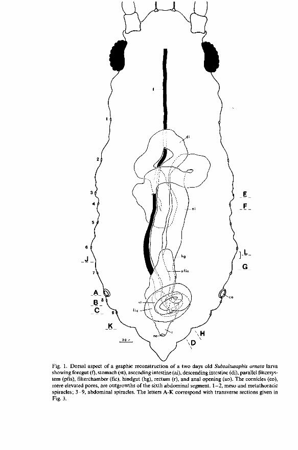
Fig. 1. Dorsal aspect of a graphic reconstruction of a two days old Subsaltusaphis ornata larva showing foregut (f), stomach (st), ascending intestine (ai), descending intestine (di), parallel filtersys-tem (pfis), filterchamber (fie), hindgut (hg), rectum (r), and anal opening (ao). The cornicles (co), mere elevated pores, are outgrowths of the sixth abdominal segment. 1-2, meso and metathoracic spiracles; 3-9, abdominal spiracles. The letters A-K correspond with transverse sections given in Fig. 3.
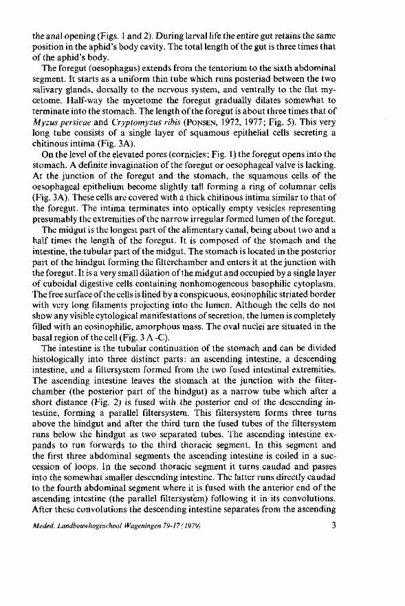
the anal opening (Figs. 1 and 2). During larval life the entire gut retains the same position in the aphid's body cavity. The total length of the gut is three times that of the aphid's body.
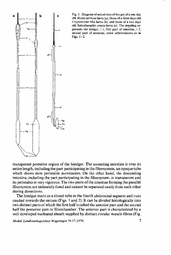
The foregut (oesophagus) extends from the tentorium to the sixth abdominal segment. It starts as a uniform thin tube which runs posteriad between the two salivary glands, dorsally to the nervous system, and ventrally to the flat my-cetome. Half-way the mycetome the foregut gradually dilates somewhat to terminate into the stomach. The length of the foregut is about three times that of Myzus persicae and Cryptomyzus ribis (PONSEN, 1972, 1977; Fig. 5). This very long tube consists of a single layer of squamous epithelial cells secreting a chitinous intima (Fig. 3A).
On the level of the elevated pores (cornicles ; Fig. 1) the foregut opens into the stomach. A definite invagination of the foregut or oesophageal valve is lacking. At the junction of the foregut and the stomach, the squamous cells of the oesophageal epithelium become slightly tall forming a ring of columnar cells (Fig. 3A). These cells are covered with a thick chitinous intima similar to that of the foregut. The intima terminates into optically empty vesicles representing presumably the extremities of the narrow irregular formed lumen of the foregut.
The midgut is the longest part of the alimentary canal, being about two and a half times the length of the foregut. It is composed of the stomach and the intestine, the tubular part of the midgut. The stomach is located in the posterior part of the hindgut forming the filterchamber and enters it at the junction with the foregut. It is a very small dilation of the midgut and occupied by a single layer of cuboidal digestive cells containing nonhomogeneous basophilic cytoplasm. The free surface of the cells is lined by a conspicuous, eosinophilic striated border with very long filaments projecting into the lumen. Although the cells do not show any visible cytological manifestations of secretion, the lumen is completely filled with an eosinophilic, amorphous mass. The oval nuclei are situated in the basal region of the cell (Fig. 3 A-C).
The intestine is the tubular continuation of the stomach and can be divided histologically into three distinct parts: an ascending intestine, a descending intestine, and a filtersystem formed from the two fused intestinal extremities. The ascending intestine leaves the stomach at the junction with the filter-chamber (the posterior part of the hindgut) as a narrow tube which after a short distance (Fig. 2) is fused with the posterior end of the descending intestine, forming a parallel filtersystem. This filtersystem forms three turns above the hindgut and after the third turn the fused tubes of the filtersystem runs below the hindgut as two separated tubes. The ascending intestine expands to run forwards to the third thoracic segment. In this segment and the first three abdominal segments the ascending intestine is coiled in a succession of loops. In the second thoracic segment it turns caudad and passes into the somewhat smaller descending intestine. The latter runs directly caudad to the fourth abdominal segment where it is fused with the anterior end of the ascending intestine (the parallel filtersystem) following it in its convolutions. After these convolutions the descending intestine separates from the ascending
Meded. Landbouwhogeschool Wageningen 79-17 ( 1979) 3
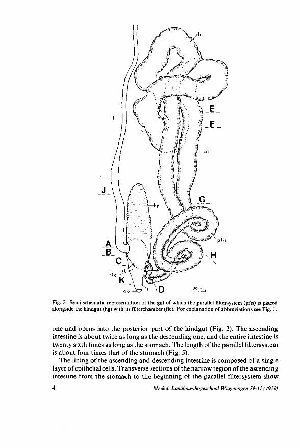
Fig. 2. Semi-schematic representation of the gut of which the parallel filtersystem (pfis) is placed alongside the hindgut (hg) with its filterchamber (fie). For explanation of abbreviations see Fig. 1.
one and opens into the posterior part of the hindgut (Fig. 2). The ascending intestine is about twice as long as the descending one, and the entire intestine is twenty sixth times as long as the stomach. The length of the parallel filtersystem is about four times that of the stomach (Fig. 5).
The lining of the ascending and descending intestine is composed of a single layer of epithelial cells. Transverse sections of the narrow region of the ascending intestine from the stomach to the beginning of the parallel filtersystem show
4 Meded. Landbouwhogeschool Wageningen 79-17 (1979)
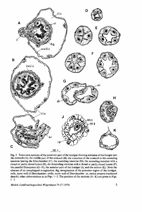
Fig. 3. Transverse sections of the posterior part of the hindgut showing entrance of the foregut into the stomach (A), the middle part of the stomach (B), the transition of the stomach to the ascending intestine leaving the filterchamber (C), the ascending intestine (D), the ascending intestine with a closed or partly closed lumen (E), the descending intestine with a closed or partly closed lumen (F), the parallel filtersystem (G-H), the anterior part of the hindgut (J), and the rectum (K). Note the absence of an oesophageal invagination, ihg, invagination of the posterior region of the hindgut; i wfic, inner wall of filterchamber ; o wfic, outer wall of filterchamber ; tp, tunica propria (nucleated sheath), other abbreviations as in Figs. 1-2. The position of the sections (A-K) are given in Figs. 1-2.
Meded. Landbouwhogeschool Wageningen 79-17 ( 1979)
N10
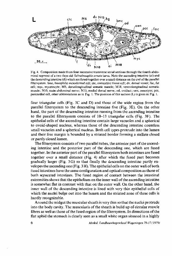
Fig. 4. Composition made from four successive transverse serial sections through the fourth abdominal segment of a two days old Subsaltusaphis ornata larva. Note the ascending intestine (ai) and the descending intestine (di) which are fused together over a small distance on the end of the parallel filtersystem. bmc, basophilic mesodermal cell; etc, connective tissue cell; dv, dorsal vessel; fac, fat cell; myc, mycetocyte; M5, dorsolongitudinal somatic muscle; M18, ventrolongitudinal somatic muscle; N10, main abdominal nerve; N13, medial dorsal nerve; od, oviduct; oen, oenocyte; pec, pericardial cell, other abbreviations as in Fig. 1. The position of this section (L) is given in Fig. 1.
four triangular cells (Fig. 3C and D) and those of the wide region from the parallel filtersystem to the descending intestine five (Fig. 3E). On the other hand, the part of the descending intestine running from the ascending intestine to the parallel filtersystem consists of 10-13 triangular cells (Fig. 3F). The epithelial cells of the ascending intestine contain large vacuoles and a spherical to ovoid-shaped nucleus, whereas those of the descending intestine countless small vacuoles and a spherical nucleus. Both cell types protrude into the lumen and their free margin is bounded by a striated border forming a stellate closed or partly closed lumen.
The filtersystem consists of two parallel tubes, the anterior part of the ascending intestine and the posterior part of the descending one, which are fused together. In the anterior part of the parallel filtersystem both intestines are fused together over a small distance (Fig. 4) after which the fused part becomes gradually larger (Fig. 3G) so that finally the descending intestine partly envelopes the ascending one (Fig. 3 H). The epithelial cells on the outer wall of both fused intestines have the same configuration and optical composition as those of both separated intestines. The fused region of contact between the intestinal extremities shows that the epithelium on the inner wall of the ascending intestine is somewhat flat in contrast with that on the outer wall. On the other hand, the inner wall of the descending intestine is lined with very thin epithelial cells of which the nuclei bulge out into the lumen and the striated zone of these cells is hardly recognizable.
Around the midgut the muscular sheath is very thin so that the nuclei protrude into the body cavity. The muscularis of the sheath is build up of circular muscle fibres as well as those of the fused region of the filtersystem. In dissections of the flat aphid the stomach is clearly seen as a small white organ situated in a highly
6 Meded. Landbouwhogeschool Wageningen 79-17 ( 1979)
k^°^ù
\
fl—
• h g _
\
Fig. 5. Diagram of actual sizes of the gut of a one day old Myzus persicae larva (a), those of a three days old Cryptomyzus ribis larva (b), and those of a two days old Subsaltusaphis ornata larva (c). The stippling represents the midgut, i 1, first part of intestine; i 2, second part of intestine, other abbreviations as in Figs. 1-2.
transparent posterior region of the hindgut. The ascending intestine is over its entire length, including the part participating in the filtersystem, an opaque tube which shows slow peristaltic movements. On the other hand, the descending intestine, including the part participating in the filtersystem, is transparent and its peristalsis is very vigorous. The two parts of the intestine forming the parallel filtersystem are intimately fused and cannot be separated easely from each other during dissections.
The hindgut starts as a closed tube in the fourth abdominal segment and runs caudad towards the rectum (Figs. 1 and 2). It can be divided histologically into two distinct parts of which the first half is called the anterior part and the second half the posterior part or filterchamber. The anterior part is characterized by a well developed nucleated sheath supplied by distinct circular muscle fibres (Fig.
Meded. Landbouwhogeschool Wageningen 79-17 (1979) 1
3 J); outside this sheath are regularly situated discrete bundles of longitudinal muscle fibres. It is supplied by a nerve originating from the main abdominal nerve. The epithelium is made of a single cell layer which consists of two ectodermal cells; their nuclei project very far into the lumen. Their apical membrane shows a mass of irregular projections and is coated by a delicate intima. In transverse and longitudinal sections the basal and apical cell membranes exhibit deep and complex infoldings presumably due to contraction of the muscles in the fixative.
The posterior part of the hindgut is the filterchamber in which the stomach is situated (Fig. 3B). The epithelium consists of very flattened ectodermal cells of which the ovoid nuclei bulge out into the lumen. It is surrounded by a muscular sheath which is much thinner than that of the anterior part. In dissections the anterior part of the hindgut is seen to be transparent. However no peristaltic movements are observed here, nor in the highly transparent posterior part.
The stomach is surrounded by an epithelial sheath of which the cells have the same structure as those of the posterior part of the hindgut (filterchamber). Transverse sections of the filterchamber (Fig. 3A-C) show the stomach epithelium with its muscular sheath (tunica propria) surrounded by the epithelial cells of the filterchamber and its muscular sheath, the lumen of the filterchamber, which in its turn is enclosed by an epithelial sheath of the filterchamber. Consequently the stomach runs through an invagination of the posterior part of the hindgut. The space between the two muscular sheaths of both the stomach and the filterchamber suggests that this invagination is in open connection with the haemocoel allowing the haemolymph to circulate freely through the invagination. The invagination guaranties the peristaltic movements of the stomach although in dissections no peristalsis is observed. In S. ornata as well as in M. persicae (PONSEN, 1972) the haemolymph is characterized by the absence of circulating haemocytes. On the other hand, numerous waxy droplets originating from fat cells are visible throughout the body cavity of the aphid during its life.
The rectum is built up of a single layer of cuboidal cells (Fig. 3 K) quite different from the epithelial cells of the hindgut. It is a very short tube and consists in longitudinal sections of three cells. The rectum continues in an epidermal invagination of which the cuticular lining is thicker than that of the rectal epithelium. Near the anal opening muscle fibres are attached to the intima, originating laterally and dorsally from the wall of the ninth abdominal segment or cauda. The rectum is innervated by a nerve originating from the medial dorsal nerve which runs alongside the dorsal vessel.
DISCUSSION
In contrast to the majority of investigated aphid species, 5. ornata possesses a very long foregut (Figs. 1 and 2) of which the length is about three times that of M. persicae and C. ribis (Fig. 5). A similar long foregut is observed in Drepanos-
8 Meded. Landbouwhogeschool Wageningen 79-17 (1979)
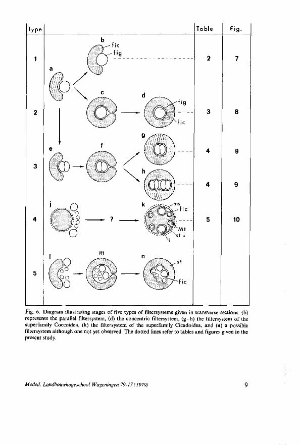
Fig. 6. Diagram illustrating stages of five types of filtersystems given in transverse sections, (b) represents the parallel filtersystem, (d) the concentric filtersystem, (g-h) the filtersystem of the superfamily Coccoidea, (k) the filtersystem of the superfamily Cicadoidea, and (n) a possible filtersystem although one not yet observed. The dotted lines refer to tables and figures given in the present study.
Meded. Landbouwhogeschool Wageningen 79-17 ( 1979)
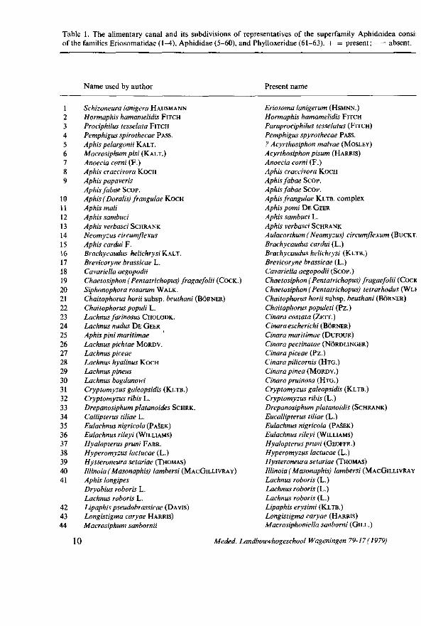
Table 1. The alimentary canal and its subdivisions of representatives of the superfamily Aphidoidea consi: of the families Eriosomatidae (1-4), Aphididae (5-60), and Phylloxeridae (61-63). + = present; - = absent.
Name used by author Present name
1 Schizoneura lanigera HAUSMANN 2 Hormaphis hamamelidis FITCH 3 Prociphilus tesselata FITCH 4 Pemphigus spirothecae PASS. 5 Aphis pelargonii KALT. 6 Macrosiphum pisi (KALT.) 7 Anoecia corni (F.) 8 Aphis craccivora KOCH 9 Aphis papaveris
Aphis fabae SCOP. 10 Aphis (Doralis) frangulae KOCH 11 Aphis mali 12 Aphis sambuci 13 Aphis verbasci SCHRANK 14 Neomyzus circumflexus 15 Aphis cardui F. 16 Brachycaudus helichrysiK-ALT. 17 Brevicoryne brassicae L. 18 Cavariella aegopodii 19 Chaetosiphon (Pentatrichopus) fragaefolii (COCK.) 20 Siphonophora rosarum WALK. 21 Chaitophorus horii subsp. beuthani (BORNER) 22 Chaitophorus populi L. 23 Lachnus farinosus CHOLODK. 24 Lachnus nudus DE GEER 25 Aphis pini rharitimae 26 Lachnus pichtae MORDV. 27 Lachnus piceae 28 Lachnus hyalinus KOCH 29 Lachnus pineus 30 Lachnus bogdanowi 31 Cryptomyzus galeopsidis (KLTB.) 32 Cryptomyzus ribis L. 33 Drepanosiphum platanoides SCHRK. 34 Callipterus tiliae L. 35 Eulachnus nigricola (PASEK) 36 Eulachnus rileyi (WILLIAMS) 37 Hyalopterus pruni FABR. 38 Hyperomyzus lactucae (L.) 39 Hysteroneura setariae (THOMAS) 40 Illinoia (Masonaphis) lambersi (MACGILLIVRAV) 41 Aphis longipes
Dryobius roboris L. Lachnus roboris L.
42 Lipaphis pseudobrassicae (DAVIS) 43 Longistigma caryae HARRIS) 44 Macrosiphum sanbornii
Eriosoma lanigerum (HSMNN.) Hormaphis hamamelidis FITCH Paraprociphilus tesselatus (FITCH) Pemphigus spyrothecae PASS. ? Acyrthosiphon malvae (MOSLEY) Acyrthosiphon pisum (HARRIS) Anoecia corni (F.) Aphis craccivora KOCH Aphis fabae Scop. Aphis fabae Scop. Aphis frangulae KLTB. complex Aphis pomi DE GEER Aphis sambuci L. Aphis verbasci SCHRANK Aulacorthum (Neomyzus) circumflexum (BUCKT. Brachycaudus cardui (L.) Brachycaudus helichrysi (KLTB.) Brevicoryne brassicae (L.) Cavariella aegopodii (SCOP.) Chaetosiphon (Pentatrichopus) fragaefolii (COCK Chaetosiphon (Pentatrichopus) tetrarhodus (WLI Chaitophorus horii subsp. beuthani (BORNER) Chaitophorus populeti (Pz.) Cinara costata (ZETT.) Cinara escherichi (BORNER) Cinara maritimae (DUFOUR) Cinara pectinatae (NÖRDLINGER) Cinara piceae (Pz.) Cinara pilicornis (HTG.) Cinara pinea (MORDV.) Cinara pruinosa (HTG.) Cryptomyzus galeopsidis (KLTB.) Cryptomyzus ribis (L.) Drepanosiphum platanoidis (SCHRANK) Eucallipterus tiliae (L.) Eulachnus nigricola (PASEK) Eulachnus rileyi (WILLIAMS) Hyalopterus pruni (GEOFFR.) Hyperomyzus lactucae (L.) Hysteroneura setariae (THOMAS) Illinoia (Masonaphis) lambersi (MACGILLIVRAY Lachnus roboris (L.) Lachnus roboris (L.) Lachnus roboris (L.) Lipaphis erysimi (KLTB.) Longistigma caryae (HARRIS) Macrosiphoniella sanborni (GILL.)
10 Meded. Landbouwhogeschool Wageningen 79-17 ( 1979)
Number. Oeso of cells in
phageal Sto Intes transverse Pyloric Hind Rec valve mach tine section valve gut Epithelium tum
Filter system Author
+ + + + + + + + + + +
+
+
+ + + + + + + + + + + + + + + + +
+ + + +
+ + + +
+ + + + + + + + + + + + + +
+ + + + + + + + + + + + + + + + + + + +
+ + + + + + + + + +
+ + + + + + + + + + + + + +
+ + + + + + + + + + + + + + + + + + + + + + + + + + + + + + +
3
12 4 -8 4 -8
5-7 3-5
4 -8
4 -8
6
4 -8
5
8-14
—
+
+
-
-
---
-
-
+ + + + + + + + + + + + + +
+ + +
+ + + + + + + + + + + + + + + + + + + + + + + + + + +
Hat
columnar flat flat
flat flat
flat
flat
flat
flat flat flat
flat flat flat flat flat
flat
flat
flat
Davidson, 1913 Lewis & Walton, 1958
- Pelton, 1938 - Witlaczil, 1882 - Witlaczil, 1882 - Schaefer, 1938 - Ponsen
Sorin, 1966 - Dufour, 1833 - Weber, 1928 - Roberti, 1946
Ramdohr, 1811 - Witlaczil, 1882
Schanderl et al., 1949 - Lindeman, 1948 - Witlaczil, 1882 - Schanderl et al., 1949 - Moericke & Mittler, 1966
Murant et al., 1976 - Ponsen
Grove, 1909 - Ponsen - Witlaczil, 1882 + Leonhardt, 1940 + Mordwilko, 1895
Dufour, 1833 + Leonhardt, 1940 + Leonhardt, 1940 + Leonhardt, 1940 + Mordwilko, 1895 + Mordwilko, 1895 + Ponsen + Ponsen, 1977 - Witlaczil, 1884 + Witlaczil, 1884, 1885 + Ponsen + Ponsen
Janiszewska, 1932 - Sylvester & Richardson, 1970
Tate, 1936 - Ponsen
Dufour, 1833 + Witlaczil, 1884, 1885 + Michel, 1942 - Moericke & Mittler, 1966 + Knowlton, 1925 - Miller, 1932
Meded. Landbouwhogeschool Wageningen 79-17 (1979) 11
Name used by author Present name
45 46 47 48 49 50
51 52 53 54 55 56 57 58 59 60 61 62 63
Macrosiphum solanifolii (ASH.) Aphis rosae Maculolachnus submacula (WLK.) Megoura viciae BUCKT. Macrosiphum tanaceti L. Aphis persicae Myzus persicae (SULZ.) Myzus persicae (SULZER) Myzus persicae (SULZ.) Aphis ribicola KALT. Plerocallis alni Pterocomma Salicis (L.) Phopalosiphoninus tulipaellus THEOB. Rhopalosiphum nympheae L. Schizaphis graminum (RONDANI) Lachnus pineti KOCH Lachnus quercus L. Trama troglodytes v. HEYD. Lachnus viminalis BOYER DE FONSC.
Phylloxera coccinea Phylloxera punctata Phylloxera vastatrix PLANCHON
Macrosiphum euphorbiae (THOS.) Macrosiphum, rosae (L.) Maculolachnus submacula (WLK.) Megoura viciae BUCKT. Metopeurum fuscoviride STROYAN Myzus persicae (SULZ.) Myzus persicae (SULZ.) Myzys persicae (SULZ.) Myzus persicae (SULZ.) Nasonovia ribisnigri (MOSLEY) Plerocallis alni (DE GEER) Pterocomma Salicis (L.) Rhopalosiphoninus tulipaellus THEOB. Rhopalosiphum nymphaeae (L.) Schizaphis graminum (ROND.) Schizolachnus pineti (F.) Stomaphis quercus (L.) Trama troglodytes v. HEYD. Tuberolachnus salignus (GMELIN) Phylloxera coccinea v. HEYD.
Viteus vitifoliae (FITCH)
iphumplatanoides (WITLACZIL, 1884; GERSCH, 1942), Callipterus tiliae, Dryobius roboris (WITLACZIL, 1884), and Plerocallis alni (GERSCH, 1942) (Table 1), as well as in the callaphidid genera Drepanosiphon ( = Drepanosiphum) and Symydobius (BORNER, 1938).
A well developed oesophageal valve, which marks the junction of the foregut and the midgut, is observed in many aphid species (Table 1). On the other hand, an oesophageal invagination is lacking in C. tiliae, as well as in representatives of Phylloxeridae (Table 1), Psylloidea (BRITTAIN, 1922; KLIMASZEWSKI and G L O W -
ACKA, 1977), Aleyroidea (WEBER, 1935 a, b), and Coccoidea (PESSON, 1944). The
oesophageal valve of S. ornata appears to be of simplest kind due to lack of invagination. The ring of columnar cells at the foregut-stomach junction seem to be clearly derived from the oesophagus secreting of intima (Fig. 3 A).
In general it was assumed that the oesophageal invagination may function in preventing the flow of plant juices back into the foregut once they have arrived in the midgut, although it is not supplied with muscle fibres (WEBER, 1928; R O B -ERTI, 1946; FORBES, 1964). The absence of such a structure in S. ornata is compensated presumably by a very long foregut. However, HARRIS and BATH (1973) stated that M. persicae can regurgitate ingested material (indian ink particles) during membrane feeding activity (ingestion-egestion theory ; HARRIS, 1977).
The dilated part of the midgut or stomach of M. persicae consists of three cell
12 Meded. Landbouwhogeschool Wageningen 79-17 ( 1979)
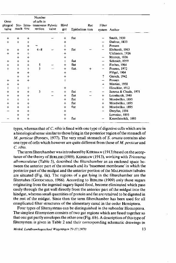
Oeso phageal valve
+ +
+ +
+ + +
+ + + + + + + + —
-
Stomach
+ +
+ + + + + + + + + + + + + + + + + + +
Intestine
+ + + + + + + + + + + +
+ + + + + + + + +
Number of cells in transverse Pyloric
section valve
_
-4-8
5 5
3 -
Hind gut
+ + + + + + + + + + + +
+ + + + + + + + +
Epithelium
Hat
Hat
Hat Hat flat.
flat flat flat flat flat
flat
Rec tum
-
+
--
Filter system
_ -+ -
---—
? ----+ + + + —
—
Author
Smith, 1939 Dufour, 1833 Ponsen Ehrhardt, 1963 Uichanco, 1924 Morren, 1836 Schmidt, 1959 Forbes, 1964 Ponsen, 1972 Flögel, 1904 Gersch, 1942 Ponsen Martini, 1958 Hirschler, 1912 Saxena & Chada, 1971 Leonhardt, 1940 Mordwilko, 1895 Mordwilko, 1895 Mordwilko, 1895 Dreyfus, 1894 Lemoine, 1893 Krassilstschik, 1893
types, whereas that of C. ribis is lined with one type of digestive cells which are in a histological sense similar to those lying in the posterior region of the stomach of M. persicae (PONSEN, 1977). The very small stomach of S. ornata contains also one type of cells which however are quite different from those of M. persicae and C. ribis.
The term filterchamber was introduced by KERSHAW (1913) based on the acceptance of the theory of BERLESE(1909). KERSHAW (1913), working with Tricentrus albomaculatus (Table 5), described the filterchamber as an enclosed space between the anterior part of the stomach and its 'basement membrane' in which the posterior part of the midgut and the anterior portion of the MALPIGHIAN tubules are situated (Fig. 6k). The regions of a gut lying in the filterchamber are the filtertubes (GOODCHILD, 1966). According to BERLESE (1909) only those sugars originating from the ingested sugary liquid food, become eliminated which pass easily through the gut wall directly from the anterior part of the midgut into the hindgut, whereas small quantities of protein and fat are retained to be digested in the rest of the midgut. Since then the term filterchamber has been used for all complicated filter structures of the alimentary canal in the order Hemiptera.
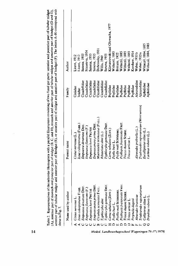
Four types of filtersystems can be distinguished in the suborder Homoptera. The simplest filtersystem consists of two gut regions which are fused together so that one gut partly envelopes the other one (Fig. 6 b). A description of this type of filtersystem is given in Table 2 and their corresponding schematic drawings in
Meded. Landbouwhogeschool Wageningen 79-17 ( 1979) 13
ga l cd'Q &
3 So >- "2 o e
& o
Crt
O O.
C M
<U
c ta
L i
ca
tx 00
.o
e
c ca
*_, 3 si;
•O
ld
uO
H ,̂
u. 3 011
•n c J 3 U i
O
ca
a L-O
^ K
•H ca L i ca a ca .e
S ca u <L>
D. O
e o X co
" O i -O
. O 3
ca
U « "3 0Û -o
e u i
o •£ ca
a L i
o a
"O e ca
o
W
^ 3 00
T3 c
_a
o
tH ca
a i -
<u e ca
-O
ca
3 00
T-t
Z 2
O- t .
«2-g 0.| i ,
_co ca •
O i n *n
oo oo
CN (N i n ^H
CT\ ON _ , « _ rt
i r i i n ON
ed Ö
«1
Os
cd
> u •£
o
u-> Wl Ov
ca
c co
X ca
t/3
>n i n O
ca
c CJ X ca
c/3
— O O
o Q
ON
rr ON
wf
S
£
l/"t i n ov.
cd G
X cd
-̂ c«
& <D N «5 cd
e 3 ,J hJ
IU U U U U U Ü cd cd cd cd cd cd cd
"O "O *Ü "O *Ü "O "O
<u S S S £ S =: := C d i D W C J C J U C J C J i U
' O c d T 3 " O T 3 * O T 3 T 3 T 3 • S * O c d c d c d c d c d c d c d x -^ o
OO (N 00 OO ÛO ^H J J « OO M 0 0 ( N O O O O C O O \ T ) ^ 0 0 0 0 — ' O N - — . ^ .—I •—• c i m ,— ••—'
—r ^ —r _" —T ~ °^ °^ ~-r _r 'N C 'N "N 'N M - - 'N 'N
£ I £ '£ % & $ $ 'Ê £
ü v u u u u u
O U O O >*>*>>>-, >~, >\ >*
ca T 3
T ) o L i 3
ca •o TJ O
3
co ca
T>
•o
si
o.
CO ra
T>
•o J3 P.
< <: < <
r/1 HH
< .H
H> _ ^ a a a es
^ U IJ Ü ^ - i
• ~ 3 c v D c- û ù Î3
a p I N
I Is s •§ -o
t, s ses
D S X
J t/3
-s g •H g .
•c - I .
a c eu
-a ^s ^ N N
P S> z> cv c^ a, a, a, m m
-si ,C s a X &-i ty -J
-1 5
"i K s CL,
eu K - 0 i
5 ^ 'S.-5 C3 K 3 ^3 . c ? S S 3 -r- •?
S ô5 j
Q c j a c ï ^ ^ ^ < j
[>i C^ t ^ C^ > ^ C , C , Ä S ^ | £ S 3 S 1 £ l .&i
'C ï -Si 'C o O
< < U U U U B U U Ï Q U Û Û Q M i . l i . O O
o, o, a, a I fcq tu tel tu O
.S .S .£ ti, a, t -
• J
K a
•Ci
,̂ u.
n m Q
5 a o
l/J •c; CI S
^ a.
u.
eu
.c X a
*
S" >. a.
•si
X
t / i
c s-<3 L i
o
I M
•2
; LV eu .^2
a -S ^
14 Meded. Landbouwhogeschool Wageningen 79-17(1979)
st —
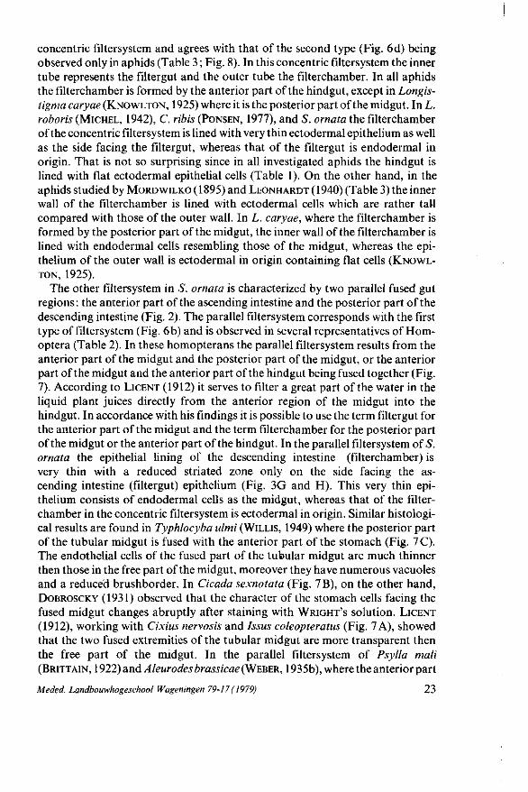
Fig. 7. Alimentary canal and its parallel filtersystem of A, Issus coleopteratus (after LICENT, 1912);B, Cicadula sexnotata (after DOBROSCKY, 1931); C, Typhlocyba ulmi (after WILLIS, 1949); D, Psyllopsis j'raxinicola (afterWITLACZIL, 1885); E, Psylliamali (after BRITTAIN, 1922); F, Aleurodes brassicae (after WEBER, 1935b); G, Callipterus tiliae (after WITLACZIL, 1884); H, Cerna nebulosa (after KLIMASZEWSKI and GLOWACKA, 1977). The stippling represents the hindgut. Mt, MALPIGHIAN tubules, other abbreviations as in Fig. 1. The letters A - H correspond with those in Table 2.
Fig. 7. In the aphids C. tiliae and D. roboris the stomach and the anterior part of the tubular midgut are fused with the anterior part of the hindgut (WITLACZIL,
1884, 1885). However, it appears that the present name of the latter aphid is Lachnus roboris belonging to the second type of filtersystem (MICHEL, 1942) (Table 3; Fig. 8).
The second type of filtersystem is only found in the family Aphididae (Table 1 and 3; Fig. 8). The anterior part of the hindgut first enfolds the anterior part of the midgut so the latter is completely surrounded (Fig. 6c) after which the two extremities fuse together. After this process the hindgut is closed (Fig. 6d) and the midgut lies inside the hindgut forming the filterchamber. The encapsulating of the stomach by the hindgut had already been supposed by MORDWILKO (1895) since in transverse sections of embryos of Trama troglodytes the stomach is
Meded. Landbouwhogeschool Wageningen 79-17 (1979) 15
M a t— etf
3 £> 3 h-
o "C (U
C es
c«
a i~ o
'ï-QJ WD
O 0. a»
J 3
c j ï
Bu »
m
S 3 t »
EU J 3
t+ -O
:•* M D. O
n
0 0 "O C
J = CU
X !
t * -O
D.
S .s
4>
(*> VH
a> S _o
(D
o c o Ü cd
• o <L>
rf S
t /3
g 3 0 0
-o
e
od oh
E c <D
O .e
•o -S o.
< t . o
e s aj <r CA
« M U
•S o H
4> O
D. O
OS
en
tu
y, E
'T? ^ 3 •2 "2 .o H S 3
Ô\ S ON Os Os Ci S
T3 ^5 'O *Q "O
J= -C -C c a c o o o
Os O N 0 0 0 0
o" o" Ü £é S S
• o - o
O O
r—
c tß C
•O
m a « M » * ON ^ ON
<N Ü. „ - * S - ° « 2 g £ -o
0 ~ H
Mic
hel,
Kon
wlt
M
ordw
L
eonh
a
i r> v - i i o 0 0 O N ON 0 \ 3" oo oo oo
- o" o" o" •S a àé a S > > S C T J T 3 - O C !— (-. t— ça o o o
z
s-' S
a su
,2 a
s- ^ * i
fi S S i 5 .s ? ». », » 5
< o o X fefe ty '£î 'C: Î3 >̂ tu
' S, .S .K a ». ». ^J ^ ^ „ s a S e c S -c -s oo ^. ^J \~ a es
1 IJ
a a 3 a a a c e s: e c c G Ö G C G G
a ». -c ;~ J? a O U - - )
t j to C5 03
s
? ^
•§ a t>j Co
-* 00 to ^t 2> *> V a o 3 $0 .a
*t a 0 0 s U 3
Ci
5
^S >i 3 a
-c <L>
a ^J
O
Q
-g 3 c to S C
-s: ^ j
a --1
> Q ce 0 (a
-S
'S. ^ 3 C
-S . <L» Q
•̂
X u 0
3 •S
-̂̂ 03 3 C
- C <u Q
<\
^ 3 c -« a -̂1
s tu
'S 0
n,-o to 3 s; -«; ^ j
a -J
^ 3 C
- C • 0 Q
-J
-i y*
b £ 0
.to
- 0 0
^ to 3 s; » -C
£ K
O O
t j
a »J
t «
5 « < X tu a a t j
S •Êf .to
a'
< tu
. S
». to 3 C
00 - c g •3
t j
a -J
X u 0
s, 6Q 3 C
-s: t j
Q <i
d,
S C
-s:
-2 ^
-s; ^ 1
co
J
C3-
a s R -s: Î J Q
^
Û
>-W
X >
-1 I3 s*
s t3
r s^ t-.
0 Pu u Q
s» 0 m
> S3 3 c
-c u Q
^ < p Q < ; < < ; o 3 o a u Q w c Q < ; p u P Q o a p Q
16 Meded. Landbouwhogeschool Wageningen 79-17 (1979)
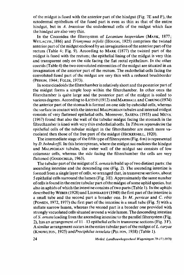
Fig. 8. Alimentary canal and its concentric filtersystem of A, Lachnus piceae (after LEONHARDT, 1940); B, Trama troglodytes (after M O R D W I L K O , 1895); C, Cryptomyzus ribis (PONSEN, 1977); D , Lachnus roboris (after M ICHEL , 1942); E, Longistigma caryae (after K N O W L T O N , 1925); F , Schizol-achnus sp. (after BRAMSTEDT, 1948). The stippling represents the hindgut. The letters A - F correspond with those in Table 3.
situated in a groove of the hindgut (Fig. 6 a) whereas in larvae the stomach lies inside the hindgut (Fig. 6d).
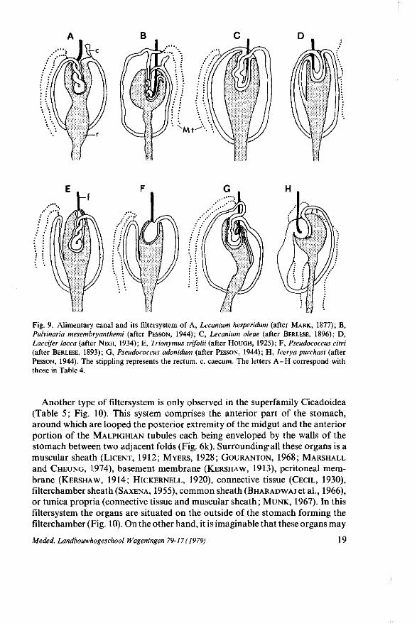
In the superfamily Coccoidea (Table 4) the filtersystem is a combination of the first and second type consisting of fused gut regions situated in an invagination of the rectum (Fig. 9). It is possible that the fused gut regions become encapsulated by the rectum on the same way as described for the second filtersystem (Fig. 6e-h).
Meded. Landbouwhogeschool Wageningen 79-17 ( 1979) 17
•s £ SP E o E S S s
•o o o
Ô c 5 > « 'S a u ?
O J3 -o
° a o
,-î o u a S K c o i M S < < « 2
B BfS " 3 • •S o Ë Ç S 3 ï B J « cd •-D, „ u t. 3 'S 2 .??£
5 C M "O <D -^ 2 S O .2 u 'C S i » » o S •S '-3 o
.y c <_ J= c o 5 o c o o -H
j-» p on e 6o a » ' S >
S'a-s — u ß
i ü >< - — T3
Cfl Vi
a. o 2 c
s cd U "2 'S o o c ^
u « a
e » ä <5 c Ë o. o u 3 o £ « U w a xi <— •£ " ° O O S O -rt DH (J ï , > c t: cd w '^ S - ^ S « g u UJ S o* "2 " ^ E'S o Ü 2 S £ S cö ( ^ O
H o
2 "3 b
- T I - *o m m ^ j - • * ,3-r t ON <N 0\ rt rf Tf
~"1 - ON * - ~"1 ~~1 "~1
o S ._- S? S n n n
ft< pa Z EC m
m Ul en t-< c/î M tfî
0> 0> 1) O cd cd a a
TJ TJ TJ "O ' o ' o '™ '™ o o o o o o o o O O O CJ
_ o o o o Ü Ü Ö H 3 3 3 3 ° .9 ,r rt * « « «
<D <D U - ^ a a a [•
T3 -O -O JH
N o H
eu
S 'S s. *
J -o z o o Vi Cfl
J s « S 'S -c -c eu
eu ~ Ö S,-S -Si 8 ;s o
2 s: 'S x
3 3 3 f
S B tu Ä, 3
1.1
5 -g 5
^ a , M
IM W a x
«j ** s -S 3 ä ^ s -̂* S .2 g ^ K, ft.
ai 'S £
a, », < B u D U ï . O Û Ï
18 Meded. Landbouwhogeschool Wageningen 79-17 (1979)
Mt-^'--
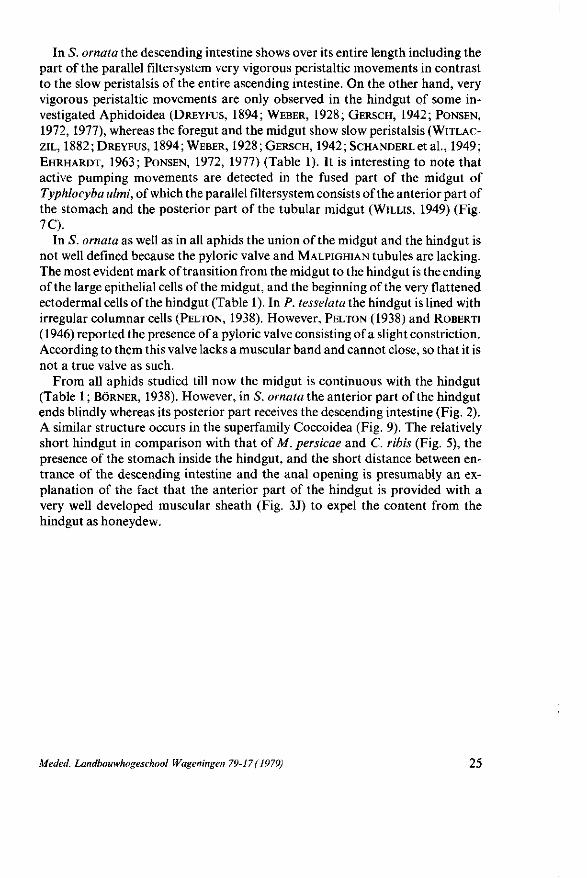
Fig. 9. Alimentary canal and its filtersystem of A, Lecanium hesperidum (after MARK, 1877); B, Pulvinaria mesembryanthemi (after PESSON, 1944); C, Lecanium oleae (after BERLESE, 1896); D, Laccifer lacca (after NEGI, 1934); E, Trionymus trifolii (after HOUGH, 1925); F, Pseudococcus citri (after BERLESE, 1893); G, Pseudococcus adonidum (after PESSON, 1944); H, Icerya purchasi (after PESSON, 1944). The stippling represents the rectum, c, caecum. The letters A - H correspond with those in Table 4.
Another type of filtersystem is only observed in the superfamily Cicadoidea (Table 5 ; Fig. 10). This system comprises the anterior part of the stomach, around which are looped the posterior extremity of the midgut and the anterior portion of the MALPIGHIAN tubules each being enveloped by the walls of the stomach between two adjacent folds (Fig. 6k). Surrounding-all these organs is a muscular sheath (LICENT, 1912; MYERS, 1928; GOURANTON, 1968; MARSHALL
and CHEUNG, 1974), basement membrane (KERSHAW, 1913), peritoneal membrane (KERSHAW, 1914; HICKERNELL, 1920), connective tissue (CECIL, 1930), filterchamber sheath (SAXENA, 1955), common sheath (BHARADWAJ et al., 1966), or tunica propria (connective tissue and muscular sheath; MUNK, 1967). In this filtersystem the organs are situated on the outside of the stomach forming the filterchamber (Fig. 10). On the other hand, it is imaginable that these organs may
Meded. Landbouwhogeschool Wageningen 79-17 (1979) 19
T3 cd
cd cj tu 43
O G
cd J3 •g ä. 2 6 ö 2 cd ( Ä i 6 — • o s
T "» •
U £ C •^ CJ •â1 s « S " 9 £ a .2 u G r
X I O u
.s e
3 s
2 H
G oo 5 D. • r^
C L cd
es O ~ cd cd
•s 0 .2 "S » • o » S n c ï 5 u cd g 3
U N ; C « I « e
Ë ."2 es CJ
c*- cd
n -° §• e 60 c j
| S O (N
E O (U CJ
' t ! CJ
O *-"
S 1 « o»
„.S ±3 "O
S 3
x> ca H
o t ' C 3 U C/5
â2
•o § U c j CX cd
J'if
«5 (fl M
0 0 Ï N os
^
i / l <N 0 0
uT 3 ,9
CN
OS
""̂ C
d M
ar:
d M
ar:
d M
ar:
•25 19
20
d M
ar:
d M
ar:
ndC
h<
55
C G C J O — G C c d o s cd cd cd c j cd cd —— ^ ^
0 0 0 0 0 0 C C 0 0 0 0 * « „ r C C C 3 h Q C j 3 2
un IT) o\
cd" G e j
•* os
cd
_ X ] X - CTv
, - , . * 1 ^ ^ — ^ **•>
c^ooo os
00 SO Os u
,_^ T-H fT) O __ OS
. . os -a -= —'
> 3 u J3 j - J* 3 D J U U ü D
CJ 3 CJ CJ
e cd 3 o
- S « -S S
cd
•S 2 cd
">5 fi fi "5 « * s> 'T £ 'T 'H « « f{ cd' cd cd i « . ï os .g •» «j -g
o Q
0S •g
• t -
"2 J o — -S 5 ta o ^ j t3
•S ^ o -H
O s j
J
o 1 3
G
•S .S 3
S a ta C3 ^ w
S S a S, « ö Ml f-S s ry
pt
yclo
ae
ar,
Ü Ü Ö
-s- < <
o •» a Q
t - S" ^
3 ^ 8 = f\s tJi - j . -W ^ o. 8 -s e
i g S s « ° •* s2 K AU §
a:
o i s s I
J 5 3 1 S ,5 -~ •= S, a
a o ° I-2 » O -S 5 U • S ^ -SP K -a
eu Qj
z, o
' Q 3 •« J
5 fcî -s " Q 3 o
H Ü
< •S -5
5s S a
STAP ^j
111 C 3 a =-» V *-
TS
=• ^i ^ ^ Q .g .3 .s
2 3 "5 S ^ s a
-s -s .a 3
S- S- s, S -s -È S -S ^ -T; U Ü
- c o s s j s j Ü
^ s" 51 5 «S.'S.
Cy CU ^ ^ : - c û . o , ûs
Q
« « ^
b & 3 !"§.£, 2
'• 1-15 a -S .= ^ 8 î S ^ s?ï i 1
Ü Ü S 1
e s -g
a « -~ .H 5 I n
a S Q S -2 !£ a 3̂
tu , b o 5,4!
S- P
f~i ^
a -c -s: o » s £ S g ^ r
a, a,,!:-« ^ X K O O O 1 -- S SV,
" • ^ ^ 2 -a -S
r n - ^ - u - i ' O f - . o o o O ' — , r s r ^ ^ - i / ~ ( , s O t ~ - 1
t i .
I s s .a -o i i : c w ^ Q cy ^ a o . Oo S o
p o 2 ^ * ts E m a, ü K ^
J ^ X
Jî
•S
-Si
3 c tu .g -S a.
J .g
5 s
3 c tu •S ^ a-
z o Q
^ t>c ^ ^ a. >o tN
Cri CO <
tL. tu O
«a S tu ta) ts X. C3 Si V? t3 O SO <N
Q 3
3 tu
£ o tS
3 S <i> •H r£ ^ r-<N
SI
Q Z < >
•S
o u # Q
a & ^
00 fN
20 Meded. Landbouwhogeschool Wageningen 79-17 (1979)
in _ "1 " i ^ Ä , « i ÏÏ 1 Ä 1 ^ ^ " i ^ ^ ^ "^ w i / - l r " - C ' - « n i n ( N f N ' < N 0 - J < N i n i / - ) i / - ) i n i n » o i n ^
es ^ - j j S s j - es M M" es M" ca M" J " C C G n - c C c c c c c c c G
~ r? T! -1—' 4—• î r n* n i n i n i n j n j nt TT " 3 c x x s t l K -
<L> (U u <*-•-, S s
OJ
- ^ T O ' T O ^ T O ^ T O ' T O ' T O ' O S " ~" TO TO • " ; - - ^ ' T " « h- • " TO TO TO TO TO TO TO ••-*
u u
Je 3
_3 a -c S-eu O
"ft*
ca £ ¥ a S0
. e u
a
3
eu eu
^
^. ffl S u crt
a!
g ¥ •S -S
<5J
Î J *n
H
5 •S3
• s : Ci,
su
H
Q
¥ •S
C .a ' S
j »i
< S
Ik.
3
Ou Î J Ci
S t u
"s •ii
•SP
^ Ç>
. < U , H H
§ 1 .S S Q .v N ~ eu ^
x T> S F*. •->
S e s «U
• à §
.60
S s -g S s~s ^ iu ^ o o -s eu eu
eu 3 £; •a o
< ft.
«I 3=S o § ï § S* c « ~ t .£p s s S a S I I §• es fcu - s : - s : ; ^ -
S S s« as
0 . ^ o ^ w J 3 t-, -J S 3 < es ^ - r,
i ^^T ^ eu eg
; g < «? -5 0, x • ~ 3
es a -S! -s: g" 5 § * .3 «a. a. 2 =- ~
tg .g _H eu
a s> ^! ~ Ü eu eu eu -r- ^ ^ *-
J
•< Un
R, es s; eu eu g Q ^
2 - 2 es ÇS es
S '5 "" a Q ? S -2 ?
s>- eu - ^ ^ ^ •S> 5; a c 3
ju "3 .g .* g
a S •S S
-s
I' eu c •S? g "~ es
es Q,
Meded. Landbouwhogeschool Wageningen 79-17 (1979) 21
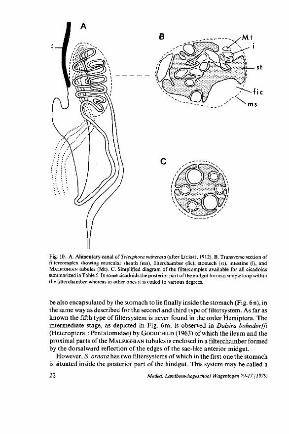
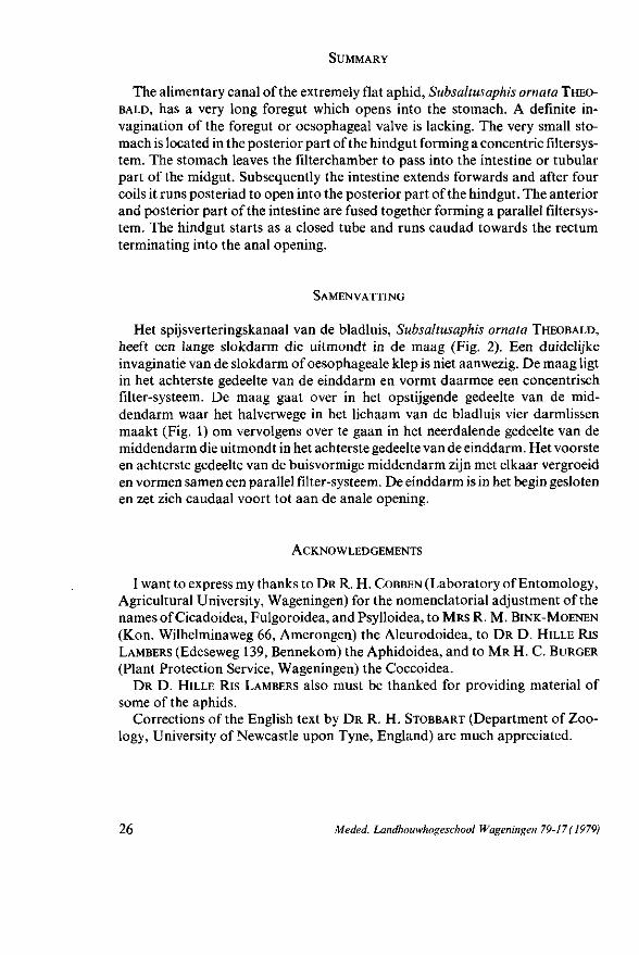
Fig. 10. A. Alimentary canal of Triecphora vulnerata (after LICENT, 1912). B. Transverse section of filtercomplex showing muscular sheath (ms), filterchamber (fie), stomach (st), intestine (i), and MALPIGHIAN tubules (Mt). C. Simplified diagram of the filtercomplex available for all cicadoids summarized in Table 5. In some cicadoids the posterior part of the midgut forms a simple loop within the filterchamber whereas in other ones it is coiled to various degrees.
be also encapsulated by the stomach to lie finally inside the stomach (Fig. 6n), in the same way as described for the second and third type of filtersystem. As far as known the fifth type of filtersystem is never found in the order Hemiptera. The intermediate stage, as depicted in Fig. 6 m, is observed in Dalsira bohndorffi (Heteroptera : Pentatomidae) by GOODCHILD (1963) of which the ileum and the proximal parts of the MALPIGHIAN tubules is enclosed in a filterchamber formed by the dorsalward reflection of the edges of the sac-like anterior midgut.
However, S. ornata has two filtersy stems of which in the first one the stomach is situated inside the posterior part of the hindgut. This system may be called a
22 Meded. Landbouwhogeschool Wageningen 79-17 ( 1979)
concentric filtersystem and agrees with that of the second type (Fig. 6d) being observed only in aphids (Table 3 ; Fig. 8). In this concentric filtersystem the inner tube represents the filtergut and the outer tube the filterchamber. In all aphids the filterchamber is formed by the anterior part of the hindgut, except in Longis-tigma caryae (KNOWLTON, 1925) where it is the posterior part of the midgut. In L. roboris (MICHEL, 1942), C. ribis (PONSEN, 1977), and S. ornata the filterchamber of the concentric filtersystem is lined with very thin ectodermal epithelium as well as the side facing the filtergut, whereas that of the filtergut is endodermal in origin. That is not so surprising since in all investigated aphids the hindgut is lined with flat ectodermal epithelial cells (Table 1). On the other hand, in the aphids studied by MORDWILKO (1895) and LEONHARDT (1940) (Table 3) the inner wall of the filterchamber is lined with ectodermal cells which are rather tall compared with those of the outer wall. In L. caryae, where the filterchamber is formed by the posterior part of the midgut, the inner wall of the filterchamber is lined with endodermal cells resembling those of the midgut, whereas the epithelium of the outer wall is ectodermal in origin containing flat cells (KNOWL
TON, 1925).
The other filtersystem in S. ornata is characterized by two parallel fused gut regions : the anterior part of the ascending intestine and the posterior part of the descending intestine (Fig. 2). The parallel filtersystem corresponds with the first type of filtersystem (Fig. 6 b) and is observed in several representatives of Hom-optera (Table 2). In these homopterans the parallel filtersystem results from the anterior part of the midgut and the posterior part of the midgut, or the anterior part of the midgut and the anterior part of the hindgut being fused together (Fig. 7). According to LICENT (1912) it serves to filter a great part of the water in the liquid plant juices directly from the anterior region of the midgut into the hindgut. In accordance with his findings it is possible to use the term filtergut for the anterior part of the midgut and the term filterchamber for the posterior part of the midgut or the anterior part of the hindgut. In the parallel filtersystem of 5*. ornata the epithelial lining of the descending intestine (filterchamber) is very thin with a reduced striated zone only on the side facing the ascending intestine (filtergut) epithelium (Fig. 3G and H). This very thin epithelium consists of endodermal cells as the midgut, whereas that of the filter-chamber in the concentric filtersystem is ectodermal in origin. Similar histological results are found in Typhlocyba ulmi (WILLIS, 1949) where the posterior part of the tubular midgut is fused with the anterior part of the stomach (Fig. 7C). The endothelial cells of the fused part of the tubular midgut are much thinner then those in the free part of the midgut, moreover they have numerous vacuoles and a reduced brushborder. In Cicada sexnotata (Fig. 7B), on the other hand, DOBROSCKY (1931) observed that the character of the stomach cells facing the fused midgut changes abruptly after staining with WRIGHT'S solution. LICENT
(1912), working with Cixius nervosis and Issus coleopteratus (Fig. 7 A), showed that the two fused extremities of the tubular midgut are more transparent then the free part of the midgut. In the parallel filtersystem of Psylla mali (BRITTAIN, 1922) and Aleurodes brassicae (WEBER, 1935b), where the anterior part
Meded. Landbouwhogeschool Wageningen 79-17(1979) 23
of the midgut is fused with the anterior part of the hindgut (Fig. 7E and F), the ectodermal epithelium of the fused part is even as thin as that of the entire hindgut, but in A. brassicae the endodermal cells of the midgut which face the hindgut are also very thin.
In the Coccoidea the filtersystem of Lecanium hesperidum (MARK, 1877; WITLACZIL,1886) and Trionymus trifolii (HOUGH, 1925) comprises the twisted anterior part of the midgut enclosed by an invagination of the anterior part of the rectum (Table 4; Fig. 9). According to MARK (1877) the twisted part of the midgut is fused with the rectum ; the epithelial lining of the midgut is very thin and transparent only on the side facing the flat rectal epithelium. In the other coccids (Table 4) the two convoluted extremities of the midgut are situated in an invagination of the anterior part of the rectum. The endothelial cells facing the convoluted fused part of the midgut are very thin with a reduced brushborder (PESSON, 1944; FOLDI, 1973).
In some cicadoids the filterchamber is relatively short and the posterior part of the midgut forms a simple loop within the filterchamber. In other ones the filterchamber is quite large and the posterior part of the midgut is coiled to various degrees. According to LICENT (1912) and MARSHALL and CHEUNG (1974) the anterior part of the stomach is formed on one side by cuboidal cells, whereas the surface in contact with the internal MALPIGHIAN tubules and internal midgut consists of very flattened epithelial cells. Moreover, SAXENA (1955) and MUNK
(1967) found that also the wall of the tubular midgut facing the stomach in the filterchamber is lined with very thin endothelial cells. In Tibicen septendecim the epithelial cells of the tubular midgut in the filterchamber are much more vacuolated then those of the free part of the midgut (HICKERNELL, 1920).
The intermediate stage of the fifth type of filtersystem (Fig. 6m) is represented by D. bohndorffi. In this heteropteran, where the midgut sac encloses the hindgut and MALPIGHIAN tubules, the outer wall of the midgut sac consists of low columnar cells, whereas the side facing the filterchamber the cells are very flattened (GOODCHILD, 1963).
The tubular part of the midgut of S. ornata is build up of two distinct parts : the ascending intestine and the descending one (Fig. 2). The ascending intestine is formed from a single layer of cells, so arranged that, in transverse sections, about 5 epithelial cells surround the lumen (Fig. 3 E). Approximately the same number of cells is found in the entire tubular part of the midgut of some aphid species, but also in aphids of which the intestine consists of two parts (Table 1). In the aphids described by WEBER (1928) and LEONHARDT (1940) the first part of the intestine is a small tube and the second part a broader one. In M. persicae and C. ribis (PONSEN, 1972, 1977) the first part of the intestine is a small tube (Fig. 5) with a stellate narrow lumen, whereas the second part is a broader one provided with strongly vacuolated cells situated around a wide lumen. The descending intestine of S. ornata leading from the ascending intestine to the parallel filtersystem (Fig. 2), has an arrangement of 11 -13 epithelial cells in transverse sections (Fig. 3 F). A similar arrangement occurs in the entire tubular part of the midgut of L. caryae (KNOWLTON, 1925) andProciphilus tesselata (PELTON, 1938) (Table 1).
24 Meded. Landbouwhogeschool Wageningen 79-17 (1979)
In S. ornata the descending intestine shows over its entire length including the part of the parallel filtersystem very vigorous peristaltic movements in contrast to the slow peristalsis of the entire ascending intestine. On the other hand, very vigorous peristaltic movements are only observed in the hindgut of some investigated Aphidoidea (DREYFUS, 1894; WEBER, 1928; GERSCH, 1942; PONSEN,
1972, 1977), whereas the foregut and the midgut show slow peristalsis (WITLAC-ZIL, 1882; DREYFUS, 1894; WEBER, 1928; GERSCH, 1942; ScHANDERLet al., 1949;
EHRHARDT, 1963; PONSEN, 1972, 1977) (Table 1). It is interesting to note that active pumping movements are detected in the fused part of the midgut of Typhlocyba ulmi, of which the parallel filtersystem consists of the anterior part of the stomach and the posterior part of the tubular midgut (WILLIS, 1949) (Fig. 7C).
In 5*. ornata as well as in all aphids the union of the midgut and the hindgut is not well defined because the pyloric valve and MALPIGHIAN tubules are lacking. The most evident mark of transition from the midgut to the hindgut is the ending of the large epithelial cells of the midgut, and the beginning of the very flattened ectodermal cells of the hindgut (Table 1). In P. tesselata the hindgut is lined with irregular columnar cells (PELTON, 1938). However, PELTON (1938) and ROBERTI (1946) reported the presence of a pyloric valve consisting of a slight constriction. According to them this valve lacks a muscular band and cannot close, so that it is not a true valve as such.
F rom all aphids studied till now the midgut is continuous with the hindgut (Table 1 ; BORNER, 1938). However, in S. ornata the anterior part of the hindgut ends blindly whereas its posterior part receives the descending intestine (Fig. 2). A similar structure occurs in the superfamily Coccoidea (Fig. 9). The relatively short hindgut in comparison with that of M. persicae and C. ribis (Fig. 5), the presence of the stomach inside the hindgut, and the short distance between entrance of the descending intestine and the anal opening is presumably an explanation of the fact that the anterior part of the hindgut is provided with a very well developed muscular sheath (Fig. 3J) to expel the content from the hindgut as honeydew.
Meded. Landbouwhogeschool Wageningen 79-17 ( 1979) 2 5
SUMMARY
The alimentary canal of the extremely flat aphid, Sub sal tus aphis ornata THEO
BALD, has a very long foregut which opens into the stomach. A definite invagination of the foregut or oesophageal valve is lacking. The very small stomach is located in the posterior part of the hindgut forming a concentric filtersy s-tem. The stomach leaves the filterchamber to pass into the intestine or tubular part of the midgut. Subsequently the intestine extends forwards and after four coils it runs posteriad to open into the posterior part of the hindgut. The anterior and posterior part of the intestine are fused together forming a parallel filtersys-tem. The hindgut starts as a closed tube and runs caudad towards the rectum terminating into the anal opening.
SAMENVATTING
Het spijsverteringskanaal van de bladluis, Subsaltusaphis ornata THEOBALD,
heeft een lange slokdarm die uitmondt in de maag (Fig. 2). Een duidelijke invaginatie van de slokdarm of oesophageale klep is niet aanwezig. De maag ligt in het achterste gedeelte van de einddarm en vormt daarmee een concentrisch fïlter-systeem. De maag gaat over in het opstijgende gedeelte van de middendarm waar het halverwege in het lichaam van de bladluis vier darmlissen maakt (Fig. 1) om vervolgens over te gaan in het neerdalende gedeelte van de middendarm die uitmondt in het achterste gedeelte van de einddarm. Het voorste en achterste gedeelte van de buisvormige middendarm zijn met elkaar vergroeid en vormen samen een parallel fïlter-systeem. De einddarm is in het begin gesloten en zet zich caudaal voort tot aan de anale opening.
ACKNOWLEDGEMENTS
I want to express my thanks to D R R. H. COBBEN (Laboratory of Entomology, Agricultural University, Wageningen) for the nomenclatorial adjustment of the names of Cicadoidea, Fulgoroidea, and Psylloidea, to MRS R. M. BINK-MOENEN
(Kon. Wilhelminaweg 66, Amerongen) the Aleurodoidea, to D R D. HILLE RIS LAMBERS (Edeseweg 139, Bennekom) the Aphidoidea, and to M R H. C. BURGER
(Plant Protection Service, Wageningen) the Coccoidea. DR D. HILLE RIS LAMBERS also must be thanked for providing material of
some of the aphids. Corrections of the English text by D R R. H. STOBBART (Department of Zoo
logy, University of Newcastle upon Tyne, England) are much appreciated.
26 Meded. Landbouwhogeschool Wageningen 79-17 ( 1979)
REFERENCES
BERLESE, A. (1893). Le Cocciniglie Italiane viventi sugli agrumi. Riv. Patol. veg., Padova 2:129-193. BERLESE, A. (1896). Le Cocciniglie Italiane viventi sugli agrumi. Riv. Patol. veg., Padova 3:129-171. BERLESE, A. (1909). Gli Insetti. Loro organizzazione sviloppo abitvdini e rapporti coll' vomo. 1.
Embridine e morfologia. Milano, 1004 pp. BHARADWAJ, R. K., D. V. R. REDDY and R. C. SINHA (966). A reinvestigation of the alimentary canal
in the leafhopper Agallia conslricta (Homöptera : Cicadellidae). Ann. ent. Soc. Am. 59:616-617. BORNER, C. (1938). Neuer Beitrag zur Systematik und Stammesgeschichte der Blattläuse. Abh.
Naturw. Ver. Bremen 30: 167-179. BORNER, C. (1952). Europae centralis Aphides (Die Blattläuse Mitteleuropas). Mitt. thüring. bot.
Ges., Beiheft 3: 1-484. BRAMSTEDT, F. (1948). Über die Verdauungsphysiologie der Aphiden. Z. Naturf. 3: 14-24. BRITTAIN, W. H. (1922). The morphology and synonymy of Psyllia mali SCHMIDBERGER. Proc.
Acadian ent. Soc. 8: 23-50. CECIL, R. (1930). The alimentary canal of Philaenus leucophthalmus L. Ohio J. Sei. 30: 120-128. CHEUNG, W. W. K. and A. T. MARSHALL (1973 a). Studies on water and ion transport in homopteran
insects : ultrastructure and cytochemistry of the Cicadoid and Cercopoid midgut. Tissue & Cell 5 : 651-669.
CHEUNG, W. W. K. and A. T. MARSHALL (1973 b). Water and ion regulation in cicadas in relation to xyleem feeding. J. Insect Physiol. 19: 1801-1816.
DAVIDSON, J. (1913). The structure and biology of Schizoneura lanigera HAUSMANN or Woolly Aphis of the apple tree. 1. The apterous viviparous female. Q. Jl microsc. Sei. 58: 653-702.
DOBROSCKY, I. D. (1931). Morphological and cytological studies on the salivary glands and alimentary tract of Cicadula sexnotata (FALLEN), the carrier of aster yellows virus. Contr. Boyce Thomson Inst. 3: 39-58.
DREYFUS, L. (1894). Zu J. KRASSILSTSCHIK'S Mittheilungen über 'die vergleichende Anatomie und Systematik der Phytophthires' mit besonderer Bezugnahme auf die Phylloxeriden. Zool. Anz. 17: 237-243.
DUFOUR, M. L. (1825). Recherches anatomiques sur les Cigales. Annls Sei. nat. 5: 155-171. DUFOUR, M. L. (1833). Recherches anatomiques et physiologiques sur les Hémiptères. Paris, Tome
4: 333 pp. EASTOP, V. F. and D. HILLE RIS LAMBERS (1976). Survey of the World's Aphids. DR. W. JUNK by,
The Hague, 573 pp. EHRHARDT, P. (1963). Untersuchungen über bau und function des Verdauungstraktes von Megoura
viciae Bückt. (Aphidae, Homotera) unter besonderer Berücksichtigung der Nahrungsaufnahme und der Honigtauabgabe. Z. Morph. Ökol. Tiere 52: 597-677.
FLÖGEL, J. H. L. (1904). Monographie der Johannisbeeren-Blattlaus, Aphis ribis L. Z. Ent. 9: 321-334.
FOLDI, I. (1973). Etude de la chambre filtrante de Planococcus citri (Insecta, Homöptera). Z. Zellforsch. 143: 549-568.
FORBES, A. R. (1964). The morphology, and fine structure of the gut of the green peach aphid, Myzus persicae (SULZER) (Homöptera: Aphididae). Mem. ent. Soc. Can. 36: 1-74.
GERSCH, M. (1942). Verteilung und Ausscheidung von Fluorescein bei Aphiden. II. Beitrag zur Exkretion bei Insekten. Z. vergl. Physiol. 29: 506-531.
GOODCHILD, A. J. P. (1963). Some new observations on the intestinal structures concerned with water disposal in sap-sucking Hemiptera. Trans. R. ent. Soc. Lond. 115: 217-237.
GOODCHILD, A. J. P. (1966). Evolution of the alimentary canal in the Hemiptera. Biol. Rev. 41: 97-140.
GOURANTON, J. (1968). Observations histochimiques et histoenzymologiques sur le tube digestif de quelques homoptères Cercopides et Jassides. J. Insect Physiol. 14: 569-579.
GROVE, A. J. (1909). The anatomy of Siphonophora rosarum WALK., the green-fly pest of the rose-tree. I. The apterous viviparous stage. Parasitology 2: l -2£.
Meded. Landbouwhogeschool Wageningen 79-17 (1979) 27
GUT, J. (1976). Chromosome numbers of parthenogenetic females of fifty five species of Aphididae (Homoptera) new to cytology. Genetica 46: 279-285.
HARRIS, K. F. (1977). An ingestion-egestion hypothesis of noncirculative virus transmission. Chapter 7 in 'Aphids as Virus Vectors' (K. F. HARRIS and K. MARAMOROSCH, eds.). Academic Press, New York.
HARRIS, K. F. and J. E. BATH (1973). Regurgitation by Myzuspersicae during membrane feeding: its likely function in transmission of nonpersistent plant viruses. Ann. ent. Soc. Am. 66: 793-796.
HICKERNELL, L. M. (1920). The digestive system of the periodical Cicada, Tibicen septendecim LINN. I. Morphology of the system in the adult insect. Ann. ent. Soc. Am. 13: 223-242.
HILLE Ris LAMBERS, D. (1935). New english aphidae (Hem.). Stylops 4: 114-120. HIRSCHLER, J. (1912). Embryologische Untersuchungen an Aphiden nebst theoretischen Erwäg
ungen über den morphologischen Wert der Dotterelemente (Dotterzellen, Vitellophagen, Dotterepithel, Merocyten, Parablast) im allgemeinen. Z. wiss. Zool. 100: 393-446.
HOUGH, W. S. (1925). The internal anatomy of the clover root mealy-bug, Trionymus trifolii FORBES (Homoptera, Coccidae). Bull. ent. Res. 16: 25-29.
JANISZEWSKA, J. (1932). Untersuchungen über die Hymenoptera Aphidius sp., Parasiten der Blattlaus Hyalopteruspruni FABR. Bull. int. Acad. pol. Sei. Lett. 7 B II: 277-293.
KERSHAW, J. G. C. (1913). Anatomical notes on a Membracid. Ann. Soc. ent. Belg. 57: 191-201. KERSHAW, J. G. C. (1914). The alimentary canal of a Cercopid. Psyche, Camb. 21: 65-72. KLIMASZEWSKI, S. M. and E. GLOWACKA (1977). Der Darmtrakt der Larven und Adulten von Cerna
nebulosa (ZETT.) (Homoptera, Aphalaridâe). Annls zool., Warsz. 33: 455-461. KNOWLTON, G. F. (1925). The digestive tract of Longistigma caryae (HARRIS). Ohio J. Sei. 25:
244-252. KRASSILSTSCHIK, J. (1893). Zur vergleichenden Anatomie und Systematik der Phytophthires. (Über
die Verwandtschaftsbeziehungen der Phylloxera zu den Aphiden und Cocciden). Zool. Anz. 16: 85-92.
LEMOINE, V. (1893). Etude comparée du développement de l'oeuf dans la forme agame aptère, dans la forme agame ailée et dans le forme sexuée du Phylloxera. Zool. Anz. 16: 145-149.
LEONHARDT, H. (1940). Beiträge zur Kenntnis der Lachniden, der wichtigsten Tannenhonigtauer-zeuger. Z. angew. Ent. 27: 208-272.
LEWIS, I. F. and L. WALTON (1958). Gall-formation on Hamamelis virginiana resulting from material injected by the aphid Hormaphis hamamelidis. Trans. Am. microsc. Soc. 77: 146-200.
LICENT, E. (1912). Recherches d'anatomie et de physiologie comparées sur le tube digestif des Homoptères supérieurs. La Cellule 28: 1-161.
LINDEMANN, C. (1948). Beitrag zur Ernährungsphysiologie der Blattläuse. Z. vergl. Physiol. 31: 112-133.
MARK, E. L. (1877). Beiträge zur Anatomie und Histologie der Pflanzenläuse, insbesondere der Cocciden. Arch, mikrosk. Anat. 13: 31-86.
MARSHALL, A. T. and W. W. K. CHEUNG (1973). Calcification in insects: the dwelling-tube and midgut of machaerotid larvae (Homoptera). J. Insect Physiol. 19: 963-972.
MARSHALL, A. T. and W. W. K. CHEUNG (1974). Studies on water and ion transport in homopteran insects : ultrastructure and cytochemistry of the cicadoid and cercopoid MALPIGHIAN tubules and filter chamber. Tissue & Cell 6: 153-171.
MARTINI, CHR. (1958). Beobachtungen über das Saugen bei Blattläusen. Z. PflKrankh. PflPath. PflSchutz 65: 90-92.
MICHEL, E. (1942). Beiträge zur Kenntnis von Lachnus (Pterochlorus) roboris L., einer wichtigen Honigtauerzeugerin an der Eiche. Z. angew. Ent. 29: 243-281.
MILLER, F. W. (1932). The digestive epithelium of the aphid, Macrosiphum sanbomii. Proc. Pa Acad. Sei. 6: 148-151.
MOERICKE, V. and T. E. MITTLER (1966). Oesophageal and stomach inclusions of aphids feeding on various cruciferae. Entomologia exp. appl. 9: 287-297.
MORDWILKO, A. (1895). Zur Anatomie der Pflanzenläuse, Aphiden. Zool. Anz. 18: 345-364. MORREN, M. CH. (1836). Mémoire sur l'émigration du puceron du pêcher (Aphispersicae), et sur les
caractères et l'anatomie de cette espèce. Annls Sei. nat. Zool. 6: 65-93.
28 Meded. Landbouwhogeschool Wageningen 79-17 (1979)

M U N K , R. (1967). Zur Morphologie und Histologie des Verdauungstraktes zweier Jassiden (Homop-tera Auchenorrhyncha) unter besonderer Berücksichtigung der sogenannten Fi l terkammer. Z. wiss. Zool. 175: 4 05 -424 .
M U R A N T , A. F . , I. M. ROBERTS, and S. ELNAGAR (1976). Association of virus-like particles with the
foregut of the aphid Cavariella aegopodii (Horn.) t ransmit t ing the semi-persistent viruses an th-riscus yellows and parsnip yellows fleck. J. gen. Virol. 3 1 : 4 7 - 5 7 .
MYERS , J. G. (1928). The morphology of the Cicadidae (Homoptera) . Proc. zool. Soc. Lond. 25: 365-472 .
N E G I , P. S. (1934). The alimentary canal, its appendages, salivary glands and the nervous system of the adult female lac insect, Laccifer lacca K E R R (Coccidae). Bull. ent. Res. 25: 541-550.
NUORTEVA, P. (1954). On the significance of proteases and amylases in the life of Empoascaflavescens (F.) (Hom., Typhlocybidae). Ann. ent. fenn. 20: 7 6 -79 .
PELTON, J. Z . (1938). The alimentary canal of the aphid Prociphilus tesselata F I T C H . Ohio J. Sei. 38 : 164-169.
PESSON, P. (1944). Contr ibut ion à l 'étude morphologique et fonctionnelle de la tête, de l 'appareil buccal et du tube digestif des femelles de Coccides. Paris, 266 pp .
PONSEN, M. B. (1972). The site of po ta to leafroll virus multiplication in i tsvector, Myzuspersicae. An anatomical study. Meded. LandbHogesch. Wageningen 72 -16 : 1-147.
PONSEN, M. B. (1977). The gut of the red currant blister aphid, Cryptomyzus ribis (Homopte ra : Aphididae). Meded. LandbHogesch. Wageningen 7 7 -11: 1-11.
RAMDOHR, K. A. (1811). Abhandlung über die Verdauungswerkzeuge der Insecten. Halle, 221 pp . ROBERTi, D . (1946). Monografia dell Aphis (Dorsalis) frangulae K O C H . I. Morfologia, anatomia ,
istologia. Boll. R. Lab . Ent . agr. Portici 6 : 127-312. SAXENA, K. N . (1955). The ana tomy and histology of the digestive organs and MALPIGHIAN tubes of
the Jassidae (Homoptera) . J. zool. Soc. India 7: 4 1 - 52 . SAXENA, P. N . and H. L. C H A D A (1971). The greenbug, Schizaphis graminum. III. Digestive system.
Ann. ent. Soc. Am. 64: 1031-1038. SCHAEFER, C. W. (1938). Physiological condit ions which produce wing development in the pea aphid.
J. agr. Res. 57: 8 25 -841 . SCHANDERL, H. , G. LAUFF and H . BECKER (1949). Studien über die Mycetom- und Darmsymbionten
der Aphiden. Z. Naturf. 4 : 5 0 - 5 3 . SCHMIDT, H . B. (1959). Beiträge zur Kenntnis der Über t ragung pflanzlicher Viren durch Aphiden.
Biol. Zbl. 78 : 889-936 . SMITH, C. F . (1939). The digestive system of Macrosiphum solanifolii ( A S H . ) (Aphidae: Homoptera) .
Ohio J. Sei. 39: 5 7 - 59 . SMITH, D . S. and V. C. LITTAU (1960). Cellular specialization in the excretory epithelia of an insect,
Macrosteles faseifrons STÂL (Homoptera) . J. biophys. biochem. Cytol. 8 : 103-133 . SORIN, M . (1966). Physiological and morphological studies on the suction mechanism of p lant juice
by aphids. Bull. Univ. Osaka Prefect. Ser. B. 18: 9 5 -137 . SYLVESTER, E. S. and J. R ICHARDSON (1970). Infection of Hyperomyzus lactucae by sowthistle yellow
vein virus. Virology 42: 1023-1042. TATE, H. D . (1936). Method of penetration, formation of stylet sheaths and source of food supply of
aphids. Iowa St. Coll. J. Sei. 11: 185-206. THEOBALD, F . N . (1915). African Aphididae. Part II. Bull. ent. Res. 6: 103-153 . THEOBALD, F . V. (1927). Notes on british Aphides with descriptions of two new species.
Entomologist 's mon . Mag. 63 : 3 0 - 34 . U ICHANCO, L. B. (1924). Studies on the embryogeny and postnatal development of the aphididae
with special reference to the history of the symbiotic organ, or mycetom. Philipp. J. Sei. 24: 143-247.
WEBER, H . (1928). Skelett, Muskulatur und Darm der schwarzen Blattlaus Aphis fabae SCOP . Zoologica, Stuttg. 28 : 1-118.
WEBER, H. (1935a). Die postembryonale Entwicklung der Aleurodinen (Hemiptera - Homoptera) . Ein Beitrag zur Kenntniss der Metamorphosen der Insekten. Z . Morph . Ökol. Tiere 29: 268 -305 .
Meded. Landbouwhogeschool Wageningen 79-17 ( 1979) 2 9
WEBER, H. (1935b). Der Bau der Imago der Aleurodinen. Zoologica, Stuttg. 89: 1-71. WILLIS, D. M. (1949). The anatomy and histology of the head, gut and associated structures of
Typhlocyba ulmi. Proc. zool. Soc. Lond. 118: 984-1001. WITLACZIL, E. (1882). Zur Anatomie der Aphiden. Arb. zool. Inst. Univ. Wien 4: 397-441. WITLACZIL, E. (1884). Entwicklungsgeschichte der Aphiden. Z. wiss. Zool. 40: 559-696. WITLACZIL, E. (1885). Die Anatomie der Psylliden. Z. wiss. Zool. 42: 569-638. WITLACZIL, E. (1886). Zur Morphologie und Anatomie der Cocciden. Z. wiss. Zool. 43: 149-174.
30 Meded. Landbouwhogeschool Wageningen 79-17 (1979)