Embed Size (px)
Citation preview

Archaeofauna 16 (2007): 33-95
The distinction of isolated bones from plaice (Pleuronectesplatessa), flounder (Platichthys flesus) and dab (Limanda
limanda): a description of the diagnostic characters
WIM WOUTERS1, LUC MUYLAERT2 & WIM VAN NEER3
1 Royal Belgian Institute of Natural Sciences, Rue Vautier 29, B-1000 Brussels, Belgium.2 Flemish Heritage Institute, Phoenix building, Koning Albert II-laan 19 box 5, B-1210 Brussels, Belgium.
3 Royal Belgian Institute of Natural Sciences, Rue Vautier 29, B-1000 Brussels, Belgium. Katholieke UniversiteitLeuven, Laboratory of Comparative Anatomy and Biodiversity,
Ch. Deberiotstraat 32, B-3000 Leuven, Belgium.
e-mail: [email protected]
(Received March 28, 2007; Revised May 10, 2007; Accepted May 14, 2007)
ABSTRACT: The osteology of 38 skeletal elements is investigated in plaice, dextral and sinis-tral flounder, and dab with the aim of defining diagnostic characters that allow species identifi-cation of isolated bones from archaeological excavations. Five of these 38 skeletal elementshave been mentioned in the literature as being diagnostic, but they appear to be unreliable foridentification. All other elements allow identification, although only 23 permit the recognitionof all three species. The individual bone elements and their diagnostic criteria are depicted anddescribed in detail. Attention is paid to individual variation, and, when relevant, size-relatedmorphological changes are also described. The keys that are developed for the various elementsare finally tested on a large flatfish bone assemblage from an archaeological site. On the basisof these results, the success rate of the identifications for the various bones is discussed.Possible strategies for identification work on this group are suggested that take into account theextent of the reference collection, the time spent on the identifications and the experience need-ed in comparative osteology of these flatfish.
KEYWORDS: OSTEOLOGY, ARCHAEOZOOLOGY, FISH, PLEURONECTIDAE
RESUMEN: Se analiza en este trabajo la osteología de 38 huesos de platijas, sollas dextrógirasy levógiras y limandas en un intento por definir rasgos diagnósticos que permitan la identifi-cación específica de piezas aisladas procedentes de excavaciones arqueológicas. Cinco de estos38 huesos se mencionan en la bibliografía como con valor diagnóstico lo que no parece ser elcaso. El resto tienen valor discriminante en mayor o menor medida aunque sólo 23 permitendiferenciar entre estas tres especies. Se representan en detalle todos los huesos considerados yse describen todos y cada uno de los criterios diagnósticos. Se tiene asimismo en cuenta lavariación de rasgos entre individuos y, siempre que ello fuese relevante, determinados cambiosmorfológicos asociados con la talla. Las claves dicotómicas que se han confeccionado para losdistintos huesos se ponen a prueba sobre una gran colección de restos de peces planos proce-dentes de un yacimiento arqueológico. Sobre la base de nuestros resultados, se valora la viabili-dad de los distintos criterios. Asimismo, se apuntan posibles estrategias para la identificación derestos de este grupo teniendo en cuenta el tamaño de las colecciones de referencia, el tiempoempleado en la identificación y la experiencia requerida para abordar la osteología comparadade estos peces planos.
PALABRAS CLAVE: OSTEOLOGÍA, ARQUEOZOOLOGÍA, PECES, PLEURONECTIDAE

INTRODUCTION
The aim of this contribution is to describe theosteological differences observed on isolated bonesof plaice (Pleuronectes platessa), flounder(Platichthys flesus) and dab (Limanda limanda).These three species, belonging to the Pleuronecti-dae family, occur frequently in archaeological sites,especially of countries neighbouring the North Seaand the Baltic, and have a rather similar osteomor-phology. Some general osteomorphological infor-mation on the Pleuronectidae has been describedby Norman (1934), but these data are of limited usewhen isolated skeletal elements need to be identi-fied. Although diagnostic characters have beendescribed for a few elements, and despite the factthat some authors seem to have undertaken com-parative work during the identification of archaeo-logical flatfish bones (see below), no systematicstudy has thus far been published on the osteologyof isolated skeletal elements. Because of the result-ing identification problems, bone remains of thisgroup are often classified as «plaice/flounder/dab»,«Pleuronectidae» or «flatfish» in faunal reports.More precise identifications would, however,enhance the possibilities of interpretation whendealing with ichthyofaunas from North-WesternEurope. Nowadays, the three species are capturedon different major fishing grounds, and also theirspawning season and the period during which theyoccur in shallow, inshore, waters is different (Poll,1947). More accurate species identifications willtherefore enable a more detailed establishment ofthe former fishing grounds and fishing seasons. Itwill also provide the means to facilitate the under-standing of the economic importance of the threespecies through time, and to verify if diachronicchanges occur in the consumption patterns and theprocessing of plaice, flounder and dab. Our under-standing of the development of fish trade will alsobenefit from more accurate identifications, as sug-gested recently on the occasion of a survey madeon this issue in the U.K. (Barrett et al., 2004: 621).It is vital, when dealing with inland trade, to distin-guish the marine species (plaice and dab) fromflounder, which is also found in estuaries and evenin fresh water. It also remains to be verified to whatextent the consumption patterns of coastal popula-tions differ from those inland: previous archaeozo-ological research in Flanders has shown, for
instance, that export to inland markets concentra-ted on a limited number of taxa, and also that aselection in function of size was carried out by thefishing communities (Van Neer & Ervynck, 2006).Both the proportion of the consumed flatfish spe-cies and their sizes therefore need more attention.
From a modern fisheries point of view, proxydata on the left- or right-sidedness of flounder maybe of interest. In Platichthys flesus the eyes areusually on the right side of the body, but up to onethird of the flounders may be reversed, and itappears that the proportion of such sinistral indi-viduals within a population shows geographicvariation. Along the south-coast of England theproportion of reversed specimens is only about 5%(Duncker, 1900; Hartley, 1940) but along the coastof Holstein this is about 25% (Duncker, 1900) andin the Baltic up to 35% of the flounder are sinistral(Strodtmann, 1906). Although the possible envi-ronmental causes for reversed asymmetry are stillpoorly understood (Norman, 1934; Fornbacke etal., 2002), inclusion of diachronic, archaeozoolog-ical data may be of interest in the future. The onlyarchaeozoological information thus far availablecomes from a medieval site near Kattegat, datedbetween 1200 and 1300 AD, where the proportionof reversed flounder, based on the frontal bone, is36% (Bødker Enghoff, 1994). It has been reportedthat hybrids between flounder and plaice canoccur, especially in the western Baltic (Nielsen,1986), but as far as we know we did not have anysuch specimens in our modern material. Possibly,this may affect the use of the keys presentedbelow.
Data in the literature dealing explicitly with thedistinction of isolated bones from plaice, flounderand dab are rare. They include differencesdescribed for the cleithrum (Heinrich, 1987), andfor the frontal, pterotic and sphenotic (BødkerEnghoff, 1989). Heinrich (1987) mentions that thepreopercular does not display consistent diagnos-tic characters. Brinkhuizen (1989) writes that thedifferences described for the os anale by Lepiksaar& Heinrich (1977) are not valid. The articular andthe dentary of the three species are illustrated anddescribed by Roselló (1986). Depictions of thedermal denticles of flounder are given in BødkerEnghoff (1986) and the same author (Bødker Eng-hoff, 1989, 1994) also illustrates neurocrania andfrontals of normal, dextral, flounder and ofreversed, sinistral, specimens.
34 W. WOUTERS, L. MUYLAERT & W. VAN NEER
Archaeofauna 16 (2007): 33-95

DISTINCTION OF ISOLATED BONES FROM PLAICE, FLOUNDER AND DAB 35
Archaeofauna 16 (2007): 33-95
TABLE 1
Overview of the studied skeletal elements of plaice, flounder and dab. For each bone, the species that can be recognised are indicatedin the second column. The third column shows the authors that have described or used the skeletal elements during their analysis [1:Norman (1934); 2: Lepiksaar & Heinrich (1977); 3: Bødker Enghoff (1986); 4: Roselló (1986); 5: Heinrich (1987); 6: Bødker Enghoff(1989); 7: Brinkhuizen (1989); 8: De Jong (1994); 9: Bødker Enghoff (1994); 10: Clavel (1997)]. The elements indicated with an aster-isk in the last two columns show straightforward diagnostic characters that are relatively easy to use.

Various other bones appear to have been usedfor the specific identification of Pleuronectidaejudging from a number of archaeozoological pub-lications, in which the intraskeletal distribution isgiven of the identified plaice, flounder and dab. Inthose cases, however, the diagnostic criteria werenot described. De Jong (1994) used the dentary,quadrate, preopercular, cleithrum and urohyal,
whereas Brinkhuizen (1989) made specific identi-fications on the basis of praefrontal, frontal,supraoccipital, articular, maxilla, ceratohyal,hyomandibular and cleithrum. Clavel (1997) madeidentifications on the basis of 18 different ele-ments (Table 1). Altogether 27 different boneshave thus far been used in the aforementionedarchaeozoological reports. In the present contribu-
36 W. WOUTERS, L. MUYLAERT & W. VAN NEER
Archaeofauna 16 (2007): 33-95
TABLE 2
Distinction between the right articulars of dextral plaice, flounder and dab.
TABLE 3
Distinction between the left articulars of dextral flounder and plaice.
TABLE 4
Distinction between the left articular of dab and sinistral flounder.
character
character
character

tion, these skeletal elements will be studied and, inaddition, 11 other bones are retained that occur fre-quently in archaeological assemblages. The sagit-tae were not studied since these otoliths havealready been described in sufficient detail inHärkönen (1986). Dab otoliths can be readily iden-tified, but the discrimination between plaice andflounder can be precarious, especially in smallerindividuals. As mentioned above, the dermal den-ticles, which only occur in flounder, have alreadybeen depicted by Bødker Enghoff (1986) and aretherefore not retained here either.
MATERIAL AND METHODS
A large number of disarticulated, dry, flatfishskeletons are available for comparative purposesin the modern reference collection housed at theRoyal Belgian Institute of Natural Sciences. Theseinclude 396 plaice skeletons measuring between5.0 cm standard length (SL)/6.2 cm total length(TL) and 56.0 cm SL (65.0 cm TL). The 121 floun-der skeletons are from individuals ranging in sizefrom 4.2 cm SL (5.0 cm TL) to 38.0 cm SL (44.0cm TL), and the 41 dab skeletons are from speci-mens measuring between 3.9 cm SL (4.8 cm TL)and 31.5 cm SL (TL unknown). All these fish arefrom the North Sea and were mainly obtainedfrom commercial landings and fish shops, but thesmaller specimens were collected either by our-selves or by colleagues working on research ves-sels. For each of the 38 analysed bone elements,modern specimens of different size classes havebeen considered. For plaice 15 individuals werestudied and for dab, dextral flounder and sinistralflounder 12 fish skeletons were selected of each.The smallest and the largest specimens availablefor each species have always been taken intoaccount. In some cases, a particular characteristicwas verified on additional specimens, for instancefor the premaxilla for which the number of teethreported in the literature deviated strongly fromour own observations.
During the analysis of paired bones the first taskwas always to attempt to define criteria enablingthe distinction between the left and right skeletalelement. In the next step, the bones of the reversedflounder and the normal (i.e., dextral) flatfish wereanalysed for their left and right elements separate-ly. Attention was paid to shape, proportions, minormorphological details and texture of the bones.
The terminology used in the description of theindividual bone elements was mainly borrowed fromLepiksaar (1983), Rojo (1991) and Cañas (1992).
RESULTS
A summary of the results of the comparativeanalysis on the 38 skeletal elements is shown inTable 1. In the paragraphs below a total of 34 pairedand unpaired elements are dealt with, albeit not inthe traditional, anatomical order. In the case of high-ly diagnostic, paired bones, the distinction ofspecies is described for left and right elements offish that are in a normal, dextral position, and ineach case, the reversed flounder is consideredsimultaneously. This has been done for the articular,the dentary, the ectopterygoid, the entopterygoid,the maxilla, the premaxilla, the palatine, the paras-phenoid, the frontal, the prefrontal, and the parietal.
For certain skeletal elements the distinctionbetween sinistral and dextral flounder cannot bemade. In that case only one drawing is provided offlounder. This was done for the interopercular, thecleithrum, the posttemporal, the pharyngob-ranchials, the pterotic and the sphenotic. Unpairedelements equally allowing the recognition of thethree species, but not the distinction between dex-tral and sinistral flounder, are the urohyal, the osanale and the first precaudal vertebra.
The next series of elements that are dealt withare those that allow the recognition of dab and ofreversed flounder. Plaice and dextral flounder can-not be discriminated, however. This is the case forthe quadrate, the vomer, the supraoccipital, theethmoid, and the right nasal.
The final series includes the elements that onlyallow the identification of dab, i.e. the preopercu-lar, the hyomandibular, the ceratohyal, the basioc-cipital, the alisphenoid, and the postcleithrum.
To facilitate identification, the users of the keyare advised to concentrate on the morphology illus-trated by the figures and on the position of thedepicted bone as such, combined with the informa-tion provided in the text and tables. It is believedthat the step-wise procedure proposed in the text foreach skeletal element is more user-friendly than adichotomic key. For most elements more than onereliable criterion is listed that can be used for iden-tification. In a few cases characters are listed thatare less clear, or that are not always observed, on all
DISTINCTION OF ISOLATED BONES FROM PLAICE, FLOUNDER AND DAB 37
Archaeofauna 16 (2007): 33-95

38 W. WOUTERS, L. MUYLAERT & W. VAN NEER
Archaeofauna 16 (2007): 33-95
FIGURE 1
Lateral view of the articular of plaice, dab, dextral and reversed flounder.

the modern specimens. Those criteria are indicatedseparately in the tables, in brackets.
Articulare (Figure 1)
When the processus anterior (1) is pointingtowards the right in lateral view, the element is aright articular.
The reversed flounder can be distinguishedfrom the three dextral species by the relative posi-tion of the processus coronoideus (2) and theangulus ventralis anterior (3). In the right articularof the reversed flounder the angulus ventralis ante-rior (3) is located below the processus coronoideus(2), whereas in the three other cases, the angulus issituated much more posteriorly. Also diagnosticfor the right articular of the reversed flounder arethe more deeply incised and longer facies articu-laris quadrate (4) and the very pronounced incision(6) between the costa inferior externa (5) and theangulus ventralis anterior (3).
The distinction between the right articulars ofdextral plaice, flounder and dab can be made on thebasis of the criteria listed in Table 2. This elementcannot be brought to species when only one charac-ter is used, especially for individuals smaller than20 cm SL. All criteria should be taken into accountwhen dealing with small flatfish and even then iden-tification appears to be sometimes impossible.
When the processus anterior (1) is pointingtowards the left in lateral view, the element is a leftarticular.
In the next step, the relative position of theprocessus coronoideus (2) and angulus ventralisanterior (3) needs to be considered: when they arelocated more or less below each other, the elementis a left articular of either dextral flounder orplaice. The distinction between the two speciescan be made as shown in Table 3.
When the angulus ventralis anterior (3) is locat-ed well behind the processus coronoideus (2) theelement is a left articular of dab or reversed floun-der. The distinction between both can be made onthe basis of the criteria listed in Table 4.
It appears that the position of the processuscoronoideus (2) relative to the angulus ventralisanterior (3) is a particularly constant and useful
criterion to distinguish dab left articulars fromthose of reversed flounder. However, it is advis-able to use the additional criteria as well, especial-ly in smaller individuals.
Dentale (Figure 2)
In lateral view the symphysis (1) is located atthe right in the right dentary.
The reversed flounder can be easily distin-guished from the others by the strong outward cur-vature of the body. In addition, the number of teethis very high: between 15 and 24, which representsa larger variation than the 17 to 18 teeth mentionedby Norman (1934). The margo inferior (2), justposterior of the symphysis (1), shows a long anddeep indentation (3). The orificiae lineae lateralis(8) are large and almost circular.
The distinction between the right dentaries ofdextral plaice, flounder and dab can be made onthe basis of the criteria listed in Table 5.
The characters used to identify this element arestraightforward, only the distinction betweensinistral and dextral flounder is sometimes unclear.It was noticed that the outward curvature of thebody was sometimes more pronounced in dextralflounder and less pronounced in reversed flounder.
In lateral view the symphysis (1) is located atthe left in the left dentary.
When the body of the left dentary is stronglycurved outwards, the element is from a dextralflatfish, if the bone is flat it is from a reversedflounder. The latter is also characterised by a fineridge on the processus aboralis superior (5), and anequal posterior extension of the processus aboralissuperior (5) and processus aboralis inferior (6).
The distinguishing characters of the left den-taries of dextral plaice, flounder and dab are indi-cated in Table 6.
The number of teeth appears to be of no use inthe distinction of the left dentaries. Norman (1934)mentions the following numbers: plaice 18-32,dab 14-28, and flounder 15-26. During the presentstudy the observations were as follows: plaice 15-31, dab 15-25, reversed flounder 12-17, and dex-
DISTINCTION OF ISOLATED BONES FROM PLAICE, FLOUNDER AND DAB 39
Archaeofauna 16 (2007): 33-95

40 W. WOUTERS, L. MUYLAERT & W. VAN NEER
Archaeofauna 16 (2007): 33-95
FIGURE 2
Lateral view of the dentary of plaice, dab, dextral and reversed flounder.

tral flounder 20-28. It was noticed that juvenileplaice (smaller than 12 cm) have another toothalignment. Instead of a single row along the entiredentary, the teeth form several parallel rows. In
flounder and dab, such a tooth alignment wasnever seen. Such small specimens of plaice canhave up to 32 teeth.
DISTINCTION OF ISOLATED BONES FROM PLAICE, FLOUNDER AND DAB 41
Archaeofauna 16 (2007): 33-95
TABLE 6
Distinction between the left dentaries of dextral plaice, flounder and dab.
TABLE 5
Distinction between the right dentaries of dextral plaice, flounder and dab.
character
character

Ectopterygoideum (Figure 3)
42 W. WOUTERS, L. MUYLAERT & W. VAN NEER
Archaeofauna 16 (2007): 33-95
FIGURE 3
Lateral and dorsal view of the ectopterygoid of plaice, dab, dextral and reversed flounder.
When the ectopterygoid is viewed laterally, i.e.with the most structural details facing towards theobserver, the crus posterior (1) is directed towardsthe right in the right ectopterygoid.
The crus posterior (1) and the crus anterior (2)are broad in the right ectopterygoid of reversedflounder, compared to their more slender outline inthose of dextral flatfish. The distinction betweenthe right ectopterygoids of dextral plaice, dab andflounder can be made on the basis of the criterialisted in Table 7.
When the ectopterygoid is viewed laterally, thecrus posterior (1) is directed towards the left in theleft ectopterygoid.
The reversed flounder can be easily distin-guished from the others by its more slender general
appearance. Additional characters are the hook (3)on the crus anterior (2), the shallow groove on thearticulation (5) with the entopterygoid with a fineridge (8) in the anterior part, and the fact that thecrus anterior (2) and crus posterior (1) are confluent.
When the ectopterygoid has a sturdier appear-ance, the element is a left ectopterygoid of a dex-tral plaice, flounder or dab. The distinctionbetween the three species can be made on the basisof the characters listed in Table 8.
In small fish of 15 cm SL or less the distinctionbetween flounder and plaice is difficult. For suchspecimens the most useful criterion seems to bethe articulation with the entopterygoid.
DISTINCTION OF ISOLATED BONES FROM PLAICE, FLOUNDER AND DAB 43
Archaeofauna 16 (2007): 33-95
TABLE 8
Distinction between the left ectopterygoids of dextral plaice, flounder and dab.
character
TABLE 7
Distinction between the right ectopterygoids of dextral plaice, flounder and dab.
character

Entopterygoideum (Figure 4)
44 W. WOUTERS, L. MUYLAERT & W. VAN NEER
Archaeofauna 16 (2007): 33-95
FIGURE 4
Lateral and ventral view of the entopterygoid of plaice, dab, dextral and reversed flounder.
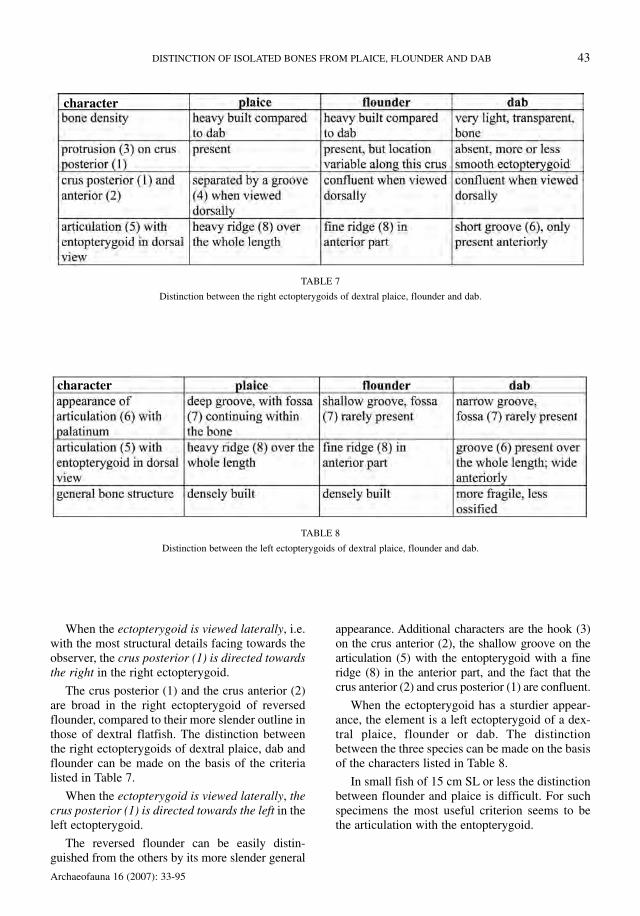
When the entopterygoid is viewed laterally,with the costa marginalis (1) below, the elementhas a pointed end directed towards the right sidein the right entopterygoid.
The right entopterygoid of reversed flounderdiffers from the others by its more oval-shapedoutline and by the presence of more structuraldetails. The margo inferior (2) is slightly curved.The right ectopterygoid of dextral plaice, flounderand dab have a more or less triangular outline, andtheir margo inferior (2) is nearly straight.
The right entopterygoids of dextral plaice andflounder cannot be distinguished from each other,but they differ from those of dab in the criteria list-ed in Table 9.
When the entopterygoid is viewed laterally,with the costa marginalis (1) below, the elementhas a pointed end directed towards the left side inthe left entopterygoid.
The left entopterygoid of reversed flounder hasa rather straight margo inferior (2), and the bonehas a relatively high and short outline. The leftentopterygoid of dextral plaice, flounder and dabare more elongate and have a rather oval outline.
The distinction between the left entopterygoidsof dextral plaice, flounder and dab can be madewith the criteria listed in Table 10.
DISTINCTION OF ISOLATED BONES FROM PLAICE, FLOUNDER AND DAB 45
Archaeofauna 16 (2007): 33-95
TABLE 10
Distinction between the left entopterygoids of dextral plaice, flounder and dab.
TABLE 9
Distinction between the right entopterygoids of dab and dextral flounder or plaice.
character
character
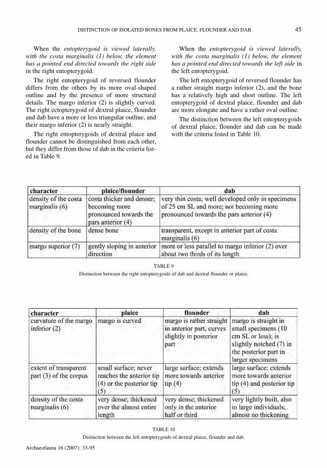
Maxillare (Figure 5)
46 W. WOUTERS, L. MUYLAERT & W. VAN NEER
Archaeofauna 16 (2007): 33-95
FIGURE 5
Medial and dorsal view of the maxilla of plaice, dab, dextral and reversed flounder.

When the maxilla is viewed medially, i.e. withthe pars caudalis (1) located right of the caputmaxillare (2), then the pars caudalis (1) is direct-ed downwards in the right maxilla. The element isa right maxilla of a reversed flounder when thestructure (4) on the collum maxillare is a clearridge. When the structure (4) is a notch, instead ofa ridge, the element is a right maxilla of a dextralplaice, flounder or dab. The listing of characters inTable 11 shows that the recognition of dab is easi-er than the distinction between plaice and flounder.
When the maxilla is viewed medially, i.e. withthe pars caudalis (1) located right of the caputmaxillare (2), then the pars caudalis (1) is direct-ed upwards in the left maxilla. The element is aleft maxilla of a reversed flounder when the struc-ture (4) on the collum maxillare is a notch. Whenthe structure (4) is a ridge, instead of a notch, theelement is a left maxilla of a dextral plaice, floun-der or dab.
It appears that dab can be easily recognisedusing the criteria listed in Table 12, but a distinc-tion of plaice and flounder was not possible fordextral specimens.
Praemaxillare (Figure 6)
When the premaxilla is viewed laterally, theprocessus anterior (1) is located at the right in theright premaxilla.
The right premaxilla of reversed flounder canbe distinguished from the others by the protrudinglower part of the symphysis (2). Another typicalfeature is the shallow incision between the proces-sus anterior (1) and the processus articularis (3). Inthe dextral flatfish both structures are more sepa-rated.
The distinction between the right premaxillaeof dextral plaice, flounder and dab can be madewith the criteria listed in Table 13.
In the right premaxilla of the reversed flounderwe observed 14 to 26 teeth in our material. It wasalso noticed that in reversed flounder smaller than12 cm SL more than one row of teeth could occur.On the basis of the modern material that we had atour disposal, the number of teeth seemed to be areliable criterion, also in the smallest individuals.Despite the fact that we doubled the number of
DISTINCTION OF ISOLATED BONES FROM PLAICE, FLOUNDER AND DAB 47
Archaeofauna 16 (2007): 33-95
TABLE 11
Distinction between the right maxillae of dextral plaice, flounder and dab.
TABLE 12
Distinction between the left maxillae of dab and dextral flounder or plaice.
character
character

48 W. WOUTERS, L. MUYLAERT & W. VAN NEER
Archaeofauna 16 (2007): 33-95
FIGURE 6
Lateral view of the premaxilla of plaice, dab, dextral and reversed flounder, and ventral view of the left premaxilla of plaice, dab anddextral flounder.

observations for this criterion, a difference in num-ber of teeth remains if compared to the data fromNorman (1934). This is no doubt due to the factthat he lumped the data for dextral and sinistralspecimens (see also below).
When the premaxilla is viewed laterally, theprocessus anterior (1) is located at the left in theleft premaxilla.
The left premaxilla of reversed flounder can bedistinguished from the others by the absence of aprotrusion in the lower part of the symphysis (2).Typical for this element are also the well-separat-ed processus anterior (1) and processus articularis(3). The number of teeth can vary between 9 and
13, and these values approach the lower part of thevariation mentioned by Norman (1934).
The distinction between the left premaxillae ofplaice, dextral flounder and dab can be made withthe criteria listed in Table 14.
Compared to the data provided by Norman(1934), we noticed differences in the number ofteeth, although less pronounced than in the den-tary. In addition, it was noticed that juvenileplaice, flounder and dab (smaller than 12 cm) haveanother tooth alignment. Instead of a single rowalong the entire premaxilla in larger fish, only theanterior part has such a single row; the more pos-teriorly located teeth form several rows.
DISTINCTION OF ISOLATED BONES FROM PLAICE, FLOUNDER AND DAB 49
Archaeofauna 16 (2007): 33-95
TABLE 13
Distinction between the right premaxillae of dextral plaice, flounder and dab.
character
TABLE 14
Distinction between the left premaxillae of dextral plaice, flounder and dab.
character

Palatinum (Figure 7)
When the palatine is viewed medially, theprocessus maxillaris (1) is located at the left, andthe pars subpterygoidea (2) is pointing towardsthe right in the right palatine. The reversed floun-der can be distinguished from the others by thesturdy appearance of the collum (3) and of the parssubpterygoidea (2). Typical are also the straightmargo superior (4), the short pars subpterygoidea
(2), the concave margo inferior (5), and the result-ing constriction of the collum (3).
The right palatine of dextral plaice, flounderand dab are slender and show almost no structuresin medial view, because the articulations are locat-ed at the lateral side. The distinction between theright palatines can be made with the criteria listed
50 W. WOUTERS, L. MUYLAERT & W. VAN NEER
Archaeofauna 16 (2007): 33-95
FIGURE 7
Medial and lateral view of the palatine of plaice, dab, dextral and reversed flounder.

in Table 15. The discrimination of dextral flounderand plaice is difficult in individuals measuring lessthan 20 cm SL.
When the palatine is viewed medially, theprocessus maxillaris (1) is located at the right, andthe pars subpterygoidea (2) is pointing towardsthe left in the left palatine. The reversed floundercan be distinguished from the others by its slender
and short collum (3). The margo superior (4) isvery concave in lateral view.
The left palatine of dextral plaice, flounder anddab has a sturdy collum (3) and the pars subptery-goidea (2) clearly shows distinct articular facetsfor the ectopterygoid (7) and entopterygoid (8).The distinction between the left palatines can bemade with the criteria listed in Table 16. The dis-crimination of dextral flounder and plaice is diffi-cult in individuals measuring less than 20 cm SL.
DISTINCTION OF ISOLATED BONES FROM PLAICE, FLOUNDER AND DAB 51
Archaeofauna 16 (2007): 33-95
TABLE 15
Distinction between the right palatines of dextral plaice, flounder and dab.
character
TABLE 16
Distinction between the left palatines of dextral plaice, flounder and dab.
character

Parasphenoideum (Figure 8)
52 W. WOUTERS, L. MUYLAERT & W. VAN NEER
Archaeofauna 16 (2007): 33-95
FIGURE 8
Ventral and lateral view of the parasphenoid of plaice, dab, dextral and reversed flounder.

Archaeofauna 16 (2007): 33-95
The prefrontal ridge (1) bordering the faciesarticularis vomeris (2) is located at the right side inthe reversed flounder (on the left side in the figure)and at the left side in dextral flatfish.
The distinction between the parasphenoids ofthe dextral flatfish can be made on the basis of thecriteria listed in Table 17. The characteristicsdescribed below for the dextral flounder are alsovalid for the reversed form.
Dab parasphenoids appear to be easily identifi-able, but the distinction between flounder andplaice may be hampered when the bones are fromfishes less than 20 cm SL.
Frontale (Figure 9)
The left frontal of reversed flounder and theright frontals of dextral flatfish have a long anteri-or processus (3). In dorsal view, this anteriorprocessus (3) is curving towards the right in theleft frontal of reversed flounder. The charactersdescribed below for the right frontal of dextralflounder are also found in the reversed form.
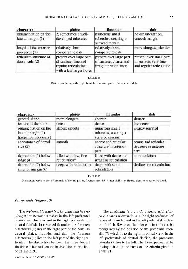
Table 18 lists the criteria that allow distinctionbetween the right frontals of dextral plaice, floun-der and dab.
The right frontal of reversed flounder and the leftfrontals of dextral flatfish have a short anteriorprocessus (3). In dorsal view, this anterior processus(3) is located on the left in the right frontal ofreversed flounder. The characters described belowfor the left frontal of dextral flounder are also foundin the reversed form.
Table 19 lists the criteria that allow distinctionbetween the left frontals of dextral plaice, flounderand dab.
The third criterion (ornamentation on the lateralmargin) cannot be accurately drawn or illustrated bya photograph and should rather be examined by pal-pation which allows one to feel whether the marginis almost smooth (plaice), weakly serrated (dab) or iscomprised of numerous small tubercles (flounder).Despite the fact that this criterion cannot be accu-rately illustrated visually, it is very diagnostic. Thismethod of examining cranial skeletal elements isalso necessary in the sphenoticum and the pteroticwhich, as the frontal, lie along the lateral skull mar-gins and have a species specific ornamentation.
DISTINCTION OF ISOLATED BONES FROM PLAICE, FLOUNDER AND DAB 53
TABLE 17
Distinction between the parasphenoids of dextral plaice, flounder and dab.
character

54 W. WOUTERS, L. MUYLAERT & W. VAN NEER
Archaeofauna 16 (2007): 33-95
FIGURE 9
Dorsal view of the frontal of plaice, dab, dextral and reversed flounder.
Praefrontale (Figure 10)
The prefrontal is roughly triangular and has noelongate posterior extension in the left prefrontalof reversed flounder and in the right prefrontal ofdextral flatfish. In reversed flounder, the foramenolfactorius (1) lies in the right part of the bone. Indextral plaice, flounder and dab, the foramenolfactorius (1) lies in the left part of the right pre-frontal. The distinction between the three dextralflatfish can be made on the basis of the criteria list-ed in Table 20.
The prefrontal is a sturdy element with elon-gate, posterior extensions in the right prefrontal ofreversed flounder and in the left prefrontal of dex-tral flatfish. Reversed flounder can, in addition, berecognised by the position of the processus later-alis (7) which is to the right in dorsal view. In theleft prefrontals of dextral flatfish, the processuslateralis (7) lies to the left. The three species can bedistinguished on the basis of the criteria given inTable 21.
DISTINCTION OF ISOLATED BONES FROM PLAICE, FLOUNDER AND DAB 55
Archaeofauna 16 (2007): 33-95
TABLE 18
Distinction between the right frontals of dextral plaice, flounder and dab.
TABLE 19
Distinction between the left frontals of dextral plaice, flounder and dab. *: not visible on figure, element needs to be tilted.
character
character

56 W. WOUTERS, L. MUYLAERT & W. VAN NEER
Archaeofauna 16 (2007): 33-95
FIGURE 10
Views of the prefrontal of plaice, dab, dextral and reversed flounder. For the right prefrontal of reversed flounder and the left prefrontalsof plaice, dab and dextral flounder, two dorsal views are provided. The left one shows the complete dorsal view, the second one is adetail of the uppermost part. For the same elements two lateral views are given as well. The second view depicts schematically the out-line of the processus lateralis and the adjacent ridge.

DISTINCTION OF ISOLATED BONES FROM PLAICE, FLOUNDER AND DAB 57
Archaeofauna 16 (2007): 33-95
TABLE 20
Distinction between the right prefrontals of dextral plaice, flounder and dab. *: size differences are more obvious when the bone isviewed under a different angle than the one on the figure.
TABLE 21
Distinction between the left prefrontals of dextral plaice, flounder and dab. *: the foramen olfactorius of dab looks smaller on the fig-ure, but its size depends on the position under which the element is viewed.
character
character

58 W. WOUTERS, L. MUYLAERT & W. VAN NEER
Archaeofauna 16 (2007): 33-95
FIGURE 11
Dorsal, ventral and lateral view of the parietal of plaice, dab, dextral and reversed flounder.
Parietale (Figure 11)
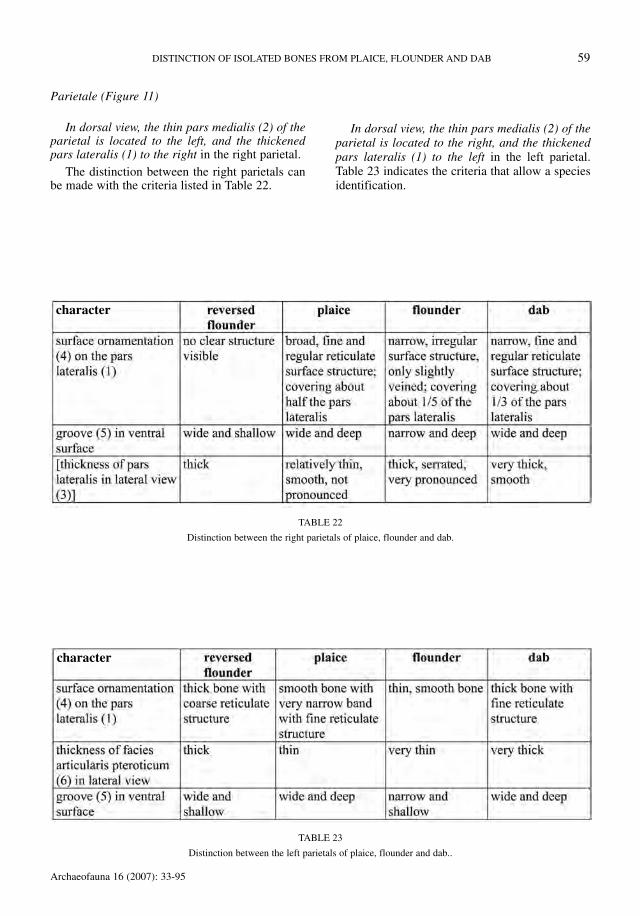
In dorsal view, the thin pars medialis (2) of theparietal is located to the left, and the thickenedpars lateralis (1) to the right in the right parietal.
The distinction between the right parietals canbe made with the criteria listed in Table 22.
In dorsal view, the thin pars medialis (2) of theparietal is located to the right, and the thickenedpars lateralis (1) to the left in the left parietal.Table 23 indicates the criteria that allow a speciesidentification.
DISTINCTION OF ISOLATED BONES FROM PLAICE, FLOUNDER AND DAB 59
Archaeofauna 16 (2007): 33-95
TABLE 22
Distinction between the right parietals of plaice, flounder and dab.
TABLE 23
Distinction between the left parietals of plaice, flounder and dab..
character
character

Interoperculare (Figure 12)
For this element it was not possible to make thedistinction between sinistral and dextral flounder.Hence, the characters below enable the recognitionat species level only. The characters allowingspecies identifications are listed in Table 24. They
are all related to the extent and location of denserparts within the element. Dab can be distinguishedfrom the two other species by the lighter build ofthe bone. For the distinction between flounder andplaice, the criteria work well for larger individuals,but in specimens of 30 cm SL and less they are lessconsistent.
60 W. WOUTERS, L. MUYLAERT & W. VAN NEER
Archaeofauna 16 (2007): 33-95
FIGURE 12
Dorsal and medial view of the interopercular of plaice, flounder and dab.

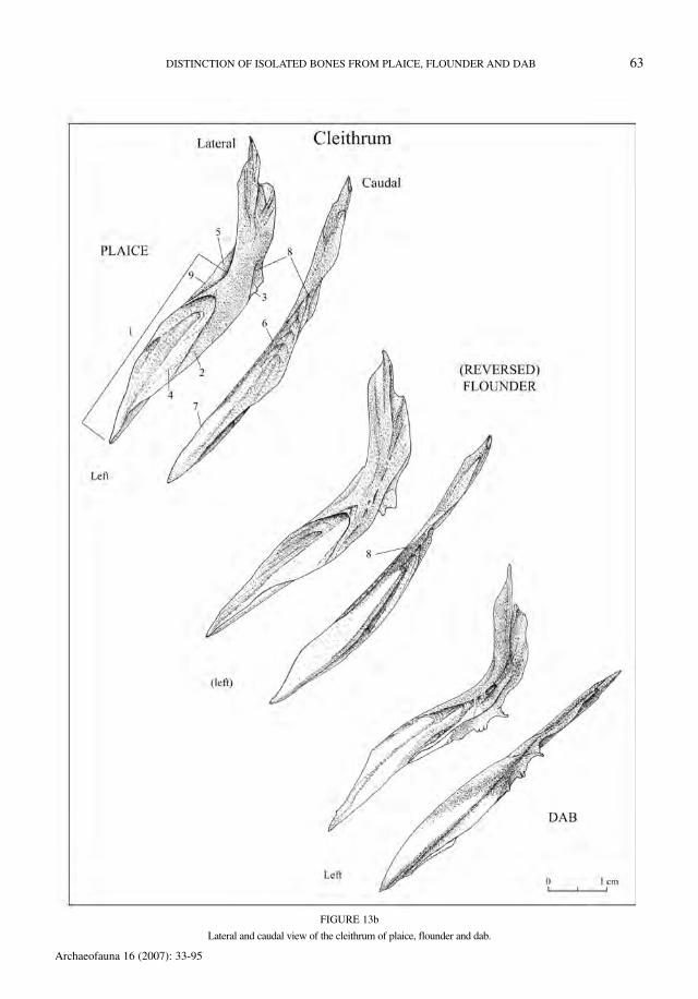
Cleithrum (Figure 13)
No difference can be made between dextral andsinistral flounder. The distinguishing charactersare listed in Table 25. The figures include lateraland caudal views of the left and right elements.
The groove (6) in the crista externa (7) is the mostreliable criterion, but other features should be takeninto account as well. For a good view of this groove,the use of a binocular microscope is necessary. Thecurvature of the margo anterior (9) was retained as adiagnostic character by Heinrich (1987), but this fea-ture appears to be very variable and not very reliable,especially in small individuals.
DISTINCTION OF ISOLATED BONES FROM PLAICE, FLOUNDER AND DAB 61
Archaeofauna 16 (2007): 33-95
TABLE 24
Distinction between interoperculars of plaice, flounder and dab.
character
TABLE 25
Distinction between cleithra of plaice, flounder and dab.
character

62 W. WOUTERS, L. MUYLAERT & W. VAN NEER
Archaeofauna 16 (2007): 33-95
FIGURE 13a
Lateral and caudal view of the cleithrum of plaice, flounder and dab.
DISTINCTION OF ISOLATED BONES FROM PLAICE, FLOUNDER AND DAB 63
Archaeofauna 16 (2007): 33-95
FIGURE 13b
Lateral and caudal view of the cleithrum of plaice, flounder and dab.
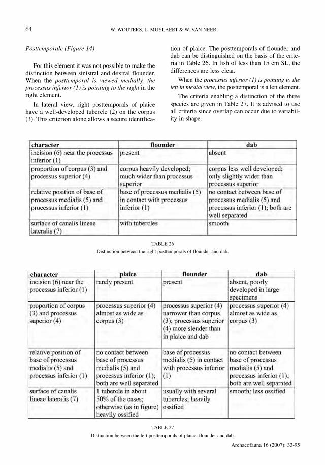
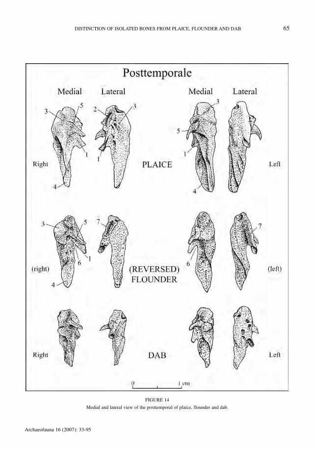
Posttemporale (Figure 14)
For this element it was not possible to make thedistinction between sinistral and dextral flounder.When the posttemporal is viewed medially, theprocessus inferior (1) is pointing to the right in theright element.
In lateral view, right posttemporals of plaicehave a well-developed tubercle (2) on the corpus(3). This criterion alone allows a secure identifica-
tion of plaice. The posttemporals of flounder anddab can be distinguished on the basis of the crite-ria in Table 26. In fish of less than 15 cm SL, thedifferences are less clear.
When the processus inferior (1) is pointing to theleft in medial view, the posttemporal is a left element.
The criteria enabling a distinction of the threespecies are given in Table 27. It is advised to useall criteria since overlap can occur due to variabil-ity in shape.
64 W. WOUTERS, L. MUYLAERT & W. VAN NEER
Archaeofauna 16 (2007): 33-95
TABLE 27
Distinction between the left posttemporals of plaice, flounder and dab.
character
TABLE 26
Distinction between the right posttemporals of flounder and dab.
character
DISTINCTION OF ISOLATED BONES FROM PLAICE, FLOUNDER AND DAB 65
Archaeofauna 16 (2007): 33-95
FIGURE 14
Medial and lateral view of the posttemporal of plaice, flounder and dab.

Pharyngobranchiale II (Figure 15)
The distinction between left and right secondpharyngobranchials of sinistral and dextral floun-der could not be made. The distinguishing charac-ters, at species level, are listed in Table 28.
The recognition of small individuals of plaiceand flounder is not easy, but it is feasible especial-ly on the basis of the outline of the margo medialis(1). An additional helpful criterion is the earlierappearance of a double tooth row in flounder, incomparison to plaice.
66 W. WOUTERS, L. MUYLAERT & W. VAN NEER
Archaeofauna 16 (2007): 33-95
TABLE 28
Distinction between the second pharyngobranchials of plaice, flounder and dab.
character
FIGURE 15
Ventral and anterior view of the second pharyngobranchials of plaice, flounder and dab.
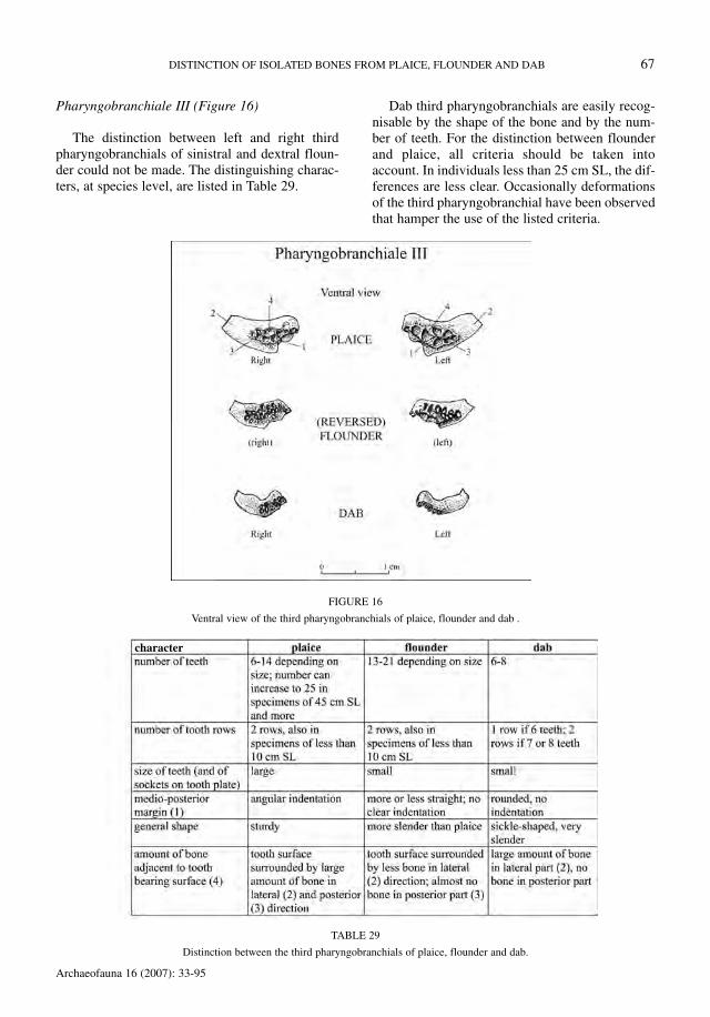
Pharyngobranchiale III (Figure 16)
The distinction between left and right thirdpharyngobranchials of sinistral and dextral floun-der could not be made. The distinguishing charac-ters, at species level, are listed in Table 29.
Dab third pharyngobranchials are easily recog-nisable by the shape of the bone and by the num-ber of teeth. For the distinction between flounderand plaice, all criteria should be taken intoaccount. In individuals less than 25 cm SL, the dif-ferences are less clear. Occasionally deformationsof the third pharyngobranchial have been observedthat hamper the use of the listed criteria.
DISTINCTION OF ISOLATED BONES FROM PLAICE, FLOUNDER AND DAB 67
Archaeofauna 16 (2007): 33-95
TABLE 29
Distinction between the third pharyngobranchials of plaice, flounder and dab.
character
FIGURE 16
Ventral view of the third pharyngobranchials of plaice, flounder and dab .
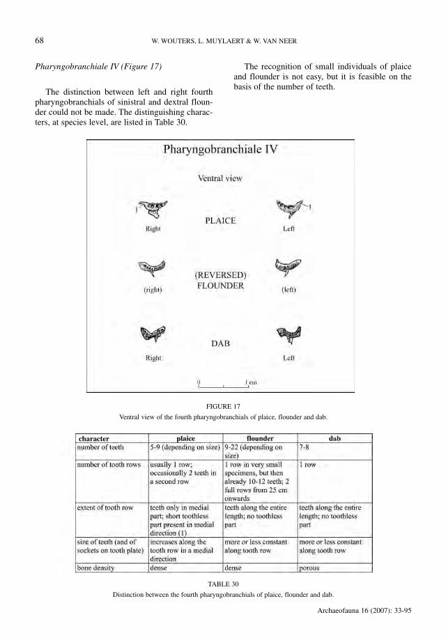
Pharyngobranchiale IV (Figure 17)
The distinction between left and right fourthpharyngobranchials of sinistral and dextral floun-der could not be made. The distinguishing charac-ters, at species level, are listed in Table 30.
The recognition of small individuals of plaiceand flounder is not easy, but it is feasible on thebasis of the number of teeth.
68 W. WOUTERS, L. MUYLAERT & W. VAN NEER
Archaeofauna 16 (2007): 33-95
FIGURE 17
Ventral view of the fourth pharyngobranchials of plaice, flounder and dab.
TABLE 30
Distinction between the fourth pharyngobranchials of plaice, flounder and dab.
character
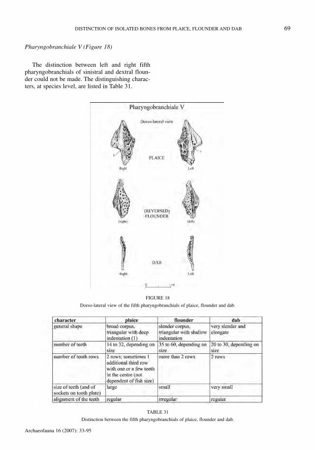
Pharyngobranchiale V (Figure 18)
The distinction between left and right fifthpharyngobranchials of sinistral and dextral floun-der could not be made. The distinguishing charac-ters, at species level, are listed in Table 31.
DISTINCTION OF ISOLATED BONES FROM PLAICE, FLOUNDER AND DAB 69
Archaeofauna 16 (2007): 33-95
FIGURE 18
Dorso-lateral view of the fifth pharyngobranchials of plaice, flounder and dab.
TABLE 31
Distinction between the fifth pharyngobranchials of plaice, flounder and dab.
character

Pteroticum (Figure 19)
When the pterotic is viewed laterally, the margoepioticum (1) and the margo exoccipitalis (2) arelocated to the right of the crista lateralis (3) in theright element. No distinction could be made betweenthe pterotics of sinistral and dextral flounder.
The criteria allowing a distinction between thethree species are listed in Table 32.
When the pterotic is viewed laterally, themargo epioticum (1) and the margo exoccipitalis
(2) are located to the left of the crista lateralis (3)in the left element. As for the elements mentionedabove, no distinction could be made between thepterotics of sinistral and dextral flounder. Appar-ently, as in the sphenotic, these bones from theposterior part of the skull are affected to a far less-er extent by the asymmetrical cranial deformationduring the post-larval development.
The criteria allowing a distinction between thethree species are listed in Table 33.
70 W. WOUTERS, L. MUYLAERT & W. VAN NEER
Archaeofauna 16 (2007): 33-95
TABLE 32
Distinction between the right pterotics of plaice, flounder and dab.
TABLE 33
Distinction between the left pterotics of plaice, flounder and dab.
character
character

DISTINCTION OF ISOLATED BONES FROM PLAICE, FLOUNDER AND DAB 71
Archaeofauna 16 (2007): 33-95
FIGURE 19
Lateral and dorsal view of the pterotic of plaice, flounder and dab.

Sphenoticum (Figure 20)
When the sphenotic is viewed laterally, the pos-terior part of the bone has a dorsal extension (1)that is directed to the left in the right element. Nodistinction could be made between the sphenoticsof sinistral and dextral flounder.
The criteria allowing a distinction between thethree species are listed in Table 34.
When the sphenotic is viewed laterally, the pos-terior part of the bone has a dorsal extension (1)that is directed to the right in the left element.Again, no distinction could be made between thesphenotics of sinistral and dextral flounder.
The criteria allowing a distinction between thethree species are listed in Table 35.
72 W. WOUTERS, L. MUYLAERT & W. VAN NEER
Archaeofauna 16 (2007): 33-95
FIGURE 20
Lateral and dorsal view of the sphenotic of plaice, flounder and dab.

Urohyale (Figure 21)
The criteria used to distinguish the species arelisted in Table 36. The bones are described as theyare positioned in Figure 21, i.e. with the incisuracollis (4) directed towards the right and with themargo ventralis pars horizontalis (5) horizontallyaligned. It is essential that the bone is held in thisposition when using the last two criteria in thetable. No distinction could be made between theurohyals of dextral and reversed flounder.
This element is very suitable for species iden-tification, even in specimens of less than 10 cmSL. Dab is easily distinguished from the twoother species by its deep incisura collis (4). Plaiceand flounder can be discriminated by the positionof the angulus inferior (1) relative to the proces-sus hypohyalis (2), and by the projection of theangulus inferior (1). However, plaice larger than35 cm SL resemble flounder for these two crite-ria. In that case, the thickness of the margo ven-tralis, measured mid-way allows discriminationof the two species: in plaice the margo ventralisis thicker than in specimens of flounder of thesame size. The thickness at this level is, however,not a useful criterion for flatfish of less than 35cm SL.
DISTINCTION OF ISOLATED BONES FROM PLAICE, FLOUNDER AND DAB 73
Archaeofauna 16 (2007): 33-95
TABLE 35
Distinction between the left sphenotics of plaice, flounder and dab.
character
TABLE 34
Distinction between the right sphenotics of plaice, flounder and dab.
character

74 W. WOUTERS, L. MUYLAERT & W. VAN NEER
Archaeofauna 16 (2007): 33-95
FIGURE 21
Lateral view of the urohyal of plaice, flounder and dab.
TABLE 36
Distinction between the urohyals of plaice, flounder and dab.
character

Os anale (Figure 22)
In this element the distinction between sinistraland dextral flounder could not be made. The criteriato distinguish the three species are listed in Table 37.
The curvature of the bone was retained as adiagnostic feature by Heinrich (1977) for the dis-crimination of dab, but it appears that small floun-der and plaice often have a curvature similar tothat seen in dab.
DISTINCTION OF ISOLATED BONES FROM PLAICE, FLOUNDER AND DAB 75
Archaeofauna 16 (2007): 33-95
TABLE 37
Distinction between the os anale of plaice, flounder and dab.
character

76 W. WOUTERS, L. MUYLAERT & W. VAN NEER
Archaeofauna 16 (2007): 33-95
FIGURE 22
Dorsal and caudal view of the os anale of plaice, flounder and dab.

First precaudal vertebra (Figure 23)
Except for the first vertebra, no consistent dis-tinguishing characters were found within the ver-tebral column. The criteria allowing species iden-tification of the first vertebra are given in Table 38.
Identification of first vertebrae from fish of lessthan 15 cm SL is not recommended. Even in largerspecimens, identification is not always straightfor-ward since the postzygapophyses tend to break off.
DISTINCTION OF ISOLATED BONES FROM PLAICE, FLOUNDER AND DAB 77
Archaeofauna 16 (2007): 33-95
FIGURE 23
Anterior and left lateral view of the first precaudal vertebra of plaice, flounder and dab.
TABLE 38
Distinction between the first precaudal vertebrae of plaice, flounder and dab.
character
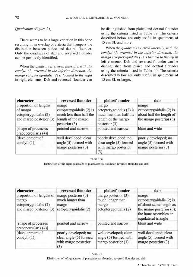
Quadratum (Figure 24)
There seems to be a large variation in this boneresulting in an overlap of criteria that hampers thedistinction between plaice and dextral flounder.Only the quadrates of dab and reversed floundercan be positively identified.
When the quadrate is viewed laterally, with thecondyli (1) oriented in the inferior direction, themargo ectopterygoidalis (2) is located to the rightin right elements. Dab and reversed flounder can
be distinguished from plaice and dextral flounderusing the criteria listed in Table 39. The criteriadescribed below are only useful in specimens of15 cm SL and more.
When the quadrate is viewed laterally, with thecondyli (1) oriented in the inferior direction, themargo ectopterygoidalis (2) is located to the left inleft elements. Dab and reversed flounder can bedistinguished from plaice and dextral flounderusing the criteria listed in Table 40. The criteriadescribed below are only useful in specimens of15 cm SL or larger.
78 W. WOUTERS, L. MUYLAERT & W. VAN NEER
Archaeofauna 16 (2007): 33-95
TABLE 39
Distinction of the right quadrates of plaice/dextral flounder, reversed flounder and dab.
TABLE 40
Distinction of left quadrates of plaice/dextral flounder, reversed flounder and dab.
character
character

DISTINCTION OF ISOLATED BONES FROM PLAICE, FLOUNDER AND DAB 79
Archaeofauna 16 (2007): 33-95
FIGURE 24
Lateral view of the quadrate of reversed flounder, dab, and plaice or dextral flounder.

Vomer (Figure 25)
In ventral view, vomers of plaice, dab and dex-tral flounder have their pars ethmoidalis (1) locat-ed at the left. This part is smaller than the parspraefrontalis (2) that is located at the right. Inreversed flounder the pars ethmoidalis (1) lies tothe right. In addition, the latter element can be
recognised by its much longer apophysis posterior(3). The ventral groove (4) in the apophysis poste-rior (3) can be absent or poorly developed in theanterior part of the apophysis.
No criteria were found allowing a distinctionbetween dextral flounder and plaice, but dab canbe distinguished from the two other species by thefeatures listed in Table 41.
80 W. WOUTERS, L. MUYLAERT & W. VAN NEER
Archaeofauna 16 (2007): 33-95
FIGURE 25
Ventral view of the vomer of reversed flounder, dab, and plaice or dextral flounder.
TABLE 41
Criteria allowing the recognition of dab vomers.
character

Supraoccipitale (Figure 26)
Identification of the supraoccipital is only pos-sible when the element is completely preserved.The supraoccipital of reversed flounder can be dis-tinguished from that of dextral flatfish by the loca-tion and the curvature of the crista supraoccipital-
is (1): in reversed flounder this crista is situated onthe right half of the bone, and it is bending towardsthe right. The crista can be single or double and iswell developed.
The supraoccipitals of dextral flounder and plaicecannot be distinguished from each other, but dab canbe identified using the criteria listed in Table 42.
DISTINCTION OF ISOLATED BONES FROM PLAICE, FLOUNDER AND DAB 81
Archaeofauna 16 (2007): 33-95
FIGURE 26
Dorsal view of the supraoccipital of reversed flounder, dab, and plaice or dextral flounder.
TABLE 42
Criteria allowing the recognition of dab supraoccipitals.
character

Ethmoid (Figure 27)
When viewed anteriorly, the ventral end (1) ofthe ethmoid is protruding towards the left inreversed flounder. In dextral flatfish the ventralprotrusion (1) is directed towards the right side ofthe figure. This sole character allows the distinc-
tion of reversed flounder. The ethmoid bone of dabcan also be easily recognised, but a distinctionbetween dextral flounder and plaice seems impos-sible due to the large amount of variation. The eth-moid bone of dab can be distinguished from floun-der and plaice using the criteria in Table 43.
82 W. WOUTERS, L. MUYLAERT & W. VAN NEER
Archaeofauna 16 (2007): 33-95
FIGURE 27
Anterior view of the ethmoid of reversed flounder, dab, and plaice or dextral flounder.
TABLE 43
Criteria allowing the recognition of dab ethmoids.
character
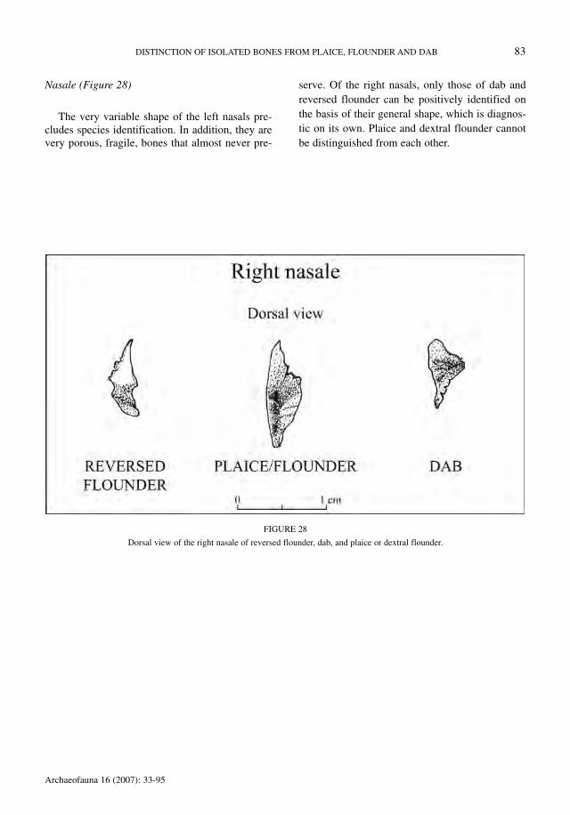
Nasale (Figure 28)
The very variable shape of the left nasals pre-cludes species identification. In addition, they arevery porous, fragile, bones that almost never pre-
serve. Of the right nasals, only those of dab andreversed flounder can be positively identified onthe basis of their general shape, which is diagnos-tic on its own. Plaice and dextral flounder cannotbe distinguished from each other.
DISTINCTION OF ISOLATED BONES FROM PLAICE, FLOUNDER AND DAB 83
Archaeofauna 16 (2007): 33-95
FIGURE 28
Dorsal view of the right nasale of reversed flounder, dab, and plaice or dextral flounder.

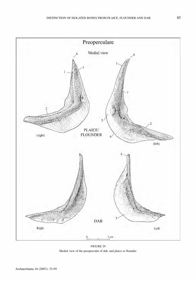
Praeoperculare (Figure 29)
The preoperculars of dab can be easily recog-nised. However, the distinction between the preop-
erculars of sinistral and dextral flounder could notbe made. There is, in addition, an overlap in crite-ria between plaice and flounder. Table 44 indicatesthe characters allowing the identification of dab.
84 W. WOUTERS, L. MUYLAERT & W. VAN NEER
Archaeofauna 16 (2007): 33-95
TABLE 44
Criteria allowing the recognition of dab preoperculars.
character
DISTINCTION OF ISOLATED BONES FROM PLAICE, FLOUNDER AND DAB 85
Archaeofauna 16 (2007): 33-95
FIGURE 29
Medial view of the preopercular of dab, and plaice or flounder.

Hyomandibulare (Figure 30)
For the hyomandibular it was not possible to
make the distinction between sinistral and dextral
flounder, and, in addition, it appeared that no dis-criminating characters are present allowing thedistinction between plaice and flounder. The crite-ria listed in Table 45, hence show only how dabcan be distinguished from plaice/flounder.
86 W. WOUTERS, L. MUYLAERT & W. VAN NEER
Archaeofauna 16 (2007): 33-95
TABLE 45
Criteria allowing the recognition of dab hyomandibulars.
character

DISTINCTION OF ISOLATED BONES FROM PLAICE, FLOUNDER AND DAB 87
Archaeofauna 16 (2007): 33-95
FIGURE 30
Lateral and medial view of the hyomandibular of dab, and plaice or flounder.

Ceratohyale (Figure 31)
The ceratohyals of dab can be easily recog-nised. However, the distinction between the cera-
tohyals of sinistral and dextral flounder could notbe made. There is, in addition, an overlap in crite-ria between plaice and flounder. Table 46 indicatesthe characters allowing the identification of dab.
88 W. WOUTERS, L. MUYLAERT & W. VAN NEER
Archaeofauna 16 (2007): 33-95
TABLE 46
Criteria allowing the recognition of dab ceratohyals.
character
FIGURE 31
Medial view of the ceratohyal of dab, and plaice or flounder.
Ceratohyale

Basioccipitale (Figure 32)
The basioccipitals of dab can be easily recog-nised (Table 47), but a distinction between floun-der and plaice seems impossible due to the largevariation. In addition, sinistral and dextral floun-der cannot be distinguished from each other.
DISTINCTION OF ISOLATED BONES FROM PLAICE, FLOUNDER AND DAB 89
Archaeofauna 16 (2007): 33-95
FIGURE 32
Ventral and lateral view of the basioccipital of dab, and plaice or flounder.
TABLE 47
Criteria allowing the recognition of dab basioccipitals.
character

Alisphenoideum (Figure 33)
The alisphenoid of dab is a rather flat bone thathas no protruding articulation (1) with the alisphe-noid of the opposite side. In plaice and flounder
this articulation (1) is well pronounced in medialview. The alisphenoids of dextral plaice and floun-der cannot be distinguished from each other, and itis also impossible to distinguish sinistral and dex-tral flounder.
90 W. WOUTERS, L. MUYLAERT & W. VAN NEER
Archaeofauna 16 (2007): 33-95
FIGURE 33
Medial view of the alisphenoid of dab, and plaice or flounder.

Postcleithrum (Figure 34)
Postcleithra with a notch (1) in the anterior mar-gin are only seen in dab. The anterior margin issmooth in plaice and flounder. In dab of large size(25 cm SL or more) the notch seems always to beabsent, while in most smaller fish it is clearly visible.
DISTINCTION OF ISOLATED BONES FROM PLAICE, FLOUNDER AND DAB 91
Archaeofauna 16 (2007): 33-95
FIGURE 34
Lateral view of the postcleithrum of dab, and plaice or flounder.

DISCUSSION
The 38 skeletal elements retained in this com-parative analysis include those that have been usedin the past by other researchers as well as 11 addi-tional bones that occur frequently on archaeologi-cal sites (Table 1). When ontogenetic series areconsidered, it appears that the diagnostic criteriaare most clearly pronounced in specimens ofrather large size (25-40 cm SL in plaice and floun-der, and 25-35 cm SL in dab). In fish exceedingthose lengths the defined criteria can sometimesbecome obscure to some extent due to irregularbone development. Heavily distorted elements arevery rare, however, except in the pharyngob-ranchials where the tooth sockets can be frequent-ly eroded or deformed.
Five of the selected elements proved to beunsuitable for species identification, namely thesupracleithrum, the second precaudal vertebra, thefirst caudal vertebra, the penultimate and the ante-penultimate vertebra. Of the nasal bones, only theright element allows species identification. Itappears that 23 elements allow an identification ofthe three species, although 6 of them are harder toidentify when dealing with smaller individuals. Oneleven of those 23 elements the distinctionbetween left- and right-sided flounder can bemade. Six skeletal elements allow only the recog-nition of dab, whereas six other bones permit theidentification of both dab and reversed flounder.Dab bones are indeed usually very distinct mor-phologically from those of plaice and flounder.The latter two species show more similarities,
which often hamper identification, especially insmaller specimens. Some elements also appear tovary considerably in morphology.
The number of skeletal elements that is usefulfor species identification is significantly higherthan that indicated in the literature, no doubtbecause of our more large-scale approach and thetime that was invested. Some of the results are incontradiction with the literature, i.e. concerningthe use of the os anale (cf. Lepiksaar & Heinrich,1977) or the cleithrum (Heinrich, 1987), and sev-eral other elements (cf. Table 1). Possible explana-tions for these conflicting results could be the lim-ited reference samples that were used previously,which may have resulted in an underestimation ofthe intraspecific variation. Also size-dependentmorphological changes may have escaped atten-tion. It is also unclear from most previous publica-tions to what extent reversed flounder has beenincluded in the analyses.
It is realised that the diagnostic criteria des-cribed in the present contribution are sometimesvery subtle and that some experience with flatfishosteology is needed for the identification of certainbones, especially small elements from the brain-case and the branchial apparatus. However, at least14 bones allow a fairly easy recognition of thethree species when the criteria described here areused (see fourth column in Table 1). Identificationof these bones should be done in combination witha reference collection consisting minimally of oneplaice, one dab, one dextral and one sinistralflounder of medium to large size. For identifica-tion of the other skeletal elements, the key can
92 W. WOUTERS, L. MUYLAERT & W. VAN NEER
Archaeofauna 16 (2007): 33-95
TABLE 48
Results of the identification of flatfish remains from the 15th-16th century AD site «Mijnplein» at Oostende.
Taxon/Group

only be used optimally when a larger collection ofmodern reference specimens is available. Ideallysuch a collection should consist of fish of differentsize classes (<10 cm; 10-20 cm; 20-30 cm and 30-40 cm SL), and this for plaice, dab, dextral andreversed flounder.
The applicability of the key was tested on theichthyofauna of a late 15th- early 16th centuryurban site along the Belgian coast (Oostende,Mijnplein) (Pieters et al., 2005). In this assem-blage, that was sieved on a series of 4, 2 and 1 mmmeshes, 2601 flatfish fragments were available, ofwhich 1826 (or 70.2%) could not be identified tospecies (Table 48). The unidentifiable fractionconsisted mainly of undiagnostic skeletal elementsthat were not retained in the present comparativestudy, i.e. fin rays, pterygiophores, branchial ele-ments and vertebrae. Among the unidentifiedbones are 52 remains (or 2% of the total) of verydiagnostic elements that could not be brought tospecies because of their fragmented state. Another161 bones could be classified as plaice/flounder. Intotal, 614 bones (or 23.6%) were identifiable tospecies. Concerning the flounder remains, itshould be noted that a high proportion of elements(44%) consisted of dermal denticles. Sixty-one
flounder bones allowed a distinction between dex-tral and sinistral specimens, and it appeared that52% of them came from reversed flounders. Ifonly the 14 skeletal elements are considered thatallow the most straightforward identifications,then only 5.9% of the remains was unidentifiable(those that were too heavily fragmented). Retain-ing these 14 elements seems to be a good strategywhen samples of the size studied here are avail-able, but a further reduction of the number ofskeletal elements considered for identification isnot recommended. Table 49 shows that there is noparticular bone that yields a relatively higher pro-portion of species identifications. Inclusion of themore difficult skeletal elements in the identifica-tion work can, however, be important when onlylimited flatfish samples are available from a givensite and will deliver the maximum of species iden-tifications. When all 34 elements are considered,the number of identified specimens doubles (614bones, instead of 273 when only 14 elements areretained). The success rate of the identificationwhen dealing with the whole series of bones is sig-nificantly lower (25.8% identifiable versus 94.1%when only the 14 elements are retained).
DISTINCTION OF ISOLATED BONES FROM PLAICE, FLOUNDER AND DAB 93
Archaeofauna 16 (2007): 33-95
TABLE 49
Unidentified and identified flatfish bones from the 15th-16th century AD site «Mijnplein» at Oostende, for the 14 most straightforwardskeletal elements.
Element

CONCLUDING REMARKS
The diagnostic characters described in the pre-sent paper should enable a more systematic identi-fication of flatfish bones from archaeological sitesin North-Western Europe. As already indicated inthe introduction, more specific identifications willmake it possible to better quantify the importanceof each species in the food provisioning of coastaland inland consumers. It is also likely that infer-ences about the season of fishing and the estab-lishment of former fishing grounds will be facili-tated. Since flounder and plaice reach larger sizesthan dab, the proportion of the latter species mayhave an influence on the average size of a flatfishassemblage. Future interpretations may thereforebenefit from an increased number of body sizereconstructions for each individual species.Although size reconstructions remain possible bydirect comparison with specimens of known bodylength, it would be preferable to use regressionformulae. It remains to be verified to what extentsuch formulae of the 3 species will be similar toeach other. For the Epinephelinae it has beenestablished that the same formulae can be used forall the taxa included in the subfamily (Desse &Desse-Berset, 1996), but in the case of the threespecies considered here there are indications thatsuch an approach may not be advisable. Norman(1934) mentions that the proportion of head lengthto total length is 3 3/4 to 4 1/2 in dab, 3 1/8 to 3 3/4in flounder and 3 to 3 7/8 in plaice. Preliminaryobservations on the modern material investigatedin the present study show, for instance, that thebasioccipital is consistently smaller in dab than inplaice or flounder of the same body size. In addi-tion, future size reconstructions of individualspecies will need to take into account the laterali-ty of the bone, and for flounder separate regres-sions may be needed for dextral and sinistral indi-viduals.
ACKNOWLEDGEMENTS
The contribution of Wim Wouters and Wim VanNeer to this paper presents research results of theInteruniversity Poles of Attraction Programme-Belgian Federal Science Policy Office. We expressour sincere thanks to Anne-Marie Wittek (RBIN-Sc) for the artwork and her patience. Aude VanDriessche (RBINSc) helped with the layout of the
plates, and Sheila Hamilton-Dyer (Southampton)is acknowledged for the correction of the Englishtext.
REFERENCES
BARRETT, J.; LOCKER, A.M. & ROBERTS, C.M. 2004:‘Dark Age Economics’ revisited: the English fish boneevidence AD 600-1600. Antiquity 78(301): 618-636.
BØDKER ENGHOFF, I. 1986: Freshwater fishing from asea-coast settlement. Journal of Danish Archaeology5: 62-76.
BØDKER ENGHOFF, I. 1989: Fishing from the Stone Agesettlement Norsminde. Journal of Danish Archaeolo-gy 8: 41-50.
BØDKER ENGHOFF, I. 1994: Fishing from medieval Hol-baek/Denmark, with notes to reversed Platichthysflesus. Offa 51: 299-302.
BRINKHUIZEN, D. 1989: Ichthyo-archeologisch Onder-zoek: Methoden en Toepassing aan de hand vanRomeins Vismateriaal uit Velsen (Nederland). Doc-toral thesis, University of Groningen.
CAÑAS, J.M. 1992: Contribución al Atlas Osteológico delos Teleósteos Ibéricos. II. Osteologia Comparada delos Lábridos Ibéricos. Unpublished doctoral thesis.Universidad Autónoma de Madrid, Madrid.
CLAVEL, B. 1997: Les restes osseux animaux du MoyenAge découverts Place de l’Hôtel de Ville à Abbeville(Somme). Revue Archéologique de Picardie 3/4:193-205.
DE JONG, T. 1994: Fish consumption at Eindhoven Cas-tle: archaeological remains versus historical sources.In: Van Neer, W. (ed.): Fish Exploitation in the Past.Proceedings of the 7th Meeting of the ICAZ FishRemains Working Group: 129-137. Annales duMusée Royal de l’Afrique Centrale, SciencesZoologiques 274, Tervuren.
DESSE, J. & DESSE-BERSET, N. 1996: Archaeozoology ofgroupers (Epinephelinae). Identification, osteometryand keys to interpretation. Archaeofauna 5: 121-127.
DUNCKER, G. 1900: Variation und Asymmetrie bei Pleu-ronectes flesus L. Wissenschaftliche Meeresunter-suchungen. Abteilung Helgoland N.F. 3: 333-406.
FORNBACKE, M.; GOMBRII, M. & LUNDBERG, A. 2002:Sidedness frequencies in the flounder Platichthys fle-sus (Pleuronectiformes) along a biogeographicalcline. Sarsia 87: 392-395.
HÄRKÖNEN, T. 1986: Guide to the Otoliths of the BonyFishes of the Northeast Atlantic. Danbui ApS,Hellerup, Denmark.
HARTLEY, P.H.T. 1940: The saltash tuck-net fishery andthe ecology of some estuarine fishes. Journal of the
94 W. WOUTERS, L. MUYLAERT & W. VAN NEER
Archaeofauna 16 (2007): 33-95
Marine Biological Society of the United Kingdom 24:1-68.
HEINRICH, D. 1987: Untersuchungen an mittelalterlichenFischresten aus Schleswig. Ausgraubungen inSchleswig, Berichte und Studien 6. Wachholtz Ver-lag, Neumünster.
LEPIKSAAR, J. 1983: Osteologia I. Pisces. Unpublisheddocument, Göteborg.
LEPIKSAAR, J. & HEINRICH, D. 1977: Untersuchungen anFischresten aus der frühmittelalterlichen SiedlungHaithabu. Neue Ausgrabungen in Haithabu 10.Wachholtz Verlag, Neumünster.
NIELSEN, J.G. 1986: Pleuronectidae. In: Whitehead,P.J.P.; Bauchot, M.-L.; Hureau, J.-C.; Nielsen, J. &Tortonese, E. (eds): Fishes of the North-easternAtlantic and the Mediterranean. Volume III: 1299-1307. Unesco, Paris.
NORMAN, J.R. 1934: A Systematic Monograph of theFlatfishes (Heterosomata). Volume I: Psettodidae,Bothidae, Pleuronectidae. British Museum, London.
PIETERS, M.; SCHIETCATTE, L.; ZEEBROEK, I.; CALUWÉ, D.;COOREMANS, B.; DEFORCE, K.; DEMERRE, I.; EECK-HOUT, J.; ERVYNCK, A.; GEVAERT, G.; HOLLEVOET, Y.;KIGHTLY, C.; TYS, D.; VANDENBRUAENE, M.; VAN-
HOUTTE, S. & VAN NEER, W. 2005: Oostende : Stad-vernieuwing en Archeologie. Een Balans van 10 Jaararcheologisch Onderzoek van het Oostendse Bode-marchief. Vlaams Instituut voor het Onroerend Erf-goed, Brussel.
POLL, M. 1947: Poissons marins. Musée Royal d’His-toire Naturelle de Belgique, Bruxelles.
ROJO, A. 1991: Dictionary of Evolutionary Fish Osteol-ogy. CRC Press, Boca Raton.
ROSELLÓ, E. 1986: Contribución al Atlas Osteológico delos Teleósteos Ibéricos. I. Dentario y Articular.Colección de Estudios nº 14. Ediciones de la Univer-sidad Autónoma de Madrid, Madrid.
STRODTMANN, S. 1906: Laichen und Wandern der Ost-seefische. Wissenschaftliche Meeresuntersuchungen.Abteilung Helgoland N.F. 7: 133-216.
VAN NEER, W. & ERVYNCK, A. 2006: The zooarchaeolog-ical reconstruction of the development of the exploita-tion of the sea: a status quaestionis for Flanders. In:Pieters, M.; Verhaeghe, F. & Gevaert, G. (eds): Fish-ery, trade and piracy. Fishermen and fishermen’s set-tlements in and around the North Sea area in the Mid-dle Ages and later: 95-103. Archeologie in VlaanderenMonografie 6. Flemish Heritage Institute, Brussel.
DISTINCTION OF ISOLATED BONES FROM PLAICE, FLOUNDER AND DAB 95
Archaeofauna 16 (2007): 33-95