Embed Size (px)
Citation preview

P1: DPI/plb P2: NBL/dat QC: NBL
April 29, 1998 13:49 Annual Reviews AR057-23
Annu. Rev. Biochem. 1998. 67:721–51Copyright c© 1998 by Annual Reviews. All rights reserved
THE DNA REPLICATION FORKIN EUKARYOTIC CELLS
Shou Waga and Bruce StillmanCold Spring Harbor Laboratory, P.O. Box 100, Cold Spring Harbor, New York 11724
KEY WORDS: polymerase switching, Simian virus 40, cell cycle, Okazaki fragments, replisomeproteins
ABSTRACT
Replication of the two template strands at eukaryotic cell DNA replication forksis a highly coordinated process that ensures accurate and efficient genome dupli-cation. Biochemical studies, principally of plasmid DNAs containing the SimianVirus 40 origin of DNA replication, and yeast genetic studies have uncovered thefundamental mechanisms of replication fork progression. At least two differentDNA polymerases, a single-stranded DNA-binding protein, a clamp-loading com-plex, and a polymerase clamp combine to replicate DNA. Okazaki fragment syn-thesis involves a DNA polymerase-switching mechanism, and maturation occursby the recruitment of specific nucleases, a helicase, and a ligase. The process ofDNA replication is also coupled to cell-cycle progression and to DNA repair tomaintain genome integrity.
CONTENTS
INTRODUCTION . . . . . . . . . . . . . . . . . . . . . . . . . . . . . . . . . . . . . . . . . . . . . . . . . . . . . . . . . . . 722
CELLULAR REPLICATION FORK PROTEINS. . . . . . . . . . . . . . . . . . . . . . . . . . . . . . . . . . . 722DNA Polymeraseα/Primase Complex. . . . . . . . . . . . . . . . . . . . . . . . . . . . . . . . . . . . . . . . . 723Replication Protein A (RPA). . . . . . . . . . . . . . . . . . . . . . . . . . . . . . . . . . . . . . . . . . . . . . . . 724Replication Factor C (RFC). . . . . . . . . . . . . . . . . . . . . . . . . . . . . . . . . . . . . . . . . . . . . . . . 726Proliferating Cell Nuclear Antigen (PCNA). . . . . . . . . . . . . . . . . . . . . . . . . . . . . . . . . . . . 728DNA Polymerasesδ andε . . . . . . . . . . . . . . . . . . . . . . . . . . . . . . . . . . . . . . . . . . . . . . . . . . 731FEN1 and RNaseHI. . . . . . . . . . . . . . . . . . . . . . . . . . . . . . . . . . . . . . . . . . . . . . . . . . . . . . . 732DNA Helicases. . . . . . . . . . . . . . . . . . . . . . . . . . . . . . . . . . . . . . . . . . . . . . . . . . . . . . . . . . . 733Molecular Links Among FEN1, Dna2 Helicase, and DNA Polymeraseα/Primase. . . . . . 735
MECHANISMS OF DNA SYNTHESIS AT A REPLICATION FORK. . . . . . . . . . . . . . . . . . 736Primosome Assembly. . . . . . . . . . . . . . . . . . . . . . . . . . . . . . . . . . . . . . . . . . . . . . . . . . . . . . 736Polymerase Switching. . . . . . . . . . . . . . . . . . . . . . . . . . . . . . . . . . . . . . . . . . . . . . . . . . . . . 738Maturation of Okazaki Fragments. . . . . . . . . . . . . . . . . . . . . . . . . . . . . . . . . . . . . . . . . . . . 739
7210066-4154/98/0701-0721$08.00
P1: DPI/plb P2: NBL/dat QC: NBL
April 29, 1998 13:49 Annual Reviews AR057-23
722 WAGA & STILLMAN
REPLICATION FORK PROTEINS AND CELL-CYCLE CONTROL. . . . . . . . . . . . . . . . . . 741S-Phase Checkpoint Control. . . . . . . . . . . . . . . . . . . . . . . . . . . . . . . . . . . . . . . . . . . . . . . . 741PCNA-p21 Interaction. . . . . . . . . . . . . . . . . . . . . . . . . . . . . . . . . . . . . . . . . . . . . . . . . . . . . 742Regulation of Telomere Length. . . . . . . . . . . . . . . . . . . . . . . . . . . . . . . . . . . . . . . . . . . . . . 743
CONCLUDING REMARKS . . . . . . . . . . . . . . . . . . . . . . . . . . . . . . . . . . . . . . . . . . . . . . . . . . . 744
INTRODUCTION
In a proliferating eukaryotic cell, the duplication of the genetic complementoccurs every S phase of the cell cycle and must occur with high accuracy onlyonce per cell cycle. Furthermore, DNA replication in a single cell must becoordinated with other cell-cycle processes such as mitosis and cytokinesis andwith DNA replication in the surrounding cells in tissue. Much of this regula-tion occurs at the level of initiation of DNA replication via interaction with thepathways that control cell cycle progression. Although we still do not fully un-derstand how initiation of DNA replication occurs in eukaryotes, rapid progressis being made and has been reviewed elsewhere (1, 2). Once initiation occurs,the replication apparatus copies each replicon in a highly efficient process.The fundamental mechanisms that operate at the eukaryotic DNA replicationfork are now quite well known and are discussed here. Because the replicationof DNA in eukaryotic cells must be coupled to DNA repair and assembly ofthe DNA into chromatin, the replication fork proteins play prominent roles inmaintaining the fidelity of DNA replication, in coordinating replication withcell-cycle progression, and in the inheritance of chromatin complexes.
CELLULAR REPLICATION FORK PROTEINS
Most of what is known about DNA replication in eukaryotes comes from exten-sive studies performed using cell extracts from mammalian cells that supportthe complete replication of plasmid DNAs containing the Simian Virus 40 DNAreplication origin (SV40ori) (3–6). SV40 DNA replication requires a singlevirus-encoded protein, the SV40 large tumor antigen (T antigen), which func-tions both as an initiator protein by binding to the SV40ori and as a DNAhelicase at the replication fork; thus the replication is achieved primarily bycellular proteins that also function to duplicate cellular DNA. The purificationand reconstitution of DNA replication with purified proteins has yielded greatinsight into the mechanism of DNA replication as well as other aspects of DNAmetabolism such as DNA repair and recombination (7–10).
Investigation of more specific enzymatic reactions and yeast genetic studieshave uncovered several proteins thought to be involved directly in DNA synthe-sis at the replication fork. This review briefly outlines the current understand-ing of the eukaryotic fork proteins (Table 1) and the reactions in which they

P1: DPI/plb P2: NBL/dat QC: NBL
April 29, 1998 13:49 Annual Reviews AR057-23
DNA REPLICATION IN EUKARYOTES 723
Table 1 Functions of DNA replication fork proteins
Proteins Functions
RPA Single-stranded DNA binding; stimulates DNA polymerases;facilitates helicase loading
PCNA Stimulates DNA polymerases and RFC ATPaseRFC DNA-dependent ATPase; primer-template DNA binding;
stimulates DNA polymerases; PCNA loadingPolα/primase RNA-DNA primer synthesisPol δ/εa DNA polymerase; 3′–5′ exonucleaseFEN1 Nuclease for removal of RNA primersRNase HI Nuclease for removal of RNA primersDNA ligase I Ligation of DNAT antigenb DNA helicase; primosome assembly
aA specific function of DNA polymeraseε in replication has not been assigned, althoughit is known to be essential for S-phase progression inS. cerevisiae(196, 197).
bT antigen is required for the replication of SV40 DNA. Its functional equivalent ineukaryotic cells has not been identified.
participate. DNA ligases (11, 12) and topoisomerases I and II (13) are also re-quired for replication, but since the function of these enzymes has been reviewedelsewhere, they are not covered extensively here.
DNA Polymeraseα/Primase ComplexThe DNA polymeraseα/primase complex (polα/primase) is the only enzymecapable of initiating DNA synthesis de novo by first synthesizing an RNA primerand then extending the primer by polymerization to produce a short DNA exten-sion (RNA-DNA primer) (14). Analyses of SV40ori–dependent replicationin vivo and in vitro has demonstrated that polα/primase can synthesize anRNA-DNA primer of approximately 40 nucleotides (nt) in length, includingabout 10 nt of RNA primer (15–20). The short RNA-DNA then serves as aprimer for extension by another polymerase for DNA synthesis on either theleading (continuously synthesized) strand or for each Okazaki fragment on thelagging (discontinuously synthesized) strand (21–26). This process involves apolymerase switch from polα/primase to either DNA polymeraseδ or ε (polδ andε) (see below). This switch occurs because, unlike other more complexpolymerases, polα/primase is not capable of processive DNA synthesis anddissociates from the template DNA following primer synthesis (27).
The human cell polα/primase consists of four subunits (p180, p70, p58 andp48), and similar subunits are found in all eukaryotes examined (for reviewsee 14, 28). cDNAs encoding all four subunits of human, mouse, or yeast (Sac-charomyces cerevisiaeandSchizosaccharomyces pombe) pol α/primase havebeen cloned, and active complexes have been reconstituted from recombinant
P1: DPI/plb P2: NBL/dat QC: NBL
April 29, 1998 13:49 Annual Reviews AR057-23
724 WAGA & STILLMAN
proteins expressed from baculovirus vectors in infected insect cells (14, 29, 30).The p180 and p48 polypeptides harbor the DNA polymerase and primase cat-alytic activities, respectively. Extensive mutational analyses of the genes en-coding both polymerase and primase catalytic subunits show that they functionin DNA replication in vivo but also suggest a regulatory role for the primase sub-unit (see below; 14, 31–33). The p58 subunit is necessary for the stability andactivity of the primase p48 subunit (29, 34, 35). Although no enzymatic activityhas been associated with p70 (also known as B subunit or p86 inS. cerevisiae)(36, 37), biochemical studies have shown that p70 plays an important role inthe assembly of the primosome (see below). In a complementary approach, ge-netic experiments with temperature-sensitive mutants ofS. cerevisiae(pol12-T)revealed an essential function for the equivalent subunit (p86) in the initiationof DNA replication (37).
The phosphorylation of polα/primase has been reported in human andS. cerevisiaecells (38–40); both human p180 and p70 were phosphorylatedpredominantly during late G2 and M phases of the cell cycle (38). Yeast p86(B subunit) is phosphorylated in a manner that depends on the stage of the cellcycle (39), but most phosphorylation occurs late in the cell cycle, suggestingthat it might play a role either in coordinating replication and mitosis or in reset-ting the replication apparatus for the next S phase. Analysis of the initiation ofSV40 DNA replication showed that the primase activity during initiation couldbe suppressed when polα/primase was phosphorylated by cyclin A-CDK2, butnot by cyclin B-CDC2 or cyclin E-CDK2, whereas DNA polymerase or primaseactivities with synthetic templates were hardly affected by this phosphorylation(41). This observation suggests that the phosphorylation of polα/primase mayplay a regulatory role in the initiation of replication, but the in vivo significanceof the phosphorylation is unknown.
Replication Protein A (RPA)Replication protein A (RPA; also reported previously as RFA or HSSB) is asingle-stranded DNA-binding protein that exists as a heterotrimeric complexconsisting of subunits with apparent masses of approximately 70, 34, and 11 kDain all eukaryotic cells examined (for review see 42, 43). The trimeric proteinwas initially identified as a factor essential for SV40 DNA replication in vitro(44–46), but subsequently it was shown to be involved in DNA recombinationand repair (43, 47).
RPA promotes extensive unwinding of duplex DNA containing the SV40oriby SV40 T antigen, which in addition to recognizing theori in a sequence-specific manner, is an RPA-stimulated DNA helicase (48–52). RPA also stimu-lates polα/primase activity under certain conditions and is required for replica-tion factor C (RFC)– and proliferating cell nuclear antigen (PCNA)–dependent

P1: DPI/plb P2: NBL/dat QC: NBL
April 29, 1998 13:49 Annual Reviews AR057-23
DNA REPLICATION IN EUKARYOTES 725
DNA synthesis by DNA polymeraseδ (25, 53–57). In support of these bio-chemical studies, some temperature-sensitive mutations in the gene encodingthe large subunit ofS. cerevisiaeRPA showed synthetic lethality, a form ofgenetic interaction, with mutations in genes encoding polα, primase, or polδ(58). Binding assays with purified proteins demonstrated that p70 binds to theprimase subunits of polα/primase and that the RPA heterotrimeric complex,but not p70 alone, binds to SV40 T antigen (59, 60). These interactions arethought to be required for the assembly of the primosome (see below).
cDNAs encoding each of the individual subunits of RPA have been clonedfrom a variety of species (43, 61). Furthermore, RPA has been produced inbacteria, and the recombinant human trimeric protein can support SV40 DNAreplication in vitro (62, 63). Functional RPA has also been produced by infec-tion of recombinant baculoviruses into insect cells (64). Although the humanp70 subunit alone can bind single-stranded DNA, it cannot support DNA repli-cation in vitro (55, 65, 66). Mutational studies to probe the structure of p70 havelocated the DNA-binding region to the N-terminal two-thirds of the subunit; theC-terminal third containing a putative zinc-finger was required for interactionswith the other two subunits (65–67).
More recent structural analysis of yeast RPA suggests that it contains a total offour potential single-stranded DNA-binding domains that are distantly relatedto each other (68). The domains are called SBDs and are made up of about 120amino acids each; two are in the large subunit and one each is in the middleand the small subunits, although the evidence for the DNA-binding domainin the small subunit is not as convincing as for the large subunit domains. Acrystal structure of the two DNA-binding domains derived from the human largesubunit (p70) revealed two structurally related subdomains, each correspondingto an SBD (69). It has been suggested that these SBDs, which contain clustersof aromatic amino acids that are similar to structures within the DNA-bindingdomain of theEscherichia colisingle-stranded DNA-binding protein (SSB), areresponsible for a higher-order assembly of the RPA-DNA complex, where RPAmight be wrapped with DNA (68). Indeed DNA-binding studies with humanRPA have demonstrated the existence of at least two distinct DNA-bindingmodes of the protein: one involving 8–10 nucleotides and another involving 30nucleotides (70, 71). There may well be higher-order interactions between RPAand longer stretches of single-stranded DNA, as occurs forE. coli SSB (72).
Both the large (p70) and the middle (p34) subunits of human and yeast RPAare phosphorylated in a cell-cycle-dependent manner (73). Increased levels ofsimilar phosphorylated forms of RPA are also seen in response to DNA dam-age (74, 75). The phosphorylation of the p34 subunit has been characterizedextensively; it is phosphorylated in the S and G2 phases of the cell cycle, andcyclin-dependent and DNA-dependent kinases have been identified as enzymes

P1: DPI/plb P2: NBL/dat QC: NBL
April 29, 1998 13:49 Annual Reviews AR057-23
726 WAGA & STILLMAN
capable of phosphorylating p34 (76–84). The phosphorylation of RPA is de-layed in cells from patients who have ataxia telangiectasia (AT), a cancer-pronedisease resulting from loss of the DNA damaging surveillance ATM gene pro-duct; and the phosphorylation of RPA is compromised in yeast lacking theMEC1gene, a gene with similarities to ATM (74, 85, 86). This is particularlyinteresting because phosphorylation of RPA seems to decrease the interactionbetween this protein and the cellular tumor suppresser protein, p53 (87). How-ever, the role of phosphorylation on the function of RPA in either SV40 DNAreplication or nucleotide-excision repair is not clear (88–90), and even the linkbetween phosphorylation of RPA and S-phase checkpoint controls has beenquestioned (91). Thus the functional significance of the RPA phosphorylationremains to be determined.
A little understood aspect of RPA in cellular DNA metabolism is its abilityto assemble into discrete foci (pre-replication center) on postmitotic, decon-densing chromosomes, which afterward serve as replication foci following theassembly of a nuclear membrane (92). A protein called FFA-1, which is requiredfor the assembly of the RPA foci, has been identified inXenopusegg extracts(93), but formation of the RPA foci required neither subunits of the cellularinitiator protein ORC (origin recognition complex), nor the Cdc6 protein (94),both of which are essential for initiation of DNA replication inXenopusextractsand in yeast (2). It is therefore not clear what relationship these replication focihave to ORC-dependent DNA replication or if they form in a normal cell cycle.It is possible that during chromosome decondensation, single-stranded DNAregions created by tortional strain might be bound by RPA, providing local as-sembly sites for polα/primase, which binds to RPA. The structure and functionof these foci and their role in normal DNA replication in cells remain to beelucidated.
Replication Factor C (RFC)One of the key proteins involved in loading the replicative polymerases to createthe replication fork is replication factor C (RFC), a complex of five subunits(p140, p40, p38, p37, and p36) that is conserved in all eukaryotes (for review see42). Functional homologs exist in bacteria, some bacteriophages, and Archea(95). The cDNAs encoding the individual subunits of human and the yeastS. cerevisiaeRFC have been cloned, and all five yeast genes are essential forcell viability (96–104). Sequence comparisons show a high degree of similarityamong all five subunits, and based on these similarities and the conservedsequences found among species, short stretches of amino acid sequences calledRFC boxes I–VIII have been defined (104). Box I is unique to the larger p140subunit, is related to sequences in prokaryote DNA ligases, and is distantlyrelated to the BRCT motif present in many proteins that respond to DNA damage
P1: DPI/plb P2: NBL/dat QC: NBL
April 29, 1998 13:49 Annual Reviews AR057-23
DNA REPLICATION IN EUKARYOTES 727
in cells (98, 105); boxes III and V are characteristic of sequence motifs presentin many ATP- and GTP-binding proteins. The other RFC sequence motifs,particularly RFC box VIII, are found in a number of other replication proteinsand proteins of unknown function, in addition to being highly conserved amongthe RFC subunits (104, 106).
RFC was first identified because it is an essential factor for SV40 DNAreplication in vitro (107). It preferentially binds to a primer-template junctioncreated by the annealing of an oligonucleotide to single stranded DNA, or bysynthesis of a DNA primer on a single-stranded DNA template. RFC can alsobind to a nick in duplex DNA. Binding requires ATP and upon binding to DNA,RFC functions as a DNA-dependent ATPase, an activity stimulated further byPCNA (56, 108–112). One study suggests that ATP hydrolysis is requiredfor the stable loading of PCNA (113), but other studies suggest that this isnot the case (56); thus the precise role of ATP in this process remains to bedetermined. The main role for RFC is to load the trimeric, ring-like structureof PCNA onto DNA at a primer-template junction or to load it onto a nickedsite in duplex DNA (56, 108, 109, 114). It has been reported that RFC can loadPCNA onto completely duplex DNA, but compared to the interactions with theabove-mentioned DNAs, this interaction is very sensitive to physiologic saltconcentrations and probably does not represent a reaction that occurs duringDNA replication in cells (115). RFC-catalyzed PCNA loading is a prerequisitefor assembly of polδ onto the template DNA to form a processive holoenzyme(25, 108–113, 116), which then functions during synthesis of both the leadingand lagging strands at a DNA replication fork (see below).
A functional human RFC complex has been reconstituted using proteins ex-pressed in recombinant baculovirus-infected insect cells (114, 117, 118), andthe yeast RFC has been overexpressed in yeast cells (119). Several mutationalanalyses of human RFC show that distinct regions of the p140 subunit are re-sponsible for DNA and PCNA binding (120, 121). The PCNA-binding domainfrom the p140 subunit inhibits DNA replication in mammalian cells (120), sup-porting a role for RFC in DNA replication in vivo. While the N-terminal regionthat includes RFC box I in p140 has a DNA-binding activity, an RFC complexlacking this region exhibits enhanced activity in a reconstituted SV40 DNAreplication reaction as well as enhanced PCNA loading activity (122). Thisobservation suggests a regulatory role for this protein domain that includes thesimilarity to the BRCT motif. The large subunit of RFC is a target for caspases,the proteases activated during apoptosis or programmed cell death (123, 124),perhaps because it is a significant ATP-regulated enzyme involved in DNAmetabolism.
Limited structure-function analysis of the small subunits has been performed,but the C-terminal sequences of each of the small subunits are required for

P1: DPI/plb P2: NBL/dat QC: NBL
April 29, 1998 13:49 Annual Reviews AR057-23
728 WAGA & STILLMAN
formation of the RFC complex (122, 125). Three small subunits (p40, p37,and p36) form a stable, core complex that has some DNA-dependent ATPaseactivity, but without the large p140 subunit, this ATPase is no longer stimulatedby PCNA (117, 118, 126, 127). The p38 small subunit seems to provide a linkbetween the p140 and the core complex (118, 126).
A cold-sensitiveS. cerevisiaemutant in the gene encoding the RFC largesubunit (cdc 44) has been isolated and characterized (100). This mutation hasan altered DNA metabolism at the nonpermissive temperature that is consistentwith a role for RFC in DNA replication or DNA repair. These defects weresuppressed by mutations in the gene encoding PCNA (pol 30), supportingthe biochemical interaction between these two proteins (128). Interestingly,a mutation in theRFC5gene encoding theS. cerevisiaep38 subunit homologcauses a defect in a DNA-damage checkpoint signal that transmits to the Rad53protein and the Tel1 protein, a yeast protein similar to the ataxia telangiectasiagene product ATM (129). This observation suggests that RFC might functionin monitoring DNA damage at the replication fork.
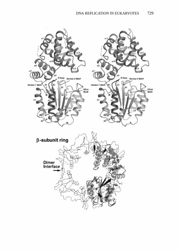
Of all the DNA polymerase accessory proteins, the subunits of RFC showboth striking sequence and functional similarities to replication fork proteinspresent inE. coli and bacteriophage T4 (γ -complex and gp44/gp62, respec-tively; 95, 106). These proteins load a ring-like DNA polymerase clamp (e.g.PCNA in eukaryotes and theE. coli β-subunit of DNA polymerase III) ontothe template DNA and, therefore, are known as clamp-loading proteins. Therecent determination of the crystal structure of a member of theγ -complex,theδ′ subunit, shows it to have a provocative C-like shape that shows strikingstructural overlap with the ring-like shape of theE. coli β-subunit and PCNA,suggesting how the clamp-loader might open the ring-like clamp and load itonto to DNA (Figure 1; 130). The clamp-loading function of RFC may be oneof the key regulated events in DNA metabolism.
Proliferating Cell Nuclear Antigen (PCNA)Perhaps one of the most intensely studied proteins is proliferating cell nuclearantigen (PCNA), the DNA polymerase clamp. Not only does this protein playa central role in DNA metabolism, but it has become a significant clinical
−−−−−−−−−−−−−−−−−−−−−−−−−−−−−−−−−−−−−−−−−−−−−−−−−−−−−−→Figure 1 Structures of a polymerase clamp and a clamp-loading protein subunit.Top: stereo viewof a ribbon diagram of the structure of the clamp-loader,δ′, a part of theE. coliDNA polymerase IIIγ -complex and a functional homolog of the RFC subunits (130).Bottom: super-position of theδ′structure and a ribbon diagram of the structure of theE. coliDNA polymerase IIIβ-subunit (133), afunctional and structural homolog of PCNA. Note the similar shape of the clamp ring surroundingthe dimer-dimer interface of the twoβ-subunits and the clamp-loader C-shape.
P1: DPI/plb P2: NBL/dat QC: NBL
April 29, 1998 13:49 Annual Reviews AR057-23
DNA REPLICATION IN EUKARYOTES 729
P1: DPI/plb P2: NBL/dat QC: NBL
April 29, 1998 13:49 Annual Reviews AR057-23
730 WAGA & STILLMAN
diagnostic marker for proliferating cells. A protein with an apparent mass onpolyacrylamide gels of 36 kDa, PCNA forms a homotrimeric complex andfunctions as a DNA polymerase accessory factor (for reviews see 42, 131, 132).The primary amino acid sequence of PCNA is not highly conserved amongspecies, but yeast and human PCNA nevertheless have an almost identical three-dimensional shape that is also very similar to the structures of the functionalhomologs of PCNA present inE. coli and bacteriophage T4 (i.e. theβ-subunitof DNA polymerase III inE. coli and the gp45 protein of T4 phage) (95, 132–135). These proteins exist as stable trimers that form a closed ring with a holein the center that encircles duplex DNA (Figure 1). Each protein monomerof PCNA consists of two structurally similar domains that are linked via aninterdomain connecting loop on the surface of the protein (134). The internalsurface of the trimer contains sixα helices, and each helix is present in astructural repeat in each PCNA monomer. Since RFC loads the PCNA trimeronto DNA, it topologically links the PCNA trimer to the DNA, allowing PCNAto track along the DNA (136). Most probably, RFC also functions to unloadthe PCNA when DNA synthesis is complete, much like the bacterial and phageT4 counterparts (137–139).
PCNA functions as a processivity factor for polδ during DNA replication(140–144). Stimulation of polε by PCNA has also been detected but onlyunder limited conditions (145–147). PCNA itself does not have DNA-bindingactivity, but it can be loaded onto the DNA by RFC in an ATP-dependent manner(56, 114). PCNA then associates with polδ/ε at the primer-template junction(56, 113, 116). Mutational analyses of PCNA have shown that distinct regionsof the trimeric ring are required for the stimulation of polδ or RFC ATPase(148–151); PCNA mutants that alter certain amino acids on the internal surfaceof the ring failed to stimulate polδ (149). In contrast, regions on the outersurface, including both the N- and C-termini and the interdomain connectingloop, are necessary for the interaction with polδ and RFC (149, 151).
PCNA also is capable of binding to other proteins, including the FEN1/Rad27/MF1 nuclease (152, 153) (see below); DNA ligase 1 (154); the p53-inducible,cyclin-dependent kinase (CDK) inhibitor protein p21 (WAF1, CIP1, sdi1)(155, 156); the p53-inducible GADD45 protein (157, 158); the nucleotide ex-cision repair protein XPG (159); DNA-(cytosine-5) methyltransferase (160);the mismatch repair proteins MLH1 and MSH2 (161); and cyclin D (162, 163).These varied interactions with DNA metabolism proteins imply that PCNA is acentral factor for the coordination of DNA replication, DNA repair, epigeneticinheritance, and cell-cycle control.
The crystal structure of PCNA bound to a peptide derived from p21 showsthat the previously unstructured p21 peptide inserts itself into a cleft in theinterdomain connecting loop surface of PCNA (164). This very stable binding

P1: DPI/plb P2: NBL/dat QC: NBL
April 29, 1998 13:49 Annual Reviews AR057-23
DNA REPLICATION IN EUKARYOTES 731
causes the inhibition of PCNA stimulation of polδ activity and competitionwith the binding of PCNA to DNA ligase, DNA-(cytosine-5) methyltransferase,XPG, and FEN1 (see below). Moreover, a monoclonal antibody that specificallybinds to this loop inhibits PCNA-activated DNA synthesis by polδ (165). Thusthe loop serves as an interface for interactions with other cellular proteins.
Immunofluorescent staining of cells with anti-PCNA antibodies has shownthat PCNA co-localizes with sites of DNA synthesis in nuclei (replication foci)(166, 167). The PCNA staining pattern varies with the stage in the S phase,corresponding with the region of the genome being replicated. In addition,PCNA becomes resistant to extraction from nuclei with detergents such asTriton X-100, only when cells enter the S phase of the cell cycle (166, 168).PCNA resistance to Triton-extraction also occurs in DNA-damaged cells, evenin the G1 phase of the cell cycle, indicating that PCNA localizes to sites ofDNA repair (168–172). The variable, detergent-resistant PCNA in nuclei isthought to reflect its topologically closed interaction with DNA at sites of DNAmetabolism.
DNA Polymerasesδ andεTwo DNA polymerases function during the S phase of the cell cycle in eukary-otic cells (14). DNA polymeraseδ (pol δ) is a heterodimer composed of p125and p50 subunits (for review see 14), although we consider it likely that thenative enzyme in mammalian cells andS. cerevisiaecontains an additional sub-unit of approximate mass of 50 kDa. Recent biochemical and genetic evidenceshows that theS. pombepol δ contains five subunits, three of which are essen-tial (173–175). p125 is the catalytic subunit for both DNA polymerase activityand a proofreading, 3′-5′ exonuclease activity. cDNAs for each subunit havebeen cloned (175–183). Moreover, an active human dimeric complex has beenreconstituted using proteins expressed in recombinant baculovirus-infected in-sect cells (184). The N-terminal region of p125 interacts with PCNA (185, 186),and although one report suggests that the polymerase activity of the large sub-unit alone can be stimulated by PCNA inS. cerevisiae(185), a recent studyof mammalian polδ shows that the p50 subunit is required for PCNA stimula-tion (184). To complicate matters further, a putative PCNA-independent polδ
was also isolated from mouse (187) andDrosophila(188). Thus further cha-racterization of the structure of DNA polymeraseδ in mammalian cells andS. cerevisiaeis necessary.
Pol δ is required for DNA synthesis of both the leading and lagging strandsduring SV40 DNA replication in vitro (22–24, 26, 189). Many studies using avariety of DNA templates support a role for polδ in RFC- and PCNA-depen-dent DNA replication (24–26, 56, 109, 113, 146, 184, 189–194). Crosslinkingof DNA polymerases to replicating SV40 DNA in vivo also supports the

P1: DPI/plb P2: NBL/dat QC: NBL
April 29, 1998 13:49 Annual Reviews AR057-23
732 WAGA & STILLMAN
involvement of both polδ and polα in SV40 DNA replication (195). Al-though some reports demonstrate a role for polε in cellular DNA replication(113, 146, 196–200), the in vivo crosslinking experiments indicate that polε isnot essential for SV40 DNA replication (195). This study, however, found thatpol ε did crosslink to replicating cell chromosomal DNA, consistent with theessential function ofS. cerevisiaepol ε gene (POL2) in cellular DNA replica-tion (196, 197). The precise role of polymeraseε in cellular DNA replicationremains to be determined.
FEN1 and RNaseHIFEN1, a 46-kDa single polypeptide in human and mouse, is a 5′-3′ exo/endonu-clease that is required for Okazaki fragment maturation (for reviews see 10, 201).The primary sequence of the protein shows similarity to the repair proteinRad2/XPG and other nucleases (202, 203). FEN1 was identified through pu-rification of the enzyme that specifically cleaves a flap-structure DNA substrate,hence the name flap endonuclease (204). Other researchers independently iden-tified the same protein in different contexts, as follows: MF1 (192), 5′-3′ exonu-clease (205), cca/exo (206), DNase IV (207), polε-associated nuclease (208),human homolog ofS. pombeRad2 (203), and factor pL (209). The homologsfrom S. cerevisiaeandS. pombewere identified as Rad27 (210, 211) and Rad2(203), respectively, indicating a role for the protein in DNA repair. FEN1 isrequired for SV40 DNA replication in vitro (192, 205).
Several biochemical studies have shown that FEN1 functions specificallyto remove the RNA primer attached to the 5′-end of each Okazaki fragment(26, 200, 212–215). Extensive studies show that removal of the RNA primerinvolves other proteins including an RNA-DNA junction endonuclease, PCNA,and Dna2 helicase (10). The mechanism for maturation of Okazaki fragmentsis described below, but removal of the RNA primer from the 5′-end of thepenultimate Okazaki fragment prior to joining to the newly synthesized Okazakifragment requires strand displacement synthesis to displace the RNA primer,creating a flap structure with a 5′ unpaired RNA-DNA strand.
The enzymatic properties of FEN1 have been examined in some detail. Whenprovided a flap structure containing a 5′-segment of DNA (or RNA) that is notpaired to a template DNA, FEN1 efficiently cleaves at the branch point, releasingthe unpaired segment (202, 204, 207, 215, 216). However, if the 5′-segment ofDNA (or RNA) is completely paired, FEN1 can degrade DNA (or RNA) onlyexonucleolyticaly from the 5′-terminus (192, 200, 202, 204, 214). A substratethat contains a 5′-triphosphorylated ribonucleotide that is annealed to DNA,such as an Okazaki fragment that is completely annealed to a single-strandedDNA, cannot be degraded by FEN1 (200). In such a situation, removal of theRNA requires a ribonuclease in addition to FEN1 (see below).

P1: DPI/plb P2: NBL/dat QC: NBL
April 29, 1998 13:49 Annual Reviews AR057-23
DNA REPLICATION IN EUKARYOTES 733
PCNA and RPA stimulate yeast FEN1 activity under certain conditions(152, 217). These interactions may be important for the maturation of Okazakifragments during lagging strand replication, as described below. The cyclin-CDK inhibitor p21 disrupts the FEN1-PCNA interaction, suggesting that thestep might be regulated (153).
While bothS. cerevisiae RAD27andS. pombe rad2+ are not essential forcell viability, both null mutants exhibit elevated chromosome loss rates andincreased UV sensitivity (203, 210, 218). Furthermore, arad27 null mutantshows temperature-sensitive lethality (210, 211). Interestingly, a novel typeof mutation can be generated in cells lacking FEN1 activity. Sequences of5–108 nucleotide pairs that are flanked by direct repeats of 3–12 nucleotidepairs become duplicated at high frequency inrad27null mutants (219). Thisunique mutation event is thought to result from a defect in lagging-strand DNAsynthesis, causing an increased recombination rate due to induced double-strand-break repair of these lesions (220, 221).
RNase H activities in eukaryotic cells have been grouped into two classes, Iand II, based on molecular mass of the enzyme and requirements for a cofactor(222). Among them, RNase HI is thought to be involved in the removal ofRNA primers during Okazaki fragment synthesis (200, 206, 223–225). Whilethe enzymatic activity of RNase HI has been known for a long time, its precisemolecular weight and subunit structure are still unclear. Nevertheless, biochem-ical characterizations of RNase HI have revealed a unique substrate specificity;it could endonucleolyticaly cleave RNA that is attached to the 5′-end of a DNAstrand, such as in an Okazaki fragment, leaving a single ribonucleotide on the5′-end of the DNA strand (200, 226–228).
DNA HelicasesDNA helicases are enzymes that promote the processive unwinding of duplexDNA, such as occurs at the DNA replication fork to create templates for the poly-merases. During SV40 DNA replication, the virus-encoded T antigen functionsas the replicative DNA helicase, but for cell chromosome replication, the natureof the replicative helicase remains unclear. Most DNA helicases required forviral or prokaryotic DNA replication form a homomultimeric complex (hexamerin most of the cases) (for review see 229). For example, T antigen functionsas a hexamer and assembles at the SV40ori as a do-decamer (two hexamers;48, 49, 229). As DNA replication proceeds, the two hexamers at the divergentreplication forks probably stay connected, forming part of a so-called replisomethat the DNA passes through (230). It is highly likely, however, that severaldistinct helicases would function in cell chromosomal replication and also thatthe role of an individual helicase might be specialized for certain steps in thereplication process. Many eukaryotic cell DNA helicases have been identified,
P1: DPI/plb P2: NBL/dat QC: NBL
April 29, 1998 13:49 Annual Reviews AR057-23
734 WAGA & STILLMAN
including the helicases associated with polδ/ε or RFC (see 229), but only a fewhave been implicated in DNA replication.
DNA2 HELICASE Yeast Dna2 helicase was identified by screening for replica-tion-defective mutants using a DNA replication assay in permeabilized cells(231, 232). TheDNA2 gene encodes a 172-kDa protein that is essential forcell viability, and the purified protein shows both DNA-dependent ATPaseand 3′-to-5′ DNA helicase activities (232, 233). Analysis of3H-labeled replica-tion intermediates from wild-type anddna2temperature-sensitive mutant cellsshowed that low-molecular-weight intermediates accumulate in thedna2mu-tants but not in wild-type cells, indicating that the defect is at the elongationstage of DNA replication (232).
MOUSE HELICASE B This mammalian helicase was identified through studiesof a temperature-sensitive mutant mouse cell line, tsFT848, which was shownto be defective in DNA replication (234–236). DNA synthesis, but not RNAand protein syntheses, in these mutant cells decreased at the nonpermissivetemperature. Comparative analyses of DNA-dependent ATPase activities infractionated extracts from wild-type and mutant cells showed that one of themajor ATPase activities, now designated helicase B, is decreased in the mutantcells. Furthermore, this helicase activity from mutant cells showed heat sensi-tivity. The DNA chain-elongation rate in the mutant cells, when analyzed byfiber autoradiography, was the same in both wild-type and mutant cells, sug-gesting that helicase B might be involved in a process that does not determinethe elongation rate of the fork.
MINI-CHROMOSOME MAINTENANCE PROTEINS The mini-chromosome mainte-nance (MCM) proteins were first identified by yeast genetic studies as proteinsrequired for replication of plasmid DNAs containing cellular origins of DNAreplication. Researchers have since determined that these proteins are essentialcomponents of the pre-replication complex established prior to the S phase atorigins of DNA replication (reviewed in 237, 238). Six MCM proteins have beenfound in all eukaryotic cells examined to date, and they share a similar aminoacid sequence motif called the MCM box, part of which contains a putative AT-Pase motif (239). Genes encoding MCM proteins have also been found in recentsequences from Archea species, suggesting that they are ancient replicationproteins. The MCM protein complex appears to be a hexamer containing equalamounts of each of the six proteins (239a). Two recent observations, when com-bined, suggest, but do not prove, that the MCM proteins function as a replicativeDNA helicase at the cellular replication fork (240, 241). A complex of threeMCM proteins (Mcm 4, 6, and 7) is capable of displacing a short oligonucleotidefrom a larger single-stranded DNA in an ATP-dependent manner, suggesting
P1: DPI/plb P2: NBL/dat QC: NBL
April 29, 1998 13:49 Annual Reviews AR057-23
DNA REPLICATION IN EUKARYOTES 735
that it contains DNA helicase activity (241). In addition, some MCM proteinsappear to be bound to different regions of a replicon in the yeast genome at dif-ferent times throughout the S phase of the cell cycle, beginning with the originsof DNA replication where they are assembled in an ORC- and Cdc6-dependentmanner prior to S-phase entry (2, 240). This suggests that the MCM proteinsmight track along the DNA with the DNA replication fork, perhaps acting tounwind the DNA or performing another essential function (240).
Molecular Links Among FEN1, Dna2 Helicase,and DNA Polymeraseα/PrimaseAccumulating evidence indicates that a multi-enzyme complex exists in cellsthat contains many of the activities discussed above (193, 242). In addition,biochemical and genetic studies of yeast DNA replication have made connec-tions linking many replication proteins into a multi-protein complex that mayfunction as a so-called replisome. Physical and genetic interactions betweenthe Dna2 helicase and FEN1 have been demonstrated; both proteins co-purifiedand co-immunoprecipitated, and the overexpression ofFEN1 suppressed thetemperature-sensitive growth of adna2-1mutant (243). Conversely, overex-pression ofDNA2suppressed the temperature-sensitive lethality of arad27nullmutant (defective in FEN1; 243). This FEN1-Dna2 helicase interaction mayplay an important role in maturation of the lagging strand.
In an independent study, another allele ofdna2 (dna2-2) was isolated ina genetic screen for mutants that show synthetic lethality with thectf4-14mutant (244). Ctf4 protein (Ctf4p), which is identical to Pob1p and Chl15p,was identified as a protein that bound to a polα catalytic subunit-affinity column(245–247). Thectf4 null mutant is viable but exhibits elevated chromosomeloss, implying a function in some process of DNA metabolism (245, 246).
Cdc68p and Pob3p have also been identified as polα–binding proteins(244). These proteins seem to compete with Ctf4p for binding to polα. Bio-chemical and genetic interactions between polα and these polα–binding pro-teins [Ctf4p, Cdc68p (248–250) and Pob3p] have also been demonstrated (244).Furthermore, thecdc68-1mutation was also synthetic lethal with thedna2-2mutation (244). These genetic and biochemical data suggest that the synthesisof an RNA-DNA primer to start an Okazaki fragment and maturation of theOkazaki fragment might be coordinately carried out during lagging strand syn-thesis by a multi-protein complex containing polα/primase, Ctf4p, Cdc68p,Pob3p, Dna2p helicase, and FEN1. Because FEN1 can bind directly to PCNA,this complex might also contain RFC and polδ, consistent with biochemicalobservations (193, 217, 242).
Another allele of RAD27 (erc 11-2) was unexpectedly isolated in a geneticscreen that sought to identify proteins that interacted with the G1 cyclins Cln1p
P1: DPI/plb P2: NBL/dat QC: NBL
April 29, 1998 13:49 Annual Reviews AR057-23
736 WAGA & STILLMAN
and Cln2p [erc (elevated requirement forCLN function) mutations; 251]. Therad27/erc11-2, cln1cln2mutant strain arrested in the S phase at nonpermissivetemperature and gradually lost viability. The temperature-sensitive lethalitycould be rescued by expression ofCLN1or CLN2but not the other G1 cyclin,CLN3. Moreover, overexpression ofDNA2(referred to asSEL1in the originalpaper) andCDC9(DNA ligase) also rescue the temperature-sensitive lethality(251). Although it is unclear how Cln1p/2p can rescue the defect in replication,it is intriguing that both proteins that rescued the defect when overexpressedcould interact with PCNA. These studies suggest that the G1 cyclins Cln1p andCln2p might affect functions at the DNA replication fork.
MECHANISMS OF DNA SYNTHESISAT A REPLICATION FORK
The above-described studies on the replication proteins in eukaryotic cells haveidentified their biochemical functions and several specific interactions amongthese proteins. These interactions underlie the mechanism of DNA synthesis ofboth the leading and lagging strands at the DNA replication fork. The followingsections describe our current understanding of how these proteins cooperate toreplicate DNA.
Primosome AssemblyOne of the first steps after recognition of the origin of DNA replication andlocal unwinding of the DNA is to load the polα/primase complex onto theDNA, a step called primosome assembly. Details about the role of T antigen inorigin recognition and local unwinding of theori have been reviewed elsewhere(229, 252). Primosome assembly normally involves a DNA helicase interact-ing with the polα/primase, but a cellular helicase that functions in cellularDNA replication in the same way that T antigen functions during SV40 DNAreplication has not been identified to date.
T antigen, polα/primase, and RPA interact with each other and cooperateto initiate DNA synthesis at the SV40ori (16, 27, 36, 60, 254). The protein-protein interactions that have been demonstrated are T antigen–polα/primase(p70 and/or p180) (36, 255–258), RPA p70–primase (p48 and p58) (59), andRPA–T antigen (59, 60). In addition, the bovine papillomavirus E1 initiator andhelicase protein also binds to polα/primase in a manner analogous to T antigen(259). Primase assays with single-stranded template DNAs have shown thatalthough RPA from either humans or yeast represses primase activity, T antigencan reverse the inhibition only when human RPA, but notS. cerevisiaeRPA, iscoating the DNA (60, 260). In addition, only human polα/primase, but not calfthymus or mouse polα/primase, can support primer synthesis in the presence
P1: DPI/plb P2: NBL/dat QC: NBL
April 29, 1998 13:49 Annual Reviews AR057-23
DNA REPLICATION IN EUKARYOTES 737
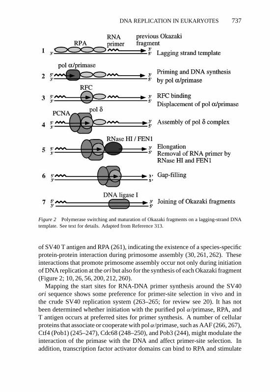
Figure 2 Polymerase switching and maturation of Okazaki fragments on a lagging-strand DNAtemplate. See text for details. Adapted from Reference 313.
of SV40 T antigen and RPA (261), indicating the existence of a species-specificprotein-protein interaction during primosome assembly (30, 261, 262). Theseinteractions that promote primosome assembly occur not only during initiationof DNA replication at theori but also for the synthesis of each Okazaki fragment(Figure 2; 10, 26, 56, 200, 212, 260).
Mapping the start sites for RNA-DNA primer synthesis around the SV40ori sequence shows some preference for primer-site selection in vivo and inthe crude SV40 replication system (263–265; for review see 20). It has notbeen determined whether initiation with the purified polα/primase, RPA, andT antigen occurs at preferred sites for primer synthesis. A number of cellularproteins that associate or cooperate with polα/primase, such as AAF (266, 267),Ctf4 (Pob1) (245–247), Cdc68 (248–250), and Pob3 (244), might modulate theinteraction of the primase with the DNA and affect primer-site selection. Inaddition, transcription factor activator domains can bind to RPA and stimulate
P1: DPI/plb P2: NBL/dat QC: NBL
April 29, 1998 13:49 Annual Reviews AR057-23
738 WAGA & STILLMAN
replication in vitro (268, 269), but the precise mechanism of this activation isunclear. In the context of cellular chromatin, these site-specific DNA-bindingproteins may aide in recruiting RPA and hence polα/primase to the DNA.
Polymerase SwitchingBiochemical studies using the SV40 DNA replication system reconstituted withpurified proteins (21, 24, 26, 54, 190, 191) have shown that two different poly-merases, polα/primase and polδ, are involved in DNA synthesis and that polδis involved in the synthesis of both the leading and lagging strands. The switch-ing from polα/primase to polδ occurs during priming of the leading strand (24)and during synthesis of every Okazaki fragment (26). The involvement of twodifferent DNA polymerases in lagging-strand DNA synthesis was suggested bythe analysis of SV40 DNA replication in vivo with DNA polymerase inhibitors(15, 17). The mechanisms of initiation of leading strand synthesis and initiationof each Okazaki fragment are apparently very similar: An RNA-DNA primeris produced by polα/primase, and the 3′-terminus of the initiator DNA (iDNA)is recognized by RFC and PCNA to expel the polα/primase and load polδ(Figure 2; 24–26).
As suggested by the model in Figure 2, polα/primase starts the synthesisof an RNA-DNA primer on an RPA-coated, single-stranded DNA template,perhaps assisted by a putative cellular loading activity (such as T antigen),to yield a short 30-nt primer RNA-DNA (18, 264, 270). Once the RNA-DNAprimer is synthesized, RFC binds to the 3′-end of the iDNA, displacing polα/primase. The turnover of polα/primase most likely occurs by the inherentnonprocessive nature of the polα catalytic activity and the tight binding ofRFC to the primer-template junction, since both RPA and RFC decrease thelength of the iDNA (25). RFC binding triggers the assembly of the primerrecognition complex, which is accomplished through the loading of PCNA andsubsequent association of PCNA with polδ. Then the relatively processive polδ holoenzyme extends the DNA strand (16, 23–25, 191). For the initiation ofleading-strand DNA replication, the synthesis by the polδ/PCNA complex isthen processive and continuous, at least for 5–10 kb of DNA. For synthesison the lagging strand, DNA synthesis of the Okazaki fragment continues untilthe polymerase encounters the previously synthesized Okazaki fragment. TheRNA primer from the preceding Okazaki fragment is then removed by a complexprocessing reaction described below, and the remaining nick is sealed by DNAligase I.
Biochemical studies in vitro (described in this review) and studies in vivo(195) show that polε is not required for SV40 DNA replication. But given theenzymatic similarities between polδ and polε, such as high processivity, and theobservation that polymeraseε is essential for DNA replication (196, 197), polε
P1: DPI/plb P2: NBL/dat QC: NBL
April 29, 1998 13:49 Annual Reviews AR057-23
DNA REPLICATION IN EUKARYOTES 739
almost certainly participates in cellular chromosomal replication. A differencebetween these two systems is the mechanism of initiation, because many cellularproteins are required to achieve what is achieved by T antigen. Therefore, polε might be involved in the initiation of DNA replication. Alternatively, eachpolymerase might be involved specifically in a separate process during DNAreplication. A genetic study utilizingS. cerevisiaestrains that contain mutationsin the proofreading exonuclease domains of either polδ or pol ε suggests thateach polymerase is involved in replicating different strands of the DNA (271).
Polymeraseε is also a unique polymerase in that it is involved in cell-cyclecheckpoint control and may therefore function at the DNA replication fork toensure accurate DNA synthesis, perhaps by a postreplicative repair mechanismor another, as yet unrecognized, mechanism (272, 273). It is also possible thatpol ε functions in replicating large chromosomes as a specialized enzyme forinitiation of DNA replication at sites where DNA synthesis has halted tem-porarily because the replication fork has to cope with the complex topology ofthe cell’s chromosomes. Clearly, more studies on the role of polε are required.
A key protein in the polymerase switching appears to be RFC, and the loadingof PCNA by RFC is an essential event for the transition from a priming mode toan extension mode of DNA synthesis. Given the functional similarities betweenRFC and theE. coli pol III γ -complex (95), it is possible that RFC coordinatesthe synthesis of both the lagging and leading strands. Further investigation ofthe mechanism for coordinating DNA synthesis of both strands at the eukaryoticcell DNA replication fork is necessary.
Maturation of Okazaki FragmentsIn maturation of Okazaki fragment synthesis, the short Okazaki fragments syn-thesized discontinuously on the lagging strand template are converted into long,ungapped DNA products. This involves several distinct steps including removalof the RNA primer, DNA gap synthesis, and sealing together of the two DNA.Recent studies, and the observation that many of the proteins involved in thisprocess bind to PCNA, suggest that these steps may be regulated coordinatelywith each other.
The analysis of lagging-strand DNA synthesis using highly purified proteinshas revealed the basis of the maturation mechanism. Two different nucleases,RNase HI and FEN1, are involved in the complete removal of the RNA primer(192, 200, 205, 206, 212–216). These nucleases are required for the completereplication of SV40 DNA and for reconstitution of lagging strand synthesison artificial templates (26, 192, 200). An in vitro assay with a model substrateshowed that PCNA binds to FEN1 and stimulates FEN1 activity (152). This ob-servation suggests that the removal of RNA (or RNA-DNA) might be triggeredeither by the upstream DNA polymerase complex or by the newly synthesized

P1: DPI/plb P2: NBL/dat QC: NBL
April 29, 1998 13:49 Annual Reviews AR057-23
740 WAGA & STILLMAN
DNA, creating a duplex DNA region upstream of the RNA at the 5′-end of theOkazaki fragment. Consistent with the latter possibility, an assay for FEN1using a synthetic oligonucleotide substrate showed that an upstream DNA caninfluence the cleavage of a downstream flap substrate (204, 213–215).
Okazaki fragment processing and genome integrity during cell chromosomereplication might require additional proteins, such as the Dna2 helicase, tocomplete the same process. Dna2 helicase has been suggested to be involvedin removal of RNA primers because of its biochemical and genetic interactionswith FEN1/Rad27 nuclease (243; also see above). Dna2 helicase, in conjunctionwith the polymerase complex that synthesizes the Okazaki fragments, mightdisplace the RNA primer from the template DNA, thereby creating a flap-likesubstrate for FEN1 endonuclease (see FEN1 and RNase HI section, Figure 3,and Reference 10). A more interesting possibility is that in addition to theRNA at the 5′-end of an Okazaki fragment, the DNA beyond the RNA-DNA
Figure 3 Two mechanisms for the removal of RNA primers. (A) RNase HI cleaves the RNAsegment attached to the 5′-end of the Okazaki fragment, leaving a single ribonucleotide adjacent tothe RNA-DNA junction. FEN1 then removes the remaining ribonucleotide. (B) Dna2 helicase dis-places the RNA segment (or RNA-DNA). FEN1 then cleaves endonucleolyticaly the branch point,releasing the displaced RNA (or RNA-DNA). Although the Dna2/FEN1-dependent mechanism hasnot been proved, recent biochemical and genetic studies strongly support this model (see 10).
P1: DPI/plb P2: NBL/dat QC: NBL
April 29, 1998 13:49 Annual Reviews AR057-23
DNA REPLICATION IN EUKARYOTES 741
junction that was synthesized by DNA polα might also be displaced by Dna2helicase and cleaved off by FEN1 (10). If this were the case, then the iDNAthat was made by polα would be removed by this Okazaki maturation process,and the gap created would be filled in by either polδ or pol ε. This would bea significant advantage for cells for maintaining genome integrity because polα/primase does not have a proofreading activity and thus could not remove anyerrors that it inserted in the iDNA, whereas if this region of the template DNAwere “re-replicated” by polδ or polε, then the proofreading exonucleases fromthese enzymes would ensure increased accuracy during replication. This mech-anism for Okazaki fragment maturation might also prevent inappropriate DNAreplication of repeated regions in the genome, such as mini-satellite repeatedsequences, a common occurrence in human cancer cells (219, 220).
REPLICATION FORK PROTEINSAND CELL-CYCLE CONTROL
S-Phase Checkpoint ControlWhen the genome is subjected to excessive DNA damage, or progression ofDNA replication forks is blocked, cells arrest progression of the cell cycle ateither the G1-S phase transition, the G2-M phase transition, or they slow downS-phase progression. This activates transcription of genes that encode proteinsrequired for the repair of DNA and facilitates the repair process itself (reviewedin 274). The signal transduction mechanisms that detect aberrant replication orDNA damage in the S phase, and then block cell-cycle progression are calledS-phase checkpoints. Previous genetic studies inS. cerevisiaeandS. pombesuggest that several replication fork proteins might be involved in these S-phasecheckpoints.
In S. cerevisiae, mutant cells that have a defect in polε ( pol 2) fail to ac-tivate the damage-inducible transcription of certain damage responsive genesin response to DNA damage by methyl methane sulfonate (MMS), or nucleotide-depletion induced by hydroxyurea during the S phase. The mutant cells enterinto the M phase without correctly completing DNA replication (272). ThusPOL2might be involved in an S-phase checkpoint signaling mechanism. Thedomain inpol ε that is responsible for its checkpoint function is separable fromits DNA polymerase catalytic domain (272). Recent studies suggest that theRad53p protein kinase is required for DNA-damage checkpoint signaling by apol ε- and Rfc5p-dependent mechanism (129, 273, 275).
DPB11, which was isolated as a multicopy suppressor of mutations in genesencoding subunits ofS. cerevisiaepolε (pol2anddpb2), has a checkpoint func-tion (276). A temperature-sensitive mutant inDPB11underwent cell division

P1: DPI/plb P2: NBL/dat QC: NBL
April 29, 1998 13:49 Annual Reviews AR057-23
742 WAGA & STILLMAN
without completion of replication at the nonpermissive temperature, and themutant cells were sensitive to hydroxyurea, MMS, and UV irradiation. Asimilar defect was demonstrated in mutants in the cut5+ gene inS. pombe,although these mutants were sensitive only to DNA damage and not to nu-cleotide depletion (277, 278). A recent report showed that the Cut5 protein,which is similar in sequence to Dpb11p fromS. cerevisiae, binds to the Chk1protein kinase that is known to control the cyclin-dependent Cdc2 kinase, theprincipal regulator for M-phase entry (279).
Other replication proteins have been implicated in checkpoint controls. Yeastmutants with defects in genes encoding RPA (S. pomberad11) (280), primase(S. cerevisiae pri1) (281), S. pombepol α and polδ (282–284), and an RFCsmall subunit (S. cerevisiae rfc5) (275, 285) have also been shown to exhibitcheckpoint defects. Some of these defects may occur because a replicationfork is not established after commitment to cell division, and others may bethe result of a real defect in signaling in response to DNA damage or a repli-cation fork block. The primase mutations are particularly interesting becausethey override a mechanism that slows down S-phase progression in the pre-sence of continuous low doses of a DNA alkylating agent (281). Interestingly,loss of theMEC1 gene, which is related to the ATM gene that is defectivein human ataxia telangiectasia, also causes the same effect (286), suggestingthat primase might be part of a signaling mechanism to the Mec1p checkpointpathway.
PCNA-p21 InteractionThe involvement of replication fork proteins in checkpoint signaling in meta-zoan species is likely, but additional controls may be imposed on DNA replica-tion in these cells. The identification of proteins that interact specifically withPCNA has led to the emergence of PCNA as a key protein required for thecoordinated regulation of replication and other events that take place at thereplication fork, such as DNA methylation and DNA repair. In addition, over-expression of PCNA inS. pomberesults in the delay of entry into the M phase(287), perhaps because PCNA is binding a protein that is essential for thisprocess.
One of the well-characterized PCNA-interacting proteins is the p21 protein,an inhibitor for cyclin-dependent kinase (CDK) (p21 has alternate names ofCIP1, WAF1, Sdi1; for review see 288, 289). p21 is induced during mitogenicstimulation of mammalian cells and in response to DNA damage by the tumorsuppression protein p53, as well as many other stimuli. The protein binds toand inhibits cyclin-dependent kinase (CDK) activities that are required for G1/Sprogression, leading to cell-cycle arrest. Thus p21 functions as part of a DNA-damage checkpoint mechanism.

P1: DPI/plb P2: NBL/dat QC: NBL
April 29, 1998 13:49 Annual Reviews AR057-23
DNA REPLICATION IN EUKARYOTES 743
Interest in p21 with respect to the DNA replication fork was triggered by theobservation that p21 can form a quaternary complex with a CDK, cyclin, andPCNA in normal cells but not in many transformed cells (162, 290), creating apossible link between cell-cycle progression and DNA replication. p21 inhibitsSV40 DNA replication in vitro and in frog cell extracts through its direct bindingto PCNA (155, 156, 291, 292). Even though PCNA is essential for nucleotide-excision repair (293, 294), p21 did not inhibit the repair in vitro (295, 296)and in vivo (168). These results suggest that through its binding to both CDKkinase and PCNA, p21 might function to coordinately regulate DNA replication,repair, and cell-cycle progression in response to DNA damage, perhaps inducinga switch from replication to repair.
The biological significance of this interaction is still unclear, even though thep21-PCNA interaction has been characterized extensively, including mappingof the binding domain in p21 (297–303), determination of a crystal structureof PCNA complexed with a p21-derived peptide (164), and investigation of theeffect of p21 on DNA synthesis and the loading of PCNA onto DNA (115, 304).A recent observation that p21 modulates the interactions between PCNA andFEN1 endonuclease, DNA (5-cytosine) methyltransferase, and DNA ligase sug-gests that the levels of p21 in the cell might control a switch from one PCNA-dependent function to another at the DNA replication fork (154, 160, 305).More evidence that the p21-PCNA interaction might be relevant in vivo isprovided by the recent observations that the E7 oncoprotein from human pa-pillomavirus type 16 can abrogate a DNA damage–induced cell-cycle arrestby binding to p21 (306, 307). These studies show that the p21-E7 interactionresults in both the reversal of p21 inhibition of CDK kinase activity and the p21inhibition of PCNA function (306).
Regulation of Telomere LengthThe appropriate regulation of telomere length and replication is also impor-tant for maintaining the integrity of the genome. In addition, the length oftelomeres correlates with the replicative potential of cells (for review see 308).Recent studies have indicated that in addition to telomerase, replication forkproteins may directly regulate the length of telomeres. The mutations in theS. cerevisiae POL1/CDC17gene encoding polymeraseα, as well as the geneencoding the large subunit of RFC (CDC44 in S. cerevisiae), cause elonga-tion of the telomeres (309, 310). Furthermore, pharmacological inhibitors ofcellular DNA polymerases disrupt coordinated DNA replication of the G-richstrand that is synthesized by telomerase and the C-rich lagging strand (311).Given that this telomere elongation requires telomerase activity and that bothpol α/primase and RFC are involved in lagging strand synthesis, both telo-mere extension and lagging strand synthesis might be regulated coordinately, in
P1: DPI/plb P2: NBL/dat QC: NBL
April 29, 1998 13:49 Annual Reviews AR057-23
744 WAGA & STILLMAN
conjunction with the function ofCDC13, a telomere-sequence-specific, single-stranded DNA-binding protein that is required for telomere maintenance (312).Further biochemical investigation of the mechanism for telomere replication isneeded to understand how both strands of the telomere are replicated.
CONCLUDING REMARKS
Modern research on the replication of DNA in eukaryotes can be groupedinto three related categories. One active area of research is to characterizethe DNA sequences that are important for replicator function and determinewhere origins of DNA replication occur in chromosomes. Another area is toidentify the proteins involved in the mechanism and control of the initiation ofDNA replication in eukaryotes. This area is the most active area at the presenttime, providing interesting links to cell-cycle research, developmental biology,control of gene expression, and chromosome structure and function. A thirdarea is to extend the already remarkable progress in understanding the eventsat the DNA replication fork, outlined above, to identify other proteins thatmay cooperate with the known replication fork proteins to ensure accurate andefficient DNA replication. Connections need to be made between the initiationproteins that establish the replication fork and those proteins discussed abovethat function to replicate DNA after initiation. Proteins that function at thereplication fork will likely play significant roles in inheritance of chromosomestructures by interacting with other proteins involved in chromatin assemblyand epigenetically inherited protein complexes. Clearly much more work needsto be done.
ACKNOWLEDGMENTS
We thank John Kuryan for Figure 1, Dr. Robert Bambara for discussions, andcolleagues who provided preprints of research papers. Our work was supportedby a grant from the National Cancer Institute (CA13106).
Visit the Annual Reviews home pageathttp://www.AnnualReviews.org.
Literature Cited
1. Diffley JFX. 1996.Genes Dev.10:2819–30
2. Stillman B. 1996.Science274:1659–643. Li JJ, Kelly TJ. 1984.Proc. Natl. Acad.
Sci. USA81:6973–774. Li JJ, Kelly TJ. 1985.Mol. Cell. Biol.
5:1238–46
5. Stillman BW, Gluzman Y. 1985.Mol.Cell. Biol.5:2051–60
6. Wobbe CR, Dean F, Weissbach L, Hur-witz J. 1985.Proc. Natl. Acad. Sci. USA82:5710–14
7. Challberg MD, Kelly TJ. 1989.Annu.Rev. Biochem.58:671–717

P1: DPI/plb P2: NBL/dat QC: NBL
April 29, 1998 13:49 Annual Reviews AR057-23
DNA REPLICATION IN EUKARYOTES 745
8. Stillman B. 1989.Annu. Rev. Cell. Biol.5:197–245
9. Bambara RA, Huang L. 1995.Prog. Nu-cleic Acid Res. Mol. Biol.51:93–122
10. Bambara RA, Murante RS, Henrick-sen LA. 1997.J. Biol. Chem.272:4647–50
11. Lindahl T, Barnes DE. 1992.Annu. Rev.Biochem.61:251–81
12. Nash R, Lindahl T. 1996. See Ref. 314,pp. 575–86
13. Andersen AH, Bendixen C, WestergaardO. 1996. See Ref. 314, pp. 587–617
14. Wang TS-F. 1996. See Ref. 314, pp.461–93
15. Nethanel T, Reisfeld S, Dinter-GottliebG, Kaufmann G. 1988.J. Virol. 6:2867–73
16. Matsumoto T, Eki T, Hurwitz J. 1990.Proc. Natl. Acad. Sci. USA87:9712–16
17. Nethanel T, Kaufmann G. 1990.J. Virol.64:5912–18
18. Bullock PA, Seo YS, Hurwitz J. 1991.Mol. Cell. Biol.11:2350–61
19. Murakami Y, Eki T, Hurwitz J. 1992.Proc. Natl. Acad. Sci. USA89:952–56
20. Salas M, Miller JT, Leis J, DePamphilisML. 1996. See Ref. 314, pp. 131–76
21. Prelich G, Stillman B. 1988.Cell 53:117–26
22. Lee S-H, Eki T, Hurwitz J. 1989.Proc.Natl. Acad. Sci. USA86:7361–65
23. Weinberg DH, Kelly TJ. 1989.Proc.Natl. Acad. Sci. USA86:9742–46
24. Tsurimoto T, Melendy T, Stillman B.1990.Nature346:534–39
25. Tsurimoto T, Stillman B. 1991.J. Biol.Chem.266:1961–68
26. Waga S, Stillman B. 1994.Nature369:207–12
27. Murakami Y, Hurwitz J. 1993.J. Biol.Chem.268:11008–17
28. Wang TS-F. 1991.Annu. Rev. Biochem.60:513–52
29. Stadlbauer F, Brueckner A, Rehfuess C,Eckerskorn C, Lottspeich F, et al. 1994.Eur. J. Biochem.222:781–93
30. Bruckner A, Stadlbauer F, Guarino LA,Brunahl A, Schneider C, et al. 1995.Mol.Cell. Biol.15:1716–24
31. Longhese MP, Jovine L, Plevani P, Luc-chini G. 1993.Genetics133:183–91
32. Copeland WC, Tan X. 1995.J. Biol.Chem.270:3905–13
33. Longhese MP, Fraaschini R, PlevaniP, Lucchini G. 1996.Mol. Cell. Biol.16:3235–44
34. Santocanale C, Foiani M, Lucchini G,Plevani P. 1993.J. Biol. Chem.268:1343–48
35. Bakkenist CJ, Cotterill S. 1994.J. Biol.Chem.269:26759–66
36. Collins KL, Russo AAR, Tseng BY,Kelly TJ. 1993.EMBO J.12:4555–66
37. Foiani M, Marini F, Gamba D, Luc-chini G, Plevani P. 1994.Mol. Cell. Biol.14:923–33
38. Nasheuer H-P, Moore A, Wahl AF, WangTS-F. 1991.J. Biol. Chem.266:7893–903
39. Foiani M, Liberi G, Lucchini G, PlevaniP. 1995.Mol. Cell. Biol.15:883–91
40. Ferrari M, Lucchini G, Plevani P, Foi-ani M. 1996.J. Biol. Chem.271:8661–66
41. Voitenleitner C, Fanning E, Nasheuer H-P. 1997.Oncogene14:1611–15
42. Hubscher U, Maga G, Podust VN. 1996.See Ref. 314, pp. 525–43
43. Wold MS. 1997.Annu. Rev. Biochem.66:61–92
44. Wobbe CR, Weissbach L, Borowiec JA,Dean FB, Murakami Y, et al. 1987.Proc.Natl. Acad. Sci. USA84:1834–38
45. Fairman MP, Stillman B. 1988.EMBOJ. 7:1211–18
46. Wold MS, Kelly T. 1988.Proc. Natl.Acad. Sci. USA85:2523–27
47. Sancar A. 1996.Annu. Rev. Biochem.65:43–81
48. Stahl H, Droge P, Knippers R. 1986.EMBO J.5:1939–44
49. Dean FB, Bullock P, Murakami Y,Wobbe CR, Weissbach L, Hurwitz J.1987.Proc. Natl. Acad. Sci. USA84:16–20
50. Dodson M, Dean FB, Bullock P, EcholsH, Hurwitz J. 1987.Science238:964–67
51. Wold MS, Li JJ, Kelly TJ. 1987.Proc.Natl. Acad. Sci. USA84:3643–47
52. Brill SJ, Stillman B. 1989.Nature342:92–95
53. Kenny MK, Lee S-H, Hurwitz J. 1989.Proc. Natl. Acad. Sci. USA86:9757–61
54. Tsurimoto T, Stillman B. 1989.EMBOJ. 8:3883–89
55. Erdile LF, Heyers W-D, Kolodner R,Kelly TJ. 1991. J. Biol. Chem.266:12090–98
56. Tsurimoto T, Stillman B. 1991.J. Biol.Chem.266:1950–60
57. Braun KA, Lao Y, He Z, Ingles CJ, WoldMS. 1997.Biochemistry36:8443–54
58. Longhese MP, Plevani P, Lucchini G.1994.Mol. Cell. Biol.14:7884–90
59. Dornreiter I, Erdile LF, Gilbert IU, vonWinkler D, Kelly TJ, Fanning E. 1992.EMBO J.11:769–76
60. Melendy T, Stillman B. 1993.J. Biol.Chem.268:3389–95
P1: DPI/plb P2: NBL/dat QC: NBL
April 29, 1998 13:49 Annual Reviews AR057-23
746 WAGA & STILLMAN
61. Ishiai M, Sanchez JP, Amin AA, Mura-kami Y, Hurwitz J. 1996.J. Biol. Chem.271:20868–78
62. Henricksen LA, Umbricht CB, WoldMS. 1994.J. Biol. Chem.269:11121–32
63. He Z, Wong JMS, Maniar HS, Brill SJ,Ingles CJ. 1996.J. Biol. Chem.271:28243–49
64. Stigger E, Dean FB, Hurwitz J, Lee S-H. 1994.Proc. Natl. Acad. Sci. USA91:579–83
65. Gomes XV, Wold MS. 1995.J. Biol.Chem.270:4534–43
66. Kim DK, Stigger E, Lee S-H. 1996.J.Biol. Chem.271:15124–29
67. Lin Y-L, Chen C, Keshav KF, Winch-ester E, Dutta A. 1996.J. Biol. Chem.271:17190–98
68. Philipova D, Mullen JR, Maniar HS, LuJA, Gu CY, Brill SJ. 1996.Genes Dev.10:2222–33
69. Bochkarev A, Pfuetzner RA, EdwardsAM, Frappier L. 1997.Nature385:176–81
70. Blackwell LJ, Borowiec JA. 1994.Mol.Cell. Biol.14:3993–4001
71. Kim C, Paulus BF, Wold MS. 1994.Bio-chemistry33:14197–206
72. Mitas M, Chock JY, Christy M. 1997.Biochem. J.324:957–61
73. Din S, Brill S, Fairman MP, Stillman B.1990.Genes Dev.4:968–77
74. Liu VF, Weaver DT. 1993.Mol. Cell.Biol. 13:7222–31
75. Carty MP, Zernik-Kobak M, McGrathS, Dixon K. 1994.EMBO J.13:2114–23
76. Dutta A, Stillman B. 1992.EMBO J.11:2189–99
77. Fotedar R, Roberts JM. 1992.EMBO J.11:2177–87
78. Brush GS, Anderson CW, Kelly TJ.1994. Proc. Natl. Acad. Sci. USA91:12520–24
79. Pan Z-Q, Amin AA, Gibbs E, Niu H,Hurwitz J. 1994.Proc. Natl. Acad. Sci.USA91:8343–47
80. Boubnov NV, Weaver DT. 1995.Mol.Cell. Biol.15:5700–6
81. Fried LM, Koumenis C, Peterson SR,Green SL, van Zijl P, et al. 1996.Proc.Natl. Acad. Sci. USA93:13825–30
82. Gibbs E, Pan Z-Q, Niu H, Hurwitz J.1996.J. Biol. Chem.271:22847–54
83. Niu H, Erdjument-Bromage H, Pan Z-Q,Lee S-H, Tempst P, Hurwitz J. 1997.J.Biol. Chem.272:12634–41
84. Zernik-Kobak M, Vasunia K, ConnellyM, Anderson CW, Dixon K. 1997.J.Biol. Chem.272:23896–904
85. Brush GS, Morrow DM, Hieter P, KellyTJ. 1996.Proc. Natl. Acad. Sci. USA93:15075–80
86. Cheng XB, Cheong N, Wang Y, Ili-akis G. 1996.Radiother. Oncol.39:43–52
87. Abramova NA, Russell J, Botchan M,Li R. 1997.Proc. Natl. Acad. Sci. USA94:7186–91
88. Henricksen LA, Wold MS. 1994.J. Biol.Chem.269:24203–8
89. Pan Z-Q, Park CH, Amin AA, HurwitzJ, Sancar A. 1995.Proc. Natl. Acad. Sci.USA92:4636–40
90. Henricksen LA, Carter T, Dutta A, WoldMS. 1996.Nucleic Acids Res.24:3107–12
91. Morgan SE, Kastan MB. 1997.CancerRes.57:3386–89
92. Adachi Y, Laemmli U. 1992.J. Cell Biol.119:1–16
93. Yan H, Newport J. 1995.Science269:1883–85
94. Coleman TR, Carpenter PB, DunphyWG. 1996.Cell 87:53–63
95. Stillman B. 1996. See Ref. 314, pp. 435–60
96. Chen M, Pan Z-Q, Hurwitz J. 1992.Proc. Natl. Acad. Sci. USA89:5211–15
97. Chen M, Pan Z-Q, Hurwitz J. 1992.Proc. Natl. Acad. Sci. USA89:2516–20
98. Bunz F, Kobayashi R, Stillman B. 1993.Proc. Natl. Acad. Sci. USA90:11014–18
99. Burbelo PD, Utani A, Pan Z-Q, YamadaY. 1993.Proc. Natl. Acad. Sci. USA90:11543–47
100. Howell EA, McAlear MA, Rose D,Holm C. 1994.Mol. Cell. Biol.14:255–67
101. Li XY, Burgers PMJ. 1994.Proc. Natl.Acad. Sci. USA91:868–72
102. Li XY, Burgers PMJ. 1994.J. Biol.Chem.269:21880–84
103. Noskov V, Maki S, Kawasaki Y, LeemS-H, Ono B-I, et al. 1994.Nucleic AcidsRes.22:1527–35
104. Cullmann G, Fien K, Kobayashi R, Still-man B. 1995.Mol. Cell. Biol.15:4661–71
105. Bork P, Hofmann K, Bucher P, NeuwaldAF, Altschul SF, Koonin EV. 1997.FASEB J.11:68–76
106. O’Donnell M, Onrust R, Dean FB, ChenM, Hurwitz J. 1993.Nucleic Acids Res.21:1–3
107. Tsurimoto T, Stillman B. 1989.Mol.Cell. Biol.9:609–19
108. Tsurimoto T, Stillman B. 1990.Proc.Natl. Acad. Sci. USA87:1023–27
P1: DPI/plb P2: NBL/dat QC: NBL
April 29, 1998 13:49 Annual Reviews AR057-23
DNA REPLICATION IN EUKARYOTES 747
109. Lee S-H, Kwong AD, Pan Z-Q, Hur-witz J. 1991.J. Biol. Chem.266:594–602
110. Yoder BL, Burgers PMJ. 1991.J. Biol.Chem.266:22689–97
111. Fien K, Stillman B. 1992.Mol. Cell.Biol. 12:155–63
112. Podust VN, Georgaki A, Strack B,Hubscher U. 1992.Nucleic Acids Res.20:4159–65
113. Burgers PMJ. 1991.J. Biol. Chem.266:22698–706
114. Cai JS, Uhlmann F, Gibbs E, Flores-Rozas H, Lee C-G, et al. 1996.Proc.Natl. Acad. Sci. USA93:12896–901
115. Podust VN, Podust LM, Goubin F,Ducommun B, Hubscher U. 1995.Bio-chemistry34:8869–75
116. Lee S-H, Hurwitz J. 1990.Proc. Natl.Acad. Sci. USA87:5672–76
117. Podust VN, Fanning E. 1997.J. Biol.Chem.272:6303–10
118. Ellison V, Stillman B. 1998.J. Biol.Chem.273:5979–87
119. Gerik KJ, Gary SL, Burgers PMJ. 1997.J. Biol. Chem.272:1256–62
120. Fotedar R, Mossi R, Fitzgerald P, Rous-selle T, Maga G, et al. 1996.EMBO J.15:4423–33
121. Mossi R, Jonsson Z, Allen BL, HardinSH, Hubscher U. 1997.J. Biol. Chem.272:1769–76
122. Uhlmann F, Cai JS, Gibbs E, O’DonnellM, Hurwitz J. 1997.J. Biol. Chem.272:10058–64
123. Rheaume E, Cohen LY, Uhlmann F,Lazure C, Alam A, et al. 1997.EMBO J.16:6346–54
124. Song QZ, Lu H, Zhang N, Luckow B,Shah G, et al. 1997.Biochem. Biophys.Res. Commun.233:343–48
125. Uhlmann F, Gibbs E, Cai JS, O’DonnellM, Hurwitz J. 1997.J. Biol. Chem.272:10065–71
126. Uhlmann F, Cai JS, Flores-Rozas H,Dean FB, Finkelstein J, et al. 1996.Proc.Natl. Acad. Sci. USA93:6521–26
127. Cai JS, Gibbs E, Uhlmann F, Phillips B,Yao N, et al. 1997.J. Biol. Chem.272:18974–81
128. McAlear MA, Howell EA, EspenshadeKK, Holm C. 1994.Mol. Cell. Biol.14:4390–97
129. Sugimoto K, Ando S, Shimomura T,Matsumoto K. 1997.Mol. Cell. Biol.17:5905–14
130. Guenther B, Onrust R, Sali A, O’Don-nell M, Kuriyan J. 1997.Cell 91:335–45
131. Jonsson ZO, H¨ubscher U. 1997.BioEs-says19:967–75
132. Kelman Z. 1997.Oncogene14:629–40
133. Kong X-P, Onrust R, O’Donnell M,Kuriyan J. 1992.Cell 69:425–37
134. Krishna TSR, Kong X-P, Gary S, Burg-ers PM, Kuriyan J. 1994.Cell 79:1233–43
135. Kelman Z, O’Donnell M. 1995.Annu.Rev. Biochem.64:171–200
136. Tinker RL, Kassavetis GA, GeiduschekEP. 1994.EMBO J.13:5330– 37
137. Hacker KJ, Alberts BM. 1994.J. Biol.Chem.269:24209–20
138. Hacker KJ, Alberts BM. 1994.J. Biol.Chem.269:24221–28
139. Stukenberg PT, Turner J, O’Donnell M.1994.Cell 78:877–87
140. Tan CK, Castillo C, So AG, DowneyKM. 1986. J. Biol. Chem.261:12310–16
141. Bravo R, Frank R, Blundell PA, Mac-Donald-Bravo H. 1987.Nature 326:515–17
142. Prelich G, Kostura M, Marshak DR,Mathews MB, Stillman B. 1987.Nature326:471–75
143. Prelich G, Tan CK, Kostura M, Math-ews MB, So AG, et al. 1987.Nature326:517–20
144. Bauer GA, Burgers PMJ. 1988.Proc.Natl. Acad. Sci. USA85:7506–10
145. Hamatake RK, Hasegawa H, Clark AB,Bebenek K, Kunkel TA, Sugino A. 1990.J. Biol. Chem.265:4072–83
146. Lee S-H, Zhen-Qiang P, Kwong AD,Burgers PMJ, Hurwitz J. 1991.J. Biol.Chem.266:22707–17
147. Chui G, Linn S. 1995.J. Biol. Chem.270:7799–808
148. Ayyagari R, Impellizzeri KJ, Yoder BL,Gary SL, Burgers PMJ. 1995.Mol. Cell.Biol. 15:4420–29
149. Fukuda K, Morioka H, Imajou S, IkedaS, Ohtsuka E, Tsurimoto T. 1995.J. Biol.Chem.270:22527–34
150. Arroyo MP, Downey KM, So AG, WangTS-F. 1996.J. Biol. Chem.271:15971–80
151. Eissenberg JC, Ayyagari R, Gomes XV,Burgers PM. 1997.Mol. Cell. Biol. 17:6367–78
152. Li XY, Li J, Harrington J, Lieber MR,Burgers PMJ. 1995.J. Biol. Chem.270:22109–12
153. Chen JJ, Chen S, Saha P, Dutta A. 1996.Proc. Natl. Acad. Sci. USA93:11597–602
154. Levin DS, Bai W, Yao N, O’Donnell M,Tomkinson AE. 1997.Proc. Natl. Acad.Sci. USA94:12863–68
155. Flores-Rozas H, Kelman Z, Dean FB,

P1: DPI/plb P2: NBL/dat QC: NBL
April 29, 1998 13:49 Annual Reviews AR057-23
748 WAGA & STILLMAN
Pan Z-Q, Harper JW, et al. 1994.Proc.Natl. Acad. Sci. USA91:8655–59
156. Waga S, Hannon GJ, Beach D, StillmanB. 1994.Nature369:574–78
157. Smith ML, Chen I-T, Zhan QM, Bae IS,Chen C-Y, et al. 1994.Science266:1376–80
158. Hall PA, Kearsey JM, Coates PJ, Nor-man DG, Warbrick E, Cox LS. 1995.Oncogene10:2427–33
159. Gary R, Ludwig DL, Cornelius HL,MacInnes MA, Park MS. 1997.J. Biol.Chem.272:24522–29
160. Chuang LS-H, Ian H-I, Koh T-W, NgH-H, Xu GL, Li BFL. 1997.Science277:1996–2000
161. Umar A, Buermeyer AB, Simon JA,Thomas DC, Clark AB, et al. 1996.Cell87:65–73
162. Xiong Y, Zhang H, Beach D. 1992.Cell71:505–14
163. Matsuoka S, Yamaguchi M, MatsukageA. 1994.J. Biol. Chem.269:11030–36
164. Gulbis JM, Kelman Z, Hurwitz J,O’Donnell M, Kuriyan J. 1996.Cell 87:297–306
165. Roos G, Jiang Y, Landberg G, NielsenNH, Zhang P, Lee MYWT. 1996.Exp.Cell Res.226:208–13
166. Bravo R, MacDonald-Bravo H. 1987.J.Cell Biol. 105:1549–54
167. Humbert C, Santisteban MS, Usson Y,Robert-Nicoud M. 1992.J. Cell Sci.103:97–103
168. Li R, Hannon GJ, Beach D, Stillman B.1996.Curr. Biol. 6:189–99
169. Celis JE, Madsen P. 1986.FEBS Lett.209:277–83
170. Toschi L, Bravo R. 1988.J. Cell Biol.107:1623–28
171. Miura M, Domon M, Sasaki T, KondoS, Takasaki Y. 1992.Exp. Cell Res.201:541–44
172. Pagano M, Theodoras AM, Tam SW,Draetta GF. 1994.Genes Dev.8:1627–39
173. Hughes DA, MacNeill SA, Fantes PA.1992.Mol. Gen. Genet.231:401–10
174. MacNeill SA, Moreno S, Reynolds N,Nurse P, Fantes PA. 1996.EMBO J.15:4613–28
175. Zuo S, Gibbs E, Kelman Z, Wang TS-F, O’Donnell M, et al. 1997.Proc. Natl.Acad. Sci. USA94:11244–49
176. Boulet A, Simon M, Faye G, Bauer GA,Burgers PMJ. 1989.EMBO J.8:1849–54
177. Pignede G, Bouvier D, Recondo A-M,Baldacci G. 1991.J. Mol. Biol.222:209–18
178. Zhang J, Chung DW, Tan C-K, Downey
KM, Davie EW, So AG. 1991.Biochem-istry 30:11742–50
179. Yang C-L, Chang L-S, Zhang P, Hao H,Zhu L, et al. 1992.Nucleic Acids Res.20:735–45
180. Cullmann G, Hindges R, BerchtoldMW, Hubscher U. 1993.Gene134:191–200
181. Park H, Francesconi S, Wang TS-F.1993.Mol. Biol. Cell4:145–57
182. Sugimoto K, Sakamoto Y, Takahashi O,Matsumoto K. 1995.Nucleic Acids Res.23:3493–500
183. Zhang J, Tan C-K, McMullen B, Down-ey KM, So AG. 1995.Genomics29:179–86
184. Zhou J-Q, He H, Tan C-K, DowneyKM, So AG. 1997.Nucleic Acids Res.25:1094–99
185. Brown WC, Campbell JL. 1993.J. Biol.Chem.268:21706–10
186. Zhang S-J, Zeng X-R, Zhang P, ToomeyNL, Chuang R-Y, et al. 1995.J. Biol.Chem.270:7988–92
187. Goulian M, Herrmann SM, Sackett JW,Grimm SL. 1990.J. Biol. Chem.265:16402–11
188. Chiang C-S, Mitsis PG, Lehman IR.1993. Proc. Natl. Acad. Sci. USA90:9105–9
189. Melendy T, Stillman B. 1991.J. Biol.Chem.266:1942–49
190. Weinberg DH, Collins KL, Simancek P,Russo A, Old MS, et al. 1990.Proc. Natl.Acad. Sci. USA87:8692–96
191. Eki T, Matsumoto T, Murakami Y, Hur-witz J. 1992.J. Biol. Chem.267:7284–94
192. Waga S, Bauer G, Stillman B. 1994.J.Biol. Chem.269:10923–34
193. Maga G, H¨ubscher U. 1996.Biochem-istry 35:5764–77
194. McConnell M, Miller H, Mozzherin DJ,Quamina A, Tan C-K, et al. 1996.Bio-chemistry35:8268–74
195. Zlotkin T, Kaufmann G, Jiang Y, LeeMYWT, Uitto L, et al. 1996.EMBO J.15:2298–305
196. Morrison A, Araki H, Clark B, Hama-take RK, Sugino A. 1990.Cell62:1143–51
197. Araki H, Ropp PA, Johnson AL, John-ston LH, Morrison A, Sugino A. 1992.EMBO J.11:733–40
198. Budd ME, Campbell JL. 1993.Mol.Cell. Biol.13:496–505
199. Podust VN, H¨ubscher U. 1993.NucleicAcids Res.21:841–46
200. Turchi JJ, Huang L, Murante RS, KimY, Bambara RA. 1994.Proc. Natl. Acad.Sci. USA91:9803–7
P1: DPI/plb P2: NBL/dat QC: NBL
April 29, 1998 13:49 Annual Reviews AR057-23
DNA REPLICATION IN EUKARYOTES 749
201. Lieber MR. 1997.BioEssays19:233–40
202. Harrington JJ, Lieber MR. 1994.GenesDev.8:1344–55
203. Murray JM, Tavassoli M, Al-Harithy R,Sheldrick KS, Lehmann AR, et al. 1994.Mol. Cell. Biol.14:4878–88
204. Harrington JJ, Lieber MR. 1994.EMBOJ. 13:1235–46
205. Ishimi Y, Claude A, Bullock P, Hur-witz J. 1988.J. Biol. Chem.263:19723–33
206. Goulian M, Richards SH, Heard CJ,Bigsby BM. 1990.J. Biol. Chem.265:18461–71
207. Robins P, Pappin DJC, Wood RD, Lin-dahl T. 1994.J. Biol. Chem.269:28535–38
208. Siegel G, Turchi J, Myers T, BambaraR. 1992.Proc. Natl. Acad. Sci. USA89:9377–81
209. Kenny MK, Balogh LA, Hurwitz J.1988.J. Biol. Chem.263:9801–8
210. Reagan MS, Pittenger C, Siede W, Fried-berg EC. 1995.J. Bacteriol.177:364–71
211. Sommers CH, Miller EJ, Dujon B,Prakash S, Prakash L. 1995.J. Biol.Chem.270:4193–96
212. Turchi JJ, Bambara RA. 1993.J. Biol.Chem.268:15136–41
213. Murante RS, Huang L, Turchi JJ, Bam-bara RA. 1994.J. Biol. Chem.269:1191–96
214. Huang L, Rumbaugh JA, Murante RS,Lin RJR, Rust L, Bambara RA. 1996.Biochemistry35:9266–77
215. Murante RS, Rumbaugh JA, Barnes CJ,Norton JR, Bambara RA. 1996.J. Biol.Chem.271:25888–97
216. Murante RS, Rust L, Bambara RA. 1995.J. Biol. Chem.270:30377–83
217. Biswas EE, Zhu FZ, Biswas SB. 1997.Biochemistry36:5955–62
218. Johnson RE, Kovvali GK, Prakash L,Prakash S. 1995.Science 269:238–40
219. Tishkoff DX, Filosi N, Gaida GM,Kolodner RD. 1997.Cell 88:253–63
220. Gordenin DA, Kunkel TA, Resnick MA.1997.Nat. Genet.16:116–18
221. Kunkel TA, Resnick MA, Gordenin DA.1997.Cell 88:155–58
222. Cathala G, Rech J, Huet J, Jeanteur P.1979.J. Biol. Chem.254:7353–61
223. Busen W. 1980.J. Biol. Chem.255:9434–43
224. DiFrancesco RA, Lehman IR. 1985.J.Biol. Chem.260:14764–70
225. Hagemeier A, Grosse F. 1989.Eur. J.Biochem.185:621–28
226. Eder PS, Walder JA. 1991.J. Biol. Chem.266:6472–79
227. Huang L, Kim Y, Turchi JJ, BambaraRA. 1994.J. Biol. Chem.269:25922–27
228. Rumbaugh JA, Murante RS, Shi S, Bam-bara RA. 1997.J. Biol. Chem.272:22591–99
229. Borowiec JA. 1996. See Ref. 314, pp.545–74
230. Wessel R, Schweizer J, Stahl H. 1992.J.Virol. 66:804–15
231. Kuo C-L, Huang C-H, Campbell JL.1983. Proc. Natl. Acad. Sci. USA80:6465–69
232. Budd ME, Campbell JL. 1995.Proc.Natl. Acad. Sci. USA92:7642–46
233. Budd ME, Choe W-C, Campbell JL.1995.J. Biol. Chem.270:26766–69
234. Matsumoto K, Seki M, Masutani C, TadaS, Enomoto T, Ishimi Y. 1995.Biochem-istry 34:7913–22
235. Saitoh A, Tada S, Katada T, EnomotoT. 1995. Nucleic Acids Res.23:2014–18
236. Seki M, Kohda T, Yano T, Tada S, Yanag-isawa J, et al. 1995.Mol. Cell. Biol.15:165–72
237. Chong JPJ, Thommes P, Blow JJ. 1996.Trends Biochem. Sci.21:102–6
238. Kearsey SE, Maiorano D, Holmes EC,Todorov IT. 1996.BioEssays18:183–90
239. Koonin EV. 1993.Nucleic Acids Res.21:2541–47
239a. Adachi Y, Usukura J, Yanagida M. 1997.Genes Cells2:467–79
240. Aparicio OM, Weinstein DM, Bell SP.1997.Cell 91:59–69
241. Ishimi Y. 1997.J. Biol. Chem.272:24508–13
242. Tom TD, Malkas LH, Hickey RJ. 1996.J. Cell. Biochem.63:259–67
243. Budd ME, Campbell JL. 1997.Mol.Cell. Biol.17:2136–42
244. Wittmeyer J, Formosa T. 1997.Mol.Cell. Biol.17:4178–90
245. Kouprina N, Kroll E, Bannikov V,Bliskovsky V, Gizatullin R, et al. 1992.Mol. Cell. Biol.12:5736–47
246. Miles J, Formosa T. 1992.Mol. Cell.Biol. 12:5724–35
247. Miles J, Formosa T. 1992.Proc. Natl.Acad. Sci. USA89:1276–80
248. Prendergast JA, Murray LE, Rowley A,Carruthers DR, Singer RA, JohnstonGC. 1990.Genetics124:81–90
249. Malone EA, Clark CD, Chiang A, Win-ston F. 1991.Mol. Cell. Biol. 11:5710–17
250. Rowley A, Singer RA, Johnston GC.1991.Mol. Cell. Biol.11:5718–26
P1: DPI/plb P2: NBL/dat QC: NBL
April 29, 1998 13:49 Annual Reviews AR057-23
750 WAGA & STILLMAN
251. Vallen EA, Cross FR. 1995.Mol. Cell.Biol. 15:4291–302
252. Fanning E, Knippers R. 1992.Annu. Rev.Biochem.61:55–85
253. Deleted in proof254. Murakami Y, Hurwitz J. 1993.J. Biol.
Chem.268:11018–27255. Smale ST, Tjian R. 1986.Mol. Cell. Biol.
6:4077–87256. Gannon JV, Lane DP. 1987.Nature
329:456–58257. Dornreiter I, H¨oss A, Arthur AK, Fan-
ning E. 1990.EMBO J.9:3329–36258. Dornreiter I, Copeland WC, Wang TS-F.
1993.Mol. Cell. Biol.13:809–20259. Park P, Copeland W, Yang L, Wang T,
Botchan MR, Mohr IJ. 1994.Proc. Natl.Acad. Sci. USA91:8700–4
260. Collins KL, Kelly TJ. 1991.Mol. Cell.Biol. 11:2108–15
261. Schneider C, Weibhart K, Guarino LA,Dornreiter I, Fanning E. 1994.Mol. Cell.Biol. 14:3176–85
262. Stadlbauer F, Voitenleitner C, BrucknerA, Fanning E, Nasheuer H-P. 1996.Mol.Cell. Biol.16:94–104
263. Denis D, Bullock PA. 1993.Mol. Cell.Biol. 13:2882–90
264. Bullock PA, Tevosian S, Jones C, DenisD. 1994.Mol. Cell. Biol.14:5043–55
265. Bullock PA, Denis D. 1995.Mol. Cell.Biol. 15:173–78
266. Goulian M, Heard CJ. 1990.J. Biol.Chem.265:13231–39
267. Goulian M, Heard CJ, Grimm SL. 1990.J. Biol. Chem.265:13221–30
268. He ZG, Brinton BT, Greenblatt J, Has-sell JA, Ingles CJ. 1993.Cell 73:1223–32
269. Li R, Botchan MR. 1993.Cell 73:1207–21
270. Eliasson R, Reichard P. 1978.J. Biol.Chem.253:7469–75
271. Shcherbakova PV, Pavlov YI. 1996.Ge-netics142:717–26
272. Navas TA, Zhou Z, Elledge SJ. 1995.Cell 80:29–39
273. Navas TA, Sanchez Y, Elledge SJ. 1996.Genes Dev.10:2632–43
274. Elledge SJ. 1996.Science274:1664–72
275. Sugimoto K, Shimomura T, HashimotoK, Araki H, Sugino A, Matsumoto K.1996. Proc. Natl. Acad. Sci. USA93:7048–52
276. Araki H, Leem S-H, Phongdara A, Sug-ino A. 1995.Proc. Natl. Acad. Sci. USA92:11791–95
277. Saka Y, Yanagida M. 1993.Cell74:383–93
278. Saka Y, Fantes P, Sutani T, Mclnerny C,
Creanor J, Yanagida M. 1994.EMBO J.13:5319–29
279. Saka Y, Esashi F, Matsusaka T, MochidaS, Yanagida M. 1997.Genes Dev.11:3387–400
280. Parker AE, Clyne RK, Carr AM, KellyTJ. 1997.Mol. Cell. Biol.17:2381–90
281. Marini F, Pellicioli A, Paciotti V, Luc-chini G, Plevani P, et al. 1997.EMBO J.16:639–50
282. D’Urso G, Grallert B, Nurse P. 1995.J.Cell Sci.108:3109–18
283. Francesconi S, De Recondo A-M, Bal-dacci G. 1995.Mol. Gen. Genet.246:561–69
284. Murakami H, Okayama H. 1995.Nature374:817–19
285. Sugimoto K, Ando S, Shimomura T,Matsumoto K. 1997.Mol. Cell. Biol.17:5905–14
286. Paulovich AG, Hartwell LH. 1995.Cell82:841–47
287. Waseem NH, Labib K, Nurse P, Lane DP.1992.EMBO J.11:5111–20
288. Sherr CJ, Roberts JM. 1995.Genes Dev.9:1149–63
289. Sherr CJ. 1996.Science274:1672–77290. Xiong Y, Zhang H, Beach D. 1993.
Genes Dev.7:1572–83291. Strausfeld UP, Howell M, Rempel R,
Maller JL, Hunt T, Blow JJ. 1994.Curr.Biol. 4:876–83
292. Jackson PK, Chevalier S, Philippe M,Kirschner MW. 1995.J. Cell Biol.130:755–69
293. Nichols AF, Sancar A. 1992.NucleicAcids Res.20:2441–46
294. Shivji MKK, Kenny MK, Wood RD.1992.Cell 69:367–74
295. Li R, Waga S, Hannon GJ, Beach D,Stillman B. 1994.Nature371:534–37
296. Shivji MKK, Grey SJ, Strausfeld UP,Wood RD, Blow JJ. 1994.Curr. Biol.4:1062–68
297. Chen J, Jackson PK, Kirschner MW,Dutta A. 1995.Nature374:386–88
298. Goubin F, Ducommun B. 1995.Onco-gene10:2281–87
299. Luo Y, Hurwitz J, Massague J. 1995.Na-ture375:159–61
300. Nakanishi M, Robetorye RS, Pereira-Smith OM, Smith JR. 1995.J. Biol.Chem.270:17060–63
301. Warbrick E, Lane DP, Glover DM, CoxLS. 1995.Curr. Biol. 5:275–82
302. Chen I-T, Akamatsu M, Smith ML, LungF-DT, Duba D, et al. 1996.Oncogene12:595–607
303. Chen J, Saha P, Kornbluth S, DynlachtBD, Dutta A. 1996.Mol. Cell. Biol.16:4673–82
P1: DPI/plb P2: NBL/dat QC: NBL
April 29, 1998 13:49 Annual Reviews AR057-23
DNA REPLICATION IN EUKARYOTES 751
304. Gibbs E, Kelman Z, Gulbis JM, O’Don-nell M, Kuriyan J, et al. 1997.J. Biol.Chem.272:2373–81
305. Warbrick E, Lane DP, Glover DM, CoxLS. 1997.Oncogene14:2313–21
306. Funk JO, Waga S, Harry JB, Espling E,Stillman B, Galloway DA. 1997.GenesDev.11:2090–100
307. Jones DL, Alani RM, M¨unger K. 1997.Genes Dev.11:2101–11
308. Greider CW, Collins K, Autexier C.1996. See Ref. 314, pp. 619–38
309. Carson MJ, Hartwell L. 1985.Cell 42:249–57
310. Adams AK, Holm C. 1996.Mol. Cell.Biol. 16:4614–20
311. Fan X, Price CM. 1997.Mol. Biol. Cell8:2145–55
312. Nugent CI, Hughes TR, Lue NF, Lund-blad V. 1996.Science274:249–52
313. Stillman B. 1994.Cell 78:725–28314. DePamphilis ML, ed. 1996.DNA Repli-
cation in Eukaryotic Cells. Cold SpringHarbor, NY: Cold Spring Harbor Lab


![Tsuguo Okazaki - Macross II Lovers Again Complete [ENG]](https://img.pdfslide.net/doc/110x75/55cf9aee550346d033a411f9/tsuguo-okazaki-macross-ii-lovers-again-complete-eng.jpg)