Embed Size (px)
Citation preview
10.1128/MCB.22.14.5089-5099.2002.
2002, 22(14):5089. DOI:Mol. Cell. Biol. Songtao Jia, Rubén D. Flores-Saaib and Albert J. Courey Active Role in Transcriptional RegulationThe Dorsal Rel Homology Domain Plays an
http://mcb.asm.org/content/22/14/5089Updated information and services can be found at:
These include:
REFERENCEShttp://mcb.asm.org/content/22/14/5089#ref-list-1at:
This article cites 45 articles, 24 of which can be accessed free
CONTENT ALERTS more»articles cite this article),
Receive: RSS Feeds, eTOCs, free email alerts (when new
http://journals.asm.org/site/misc/reprints.xhtmlInformation about commercial reprint orders: http://journals.asm.org/site/subscriptions/To subscribe to to another ASM Journal go to:
on May 16, 2014 by C
OLU
MB
IA U
NIV
ER
SIT
Yhttp://m
cb.asm.org/
Dow
nloaded from
on May 16, 2014 by C
OLU
MB
IA U
NIV
ER
SIT
Yhttp://m
cb.asm.org/
Dow
nloaded from
MOLECULAR AND CELLULAR BIOLOGY, July 2002, p. 5089–5099 Vol. 22, No. 140270-7306/02/$04.00�0 DOI: 10.1128/MCB.22.14.5089–5099.2002Copyright © 2002, American Society for Microbiology. All Rights Reserved.
The Dorsal Rel Homology Domain Plays an Active Role inTranscriptional Regulation
Songtao Jia, Ruben D. Flores-Saaib,† and Albert J. Courey*Department of Chemistry and Biochemistry, University of California, Los Angeles,
Los Angeles, California 90095-1569
Received 26 December 2001/Returned for modification 22 February 2002/Accepted 1 April 2002
The Dorsal morphogen directs formation of the Drosophila dorsoventral axis by both activating and repress-ing transcription. It contains an N-terminal Rel homology domain (RHD), which is responsible for DNAbinding and regulated nuclear import, and a C-terminal domain (CTD) that contains activation and repressionmotifs. To determine if the RHD has a direct role in transcriptional control, we analyzed a series of RHDmutations in S2 cells and embryos. Two classes of mutations (termed class I and class II mutations) that alteractivation without affecting DNA binding or nuclear import were identified. The two classes appear to definedistinct protein interaction surfaces on opposite faces of the RHD. Class I mutations enhance an apparentlyinhibitory interaction between the RHD and the CTD and eliminate both activation and repression by Dorsal.In contrast, class II mutations result in increased activation in S2 cells but severely decreased activation inembryos and have little effect on repression. Analysis of the cuticles of class II mutant embryos suggests that,in the absence of Dorsal-mediated activation, Dorsal-mediated repression is not sufficient to pattern theembryo. These results provide some of the first evidence that the RHD plays an active role in transcriptionalregulation in intact multicellular organisms.
Dorsal is a maternal morphogen that is crucial for the es-tablishment of the dorsoventral axis during Drosophila embry-ogenesis (12, 16, 35). In the blastoderm embryo, Dorsal formsa dorsoventral nuclear concentration gradient, with the highestconcentrations on the ventral side of the embryo. Dorsal di-rects pattern formation by directly regulating the transcriptionof a number of zygotically active genes, which in turn direct thedifferentiation of the germ layers. For example, Dorsal acti-vates twist (twi) and snail (sna) in ventral nuclei, thus deter-mining the mesoderm. At the same time, this factor represseszerknullt (zen) and decapentaplegic (dpp) in ventral nuclei. Thisrestricts their expression to dorsal nuclei, thereby establishingthe spatial limits of the dorsal ectoderm and amnioserosa.
Dorsal is a member of the Rel family of transcription factors(22). In addition to Dorsal, members of this family include thevertebrate protein NF-�B and the Drosophila proteins Dif andRelish. Rel family proteins are characterized by an N-terminal�300-amino-acid Rel homology domain (RHD), which is re-sponsible for protein dimerization, DNA binding, and regu-lated nuclear import. Rel family proteins can function as eitherhomodimers or heterodimers. For example, Dorsal functionsin the embryo as a homodimer (23, 26), while NF-�B, a ver-tebrate Rel family factor, is a heterodimer of p50 and p65 (44).Not only do Dorsal and NF-�B exhibit sequence homology,they are also regulated by a conserved pathway. Both areinitially retained in the cytoplasm due to an interaction with acytoplasmic inhibitor. The regulated phosphorylation and con-sequent ubiquitin- and proteasome-dependent destruction of
the inhibitor then allows nuclear uptake of the Rel familyprotein.
While the RHD mediates dimerization, DNA binding, andregulated nuclear import, the transcriptional regulatory func-tions of Rel family factors are generally thought to resideoutside the RHD. For example, the NF-�B subunit p65 con-tains multiple activation domains in the region C terminal tothe RHD (5, 19). Likewise, a number of studies indicate thatthe region of Dorsal C terminal to the RHD (termed theC-terminal domain [CTD]) is required for transcriptional ac-tivation. For example, deletion of the extreme C-terminal endof Dorsal results in decreased levels of activation in yeast cells(3), cultured Drosophila cells (40), and Drosophila embryos(27). In addition, fusion proteins consisting of the Gal4 DNA-binding domain fused to the Dorsal CTD mediate activation ofGal4 binding site-containing reporters in Drosophila embryos(17). Furthermore, the CTD has been found to interact withthe TAFII110 and TAFII60 subunits of the TFIID complex,and this interaction appears to be required for Dorsal-medi-ated activation in vitro (38, 46).
While studies of activation by Rel family proteins have gen-erally focused on the activation domains outside the RHD, anumber of studies suggest that the RHD does more than pas-sively tether activation domains to the template. For example,while the Dorsal RHD is not sufficient for simple activation ofa Dorsal binding site-containing reporter, it interacts withTwist synergistically to activate a reporter containing adjacentDorsal and Twist binding sites (40). Furthermore, the RHDs inboth Dorsal (1) and p65 (45) have the inherent ability tointeract with Drosophila CREB binding protein (dCBP), atranscriptional coactivator protein, and these interactions ap-pear to play roles in transcriptional activation.
To dissect the roles of the RHD in activation from its rolesin dimerization, DNA binding, and nuclear import, we have
* Corresponding author. Mailing address: Department of Chemistryand Biochemistry, University of California, Los Angeles, 405 HilgardAvenue, Los Angeles, CA 90095-1569. Phone: (310) 825-2530. Fax:(310) 206-4038. E-mail: [email protected].
† Present address: BD Biosciences, San Diego, CA 92121.
5089
on May 16, 2014 by C
OLU
MB
IA U
NIV
ER
SIT
Yhttp://m
cb.asm.org/
Dow
nloaded from
carried out an alanine scan mutagenesis of this domain. Anal-ysis of the mutant proteins in cultured cells and in Drosophilaembryos resulted in the identification of two classes of RHDmutations that resolve activation from DNA binding and nu-clear import. The two classes of mutations appear to define twoseparate surfaces on the RHD. One of the classes of mutationsmay block activation, at least in part, by strengthening aninhibitory interaction between the RHD and the CTD. Thisanalysis adds to a growing body of evidence suggesting thateukaryotic transcription factors are not strictly modular enti-ties with independent DNA-binding and regulatory domains.Rather, it appears that many DNA-binding domains may ac-tively participate in transcriptional control, at least in part bymodulating the activity of linked regulatory domains.
MATERIALS AND METHODS
Site-directed mutagenesis. Mutations in the Dorsal RHD were generated withthe QuickChange site-directed mutagenesis kit (Stratagene) according to themanufacturer’s protocol with pAR-Dorsal(1-379) (40) as the template. The mu-tations and the integrity of the entire coding region for each mutant wereconfirmed by DNA sequencing.
Cotransfection assays. Plasmid pPac-Dorsal (40) was used for the expressionof Dorsal in S2 cells. Mutants were introduced into this plasmid with a BstXIfragment containing the mutation to replace the wild-type counterpart. pPac-Twist (40) and pPac-Cactus (3) were used for the expression of Twist and Cactus,respectively, in S2 cells. The DE5 reporter, G5 reporter, control Renilla lucif-erase reporter, and pPac-Gal4(1-147) have been described previously (9). pPac-Gal4-CTD was constructed by inserting a PCR fragment encoding residues 330to 678 of Dorsal between the BamHI and KpnI sites of pPac-Gal4(1-147).Plasmids were introduced into S2 cells by using calcium phosphate as describedpreviously (11).
For reporter assays, 5 �g of luciferase reporter was used for each transfection,and 0.1 �g of the control Renilla luciferase reporter was also included as aninternal control to normalize transcription efficiency. The amounts of pPac-Dorsal, pPac-Twist, and pPac-Cactus are indicated in the figure legends. Totalplasmid DNA was brought to 20 �g with pBluescript carrier DNA. The dualluciferase reporter assay (Promega) was performed following the manufacturer’sprotocol. All firefly luciferase activities were normalized to the control Renillaluciferase activities, and the basal activity was set to 1. All cotransfection exper-iments were done in duplicate, with the standard deviation indicated.
Integrated reporter assays. The DE5 reporter was integrated into the genomeof S2 cells with the Drosophila expression system (Invitrogen) following themanufacturer’s instructions, and 300 �g of hygromycin per ml was used forselection. Dorsal and mutants were cloned into the SmaI site of pRM-Ha-3 (3)and integrated into the genome together with the reporter. A total of 107 cellswere treated with 0, 100, or 500 �M CuSO4, and luciferase activity was measured2 days postinduction.
Protein-protein interaction assays. pGEX-dCBP(781-1159) (1), pGEX-Cac-tus (41), pGEX-Twist (25), and pGEX-CTD (17) were used to express glutathi-one S-transferase (GST) fusion proteins in Escherichia coli. Purification of fusionproteins was performed as described previously (40). Baculovirus expression andpurification of Flag-Groucho was performed as described previously (8). Dor-sal380 and mutants were labeled with [35S]methionine with the TNT T7-coupledreticulocyte lysate system (Promega) according to the manufacturer’s protocols.pAR-Dorsal(1-379) (40) or mutants were used as the templates.
Binding assays were performed essentially as described previously (40); 2 �g ofa fusion protein was immobilized on glutathione-agarose beads (GST-dCBP,GST-Cactus, GST-Twist, and GST-CTD) or Flag-agarose beads (Flag-Groucho)in 600 �l of HEMNK buffer (40 mM HEPES [pH 7.5], 0.2 mM EDTA, 5 mMMgCl2, 0.2 mM EDTA, 0.5% NP-40, 100 mM KCl, 1 mM dithiothreitol) andincubated with 10 �l of in vitro translation product for 1 h at 4°C. The beads werethen washed five times with 1 ml of HEMNK, eluted with 40 �l of sodiumdodecyl sulfate-polyacrylamide gel electrophoresis (SDS-PAGE) sample buffer,and resolved by SDS–10% PAGE. The gel was then dried and exposed to aphosphorimaging screen. The scanned image was visualized, and the intensity ofthe bands was calculated with Image Quant software (Molecular Dynamics).
DNase I footprinting assays. Flag-tagged Dorsal380 or Dorsal380 mutantswere made by inserting the Dorsal380 coding sequences between the NotI andXbaI sites of the transfer vector pVL1392 (PharMingen). Recombinant baculo-
virus was obtained with the baculovirus expression vector system (PharMingen)according to the manufacturer’s protocol. Protein expression and purification ofFlag-Dorsal380 or mutants with anti-Flag affinity beads were performed as pre-viously described (8). The concentration of purified proteins was determined bycomparing the intensity of proteins bands in Coomassie-stained gels to a bovineserum albumin standard.
The DNase I footprinting assays were performed as described previously (37).A �500-bp NheI/NcoI fragment from the DE5 reporter and a �500-bp XhoI/SacI fragment from pBS-zenVRR(2 � 180) (generously provided by M. Levine)were used as probes.
Generation of transgenic flies. The P-element expression vector was con-structed as follows: 4.5 kb of genomic DNA from the dorsal (dl) 5�-flankingregion (13) was fused to sequences encoding wild-type or mutant forms of Dorsalfollowed by the nt1 epitope (17). These fragments of DNA were then insertedbetween the KpnI and BamHI sites of pHWZ128 (32), leaving the hsp70 poly(A)signal intact.
Fly stocks of dl1/SM6; P[Dorsal/nt1]/TM3 were generated by P-element trans-formation of w1118 flies, followed by crossing with dl1/SM6 flies as describedpreviously (17). dl1/dl1 females were selected to collect embryos that were devoidof endogenous Dorsal. dl1 is a null allele.
Antibody staining and in situ hybridization of whole-mount embryos. Em-bryos (0 to 3 h) from multiple independent transformation lines of each constructwere collected and fixed. Antisense RNA probes were labeled with digoxigenin,and in situ hybridization was carried out as described previously (42). Whole-mount antibody staining was carried out with the Vectastain ABC kit (VectorLaboratories, Inc.) with anti-nt1 monoclonal antibody (7).
RESULTS
Mutagenesis of Dorsal RHD. To explore the role of theDorsal RHD in activation, we mutagenized this domain. Sincethe �300-amino-acid RHD folds into a compact globularstructure stabilized by long-range interactions (10, 21, 36), wedecided against deletion analysis for fear that deletions wouldcompromise the integrity of the RHD. Instead, we generatedmultiple point mutations targeting residues on the surface ofthe domain. The three-dimensional structure of Dorsal has notbeen determined. However, the structures of the closely re-lated p50 and p65 RHDs are available (10, 21, 36) and wereused for planning the mutagenesis of the Dorsal RHD. Ingeneral, residues with side chains that are more than 50%surface exposed were selected for mutagenesis. To reduce thenumber of mutants that needed to be analyzed, amino acidsthat cluster together were mutated in groups. In most cases, nomore than three amino acids were mutated at a time, althoughin a few cases mutants containing either four or five alteredamino acids were generated.
Altogether, we generated 27 mutants, targeting a total of 61amino acid residues covering a large fraction of the surface ofthe RHD (Table 1 and Fig. 1). Most of the mutations con-verted wild-type residues to alanines, although in four cases(M4�, M7�, M16, and M18) charged residues were converted toresidues of the opposite charge. Amino acid residues close tothe DNA-binding and dimerization interfaces of the RHDwere avoided in the mutagenesis.
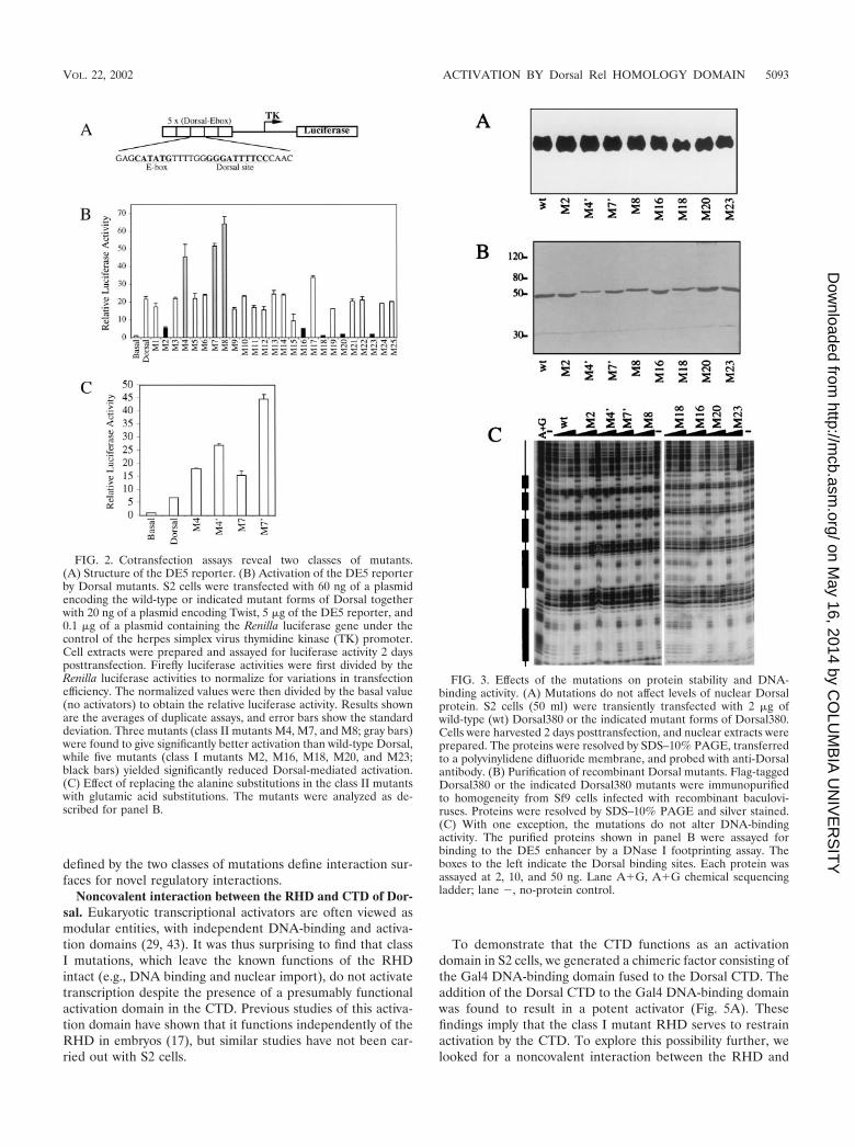
Cotransfection assays reveal two classes of mutants withaltered ability to activate transcription. The Dorsal mutantswere examined for their ability to activate transcription in atransient-transfection assay with the DE5 reporter (Fig. 2A)(3, 40). This reporter contains both Twist and Dorsal bindingsites. It is weakly activated by Dorsal alone but is stronglyactivated by the combination of Dorsal and Twist. The reporterwas cotransfected into S2 cells along with vectors encodingTwist and wild-type Dorsal or one of the Dorsal mutants (Fig.
5090 JIA ET AL. MOL. CELL. BIOL.
on May 16, 2014 by C
OLU
MB
IA U
NIV
ER
SIT
Yhttp://m
cb.asm.org/
Dow
nloaded from

2B). The majority of the mutants gave approximately wild-typelevels of stimulation. However, five mutants (M2, M16, M18,M20, and M23) (Fig. 2B) were compromised in their ability toactivate transcription in this assay. An examination of a mo-lecular model of the RHD (Fig. 1B) shows that the alteredamino acids in these mutants, hereafter referred to as class Imutants, are all on the same face of the RHD, suggesting thatthey define a positively acting interaction surface.
The cotransfection assays also revealed a second class ofmutants with altered ability to activate transcription. Three ofthe mutants (M4, M7, and M8) (Fig. 2B) consistently yieldedtwo- to threefold greater activation than that achieved withwild-type Dorsal. Examination of the RHD model (Fig. 1B)reveals that the amino acids affected in these superactive mu-tants cluster together on the opposite face of the domain fromthe amino acids altered in the class I mutants. Thus, thesemutants, hereafter referred to as class II mutants, may definea second interface on the RHD. All three class II mutationseliminated lysine side chains, suggesting that electrostatic in-teractions are important for stabilizing the interaction with thehypothetical target of this interacting surface. This conclusionis supported by the finding that replacement of two of theselysines, K84 and K121, by glutamate, an amino acid of theopposite charge, resulted in an even stronger superactive phe-notype than did the corresponding alanine substitutions (Fig.2C).
Class I and II mutations do not generally inactivate theRHD. Although we exercised caution in designing our mutantsby trying to select surface amino acids that would not playcritical roles in stabilizing the RHD fold, it was still possiblethat some of our mutations destabilized the RHD or perturbedits folding. To address these possibilities, we examined theexpression, nuclear localization, and DNA-binding activity ofthe mutants. When nuclear extracts of S2 cells transfected withequal amounts of DNA encoding Dorsal RHD mutants wereanalyzed by anti-Dorsal immunoblotting (Fig. 3A), the wild-type and mutant proteins were all found to accumulate in thenucleus to similar levels.
To assess the effects of the mutations on DNA-binding ac-tivity, we carried out DNase I footprinting assays. Recombi-nant mutant proteins were purified to homogeneity (Fig. 3B).With either the DE5 enhancer (Fig. 3C) or the zen ventralrepression region (data not shown) as a probe, we observednearly wild-type DNA-binding activity for all the class II mu-tants and four of the five class I mutants. Only mutation M18showed severely reduced DNA binding. Thus, the expression,localization, and DNA-binding data indicate that, with thepossible exception of M18, the mutants do not have reducedstability or structural integrity.
Mutations do not interfere with binding to known Dorsal-interacting proteins. As discussed above, the mutations in theRHD may define surfaces for interaction with other regulatoryproteins. We therefore examined the effects of these mutationson proteins thought to interact with the Dorsal RHD andpositively or negatively regulate Dorsal function.
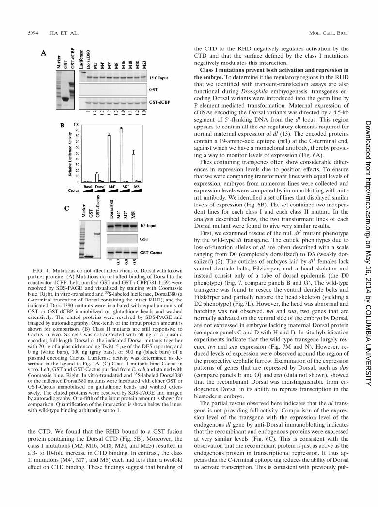
The RHD binds to the coactivator protein dCBP and maystimulate Dorsal-mediated activation in S2 cells and in theembryo (1). It is possible that the class I mutations impairDorsal-mediated activation by interfering with the interactionbetween Dorsal and dCBP. We therefore examined the abilityof the RHD mutants to bind dCBP in vitro. In agreement withprevious findings, we detected a specific interaction betweenthe wild-type Dorsal RHD (Dorsal380) and a GST fusionprotein containing a fragment of dCBP (amino acids 781 to1159). However, none of the mutations that altered activationin S2 cells had a significant effect on this interaction (Fig. 4A).Therefore, it is unlikely that our mutations altered activationby altering the affinity of Dorsal for dCBP.
The superactivity of the class II mutants suggests that theyare impaired in their ability to interact with a negative regu-lator of Dorsal activity. One well-characterized negative regu-lator of Dorsal is Cactus, which binds the RHD and inhibitsDorsal nuclear uptake (20, 30). In cotransfection assays, acti-vation by Dorsal was blocked by simultaneous overexpressionof Cactus (Fig. 4B). However, the class II mutants respondedto Cactus as well as wild-type Dorsal. Furthermore, GST pull-down assays confirmed that the wild-type RHD and the class IImutants bound to Cactus with comparable affinity (Fig. 4C).Thus, these mutations do not impair the interaction with Cac-tus.
We also examined the interaction between the RHD mu-tants and three other factors known to interact with the RHD,Ubc9 (3), Twist (40), and Groucho (14). We did not detect anyeffects of the mutations on these interactions (data not shown).In conclusion, our results strongly suggest that the two surfaces
TABLE 1. Mutagenesis of the Dorsal RHD
Mutant Amino acid(s) mutated Exposedsurfacea
Phenotypicclassb
M1 P47A, W48A, K50A 0.65 NeutralM2 E53A, Q54A, K58A 0.60 IM3 E65A, R69A 0.78 NeutralM4 K84A 0.83 IIM4� K84E 0.83 IIM5 W94A, K95A, R97A 0.56 NeutralM6 K107A, D108A, T109A 0.81 NeutralM7 K121A 0.85 IIM7� K121E 0.85 IIM8 C124A, K125A, K126A 0.87 IIM9 E132A, N134A, S135A 0.60 NeutralM10 E136A, T137A, M138A, R139A 0.74 NeutralM11 S143A, N144A 0.54 NeutralM12 D154A, E156A 0.73 NeutralM13 E163A, E164A, R166A 0.81 NeutralM14 S175A, H176A, R177A 0.54 NeutralM15 S181A, S182A 0.88 NeutralM16 D184K 0.81 IM17 S187A 0.43 NeutralM18 R189E 0.63 Ic
M19 E197A, S198A, E199A, K201, R203A 0.81 NeutralM20 E253A, D254A 0.64 IM21 Q266A, S267A 0.98 NeutralM22 D274A, Q276A 0.81 NeutralM23 H294A, T295A, L296A, D297A 0.71 IM24 T299A, E300A, P301A, K303A 0.94 NeutralM25 E318A 0.91 Neutral
a Fraction of the total surface area of the mutated residues accessible to water.The calculation (6) was carried out by using the structure of the p50 homodimer(21).
b Phenotypic class assignments are based on the data in Fig. 2B. Mutants thatwere at least twofold more active than the wild type are defined as class II.Mutants that were at least twofold less active than the wild type are defined asclass I. The remaining mutants are defined as neutral.
c This mutant showed severely reduced DNA-binding activity.
VOL. 22, 2002 ACTIVATION BY Dorsal Rel HOMOLOGY DOMAIN 5091
on May 16, 2014 by C
OLU
MB
IA U
NIV
ER
SIT
Yhttp://m
cb.asm.org/
Dow
nloaded from
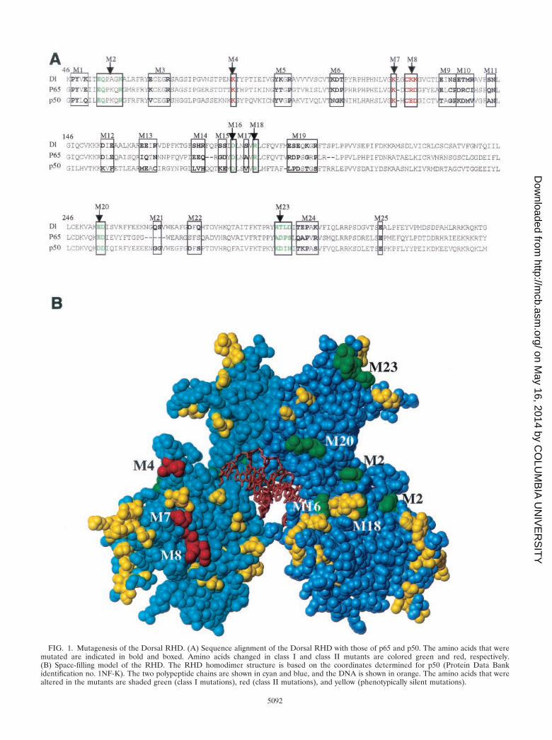
FIG. 1. Mutagenesis of the Dorsal RHD. (A) Sequence alignment of the Dorsal RHD with those of p65 and p50. The amino acids that weremutated are indicated in bold and boxed. Amino acids changed in class I and class II mutants are colored green and red, respectively.(B) Space-filling model of the RHD. The RHD homodimer structure is based on the coordinates determined for p50 (Protein Data Bankidentification no. 1NF-K). The two polypeptide chains are shown in cyan and blue, and the DNA is shown in orange. The amino acids that werealtered in the mutants are shaded green (class I mutations), red (class II mutations), and yellow (phenotypically silent mutations).
5092
on May 16, 2014 by C
OLU
MB
IA U
NIV
ER
SIT
Yhttp://m
cb.asm.org/
Dow
nloaded from
defined by the two classes of mutations define interaction sur-faces for novel regulatory interactions.
Noncovalent interaction between the RHD and CTD of Dor-sal. Eukaryotic transcriptional activators are often viewed asmodular entities, with independent DNA-binding and activa-tion domains (29, 43). It was thus surprising to find that classI mutations, which leave the known functions of the RHDintact (e.g., DNA binding and nuclear import), do not activatetranscription despite the presence of a presumably functionalactivation domain in the CTD. Previous studies of this activa-tion domain have shown that it functions independently of theRHD in embryos (17), but similar studies have not been car-ried out with S2 cells.
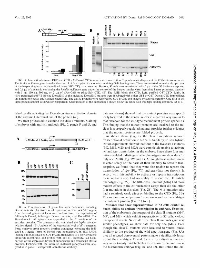
To demonstrate that the CTD functions as an activationdomain in S2 cells, we generated a chimeric factor consisting ofthe Gal4 DNA-binding domain fused to the Dorsal CTD. Theaddition of the Dorsal CTD to the Gal4 DNA-binding domainwas found to result in a potent activator (Fig. 5A). Thesefindings imply that the class I mutant RHD serves to restrainactivation by the CTD. To explore this possibility further, welooked for a noncovalent interaction between the RHD and
FIG. 2. Cotransfection assays reveal two classes of mutants.(A) Structure of the DE5 reporter. (B) Activation of the DE5 reporterby Dorsal mutants. S2 cells were transfected with 60 ng of a plasmidencoding the wild-type or indicated mutant forms of Dorsal togetherwith 20 ng of a plasmid encoding Twist, 5 �g of the DE5 reporter, and0.1 �g of a plasmid containing the Renilla luciferase gene under thecontrol of the herpes simplex virus thymidine kinase (TK) promoter.Cell extracts were prepared and assayed for luciferase activity 2 daysposttransfection. Firefly luciferase activities were first divided by theRenilla luciferase activities to normalize for variations in transfectionefficiency. The normalized values were then divided by the basal value(no activators) to obtain the relative luciferase activity. Results shownare the averages of duplicate assays, and error bars show the standarddeviation. Three mutants (class II mutants M4, M7, and M8; gray bars)were found to give significantly better activation than wild-type Dorsal,while five mutants (class I mutants M2, M16, M18, M20, and M23;black bars) yielded significantly reduced Dorsal-mediated activation.(C) Effect of replacing the alanine substitutions in the class II mutantswith glutamic acid substitutions. The mutants were analyzed as de-scribed for panel B.
FIG. 3. Effects of the mutations on protein stability and DNA-binding activity. (A) Mutations do not affect levels of nuclear Dorsalprotein. S2 cells (50 ml) were transiently transfected with 2 �g ofwild-type (wt) Dorsal380 or the indicated mutant forms of Dorsal380.Cells were harvested 2 days posttransfection, and nuclear extracts wereprepared. The proteins were resolved by SDS–10% PAGE, transferredto a polyvinylidene difluoride membrane, and probed with anti-Dorsalantibody. (B) Purification of recombinant Dorsal mutants. Flag-taggedDorsal380 or the indicated Dorsal380 mutants were immunopurifiedto homogeneity from Sf9 cells infected with recombinant baculovi-ruses. Proteins were resolved by SDS–10% PAGE and silver stained.(C) With one exception, the mutations do not alter DNA-bindingactivity. The purified proteins shown in panel B were assayed forbinding to the DE5 enhancer by a DNase I footprinting assay. Theboxes to the left indicate the Dorsal binding sites. Each protein wasassayed at 2, 10, and 50 ng. Lane A�G, A�G chemical sequencingladder; lane �, no-protein control.
VOL. 22, 2002 ACTIVATION BY Dorsal Rel HOMOLOGY DOMAIN 5093
on May 16, 2014 by C
OLU
MB
IA U
NIV
ER
SIT
Yhttp://m
cb.asm.org/
Dow
nloaded from
the CTD. We found that the RHD bound to a GST fusionprotein containing the Dorsal CTD (Fig. 5B). Moreover, theclass I mutations (M2, M16, M18, M20, and M23) resulted ina 3- to 10-fold increase in CTD binding. In contrast, the classII mutations (M4�, M7�, and M8) each had less than a twofoldeffect on CTD binding. These findings suggest that binding of
the CTD to the RHD negatively regulates activation by theCTD and that the surface defined by the class I mutationsnegatively modulates this interaction.
Class I mutations prevent both activation and repression inthe embryo. To determine if the regulatory regions in the RHDthat we identified with transient-transfection assays are alsofunctional during Drosophila embryogenesis, transgenes en-coding Dorsal variants were introduced into the germ line byP-element-mediated transformation. Maternal expression ofcDNAs encoding the Dorsal variants was directed by a 4.5-kbsegment of 5�-flanking DNA from the dl locus. This regionappears to contain all the cis-regulatory elements required fornormal maternal expression of dl (13). The encoded proteinscontain a 19-amino-acid epitope (nt1) at the C-terminal end,against which we have a monoclonal antibody, thereby provid-ing a way to monitor levels of expression (Fig. 6A).
Flies containing transgenes often show considerable differ-ences in expression levels due to position effects. To ensurethat we were comparing transformant lines with equal levels ofexpression, embryos from numerous lines were collected andexpression levels were compared by immunoblotting with anti-nt1 antibody. We identified a set of lines that displayed similarlevels of expression (Fig. 6B). The set contained two indepen-dent lines for each class I and each class II mutant. In theanalysis described below, the two transformant lines of eachDorsal mutant were found to give very similar results.
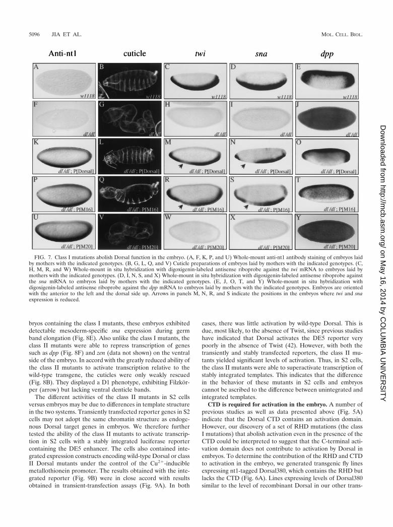
First, we examined rescue of the null dl1 mutant phenotypeby the wild-type dl transgene. The cuticle phenotypes due toloss-of-function alleles of dl are often described with a scaleranging from D0 (completely dorsalized) to D3 (weakly dor-salized) (2). The cuticles of embryos laid by dl1 females lackventral denticle belts, Filzkorper, and a head skeleton andinstead consist only of a tube of dorsal epidermis (the D0phenotype) (Fig. 7, compare panels B and G). The wild-typetransgene was found to rescue the ventral denticle belts andFilzkorper and partially restore the head skeleton (yielding aD2 phenotype) (Fig.7L). However, the head was abnormal andhatching was not observed. twi and sna, two genes that arenormally activated on the ventral side of the embryo by Dorsal,are not expressed in embryos lacking maternal Dorsal protein(compare panels C and D with H and I). In situ hybridizationexperiments indicate that the wild-type transgene largely res-cued twi and sna expression (Fig. 7M and N). However, re-duced levels of expression were observed around the region ofthe prospective cephalic furrow. Examination of the expressionpatterns of genes that are repressed by Dorsal, such as dpp(compare panels E and O) and zen (data not shown), showedthat the recombinant Dorsal was indistinguishable from en-dogenous Dorsal in its ability to repress transcription in theblastoderm embryo.
The partial rescue observed here indicates that the dl trans-gene is not providing full activity. Comparison of the expres-sion level of the transgene with the expression level of theendogenous dl gene by anti-Dorsal immunoblotting indicatesthat the recombinant and endogenous proteins were expressedat very similar levels (Fig. 6C). This is consistent with theobservation that the recombinant protein is just as active as theendogenous protein in transcriptional repression. It thus ap-pears that the C-terminal epitope tag reduces the ability of Dorsalto activate transcription. This is consistent with previously pub-
FIG. 4. Mutations do not affect interactions of Dorsal with knownpartner proteins. (A) Mutations do not affect binding of Dorsal to thecoactivator dCBP. Left, purified GST and GST-dCBP(781-1159) wereresolved by SDS-PAGE and visualized by staining with Coomassieblue. Right, in vitro-translated and 35S-labeled luciferase, Dorsal380 (aC-terminal truncation of Dorsal containing the intact RHD), and theindicated Dorsal380 mutants were incubated with equal amounts ofGST or GST-dCBP immobilized on glutathione beads and washedextensively. The eluted proteins were resolved by SDS-PAGE andimaged by autoradiography. One-tenth of the input protein amount isshown for comparison. (B) Class II mutants are still responsive toCactus in vivo. S2 cells was cotransfected with 60 ng of a plasmidencoding full-length Dorsal or the indicated Dorsal mutants togetherwith 20 ng of a plasmid encoding Twist, 5 �g of the DE5 reporter, and0 ng (white bars), 100 ng (gray bars), or 500 ng (black bars) of aplasmid encoding Cactus. Luciferase activity was determined as de-scribed in the legend to Fig. 1A. (C) Class II mutants bind Cactus invitro. Left, GST and GST-Cactus purified from E. coli and stained withCoomassie blue. Right, In vitro-translated and 35S-labeled Dorsal380or the indicated Dorsal380 mutants were incubated with either GST orGST-Cactus immobilized on glutathione beads and washed exten-sively. The eluted proteins were resolved by SDS-PAGE and imagedby autoradiography. One-fifth of the input protein amount is shown forcomparison. Quantification of the interaction is shown below the lanes,with wild-type binding arbitrarily set to 1.
5094 JIA ET AL. MOL. CELL. BIOL.
on May 16, 2014 by C
OLU
MB
IA U
NIV
ER
SIT
Yhttp://m
cb.asm.org/
Dow
nloaded from
lished results indicating that Dorsal contains an activation domainat the extreme C-terminal end of the protein (40).
We then proceeded to examine the class I mutants. Stainingof embryos with anti-nt1 antibody (Fig. 7, panels P and U, and
data not shown) showed that the mutant proteins were specif-ically localized to the ventral nuclei in a pattern very similar tothat observed for the wild-type recombinant protein (panel K).This finding that the mutant proteins are localized to the nu-cleus in a properly regulated manner provides further evidencethat the mutant proteins are folded properly.
As shown above (Fig. 2), the class I mutations reducedtranscriptional activation in S2 cells. Similarly, in situ hybrid-ization experiments showed that four of the five class I mutants(M2, M18, M20, and M23) were completely unable to activatetwi and sna transcription in the embryo. Since these four mu-tations yielded indistinguishable phenotypes, we show data foronly one (M20) (Fig. 7W and X). Although these mutants wereselected solely on the basis of their inability to activate tran-scription, we found that they were also unable to repress thetranscription of dpp (Fig. 7Y) and zen (data not shown). Inaccord with this inability to activate or repress transcription,these mutants also had no ability to rescue the D0 cuticlephenotype (Fig. 7V). The fifth class I mutant (M16) had moremodest effects in the cotransfection assays than did the otherfour mutations in this class (Fig. 2B). The M16 mutation alsohad a relatively weak effect on binding to the CTD (Fig. 5B).This mutant rescued pattern formation as well as the wild-typerecombinant protein (Fig. 7Q to T).
Mutants that show superactivation in S2 cells exhibit re-duced ability to activate transcription in embryos. Examina-tion of the embryonic phenotypes of the class II mutants (M4�,M7�, and M8), which exhibit superactivity in S2 cells, yieldedunexpected results. Since all three class II mutants gave verysimilar phenotypes, we show data for only one (M4�). Eventhough the class II mutants were localized to ventral nucleisimilarly to the product of the wild-type transgene (Fig. 8A),they all rescued dorsoventral patterning to a significantly lesserextent than wild-type Dorsal. In situ hybridization revealedvery weak (nearly undetectable) expression of twi and sna inthe blastoderm embryo (Fig. 8C and D). But unlike the em-
FIG. 5. Interaction between RHD and CTD. (A) Dorsal CTD can activate transcription. Top, schematic diagram of the G5 luciferase reporter.The firefly luciferase gene is under the control of five copies of a module containing Gal4 binding sites. These are inserted immediately upstreamof the herpes simplex virus thymidine kinase (HSV TK) core promoter. Bottom, S2 cells were transfected with 5 �g of the G5 luciferase reporterand 0.1 �g of a plasmid containing the Renilla luciferase gene under the control of the herpes simplex virus thymidine kinase promoter, togetherwith 0 ng, 150 ng, 500 ng, or 2 �g of pPac-Gal4 or pPac-Gal4-CTD. (B) The RHD binds the CTD. Left, purified GST-CTD. Right, invitro-translated and 35S-labeled Dorsal380 or the indicated Dorsal380 mutants were incubated with either GST or GST-Dorsal-CTD immobilizedon glutathione beads and washed extensively. The eluted proteins were resolved by SDS-PAGE and imaged by autoradiography. One-fifth of theinput protein amount is shown for comparison. Quantification of the interaction is shown below the lanes, with wild-type binding arbitrarily set to 1.
FIG. 6. Transformation of germ line with P-elements encodingDorsal mutants. (A) Structure of expression vectors. A 4.5-kb regionfrom the endogenous dl locus was used to direct the expression offull-length Dorsal, full-length Dorsal mutants, and Dorsal380. The19-amino-acid nt1 epitope was appended to the C terminus of theencoded proteins. The constructs also contained the hsp70 polyade-nylation signal. (B) Analysis of the expression level of dl transgenes.Forty embryos from mothers bearing transgenes encoding the indi-cated nt1-tagged forms of Dorsal were homogenized in SDS-PAGEloading buffer, resolved by SDS-PAGE, transferred to a polyvinylidenedifluoride membrane, and probed with anti-nt1 antibody. (C) Com-parison of the expression levels of endogenous and transgenic Dorsalproteins. Embryos with the indicated maternal genotypes were ana-lyzed as in panel B except with anti-Dorsal antibody.
VOL. 22, 2002 ACTIVATION BY Dorsal Rel HOMOLOGY DOMAIN 5095
on May 16, 2014 by C
OLU
MB
IA U
NIV
ER
SIT
Yhttp://m
cb.asm.org/
Dow
nloaded from
bryos containing the class I mutants, these embryos exhibiteddetectable mesoderm-specific sna expression during germband elongation (Fig. 8E). Also unlike the class I mutants, theclass II mutants were able to repress transcription of genessuch as dpp (Fig. 8F) and zen (data not shown) on the ventralside of the embryo. In accord with the greatly reduced ability ofthe class II mutants to activate transcription relative to thewild-type transgene, the cuticles were only weakly rescued(Fig. 8B). They displayed a D1 phenotype, exhibiting Filzkor-per (arrow) but lacking ventral denticle bands.
The different activities of the class II mutants in S2 cellsversus embryos may be due to differences in template structurein the two systems. Transiently transfected reporter genes in S2cells may not adopt the same chromatin structure as endoge-nous Dorsal target genes in embryos. We therefore furthertested the ability of the class II mutants to activate transcrip-tion in S2 cells with a stably integrated luciferase reportercontaining the DE5 enhancer. The cells also contained inte-grated expression constructs encoding wild-type Dorsal or classII Dorsal mutants under the control of the Cu2�-induciblemetallothionein promoter. The results obtained with the inte-grated reporter (Fig. 9B) were in close accord with resultsobtained in transient-transfection assays (Fig. 9A). In both
cases, there was little activation by wild-type Dorsal. This isdue, most likely, to the absence of Twist, since previous studieshave indicated that Dorsal activates the DE5 reporter verypoorly in the absence of Twist (42). However, with both thetransiently and stably transfected reporters, the class II mu-tants yielded significant levels of activation. Thus, in S2 cells,the class II mutants were able to superactivate transcription ofstably integrated templates. This indicates that the differencein the behavior of these mutants in S2 cells and embryoscannot be ascribed to the difference between unintegrated andintegrated templates.
CTD is required for activation in the embryo. A number ofprevious studies as well as data presented above (Fig. 5A)indicate that the Dorsal CTD contains an activation domain.However, our discovery of a set of RHD mutations (the classI mutations) that abolish activation even in the presence of theCTD could be interpreted to suggest that the C-terminal acti-vation domain does not contribute to activation by Dorsal inembryos. To determine the contribution of the RHD and CTDto activation in the embryo, we generated transgenic fly linesexpressing nt1-tagged Dorsal380, which contains the RHD butlacks the CTD (Fig. 6A). Lines expressing levels of Dorsal380similar to the level of recombinant Dorsal in our other trans-
FIG. 7. Class I mutations abolish Dorsal function in the embryo. (A, F, K, P, and U) Whole-mount anti-nt1 antibody staining of embryos laidby mothers with the indicated genotypes. (B, G, L, Q, and V) Cuticle preparations of embryos laid by mothers with the indicated genotypes. (C,H, M, R, and W) Whole-mount in situ hybridization with digoxigenin-labeled antisense riboprobe against the twi mRNA to embryos laid bymothers with the indicated genotypes. (D, I, N, S, and X) Whole-mount in situ hybridization with digoxigenin-labeled antisense riboprobe againstthe sna mRNA to embryos laid by mothers with the indicated genotypes. (E, J, O, T, and Y) Whole-mount in situ hybridization withdigoxigenin-labeled antisense riboprobe against the dpp mRNA to embryos laid by mothers with the indicated genotypes. Embryos are orientedwith the anterior to the left and the dorsal side up. Arrows in panels M, N, R, and S indicate the positions in the embryos where twi and snaexpression is reduced.
5096 JIA ET AL. MOL. CELL. BIOL.
on May 16, 2014 by C
OLU
MB
IA U
NIV
ER
SIT
Yhttp://m
cb.asm.org/
Dow
nloaded from
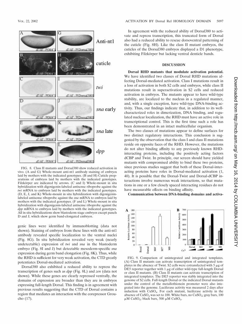
genic lines were identified by immunoblotting (data notshown). Staining of embryos from these lines with the anti-nt1antibody revealed specific localization to the ventral nuclei(Fig. 8G). In situ hybridization revealed very weak (nearlyundetectable) expression of twi and sna in the blastodermembryo (Fig. 8I and J) but detectable mesoderm-specific snaexpression during germ band elongation (Fig. 8K). Thus, whilethe RHD is sufficient for very weak activation, the CTD greatlypotentiates Dorsal-mediated activation.
Dorsal380 also exhibited a reduced ability to repress thetranscription of genes such as dpp (Fig. 8L) and zen (data notshown). While these genes are clearly repressed ventrally, thedomains of expression are broader than they are in embryosexpressing full-length Dorsal. This finding is in agreement withprevious results suggesting that the CTD of Dorsal contains aregion that mediates an interaction with the corepressor Grou-cho (17).
In agreement with the reduced ability of Dorsal380 to acti-vate and repress transcription, this truncated form of Dorsalalso had a reduced ability to rescue dorsoventral patterning ofthe cuticle (Fig. 8H). Like the class II mutant embryos, thecuticles of the Dorsal380 embryos displayed a D1 phenotype,exhibiting Filzkorper but lacking ventral denticle bands.
DISCUSSION
Dorsal RHD mutants that modulate activation potential.We have identified two classes of Dorsal RHD mutations af-fecting Dorsal-mediated activation. Class I mutations result ina loss of activation in both S2 cells and embryos, while class IImutations result in superactivation in S2 cells and reducedactivation in embryos. The mutants appear to have wild-typestability, are localized to the nucleus in a regulated manner,and, with a single exception, have wild-type DNA-binding ac-tivity. Thus, our findings indicate that, in addition to its well-characterized roles in dimerization, DNA binding, and regu-lated nuclear localization, the RHD must have an active role intranscriptional control. This is the first time such a role hasbeen demonstrated in an intact multicellular organism.
The two classes of mutations appear to define surfaces fortwo distinct regulatory interactions. This conclusion is sug-gested by the observation that the class I and class II mutationsreside on opposite faces of the RHD. However, the mutationsdo not alter binding affinity to any previously known RHD-interacting proteins, including the positively acting factorsdCBP and Twist. In principle, our screen should have yieldedmutants with compromised ability to bind these two proteins,since previous studies suggest that both of these Dorsal-inter-acting proteins have roles in Dorsal-mediated activation (1,40). It is possible that the Dorsal-Twist and Dorsal-dCBP in-teractions involve numerous weak interactions, so that muta-tions in one or a few closely spaced interacting residues do nothave measurable effects on binding affinity.
Communication between DNA-binding domains and activa-
FIG. 8. Class II mutants and Dorsal380 show reduced activation invivo. (A and G) Whole-mount anti-nt1 antibody staining of embryoslaid by mothers with the indicated genotypes. (B and H) Cuticle prep-arations of embryos laid by mothers with the indicated genotypes.Filzkorper are indicated by arrows. (C and I) Whole-mount in situhybridization with digoxigenin-labeled antisense riboprobe against thetwi mRNA to embryos laid by mothers with the indicated genotypes.(D, E, J, and K) Whole-mount in situ hybridization with digoxigenin-labeled antisense riboprobe against the sna mRNA to embryos laid bymothers with the indicated genotypes. (F and L) Whole-mount in situhybridization with digoxigenin-labeled antisense riboprobe against thedpp mRNA to embryos laid by mothers with the indicated genotypes.All in situ hybridizations show blastoderm stage embryos except panelsD and J, which show germ band-elongated embryos.
FIG. 9. Comparison of unintegrated and integrated templates.(A) Class II mutants can activate transcription of unintegrated tem-plates in the absence of Twist. S2 cells were cotransfected with 5 �g ofDE5 reporter together with 1 �g of either wild-type full-length Dorsalor class II mutants. (B) Class II mutants can activate transcription ofintegrated templates. The DE5 reporter was stably integrated into thegenome of S2 cells. Full-length Dorsal or the indicated Dorsal mutantsunder the control of the metallothionein promoter were also inte-grated into the genome. Luciferase activity was measured 2 days afterinduction with CuSO4. For each mutant, luciferase activity in theabsence of CuSO4 was set to 100. White bars, no CuSO4; gray bars, 100�M CuSO4; black bars, 500 �M CuSO4.
VOL. 22, 2002 ACTIVATION BY Dorsal Rel HOMOLOGY DOMAIN 5097
on May 16, 2014 by C
OLU
MB
IA U
NIV
ER
SIT
Yhttp://m
cb.asm.org/
Dow
nloaded from
tion domains. Our class I mutations block activation in thecontext of full-length Dorsal protein despite the presence of anactivation domain in the CTD. As we have demonstrated here,this activation domain can function independently of theRHD. These findings are inconsistent with a simple model fortranscriptional control in which the sole function of a DNA-binding domain is to tether one or more activation domains tothe template to allow them to interact either directly or indi-rectly with the transcriptional machinery or chromatin tem-plate. However, our findings are in accord with previous stud-ies suggesting that DNA-binding domains often containinteraction surfaces that send regulatory signals to attachedactivation domains (31). For example, certain mutations in theglucocorticoid receptor DNA-binding domain prevent activa-tion while not interfering with DNA binding despite the pres-ence of independent activation domains in the factor (39). Ithas been suggested that the amino acids affected in theseglucocorticoid receptor mutants play roles in transmitting anallosteric signal from the DNA to the activation domain, whichserves to stimulate activation domain function. Rel homologydomains are also thought to undergo conformational changesupon binding to DNA, as shown by changes in both proteasesensitivity and circular dichroism spectra, which could modu-late the activity of linked activation domains (19, 24, 34).
The analysis of the Dorsal RHD class I mutations presentedhere suggests a novel mechanism by which a DNA-bindingdomain could regulate a linked activation domain. In particu-lar, we have found that the RHD binds the CTD and thatmutations that increase the affinity of this interaction weakenthe ability of Dorsal to function as a transcriptional activator.These findings strongly suggest that noncovalent interactionsbetween the RHD and the CTD downregulate activation bythe CTD. In effect, the RHD may be serving as a decoy for theactivation domain. When the activation domain interacts withthe RHD, it may be unavailable to interact with the generalmachinery.
While the class I mutant RHD binds the CTD, the wild-typeRHD also binds the CTD, albeit with a lower affinity. Thus, thewild-type RHD may also be able to downregulate CTD func-tion, and the phenotype of the class I mutants may represent aheightening of this wild-type function. Given the complexity ofeukaryotic genomes and the relatively low DNA-binding spec-ificity of sequence-specific factors such as Dorsal, this down-regulation may be necessary to prevent high levels of inappro-priate activation by Dorsal when it binds to adventitious sitesthat occur throughout the genome. When Dorsal binds to bonafide Dorsal-responsive enhancers, however, it may find itself inthe context of other transcriptional activators that can coop-erate with Dorsal via enhanceosome formation to yield highlevels of activation.
In addition to preventing activation, class I mutations alsoprevent repression. One possible interpretation of this obser-vation is that the same RHD-CTD interaction that preventsactivation also prevents repression, possibly by interfering withthe repression domain in the CTD (17).
Uncoupling of Dorsal-mediated repression from Dorsal-me-diated activation. In contrast to the class I mutants, whichbehave similarly in S2 cells and embryos, the class II mutantsdisplayed significantly different behavior in the two settings.While the class II mutants are more potent activators than
wild-type Dorsal in S2 cells, they have a severely attenuatedability to activate transcription in embryos. These findings sug-gest that the surface defined by the class II mutations interactswith a variety of coregulatory proteins, including both positiveand negative regulators. In embryos, a positive coregulatormay be the dominant interacting protein, and thus perturbingthe interaction surface results in reduced levels of activation.In contrast, in S2 cells, a negative coregulator may dominate,resulting in superactivation upon disruption of the interactionsurface. These mutations may interfere with multiple interac-tions by influencing the conformation of the RHD. This pos-sibility is suggested by previous work showing that binding ofthe RHD to DNA induces a change in the rate of proteasecleavage at certain protease-hypersensitive sites in the RHD.These hypersensitive sites map to the region of the RHDdefined by our class II mutations (34). Perhaps the class IImutations alter the ability of the RHD to undergo a DNA-induced conformational change, thereby altering the affinity ofthe factor for multiple coregulators.
Because the class II Dorsal mutants are able to repress butunable to activate transcription in the blastoderm embryo, theyallow us to assess the developmental consequences of uncou-pling Dorsal-mediated activation from Dorsal-mediated re-pression. Despite the ability of class II mutants to repress dppin the ventral ectoderm, these mutants are unable to rescue theventral ectoderm, as evidenced by the absence of ventral den-ticle belts. These findings support the idea that Dpp can diffuseventrally to block ventral ectoderm formation in the ventrolat-eral region (4, 15). In the wild-type embryo, Dorsal is compe-tent to activate transcription and therefore turns on short gas-trulation (sog) and brinker in the ventrolateral region (33). Sogthen functions upstream while Brinker functions downstreamof Dpp receptors to block Dpp signaling and allow ventralectoderm formation in the ventolateral region (4, 18, 28). Ourfinding that a repression-competent but activation-defectiveform of Dorsal is not sufficient to allow ventral ectoderm for-mation suggests that Dorsal-mediated activation is of primaryimportance in the subdivision of the embryo into developmen-tal domains. In contrast, Dorsal-mediated repression may be arelatively recent adaptation that ensures the complete shut-down of Dpp signaling in the ventral and ventrolateral regions.The development of Dorsal alleles that are able to activate butunable to repress transcription would allow further testing ofthese ideas.
ACKNOWLEDGMENTS
We thank Stephen Smale and Uptal Banerjee for critical reading ofthe manuscript, Ruth Steward for providing us with a genomic clonecontaining the dl 5�-flanking region, and Larry Zipursky for providingthe anti-nt1 antibody. We also thank Benjamin Chen and JonathanWojciak for technical assistance.
This work was supported by National Institutes of Health grantGM44522.
REFERENCES
1. Akimaru, H., D. X. Hou, and S. Ishii. 1997. Drosophila CBP is required fordorsal-dependent twist gene expression. Nat. Genet. 17:211–214.
2. Anderson, K. V., and C. Nusslein-Volhard. 1986. Dorsal-group genes ofDrosophila, p. 177–194. In J. Gall (ed.), Gametogenesis and the early em-bryo. A. R. Liss, New York, N.Y.
3. Bhaskar, V., S. A. Valentine, and A. J. Courey. 2000. A functional interactionbetween dorsal and components of the Smt3 conjugation machinery. J. Biol.Chem. 275:4033–4040.
5098 JIA ET AL. MOL. CELL. BIOL.
on May 16, 2014 by C
OLU
MB
IA U
NIV
ER
SIT
Yhttp://m
cb.asm.org/
Dow
nloaded from
4. Biehs, B., V. Francois, and E. Bier. 1996. The Drosophila short gastrulationgene prevents Dpp from autoactivating and suppressing neurogenesis in theneuroectoderm. Genes Dev. 10:2922–2934.
5. Blair, W. S., H. P. Bogerd, S. J. Madore, and B. R. Cullen. 1994. Mutationalanalysis of the transcription activation domain of RelA: identification of ahighly synergistic minimal acidic activation module. Mol. Cell. Biol. 14:7226–7234.
6. Bowie, J. U., R. Luthy, and D. Eisenberg. 1991. A method to identify proteinsequences that fold into a known three-dimensional structure. Science 253:164–170.
7. Cagan, R. L., H. Kramer, A. C. Hart, and S. L. Zipursky. 1992. The bride ofsevenless and sevenless interaction: internalization of a transmembrane li-gand. Cell 69:393–399.
8. Chen, G., and A. J. Courey. 1999. Baculovirus-transfer vector for eukaryoticexpression and immunoaffinity purification of Gal4-fusion proteins. Bio-Techniques 26:808–810, 812, 814.
9. Chen, G., P. H. Nguyen, and A. J. Courey. 1998. A role for Groucho tet-ramerization in transcriptional repression. Mol. Cell. Biol. 18:7259–7268.
10. Chen, Y. Q., S. Ghosh, and G. Ghosh. 1998. A novel DNA recognition modeby the NF-�B p65 homodimer. Nat. Struct. Biol. 5:67–73.
11. Courey, A. J., D. A. Holtzman, S. P. Jackson, and R. Tjian. 1989. Synergisticactivation by the glutamine-rich domains of human transcription factor Sp1.Cell 59:827–836.
12. Courey, A. J., and J. D. Huang. 1995. The establishment and interpretationof transcription factor gradients in the Drosophila embryo. Biochim. Bio-phys. Acta 1261:1–18.
13. Drier, E. A., L. H. Huang, and R. Steward. 1999. Nuclear import of theDrosophila Rel protein Dorsal is regulated by phosphorylation. Genes Dev.13:556–568.
14. Dubnicoff, T., S. A. Valentine, G. Chen, T. Shi, J. A. Lengyel, Z. Paroush, andA. J. Courey. 1997. Conversion of dorsal from an activator to a repressor bythe global corepressor Groucho. Genes Dev. 11:2952–2957.
15. Ferguson, E. L., and K. V. Anderson. 1992. Decapentaplegic acts as a mor-phogen to organize dorsal-ventral pattern in the Drosophila embryo. Cell71:451–461.
16. Flores-Saaib, R. D., and A. J. Courey. 2000. Regulation of dorso/ventralpatterning in the Drosophila embryo by multiple dorsal-interacting proteins.Cell Biochem. Biophys. 33:1–17.
17. Flores-Saaib, R. D., S. Jia, and A. J. Courey. 2001. Activation and repressionby the C-terminal domain of Dorsal. Development 128:1869–1879.
18. Francois, V., M. Solloway, J. W. O’Neill, J. Emery, and E. Bier. 1994.Dorsal-ventral patterning of the Drosophila embryo depends on a putativenegative growth factor encoded by the short gastrulation gene. Genes Dev.8:2602–2616.
19. Fujita, T., G. P. Nolan, S. Ghosh, and D. Baltimore. 1992. Independentmodes of transcriptional activation by the p50 and p65 subunits of NF-�B.Genes Dev. 6:775–787.
20. Geisler, R., A. Bergmann, Y. Hiromi, and C. Nusslein-Volhard. 1992. cactus,a gene involved in dorsoventral pattern formation of Drosophila, is relatedto the I�B gene family of vertebrates. Cell 71:613–621.
21. Ghosh, G., G. van Duyne, S. Ghosh, and P. B. Sigler. 1995. Structure ofNF-�B p50 homodimer bound to a �B site. Nature 373:303–310.
22. Ghosh, S., M. J. May, and E. B. Kopp. 1998. NF-�B and Rel proteins:evolutionarily conserved mediators of immune responses. Annu. Rev. Im-munol. 16:225–260.
23. Govind, S., A. M. Whalen, and R. Steward. 1992. In vivo self-association ofthe Drosophila rel-protein Dorsal. Proc. Natl. Acad. Sci. USA 89:7861–7865.
24. Hay, R. T., and J. Nicholson. 1993. DNA-binding alters the protease sus-ceptibility of the p50 subunit of NF-�B. Nucleic Acids Res. 21:4592–4598.
25. Ip, Y. T., R. E. Park, D. Kosman, E. Bier, and M. Levine. 1992. The dorsalgradient morphogen regulates stripes of rhomboid expression in the pre-
sumptive neuroectoderm of the Drosophila embryo. Genes Dev. 6:1728–1739.
26. Isoda, K., and C. Nusslein-Volhard. 1994. Disulfide cross-linking in crudeembryonic lysates reveals three complexes of the Drosophila morphogendorsal and its inhibitor cactus. Proc. Natl. Acad. Sci. USA 91:5350–5354.
27. Isoda, K., S. Roth, and C. Nusslein-Volhard. 1992. The functional domainsof the Drosophila morphogen dorsal: evidence from the analysis of mutants.Genes Dev. 6:619–630.
28. Jazwinska, A., C. Rushlow, and S. Roth. 1999. The role of brinker in medi-ating the graded response to Dpp in early Drosophila embryos. Develop-ment 126:3323–3334.
29. Johnson, P. F., and S. L. McKnight. 1989. Eukaryotic transcriptional regu-latory proteins. Annu. Rev. Biochem. 58:799–839.
30. Kidd, S. 1992. Characterization of the Drosophila cactus locus and analysisof interactions between cactus and dorsal proteins. Cell 71:623–635.
31. Lefstin, J. A., and K. R. Yamamoto. 1998. Allosteric effects of DNA ontranscriptional regulators. Nature 392:885–888.
32. Liaw, G. J., K. M. Rudolph, J. D. Huang, T. Dubnicoff, A. J. Courey, and J. A.Lengyel. 1995. The torso response element binds GAGA and NTF-1/Elf-1,and regulates tailless by relief of repression. Genes Dev. 9:3163–3176.
33. Markstein, M., P. Markstein, V. Markstein, and M. S. Levine. 2002. Ge-nome-wide analysis of clustered Dorsal binding sites identifies putative tar-get genes in the Drosophila embryo. Proc. Natl. Acad. Sci. USA 99:763–768.
34. Matthews, J. R., J. Nicholson, E. Jaffray, S. M. Kelly, N. C. Price, and R. T.Hay. 1995. Conformational changes induced by DNA binding of NF-�B.Nucleic Acids Res. 23:3393–3402.
35. Morisato, D., and K. V. Anderson. 1995. Signaling pathways that establishthe dorsal-ventral pattern of the Drosophila embryo. Annu. Rev. Genet.29:371–399.
36. Muller, C. W., F. A. Rey, M. Sodeoka, G. L. Verdine, and S. C. Harrison.1995. Structure of the NF-�B p50 homodimer bound to DNA. Nature 373:311–317.
37. Pan, D. J., J. D. Huang, and A. J. Courey. 1991. Functional analysis of theDrosophila twist promoter reveals a dorsal-binding ventral activator region.Genes Dev. 5:1892–1901.
38. Pham, A. D., S. Muller, and F. Sauer. 1999. Mesoderm-determining tran-scription in Drosophila is alleviated by mutations in TAF(II)60 andTAF(II)110. Mech. Dev. 84:3–16.
39. Schena, M., L. P. Freedman, and K. R. Yamamoto. 1989. Mutations in theglucocorticoid receptor zinc finger region that distinguish interdigitatedDNA-binding and transcriptional enhancement activities. Genes Dev.3:1590–1601.
40. Shirokawa, J. M., and A. J. Courey. 1997. A direct contact between theDorsal Rel homology domain and Twist may mediate transcriptional syn-ergy. Mol. Cell. Biol. 17:3345–3355.
41. Tatei, K., and M. Levine. 1995. Specificity of Rel-inhibitor interactions inDrosophila embryos. Mol. Cell. Biol. 15:3627–3634.
42. Tautz, D., and C. Pfeifle. 1989. A nonradioactive in situ hybridizationmethod for the localization of specific RNAs in Drosophila embryos revealstranslational control of the segmentation gene hunchback. Chromosoma98:81–85.
43. Triezenberg, S. J. 1995. Structure and function of transcriptional activationdomains. Curr. Opin. Genet. Dev. 5:190–196.
44. Urban, M. B., R. Schreck, and P. A. Baeuerle. 1991. NF-�B contacts DNA bya heterodimer of the p50 and p65 subunit. EMBO J. 10:1817–1825.
45. Zhong, H., R. E. Voll, and S. Ghosh. 1998. Phosphorylation of NF-�B p65 byPKA stimulates transcriptional activity by promoting a novel bivalent inter-action with the coactivator CBP/p300. Mol. Cell 1:661–671.
46. Zhou, J., J. Zwicker, P. Szymanski, M. Levine, and R. Tjian. 1998. TAFIImutations disrupt Dorsal activation in the Drosophila embryo. Proc. Natl.Acad. Sci. USA 95:13483–13488.
VOL. 22, 2002 ACTIVATION BY Dorsal Rel HOMOLOGY DOMAIN 5099
on May 16, 2014 by C
OLU
MB
IA U
NIV
ER
SIT
Yhttp://m
cb.asm.org/
Dow
nloaded from