Embed Size (px)
Citation preview
* Corresponding author: Jerry M. Obaldo. Philippine Heart Center, East Avenue, Quezon City 1100, Philippine. Tel: +63 920
2737297; Fax: +63 2 89252401; Email: [email protected]
© 2021 mums.ac.ir All rights reserved.
This is an Open Access article distributed under the terms of the Creative Commons Attribution License
(http://creativecommons.org/licenses/by/3.0), which permits unrestricted use, distribution, and reproduction in
any medium, provided the original work is properly cited.
The early years of nuclear medicine: A Retelling Jerry M. Obaldo*1, Barbara E. Hertz2 1Department of Nuclear Medicine, Philippine Heart Center, Philippine General Hospital, University of the Philippines, Philippines 2Curator, Dr. Saul Hertz Archives, United States of America
A R T I C L E I N F O
Article type: History and perspective Article history:
Received: 6 Feb 2021
Revised: 9 Mar 2021
Accepted: 13 Mar 2021
Keywords:
Nuclear medicine
History
Radioactive iodine
A B S T R A C T
Nuclear medicine history has its share of captivating personalities, controversial claims, and forgotten pioneers. Publications and documents that came out relatively recently, provide us with new perspectives on its history. Primary sourced material might contradict some of the long-held beliefs of the reader who only has a casual familiarity with the events, including basics such as who discovered radioactivity. Because of the nature of the specialty, the importance of the contributions of colleagues in related fields, like physics and chemistry, cannot be overstated. Many of the important discoveries were marked by serendipity, but the pioneers must be given credit for having the necessary insights to interpret the new phenomena correctly, sometimes turning perceived “failure” into novel scientific principles. In addition, most of our pioneers had to deal with inadequate facilities and funding, religious and racial discrimination, and even misogynism. The early history of nuclear medicine is presented in this article as a series of its most interesting anecdotes, from the early work on radioactivity, to the conception of the tracer principle, until the development of radioactive iodine therapy.
Please cite this paper as:
Obaldo J M, Hertz B E. The early years of nuclear medicine: A Retelling. Asia Ocean J Nucl Med Biol. 2021; 9(2): 207-219. doi: 10.22038/AOJNMB.2020.52375.1358
Introduction Historical articles often become controversial because some of the “facts” may become disputed over time. Occasionally, there are even deliberate attempts to change history in order to achieve academic or scientific renown. Such is the case with the history of nuclear medicine . Over the past decade or so, a number of publications allowed us to settle a few of these controversies but also to realize that events we took for granted may not be as straightforward as they seem. New biographies of the nuclear pioneers were published quite recently. We weaved the earliest and most remarkable stories together in order to get new insights, while hopefully making history a bit more interesting for the modern-day nuclear medicine practitioner.
Who discovered radioactivity? Wilhelm Roentgen was experimenting with a Crooke’s tube, or cathode ray tube, on November 8, 1895. He noticed that a fluorescent, platinobarium- covered cardboard screen some distance away would glow every time he turned on the tube, which itself was covered with heavy black paper (1). He surmised correctly that the finding was caused by penetrating radiation being emitted from the tube. Over the next seven weeks, Roentgen continued his experiments and found that various substances blocked the x-rays by different degrees. The variable attenuation by soft tissues and denser bones produced the medical images we are very familiar with today, one of the first of which was his wife’s hand wearing her wedding ring. He presented his findings to the Wurzburg Physico-Medical Society
Obaldo J M et al Early Years of Nuclear Medicine
208 Asia Ocean J Nucl Med Biol. 2021; 9(2):207-219
in January 1896, and understandably, to great acclaim (2). In the 1800s, reversibility of scientific phenomena was still considered a reasonable principle (3). If x-rays made phosphorescent material glow (emit visible light), can exposure to strong visible light make them emit x-rays? This hypothesis was tested by Antoine Henri Becquerel. In February 1986, he exposed uranium salts to strong sunlight, placed them on top of photographic plates wrapped in black paper to block visible light, then developed the plates after some time. He saw the outline of the salts on the photographic plates, and he concluded (incorrectly) that sunlight was absorbed by the uranium salts, and x-rays were emitted by phosphorescence. Becquerel presented these initial findings to the French Academy of Science meeting on February 24, 1896 (4). He tried to confirm the findings on subsequent days, using metal objects like coins or medals to
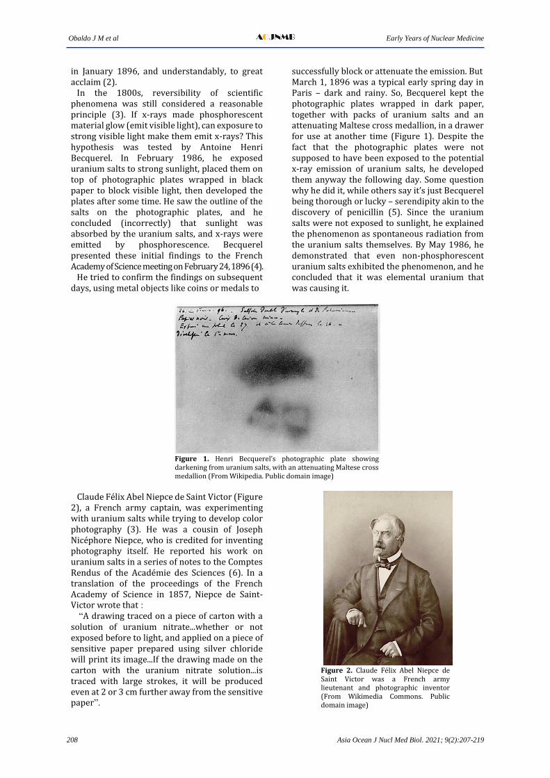
successfully block or attenuate the emission. But March 1, 1896 was a typical early spring day in Paris – dark and rainy. So, Becquerel kept the photographic plates wrapped in dark paper, together with packs of uranium salts and an attenuating Maltese cross medallion, in a drawer for use at another time (Figure 1). Despite the fact that the photographic plates were not supposed to have been exposed to the potential x-ray emission of uranium salts, he developed them anyway the following day. Some question why he did it, while others say it’s just Becquerel being thorough or lucky – serendipity akin to the discovery of penicillin (5). Since the uranium salts were not exposed to sunlight, he explained the phenomenon as spontaneous radiation from the uranium salts themselves. By May 1986, he demonstrated that even non-phosphorescent uranium salts exhibited the phenomenon, and he concluded that it was elemental uranium that was causing it.
Figure 1. Henri Becquerel’s photographic plate showing darkening from uranium salts, with an attenuating Maltese cross medallion (From Wikipedia. Public domain image)
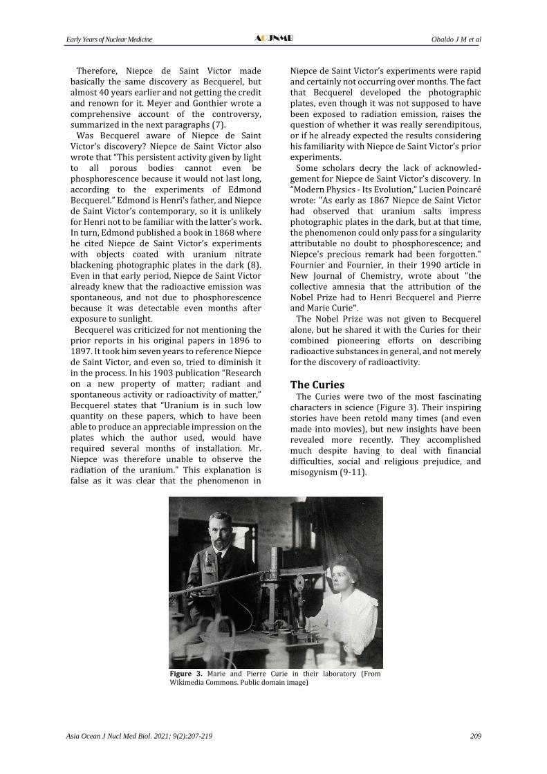
Claude Felix Abel Niepce de Saint Victor (Figure 2), a French army captain, was experimenting with uranium salts while trying to develop color photography (3). He was a cousin of Joseph Nicephore Niepce, who is credited for inventing photography itself. He reported his work on uranium salts in a series of notes to the Comptes Rendus of the Academie des Sciences (6). In a translation of the proceedings of the French Academy of Science in 1857, Niepce de Saint-Victor wrote that : “A drawing traced on a piece of carton with a solution of uranium nitrate...whether or not exposed before to light, and applied on a piece of sensitive paper prepared using silver chloride will print its image...If the drawing made on the carton with the uranium nitrate solution...is traced with large strokes, it will be produced even at 2 or 3 cm further away from the sensitive paper”.
Figure 2. Claude Felix Abel Niepce de Saint Victor was a French army lieutenant and photographic inventor (From Wikimedia Commons. Public domain image)
Early Years of Nuclear Medicine Obaldo J M et al
Asia Ocean J Nucl Med Biol. 2021; 9(2):207-219 209
Therefore, Niepce de Saint Victor made basically the same discovery as Becquerel, but almost 40 years earlier and not getting the credit and renown for it. Meyer and Gonthier wrote a comprehensive account of the controversy, summarized in the next paragraphs (7). Was Becquerel aware of Niepce de Saint Victor’s discovery? Niepce de Saint Victor also wrote that “This persistent activity given by light to all porous bodies cannot even be phosphorescence because it would not last long, according to the experiments of Edmond Becquerel.” Edmond is Henri’s father, and Niepce de Saint Victor’s contemporary, so it is unlikely for Henri not to be familiar with the latter’s work. In turn, Edmond published a book in 1868 where he cited Niepce de Saint Victor’s experiments with objects coated with uranium nitrate blackening photographic plates in the dark (8). Even in that early period, Niepce de Saint Victor already knew that the radioactive emission was spontaneous, and not due to phosphorescence because it was detectable even months after exposure to sunlight. Becquerel was criticized for not mentioning the prior reports in his original papers in 1896 to 1897. It took him seven years to reference Niepce de Saint Victor, and even so, tried to diminish it in the process. In his 1903 publication “Research on a new property of matter; radiant and spontaneous activity or radioactivity of matter,” Becquerel states that “Uranium is in such low quantity on these papers, which to have been able to produce an appreciable impression on the plates which the author used, would have required several months of installation. Mr. Niepce was therefore unable to observe the radiation of the uranium." This explanation is false as it was clear that the phenomenon in
Niepce de Saint Victor’s experiments were rapid and certainly not occurring over months. The fact that Becquerel developed the photographic plates, even though it was not supposed to have been exposed to radiation emission, raises the question of whether it was really serendipitous, or if he already expected the results considering his familiarity with Niepce de Saint Victor’s prior experiments. Some scholars decry the lack of acknowled-gement for Niepce de Saint Victor’s discovery. In “Modern Physics - Its Evolution,” Lucien Poincare wrote: "As early as 1867 Niepce de Saint Victor had observed that uranium salts impress photographic plates in the dark, but at that time, the phenomenon could only pass for a singularity attributable no doubt to phosphorescence; and Niepce's precious remark had been forgotten." Fournier and Fournier, in their 1990 article in New Journal of Chemistry, wrote about "the collective amnesia that the attribution of the Nobel Prize had to Henri Becquerel and Pierre and Marie Curie". The Nobel Prize was not given to Becquerel alone, but he shared it with the Curies for their combined pioneering efforts on describing radioactive substances in general, and not merely for the discovery of radioactivity.
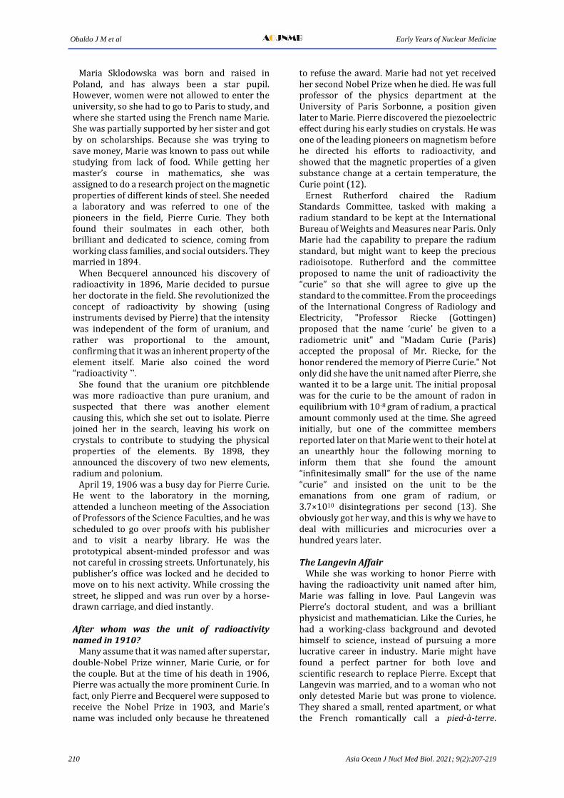
The Curies The Curies were two of the most fascinating characters in science (Figure 3). Their inspiring stories have been retold many times (and even made into movies), but new insights have been revealed more recently. They accomplished much despite having to deal with financial difficulties, social and religious prejudice, and misogynism (9-11).
Figure 3. Marie and Pierre Curie in their laboratory (From Wikimedia Commons. Public domain image)
Obaldo J M et al Early Years of Nuclear Medicine
210 Asia Ocean J Nucl Med Biol. 2021; 9(2):207-219
Maria Sklodowska was born and raised in Poland, and has always been a star pupil. However, women were not allowed to enter the university, so she had to go to Paris to study, and where she started using the French name Marie. She was partially supported by her sister and got by on scholarships. Because she was trying to save money, Marie was known to pass out while studying from lack of food. While getting her master’s course in mathematics, she was assigned to do a research project on the magnetic properties of different kinds of steel. She needed a laboratory and was referred to one of the pioneers in the field, Pierre Curie. They both found their soulmates in each other, both brilliant and dedicated to science, coming from working class families, and social outsiders. They married in 1894. When Becquerel announced his discovery of radioactivity in 1896, Marie decided to pursue her doctorate in the field. She revolutionized the concept of radioactivity by showing (using instruments devised by Pierre) that the intensity was independent of the form of uranium, and rather was proportional to the amount, confirming that it was an inherent property of the element itself. Marie also coined the word “radioactivity ”. She found that the uranium ore pitchblende was more radioactive than pure uranium, and suspected that there was another element causing this, which she set out to isolate. Pierre joined her in the search, leaving his work on crystals to contribute to studying the physical properties of the elements. By 1898, they announced the discovery of two new elements, radium and polonium. April 19, 1906 was a busy day for Pierre Curie. He went to the laboratory in the morning, attended a luncheon meeting of the Association of Professors of the Science Faculties, and he was scheduled to go over proofs with his publisher and to visit a nearby library. He was the prototypical absent-minded professor and was not careful in crossing streets. Unfortunately, his publisher’s office was locked and he decided to move on to his next activity. While crossing the street, he slipped and was run over by a horse-drawn carriage, and died instantly. After whom was the unit of radioactivity named in 1910? Many assume that it was named after superstar, double-Nobel Prize winner, Marie Curie, or for the couple. But at the time of his death in 1906, Pierre was actually the more prominent Curie. In fact, only Pierre and Becquerel were supposed to receive the Nobel Prize in 1903, and Marie’s name was included only because he threatened
to refuse the award. Marie had not yet received her second Nobel Prize when he died. He was full professor of the physics department at the University of Paris Sorbonne, a position given later to Marie. Pierre discovered the piezoelectric effect during his early studies on crystals. He was one of the leading pioneers on magnetism before he directed his efforts to radioactivity, and showed that the magnetic properties of a given substance change at a certain temperature, the Curie point (12). Ernest Rutherford chaired the Radium Standards Committee, tasked with making a radium standard to be kept at the International Bureau of Weights and Measures near Paris. Only Marie had the capability to prepare the radium standard, but might want to keep the precious radioisotope. Rutherford and the committee proposed to name the unit of radioactivity the “curie” so that she will agree to give up the standard to the committee. From the proceedings of the International Congress of Radiology and Electricity, "Professor Riecke (Gottingen) proposed that the name ‘curie’ be given to a radiometric unit" and "Madam Curie (Paris) accepted the proposal of Mr. Riecke, for the honor rendered the memory of Pierre Curie." Not only did she have the unit named after Pierre, she wanted it to be a large unit. The initial proposal was for the curie to be the amount of radon in equilibrium with 10-8 gram of radium, a practical amount commonly used at the time. She agreed initially, but one of the committee members reported later on that Marie went to their hotel at an unearthly hour the following morning to inform them that she found the amount “infinitesimally small” for the use of the name “curie” and insisted on the unit to be the emanations from one gram of radium, or 3.7×10¹⁰ disintegrations per second (13). She obviously got her way, and this is why we have to deal with millicuries and microcuries over a hundred years later. The Langevin Affair While she was working to honor Pierre with having the radioactivity unit named after him, Marie was falling in love. Paul Langevin was Pierre’s doctoral student, and was a brilliant physicist and mathematician. Like the Curies, he had a working-class background and devoted himself to science, instead of pursuing a more lucrative career in industry. Marie might have found a perfect partner for both love and scientific research to replace Pierre. Except that Langevin was married, and to a woman who not only detested Marie but was prone to violence. They shared a small, rented apartment, or what the French romantically call a pied-à-terre.
Early Years of Nuclear Medicine Obaldo J M et al
Asia Ocean J Nucl Med Biol. 2021; 9(2):207-219 211
Langevin’s wife hired a private investigator, who stole the lovers’ letters from their apartment (10, 14). All these came to a head during the prestigious 1911 Solvay Physics Conference in Brussels, Belgium, a by-invitation only event attended by the world’s most eminent scientists. Marie was not very popular during the conference, firstly because she was a woman; and secondly, because her affair with Langevin (who was also attending the conference) became publicly known, after his wife Jeanne exposed their love letters to the press. Marie was pilloried by the news media for sullying Pierre’s name, to the extent of calling her a Jew (not true, she was an atheist). These overshadowed the news Marie received by telegram during the conference that she was to receive her second Nobel Prize, this time in chemistry for her discovery of radium and polonium. This made her the first person, male or female, to receive two Nobels, and in two different fields (physics and chemistry) to boot. But because of the scandals, the Swedish Academy discouraged her from going to Stockholm to receive the prize as they did not want an adulteress to be shaking hands with King Gustav V, and to be partaking of the banquet with the other royals. Marie insisted on going, encouraged by her friend Albert Einstein. She wrote back that “I believe that there is no connection between my scientific work and the facts of private life.” During the 11-course dinner, things went smoothly and Marie and the king got along very well. And it was probably because they shared something in common. Before his reign was over, Gustav also had an affair, and like Marie Curie, also with a married man (10). Actually, more of a sting operation by a con man and murderer, costing the royal family a fortune in hush money that obviously went to waste because the affair became common knowledge anyway. While Marie Curie and Paul Langevin did not end up together, their grandchildren did. Helene Joliot, Irene Curie’s daughter and a noted scientist herself, married Michel Langevin. What did Marie Curie die of? The default assumption is that she died of aplastic anemia as a result of exposure from her work with radium. Both she and Pierre certainly had radiation effects from these, including burns, cataracts and sores. But Marie actually had a second career as a radiologist. Frustrated from the lack of support from the government, she used her personal contacts to raise funds and worked to set up x-ray units in military hospitals during the First World War. In all, she created about 200 x-ray facilities, 20 of them mobile units called “Petite Curies” or “Little Curies,” which she
sometimes drove to the frontlines herself. The philanthropic organization Union of Women of France provided the first car. Marie designed the conversion to a mobile unit, adding a dynamo to provide the needed high-voltage electricity (15). Equipment and safety practices at the time were expectedly not up to modern-day standards, and overexposure to x-rays was common. It was estimated that about a million French soldiers benefited from these facilities, so the radiation exposure to the radiographers must have been staggering. As important as establishing the radiology units was Marie’s training the people who will run the machines, who included her daughter Irene, also a Nobel laureate for discovering artificial radioactivity that is very crucial for the development of nuclear medicine. Probably because of her efforts during the war, the French eventually forgave her for the Langevin affair. Her remains were moved in 1995 from the family-owned plot in Sceaux to a crypt in the Pantheon in Paris, an honor reserved for the most revered of French citizens. And Marie was the first woman to receive the honor on her own merits. When her (and Pierre’s) remains were exhumed, these were measured for radiation, probably because the coffins were going to be on public display. And unexpectedly, her remains only showed background levels. So, the thinking now is that Marie may have suffered from x-ray radiation as much as from gamma rays (16). In her later years, she herself attributed her illness to the high X-ray exposures she had received during the war (15). And yes. Marie and Pierre Curie, joined Paul Langevin, who was entombed in the Pantheon earlier in 1948.
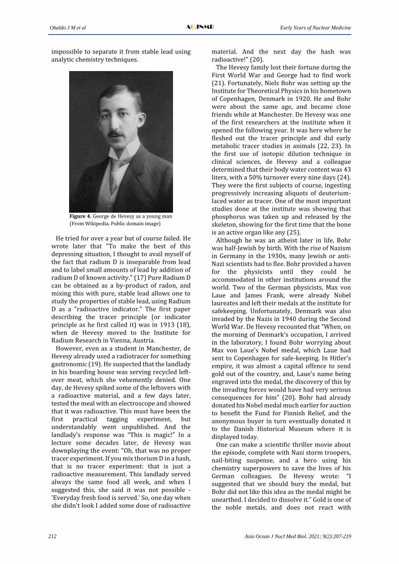
The tracer principle The celebrated scientist Ernest Rutherford, was head of the physics department at the University of Manchester in England. Together with his star doctorate student, Niels Bohr, they worked out the structure of the atom, a model that was unchanged for decades. Of all things to say, he once declared that he has “never given one of his students a hopeless problem ”. Rutherford had a problem that might be hopeless though. He had a large quantity of Radium D that was mixed up in a large quantity of lead. Rutherford issued a challenge, "My boy, if you are worth your salt, you separate Radium D from all that nuisance lead." (17) The boy he was referring to was George de Hevesy (Figure 4), scion of a Hungarian-Jewish noble family, who were wealthy industrialists. The problem was that Radium D is not radium. He did not know it at the time but Radium D turned out that to be a radioisotope of lead (Pb-210), so it was
Obaldo J M et al Early Years of Nuclear Medicine
212 Asia Ocean J Nucl Med Biol. 2021; 9(2):207-219
impossible to separate it from stable lead using analytic chemistry techniques.
Figure 4. George de Hevesy as a young man
(From Wikipedia. Public domain image)
He tried for over a year but of course failed. He wrote later that "To make the best of this depressing situation, I thought to avail myself of the fact that radium D is inseparable from lead and to label small amounts of lead by addition of radium D of known activity." (17) Pure Radium D can be obtained as a by-product of radon, and mixing this with pure, stable lead allows one to study the properties of stable lead, using Radium D as a "radioactive indicator." The first paper describing the tracer principle (or indicator principle as he first called it) was in 1913 (18), when de Hevesy moved to the Institute for Radium Research in Vienna, Austria. However, even as a student in Manchester, de Hevesy already used a radiotracer for something gastronomic (19). He suspected that the landlady in his boarding house was serving recycled left-over meat, which she vehemently denied. One day, de Hevesy spiked some of the leftovers with a radioactive material, and a few days later, tested the meal with an electroscope and showed that it was radioactive. This must have been the first practical tagging experiment, but understandably went unpublished. And the landlady’s response was “This is magic!” In a lecture some decades later, de Hevesy was downplaying the event: "Oh, that was no proper tracer experiment. If you mix thorium D in a hash, that is no tracer experiment: that is just a radioactive measurement. This landlady served always the same food all week, and when I suggested this, she said it was not possible - 'Everyday fresh food is served.' So, one day when she didn't look I added some dose of radioactive
material. And the next day the hash was radioactive!" (20). The Hevesy family lost their fortune during the First World War and George had to find work (21). Fortunately, Niels Bohr was setting up the Institute for Theoretical Physics in his hometown of Copenhagen, Denmark in 1920. He and Bohr were about the same age, and became close friends while at Manchester. De Hevesy was one of the first researchers at the institute when it opened the following year. It was here where he fleshed out the tracer principle and did early metabolic tracer studies in animals (22, 23). In the first use of isotopic dilution technique in clinical sciences, de Hevesy and a colleague determined that their body water content was 43 liters, with a 50% turnover every nine days (24). They were the first subjects of course, ingesting progressively increasing aliquots of deuterium-laced water as tracer. One of the most important studies done at the institute was showing that phosphorus was taken up and released by the skeleton, showing for the first time that the bone is an active organ like any (25). Although he was an atheist later in life, Bohr was half-Jewish by birth. With the rise of Nazism in Germany in the 1930s, many Jewish or anti-Nazi scientists had to flee. Bohr provided a haven for the physicists until they could be accommodated in other institutions around the world. Two of the German physicists, Max von Laue and James Frank, were already Nobel laureates and left their medals at the institute for safekeeping. Unfortunately, Denmark was also invaded by the Nazis in 1940 during the Second World War. De Hevesy recounted that "When, on the morning of Denmark's occupation, I arrived in the laboratory, I found Bohr worrying about Max von Laue's Nobel medal, which Laue had sent to Copenhagen for safe-keeping. In Hitler's empire, it was almost a capital offence to send gold out of the country, and, Laue's name being engraved into the medal, the discovery of this by the invading forces would have had very serious consequences for him” (20). Bohr had already donated his Nobel medal much earlier for auction to benefit the Fund for Finnish Relief, and the anonymous buyer in turn eventually donated it to the Danish Historical Museum where it is displayed today. One can make a scientific thriller movie about the episode, complete with Nazi storm troopers, nail-biting suspense, and a hero using his chemistry superpowers to save the lives of his German colleagues. De Hevesy wrote: “I suggested that we should bury the medal, but Bohr did not like this idea as the medal might be unearthed. I decided to dissolve it.” Gold is one of the noble metals, and does not react with
Early Years of Nuclear Medicine Obaldo J M et al
Asia Ocean J Nucl Med Biol. 2021; 9(2):207-219 213
ordinary solutions. So, de Hevesy used aqua regia (“royal water,” a potent mixture of hydrochloric acid and nitric acid), the only chemical able to dissolve gold, and placed the jar of gold solution on a shelf together with hundreds of glass beakers and flasks in the laboratory. De Hevesy wrote to von Laue that the task of dissolving the medals had not been easy, as gold is “exceedingly unreactive and difficult to dissolve.” The wait and the feeling of urgency must have been excruciating, as he wrote: “While the invading forces marched in the streets of Copenhagen, I was busy dissolving Laue’s and also James Franck’s medals.” 26. Since the institute was known to be welcoming to German Jews, as expected, it was raided and searched carefully. But the orange-colored solution was overlooked by the Nazis, and waited out the war on the shelf. Because of the increasing likelihood of their being arrested, Bohr himself was later spirited out to Sweden (27), and to avoid detection and enemy fire from occupied territories, was brought by high-altitude plane to Britain (28). De Hevesy used his Nazi-friendly Hungarian passport to escape by train to Sweden, where he worked and lived for the rest of his life (29). He received the 1943 Nobel Prize in Chemistry for developing the tracer principle and the early metabolic studies that started the development of nuclear medicine. Both men returned to the institute after the war to find the jar with the dissolved medals undisturbed on the shelf. The gold was precipitated out of the solution and sent back to the Swedish Academy (Nobel Foundation) in Stockholm early in 1950, and by 1952 the recast medals were presented back to the German scientists, with Franck receiving his at a ceremony at the University of Chicago (30). Who is the Father of Nuclear Medicine? It is understandable that academics want to be considered pioneers in their field, as they can be as competitive as any athlete. Colleagues often honor these pioneers, not only because they are friends, but also because the honor extends to their institutions. The Wikipedia entry of nuclear medicine has John Lawrence for the honor (31), for his pioneering work on phosphorus-32 to treat leukemia, first in rats and shortly after, in humans (32). The Berkeley Laboratory, where he worked, of course claims to be the birthplace of nuclear medicine, and John Lawrence as the father (33). Others also try to claim the title by their innovating specific nuclear medicine techniques. As previously mentioned, de Hevesy not only came up with the tracer principle, the very core
of nuclear medicine theranostics, but he pioneered actual metabolic studies while at Bohr’s institute. Bone scintigraphy can trace its lineage to the understated study of phosphorus metabolism in rat skeleton. This was actually a revolutionary paper that pioneered the concept that bodily organs were in dynamic equilibrium, and "figuratively shook the earth of biology” (34). The study was made possible by the production of artificially radioactive “vital” or biological elements like phosphorus, to study its metabolism, and it was just a matter of technology needing to catch up so that we can use it for scintigraphy. This was the first biomedical experiment with artificial radioactivity, much like how we use I-131 for iodine metabolism for the thyroid, or F-18 to substitute for oxygen in glucose, today. He also developed a method for measuring blood volume by labeling red blood cells (35), a nuclear medicine procedure we have performed with some modifications until the 1990s. For these, de Hevesy truly deserves the sobriquet “Father of Nuclear Medicine,” (19) and the Institute for Theoretical Physics, as its birthplace.
Radioactive Iodine Radioactive iodine (RAI) therapy is a seminal medical discovery of the 20th century. The story of its development is compelling, but not because of serendipity, which was crucial in the discovery of radioactivity, the tracer principle, and radioimmunoassay. Rather, aside from the progressive build-up in related scientific discoveries that led to an “Aha!” moment, there was the posturing afterwards to get credit for coming up with the idea. The history of RAI has sometimes been falsely put forth in the literature, with books, articles and presentations citing inaccurate content.
The Pivotal Question to launch the RAI Research Saul Hertz served as director of the Thyroid Clinic from 1931-1943 at Boston’s Massachusetts General Hospital (MGH), then the premier hospital of the Harvard Medical School. Dr Hertz had been using iodine to explore a nonsurgical method of treating Graves’ disease. Hertz attended a lecture at Harvard Medical School’s Vanderbilt Hall, on November 12, 1936, given by the president of the Massachusetts Institute of Technology (MIT), Karl Compton. Compton’s topic was, “What Physics can do for Biology and Medicine.” In that meeting were MGH’s Chief of Medicine, James H. Means, founder of the MGH Thyroid Unit, and MIT’s Robley Evans, the administrative director of the physics laboratory.
Obaldo J M et al Early Years of Nuclear Medicine
214 Asia Ocean J Nucl Med Biol. 2021; 9(2):207-219
After the lecture, Hertz spontaneously asked Compton, the pivotal question, “Could iodine be made radioactive artificially?” (36, 37). Hertz’s question leapfrogged the development of radiopharmaceutical therapy. The question could be considered as the logical next step after a number of fairly recent scientific advances: the description of the role of iodine in thyroid metabolism by Marine in 1915 (38), application of the radiotracer principle in metabolic studies by de Hevesy in the 1920s and 1930s, production of artificial radioactivity by the Joliot-Curies in 1934, and creation of progressively larger cyclotrons by Ernest Lawrence in the 1930s. A month later on December 15, 1936 Compton replied by letter to Hertz (36), writing, “Iodine can be made artificially radioactive” and “emits gamma rays and beta rays” (demonstrating the potential use of RAI as a theranostic agent). President Compton’s correspondence also stated, “… It has a half period of twenty-five minutes.” Additionally, indicating that there may be other periods of decay. Hertz replied, “…to hope that iodine that is made radioactive…will be a useful method of therapy in cases of overactivity of the thyroid gland”. (37). Unfortunately, there are articles crediting Means with coming up with the idea for RAI or that the question came as a result of a group discussion. Ell (39) had an alternative account of the November 12, 1936 lecture by Compton, where it was Means who asked “Is there a radioisotope of iodine?” and it was Evans who replied “We can make some.” Fahey says this conversation did not occur (37). An exchange of letters between Compton and Hertz clearly showed that it was Hertz who posed the RAI question. Means also wrote in a letter to the Markle Foundation that granted funds to build the MIT medical cyclotron stating, “… it at once occurred to Hertz that we might solve a problem we were already working on (treatment of Graves’ disease without surgery”). Animal Studies In early 1937, a collaboration was established between Cambridge’s MIT and Boston’s MGH. A young physicist, Arthur Roberts, Ph.D., was hired by MIT to work in the physics lab. But Roberts found no counting equipment and no evidence that radioiodine had been made at MIT (40). He had to devise a way to produce I-128 in small quantities, using a radium-beryllium neutron generator, based on Enrico Fermi’s work. Hertz and Roberts began the first studies on four dozen rabbits to evaluate the effects of RAI, on the thyroid. MGH’s Means arranged for funding from Harvard’s Milton Fund.
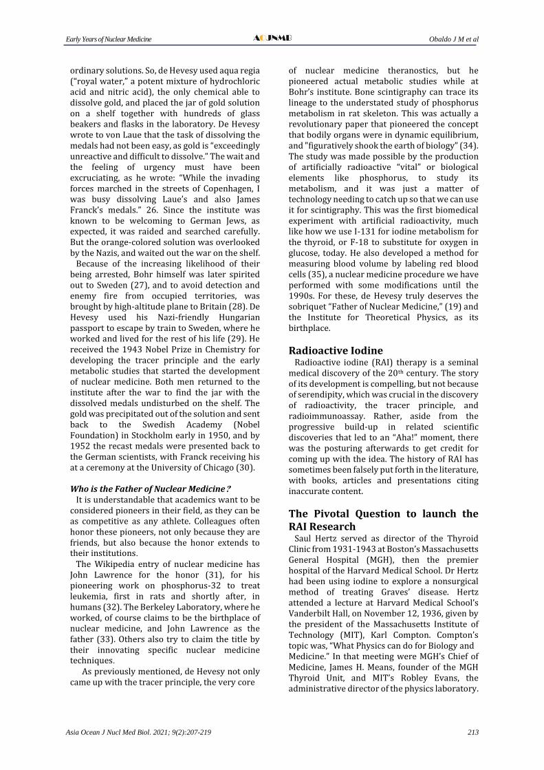
Because of the very short half-life of I-128, they only had about an hour to anesthetize the rabbit, inject the radioisotope, remove the thyroid, and measure the radioactivity (41) (Figure 5).
Figure 5. Arthur Roberts (left) and Saul Hertz (right) performing early RAI experiments on rabbits (Courtesy of the Dr. Saul Hertz Archive and SaulHertzMD.com)
Hertz and Roberts proved that RAI was a useful physiological indicator, or biomarker. They reported in with their first of a series of publications that hyperplastic thyroid glands retained more RAI than normal thyroid glands (42). The animal studies demonstrated the principle that RAI could be used to investigate thyroid gland physiology, demonstrating the tracer capabilities of RAI and its delineation of function of the thyroid gland. The original manuscript describing their rabbit study findings had Hertz and Roberts as the coauthors as they had done the work and written the paper. Primary sources document Roberts’ production of I-128 at the MIT laboratory, while Hertz and Roberts solely administered and analyzed the biodistribution of the radioisotope in the rabbits. After the article was accepted for publication, MIT’s Evans insisted that his name be added to the paper while it was at the publishers. When Roberts was hired, Evans had included a condition of Roberts’ employment that Evans’ name be added to any papers that might be forthcoming. Evans dictated a letter to the editor for Hertz to sign that Evans’ name be added although Evans made no contribution (43, 44). Roberts wrote a scathing letter in 1991, to MGH’s Dr John Stanbury, author of a book on the history of the MGH Thyroid Clinic, correcting Stanbury for highlighting the non-existent academic contributions of Means and Evans to
Early Years of Nuclear Medicine Obaldo J M et al
Asia Ocean J Nucl Med Biol. 2021; 9(2):207-219 215
the scientific development of RAI therapy. Neither Evans nor Means took part in the RAI experimental work (40). Stanbury took no heed of Roberts’s impassioned pleas to present the history honestly. Stanbury’s book fictitiously stated that there had been a group discussion and that Hertz just happened to ask Compton the pivotal question on RAI. The book has been available for decades, and is probably responsible for much of the misinformation about RAI therapy history. Clinical Studies Hertz and Roberts indicated that essential to clinical trials was a longer lasting radioisotope of iodine. Information was exchanged with the University of California Berkeley, where Ernest Lawrence had invented the cyclotron for which he received a Nobel Prize in 1936. Hertz’s former Boston colleague, Mayo Soley, reached out to Hertz from California. Joseph Hamilton traveled from California to Boston, to see firsthand the
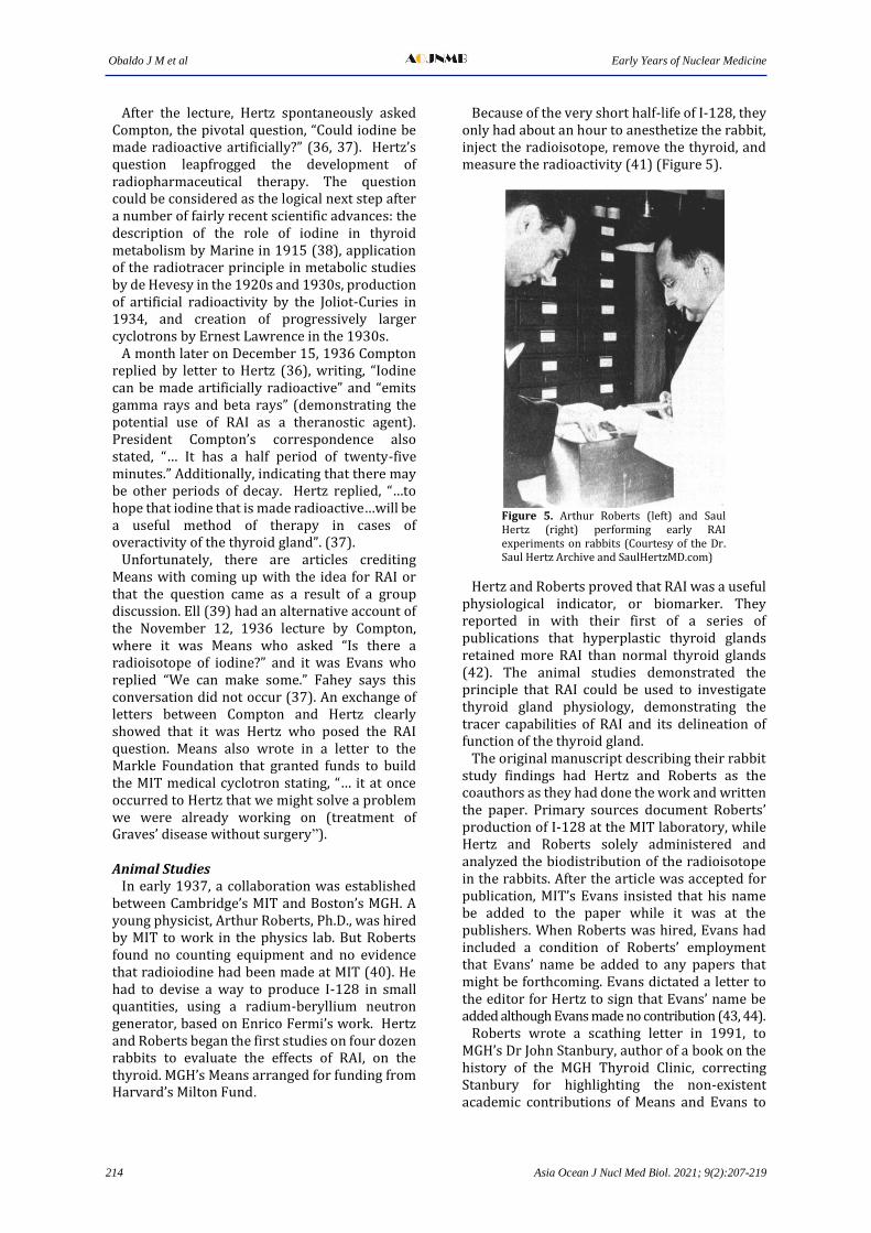
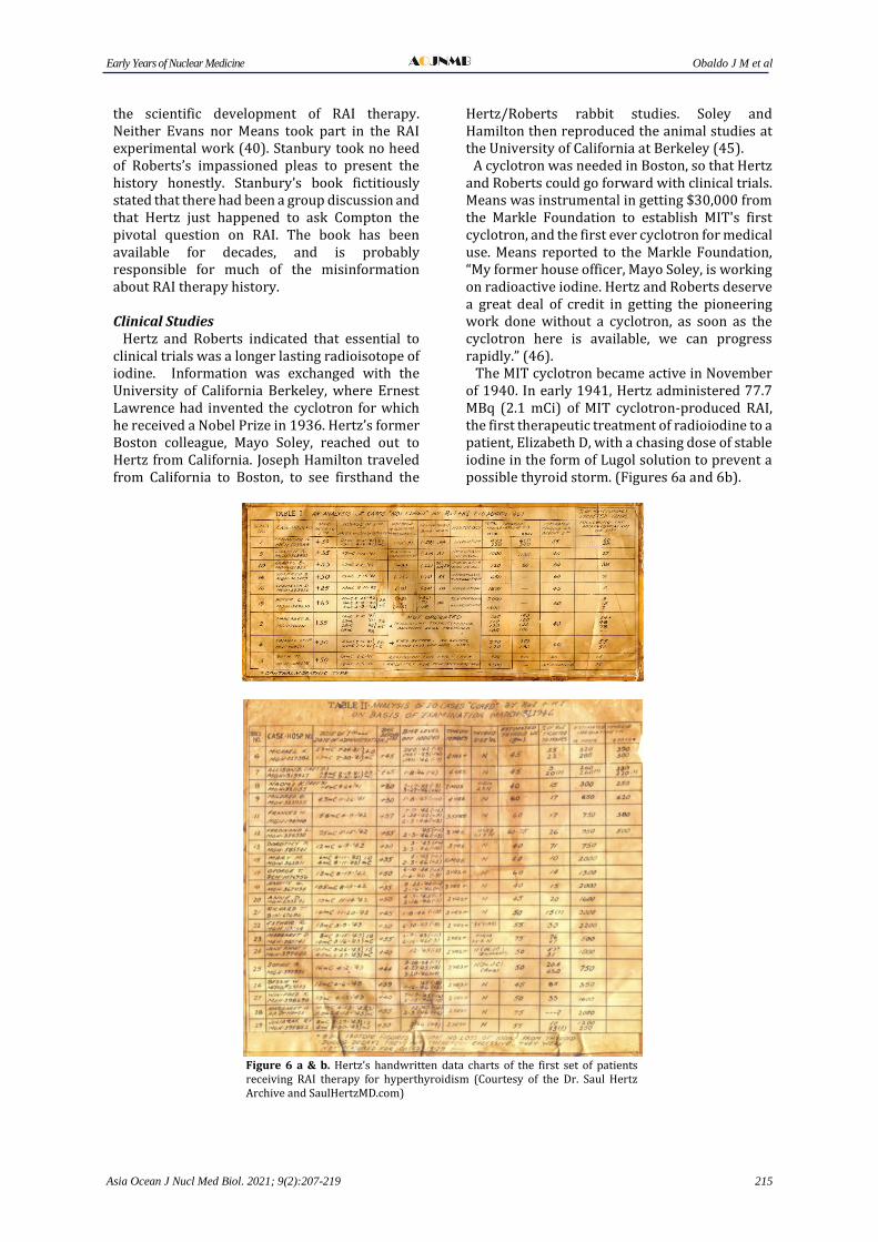
Hertz/Roberts rabbit studies. Soley and Hamilton then reproduced the animal studies at the University of California at Berkeley (45). A cyclotron was needed in Boston, so that Hertz and Roberts could go forward with clinical trials. Means was instrumental in getting $30,000 from the Markle Foundation to establish MIT's first cyclotron, and the first ever cyclotron for medical use. Means reported to the Markle Foundation, “My former house officer, Mayo Soley, is working on radioactive iodine. Hertz and Roberts deserve a great deal of credit in getting the pioneering work done without a cyclotron, as soon as the cyclotron here is available, we can progress rapidly.” (46). The MIT cyclotron became active in November of 1940. In early 1941, Hertz administered 77.7 MBq (2.1 mCi) of MIT cyclotron-produced RAI, the first therapeutic treatment of radioiodine to a patient, Elizabeth D, with a chasing dose of stable iodine in the form of Lugol solution to prevent a possible thyroid storm. (Figures 6a and 6b).
Figure 6 a & b. Hertz’s handwritten data charts of the first set of patients receiving RAI therapy for hyperthyroidism (Courtesy of the Dr. Saul Hertz Archive and SaulHertzMD.com)
Obaldo J M et al Early Years of Nuclear Medicine
216 Asia Ocean J Nucl Med Biol. 2021; 9(2):207-219
After two weeks, she received another 48.1 MBq (1.3 mCi), because of persistent hyperthyroidism (43). Details are a bit vague, but she eventually underwent thyroidectomy, and histopathology showed involution of the thyroid. The activity given was inadequate because of the relatively short half-life of I-130, but as proof of concept, it was a success as shown by histology. Probably because it was the first time to use in humans, Hertz tended to use lower RAI activities and used stable iodine after RAI to control symptoms, the latter at the insistence of Means. Gradually, the first series of 29 patients was developed. Hertz and Roberts continued to treat hyperthyroid patients in 1942. In April of 1943, Hertz received an MGH Military Leave of Absence to serve in the Navy during World War II. Hertz asked Earle Chapman, who had a private practice treating wealthy patients in Boston, and was ineligible for military service, to take over his clinical trials with an understanding and agreement that Chapman would continue using the Hertz and Roberts protocol. The JAMA Papers Chapman, however, used a different protocol in subsequent patients. He, together with Evans, submitted their article on 22 patients, for publication to the Journal of the American Medical Association (JAMA) without Dr. Hertz's name and before Hertz completed his service to his country. JAMA returned the Chapman and Evans paper indicating that it needed to be more concise. The editor of JAMA, Morris Fishbein, reached out to Hertz and Roberts requesting a seventh paper on the subject that would report the results of the Hertz’s and Roberts’ first clinical trial of RAI to successfully diagnose and treat Graves’ disease. Means, wanted a manuscript “in print at an early date,” referring to the two articles because of his concern about "the California and perhaps other competition,” referring to the group at Berkeley (40). The Solomonic solution was to publish both in JAMA on May 11, 1946. The Hertz/Roberts paper (47) preceded the Chapman/ Evans paper (48) in pagination, an acknowledgement of their pioneer status. At the MGH Russell Museum presentation in April, 2016, by Chairman Emeritus of MGH Department of Radiology (and author of a popular nuclear medicine textbook), Dr. James Thrall, who stated that “Chapman and Evans had basically stolen his (Hertz’s) work ... the most flagrant, I think, unethical academically reprehensible behavior...worst yet. Saul Hertz died ...in 1950 and these two gentlemen
(Chapman and Evans) spent a great deal of time and effort rewriting history.” (49). The use of dosimetry was essential in preparing a personalized treatment plan, and was a distinguishing feature of the Hertz and Roberts paper. Dosimetry expert, Glenn Flux writes, “... to calculate patient-specific dosimetry showed an early understanding of the use of radionuclides. Hertz and Roberts were truly visionaries.” (50). Use of RAI to Diagnose and Treat Thyroid Carcinoma Although Hertz was credited with pioneering RAI therapy for hyperthyroidism, he also conceived the use of RAI in the treatment of thyroid carcinoma. Hertz wrote, “At the onset of the above experiments (animal studies) in 1937, it was thought that there might be promising therapeutic possibilities in the treatment of carcinoma of the thyroid.” Samuel Seidlin of New York’s Montefiore Hospital, consulted Hertz in 1943, prior to treating a celebrated patient with metastatic thyroid cancer with RAI. Hertz wrote in 1946 that his research would focus on “cancer of the thyroid which I believe holds the key to the larger problem of cancer in general”, and that, after the War, “(new) demand expected in the fields of cancer and leukaemia for other radioactive medicines”. (51). After WWII Dr Hertz’s Military Leave of Absence was not honored at MGH. In 1946, Hertz joined Boston’s Beth Israel Hospital coming with a grant from the Navy to utilize RAI to diagnose and treat thyroid cancer. Jews were starting to receive medical training, however, they were excluded from many hospitals. Boston’s Beth Israel Hospital was expanding and welcoming Jews. Hertz joined the staff of the Beth Israel Hospital, where he refined the successful use of RAI to diagnose and treat thyroid cancer (36). In September, 1946, Hertz founded The Radioactive Isotope Research Institute with clinics and laboratories in Boston and New York City. The New York facilities were run by Samuel Seidlin, who was the associate director. The institute was devoted to the application of nuclear physics to medical investigation, diagnosis and treatment. (50). Hertz developed the Multiscaler at MIT, where he continued to teach after the war. The Multiscaler, combined with multidetector Geiger counters, facilitated uptake testing that was essential for establishing a safe and effective administration of RAI. The headline of an article in The Harvard Crimson, May 24, 1949 read, “Hertz to Use Nuclear Fission in Cure for Cancer.” There was an advertisement for The Multiscaler on the same page as the article. Hertz is quoted as stating,
Early Years of Nuclear Medicine Obaldo J M et al
Asia Ocean J Nucl Med Biol. 2021; 9(2):207-219 217
“However he (Hertz) emphasized this example of therapeutic application (RAI to treat Graves’ disease) as a beacon in utilizing the tracer method in other organs than the thyroid.” Hertz also encouraged the Atomic Energy Commission to take RAI off of the atomic piles to reduce the cost (to less than $5) and expand the RAI distribution (52). Even in those early years, Hertz’s vision of treating thyroid cancer was clear, and quite similar to how we do things today. He wrote: “The patient should have as radical a removal of (thyroid) tissue as is possible and have subsequent dosage of radioactive iodine administered as long as there is significant retention of radioactive iodine within the body… in the absence of any metastatic lesion is placed upon thyroid medication and requires it as a permanent form of substitution therapy.” (53). In 1949, Saul Hertz established the first Nuclear Medicine Department at The Massachusetts Women’s Hospital. It was reported as “Opening a new division where radioactive isotopes will be used to study and treat disease.” He served as the director until his passing in 1950 from a heart attack (54). Tributes In 2016, The Society of Nuclear Medicine and Molecular Imaging (SNMMI) established The Saul Hertz Award and Symposium. Starting in February 2021 the South African Society of Nuclear Medicine (SASNM) will present a Saul Hertz Young Investigator Award. The American Chemical Society (ACS) will in October of 2021 install an ACS’s, “Saul Hertz and The Medical Uses of Radioiodine” historic landmark, at the Massachusetts General Hospital. Thanks to the efforts of prominent medical historians, archivists, thyroid specialists, nuclear physicians, physicists and others, we now know the reality behind one of the most epochal discoveries of nuclear medicine and its history .
References 1. Department of Radiology, University of
Washington. Featured History: Wilhelm Roentgen. https://rad.washington.edu/ blog/ featured history-wilhelm-rontgen. Accessed 2 January 2021.
2. Chodos A, Ouellette J. November 8, 1895: Roentgen’s Discovery of X-rays. American Physical Society APS News. Nov 2001; 10(10):2.
3. Habashi F. Niepce de Saint-Victor and the Discovery of Radioactivity. Bull Hist Chem 2001; 26:104-105 .
4. Chodos A, Ouellette J, Tretkoff E. March 1, 1896: Henri Becquerel Discovers Radio-
activity. American Physical Society APS News. Mar 2008; 17(3):2.
5. Strauss HW, Zaret B, Pieri G, Lahiri A. History corner: Antoine Henri Becquerel. J Nucl Cardiol 2017; 24:1515-6.
6. Niepce de Saint Victor CFA. On a new action of light. CR Acad Sci 1857; 45: 811-5.
7. Meyer M, Gonthier E. Is there still controversy surrounding the discovery of so-called radioactive phenomena? http://www.tribunes. com/tribune/ art97 /meyer.htm. Accessed 10 July 2017.
8. Becquerel E. La lumière: Ses causes et ses effets, (Light: Its causes and effects) vol. 2. 1st ed. Paris: F. Didot; 1868.
9. Naomi Pasachoff. Marie Curie and the Science of Radioactivity. 1st ed. Oxford: Oxford University Press; 1996.
10. Redniss L. Radioactive: Marie & Pierre Curie: A Tale of Love and Fallout. 1sted. New York: It Books; 2010.
11. Wilkie T. The Secret Sex Life of Marie Curie. The Independent. 12 June 1995. https: //www.independent.co.uk/news/science/ secret-sex-life-marie-curie-1586244.html. Accessed 10 September 2017.
12. Nobel Media AB. Pierre Curie Biographical. https://www.nobelprize.org/ prizes/ physics/ 1903/Pierre curie/ biographical/. Accessed 15 September 2019.
13. Frame P. How the Curie Came to Be. Health Physics Society's Newsletter. 1996. https: //www.orau.org/ptp/articles stories/ the curie.htm. Accessed 2 January 2021.
14. Krulwich R. Don’t Come To Stockholm! Madame Curie’s Nobel Scandal. https: //www.npr.org/sections/krulwich/2010/12/14/132031977/dont-come -to-stock holm- madame-curie-s-nobel-scandal. Accessed 11 September 2017.
15. Jorgensen TJ. How Marie Curie Brought X-Ray Machines to the Battlefield. Smit-hsonianmag.com. October 11, 2017. Accessed 2 January 2021.
16. Valiunas A. The Marvelous Marie Curie. The New Atlantis, Number 37, Fall 2012, pp. 51-70.
17. Levi H. George de Hevesy. Life and Work. Copenhagen: Rhodos Press; 1985. Pp 147 .
18. Hevesy G, Paneth F. The Solubility of Lead Sulphide and Lead Chromate. Z Anorg Chem 1913; 82:322
19. Myers WG. Georg Charles de Hevesy: The Father of Nuclear Medicine. J Nucl Med 1979; 20(6):590-4.
20. Niels Bohr Archive. Hevesy Anecdotes 2010. http://www.nba-old.nbi.dk/ hevesyanec.html. Accessed 6 January 2021.
21. Niels Bohr Institute. George de Hevesy: The Origin of Nuclear Medicine. 16 February
Obaldo J M et al Early Years of Nuclear Medicine
218 Asia Ocean J Nucl Med Biol. 2021; 9(2):207-219
2010).https://www.nbi.ku.dk/english/ www/george/dehevesy/del1/. Accessed 6 January 2021.
22. Christiansen IA, Hevesy G, Lomholt S. Radiochemical Method of Studying the Circulation of Bismuth in the Body. C. R. Acad. Sci. 1924; 178:1324.
23. Christiansen IA, Hevesy G, Lomholt S. Radiochemical Method of Studying the Circulation of Lead in the Body. C. R. Acad. Sci. 1924; 179:291.
24. Hevesy, G, Hofer, E. Elimination of water from the human body. Nature 1934; 134: 879.
25. Chievitz O, Hevesy G. Radioactive Indicators in the Study of Phosphorus Metabolism in Rats. Nature 1935; 13:754.
26. Hevesy G. A Scientific Career. Perspectives in Biology and Medicine. 1958; 1(4):345-365.
27. Gratzer W. Eurekas and Euphorias. The Oxford Book of Scientific Anecdotes. New York; Oxford University Press: 2002. Pp 220-2.
28. Wikipedia.org. Niels Bohr. https://en. wikipedia.org/wiki/Niels_Bohr. Accessed 6 January 2021.
29. Bonilis L. George de Hevesy. Lindau Nobel Laureate Meetings. https://www. mediathe-que.lindau-nobel.org/research-profile/laure ate-de-hevesy# page=all. Accessed 6 January 2021.
30. Nobel Media AB. A unique gold medal. https://www.nobelprize.org/prizes/about/the-nobel-medals-and-the-medal-for-the-prize-in-economic-sciences/. Accessed 6 January 2021.
31. Wikipedia.org. Nuclear Medicine. https://en. wikipedia.org/wiki/ Nuclear medicine. Accessed 6 January 2021.
32. Distinguished Nuclear Pioneer, John Hundale Lawrence. J Nucl Med 1970; 11:292-293.
33. Berkley Lab. From Radioisotopes to Medical Imaging, History of Nuclear Medicine Written at Berkeley. https:// www. lbl. gov/ Science- Articles/ Archive/nuclear-med -history.html. Accessed 8 January 2021.
34. Frame P. Tales from the Atomic Age. Four Tales of George de Hevesy. https://www. orau.org/ptp/articles-stories/hevesy.htm. Accessed 15 May 2017.
35. Hahn, L, Hevesy G. A method of blood volume determination. Acta Physiol. Scandinav., 1940-41:1, 3.
36. Hertz BE, Schuller KR. Saul Hertz, MD (1905-1950): A Pioneer in the Use of Radioactive Iodine. Endocrine Practice 2010; 16(4):713-6.
37. Fahey FH, Grant FD, Thrall JH. Saul Hertz, MD, and the birth of radionuclide therapy. EJNMMI Physics 2017; 4:15-21.
38. Marine D. Quantitative studies on the in vivo absorption of iodine by dogs' thyroid glands. J Biol Chem 1915; 22:547.
39. Ell PJ. The contribution of medical physics to nuclear medicine: a physician's perspective. EJNMMI Physics 2014; 1:3-9.
40. Sawin CT and Becker DV. Radioiodine and the Treatment of Hyperthyroidism: The Early History. Thyroid 1997; 7(2):163-176.
41. Remembering the Early Days of Nuclear Medicine. MGH News, Sep 1987; 46(6):1, 5, 6.
42. Hertz, S., Roberts, A., Evans, R. D., Radioactive iodine as an indicator in thyroid physiology. Proc. Soc. Exp. Biol. and Med., 1938; 38:510-3.
43. Hertz B. Dr. Saul Hertz (1905–1950) Discovers the Medical Uses of Radioactive Iodine: The First Targeted Cancer Therapy. In: Ahmadzadehfar H, editor. Thyroid Cancer: Advances in Diagnosis and Therapy. 2016.
44. Hertz B, A tribute to Dr. Saul Hertz: The discovery of the medical uses of radioiodine. World J Nucl Med. 2019; 18:8-12.
45. Hertz B, Bharadwaj P, Greenspan B, A historic review of the discovery of the medical uses of radioiodine, Pak J Nucl Med 2020;10(1):60–63.
46. Hertz B. Dr. Saul Hertz Discovers the Medical Uses of Radioiodine (RAI). J Radiol Oncol. 2018; 2: 053-054.
47. S Hertz and A Roberts. The Use of Radioactive Iodine Therapy in Hyper-thyroidism. JAMA. 1946; 131(2):81-86.
48. E Chapman and R Evans. The Treatment of Hyperthyroidism with Radioactive Iodine Therapy. JAMA. 1946; 131(2):86-91.
49. Thrall JH, MGH Russell Museum presentation, Saul Hertz: radioiodine and the origins of nuclear medicine." Youtube.com; 2016. Available at www. youtube.com/watch?v=34 Qhm8CeMuc (time index 24:40-25:08). Accessed 27 Novem-ber 2020.
50. Hertz B. Yale Medical School Beaumont Club Presentation 2016, Dr Saul Hertz discovers the medical uses of radioiodine: challenges and legacy. Youtube.com; 2016. Available at www.youtube.com/watch? v=Ssd5K_-rDJQ (time index 46:12- 46:45). Accessed 27 November 2020.
51. Ehrhardt JD, Güleç S. A Review of the History of Radioactive Iodine Theranostics: The Origin of Nuclear Ontology. Mol Imaging Radionucl Ther 2020; 29:88-97.
52. Vincent DG. Hertz to use nuclear fission in cure for cancer. Harvard Crimson May 24,
Early Years of Nuclear Medicine Obaldo J M et al
Asia Ocean J Nucl Med Biol. 2021; 9(2):207-219 219
1949. https://www.thecrimson.com /article /1949/5/24/hertz-to-use-nuclear-fission-in/. Accessed 28 November 2020.
53. Hertz S. Use of Radioactive Iodine in the Diagnosis, Study and Treatment of Diseases of the Thyroid. In: Soskin S, editor. Progress
in Clinical Epidemiology. 1st ed. Amsterdam: Elsevier Ltd; 1950. pp 65-78.
54. Lynch J, Radioactive isotope study starts at women’s hospital, Boston Traveler, Sep 30, 1949, p.14.