Embed Size (px)
Citation preview
The Effect of Hydroxyapatite-Chitosan Block
Bone Graft on the Periodontal Regeneration in
One-wall Intrabony Defect of Beagle Dogs
Sung-Koo Kim
The Graduate School
Yonsei University
Department of Dental Science
The Effect of Hydroxyapatite-Chitosan Block
Bone Graft on the Periodontal Regeneration in
One-wall Intrabony Defect of Beagle Dogs
A Dissertation Thesis
Submitted to the Department of Dental Science
and the Graduate School of Yonsei University
in partial fulfillment of the
requirements for the degree of
Doctor of Philosophy of Dental Science
Sung-Koo Kim
April 2007
This certifies that the dissertation thesis
of Sung-Koo Kim is approved
Thesis Supervisor : Seong-Ho Choi
Chong-Kwan Kim
Jung-Kiu Chai
Kwang-Mahn Kim
Yong-Keun Lee
The Graduate School
Yonsei University
April 2007
감사의감사의감사의감사의 글글글글
본 논문이 완성되기까지 부족한 저를 항상 격려해 주시고 사랑과 관심으로
이끌어 주신 최성호 교수님께 깊은 감사를 드립니다. 그리고, 많은 조언과 따뜻한
관심으로 지켜봐 주신 김종관 교수님, 채중규 교수님, 조규성 교수님, 김광만
교수님, 이용근 교수님, 김창성 교수님, 정의원 교수님께 진심으로 감사 드립니다.
연구 내내 많은 도움을 주신 치주과 교실원 여러분, 특히 채경준 선생님,
이중석 선생님께 고마움을 전합니다.
늘 변함없는 사랑과 헌신적인 도움으로 어려운 생활 속에서도 지팡이가 되어준
아내 태정과, 이제는 든든한 친구가 된 아들 병현에게 무한한 고마움의 마음을
전합니다.
오늘이 있기 까지 변함없는 믿음과 사랑으로 이해해 주시며, 물심양면으로 후원해
주신 어머님과 장인, 장모님께 감사의 마음을 드립니다.
마지막으로, 저의 마음속 영원한 정신적 기둥 이며, 하늘에서 항상 저를 지켜보고
계실 아버지께 이 논문을 바칩니다.
모든 분들께 진심으로 감사 드립니다.
2007년 6월
저자 씀
i
Table of Contents
Abstract (English) �������������������������������������������������������������������� iii
I. Introduction ������������������������������������������������������������������������� 1
II. Materials and Methods ��������������������������������������������������������� 5
A. Materials �������������������������������������������������������������������������� 5
1. Animals ������������������������������������������������������������������������� 5
2. Hydroxyapatite chitosan block bone ������������������������������������� 5
3. Chitosan membrane���������������������������������������������������������� 5
B. Experimental Procedures ������������������������������������������������������� 6
1. Surgical procedures ���������������������������������������������������������� 6
2. Histologic and histometric analysis ��������������������������������������� 6
3. Statistical analysis ������������������������������������������������������������ 7
III. Results ������������������������������������������������������������������������������ 8
A. Clinical observations ����������������������������������������������������������� 8
B. Histologic observations �������������������������������������������������������� 8
C. Histometric observations ������������������������������������������������������ 9
IV. Discussion �������������������������������������������������������������������������� 12
V. Conclusion �������������������������������������������������������������������������� 19
References ���������������������������������������������������������������������������� 20
Figures ��������������������������������������������������������������������������������� 32
Abstract (Korean) ������������������������������������������������������������������ 36
ii
List of Figures
Figure 1. A schematic diagram depicting the experimental design�������������� 7
Figure 2. Periodontal healing illustrated in percentage of the defect height 11
Figure 3. Control group (X20) ������������������������������������������������������ 32
Figure 4. Control group (X20) ������������������������������������������������������ 32
Figure 5. Hydroxyapatite-chitosan bone (X20) ��������������������������������������� 33
Figure 6. Hydroxyapatite-chitosan bone (X20) ������������������������������������ 33
Figure 7. Hydroxyapatite-chitosan bone (X200) ���������������������������������� 33
Figure 8. Chitosan membrane (X20) ����������������������������������������������� 34
Figure 9. Chitosan membrane (X20) ����������������������������������������������� 34
Figure 10. Chitosan membrane (X100) ���������������������������������������������� 34
Figure 11. Chitosan membrane (X100) ���������������������������������������������� 34
Figure 12. Hydroxyapatite-chitosan block bone and chitosan membrane (x20) 35
Figure 13. Hydroxyapatite-chitosan block bone and chitosan membrane (x100) 35
Figure 14. Hydroxyapatite-chitosan block bone and chitosan membrane (x100) 35
List of Tables
Table 1. Histometric Analysis: height(mean ± standard deviation(mm)) ������ 30
Table 2. Histometric Analysis (percentage of the entire depth of defects) ���� 31
iii
Abstract
The Effect of Hydroxyapatite-Chitosan Block Bone Graft on the Periodontal
Regeneration in One-wall Intrabony Defect of Beagle Dogs
This study evaluated a periodontal repair and a biomaterial reaction following
implantation of a newly fabricated Hydroxyapatite-chitosan block bone and a chitosan
membrane on the regeneration of one-wall intrabony defects in beagle dogs. The
surgical control groups received a flap operation only, while experimental groups
were treated with the Hydroxyapatite-chitosan block bone and/or a chitosan
membrane. In new bone formation, there was a statistically significant difference
between the chitosan membrane group and other treatment groups (P<0.05) (Kruskal
Wallis Test). The amount of new bone of the hydroxyapatite-chitosan block bone
group and the Hydroxyapatite-chitosan block bone and chitosan membrane group was
greater than control group, but there was no statistically significant different from the
control group. Comparing the amount of new cementum with that of new bone in the
four groups of our study, no cemental growth surpassed the bone growth. This result
didn’t suggest that new bone follows the coronal growth of new cementum, which
disagreed with the results of previous studies. Therefore, the further study for the
mechanism seems good to follow. The amount of new cementum of the
hydroxyapatite-chitosan block bone group and/or the chitosan membrane group was
greater than control group, but there was no statistically significant different from the
control group. In chitosan membrane group, the amount of new bone formation
was greater than other groups, which suggest that the potency of chitosan
membrane induce the new bone regeneration. The results of the present study
iv
did support the potential of chitosan in the guided tissue regeneration, but did not
showed enough capacity to bear load. Therefore, the further studies to enhance the
mechanical properties appear to be necessary.
KEY WORDS: Guided tissue regeneration, Hydroxyapatite-chitosan block bone,
chitosan membrane
1
The Effect of Hydroxyapatite-Chitosan Block Bone Graft
on the Periodontal Regeneration in One-wall Intrabony
Defect of Beagle Dogs
Sung-Koo Kim, D.D.S. , M.S.D.
Department of Dental Science
Graduate School, Yonsei University
(Directed by Prof. Seong-Ho Choi, D.D.S., M.S.D., PhD.)
I. INTRODUCTION
The regeneration of bone has long been the critical issue in the field of periodontal
and implant surgery. Many procedures have been developed for the purpose of
promoting regeneration, including guided tissue regeneration and bone graft. All of
them, however, have limitations.
Guided tissue regeneration (GTR) therapy using barrier membranes has been
introduced to induce selective repopulation of undifferentiated cells that originate
from the periodontal ligament, and has been shown to improve the periodontal
regeneration in both animal and human experiments (Caffesse et al., 1993; Karring,
1983; Kim et al., 1998; Selvig et al., 1993). For periodontal defects, it has been
suggested that the periodontal ligament cells and their migration potential are crucial
for periodontal regeneration, and that regeneration might occur if the gingival
connective tissue and epithelial cells are prevented from accessing the tooth surface.
2
Both the application of bone grafts, synthetic implant materials or inorganic bone
graft materials in combination with GTR have been reported to favour the formation
of bone (Dahlin et al., 1991; Alberius et al., 1992; Wetzel et al., 1995). Different types
of biocompatible materials such as hydroxyapatites, calcium phosphates and inorganic
bone graft materials have been used alone or in combination with GTR with varying
results in terms of bone formation ( Pinholt et al., 1991; Klinge et al., 1992; Fukuta et
al., 1992; Hislop et al., 1993; Dahlin et al., 1991; Alberius et al., 1992; Wetzel et al.,
1995 ).
The membrane barrier used in GTR should satisfy the following factors. It should
be histocompatible, biocompatible and capable of space maintenance (Magnusson et
al., 1988). It also should avoid cell migration and be easy to handle. Non-absorbable
membrane ePTFE almost fulfills the factors mentioned above and is most widely used
so far giving out good results (Nyman et al., 1982; Gottlow et al., 1986; Blumental,
1993; Becker et al., 1988; Pontoriero et al., 1988; Handelsman et al., 1991; Becker et
al., 1993 ).
Chitosan has been reported to enhance the healing of injured connective tissue
(Muzzarelli et al., 1988). Recently, a tissue engineering strategy has been suggested as
a possible alternative to conventional regenerative therapy. Chitin is a natural polymer
of N-acetylglucosamine, and is a component of the exoskeleton of a greatnumber of
organisms such as shells and cuticles of arthropods including crustaceans and insects
(Cabib, 1987). Chitosan has excellent potential as a structural base material for a
variety of engineered tissue system (Madihally et al., 1999). Chitosan has been
reported to enhance peridontal tissue regeneration (Madihally et al., 1999; Mukherjee
et al., 2003; Park et al., 2003).
3
Chemical mediators or substances that enhance bone formation are thought to be
conductive to periodontal regeneration. Among these materials, the influence of
chitosan on bone regeneration is of a particular interest. The use of chitosan and mesh
progressively improved the mechanical properties. These strong and cell-seeded
hydroxyapatite cements may have potential for bone tissue engineering in moderate
stress bearing applications (Michael et al., 2006). Chitosan takes an increasing interest
for its non-toxic, immune enhancing, antimicrobial, and wound healing properties.
Different scaffold materials have been used with varying success to generate tissue-
engineered bone formation in vitro. Ishaug et al. investigated bone formation in vitro
by culturing stromal osteoblasts in a three dimensional, biodegradable poly(lactic-co-
glycolic acid) foam (Ishaug et al., 1997). Chitosan/tricalcium phosphate sponges,
transplanted into a site for bone regeneration, can be used as a scaffolding material to
allow growth of osteoblasts in three-dimensional structure (Lee et al., 2000).
Chitosan nonwoven membrane is shown to accelerate the wound healing and
infection control as well (Risbud et al., 2001; Wang et al., 2002; Mizuno et al., 2003;
de Queiroz et al., 2003). Chitosan nonwoven membrane has been on the market for
periodontal treatment. It has an embossed surface and a yellowish-white microporous
structure, and was reported to encourage tissue attachment and first recovery. This
membrane is absorbed via hydrolysis, and is eliminated through the Kreb’s cycle as
carbon dioxide and water, and is biodegraded completely in 6 months (Suk et al.,
2002).
The placement of chitin gel in infrabony defects during periodontal surgery resulted in
proper tissue integration followed by gradual resorption of the material without any
acute inflammatory reactions (Muzzarelli et al., 1989).
4
However, chitosan has a low physical property leading to an improper use in the
areas where it receives a lot of force. Several inherent disadvantages have been
observed with these scaffold materials including weak structural integrity, variable
degradation rates, inadequate tissue peneration, and host immune reactivity.
The aim of this study was to evaluate the regenerative effects of a chitosan
membrane and/or calcium phosphate chitosan block bone applied to preclinical one
wall defects surgically created in beagle dogs.
5
ⅡⅡⅡⅡ. MATERIAL AND METHODS
A. Materials
1. Animals
A total of six male beagle dogs, each weighing about 15 kg, were used in this
study. The animals had intact dentition and a healthy periodontium. Animal selection
and management, surgical protocol, and preparation followed routines approved by the
Institutional Animal Care and Use Committee, Yonsei Medical Center, Seoul, Korea.
The animals were fed a soft diet throughout study, in order to reduce chance of
mechnical interference with the healing process during food intake.
2. Hydroxyapatite/chitosan block bone
Hydroxyapatite/chitosan hybrid scaffold was manufactured by freeze-dried
method. Chitosan solution was prepared by dissolving chitosan in 0.2 M acetic
solution. Hydroxyapatite/chitosan solution was made by dissolving 10-70wt%
hydroxyapatite in the chitosan solution. Hydroxyapatite/chitosan solution was poured
in Φ 6 mm × 12 mm teflon mold. Above 5 hours, it was refrigerated in –70 ℃. It was
freeze-dried under 6 mTorr by freezing dehydrator, above 3 days. The residual acetic
acid was neutralized by 1 M NaOH solution, and then washed with the distilled water
(above 3 times). The solvent was completely dried by freeze dry, during 3 days.
Hydroxyapatite/chitosan hybrid scaffold was manufactured.
3. Chitosan membrane
Chitosan solution was prepared by dissolving 5 wt% of chitosan in 3 wt% acetic
acid. The prepared chitosan solution was poured in mold, and feeze-dried. Then the
solution was soaked in 5 wt% NaOH aqueous solution. It was washed with the
distilled water (5-6 times). The chitosan membrane was constructed on 40℃ during 24
6
hours.
B. Experimental Procedures
1. Surgical procedures
Six male beagle dogs were used. 4X4 mm one-wall intrabony periodontal defects
were surgically created bilaterally at the distal sides of the mandibular second
premolars and mesial sides of the fourth premolars. The surgical control group
received a flap operation only. The first group was treated with
Hydroxyapatite/chitosan block bone. The second group was treated with chitosan
membrane. The third group was treated with both Hydroxyapatite/chitosan block bone
and chitosan membrane. The dogs were sacrificed at 8 weeks after the experimental
surgery.
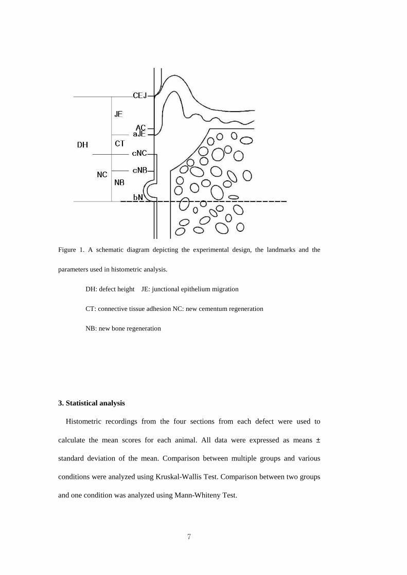
2. Histologic and histometric Analysis (Figure 1.)
Tissue blocks, which included teeth, bone, and tissue, were removed, rinsed in
saline, then fixed in 10% buffered formalin for 10 days. After being rinsed in water,
the block section were decalcified in 5 % formic acid for 14 days, and embedded in
paraffin. Serial sections, 5 um thick, were prepared at intervals of 80 µm. The four
most central sections from each block were stained with hematoxylin/eosin (H-E) and
examined using a light microscope. The most central section from each block was
selected to compare histologic findings between groups. Computer-assisted
histometric measurements were obtained using an automated image analysis system††
coupled with a video camera on a light microscope‡‡. Sections were examined at 20x
magnification.
†† Image-Pro Plus®, Media Cybernetics, Silver Spring, MD, U.S.A
‡‡ Olympus BX50, Olympus Optical Co., Tokyo, Japan
7
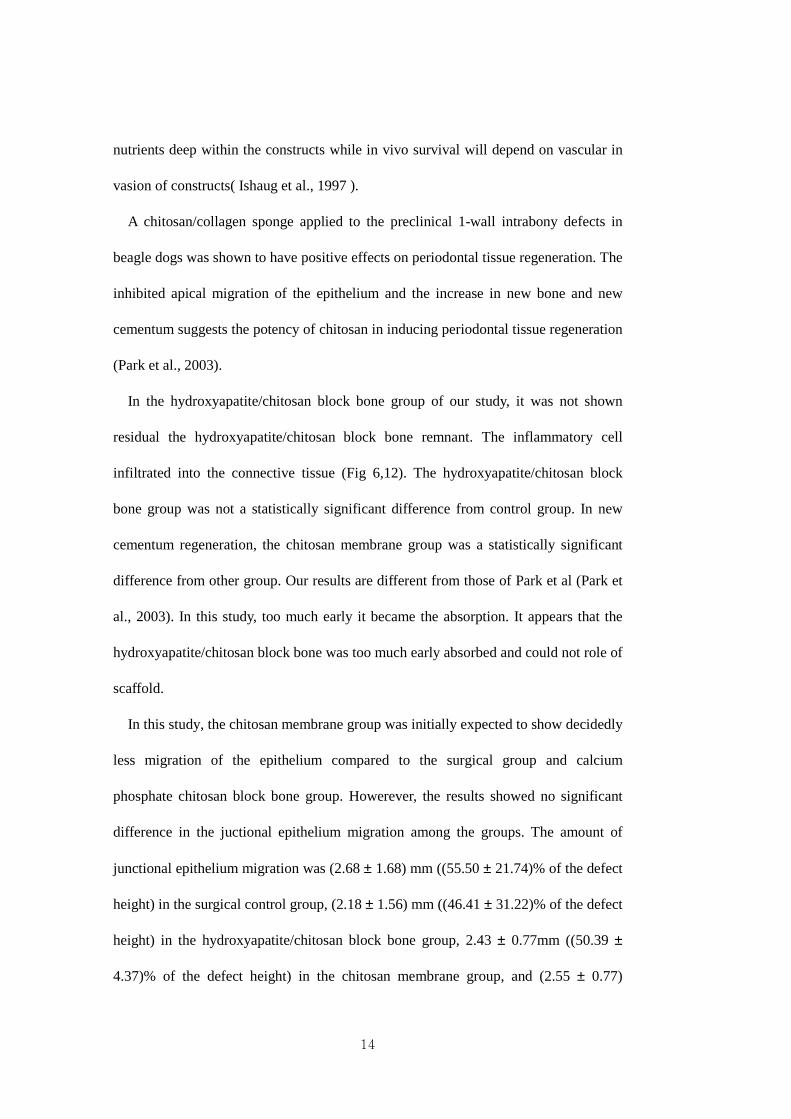
Figure 1. A schematic diagram depicting the experimental design, the landmarks and the
parameters used in histometric analysis.
DH: defect height JE: junctional epithelium migration
CT: connective tissue adhesion NC: new cementum regeneration
NB: new bone regeneration
3. Statistical analysis
Histometric recordings from the four sections from each defect were used to
calculate the mean scores for each animal. All data were expressed as means ±
standard deviation of the mean. Comparison between multiple groups and various
conditions were analyzed using Kruskal-Wallis Test. Comparison between two groups
and one condition was analyzed using Mann-Whiteny Test.
8
ⅢⅢⅢⅢ. RESULT
A. Clinical observations
Surgical procedures were uneventful and without complication in all dogs. Wound
closure was successfully maintained throughout the experiment for all defects. Healing
process was generally uneventful.
B. Histologic findings
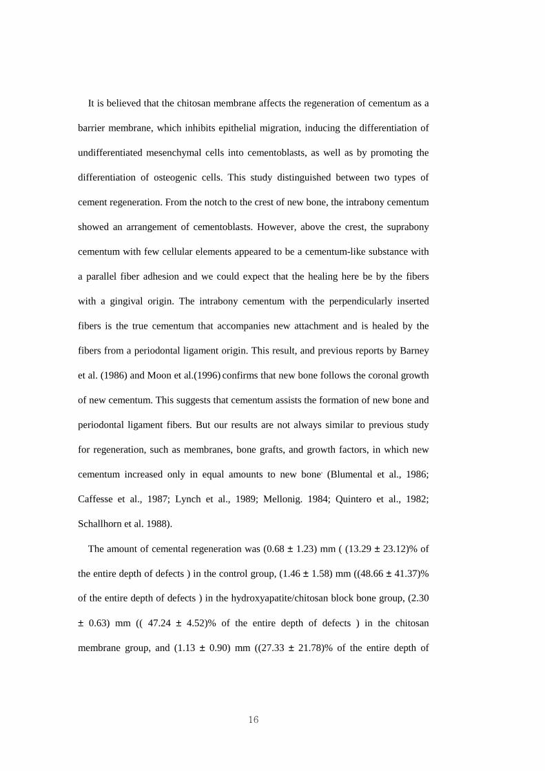
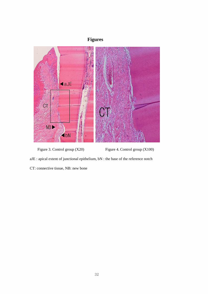
In the control group, the apical migration of junctional epithelium was observed
(Figure 3). The periodontal ligament fibers were generally oriented in a direction
parallel to the root surface in suprabony area. Dense connective tissue was shown
(Figure 4). No site showed signs of ankylosis. There was little or no sign of
inflammatory cell infiltration. Above the apical notch, a small amount of new
cementum and bone had formed along the root surface (Figure 3).
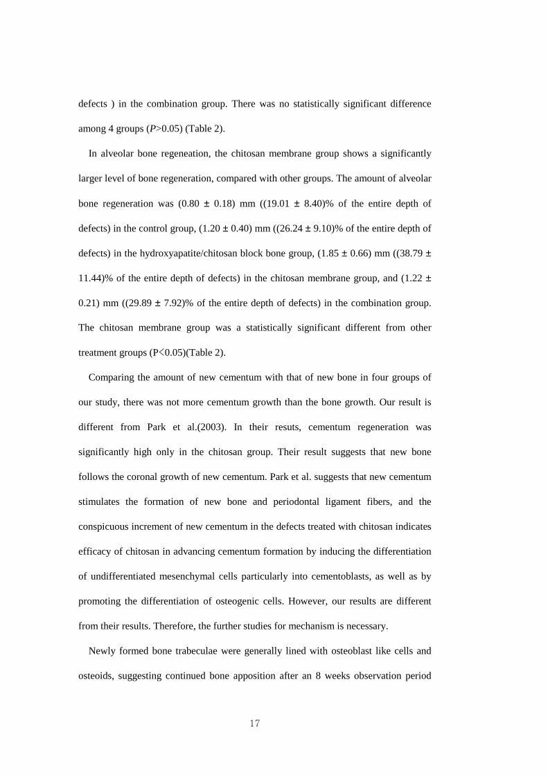
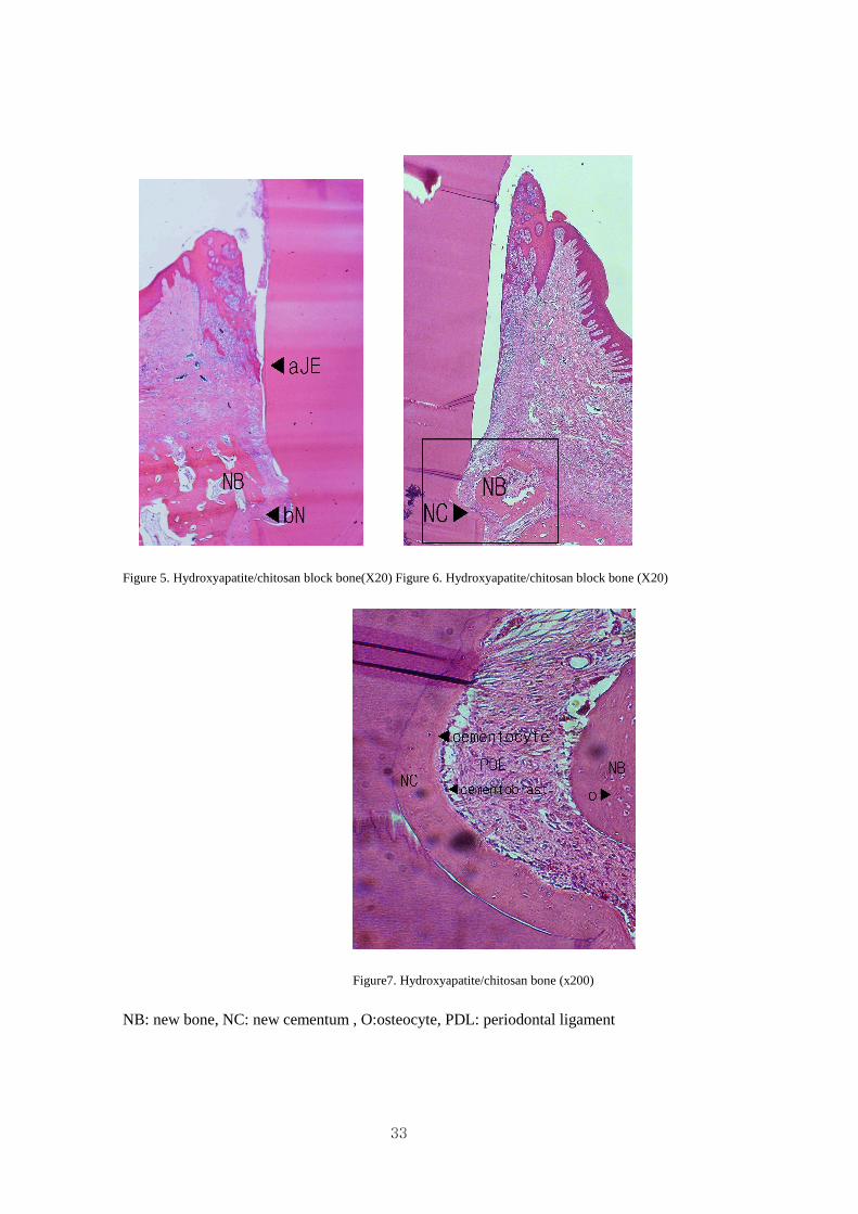
In the Hydroxyapatite/chitosan block bone group, all the Hydroxyapatite/chitosan
block bone was resorbed (Figure 5, 6). In some parts of the slide, osteoblast and
osteoids were observed around the bone marrow (Figure 7). The lamella bone could be
distinguished and periodontal ligament fibers were observed between new bone and
new cementum(Figure 7). In addition some inflammatory cell infiltration was in the
connective tissue. The periodontal ligament fiber orientation was perpendicular to root
surfaces around the reference notch area (Figure 7).
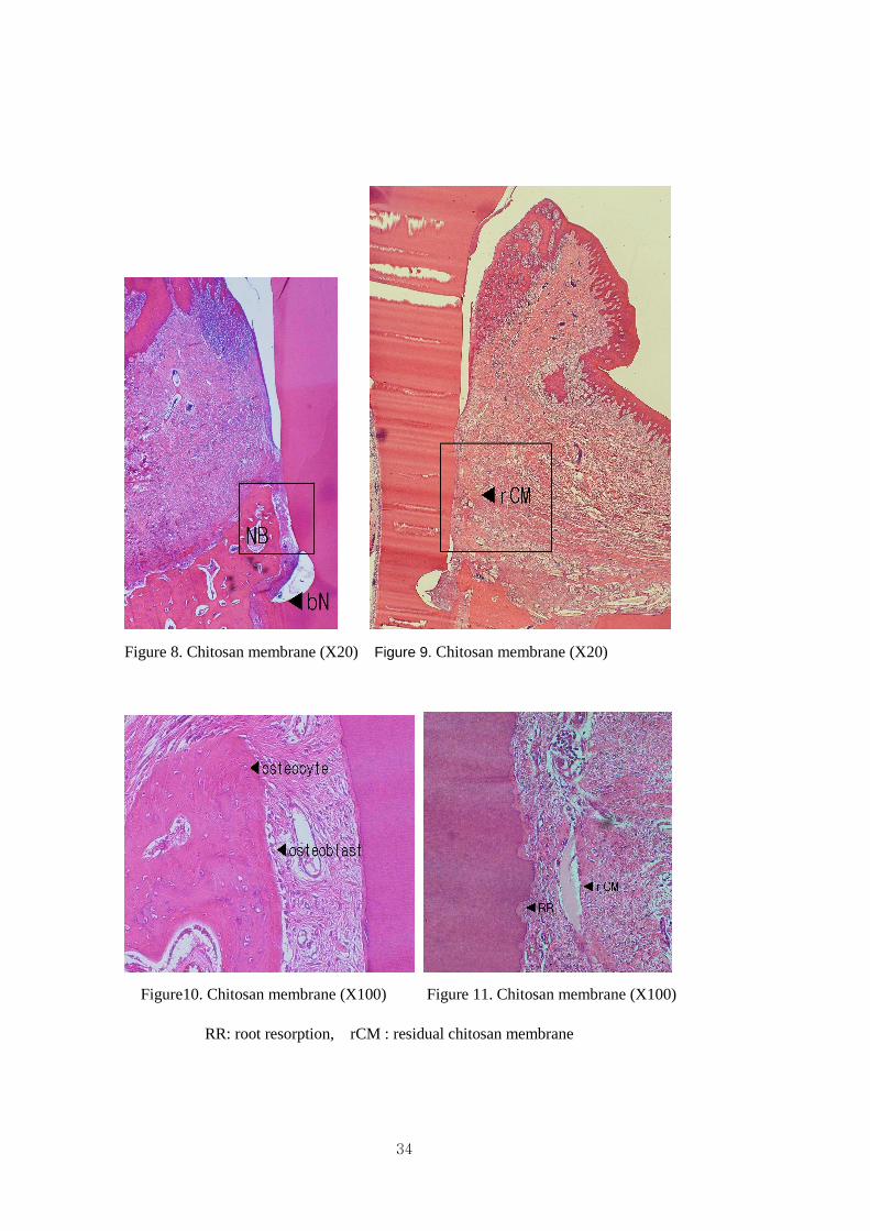
In chitosan membrane group, residual chitosan membrane remnants were observed
at the connective tissue area (Figure 9, 11). In some parts of the slide, osteoblast and
osteoids were observed around the bone marrow (Figure 10). The lamella bone could
be distinguished and periodontal ligament fibers were observed between new bone and
9
new cementum (Figure 10). The inflammatory cell infiltration was lesser than other
groups.
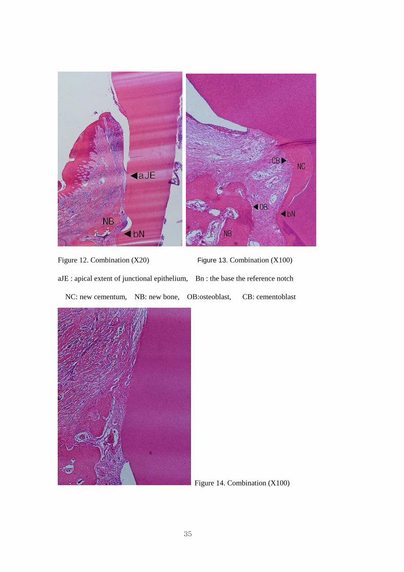
The chitosan membrane and the hydroxyapatite/chitosan block bone group revelaed
similar results to that of the chitosan membrane or block bone group (Figure 12,13,14).
The inflammatory cell infiltration into the connective tissue was greater than chitosan
membrane group.
C. Histometric Analysis
Table 1, Table 2 and Figure 2 show the results of the histometric analysis. The
amount of new alveolar bone formation in the chitosan membrane group was greater
than those of the other groups, and there was a statiscally significant difference
(P<0.05). The amount of new cementum formation of chitosan membrane group was
greater than those of the other groups, but there was no significant difference from
other groups (P>0.05). In apical migration of the junctional epithelium, no statistically
significant difference was observed between the control group and the other groups
(P>0.05). The amount of connective tissue adhesion in surgical control group was
greater than those of the other groups, but there was no significant difference (P>0.05).
(Tables 1, 2, Figure 2)
The amount of alveolar bone regeneration was (0.80 ± 0.18) mm in the control
group, (1.20 ± 0.40) mm in the hydroxyapatite/chitosan block bone group, (1.85 ±
0.66) mm in the chitosan membrane group, and (1.22 ± 0.21) mm in the combination
group, which showed a statisticcally significant difference for the chitosan membrane
group. Chitosan membrane group was a statistically significant different from other
treatment groups(P<0.05). The amount of cemental regeneration was (0.68 ± 1.23)
10
mm in the control group, (1.46 ± 1.58) mm in Hydroxyapatite/chitosan block bone
group, (2.30 ± 0.63) mm in Chitosan membrane group, and (1.13 ± 0.90) mm in
combination group. There was no statistically significant difference among 4 groups
(P>0.05). The amount of epithelial migration was (2.68 ± 1.68) mm in the control
group, (2.18 ± 1.56) mm in the hydroxyapatite/chitosan block bone group, (2.43 ±
0.56) mm in the chitosan membrane group, and (2.55 ± 0.77) mm in the combination
group, which showed no statistically significant difference among the four groups
(P>0.05). The amount of connective tissue adhesion was (1.22 ± 0.71) mm in the
control group, (0.93 ± 1.46) mm in the hydroxyapatite/chitosan block bone group,
(0.27 ± 0.36) mm in the chitosan membrane group, and (0.79 ± 0.83) mm in the
combination group. There was no statistically significant difference among 4 groups
(P>0.05) (Table 1).
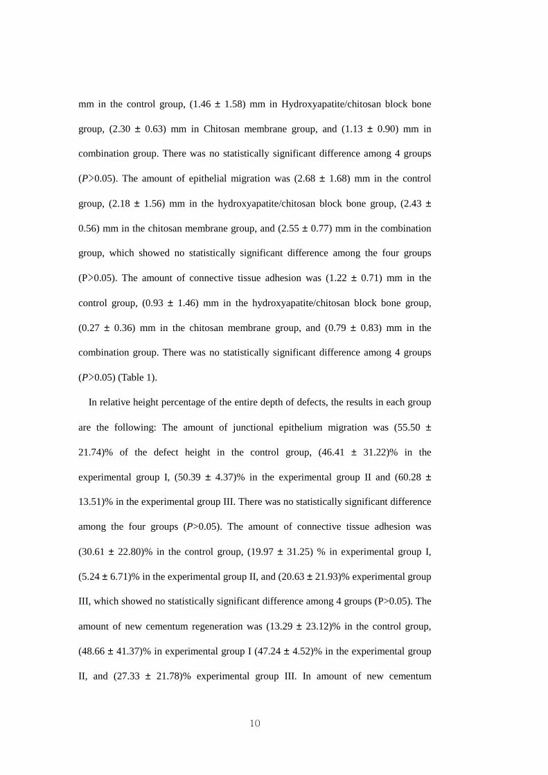
In relative height percentage of the entire depth of defects, the results in each group
are the following: The amount of junctional epithelium migration was (55.50 ±
21.74)% of the defect height in the control group, (46.41 ± 31.22)% in the
experimental group I, (50.39 ± 4.37)% in the experimental group II and (60.28 ±
13.51)% in the experimental group III. There was no statistically significant difference
among the four groups (P>0.05). The amount of connective tissue adhesion was
(30.61 ± 22.80)% in the control group, (19.97 ± 31.25) % in experimental group I,
(5.24 ± 6.71)% in the experimental group II, and (20.63 ± 21.93)% experimental group
III, which showed no statistically significant difference among 4 groups (P>0.05). The
amount of new cementum regeneration was (13.29 ± 23.12)% in the control group,
(48.66 ± 41.37)% in experimental group I (47.24 ± 4.52)% in the experimental group
II, and (27.33 ± 21.78)% experimental group III. In amount of new cementum
11
regeneration, which showed no statistically significant difference among 4 groups
(P>0.05). The amount of new alveolar bone regeneration was (19.01 ± 8.40)% in the
control group, (26.24 ± 9.10)% in experimental group I, (38.79 ± 11.44)% in the
experimental group II, and (29.89 ± 7.92)% in experimental group III. Experimental
group II was a statistically significant difference from the control group (P<0.05)
(Table 2).
The illustration of periodontal healing in percentage of the defect height is shown
in Figure 2.
bN bN bN bN 0000
25252525
50505050
75757575
CEJ 100CEJ 100CEJ 100CEJ 100
ControlControlControlControl Exp IExp IExp IExp I
%%%%
Exp IIExp IIExp IIExp II Exp IIIExp IIIExp IIIExp III
Figure 2. Periodontal healing illustrated in percentage of the defect height.
Control group: surgical control group received a flap operation only
Exp I : Hydroxyapatite/chitosan block bone group,
Exp II: the chitosan membrane group,
Exp III: Hydroxyapatite/chitosan block bone and chitosan membrane
CEJ: cemento-enamel junction, bN : base of the reference notch
Blue: new bone, Green: new cementum, Red: Junctional epithelium
12
IV. DISCUSSION
The ultimate goal of periodontal therapy is to regenerate the supporting tissue that was
destroyed. Although, various procedures such as guide tissue regeneration (Kim et al.,
1996; Kim et al. 1998; Moon et al., 1996; Trombelli et al., 1997), autografts
(Schallhorn, 1972), other bone grafts (Mellonig et al., 1976; Mellonig, 1984; Kim et
al., 1998a; Kim et al., 1998b), and the application of growth factor (Lynch et al., 1989;
Becked et al., 1992; Wikesjö et al., 1999; Choi et al., 2002) has been already
developed and used to help regeneration, each has its shortcomings.
The aim of this study was to evaluate the periodontal repair and the biomaterial
reaction following implantation of a newly fabricated hydroxyapatite-chitosan block
bone and chitosan membrane on the regeneration of 1- wall intrabony defects in the
beagle dogs. Based on the results of Wikesjo et al’s study, which found that naturally
or ligature induced loss of attachment and surgically induced loss of attachment
showed no difference in healing (Wikesjö, 1991).
In previous similar study, the tissue regeneration effect by chitosan was good. The
inhibited apical migration of epithelium and increase in amount of new bone and new
cementum suggest the potency of chitosan in inducing periodontal tissue regeneration
(Park et al., 2003).
The chitosan nonwoven membrane has the potential to support the cementum and
bone regeneration, possibly by providing the conditions needed for guided tissue
regeneration in the one wall intrabony periodontal defects of beagle dogs (Yeo et al.,
2005). The biocompatibility of the chitosan nanofiber membrane was confirmed, with
enhanced bone regeneration and no evidence of an inflammatory reaction. The
experiment shows that the novel biodegradable chitosan nanofiber membrane may be
13
useful as a tool for guided bone regeneration (Shin et al., 2005). Consequently, interest
in the search for a new non-toxic, biodegradable material that would be free from any
side effect has been growing. Among those considered, chitin and its extract, chitosan
(poly-N-acetyl glucosaminoglycan), are attracting particular attention. It was reported
that chitosan has biocompatibility with the host in a previous animal study and the
host progressively reabsorbs chitosan almost 2 months after the treatment (Nakajima
et al., 1986).
An ideal scaffolding material for bone tissue engineering should promote the
expression of the osteoblastic phenotype. Calcium phosphate-chitosan composite
scaffolds have been reported to support the proliferation and differentiation of seeded
oseoblast cells as indicated by heigh alkaline phosphatase activities and the formation
of mineralizes matrices (Lee et al., 2000).
Osteoblasts usually exhibit higher basal levels of alkaline phosphatase, a relatively
early differentiation marker, than do cells that do not produce mineralized
extraxellular matrix, such as fibroblasts (Sein et al., 1990). The in vitro engineering of
bone tissue requires appropriate carriers that allow a three dimensional distribution of
cells. Ishaug et al. suggested that a scaffold material used for bone formation should
meet the following: Osetoblast proliferation and function were not affected by
polymer foam size in the range of 150-710 µm increased over time for all constructs.
Cell seeding density affected initial osteoblast attachment and proliferation rate, but
differences became less significant over time with no measurable difference in
function. Viable cells may be supported for only short distances into the 3-D matrices
under static culture conditions. Achieving cell survival beyond the surface of large 3-D
porous scaffolds may require altering culture conditions to improve delivery of
14
nutrients deep within the constructs while in vivo survival will depend on vascular in
vasion of constructs( Ishaug et al., 1997 ).
A chitosan/collagen sponge applied to the preclinical 1-wall intrabony defects in
beagle dogs was shown to have positive effects on periodontal tissue regeneration. The
inhibited apical migration of the epithelium and the increase in new bone and new
cementum suggests the potency of chitosan in inducing periodontal tissue regeneration
(Park et al., 2003).
In the hydroxyapatite/chitosan block bone group of our study, it was not shown
residual the hydroxyapatite/chitosan block bone remnant. The inflammatory cell
infiltrated into the connective tissue (Fig 6,12). The hydroxyapatite/chitosan block
bone group was not a statistically significant difference from control group. In new
cementum regeneration, the chitosan membrane group was a statistically significant
difference from other group. Our results are different from those of Park et al (Park et
al., 2003). In this study, too much early it became the absorption. It appears that the
hydroxyapatite/chitosan block bone was too much early absorbed and could not role of
scaffold.
In this study, the chitosan membrane group was initially expected to show decidedly
less migration of the epithelium compared to the surgical group and calcium
phosphate chitosan block bone group. Howerever, the results showed no significant
difference in the juctional epithelium migration among the groups. The amount of
junctional epithelium migration was (2.68 ± 1.68) mm ((55.50 ± 21.74)% of the defect
height) in the surgical control group, (2.18 ± 1.56) mm ((46.41 ± 31.22)% of the defect
height) in the hydroxyapatite/chitosan block bone group, 2.43 ± 0.77mm ((50.39 ±
4.37)% of the defect height) in the chitosan membrane group, and (2.55 ± 0.77)
15
mm( (60.28 ± 13.51)% of the defect height) in the combination group(the chitosan
membrane group with the hydroxyapatite/chitosan block bone). There was no
statistically significant difference among 4 groups (P>0.05)(Table 2).
These results are different from those of Park et al (Park et al., 2003). The high rate
of migration indicates the wound instability of the 1-wall intrabony defects. Loss of
tight adaptation of the chitosan membrane during the early postsurgical period may
have contributed to the failure of controlling epithelium migration.This is not similar
to the previous reports (Kim & Chai et al., 1998; Wikesjö et al., 1991).
In the hydroxyapatite/chitosan block bone, results differed from initial expectations.
Since previous studies have reported that collagen has an ability to stabilize the fibrin-
clot on the root surface by concentrating platelets on the site and subsequently
constrain the apical migration of epithelium through their contact-inhibition effects
(Beachey et al., 1979; Mason & Read, 1974; Ueno et al., 1999; Wikesjö & Nilvéus,
1990; Winter, 1974), it was expected that the migration of the epithelium would be
largely impaired. Yet the outcome turned out controversial, the effect varying among
the subjects. This outcome subsequently calls for further research involving more
subjects.
The amount of connective tissue adhesion was (1.22 ± 0.71) mm ((30.61 ± 22.80)%
of the entire depth of defects) in the control group, (0.93 ± 1.46) mm ((19.97 ±
31.25)% of the entire depth of defects) in the hydroxyapatite/chitosan block bone
group, (0.27 ± 0.36) mm ((5.24 ± 6.71)% of the entire depth of defects) in the chitosan
membrane group, and (0.79 ± 0.83) mm ((20.63 ± 21.93)% of the entire depth of
defects) in the combination group. There was no statistically significant difference
among 4 groups (P>0.05) (Table 2).
16
It is believed that the chitosan membrane affects the regeneration of cementum as a
barrier membrane, which inhibits epithelial migration, inducing the differentiation of
undifferentiated mesenchymal cells into cementoblasts, as well as by promoting the
differentiation of osteogenic cells. This study distinguished between two types of
cement regeneration. From the notch to the crest of new bone, the intrabony cementum
showed an arrangement of cementoblasts. However, above the crest, the suprabony
cementum with few cellular elements appeared to be a cementum-like substance with
a parallel fiber adhesion and we could expect that the healing here be by the fibers
with a gingival origin. The intrabony cementum with the perpendicularly inserted
fibers is the true cementum that accompanies new attachment and is healed by the
fibers from a periodontal ligament origin. This result, and previous reports by Barney
et al. (1986) and Moon et al.(1996) confirms that new bone follows the coronal growth
of new cementum. This suggests that cementum assists the formation of new bone and
periodontal ligament fibers. But our results are not always similar to previous study
for regeneration, such as membranes, bone grafts, and growth factors, in which new
cementum increased only in equal amounts to new bone, (Blumental et al., 1986;
Caffesse et al., 1987; Lynch et al., 1989; Mellonig. 1984; Quintero et al., 1982;
Schallhorn et al. 1988).
The amount of cemental regeneration was (0.68 ± 1.23) mm ( (13.29 ± 23.12)% of
the entire depth of defects ) in the control group, (1.46 ± 1.58) mm ((48.66 ± 41.37)%
of the entire depth of defects ) in the hydroxyapatite/chitosan block bone group, (2.30
± 0.63) mm (( 47.24 ± 4.52)% of the entire depth of defects ) in the chitosan
membrane group, and (1.13 ± 0.90) mm ((27.33 ± 21.78)% of the entire depth of
17
defects ) in the combination group. There was no statistically significant difference
among 4 groups (P>0.05) (Table 2).
In alveolar bone regeneation, the chitosan membrane group shows a significantly
larger level of bone regeneration, compared with other groups. The amount of alveolar
bone regeneration was (0.80 ± 0.18) mm ((19.01 ± 8.40)% of the entire depth of
defects) in the control group, (1.20 ± 0.40) mm ((26.24 ± 9.10)% of the entire depth of
defects) in the hydroxyapatite/chitosan block bone group, (1.85 ± 0.66) mm ((38.79 ±
11.44)% of the entire depth of defects) in the chitosan membrane group, and (1.22 ±
0.21) mm ((29.89 ± 7.92)% of the entire depth of defects) in the combination group.
The chitosan membrane group was a statistically significant different from other
treatment groups (P<0.05)(Table 2).
Comparing the amount of new cementum with that of new bone in four groups of
our study, there was not more cementum growth than the bone growth. Our result is
different from Park et al.(2003). In their resuts, cementum regeneration was
significantly high only in the chitosan group. Their result suggests that new bone
follows the coronal growth of new cementum. Park et al. suggests that new cementum
stimulates the formation of new bone and periodontal ligament fibers, and the
conspicuous increment of new cementum in the defects treated with chitosan indicates
efficacy of chitosan in advancing cementum formation by inducing the differentiation
of undifferentiated mesenchymal cells particularly into cementoblasts, as well as by
promoting the differentiation of osteogenic cells. However, our results are different
from their results. Therefore, the further studies for mechanism is necessary.
Newly formed bone trabeculae were generally lined with osteoblast like cells and
osteoids, suggesting continued bone apposition after an 8 weeks observation period
18
(Siguardsson et al. 1994). The coronal thining of new bone apparent in all group
confirms the reports that collagen is deficient in its ability to both make and maintain
the space for cellular regeneration. In this study, only a small amount of new bone was
coronally formed. This result verifies the reports that osteogenesis occurs only within
the given space in periodontal defects and suggests the need for research into other
carrier system.
Root resorption appears to be a common sequela of repair in experimental
periodontal defects. Wikesjö et al. suggested that the initial resorption is caused by
tissue remodeling during periodontal reconstructive surgery (Wikesjö et al., 1991).
Most teeth in the present study exhibited superficial resorption, appearing more
pronounced when the connective tissue was directly opposed to dentin rather than
when the root surface was covered by cementum. This is in agreement with the
suggestion that cementum matrix formation may prevent resorption
Ankylosis was not observed in any of the 4 groups. Ankylosis often occurs in faster
sites of osteogenesis without the regeneration of the periodontal ligaments. Caffesse et
al. noted that the periodontal ligament cells are far faster in repopulation the root
surface than osteogenic cells (Caffesse et al., 1987). Similarly, these results revealed
how the early colonization of defects by undifferentiated periodontal ligament cells
prevented ankylosis.
The results of the present study did not appear to support the potential of chitosan to
enhance bone formation, but still show low load bearing capacity. Therefore, the
additional study to enhance the mechanical properties appears to be necessary.
19
V. CONCLUSION
This study evaluated the periodontal repair and biomaterial reaction following
implantation of a newly fabricated hydroxyapatite/chitosan block bone and/or chitosan
membrane on the regeneration of 1- wall intrabony defects in the beagle dogs. In
chitosan membrane group, the amount of new bone formation was greater than the
other groups, which suggests that the potency of chitosan membrane induce the new
bone regeneration. The results of the present study did support the potential of
chitosan membrane in the guided tissue regeneration, but did not showed enough
capacity to regeneration of new cementum. Therefore, the further study is to be
necessary.
20
REFERENCES
Alberius P, Dahlin C, Linde A: Role of osteopromotion in experimental bone grafting
to the skull: a study in adult rats using the membrane technique. J Oral and Maxillofac
Surg 50: 829-834, 1992.
Barney VC, Levin MP, Adams DF: Bioceramic implants in surgical periodontal
defects: A comparison study. J Periodontol 57: 764-769, 1986.
Beachey EH., Chiang TM, Kang AHL, Collagen platelet interaction. Int. Review of
Connective Tissue Research 8: 1-21, 1979.
Becker W, Becker B, Berg L: New attachment after treatment with root isolation
procedure: report for treated Class III and Class II furcation and vertical osseous
defects. Int.J.Periodont. Res.Dent 8: 2-16, 1988.
Becker W, Becker B: Treatment of mendibular 3-wall intrabony defects by flap
debridement and expanded polytetra fluoroethylene barrier membranes. Long term
evaluation of 32 treated patients. J Periodontol 64: 1138-1144, 1993.
Becker W, Lynch SE, Lekholm U, Becker B, Caffesse R, Donath C, Sanchez R: A
comparison of PTFE membranes alone or in combination with platelet derived growth
factor-I, or demineralized freeze-dried bone in promoting bone formation around
immediate extraction socket implants: A study in dogs. J Periodontol 63: 929-940,
1992.
21
Blumental NM: A clinical comparison of collagen membranes with e-PTFE
membranes in the treatment of human mandibular buccal class II furcation defects. J
Periodontol 64: 925-933, 1993.
Blumenthal NM, Sabet, T, Barrington E: Healing responses to grafting of combined
collagen decalcified bone in periodontal defects in dogs. J Periodontology 57: 84-90,
1986
Cabib E: The synthesis and degradation of chitin. Advances in Enzymology and
Related Areas of Molecular Biology 59:59-101, 1987
Caffesse RG, Quinones CR: Polypeptide growth factors and attachment proteins in
periodontal wound healing and regeneration. Periodontol 2000 2:69-79, 1993.
Caffesse RG, Smith BA, Castlelli WA, Lopatin DE: Cell proliferation after flap surgery,
root conditioning and fibronectin application. J Periodontol 58:661-666, 1987.
Choi SH, Kim CK, Cho KS, Huh JS, Sorensen RG, Wozney JM, Wikesjö UME: Effect
of rhBMP-2/ACS on healing in 3-wall intrabony defects in dogs. J Periodontol 73: 65-
74, 2002
Dahlin C, Alberius P, Linde A: Osteopromotion for cranioplasty. An experimental
study in rats using a membrane technique. J Neurosurg 74: 487-491, 1991.
22
de Queiroz AA, Ferraz HG, Abraham GA, del Mar Fernandez M, Bravo AL, Roman
JS: Development of new hydroactive dressings based on chitosan membranes:
characterization and in vivo behavior. J Biomed Mat Res 64A: 147-154, 2003
Fukuta K, Har-Shai Y, Collares MV, Jackson IT: Comparison of inorganic bovine bone
mineral particles with porous hydroxylapatite granules and cranial bone dust in the
reconstruction of full-thickness skull defect. J Craniofac Surg 3: 25-29, 1992.
Gottlow J, Nyman S, Karring T, Wennstrom J: New attachment formation in the
human periodontium by guided tissue regeneration. Case reports, J Clin Periodontol
13(6): 604-616, 1986
Handelsman M, Davarpanah M, Celletti R: Guided tissue regeneration with and
without citric acid treatment in vertical osseous defects. Int J Periodont Res Dent, 11:
350-363, 1991.
Hislop WS, Finlay PM, Moos KF, A preliminary study into the uses of anorganic
bone in oral and maxillofacial surgery. British J Oral Maxillofac Surg 31: 149-153,
1993
Ishaug SL, Crane GM, Miller MJ, Yasko AW, Yaszemski MJ, Mikos AG: Bone
formation by three-dimensional stromal osteoblast culture in biodegradable polymer
scaffolds. J Biomed Mater Res 36: 17-28, 1997
23
Karring T, Nyman S, Gottlow J, Laurell L: Development of the biological concepts of
guided tissue regeneration - animal and human studies. Periodontol 2000 1: 26-35,
1993.
Kim CK, Chai JK, Cho KS, Choi SH: Effect of calcium sulfate on the healing of
periodontal intrabony defects. Int Dent J 48: 330-337, 1998a.
Kim CK, Chai JK, Cho KS, Moon IS, Choi SH, Sottosanti JS, Wikesjö UME:
Periodontal repair in dogs: Effect of allogenic freeze-dried demineralized bone matrix
implants on alveolar bone and cementum regeneration. J Periodontol 69: 26-33, 1998b.
Kim CK, Choi EJ, Cho KS, Chai JK, Wikesjö UME: Periodontal repair in intrabony
defects treated with a calcium carbonate implant and guided tissue regeneration. J
Periodontol 61: 1301-1306, 1996.
Kim CK, Kim HY, Choi EJ, Chai JK, Cho ks, Moon IS, Choi SH, Sottosanti JS,
Wikesjö UME: Effect of calcium sulfate implant with calcium sulfate barrier on
periodontal healing in 3 wall intrabony defects in dogs. J Periodontol 69: 982-988,
1998.
Klinge B, Alberius P, Isaksson S, Jönsson J: Osseous response to implanted natural
bone mineral and synthetic hydroxylapatite ceramic in the repair of experimental skull
bone defects. J Oral Maxillofac Surg 50: 241-249, 1992.
24
Lee YM, Park YJ, Lee SJ, Ku Y, Han SB, Klokkevold PR, Chung CP: The bone
regenerative effects of platelet/tricalcium phosphate sponge carrier. J Peridontol 71:
418-424, 2000.
Lee YM, Park YJ, Lee SJ, Ku Y, Han SB, Choi SM, Klokkevold PR, Chung CP:
Tissue engineered bone formation using chitosan/tricalcium phosphate sponge carrier.
J Periodontol 71: 410- 417, 2000.
Lynch SE, Williams RC, Polson AM, Howell TH, Reddy MS, Zappa, UE,
Antoniades HN: A combination of platelet derived and insulin-like growth factors
enhances periodontal regeneration. J Clinic Periodontol 16: 545-548, 1989.
Madihally SV, Matthew HW: Porous chitosan scaffolds for tissue engineering.
Biomaterials 20:1133-1142, 1999.
Magnusson I, Batich C, Collins BR: New attachment formation following controlled
tissue regeneration using biodegradable membranes. J Periodontol. 59: 1-6, 1988.
Mason RG, Read MSL: Some effects of a microcrystalline collagen preparation on
blood. Hemostasis 3: 31-45, 1974.
Mellonig TJ, Browers GM, Bright RW, Lawrence JT: Clinical evaluation of freeze-
dried bone allograft in periodontal osseous defects. J Periodontol 47:125-131, 1976.
25
Mellonig TJ, decalcified freeze-dried bone allografts as an implant material in human
periodontal defects. Int J Peiodont rest Dent 6: 41-45, 1984
Michael D, Weir Hockin HK, Xu Carl G., Simon Jr: Strong calcium phosphate cement-
chitosan-mesh construct containing cell-encapsulating hyerogel beads for bone tissue
engineering. J Biomed Mater Research 77A: 487-496, 2006.
Mizuno K, Yamamura K, Yano K, Osada T, Saeki S, Takimoto N, Sakurai T, Nimura
Y: Effect of chitosan film containing basic fibroblast growth factor on wound healing
in genetically diabetic mice. J Biomed Mater Res 64A: 177-181, 2003.
Moon IS, Chai JK, Cho KS, Wikesjö UME, Kim CK: Effects of polyglycan mesh
combined with resorbable calcium carbonate or replamineform hydroxapatite on
periodontal repair in dogs. J clin Periodontol 23: 945-951, 1996.
Mukherjee DP, Tunkle AS, Roberts RA, Clavenna A, Rogers s, Smith D: An animal
evaluation of a paste of chitosan glutamate and hydroxyapatite as a synthetic bone
graft material. J Biomed Mater Res 67B: 603-609, 2003.
Muzzarelli R, Baldassarre M., Conti F, Ferrare P, Biagini B: Biological activity of
chitosan: ultrastructural study. Biomaterials 14: 39-43, 1988.
Muzzarelli, R, Biagini G, Pugnaloni A, Filippini O, Baldassarrc V, Castaldini C,
26
Rizzoli C: Reconstruction of parodontal tissue with chitosan. Biomaterials 10: 598-
603, 1989.
Nakajima M, Atsumi K, Kifume K, Miupa K, Kunanaru M: Chitin is an effective
material for sutures. Japan J Surg 236: 313-325, 1986.
Nyman S, Lindhe J, Karring T, Rylander H: New attachment following surgical
treatment of human periodontal disease. J Clin Periodontol 9(4): 290-296, 1982.
Park J-S, Choi S-H, Moon I-S, Cho K-S, Chai J-K, Kim C-K: Eight-week historical
analysis on the effect of chitosan on surgically created one wall intrabony defects in
beagle dogs. J Clin Periodontol 30: 443-453, 2003.
Pinholt EM, Bang G, Haanacs HR: Alveolar ridge augumentation in rats by Bio-Oss.
Scandinavian J Dent Resear 99:154-161, 1991.
Pontoriero R, Lindhe J, Nyman S, Karring T, Rosenberg E, Sanavi F: Guided tissue
regeneration in degree II furcation- involved mandibular molar. A Clinical study. J
Clin periodontal 15:247-254, 1988.
Quintero G, Mellonig JT, Gambill VM, Pelleu GBJr: A six-months clinical evaluation
of decalcified freeze-dried bone allografts in periodontal osseous defects. J
Periodontol 53: 726-730. 1982.
27
Risbud MV, Bhonde MR, Bhonde RR: Effect of chitosan-polyvinyl pyrrolidone
hydrogel on proliferation and cytokine expression of endothelial cells: implications in
islet immunoisolation. J biomed Mater Res 57: 300-305, 2001.
Schallhorn RG,: Postoperative problems associated with iliac transplants. J
Periodontol 43: 3-9, 1972
Schallhorn RG, McClain PK, Combined osseous composite grafting, root
conditioningand guided tissue regeneration. Int J Periodont Rest Dent 8: 8-31, 1988.
Schroeder, HE: Biologic problems of regenerative cementogenesis: Synthesis and
attachment of collagenous matrices on growing and established tooth surface, Int
Review Cytology/A Survey of Cell Biology 142: 1-52, 1992.
Selvig KA, Kersten BG, Wikesjö UME: Surgical treatment of intrabony periodontal
defects using ePTFE barrier membranes: Influence of defect configuration on healing
response. J Periodontol 64: 730-733, 1993.
Shin SY, Park HN, Kim KH, Lee MH, Choi YS, Park YJ, Lee YM, Ku Y, Rhyu IC,
Han SB, Lee SJ, Chung CP: Biological evaluation of chitosan nanofiber membrane for
guided bone regeneration. J. Periodontol. 76(10): 1778-1784, 2005.
Sigurdsson TJ, Hardwick R, Bogle GC, Wikesjö, UME: Periodontal repair in dogs:
Space provision by reinforced ePTFE membranes enhances bone and cementum
28
regeneration in large supraalveolar defects. J Periodontol 65: 350-356, 1994.
Stein GS, Lian JB, Owen TA: Relationship of cell growth to the regulation of tissue-
specific gene expression during osteoblast differentiation. The FASEB J 4: 3111-3123,
1990.
Suk HJ, Kwon SH, Kim CS, Choi SH, Jeon DW, Kim CK: Resorbability and
histological reaction of bioabsorbable membranes. J Korean Acad Periodontol 32:
781-800, 2002.
Trombelli L, Kim CK, Zimmerman GJ, Wikesjö UME: Retrospective analysis of
factors related to clinical outcome of guided tissue regenerationprocedures in
intrabony defects. J clinic Periodontol 24: 366-371, 1997.
Ueno H., Yamada H., Tanaka I, Kaba N, Matsuura M, Okumura M, Kadosawa T,
Fujinaga T: Accelerating effects of chitosan for healing at early phase of experimental
open wound in dogs. Biomaterials 20: 1407-1414, 1999.
Wang L, Khor E, Wee A, Lim LY: Chitosan-alginate PEC membrane as awound
dressing: Assessment of incisional wound healing. J Biomed Mater Res 63: 610-618,
2002;
Young-Ju Yeo, Dong-Won Jeon, Chang-sung Kim, Seong-Ho Choi, Kyoo-Sung Cho,
Yong-Keun Lee, Chong-Kwan Kim: Effects of Chitosan Nonwoven Membrane on
29
Periodontal Healing of Surgically Created One-wall Intrabony Defects in Beagle Dogs,
J Biomed Mater Res Part B : Appl Biomater 72B: 86-93, 2005.
Wikesjö UME, Guglielmoni P, Promsudthi A, Cho KS, Trombelli L, Selvig KA, Jin L,
Wozney JM: Periodontal repair in dogs: Effect of rhBMP-2 concentration on
regeneration of alveolar bone and periodontal attachment. J Clinic Periodontol 26:
392-400, 1999.
Wikesjö UME, Nilvéus RE, Periodontal repair in dogs: Effect of wound stabilization
on healing. J Periodontol 61: 719-724, 1990.
Wikesjö UME, Nilvéus RE: Periodontal repair in dogs Healing patterns in large
circumferential periodontal defects. J Clin. Periodontol. 18: 49-59, 1991
Winter GD, Transcutaneous implants: Reactions of the skin-implant interface. Journal
of Biomedical Materials Research 1974;8: 99-113.
Yeo YJ, Jeon DW, Kim CS, Choi SH, Cho KS, Lee YK, Kim CK: Effects of chitosan
nonwoven membrane on periodontal healing of surgically created one-wall intrabony
defects in beagle dogs. J Biomed Mater Res 72: 86-93, 2005.

30
Table 1. Histometric Analysis: Height (mean ± standard deviation (mm))
DH NB NC JE CT
Control 4.61 ± 1.16 0.80 ± 0.18 0.68 ± 1.23 2.68 ± 1.68 1.22 ± 0.71
Exp I 4.58 ± 0.42 1.20 ± 0.40 1.46 ± 1.58 2.18 ± 1.56 0.93 ± 1.46
Exp II 4.84 ± 1.11 1.85±0.66* 2.30 ± 0.63 2.43 ± 0.56 0.27 ± 0.36
Exp III 4.18 ± 0.53 1.22 ± 0.21 1.13 ± 0.90 2.55 ± 0.77 0.79 ± 0.83
Control group: surgical control group received a flap operation only
Exp I : Hydroxyapatite/chitosan block bone group.
Exp II : the chitosan membrane group.
Exp III : Combination group of Hydroxyapatite/chitosan block bone and chitosan membrane.
* a statistically significant difference from other group (P<0.05).
DH: defect height; JE: junctional epithelium migration; CT: connective tissue adhesion; NC:
new cementum regeneration; NB: new bone regeneration
31
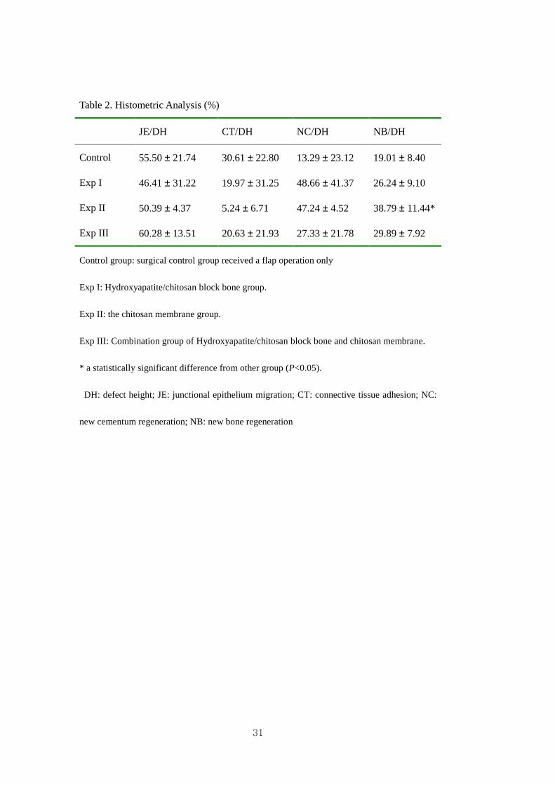
Table 2. Histometric Analysis (%)
JE/DH CT/DH NC/DH NB/DH
Control 55.50 ± 21.74 30.61 ± 22.80 13.29 ± 23.12 19.01 ± 8.40
Exp I 46.41 ± 31.22 19.97 ± 31.25 48.66 ± 41.37 26.24 ± 9.10
Exp II 50.39 ± 4.37 5.24 ± 6.71 47.24 ± 4.52 38.79 ± 11.44*
Exp III 60.28 ± 13.51 20.63 ± 21.93 27.33 ± 21.78 29.89 ± 7.92
Control group: surgical control group received a flap operation only
Exp I: Hydroxyapatite/chitosan block bone group.
Exp II: the chitosan membrane group.
Exp III: Combination group of Hydroxyapatite/chitosan block bone and chitosan membrane.
* a statistically significant difference from other group (P<0.05).
DH: defect height; JE: junctional epithelium migration; CT: connective tissue adhesion; NC:
new cementum regeneration; NB: new bone regeneration
32
Figures
Figure 3. Control group (X20) Figure 4. Control group (X100)
aJE : apical extent of junctional epithelium, bN : the base of the reference notch
CT: connective tissue, NB: new bone
33
Figure 5. Hydroxyapatite/chitosan block bone(X20) Figure 6. Hydroxyapatite/chitosan block bone (X20)
Figure7. Hydroxyapatite/chitosan bone (x200)
NB: new bone, NC: new cementum , O:osteocyte, PDL: periodontal ligament
34
Figure 8. Chitosan membrane (X20) Figure 9. Chitosan membrane (X20)
Figure10. Chitosan membrane (X100) Figure 11. Chitosan membrane (X100)
RR: root resorption, rCM : residual chitosan membrane
35
Figure 12. Combination (X20) Figure 13. Combination (X100)
aJE : apical extent of junctional epithelium, Bn : the base the reference notch
NC: new cementum, NB: new bone, OB:osteoblast, CB: cementoblast
Figure 14. Combination (X100)
36
국문요약국문요약국문요약국문요약
성견성견성견성견 1111면면면면 골골골골 결손부에서결손부에서결손부에서결손부에서 하이드록시아파타이트하이드록시아파타이트하이드록시아파타이트하이드록시아파타이트----키토산키토산키토산키토산
block boneblock boneblock boneblock bone과과과과 키토산키토산키토산키토산 차단막차단막차단막차단막 의의의의 치주조직치주조직치주조직치주조직 재생에재생에재생에재생에
대한대한대한대한 연구연구연구연구
< 지도교수 최성호최성호최성호최성호 >
연세대학교 대학원 치의학과
김김김김 성성성성 구구구구
키토산을 이용한 재료는 치주 조직 재생 및 신생골 형성에 효과적인 물질로
알려져 왔다. 이 연구는 새로 개발된 Hydroxyapatite-chitosan block bone 과
Chitosan membrane 을 이용하여, 성견 1 면 골내낭에서의 치주조직 재생에
미치는 영향을 조사하였다. 4X4mm 1 면 골 결손부에 Hydroxyapatite-chitosan
block bone 을 이식한 후 조직유도재생술을 시행한 군과, Chitosan membrane 을
이용하여 조직유도재생술을 시행한 군, 그리고 두 가지를 모두 사용한
군(combination 군)을 치은박리소파술만 시행한 군과 비교하여 다음과 같은 결과를
얻었다. Chitosan membrane 을 사용한 군은 Hydroxyapatite-chitosan block
bone 만을 이용한 군 혹은 Chitosan membrane 과 함께 사용한 군 그리고
치은박리소파술만 사용한 군에 비해 신생골 형성에 있어서 통계학적으로 유의성
있는 증가량의 차이를 보였다 (P<0.05) (Kruskal Wallis test). 신생백악질과
신생골의 생성량은 연관성이 있었다. 기존의 연구결과들은 신생골 생성량이
신생백악질의 생성량을 따르는 양상이었으나 이번 연구결과는 일정하지
않았으므로, 이에대한 연구가 필요하다고 사료된다. Hydroxyapatite-chitosan
block bone 만을 사용한 군과 이것을 Chitosan membrane 과 함께 사용한 군은
37
치은박리소파술만 사용한 군에 대해 신생골과 신생백악질의 생성에 있어서 수치상
우월적 차이를 보였으나 통계학적 유의성 있는 차이를 보이진 않았다 (p>0.05).
이러한 연구결과로 볼 때, Hydroxyapatite-chitosan block bone 보다는 Chitosan
membrane 이 조직 유도 재생에 보다 긍정적인 효과를 보인다는 것을 알 수 있다.
이는 그간, 발표된 충전방식의 키토산을 이용한 이식재의 결과와 다소 상충되는
결과로 판단된다. 따라서, 조직재생에 대한 키토산을 이용한 충전식 접근은 앞으로
연구가 더욱 필요하다고 판단된다. Chitosan membrane 은 신생골형성에 있어서
양호한 결과를 얻을 수 있었다. 그러나, 임상에서 적용되기 위해서는 앞으로
연구가 더욱 필요하다고 사료된다.
핵심되는핵심되는핵심되는핵심되는 말말말말: 조직유도재생, 하이드록시아파타이트 키토산 블록형 골, 키토산 차단막