Embed Size (px)
Citation preview

The effect of processing techniques on the microbiological and nutritional qualities of the leafy vegetables Vigna unguiculata and Moringa oleifera grown in South Africa
by
OLUWATOBI SARAH OTUN
submitted in accordance with the requirements for the degree of
MASTER OF SCIENCE
In the subject
LIFE SCIENCES
at the
University of South Africa
SUPERVISOR: PROF J DEWAR
CO-SUPERVISORS: DR N DLAMINI
FEBRUARY 2015
ii
DEDICATION
This project is dedicated to God, the author and the finisher of my faith, for the wisdom,
strength and support He’s given me to successfully complete this research, and to my
husband (Stephen) and my children (King, Anointed and Olive-virtue) for their immeasurable
support, love and understanding.
iii
DECLARATION
Student number: 50828576
I declare that The effect of processing techniques on the microbiological and
nutritional qualities of the leafy vegetables Vigna unguiculata and Moringa oleifera
grown in South Africa is my own work and that all the sources that I have used or quoted
have been indicated and acknowledged by means of complete references.
15.09.2014
SIGNATURE DATE
(Mrs Sarah O. Otun)
iv
ABSTRACT
Cowpea (Vigna unguiculata) and moringa (Moringa oleifera) are nutritious and medicinal
vegetables, but could also harbor harmful microbial contaminants.
The main aim of the project was to determine the effect of each processing techniques on the
microbiology, proximate nutrients and shelf life of these vegetables to produce nutritious,
tasty, safe and long lasting vegetable products.
The processing techniques used were: washing, blanching, and drying. Leaf samples were
collected at each stage of processing and were analysed for total viable count, coliform
count, yeast and mould count and nutritional content.
Microbial plate analysis showed the presence, particularly on cowpea leaves, of yeasts and
bacteria such as Pseudomonas, Klebsiella, Staphylococci, Streptococci, and enterobacter
including enteropathogens such as Salmonella spp., Shigella dysenteriae and E coli. The
presence of E.coli on the leaves was also confirmed using polymerase chain reaction-
amplified ribosomal DNA analysis. The most effective processing technique which reduced
microbial load to below SABS standards while retaining nutritional quality was the washing of
the leaves twice with tap water followed by steam tunnel blanching at 94oC for 12 minutes.
Oven drying the leaves at 60oC gave satisfactory and extended shelf life results. Proximate
analysis comparison of the two leaf types showed that on average moringa leaves contained
more ash (2.37 vs 1.1 g), protein (6,9 vs 3,6 g), fat (0,41 vs 0.2 g) and energy (305,1 vs 70
KJ) but less dietary fibre (0,9 vs 7,5 g) than cowpea leaves. No significant differences were
noted in these values following washing and steam blanching. These results indicate that
washing of these leaves is effective as to reducing microbial load and maintaining proximate
values in the short term (up to 4 days) but that oven drying is effective for longer-term
storage.
Key Words: Cowpea, moringa, vegetables, nutritional content, microbiological analysis,
processing techniques, pathogen, coliform, yeast, shelf life.
v
ACKNOWLEDGEMENTS
According to a wise saying from Mahatma Gandhi “Live as if you were to die tomorrow, learn
as if you were to live forever.” Today I am filled with so much joy that I took that huge step to
further my studies despite all oppositions I encountered before and during my research.
Words alone cannot describe my utmost gratitude to God, for his grace, mercy, love, and
favor He bestowed on me through-out my academic journey, all I can say is “THANK YOU
LORD” There were so many people who helped me along the way and my gratitude is
beyond measure.
It would have been just been a mere dream that would never have been actualised if I
did not have the support of UNISA (for bursary funding) and CSIR.
A big thank you to Prof. John Dewar, meeting you has been a blessing to my life - you
have been an answer to my prayers. Thank you for believing so much in me even when I had
no faith in myself. Your academic, moral, and financial support kept me strong all through.
To Dr. Nomusa Dlamini, you have always been there for me, making sure that I am as
comfortable as possible even under the heaviest workload.
To Ms. Annali Jacobs, words alone cannot express how grateful I am to you, for all the
late nights and extra time you had to put in my research especially in the Laboratory and
proof-reading of my write-up. Thank you so much.
Dr Luke Menlo, Dr Zodwa Mambo, Dr Kyahaletu Ntushelo, I am so grateful to each and
every one of you for the role you played in my career. You all took time out of your busy
schedule to mentor and assist me with my research. May you continue with these good
deeds because it will surely be rewarded by God and posterity.
To my husband, Pastor Stephen Otun who supported and encouraged me throughout
the studies. He was always optimistic and involved in my studies to the point of becoming a
lover of all moringa products.
vi
My children; King, Anointed and Olive-virtue whom I thank so much for their motivation.
Just because they look up to me, I could not afford to fail.
To my mom; you are the best. Thank you for all the sacrifice you made (single-
handedly) to give me the best education. Even for travelling down from Nigeria to help ease
the burden of taking care of my kids during the last few months of this study.
Finally, to all members of “The Prayer Stronghold International Ministries” family friends
and course mates, office mates, I wish to express my heartfelt appreciation to you for your
prayers and support throughout my academic pursuit.
vii
TABLE OF CONTENTS DEDICATION ii
DECLARATION iii
ABSTRACT iv
ACKNOWLEDGEMENTS v
TABLE OF CONTENTS vii
LIST OF FIGURE xi
LIST OF TABLES xii
ABBREVATIONS AND ACRONYMS xiii
CHAPTER 1 1
1.1. Introduction 1
1.2. Problem Statement 2
1.3. Aim 3
1.4. Objectives 3
CHAPTER 2 4
Literature Review 4
2.1. Cowpea (Vigna unguiculata) 4
2.1.1. Scientific Classification 4
2.1.2. General background and History 5
2.1.3. Background 5
2.1.4. Uses of cowpea plant 6
2.1.5. Medicinal Value 7
viii
2.2. Moringa Leaves (Moringa oleifera) 8
2.2.1. General 8
2.2.2. Classification 8
2.2.3. Cultivation 9
2.2.4. Moringa oleifera In South Africa 10
2.2.5. Uses of Moringa oleifera plant 10
2.3. General background on the processing and analysis of leafy vegetables 13
2.3.1. Processing Techniques 13
2.3.2 Microbiological analysis 15
2.3.2.1 Microflora: 15
CHAPTER 3 18
Research Methodology: Part A 18
3.1. Processing Techniques 18
3.1.1. Harvesting of leafy vegetables 18
3.1.2. Receiving of cowpea and moringa leaves: 18
3.1.3. Sorting of leafy vegetables: 19
3.1.4. Washing of the leafy vegetables: 19
3.1.5. Blanching 20
3.1.6. Drying 21
Research Methodology: Part B 23
3.2. Microbiological Analyses 23
3.2.1. Microbial Enumeration and Isolation 23
3.2.2. Gram Staining 25
ix
3.3. Shelf – life study 26
3.4. Biochemical Analysis 27
Research Methodology: Part C 29
3.5. Proximate Analysis 29
3.5.1. Moisture Content 29
3.5.2. Ash content 29
3.5.3. Fat Content 31
3.5.4. Protein Content 31
3.5.5. Carbohydrate Content 31
3.5.6. Crude Fibre Content 31
3.5.7. Energy Content (by calculations) 31
3.6. Sensory Evaluation 32
Research Methodology: Part D 33
Molecular Biology 33
3.7. Identification and Isolation of Coliforms 33
3.7.1. Molecular Analysis 33
3.7.2. DNA extraction 34
3.7.3. DNA quantification 34
3.7.4. Polymerase Chain Reaction 35
3.7.5. Electrophoresis 36
CHAPTER 4 38
RESULTS 38
DISCUSSION 53
x
CONCLUSION 57
RESEARCH DATA ANALYSES 58
ETHICAL CONSIDERATIONS 58
QUALITY ASSURANCE 59
REFERENCES 60
APPENDIX 1 67
APPENDIX 2 69
APPENDIX 3 71
xi
Table of Figures
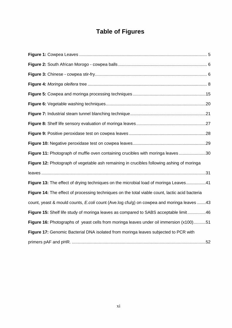
Figure 1: Cowpea Leaves ......................................................................................................... 5
Figure 2: South African Morogo - cowpea balls ......................................................................... 6
Figure 3: Chinese - cowpea stir-fry ............................................................................................ 6
Figure 4: Moringa oleifera tree .................................................................................................. 8
Figure 5: Cowpea and moringa processing techniques ............................................................15
Figure 6: Vegetable washing techniques ..................................................................................20
Figure 7: Industrial steam tunnel blanching technique ..............................................................21
Figure 8: Shelf life sensory evaluation of moringa leaves .........................................................27
Figure 9: Positive peroxidase test on cowpea leaves ...............................................................28
Figure 10: Negative peroxidase test on cowpea leaves ............................................................29
Figure 11: Photograph of muffle oven containing crucibles with moringa leaves ......................30
Figure 12: Photograph of vegetable ash remaining in crucibles following ashing of moringa
leaves .......................................................................................................................................31
Figure 13: The effect of drying techniques on the microbial load of moringa Leaves ................41
Figure 14: The effect of processing techniques on the total viable count, lactic acid bacteria
count, yeast & mould counts, E.coli count (Ave.log cfu/g) on cowpea and moringa leaves .......43
Figure 15: Shelf life study of moringa leaves as compared to SABS acceptable limit ...............46
Figure 16: Photographs of yeast cells from moringa leaves under oil immersion (x100) ..........51
Figure 17: Genomic Bacterial DNA isolated from moringa leaves subjected to PCR with
primers pAF and pHR. ..............................................................................................................52
xii
List of Tables
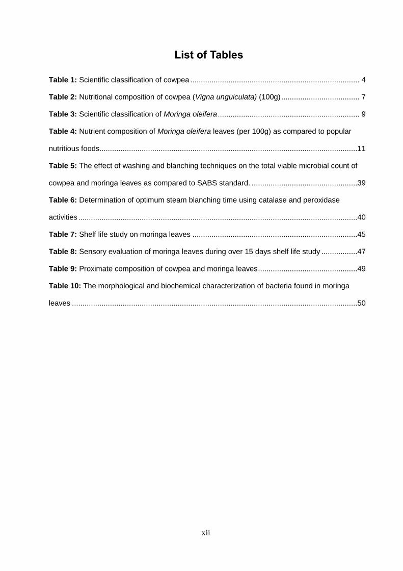
Table 1: Scientific classification of cowpea ................................................................................ 4
Table 2: Nutritional composition of cowpea (Vigna unguiculata) (100g) ..................................... 7
Table 3: Scientific classification of Moringa oleifera ................................................................... 9
Table 4: Nutrient composition of Moringa oleifera leaves (per 100g) as compared to popular
nutritious foods..........................................................................................................................11
Table 5: The effect of washing and blanching techniques on the total viable microbial count of
cowpea and moringa leaves as compared to SABS standard. ..................................................39
Table 6: Determination of optimum steam blanching time using catalase and peroxidase
activities ....................................................................................................................................40
Table 7: Shelf life study on moringa leaves ..............................................................................45
Table 8: Sensory evaluation of moringa leaves during over 15 days shelf life study .................47
Table 9: Proximate composition of cowpea and moringa leaves ...............................................49
Table 10: The morphological and biochemical characterization of bacteria found in moringa
leaves .......................................................................................................................................50
xiii
ABBREVATIONS AND ACRONYMS
oC degree Celsius
% Percentage
+ve Positive
-ve Negative
AOAC Association of Official Analytical Chemists
Ave Average
CDC Center of Disease Control
cm Centimetre
CSIR Council for Scientific and Industrial Research
f Frequency
g Gram
H2O Water
hrs Hours
HPLC high performance liquid chromatography –
kg Kilogram
L Litre
min Minutes
ml Millilitre
mM milli-molar
mg Milligram
PCR Polymerase chain reaction
pH hydrogen ion concentration / measure of alkalinity or
acidity
Prep Preparative
rpm revolutions per minute
RSA Republic of South Africa
xiv
SABS South African Bureau of Standards
SANAS South African National Accreditation System
SANBI South African National Biodiversity Institute
sec Seconds
UNISA University of South Africa
UV Ultraviolet
1
CHAPTER 1
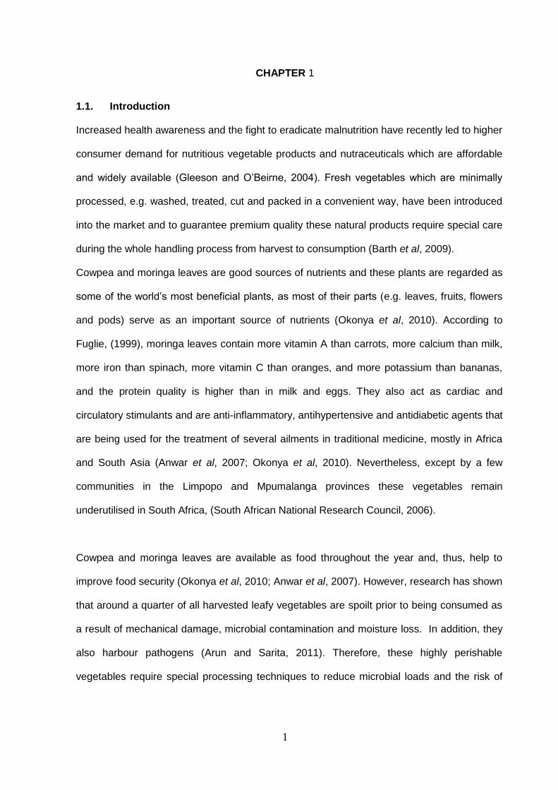
1.1. Introduction
Increased health awareness and the fight to eradicate malnutrition have recently led to higher
consumer demand for nutritious vegetable products and nutraceuticals which are affordable
and widely available (Gleeson and O’Beirne, 2004). Fresh vegetables which are minimally
processed, e.g. washed, treated, cut and packed in a convenient way, have been introduced
into the market and to guarantee premium quality these natural products require special care
during the whole handling process from harvest to consumption (Barth et al, 2009).
Cowpea and moringa leaves are good sources of nutrients and these plants are regarded as
some of the world’s most beneficial plants, as most of their parts (e.g. leaves, fruits, flowers
and pods) serve as an important source of nutrients (Okonya et al, 2010). According to
Fuglie, (1999), moringa leaves contain more vitamin A than carrots, more calcium than milk,
more iron than spinach, more vitamin C than oranges, and more potassium than bananas,
and the protein quality is higher than in milk and eggs. They also act as cardiac and
circulatory stimulants and are anti-inflammatory, antihypertensive and antidiabetic agents that
are being used for the treatment of several ailments in traditional medicine, mostly in Africa
and South Asia (Anwar et al, 2007; Okonya et al, 2010). Nevertheless, except by a few
communities in the Limpopo and Mpumalanga provinces these vegetables remain
underutilised in South Africa, (South African National Research Council, 2006).
Cowpea and moringa leaves are available as food throughout the year and, thus, help to
improve food security (Okonya et al, 2010; Anwar et al, 2007). However, research has shown
that around a quarter of all harvested leafy vegetables are spoilt prior to being consumed as
a result of mechanical damage, microbial contamination and moisture loss. In addition, they
also harbour pathogens (Arun and Sarita, 2011). Therefore, these highly perishable
vegetables require special processing techniques to reduce microbial loads and the risk of
2
food poisoning and to prevent post-harvest losses thus prolonging their shelf-life (Barth et al,
2009).
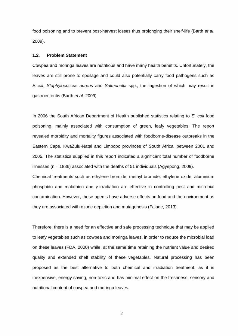
1.2. Problem Statement
Cowpea and moringa leaves are nutritious and have many health benefits. Unfortunately, the
leaves are still prone to spoilage and could also potentially carry food pathogens such as
E.coli, Staphylococcus aureus and Salmonella spp., the ingestion of which may result in
gastroenteritis (Barth et al, 2009).
In 2006 the South African Department of Health published statistics relating to E. coli food
poisoning, mainly associated with consumption of green, leafy vegetables. The report
revealed morbidity and mortality figures associated with foodborne-disease outbreaks in the
Eastern Cape, KwaZulu-Natal and Limpopo provinces of South Africa, between 2001 and
2005. The statistics supplied in this report indicated a significant total number of foodborne
illnesses (n = 1886) associated with the deaths of 51 individuals (Agyepong, 2009).
Chemical treatments such as ethylene bromide, methyl bromide, ethylene oxide, aluminium
phosphide and malathion and γ-irradiation are effective in controlling pest and microbial
contamination. However, these agents have adverse effects on food and the environment as
they are associated with ozone depletion and mutagenesis (Falade, 2013).
Therefore, there is a need for an effective and safe processing technique that may be applied
to leafy vegetables such as cowpea and moringa leaves, in order to reduce the microbial load
on these leaves (FDA, 2000) while, at the same time retaining the nutrient value and desired
quality and extended shelf stability of these vegetables. Natural processing has been
proposed as the best alternative to both chemical and irradiation treatment, as it is
inexpensive, energy saving, non-toxic and has minimal effect on the freshness, sensory and
nutritional content of cowpea and moringa leaves.

3
1.3. Aim
To investigate the effect of various processing techniques on the microflora and nutrient
content of cowpea and moringa leaves.
1.4. Objectives
To determine the effect of processing techniques such as washing and hot water or
steam blanching on the microbial load, nutrient content and sensory quality of fresh
cowpea and moringa leaves
To scale up such techniques so as to investigate the most effective commercial
processing and preservation techniques that may be used to reduce the microbial
load of fresh cowpea and moringa leaves
To investigate the effect of various leaf drying techniques on microbial load and
nutrient content of dried cowpea and moringa leaves
To study the shelf-life of tap water washed, steam blanched and milled moringa
leaves
To isolate and identify using standard microbiological methods those bacterial and
fungal contaminants found on treated and untreated moringa and cowpea leaves
To confirm the presence of coliform bacteria on moringa leaves using the polymerase
chain reaction
A brief description of the research design that was followed in order to attain the above aim
and objectives is discussed below in research materials and methods (chapter 3).
4
CHAPTER 2
Literature Review
2.1. Cowpea (Vigna unguiculata)
2.1.1. Scientific Classification
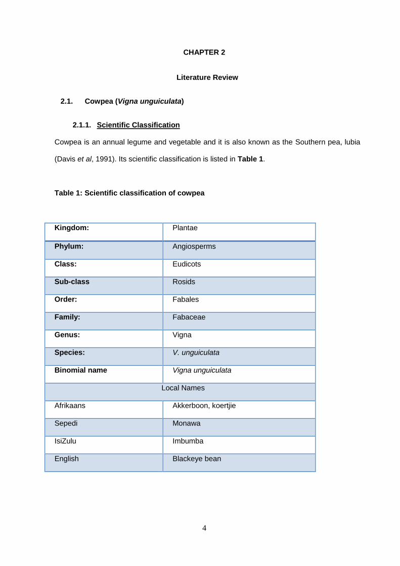
Cowpea is an annual legume and vegetable and it is also known as the Southern pea, lubia
(Davis et al, 1991). Its scientific classification is listed in Table 1.
Table 1: Scientific classification of cowpea
Kingdom: Plantae
Phylum: Angiosperms
Class: Eudicots
Sub-class Rosids
Order: Fabales
Family: Fabaceae
Genus: Vigna
Species: V. unguiculata
Binomial name Vigna unguiculata
Local Names
Afrikaans Akkerboon, koertjie
Sepedi Monawa
IsiZulu Imbumba
English Blackeye bean
5
2.1.2. General background and History
Cowpea (Vigna unguiculata) is an annual legume that was domesticated in West Africa. It is
an important grain legume and leafy vegetable in most parts of Africa and Asia. It is produced
on some commercial scale in eighteen African countries and seven Asian countries (Davis et
al, 1991).
The cowpea plant originated and was domesticated in western Africa and was later moved to
East and Southern Africa, Asia, Europe, United States and Central and South America.
Cowpea is chiefly used as a grain crop for animal fodder or as a vegetable (Davis et al,
1999). The name "cowpea" was probably derived from when it was an important livestock
feed for cows in the United States.
2.1.3. Background
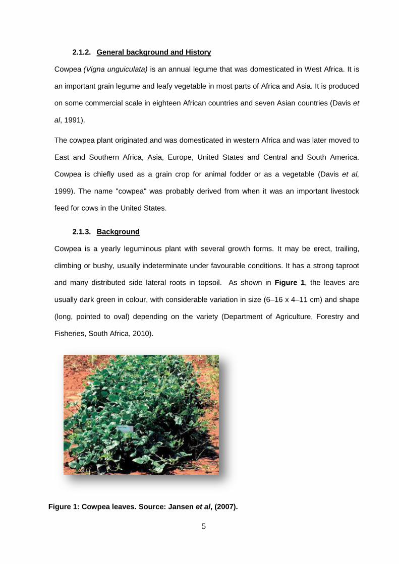
Cowpea is a yearly leguminous plant with several growth forms. It may be erect, trailing,
climbing or bushy, usually indeterminate under favourable conditions. It has a strong taproot
and many distributed side lateral roots in topsoil. As shown in Figure 1, the leaves are
usually dark green in colour, with considerable variation in size (6–16 x 4–11 cm) and shape
(long, pointed to oval) depending on the variety (Department of Agriculture, Forestry and
Fisheries, South Africa, 2010).
Figure 1: Cowpea leaves. Source: Jansen et al, (2007).
6
2.1.4. Uses of cowpea plant
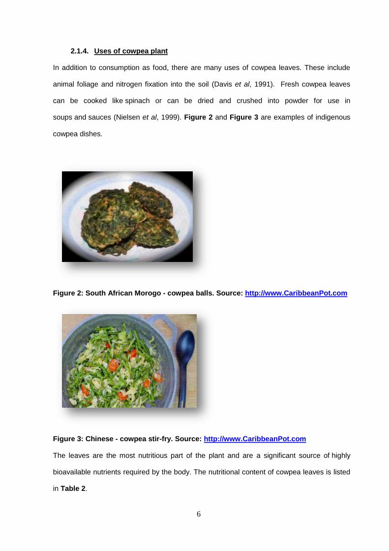
In addition to consumption as food, there are many uses of cowpea leaves. These include
animal foliage and nitrogen fixation into the soil (Davis et al, 1991). Fresh cowpea leaves
can be cooked like spinach or can be dried and crushed into powder for use in
soups and sauces (Nielsen et al, 1999). Figure 2 and Figure 3 are examples of indigenous
cowpea dishes.
Figure 2: South African Morogo - cowpea balls. Source: http://www.CaribbeanPot.com
Figure 3: Chinese - cowpea stir-fry. Source: http://www.CaribbeanPot.com
The leaves are the most nutritious part of the plant and are a significant source of highly
bioavailable nutrients required by the body. The nutritional content of cowpea leaves is listed
in Table 2.

7
Table 2: Nutritional composition of cowpea leaves (Vigna unguiculata). Adapted from
Nielsen et al, (1999).
Nutrient component Content per 100 g
Water (%) 88.4
Energy (cal) 3.4
Protein (g) 4.2
Calcium (mg) 110
Iron (mg) 4.7
Carotene (mg) 2.4
Ascorbic acid (mg) 35
2.1.5. Medicinal Value
Research has revealed that countries that preserve indigenous vegetable diets and have
high consumption of these vegetables are much less likely to be affected by cardiovascular
diseases, diabetes and other effects of malnutrition. Hence, there is a possibility that
indigenous vegetables such as cowpea could contribute towards an alleviation of diabetes,
gout, hyperlipidemia, gastro-intestinal tract infections, protozoan parasites and other
diseases. This motivates for an intervention geared towards encouraging individuals to
increase the consumption of indigenous leafy vegetables (Kimiywe et al, 2007). There is a
need for further investigation to establish the basis of the above-mentioned perceptions.
8
2.2. Moringa Leaves (Moringa oleifera)
2.2.1. General
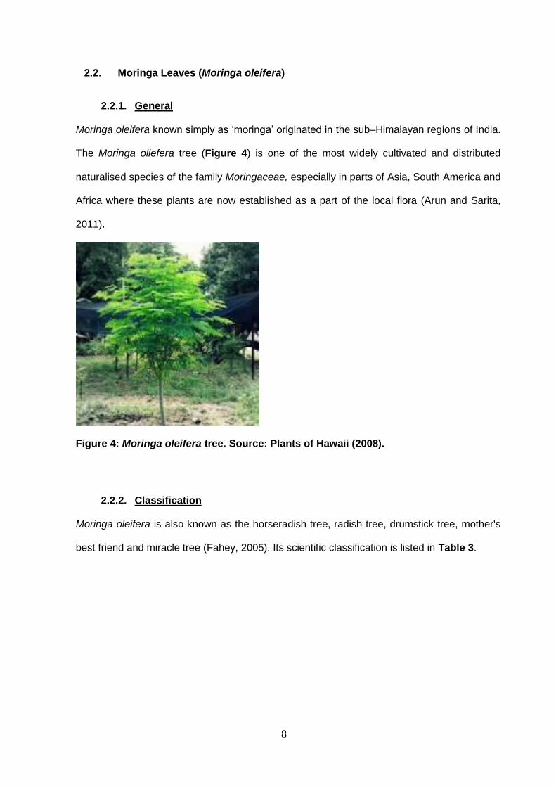
Moringa oleifera known simply as ‘moringa’ originated in the sub–Himalayan regions of India.
The Moringa oliefera tree (Figure 4) is one of the most widely cultivated and distributed
naturalised species of the family Moringaceae, especially in parts of Asia, South America and
Africa where these plants are now established as a part of the local flora (Arun and Sarita,
2011).
Figure 4: Moringa oleifera tree. Source: Plants of Hawaii (2008).
2.2.2. Classification
Moringa oleifera is also known as the horseradish tree, radish tree, drumstick tree, mother's
best friend and miracle tree (Fahey, 2005). Its scientific classification is listed in Table 3.
9
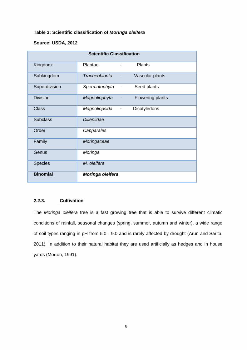
Table 3: Scientific classification of Moringa oleifera
Source: USDA, 2012
Scientific Classification
Kingdom: Plantae - Plants
Subkingdom Tracheobionta - Vascular plants
Superdivision Spermatophyta - Seed plants
Division Magnoliophyta - Flowering plants
Class Magnoliopsida - Dicotyledons
Subclass Dilleniidae
Order Capparales
Family Moringaceae
Genus Moringa
Species M. oleifera
Binomial Moringa oleifera
2.2.3. Cultivation
The Moringa oleifera tree is a fast growing tree that is able to survive different climatic
conditions of rainfall, seasonal changes (spring, summer, autumn and winter), a wide range
of soil types ranging in pH from 5.0 - 9.0 and is rarely affected by drought (Arun and Sarita,
2011). In addition to their natural habitat they are used artificially as hedges and in house
yards (Morton, 1991).
10
2.2.4. Moringa oleifera In South Africa
In India and parts of West Africa and Central Africa, use of the moringa plant has become
very common among local people who have included it as part of their traditional cuisine
(Fuglie, 1999). Although the moringa plant grows in some parts of South Africa (e.g. in the
Mokopane district in the Limpopo Province), most South Africans are still unaware of the
plant and its numerous uses and benefits (National Research Council, 2006).
The results of a literature study as well as a few informal discussions held in Tshwane in the
Gauteng province of South Africa revealed that very few South Africans, except for mostly
members of the Indian community, use moringa in their diets. A publication indicated that the
moringa plant is listed as a naturalised plant in South Africa and this might indicate that the
popularity and general acceptability of the plant in South Africa is on the increase (Agyepong,
2009).
2.2.5. Uses of Moringa oleifera plant
In addition to food, there are many products derived from various parts of the moringa tree.
These include biomass and biogas production, domestic cleaning agents, fertilizer and
manure, honey, medicine and bio-pesticides (Fahey, 2005; Agyepong, 2009). Please refer to
Appendix 1 for a more complete list of products associated with the moringa tree. A list of
the nutrient composition of moringa leaves as compared to other foods is shown in Table 4.
11
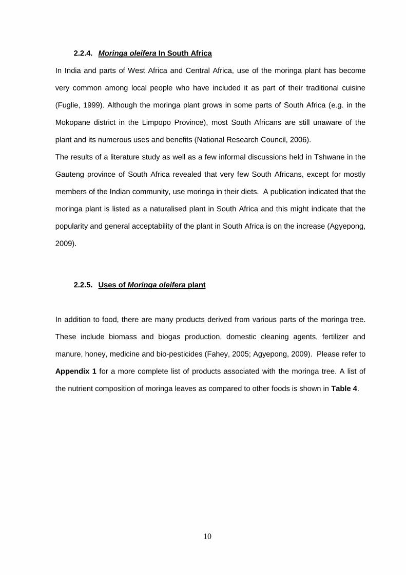
Table 4: Nutrient Composition of Moringa oleifera leaves (per 100g) as compared to
popular nutritious foods. Adapted from: Fahey, (2005), Fuglie, (1999) and McCance and
Widdowson (1992).
Nutrient
Content
Moringa (raw) Per 100g Other foods (raw) Per 100g
Vitamin A 6.8mg 1.8mg in Carrot
Calcium 440mg 120mg in Milk
Potassium 259mg 88mg in Banana
Protein 6.7g 3.1g in Yoghurt
Vitamin C 220 mg 30mg in Orange
Phosphorous 70mg 61mg in Curly-kale
Energy 95Kcal 98Kcal in Garlic
Carbohydrates 8.28 g 7.6g in Beetroot (root)
Sodium 9mg 3mg in Cucumber
Zinc 0.6mg 0.6mg in Broccoli
Iron 4.00mg 2.1mg in Spinach
Magnesium 147mg 71mg in Okra
Manganese 0.36mg 0.1mg in Onion
Dietary fibre 2.0g 1.9mg in Cauliflower
Fat 1.40g 1.4g in Brussels sprouts
Protein 9.40g 7.9g in Garlic
Water 78.66g 89.7g in Spinach
Riboflavin (vit. B2) 0.660mg 0.11mg in Brussels sprouts
Pantothenic acid (B5) 0.125mg 0.3mg in Cucumber
Vitamin B6 1.200mg 0.28mg in Cauliflower
12
The moringa plant also supplies a host of prophylactic and therapeutic health benefits (Arun
and Sarita,(2011); Anwar et al, (2007); Fuglie, (1999, 2000)) These include:
Helping to balance cholesterol levels in the body.
Assisting in balancing sugar levels associated with diabetes.
Stimulating the immune system.
Stimulating metabolism.
Aiding in digestion and acting as a natural laxative.
Serving as a nutrition booster and a non-sugar based energiser.
Assisting in weight loss.
Increasing breast milk production.
Acting as a skin tonic.
For many years, parts of the moringa plant have been used to treat a number of health
conditions or diseases in Ayurvedic medicine as well as other traditional systems where,
particularly the roots, seeds, leaves and flowers have been utilised (Arun and Sarita, 2011).
A list of these therapeutic uses is provided in Appendix 2.
It should be noted that there are potential hazards associated with using moringa as a food
source. Thus, consumption of moringa should be avoided by people on blood-thinning
medication or pregnant women without medical consent, because of the effect of moringa
leaves on improving blood coagulation (Fahey, 2005).

13
2.3. General background on the processing and analysis of leafy vegetables
2.3.1. Processing Techniques
Based on published statistics (South African Department of Health, 2006) relating to E. coli
food poisoning that year, mainly associated with consumption of green, leafy vegetables, 51
people died of food borne-related illness out of the 1886 people who experienced food-borne
illness (Agyepong, 2009). A similar outbreak of bacterial food-borne disease began
in Germany in May 2011 that resulted from the consumption of contaminated leafy
vegetables. One E. coli strain, E. coli O104:H4 was identified as the causative organism. As
reported to the WHO, this outbreak was not restricted to Germany, but included 11 other
countries including Denmark, France, Netherlands Sweden and the United States of America
(www.sciencedaily.com).
Therefore, there is a need for effective processing at every step from planting to consumption
of leafy vegetables, in order to reduce microbial contamination which might eventually lead to
food poisoning and food spoilage (FDA, 2000).
However, if current processing techniques are not controlled, it could reduce the levels of
some of the health-benefiting nutrients, or the availability of these compounds, so minimal
processing is desirable to ensure product safety and good quality over a prolonged shelf-life
period.
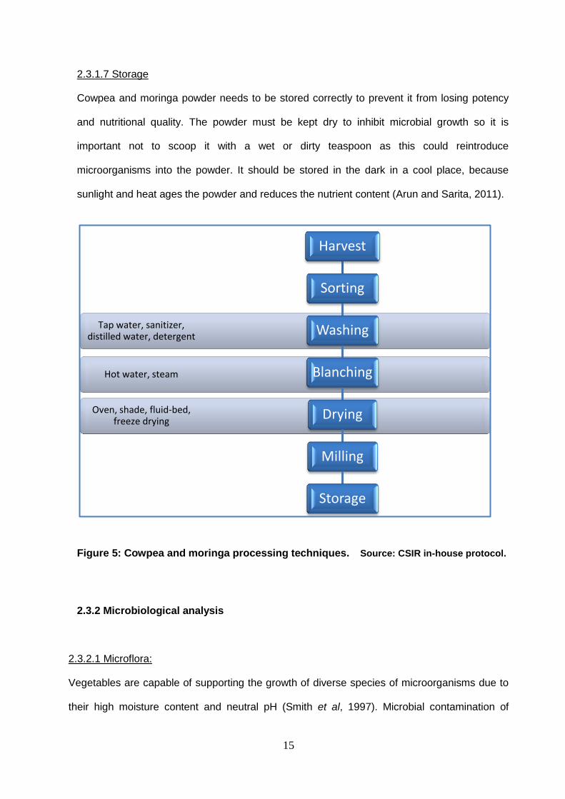
As shown in Figure 5, the following processing techniques have been used for cowpea and
moringa leaves (CSIR in-house protocol):

14
2.3.1.1 Sorting
This step involves the picking out of the healthy leaves and detaching them from their stems.
2.3.1.2 Washing
Washing is not only used to remove field soil and surface micro-organisms, but also to
remove fungicides, insecticides and other pesticides from leafy vegetables. The washing
water may contain detergents or other sanitizers that can assist in removing these residues.
2.3.1.3 Blanching
This is a special heat treatment to inactivate enzymes such as catalase and peroxidase
present in vegetables, where the leaves are submerged in boiling water 94°C (hot water
blanching) or steam at 85-95°C (steam blanching). The leaves are removed after a brief,
timed interval (too little heat is ineffective while excess heat damages the vegetables) and
finally placed under cold running water to stop the cooking process.
2.3.1.4 Drying
During this step, the moisture is removed from the leaves by simultaneous heat and mass
transfer. The drying techniques of vegetables require a dependable model to predict its
drying behaviour (Premi, 2010). Methods of drying could involve sun-drying, spray drying,
freeze drying and oven drying.
2.3.1.5 Milling
This is the stage where the vegetables are broken down by course milling into a smaller size
or into a powdered form.
2.3.1.6 Packaging
At this stage the final product of cowpea and moringa leaves that is required for distribution,
storage, sale and use is enclosed or protected and sealed. Exposure of the final products to
oxygen hastens leaf degradation and nutritional value loss (Premi et al, 2010).
13
15
2.3.1.7 Storage
Cowpea and moringa powder needs to be stored correctly to prevent it from losing potency
and nutritional quality. The powder must be kept dry to inhibit microbial growth so it is
important not to scoop it with a wet or dirty teaspoon as this could reintroduce
microorganisms into the powder. It should be stored in the dark in a cool place, because
sunlight and heat ages the powder and reduces the nutrient content (Arun and Sarita, 2011).
Figure 5: Cowpea and moringa processing techniques. Source: CSIR in-house protocol.
2.3.2 Microbiological analysis
2.3.2.1 Microflora:
Vegetables are capable of supporting the growth of diverse species of microorganisms due to
their high moisture content and neutral pH (Smith et al, 1997). Microbial contamination of
Hot water, steam
Oven, shade, fluid-bed, freeze drying
Tap water, sanitizer, distilled water, detergent
Harvest
Sorting
Washing
Blanching
Drying
Milling
Storage
16
vegetables occur at various stages of production such as the use of contaminated water for
irrigation or washing of the vegetables, soil containing manure from human or animal origin,
animals including insects and birds, handling of the vegetable product, contaminated
harvesting and processing equipment and during transportation ( Barth et al, 2009).
Microorganisms found on vegetable leaves include bacteria or fungi that have grown on and
colonized the leaf surface by utilizing nutrients exuded from plant tissues. These could be
pathogenic or spoilage-causing microorganisms. Many of these agents enter the plant tissue
through mechanical or chilling injuries, or after the plant surface barrier has been broken down
by other organisms (Joshi et al, 2010). Besides causing huge economic losses, some fungal
species could produce toxic metabolites in the affected sites, constituting a potential health
hazard for humans. In order to slow down vegetable spoilage and minimize the associated
adverse health effects, great caution should be taken to follow strict hygiene, good agricultural
practices and good manufacturing practices during cultivation, harvest, storage, transport, and
marketing (Joshi et al, 2010; Barth et al, 2009).
2.3.2.2 Spoilage Microorganisms
After harvest, vegetables are often spoiled by a wide variety of microorganisms including
many bacterial and fungal species. Fungi commonly causing spoilage of fresh vegetables
include Botrytis cinerea and various species of the genera Aspergillus, Cladosporium,
Fusarium, Penicillium, Phoma, Phytophthora, Pythium and Rhizopus. These affect a wide
variety of vegetables causing devastating losses (Joshi et al, 2010).
According to a USDA-Economic Research Service study in 2010, 18.9 billion pounds of fresh
fruits and vegetables were lost annually due to spoilage, which was 19.6% of all losses of
edible foods that year in the United States (Kantor et al, 2010). The most common bacterial
agents are Gram positive bacteria such as lactic acid bacteria originating mainly from
contamination from the hands of employees during harvesting or processing, as well as
17
Erwinia, carotovora, Pseudomonas spp., Corynebacterium and Xanthomonas, campestris
which attack virtually every vegetable type (Garg et al, 1990).
2.3.2.3 Pathogenic Microorganisms
There have been a number of human infections associated with the consumption of raw fruits
and vegetables. Outbreaks with identified etiology were predominantly of bacterial origin,
primarily Salmonella, Escherichia coli, Bacillus cereus, Clostridium spp, Listeria
monocytogenes, Salmonella spp, Shigella spp and Staphylococcus spp which have the
potential for growth prior to consumption or which have a low infectious dose (Buck et al,
2002). The presence of high coliform bacteria counts are used as indicators of the level of
hygiene, the possible presence of pathogens and the quality of the vegetables. Besides
causing huge economic losses, some fungal species could produce toxic metabolites in the
affected sites, constituting a potential health hazard for humans (Barth et al, 2009).
18
CHAPTER 3
Research Methodology: Part A
3.1. Processing Techniques
Cowpea and moringa leaves were processed using industrial methods performed on a semi-
industrial scale as well as on a smaller scale in a controlled environment under aseptic
conditions in the laboratory. All experiments were done in triplicate.
3.1.1. Harvesting of leafy vegetables
Cowpea leaves were harvested from plants grown at the Agricultural Research Council
(ARC), Roodeplaat, Pretoria, South Africa while Moringa oleifera leaves were harvested from
plants grown on farms in Atteridgeville and Hammanskraal near Pretoria, South Africa.
Harvesting was done by picking the new leaves at the end of the stem while avoiding long
and thick stems that would affect the drying process. The harvested leaves were stored in
breathable woven bags (not plastic bags) and the harvesters completed a Collectors Form.
Each bag was weighed and labelled and data were filled in on a transport dispatch note. The
bags containing harvested vegetables were transported to the CSIR in Pretoria on the day of
harvest.
3.1.2. Receiving of cowpea and moringa leaves:
At the CSIR a batch number was allocated to each received bag containing the cowpea and
moringa leaves. Each bag was weighed and the mass was recorded. The moisture content of
the vegetables was checked as described below and random samples were aseptically
selected from the bags, labelled and then taken to the laboratory for full microbiological and
nutrients analyses as well as sensory evaluations. The portions of the cowpea and moringa
samples not used for analyses were stored in a cold room at 4oC.
19
3.1.3. Sorting of leafy vegetables:
The required number of bags containing a sufficient mass (around 50kg) of leaves for
processing. The leaves were inspected for fungal and other contaminants and the size,
colour and appearance of the leaves was recorded. Defective, insect-infested and
contaminated leaves were discarded.
3.1.4. Washing of the leafy vegetables:
Laboratory scale: Approximately 20g of leaves with a healthy appearance was selected and
about 5g of these leaves was removed and placed in a sterile petri dish as control. The
remaining 15g of leaves were washed for 1min to remove soil and other dirt. These leaves
were divided into three portions and each of these was placed into separate petri dishes –
around 5g per dish. Apart from the control, unwashed leaves, each portion of leaves was
then rinsed for 1min by the addition of washing fluid to the leaves that were then gently
swirled for 10 seconds. Three different washing treatments per 5g of leaves included 50ml of
tap water, 50ml of distilled water or 50ml of soapy water. The soapy water was prepared by
adding 1g of household food-grade detergent to 99ml of tap water. The petri dishes with
rinsed leaves were then inverted and allowed to drip dry for 1min. Control and treated leaf
samples were then analysed to determine levels of microbial contaminants. The leaves from
the tap water washing method were then selected for nutrient analysis and sensory
evaluation.
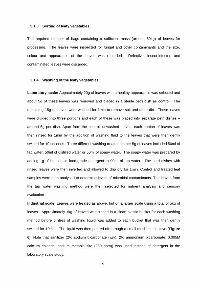
Industrial scale: Leaves were treated as above, but on a larger scale using a total of 5kg of
leaves. Approximately 1kg of leaves was placed in a clean plastic bucket for each washing
method before 5 litres of washing liquid was added to each bucket that was then gently
swirled for 10min. The liquid was then poured off through a small mesh metal sieve (Figure
6). Note that sanitizer (2% sodium bicarbonate (w/v), 2% ammonium bicarbonate, 0.005M
calcium chloride, sodium metabisulfite (250 ppm)) was used instead of detergent in the
laboratory scale study.
20
Figure 6: Photograph of leaf washing in tap water. Source: Photograph taken by
author
3.1.5. Blanching
Laboratory scale: Approximately 10g of tap water-rinsed leaves were selected (after drip
drying for 1min) and a 5g portion of these leaves was moved to a sterile petri dish as a
control. Then, 50ml of hot water (95oC) was added to the remaining 5g of leaves before
gently swirling for 2min. Control and treated leaf samples were then analysed for microbial
contamination, nutrient analysis and sensory evaluation.
Industrial scale: Leaves were treated as above, but at a larger scale using a total of 2kg of
leaves. Approximately 1kg of tap water-washed leaves was immersed in hot water at 94oC
+1 for 6-8 minutes (hot water blanching). The other 1kg of tap water washed leaves were
divided into approximately 100g portions each of which were placed on perforated stainless
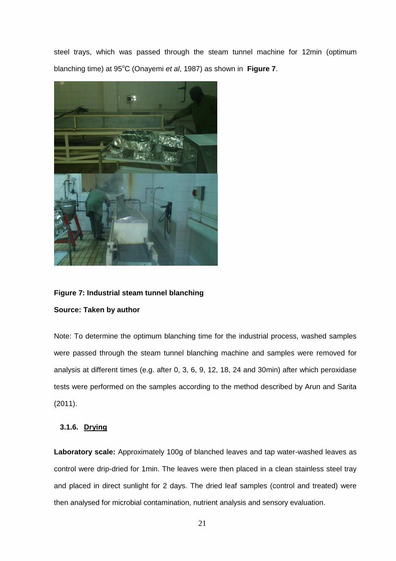
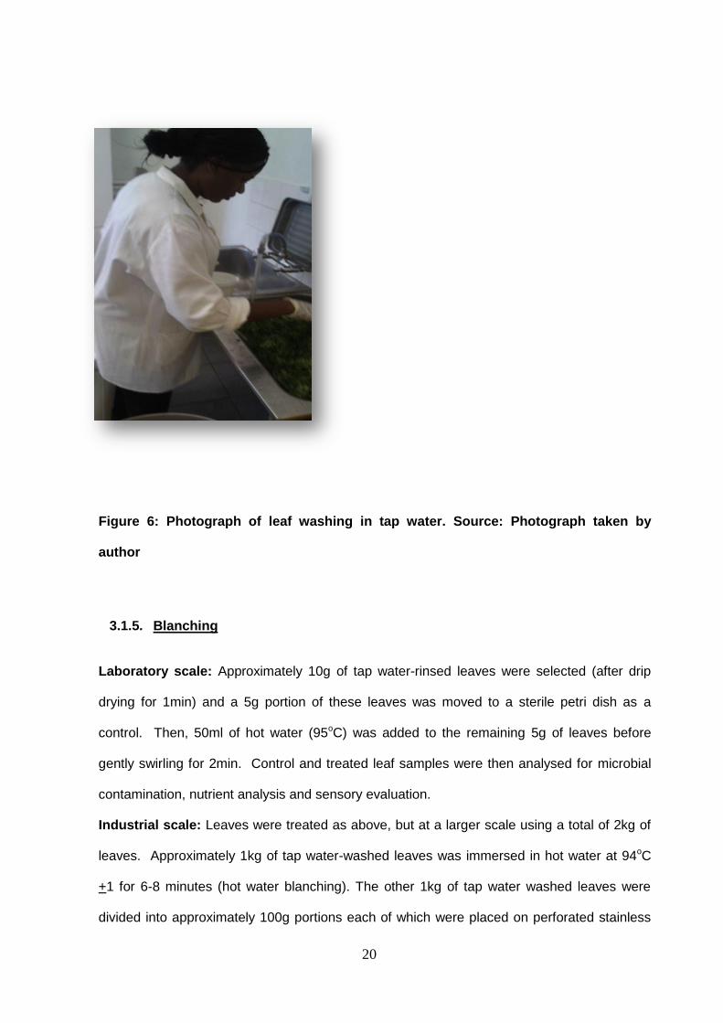
21
steel trays, which was passed through the steam tunnel machine for 12min (optimum
blanching time) at 95oC (Onayemi et al, 1987) as shown in Figure 7.
Figure 7: Industrial steam tunnel blanching
Source: Taken by author
Note: To determine the optimum blanching time for the industrial process, washed samples
were passed through the steam tunnel blanching machine and samples were removed for
analysis at different times (e.g. after 0, 3, 6, 9, 12, 18, 24 and 30min) after which peroxidase
tests were performed on the samples according to the method described by Arun and Sarita
(2011).
3.1.6. Drying
Laboratory scale: Approximately 100g of blanched leaves and tap water-washed leaves as
control were drip-dried for 1min. The leaves were then placed in a clean stainless steel tray
and placed in direct sunlight for 2 days. The dried leaf samples (control and treated) were
then analysed for microbial contamination, nutrient analysis and sensory evaluation.
22
Industrial scale: Leaves were treated as above, but at a larger scale using a total of 15kg of
leaves. The blanched (hot water-blanched and steam-blanched) leaves were divided into
three portions of 5kg each. The first portion was dried in an oven at 60oC +1oC for 48hrs, the
second portion was spread on a wooden tray and placed under direct sunlight for 3 days and
the third portion was freeze dried. These dried leaves were then collected, weighed, labelled
and samples were taken randomly from each portion for full microbial analysis, nutrient
analysis and sensory evaluations. The remaining dried samples were milled into fine powder
and stored in air-tight thermally sealed plastic containers at room temperature.

23
Research Methodology: Part B
3.2. Microbiological Analyses
This section describes the microbiological analysis on cowpea and moringa leaves. The
following microbiological analyses were carried out on both leafy vegetables after each
processing step.
3.2.1. Microbial Enumeration and Isolation
3.2.1.1. Sample treatment
Five grams of the vegetable leaves were diluted with 45ml sterile Maximum Recovery Diluent
(MRD) solution (Oxoid LTD, Basingstoke, Hampshire, England) and homogenized for 2min at
normal speed in a stomacher. A 10-1 to 10-8 dilution series was then prepared in MRD and
enumerated on each of the different agar media. This serial dilution was performed as
follows: 5 g of each sample was diluted by aseptically adding 45ml of MRD and then
homogenised in the Stomacher for 2min. One ml (10-1 dilution) of each sample was then
aseptically transferred into the 10-2 dilution and similarly serial dilutions were aseptically
prepared up to 10-8 in a 76% ethanol sanitised laminar flow cabinet.
3.2.1.2. Determination of total viable count:
The Total Viable Count (TVC) enumerates the total population of viable, non-fastidious
microorganisms that grow at mesophilic temperature (i.e. 30oC). Such microorganisms
include bacteria, yeast and moulds.
Determination of Total Viable Count in vegetable samples was performed using the Plate
Count Method using Tryptone soy agar (TSA) (Oxoid LTD, Basingstoke, Hampshire,
England) that was prepared according to supplier’s instructions.

24
For pour plating, an aliquot of 1ml of each dilution (10-3 to 10-8) was pipetted into a sterile
plate and 20ml of TSA (at 48oC) was added to each, before being gently gently mixed. The
labelled plates were then left to solidify before transferring to the incubator. The plates were
incubated at 30oC for 48hrs before being assessed. This involved counting colonies on
plates with between 25 to 250 colonies and the colony count was calculated from dilution
factors to give the Total Viable Count according to ISO Method 21527 (Rahman et al, 2010).
3.2.1.3. Determination of Yeast and Mould Count
To determine the total viable count of yeast and moulds in vegetable samples using the plate
count method, potato dextrose agar (PDA) (Oxoid LTD, Basingstoke,Hampshire, England)
was acidified with 10% Tartaric acid (Sigma-Aldrich Inc., St Louis, USA). Serial dilutions were
prepared of each sample and 1ml of each dilution was transferred into each plate.
Thereafter, approximately 20ml of acidified PDA was added to each plate and gently swirled
before allowing the medium to solidify before incubating at 25oC for 5 days. The number of
growing colonies was recorded as a combined count according to SABS Method 758.
3.2.1.4. Determination of Coliform Count
Scope: Enumeration of coliforms in vegetable leaves using the Most Probable Number
(MPN) method.
Principle: Determination of the number of coliforms by means of a MPN table according to
the number of inoculated tubes which gave rise to gas formation in the selective coliform
medium. The MPN procedure involves a multiple tube fermentation technique where each of
three decimal dilutions of the sample are inoculated into separate tubes of broth medium and
incubated at a specific temperature and for a specific time. The method is progressive; i.e.,
first determining the presence of coliforms in the tubes (presumptive coliforms), then
determining if these tubes also contain faecal coliforms (confirmed coliforms), and then
25
confirming whether E. coli is present (Tryptone water Indole test - E.coli confirmed). Based
on the number of tubes indicating the presence / absence of the three groups of organisms,
the most probable number (MPN) present can be estimated from a standard statistical MPN
table. The method has been shown to produce satisfactory results with naturally-
contaminated foods and water for the detection of coliforms, faecal coliforms and aerogenic
E. coli (Rahman et al, 2010). Briefly, lauryl sulphate tryptose broth (LTB), brilliant green bile
broth (BgBB) (Oxoid LTD, Basingstoke, Hampshire, England), were separately dispensed as
9ml aliquots with inverted Durham tubes. Tryptone water was also dispensed in 5ml volumes
with inverted Durham tubes.
The MPN Method –Three tube method – involves preparing three serial dilutions of the
cowpea samples and for each dilution, inoculate 1ml into each of the tubes of LTB; then after
24hrs of incubation, inoculate 1ml from each positive test tube into BgBB tubes.
E.coli
Positive E.coli culture was sub-cultured into Tryptone water and tested for indole production
using Kovac’s (4(p)-dimethylamino-benzaldehyde) following incubating in a 44oC water bath
for 2 days.
3.2.2. Gram Staining
Protocol:
A loopful of sterile distilled water was placed on a slide; using a sterile cool loop a small
sample of an isolated bacterial colony was transferred to the drop, and emulsified. The film
was allowed to air-dry by passing it briefly through the Bunsen flame two or three times
without exposing the dried film directly to the flame. The slide was flooded with crystal violet
solution for up to one minute, washed off briefly with distilled water and drained. This was
26
followed by flooding the slide with Gram's Iodine solution, and allowed to act as a mordant for
one minute, washed off with distilled water and drained. The slides were then flooded with
95% acetone for 10 seconds, washed off with distilled water and allowed to drain. Lastly the
slides was flooded with safranin solution and allowed to counterstain for 30 seconds, washed
off with distilled water, drained and blotted dry. All slides were examined under the oil
immersion lens (CSIR in-house protocol).
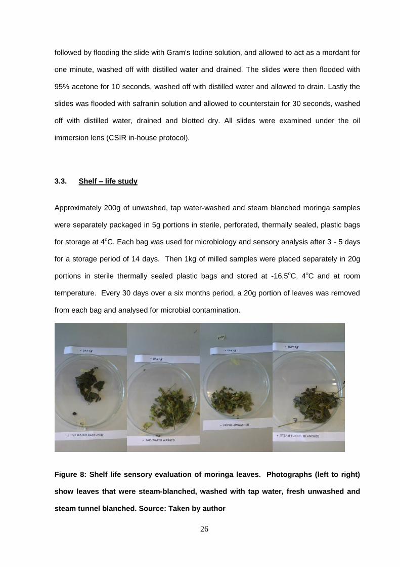
3.3. Shelf – life study
Approximately 200g of unwashed, tap water-washed and steam blanched moringa samples
were separately packaged in 5g portions in sterile, perforated, thermally sealed, plastic bags
for storage at 4oC. Each bag was used for microbiology and sensory analysis after 3 - 5 days
for a storage period of 14 days. Then 1kg of milled samples were placed separately in 20g
portions in sterile thermally sealed plastic bags and stored at -16.5oC, 4oC and at room
temperature. Every 30 days over a six months period, a 20g portion of leaves was removed
from each bag and analysed for microbial contamination.
Figure 8: Shelf life sensory evaluation of moringa leaves. Photographs (left to right)
show leaves that were steam-blanched, washed with tap water, fresh unwashed and
steam tunnel blanched. Source: Taken by author

27
3.4. Biochemical Analysis
The following tests are importance in order to determine the effectiveness of vegetable
blanching treatments (temperature and time). Incomplete enzyme inactivation has a negative
effect on the finished product quality (Rahman et al, 2010). The biochemical analyses carried
out were:
Indole test
This is a confirmatory test for the presence of E.coli. Tryptone water was prepared according
to manufacturer’s instruction. Briefly, 5ml of broth was dispensed per test tube and
autoclaved at 121oC for 15min. The bacteria was aseptically inoculated into the broth and
incubated for 24hrs at 37oC (Rahman et al, 2010). A pinkish-reddish colour change after the
addition of a few drops of Kovac’s reagent indicates a positive reaction while no colour
change indicates a negative reaction (Prabhun et al, 2011).
Peroxidase test
A few drops of 1% guaiacol solution and 0.3% peroxide solution were added directly onto
blanched and crushed vegetables leaves. A rapid and intensive brown-reddish tissue
colouring indicates a high peroxidase activity (positive reaction) while no colour change after
5min indicates inactivation of the enzyme peroxidase (negative reaction). These results are
indicated in Figure 9 and Figure 10, respectively (Rahman et al, 2010).
Catalase test
Two grams of dehydrated vegetables were well crushed and mixed with about 20ml of
distilled water. After 15min softening, 3% peroxide solution was poured on the prepared
vegetables. The formation of bubbles of free oxygen gas after 2-3 minutes represents a
positive reaction (Prabhun et al, 2011, Rahman et al, 2010).

28
Figure 9: Positive peroxidase test on cowpea leaves. Source: Photograph taken by
author.
Figure 10: Negative peroxidase test on cowpea leaves. Source: Photograph taken by
author.
29
Research Methodology: Part C
3.5. Proximate Analysis
The moisture, ash, fat, carbohydrate, protein and fibre content of cowpea and moringa
samples were determined at the CSIR Food Science Analysis Laboratory in Pretoria, South
Africa.
3.5.1. Moisture Content
The moisture content of cowpea and moringa samples were checked using a halogen
moisture analyser (HX204 Mettler Toledo) where 3g of each sample was weighed into an
aluminium dish and placed in the automatic halogen moisture analyser. The results were
recorded as a percentage (CSIR-AM020)
3.5.2. Ash content
Clean crucibles were dried inside a muffle furnace for 2hrs at 55oC after which the crucibles
were placed in the desiccator to cool. Each crucible was weighed and 3g of the vegetable
samples were dispensed per crucible and placed in a muffle oven (Furnace Nabertherm,
Germany) at 550oC overnight as shown in and Fig 11, after which the crucible was weighed
and the ash (Fig 12) percentage was calculated (AOAC, 1990).
% Ash content = Mass of crucible after ashing –Mass of crucible before ashing x 100
Mass of sample

30
Figure 11: Photograph of muffle oven containing crucibles with moringa leaves
Source: Photograph taken by author
Figure 12: Photograph of Vegetable ash remaining in crucibles following ashing of
moringa leaves. Source: Photograph taken by author.
3.5.3. Fat Content
The fat content was determined by weighing 5g of vegetable samples into extraction thimbles
and weighing the labelled extraction cups before and after the extraction. The thimbles were
connected to a Soxhlet extractor (Gerhadt, Germany) and extracted with petroleum ether for
4hrs. The residue in the extraction bottle after solvent removal represents the fat content of
31
the samples (Sanjukta et al, 2013). The result was reported as a percentage and was
calculated as:
% Fat content = Mass of thimble after extraction –Mass of thimble before extraction x 100
Mass of sample
3.5.4. Protein Content
The protein analysis was performed in duplicate using a Trumac Nitrogen Analyser according
to the manufacturer’s instructions (LECO Africa). The nitrogen value is an indicator of the
protein content of a substance and was determined by digestion, distillation and finally
titration of the sample. The nitrogen value was converted to protein by multiplying by a factor
of 6.25 (AOAC, 1990).
3.5.5. Carbohydrate Content
Carbohydrate content was determined by subtracting the total sum of the moisture, ash,
crude fibre, fat and crude protein from 100.
3.5.6. Crude Fibre Content
Crude fibre was determined by treating oil-free sample with sulphuric acid (0.26 N) and
potassium hydroxide (0.23 N) solution in refluxing systems, followed by oven drying and
muffle furnace incineration (AOAC, 1990).
3.5.7. Energy Content (by calculations)
The energy value was calculated by multiplying the mean values for the crude fat, protein
and total carbohydrates by 37, 17 and 17, respectively (Akinyele et al., 2011).
32
3.6. Sensory Evaluation
The main reason for sensory testing was to provide data on which sound decisions may be
made. It is an integrated, multi-dimensional measure with three important advantages: it
rapidly identifies the presence of notable differences, identifies and quantifies important
sensory characteristics and identifies specific problems that cannot be detected by other
analytical procedures, such as consumer preference.
Sensory evaluation was carried out in the laboratory by 10 panellists, who were used to
evaluating the qualities of colour, texture (crispness), odour, and general acceptability of
fresh, blanched, dried and milled vegetable (moringa) samples. A simple questionnaire was
given to each panel to record their observations about the samples. Panellists were asked to
compare the quality attributes of the vegetables using a preference scale with 1= extremely
unacceptable and 5= extremely acceptable (Onayemi et al, 1987). Appendix 3 is a copy of
sensory evaluation questionnaire used.
33
Research Methodology: Part D
MOLECULAR BIOLOGY
The molecular biology aspect of this study was carried out in the molecular laboratory at
UNISA Florida campus, Johannesburg under the supervision of Dr.Khayaletu Ntushelo.
3.7. Identification and Isolation of Coliforms
Presumptive coliforms were cultured in Lauryl Tryptose Broth (LTB) and Brilliant Green Bile
Broth (BgBB) to confirm the presence of coliforms, as described in section 3.2.1.4.
A sterile, flamed loop was dipped into the cultured BgBB after 24hrs of inoculation and was
transferred into a tube containing 5ml of Tryptone water (prepared according to
manufacturer’s instructions) and incubated in a water bath at 44oC for 48 hrs, after which a
loop of the broth was plated on Tryptone Soy agar (TSA) for 24hrs at 37oC (3.2.1.4). This
protocol is selective only for coliforms, Enterobacteriaceae and E.coli.
3.7.1. Molecular Analysis
Strain conservation of coliforms and yeasts
Surface coliforms and yeasts from fresh unwashed sample and milled sample were plated
and incubated and counted. From these enumeration plates, ten coliform and ten yeast
colonies were selected at random. The coliform colonies were inoculated in 10ml of Tryptone
soy broth (TSB) (Oxoid LTD, Basingstoke, Hampshire, England) and incubated at 37˚C
overnight. Cells were harvested by centrifugation at 4500xg for three minutes and the pellet
was suspended in cryoprotective medium [K2HPO4 0,82 g.l-1, KH2 PO4 0,18 g.l-1,
C6H5Na3O7.H2O 0,67 g.l-1,MgSO4.7H2O 0,25 g.l-1 and sterile glycerol to 15% of the total
volume (all substances from Merck KGaA, Darmstadt, Germany)] and frozen at -70˚C.
The yeast colonies from these samples were inoculated in 5ml Yeast Peptone D-glucose
(YPD) broth [yeast extract 10g.l-1 (Oxoid LTD, Basingstoke, Hampshire, England),

34
bacteriological peptone 20g.l-1 (Oxoid Ltd)) and D glucose 20g.l-1 and incubated overnight at
25˚C. One ml of cell suspension was mixed with an equal volume of sterile glycerol and
frozen at -70˚C.
3.7.2. DNA extraction
Coliform bacterial DNA was isolated by using the DNeasy tissue kit (Qiagen,
Hilden,Germany) following the manufacturer’s instructions.
Procedure:
A volume of 180µl of extraction buffer 1 (ATL) and 20µl of Proteinase K was added to 2ml
tubes containing pure overnight cultured bacteria cells, which was then vortex mixed and
incubated at 56oC for 1hr after which it was micro centrifuged briefly. Then, 200µl of lysis
buffer (AL) was added to each tube, vortex mixed and incubated at 70oC for 10min.The
mixture was centrifuged and 200µl of 100% ethanol was added to precipitate the DNA. Using
a micropipette, the entire contents (~600 µl) of tube was transferred to a labelled spin
column. This was followed by centrifuging at 8000rpm for 1min. The eluate was discarded
and 500µl of wash buffer 1 (BW1) was added above the filter. This was followed by
centrifuging at 8000 rpm for 1min. The process was repeated using wash buffer 2 (BW2) and
centrifuging at 14000rpm for 3min. The eluate was also discarded and the filter was placed in
a new 1.5ml tube (labelled accordingly). Lastly, 200µl of elution buffer (AE) was added and it
was centrifuged at 18000 for 3min. The elution buffer eluted the DNA into the clean tubes
that were then stored appropriately (4°C for short term and -20°C for long term storage).
3.7.3. DNA quantification
DNA Quantification by Fluorometer
The fluorometer used at UNISA’s molecular laboratory is the QuantusTMfrom Promega. The
following instructions were used for this specific fluorometer.
Procedure:
35
Preparation of a working solution was performed by diluting the supplied QuantiFluor®
dsDNA Dye with I x TE buffer in a ratio 1:200. A blank solution was also prepared by adding
100µl of QuantiFluor® dsDNA Dye working solution and 100µl of 1X TE buffer to an empty
0.5ml PCR tube, mixing and while protecting from light. A standard solution was prepared by
diluting the DNA Standard to 2ng/µl by adding 2µl of the provided DNA Standard to 98µl of
1X TE buffer, and mixed. Then, 100µl of QuantiFluor® dsDNA Dye working solution was
added, and mixed.
Finally, preparation of the DNA samples was done by adding 100µl of the DNA sample and
100µl of QuantiFluor®dsDNA Dye working solution to a 0.5ml PCR tube, and mixing. The
prepared samples were incubated at room temperature for 5 min, protected from light.
The dsDNA protocol on the Quantus™ Fluorometer was selected and the Quantus™
Fluorometer was calibrated by reading the blank and standard samples in the calibration
screen and then selecting “Save”. The volume of the unknown sample and desired
concentration units was entered and fluorescence of the DNA samples were determined.
3.7.4. Polymerase Chain Reaction
Repetitive-DNA-element PCR fingerprinting made use of the following primers:
pA Forward primer ( 5' AGA GTT TGA TCC TGG CTC AG 3',)
pH reverse primer (5' AAG GAG GTG ATC CAG CCG CA 3')
These primers were purchased locally (Inqaba Biotechnology, South Africa) and used to
target the 16S rRNA to obtain genotypic differences as described by Bruce et al, (1992). The
PCR samples were prepared in a total of 25µl and mixed according to the supplier’s
recommendations (Inqaba Biotech, Pretoria, South Africa). Each contained 7.5µl of nuclease
free water, 1µl each of forward and reverse primers, 3µl of crude DNA extract and 12.5µl of
Taq Master Mix (Qiagen, Hilden, Germany).
Thermal cycling was performed in an MJ Mini Personal Thermal Cycler (Bio-Rad Laboratories
Inc., USA). The reaction conditions for coliform amplification was modified from Sakallah et al,
(2013) and Bruce et al, (1992) started with denaturation at 95˚C for 1 min, followed by 45
36
cycles of 95˚C for 30 seconds, 62˚C for 30 seconds, 72˚C for 30 seconds, with a final
extension of 72˚C for 10 min then stored at 4oC. The results of such a PCR are shown in
Figure 17.
3.7.5. Electrophoresis
Electrophoresis is a method of separating substances based on the rate of movement of
charged molecules while under the influence of an electric field. During electrophoresis, the
gel is submerged under buffer in a chamber that has a positive and a negative electrode.
After attaching a power pack to the electrodes and applying the appropriate electrical current,
the DNA molecules move through the pores of the gel towards the positive electrode (red)
and away from the negative electrode (black). Several factors influence how fast the DNA
moves, including the strength of the electrical field, the concentration of agarose in the gel
and most importantly, the size of the DNA molecules. Smaller DNA molecules move through
the agarose faster than larger molecules. DNA itself is not visible within an agarose gel. The
DNA is visualized by the use of a dye that binds to DNA.
Protocol
Coliform PCR amplification products were electrophoresed through 1% agarose gels. These
gels were prepared by adding 1g of agarose powder to 100ml of 1 x Tris-acetate (TAE) buffer
(pH 8), and dissolving by microwaving for 2min until the solution was clear. The gel was
allowed to cool about 45˚C with gentle swirling of the flask. Then, 3µl of ethidium bromide
was added to the liquid agarose and was mixed thoroughly but gently to avoid bubbles in it
(Sakallah et al, 2013). The gel was then poured into the gel casting tray that has prepared
with rubber gates and the appropriate gel comb. The gel was allowed to cool until it set before
the comb and rubber gates were carefully removed from the gel casting tray and the gel
placed into the electrophoresis chamber ensuring that the wells were placed at the negative
36

37
electrode charge. The chambers were then filled with 1X TAE running buffer so that the gel
was just covered with buffer.
Loading the gel
A volume of 1l of loading dye was added to each sample before 3l of each sample was
loaded into each well of the gel. The solution containing the DNA ladder was appropriately
diluted with loading dye before the DNA ladder was carefully loaded into the first and last well
of the gel.
Running the gel
The lid was placed on the gel box, the electrode wires were connected to the power supply,
making sure the positive (red) and negative (black) are correctly connected. The power
supply was switched on, and calibrated as 100 amp, 65volts and 60min. The whole process
was monitored ensuring that;-
the current is running through the buffer by looking for bubbles forming on each
electrode.
that the current is running in the correct direction by observing the movement of the
blue loading dye – this will take a couple of minutes (it will run in the same direction as the
DNA).
the power run until the dye approaches the end of the gel.
After completion of the process the power was switched off, the wires disconnected and the
lid was removed. Using gloves, the tray and gel were carefully removed and the gel was
visualized with UV light and photographed with a Polaroid Photo documentation camera. The
gel was then appropriately disposed of.

38
CHAPTER 4
RESULTS
4.1. The effect of processing techniques such as washing and hot water or steam
blanching on the microflora of cowpea and moringa leaves
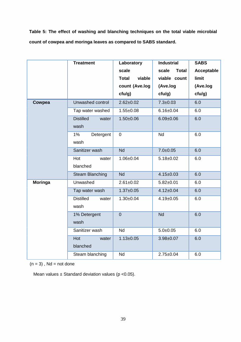
The washing technique is not only used to remove field soil and surface microorganisms, but
also to remove fungicides, insecticides and other pesticides from the leafy vegetables. Tap
water, sanitizer, 1% detergent solution and distilled water were used to wash the different
batches of the cowpea and moringa leaves, so as to determine the most effective washing
techniques (Table 5). Washing the vegetables twice was found to be the most effective
washing technique because it gave a relatively lower total viable microbial count of
vegetables, when compared to other washing techniques. Although washing with 1%
detergent water resulted in the lowest total viable microbial count, it affected the sensory
quality of the vegetables negatively, making it slimy and discoloured. Blanching is a short
heat treatment to inactivate enzymes catalase and peroxidase present in the vegetables,
wherein a portion of the 2x tap water washed leaves was plunged into boiling water 94°C (hot
water blanching) for 5min and the second portion was passed through a steam tunnel at
85oC-94°C (steam blanching), removed after 12min the optimum steam blanching time (the
shortest treatment time which gave a negative result to the peroxidase and catalase test).
These results are indicated in Table 6.
39
Table 5: The effect of washing and blanching techniques on the total viable microbial
count of cowpea and moringa leaves as compared to SABS standard.
Treatment Laboratory
scale
Total viable
count (Ave.log
cfu/g)
Industrial
scale Total
viable count
(Ave.log
cfu/g)
SABS
Acceptable
limit
(Ave.log
cfu/g)
Cowpea Unwashed control 2.62±0.02 7.3±0.03 6.0
Tap water washed 1.55±0.08 6.16±0.04 6.0
Distilled water
wash
1.50±0.06 6.09±0.06 6.0
1% Detergent
wash
0 Nd 6.0
Sanitizer wash Nd 7.0±0.05 6.0
Hot water
blanched
1.06±0.04 5.18±0.02 6.0
Steam Blanching Nd 4.15±0.03 6.0
Moringa Unwashed 2.61±0.02 5.82±0.01 6.0
Tap water wash 1.37±0.05 4.12±0.04 6.0
Distilled water
wash
1.30±0.04 4.19±0.05 6.0
1% Detergent
wash
0 Nd 6.0
Sanitizer wash Nd 5.0±0.05 6.0
Hot water
blanched
1.13±0.05 3.98±0.07 6.0
Steam blanching Nd 2.75±0.04 6.0
(n = 3) , Nd = not done
Mean values ± Standard deviation values (p <0.05).

40
Table 6: Determination of optimum steam blanching time using catalase and
peroxidase activities
Sample
Treatment
period
0
min
3
min
6
min
9
min
12
min
18
min
24
min
30
min
36
min
Total Viable
count
7.7
5
7.57 7.9 7.34 3.57 4 3.7 3 3.33
Catalase
test
+ve +ve +ve +ve -ve -ve -ve -ve -ve
Peroxidase
test
+ve +ve +ve +ve -ve -ve -ve -ve -
(n = 3)
4.1. Up-scaled processing techniques:
Washing twice with tap water and steam blanch techniques gave best results for both lab and
pilot-scale which conforms to SABS standard on the level of vegetables microbial load. Table
5 shows the results of the average log of the total viable microbial count.
Table 6 reveals the results of peroxidase and catalase test on cowpea leaves after steam
blanching at different time internals. At 12min the leaves tested negative for both catalase
and peroxidase test, indicating the optimum blanching time for enzyme inactivation.
41
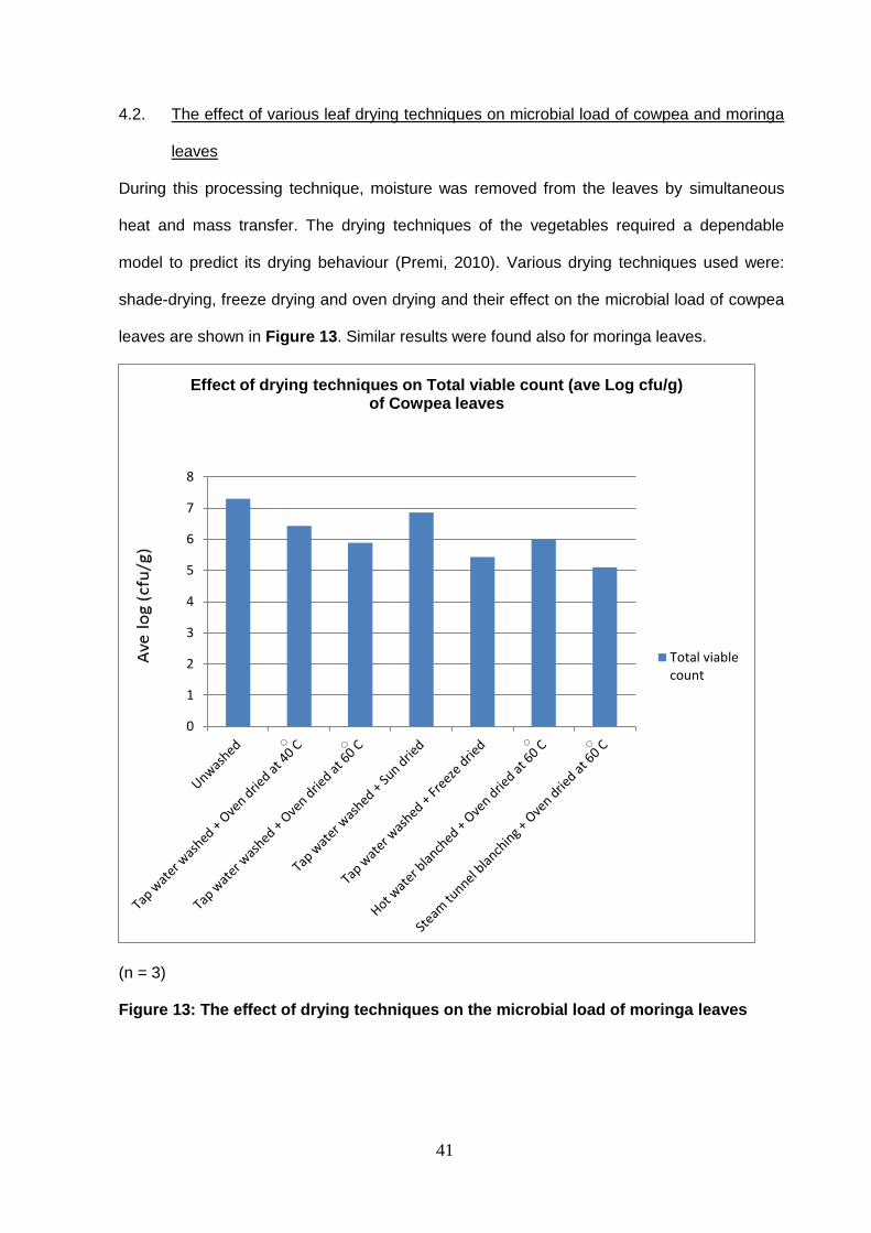
4.2. The effect of various leaf drying techniques on microbial load of cowpea and moringa
leaves
During this processing technique, moisture was removed from the leaves by simultaneous
heat and mass transfer. The drying techniques of the vegetables required a dependable
model to predict its drying behaviour (Premi, 2010). Various drying techniques used were:
shade-drying, freeze drying and oven drying and their effect on the microbial load of cowpea
leaves are shown in Figure 13. Similar results were found also for moringa leaves.
(n = 3)
Figure 13: The effect of drying techniques on the microbial load of moringa leaves
0
1
2
3
4
5
6
7
8
Effect of drying techniques on Total viable count (ave Log cfu/g) of Cowpea leaves
Total viablecount

42
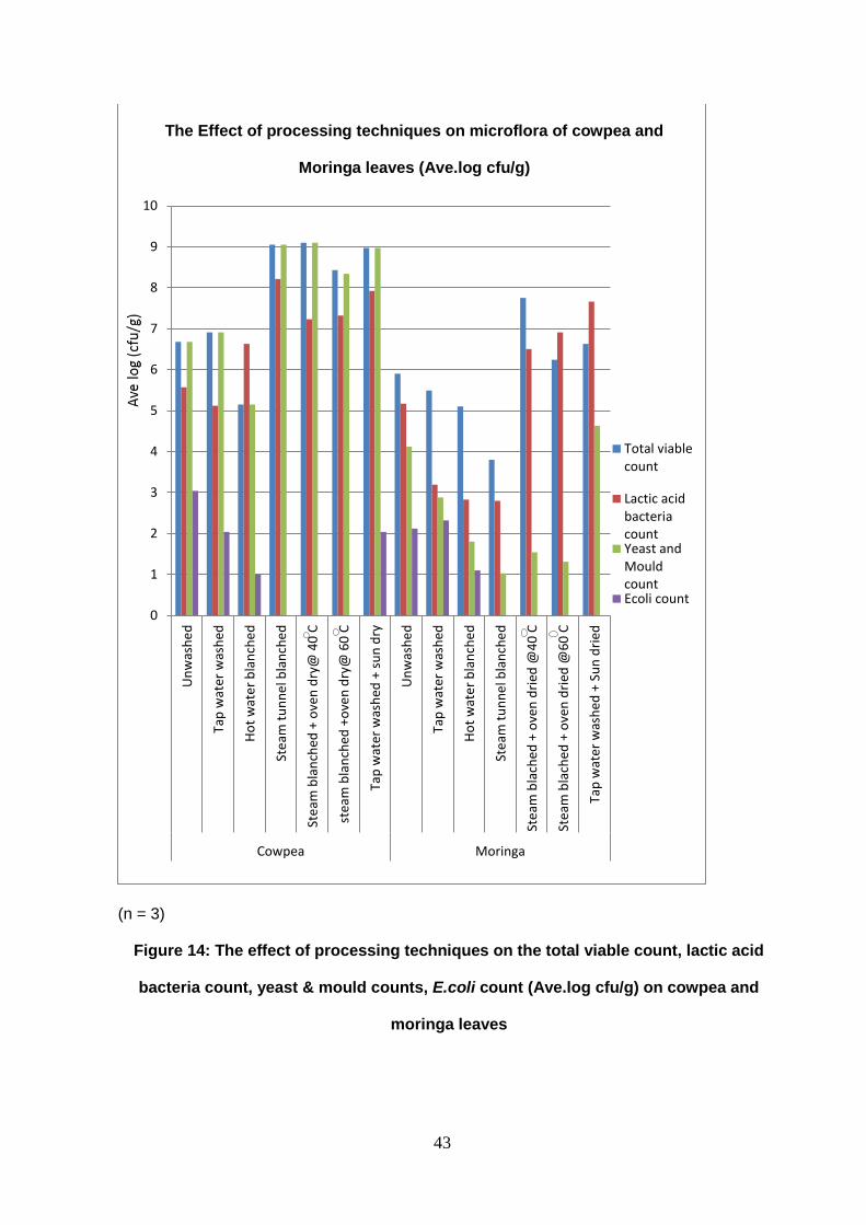
4.3. The effect of processing techniques on the microflora of cowpea and moringa leaves
The results of total viable count, lactic acid bacteria count, yeast and mould count and E.coli
count for cowpea and moringa leaves (Figure 14) show the survival, growth or reduction of
the total viable count, total lactic acid bacteria count, total yeast and moulds count and total
E.coli count under different processing technique conditions.
Steam blanching at 94oC was the most effective technique in terms of reduction of microbial
load, and it also eliminated all presence of E.coli from the vegetable samples.
43
(n = 3)
Figure 14: The effect of processing techniques on the total viable count, lactic acid
bacteria count, yeast & mould counts, E.coli count (Ave.log cfu/g) on cowpea and
moringa leaves
0
1
2
3
4
5
6
7
8
9
10
Un
was
he
d
Tap
wat
er w
ash
ed
Ho
t w
ate
r b
lan
che
d
Ste
am t
un
ne
l bla
nch
ed
Ste
am b
lan
ched
+ o
ven
dry
@ 4
0 C
stea
m b
lan
ched
+o
ven
dry
@ 6
0 C
Tap
wat
er w
ash
ed +
su
n d
ry
Un
was
he
d
Tap
wat
er w
ash
ed
Ho
t w
ate
r b
lan
che
d
Ste
am t
un
ne
l bla
nch
ed
Ste
am b
lach
ed +
ove
n d
ried
@4
0 C
Ste
am b
lach
ed +
ove
n d
ried
@6
0 C
Tap
wat
er w
ash
ed +
Su
n d
ried
Cowpea Moringa
Total viablecount
Lactic acidbacteriacountYeast andMouldcountEcoli count
The Effect of processing techniques on microflora of cowpea and
Moringa leaves (Ave.log cfu/g)

44
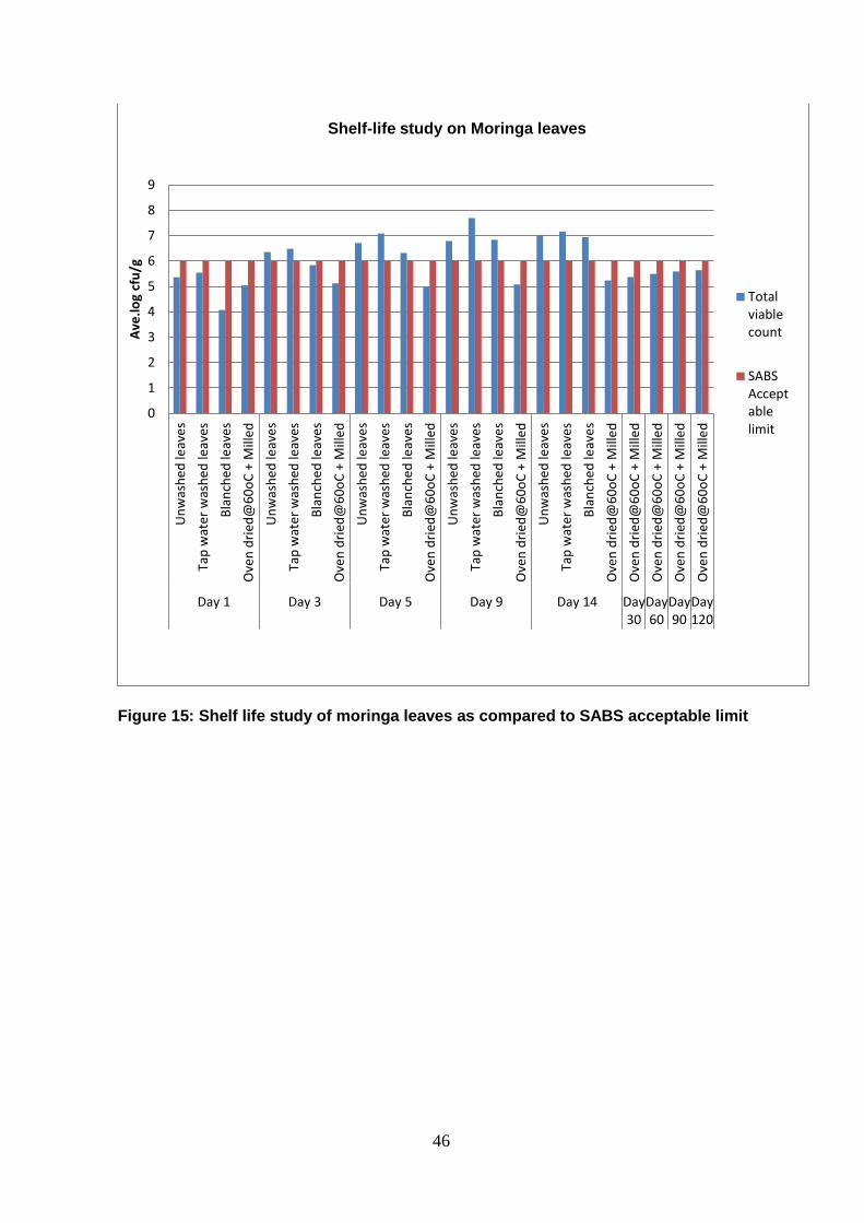
4.3.1. Shelf –life study
The shelf–life of tap water washed, steam blanched and milled moringa leaves were studied
over a period of time, and the respective microbial load is listed in Table 7. The sensory
evaluation result on the shelf life of moringa leaves are also listed in Table 8. These shelf life
studies revealed that there was a significant increase in the microbial load of the washed and
blanched leaves after day 4; this could be as a result of its higher moisture content than the
unwashed leaves or maybe mechanical damage during washing and handling which could
have led to contaminants finding entry into the leaves. Dried stored leaves maintained a
constant level of microbial load for the first six months only with an insignificant variation
(Table 7).
Figure 15 shows the level of the microbial load over the period of shelf life study as
measured against the acceptable level of South African Bureau of Standards in regards to
food safety, acceptable limit of microbial load (https://www.capetown.gov.za/en
/CityHealth/Documentation).
This means on day 1 both processed and unprocessed moringa leaves are safe for
consumption after which only the dried leaves are safe for consumption.

45
Table 7: Shelf life study on moringa leaves
Treatment/ Total viable count (Ave. log cfu/g)
Day(s) Unwashed
leaves
Tap water
washed
leaves
Steam
blanched
leaves
Oven dried
leaves @ 60oC
1 5.4±0.04 5.5±0.06 4.1±0.07 5.0±0.03
3 6.4±0.03 6.2±0.03 5.8±0.05 5.1±0.04
5 6.7±0.05 7.1±0.07 6.3±0.09 5.0±0.05
9 6.8±0.08 7.7±0.05 6.8±0.04 5.1±0.04
14 7±0.02 7.2±0.05 6.9±0.06 5.2±0.03
30 >1000 >1000 >1000 5.4±0.01
60 >1000 >1000 >1000 5.5±0.01
90 >1000 >1000 >1000 5.6±0.03
120 >1000 >1000 >1000 5.6±0.02
(n = 3) Mean values ± Standard deviation values (p <0.05).
46
Figure 15: Shelf life study of moringa leaves as compared to SABS acceptable limit
0
1
2
3
4
5
6
7
8
9U
nw
ash
ed
leav
es
Tap
wat
er w
ash
ed le
aves
Bla
nch
ed
leav
es
Ove
n d
ried
@6
0o
C +
Mill
ed
Un
was
he
d le
ave
s
Tap
wat
er w
ash
ed le
aves
Bla
nch
ed
leav
es
Ove
n d
ried
@6
0o
C +
Mill
ed
Un
was
he
d le
ave
s
Tap
wat
er w
ash
ed le
aves
Bla
nch
ed
leav
es
Ove
n d
ried
@6
0o
C +
Mill
ed
Un
was
he
d le
ave
s
Tap
wat
er w
ash
ed le
aves
Bla
nch
ed
leav
es
Ove
n d
ried
@6
0o
C +
Mill
ed
Un
was
he
d le
ave
s
Tap
wat
er w
ash
ed le
aves
Bla
nch
ed
leav
es
Ove
n d
ried
@6
0o
C +
Mill
ed
Ove
n d
ried
@6
0o
C +
Mill
ed
Ove
n d
ried
@6
0o
C +
Mill
ed
Ove
n d
ried
@6
0o
C +
Mill
ed
Ove
n d
ried
@6
0o
C +
Mill
ed
Day 1 Day 3 Day 5 Day 9 Day 14 Day30
Day60
Day90
Day120
Ave
.lo
g cf
u/g
Totalviablecount
SABSAcceptablelimit
Shelf-life study on Moringa leaves
47
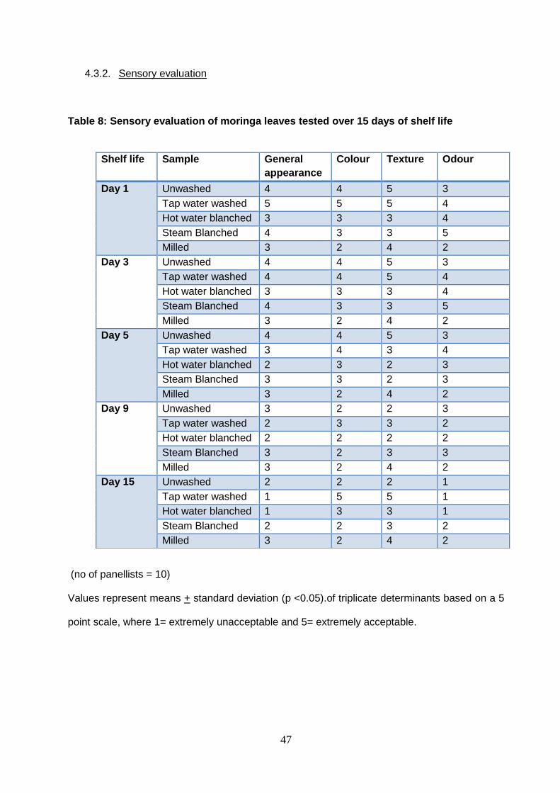
4.3.2. Sensory evaluation
Table 8: Sensory evaluation of moringa leaves tested over 15 days of shelf life
(no of panellists = 10)
Values represent means + standard deviation (p <0.05).of triplicate determinants based on a 5
point scale, where 1= extremely unacceptable and 5= extremely acceptable.
Shelf life Sample General
appearance
Colour Texture Odour
Day 1 Unwashed 4 4 5 3
Tap water washed 5 5 5 4
Hot water blanched 3 3 3 4
Steam Blanched 4 3 3 5
Milled 3 2 4 2
Day 3 Unwashed 4 4 5 3
Tap water washed 4 4 5 4
Hot water blanched 3 3 3 4
Steam Blanched 4 3 3 5
Milled 3 2 4 2
Day 5 Unwashed 4 4 5 3
Tap water washed 3 4 3 4
Hot water blanched 2 3 2 3
Steam Blanched 3 3 2 3
Milled 3 2 4 2
Day 9 Unwashed 3 2 2 3
Tap water washed 2 3 3 2
Hot water blanched 2 2 2 2
Steam Blanched 3 2 3 3
Milled 3 2 4 2
Day 15 Unwashed 2 2 2 1
Tap water washed 1 5 5 1
Hot water blanched 1 3 3 1
Steam Blanched 2 2 3 2
Milled 3 2 4 2
48
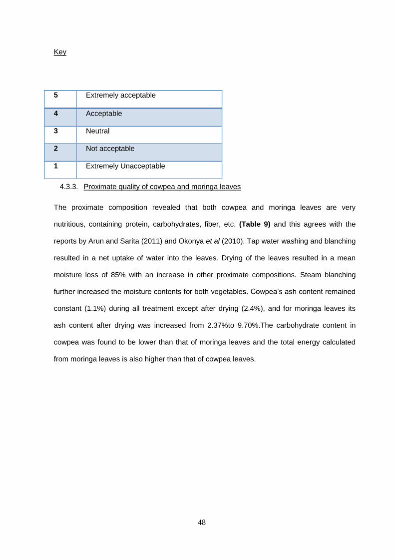
Key
4.3.3. Proximate quality of cowpea and moringa leaves
The proximate composition revealed that both cowpea and moringa leaves are very
nutritious, containing protein, carbohydrates, fiber, etc. (Table 9) and this agrees with the
reports by Arun and Sarita (2011) and Okonya et al (2010). Tap water washing and blanching
resulted in a net uptake of water into the leaves. Drying of the leaves resulted in a mean
moisture loss of 85% with an increase in other proximate compositions. Steam blanching
further increased the moisture contents for both vegetables. Cowpea’s ash content remained
constant (1.1%) during all treatment except after drying (2.4%), and for moringa leaves its
ash content after drying was increased from 2.37%to 9.70%.The carbohydrate content in
cowpea was found to be lower than that of moringa leaves and the total energy calculated
from moringa leaves is also higher than that of cowpea leaves.
5 Extremely acceptable
4 Acceptable
3 Neutral
2 Not acceptable
1 Extremely Unacceptable
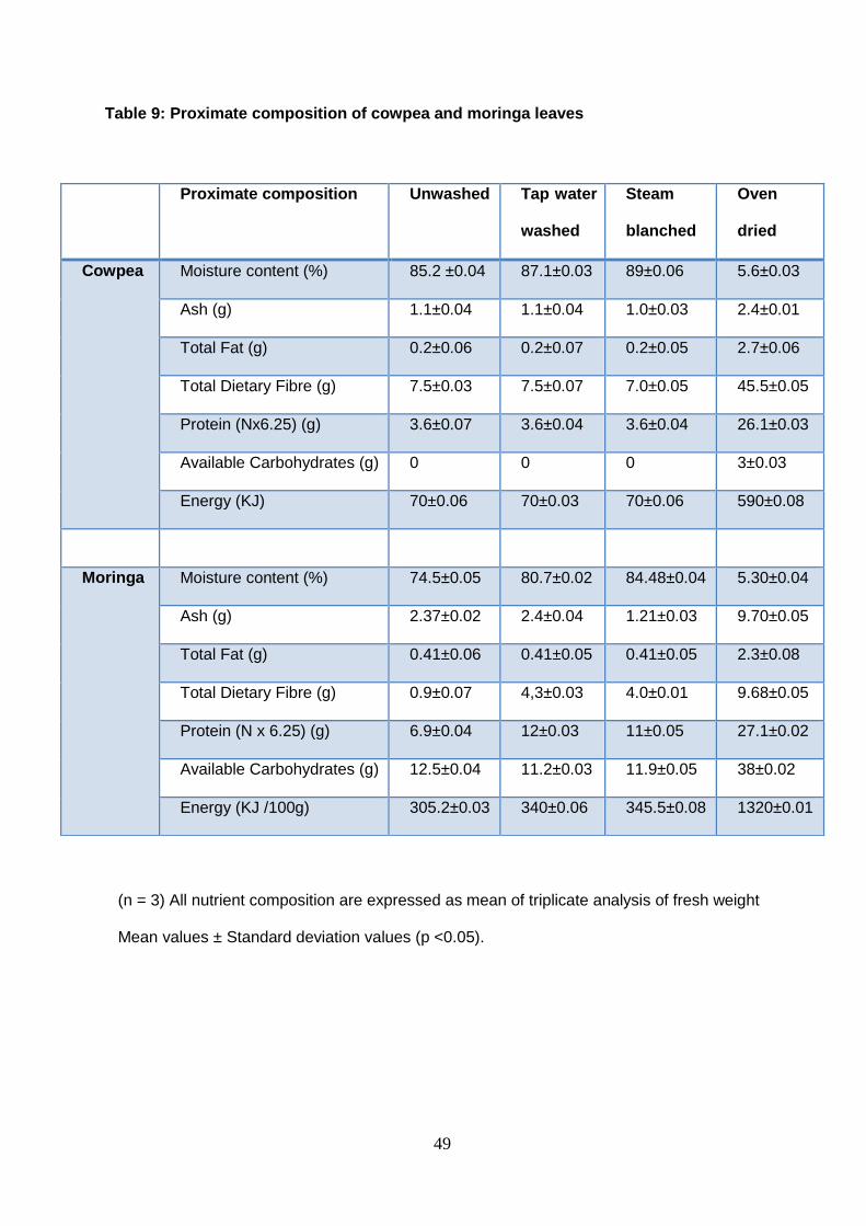
49
Table 9: Proximate composition of cowpea and moringa leaves
(n = 3) All nutrient composition are expressed as mean of triplicate analysis of fresh weight
Mean values ± Standard deviation values (p <0.05).
Proximate composition Unwashed Tap water
washed
Steam
blanched
Oven
dried
Cowpea Moisture content (%) 85.2 ±0.04 87.1±0.03 89±0.06 5.6±0.03
Ash (g) 1.1±0.04 1.1±0.04 1.0±0.03 2.4±0.01
Total Fat (g) 0.2±0.06 0.2±0.07 0.2±0.05 2.7±0.06
Total Dietary Fibre (g) 7.5±0.03 7.5±0.07 7.0±0.05 45.5±0.05
Protein (Nx6.25) (g) 3.6±0.07 3.6±0.04 3.6±0.04 26.1±0.03
Available Carbohydrates (g) 0 0 0 3±0.03
Energy (KJ) 70±0.06 70±0.03 70±0.06 590±0.08
Moringa Moisture content (%) 74.5±0.05 80.7±0.02 84.48±0.04 5.30±0.04
Ash (g) 2.37±0.02 2.4±0.04 1.21±0.03 9.70±0.05
Total Fat (g) 0.41±0.06 0.41±0.05 0.41±0.05 2.3±0.08
Total Dietary Fibre (g) 0.9±0.07 4,3±0.03 4.0±0.01 9.68±0.05
Protein (N x 6.25) (g) 6.9±0.04 12±0.03 11±0.05 27.1±0.02
Available Carbohydrates (g) 12.5±0.04 11.2±0.03 11.9±0.05 38±0.02
Energy (KJ /100g) 305.2±0.03 340±0.06 345.5±0.08 1320±0.01

50
4.3.4. Identification of bacteria and fungi contaminant found on the leaves of treated and
untreated moringa and cowpea leaves
Figure 16 represents the morphology of the yeast cells obtained in leaves of Moringa
oleifera. Morphological and biochemical tests such as the catalase, oxidase and indole tests
were used to identify the bacteria derived from the moringa leaves as indicated in Table 10.
Table 10: The morphological and biochemical characterization of bacteria found on
moringa leaves
Colony
Description
Gram
stain
Cell
Shape
Indole
test
Catalase
test
Suspected
Bacteria
Smooth pink -ve Short Rod +ve +ve E.coli
Mucoid pink -ve Short Rod -ve +ve Klebsiellia
pneumonia
Mucoid pink -ve Rod -ve +ve Enterobacter spp.
Red pink -ve Short Rod -ve +ve Serratia
marcescens
Pale colored -ve Rod +ve +ve Salmonella spp.
Pale colored -ve Rod -ve +ve Shigella
dysenteriae
Mucoid pale +ve Rod -ve +ve Bacillus
Yellow +ve Round -ve -ve Staphylococcus
Joined
together in a
chain
+ve Round -ve -ve Streptococcus
Cream/ yellow -ve Rod -ve +ve Pseudomonas
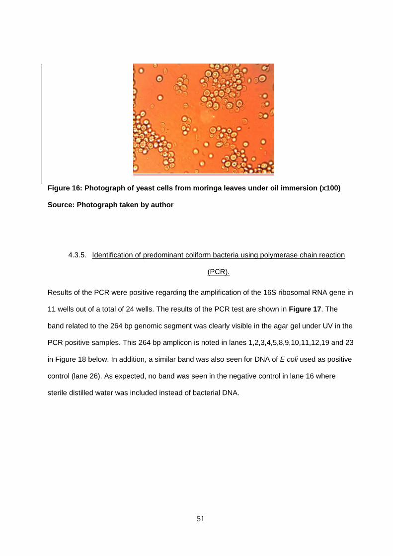
51
Figure 16: Photograph of yeast cells from moringa leaves under oil immersion (x100)
Source: Photograph taken by author
4.3.5. Identification of predominant coliform bacteria using polymerase chain reaction
(PCR).
Results of the PCR were positive regarding the amplification of the 16S ribosomal RNA gene in
11 wells out of a total of 24 wells. The results of the PCR test are shown in Figure 17. The
band related to the 264 bp genomic segment was clearly visible in the agar gel under UV in the
PCR positive samples. This 264 bp amplicon is noted in lanes 1,2,3,4,5,8,9,10,11,12,19 and 23
in Figure 18 below. In addition, a similar band was also seen for DNA of E coli used as positive
control (lane 26). As expected, no band was seen in the negative control in lane 16 where
sterile distilled water was included instead of bacterial DNA.

52
Figure 17: Genomic Bacterial DNA isolated from moringa leaves subjected
to PCR with primers pAF and pHR. The horizontal arrows indicate the
position of the 264 bp amplicon. Lanes 1 to 15 and 18 to 26 show the results
of amplification of bacterial DNA. Samples in lanes 1 to 5, 8 to 12, 19, 23 and
26 were successfully amplified with this primer pair and thermal conditions.
Lanes marked L contains 50 bp molecular weight ladder DNA. Lane 16 is a
negative control reaction and lane 26 the positive control.
.
L 1 2 3 4 5 6 7 8 9 10 11 12 13 14 15 16 L
L 17 18 19 20 21 22 23 24 25 26 27 L

53
DISCUSSION
Cowpea and moringa leaves have been reported to be highly nutritious and to have
medicinal properties. However, they are still prone to microbial contamination resulting from
pre-harvest or post-harvest contamination. Possible sources of contamination are manure-
contaminated soil, irrigation water, water used to apply fungicides and insecticides, dust,
insects, inadequately composted manure, human handling, harvesting equipment, transport
containers, transport vehicles, processing equipment. The practice of animal grazing may
result in the introduction into the soil of enteric bacteria such as Salmonella, E. coli O157:H7,
and Listeria monocytogenes (Barth et al, 2010; Ibrahim and Jude 2009).
To reduce microbial contamination of the vegetables, good agricultural practices, high levels
of worker hygiene and proper human waste management at production sites should be
enforced, but most importantly, effective processing techniques must be utilized in eliminating
these microbial contaminants from the vegetables (Ibrahim and Jude, 2009).
Microbiological analysis
As part of the objectives of this research, the effect of processing techniques on microflora
and nutrient contents of cowpea and moringa leaves were investigated. The high average log
cfu/g of viable counts from the unwashed cowpea leaves could be attributed to unhygienic
farm conditions, contamination from the soil, and contamination from harvesting, packaging
and transportation (Nielsen et al, 2000). The high moisture content of the fresh leaves also
provided a favourable environment for the growth of microorganisms (Sanjukta et al, 2013).
The relatively lower microbial load on unwashed moringa leaves as compared to cowpea
leaves could be because the moringa leaves grow well above the soil as compared to the
cowpea leaves that grow very close to the soil. The reduced bacterial count may also be due
to its reported anti-microbial properties (Abalaka et al, 2012). All the processing techniques
showed a reduction in the average log of microbial density. Washing with detergent was the

54
most effective of the washing modes, but it made the leaves soapy and slimy, an undesirable
characteristic. Thus, tap water washing was the preferred method, while the use of the
sanitizer wash had the lowest effect on the microbial load of the vegetables. Steam blanching
was more effective than hot water blanching, and oven drying at 60oC was the most effective
preservation technique when compared to other drying techniques.
The effect of drying techniques on the microflora of cowpea and moringa were also studied.
High moisture content provide a favourable environment for the growth of microorganisms,
thus bacterial growth was reduced during drying. Sun-drying in direct sunshine and under
shade is the common practices used in most parts of the world to preserve vegetables for dry
season consumption (Nielson et al, 200). However, vegetable processing and preservation
techniques may significantly affect the concentration and availability of nutrients and other
essential compounds in food.
Different numbers and types of microorganisms were found to be present on the surface of
cowpea and moringa leaves. Bacterial and fungal colonies were found when it was plated on
Tryptose soy agar media and acidified Potato Dextrose media, respectively. The results
obtained for bacterial colonies and fungal cells are shown in Table 10 and Figure 16. Spore
formers were abundant in cowpea (Average log cfu/g <9) and moringa leaves (Average
log,cfu/g <7 ), several species of Bacillus were isolated and this could be as a result of the
soil flora associated with these vegetables during growth. This agrees with the findings of
Mpuchane and Gashe (1998) who reported several species of bacillus (<107) from traditional
leafy vegetables in Botswana.
Coliform bacteria counts are used for monitoring the bacteriological safety of food, based on
the fact that the presence of coliform bacteria in food, water or vegetables is an indicator of
level of hygiene and potential human fecal contamination and therefore the possible
presence of enteric pathogens (Asim et al, 1989).The presence of pathogenic organism
55
which belong to this family has been reported in many fresh leafy vegetables (Mpuchane and
Gashe, 1998), and this is mostly associated with the use of faeces-contaminated water and
soil and crops contaminated by faeces from wild and farm animals and birds.
Proximate Analysis
The proximate composition revealed that both cowpea and moringa leaves are very nutritious
(Table 9) and this agrees with the reports by Arun and Sarita (2011) and Okonya et al
(2010).
Table 9 reveals values for moisture content and shows that cowpea has the highest value
(87.1%) (tap water washed cowpea leaves) making cowpea leaves more prone to spoilage
since foods with high moisture content are more prone to perishability (Sanjukta et al, 2013).
The ash content of moringa leaves was higher than that of the cowpea leaves. The high ash
content of the moringa leaves is a reflection of the mineral contents preserved in the food
materials. The results, therefore, suggest a high deposit of mineral elements in the leaves.
The total fat content of water-treated cowpea leaves (0.2g) was lower than that of the
moringa leaves (0.41g). A diet including moringa leaves may be more palatable than that of
cowpea leaves because dietary fats function to increase food palatability by absorbing and
retaining flavours (Sanjukta et al, 2013). A diet providing 1 - 2% of its caloric energy as fat is
said to be sufficient for human beings, as excess fat consumption contributes to certain
cardiovascular disorders such as atherosclerosis, cancer and aging (Ogbe and John, 2011).
These vegetables are low-fat foods.
In comparison, moringa leaves had a higher protein content than the cowpea leaves. This
makes moringa leaves a good source of protein. The protein content of moringa samples
varied and are lower than those described by Ogbe et al, (2012) and Oduro et al (2008).
These differences could be attributed to geographical locations of growth and stage of
maturity of the plant (Ogbe et al, 2012).
56
The crude fibre content of moringa leaves was higher than that of cowpea leaves, and this
makes it a more favourable vegetable since high fibre content of foods help in digestion and
prevention of colon cancer (Mepha et al, 2007). Non-starchy vegetables are the richest
sources of dietary fibre (Sanjukta et al, 2013) and are employed in the treatment of diseases
such as obesity, diabetes and gastrointestinal disorders. The caloric value obtained showed
fresh M. oleifera as having the lowest value (305.62 cal/g). The caloric value of these leaves
make them a good source of energy for all.
Shelf life studies revealed that there was a significant increase in the microbial load of the
washed and blanched leaves after day 4. This could result from its high moisture contents
compared to the unwashed leaves (Figure 15). Dried stored leaves maintained a constant
level of microbial load for the first six months only with an insignificant variation (+0.02).
The sensory evaluation revealed that on the 15th day, both the processed and unwashed
moringa leaves were spoilt and unacceptable for consumption. This means that the shelf life
of fresh moringa leaves whether processed or not is 9 days, except for the steam blanched
that has a longer shelf life of 15 days. However, from the 3rd day, it is no longer safe
microbiologically to consume the processed vegetable with the exception of the milled
sample that had a constant microbial level and sensory test over a period of 120 days.
57
Conclusion
According to the South African regulations governing microbiological standards for foodstuffs
and related matters in 2002 that state that “in the food industry, a product shall be deemed to
be contaminated, impure, decayed or harmful or injurious to human health if any such
product contains more than 106cfu per gram for total viable count, 104cfu per gram of yeasts
and moulds and 103cfu per gram of coliforms (https://www.capetown.gov.za/e/CityHealth).
Although limitations exist, the results of this research revealed that cowpea and moringa
leaves are good sources of nutrients, but that they harbour microbial contaminants and are,
therefore, unsafe to eat without using proper processing techniques. Most South Africans
prefer to utilise fresh vegetables even though the nutritional quality of dry vegetables is
better, as this concentrates the nutrients and has a longer shelf-life. Hence, the quality of
fresh produce of the local market must be maintained in hygienic conditions and proper
handling, transport, storage must be ensured so that risk of contaminants decreases and
chances of food borne disease outbreaks can be minimized. This can be achieved by pre-
treatment of fresh vegetables by tap water washing twice and steam blanching so as to
decrease the density of microbial contaminants on the surface of the vegetables, while oven
drying at 60oC, followed by milling and packaging in an air tight container will prolong the
product shelf life. The consumption of these processed vegetables should be encouraged
and promoted as vegetables in South Africa and other developing countries, due to their high
nutrient content and other nutraceutical benefits.
58
RESEARCH DATA ANALYSES
Four groups of microorganisms were evaluated at five different processing points (fresh
leaves, washing, drying, milling and storage (shelf-life). The experiment was repeated three
times, once per month consecutively for four months. The results are presented with their
average log values, means, standard deviation and standard error.
The data were analysed using the Microsoft Excel package and compared by ANOVA using
the GLM procedure.
ETHICAL CONSIDERATIONS
Research work was carried out in the CSIR Food Analysis Laboratory. This is a government-
and UNISA-approved institution that may execute all necessary procedures required by the
study, under supervision of Ms Annali Jacobs (CSIR Senior Researcher) and by Dr
Khayaletu Ntushelo at the UNISA molecular laboratory. Ethics approval was obtained from
the UNISA Ethics Committee under the number 2013/CAES/060. The laboratory code of
conduct and safety regulations was strictly adhered to, so as to prevent any harm or injuries
to personnel.
Plagiarism was avoided and all citations were done in the form of interpretation and
reconstruction of published works. Any references to uses of moringa by communities
acknowledge and respect the present indigenous knowledge and dietary behaviour.
All research work in the CSIR would be kept strictly confidential until published as a
completed MSc dissertation.
59
Finally, the study plants (Vigna unguiculata and Moringa oleifera) needed for the research
was supplied by CSIR from cultivated crops. This ensured sustainable use of the plant
without damaging effects on the environment or biodiversity.
QUALITY ASSURANCE:
All methods used in this research project are CSIR approved methods which have been
tested and proven to be the most reliable analytical methods. These methods are also
accredited by SABS: South African Bureau of Standards, ISO: International Standards
Organisation and SANAS.

60
REFERENCES
ABALAKA M.E., DANIYAN S.Y., OYELEKE S.B. and ADEYEMO S.O. (2012). The
Antibacterial Evaluation of Moringa Oleifera Leaf Extracts on Selected Bacterial Pathogens.
Journal of Microbiology Research. Vol 2 pp 1 - 4.
AGYEPONG A.O. (2009). The Possible Contributions of Moringa oleifera LAM Leaves to
Dietary quality in Two Bapedi Communities in Makopane, Limpopo Province. Masters of
Human Ecology, Dissertation at University of South Africa.
AKINYELE R.O., OLUWADUNSIN A. and OMOYENI A. (2010). Proximate, mineral, anti-
nutrients and phytochemical screening and amino acid composition of the leaves of
Pterocarpusmildbraedi Harms. Journal of Environmental, Agricultural and Food Chemistry
(EJEAFChe). Vol 9(8) pp 1322-1333.
ANWAR F., LATIF S., ASHRAF M. and GILANI, A.H. (2007). Moringa oleifera: a food plant with
multiple medicinal uses. Department of Chemistry, University of Agriculture, Pakistan. Vol 21(1)
pp 17 - 25.
AOAC (1990). Official Methods of Analysis of the Association of Official Analytical Chemists
(15th edition), Washington DC. pp 992 – 995.
ARUN P.R. and SARITA S. (2011). Comparative analysis of preservation techniques on
Moringa Oleifera.International Journal of Agricultural and Food Science. Accepted 02 June
2011, Available online at http://www.urpjournals.com

61
AWANISH P., PANDEY R.D., TRIPATHI P., GUPTA P.P. and HAIDER J.A. (2012). Moringa
Oleifera Lam. (Sahijan) - A Plant with a Plethora of Diverse Therapeutic Benefits: An Updated
Retrospection. Medicinal Aromatic Plants. Vol 1, pp 101.
BARTH M., THOMAS R.H., ZHUANG H. and BREIDT F. (2009). Microbiological spoilage of
fruits and vegetables. Food Microbiology and Food Safety Journal. Vol? pp?
BORLING J. (2010). Feed Improvement by Energy efficient Storage Using Pichiaanomalia
Inoculated ensiled Cereal grain. Independent project / degree project in Biology, Swedish
University of Agricultural Science. ISSN 1101-8151.
BRUCE K.D., HIORNS W.D., HOBMAN J.L., OSBORN A.M., STRIKE P. and RITCHIE D.A.
(1992). Amplification of DNA from native populations of soil bacteria using Polymerase Chain
Reaction. Applied Environmental Microbiology. Vol 58 pp 3413 -3416.
DAVIS D.W., OELKE E.A. OPLINGER E.S. , DOLL J.D., HANSON C.V. and PUTNAM D.H.
(1991). Cowpea; Alternative field crop manual. Departments of Horticultural Science,
Agronomy and Plant Genetics, Centre for Alternative Plant and Animal Products, Minnesota
Extension Service, University of Minnesota, St. Paul. pp?
FAHEY J.W. (2005). ‘Moringa oleifera: A review of the Medical Evidence for its Nutritional,
Therapeutic, and Prophylactic Properties Part 1’, Trees for Life Journal. Pp?
FALADE K. and KOLAWOLE T. (2013). Effect of Irradiation dose on the Physical, Functional
and pasting properties of Cowpea (Vigna Unguiculata (L) Walp) Cultivars.Journal of Food
Process Engineering. Vol 36(2) pp 147-159.
62
FOOD AND AGRICULTURE ORGANIZATION OF THE UNITED NATIONS (1995). Vegetable
Specific Processing Technologies, Chap 9. FAO Agricultural Service Bulletin. ISBN: 92-5-
103657
FUGLIE L.J. (1999).The Miracle Tree: Moringa oleifera: Natural Nutrition for the Tropics.
Church World Service, Dakar. 68 pp.; revised in 2001 and published as The Miracle Tree: The
Multiple Attributes of Moringa. pp 172. http://www.echotech.org/bookstore/advance
FUGLIE L.J. (2005). The Moringa Tree: a local solution to malnutrition? Church World Service
in Senegal. GARG N., CHUREY J.Jand SPLITTSTOESSER D.F. (1990). Effect of processing
conditions on the microflora of fresh cut vegetables. Journal for food production, Vol 53 pp 701-
703.
GLEESON E. and O’BEIRNE D. (2004). Effect of process severity on the survival and growth
of E.coli, Listeria on minimally processed vegetables. Journal of Food Control. Vol 16(8) pp
677-685
Guide to minimize microbial food safety hazards for fresh fruits & vegetable. (2000). By U.S
Food and Drug Administration (FDA).
https://www.capetown.gov.za/en/CityHealth/Documentation/Documents/Regul_Microb_standar
ds_Regul_governing_microb_standards_for_foodstuffs_related_matters.pdf
IBRAHIM T.A. and JUDE-OJEI B. (2009). Microbiological Analysis And Effect of Selected
Antibacterial Agents on Microbial Load of Fluted Pumpkin, Cabbage and Bitter leaves. The
internet journal of Microbiology. Vol 7(2) ISSN: 1037 – 8289.
63
INNIS M., GELFAND D. SNINSKY J. and WHITE T. (1990). PCR protocols: a guide to
methods and applications. Book, pp. xviii + 482 pp. ISBN: 0-12-372180-6
JANSEN VAN RENSBURG, AVERBEKE W. VAN., FABER M., JAARSVELD P. VAN.,
HEERDEN, I. VAN., WENHOLD F. and OELOFSE A. (2007). African Leafy Vegetables in
South Africa. Agricultural Research Council- Vegetable and ornamental plant Institute. Private
bag X293, Pretoria 001, South Africa. pp?
JOSHI P.A. and SONALI P.P. (2010). Microbiological Analysis of Fresh Vegetables and Fruits
and Effect of Anti microbial agent on Microbial Load.Department of Microbiology, Birla College
of Art, Science and Commerce, Kalyan, India.
KANTOR LS, LIPTON K, and MANCHESTER A. (2010). Estimating and addressing America’s
food losses. Food Review. Jan-Apr: 2–12.
KIMIYWE J., WAUDO J., MBITHE D. and MAUDUN P.. (2007). Utilization and medicinal value
of indigenous leafy vegetables consumed in urban and per-urban Nairobi. African Journal of
Food, Agriculture, Nutrition and Development. Vol? pp?
McCANCE and WIDDOWSON Eds. (1992).The Composition of Foods. The Royal Society of
Chemistry and Ministry of Agriculture, Fisheries and Food. Xerox Ventura Publisher,
Cambridge, UK. pp 224-270.
MEPBA H.D., EBOH L. and BANIGO D.E.B. (2007). Effect of processing treatment on the
nutritive composition and consumer acceptance of some Nigerian edible leafy vegetables.
African Journal of Food, Agriculture, Nutrition and Development.Vol 7 (ISSN 1684 -5374).
64
MORTON J.F. (1991). The Horseradish tree, Moringa pterygosperma (Moringaceae) - A boon
to arid lands? Econ. Bot. Vol 45 pp 318-333.
MPUCHANE S.F and GASHE B.A. (1998). An Investigation of the Microbial Ecology of Four
Traditional Dried Leafy Vegetables Consumed in Botswana.Botswana Society. Vol.30 pp.139-
146. http://www.jstor.org/stable/40980210. Accessed: 21/01/2014
Natural Resources Conservation Service Available at: http://plants.usda.gov/ (Accessed:
28/08/2012).
NIELSEN S.S., OHLER T.A., MITCHELL C.A. (2000). Cowpea leaves for human consumption;
production, utilization and nutrient composition, West Lafyette, USA: s.n.
ODURO I., ELLIS W.O. and OWUSU D. (2008). Nutritional potential of two leafy vegetables:
Moringa Oleifera and Ipomoea batatas leaves. Scientific Research and Essay. Vol.3(2) pp.
057-060. ISSN: 1992-2248.
OGBE A.O and AFFIKU J.P. (2011). Proximate Study, Mineral and Anti-nutrient Composition of
Moringa Oleifera Leaves Harvested from Lafia, Nigeria Potential Benefit in Poultry Nutrition
and Health. Journal of Microbiology, Biotechnology and Food Sciences. Vol 1 pp 296 - 308.
OKONYA J., MAASS B.L. (2010). Can traditional leafy vegetable reduce protein and
micronutrient malnutrition? A look at the nutrition quality of Cowpea leaves for two landraces
grown in Uganda. Seminar paper from Tropentag 2010, Zurich, Germany “world food system –
A contribution from Europe.
ONAYEMI O. and BADIFU G.I.O. (1987). Effect of Blanching and Drying methods on the
nutitional and sensory quality of Leafy Vegetables. Plant food for Human Nutrition. Vol 37 pp
291 - 298.
65
PLANTS OF HAWAII (2008). Available online at:
http://www.hear.org/starr/images/image/?q=080609-7907&o=plants. (Accessed 14/08/2012)
PREMI M., SHARMA H.K., SARKAR B.C. and SINGH C. (2010). Kinetics of drumstick leaves
(Moringa oleifera) during convectional drying. African Journal of Plant Science. Vol 4(10) pp.
391 - 400. ISSN 1996 – 0824.
RAHMAN M.M., RAHMAN M.M., AKHTER M.J., PANDEYA D.R ., HAQUE M.A., ALAM M.F.
and RAHMAN A. (2010). Control of coliform bacteria detected from diarrhea associated
patients by extracts of Moringa oleifera. Nepal Medical College Journal. Vol12(1) pp 12-19.
JANSEN VAN RENSBURG W.S., AVERBEKE W., SLABBERT R., FABER M., VAN
JAARSVELD ., VAN HEERDEN I., WENHOLD F. and OELOFSE A .(2007). African leafy
vegetables in South Africa. ISSN 0378-4738 = Water SA. Vol 33(3).
SAKALLAH A.S., LANNING W.R., COOPER L. Dial Lysates D., (2013). DNA Fingerprinting of
Crude Bacterial Lysates Using Degenerate RAPD Primers. Genome Research. .Cold Spring
Harbor Laboratory Press.
SANJUKTA K., ATRAYEE M., GHOSH M. and BHATTACHARYYA D.K., (2013). Utilization of
Moringa leaves as Valuable Food Ingredient in Biscuit Preparation. International Journal of
Applied Sciences & Engineering. Vol 1 pp 29-37.
SOUTH AFRICAN DEPARTMENT OF AGRICULTURE, FORESTRY AND FISHERIES., n.d.
Production guidlines for Cowpeas, Republic of South Africa:
66
SOUTH AFRICAN NATIONAL RESEARCH COUNCIL (NRC), (2006).Lost Crops of Africa:
Volume II: Vegetables. [Online]. Available at: http://www.nap.edu/catalog/11763.html
(Accessed: 13/08/2012)
http://www.CaribbeanPot.com
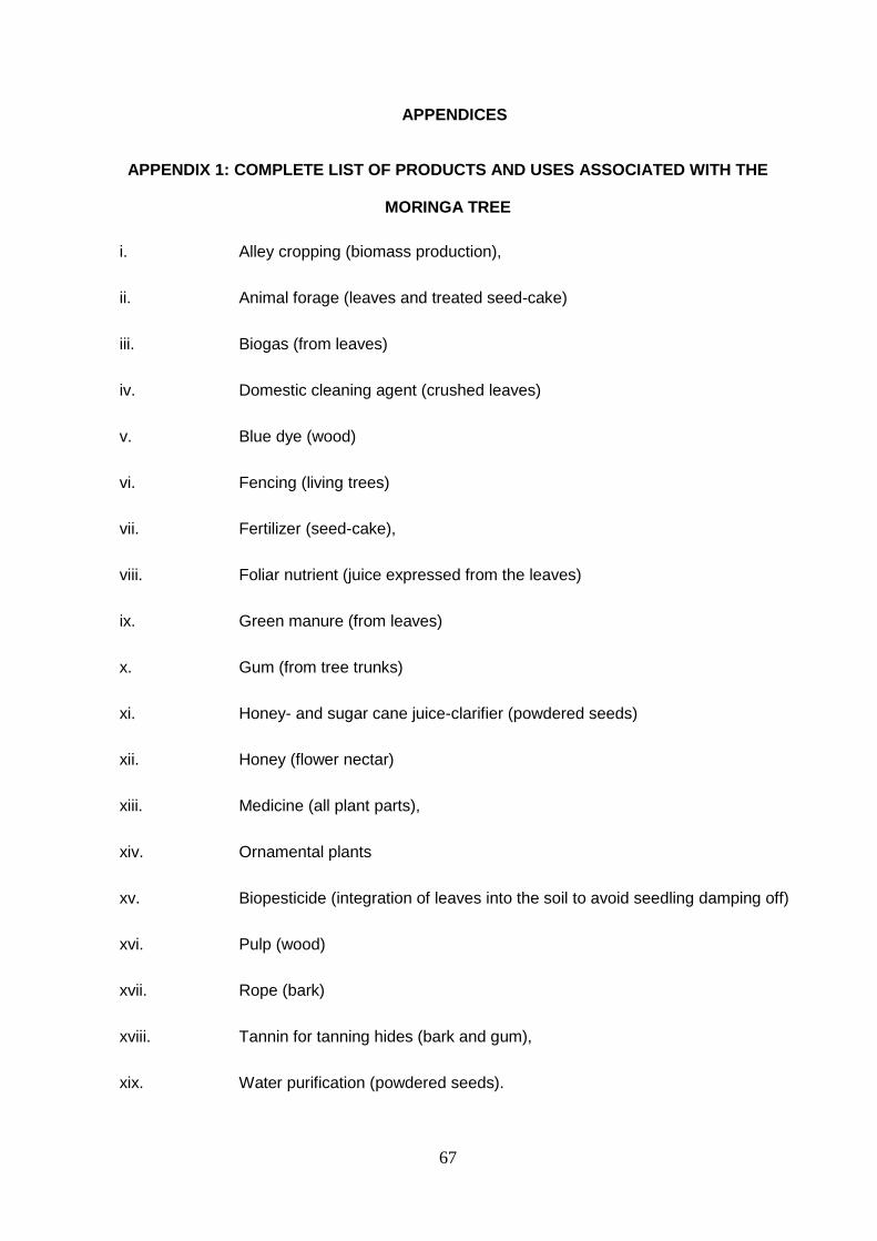
67
APPENDICES
APPENDIX 1: COMPLETE LIST OF PRODUCTS AND USES ASSOCIATED WITH THE
MORINGA TREE
i. Alley cropping (biomass production),
ii. Animal forage (leaves and treated seed-cake)
iii. Biogas (from leaves)
iv. Domestic cleaning agent (crushed leaves)
v. Blue dye (wood)
vi. Fencing (living trees)
vii. Fertilizer (seed-cake),
viii. Foliar nutrient (juice expressed from the leaves)
ix. Green manure (from leaves)
x. Gum (from tree trunks)
xi. Honey- and sugar cane juice-clarifier (powdered seeds)
xii. Honey (flower nectar)
xiii. Medicine (all plant parts),
xiv. Ornamental plants
xv. Biopesticide (integration of leaves into the soil to avoid seedling damping off)
xvi. Pulp (wood)
xvii. Rope (bark)
xviii. Tannin for tanning hides (bark and gum),
xix. Water purification (powdered seeds).

68
xx. Moringa seed oil used in salads,
xxi. Machine lubrication (Seed oil)
xxii. Manufacture of perfume and hair care products (Seed oil).
xxiii. The seeds are also eaten raw, roasted
Adapted from Fahey, (2005) and Agyepong, (2009)
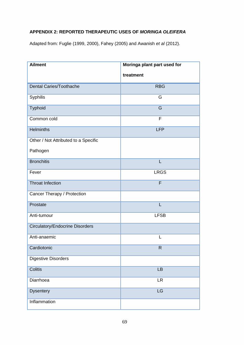
69
APPENDIX 2: REPORTED THERAPEUTIC USES OF MORINGA OLEIFERA
Adapted from: Fuglie (1999, 2000), Fahey (2005) and Awanish et al (2012).
Ailment Moringa plant part used for
treatment
Dental Caries/Toothache RBG
Syphilis G
Typhoid G
Common cold F
Helminths LFP
Other / Not Attributed to a Specific
Pathogen
Bronchitis L
Fever LRGS
Throat Infection F
Cancer Therapy / Protection
Prostate L
Anti-tumour LFSB
Circulatory/Endocrine Disorders
Anti-anaemic L
Cardiotonic R
Digestive Disorders
Colitis LB
Diarrhoea LR
Dysentery LG
Inflammation
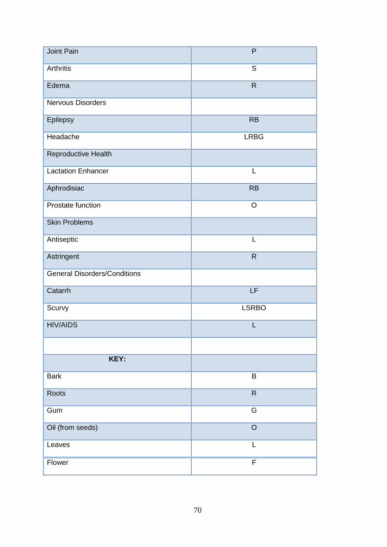
70
Joint Pain P
Arthritis S
Edema R
Nervous Disorders
Epilepsy RB
Headache LRBG
Reproductive Health
Lactation Enhancer L
Aphrodisiac RB
Prostate function O
Skin Problems
Antiseptic L
Astringent R
General Disorders/Conditions
Catarrh LF
Scurvy LSRBO
HIV/AIDS L
KEY:
Bark B
Roots R
Gum G
Oil (from seeds) O
Leaves L
Flower F
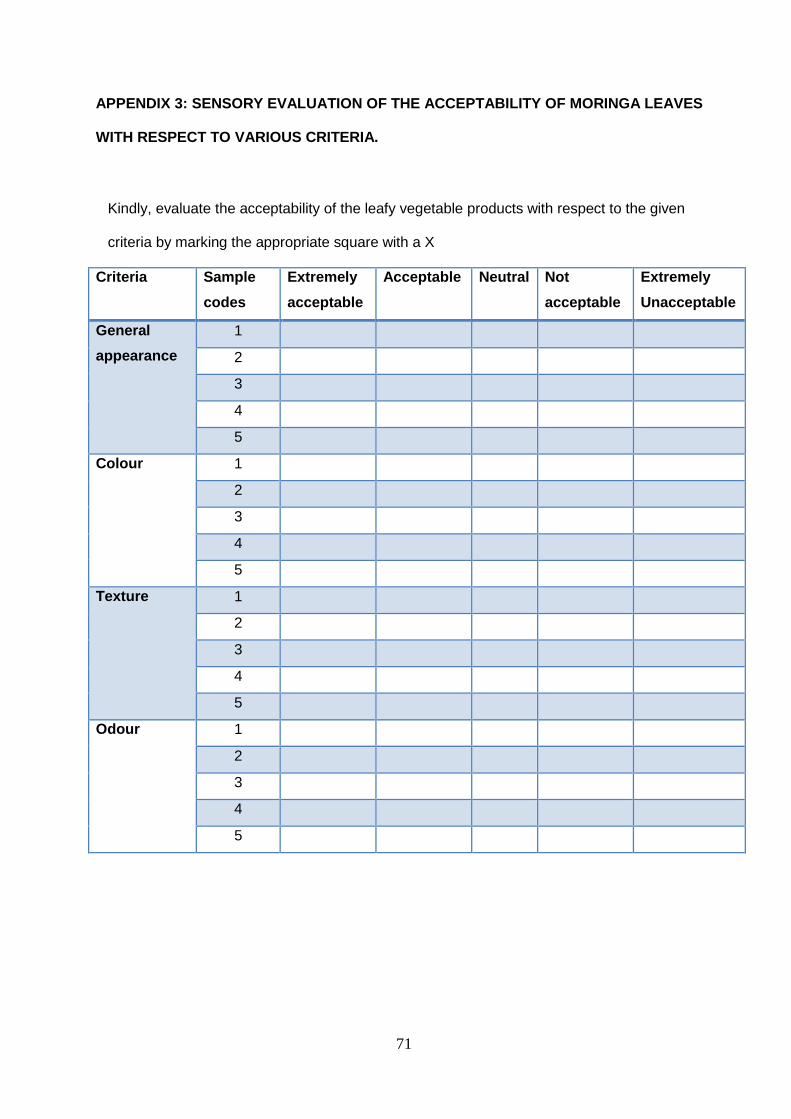
71
APPENDIX 3: SENSORY EVALUATION OF THE ACCEPTABILITY OF MORINGA LEAVES
WITH RESPECT TO VARIOUS CRITERIA.
Kindly, evaluate the acceptability of the leafy vegetable products with respect to the given
criteria by marking the appropriate square with a X
Criteria Sample
codes
Extremely
acceptable
Acceptable Neutral Not
acceptable
Extremely
Unacceptable
General
appearance
1
2
3
4
5
Colour 1
2
3
4
5
Texture 1
2
3
4
5
Odour 1
2
3
4
5