Embed Size (px)
Citation preview

1
The Effect of Sympathetic Nervous System Stimulation on Skeletal Muscle Reflex Response
Time
Anna Batley, Rebeca Cerda, William Dyke, Celena Ho, Abigail Moeller
University of Wisconsin-Madison
Physiology 435
Lab 602, Group 10
May 1, 2018
Key Terms : Sympathetic Nervous System, Heart Rate, Reaction Time, Auditory Stimulus,
Respiration Rate, Sound, Skeletal Muscle
Word Count: 3,628

2
Abstract:
The sympathetic response, also known as fight or flight, is a physiological response that occurs due to a perceived threat to survival. It can physiologically be observed through an increase in heart rate, respiratory rate, blood pressure, oxygen availability, and other mechanisms. In this experiment, we investigate if the fight or flight response induced by an auditory stimuli will quicken reaction time. Thirty participants completed a series of three mazes. A surprise auditory stimuli was played during the third maze to induce a fight or flight response. Throughout the completion of all of the mazes, the respiratory rate and heart rate were measured. After the completion of each maze, participants were asked to complete the Reaction Time Test, which measured response time. It was hypothesized that inducing a fight or flight response, which increases available oxygen and mobilization of energy stores for muscle contraction, would enable muscles to act more quickly resulting in a decreased skeletal muscle response time. A paired t-test with a 95% confidence interval was used to compare all three mazes where a significance criterion of p<0.05 was significant compared to the baseline measurement. Maze 1 to maze 2, maze 1 to maze 3, and maze 2 to maze 3 had p>0.05 indicative that the data did not have a significant difference. Overall, the reaction times were not shown to decrease despite an increase in respiration rate and heart rate among some participants indicative that there was a physiological change in response to the auditory stimuli. However, the physiological changes were not sufficient to cause a decrease in reaction times. Due to the lack of significance in reaction times, the null hypothesis cannot be rejected. Despite the lack of statistical significance, the data can be used to better understand the activation of the sympathetic nervous system, specifically the activation in respect to an auditory stimuli.

3
Introduction
Sympathetic nervous system stimulation increases heart rate, respiratory rate, blood
pressure, and other mechanisms that contribute to a “fight or flight” response. The sympathetic
nervous system is triggered in response to stressors that indicate danger, and its major
components increase an organism’s ability to have a fight or flight response to that danger.
However, it is unclear if the activation of the sympathetic nervous system actually leads to a
quicker skeletal muscle reaction response time. Muscle reaction time is defined as “the time
between the presentation of a sudden stimulus and the onset of a person’s motor response”
(Schmidt & Wrisberg , 2008). In this study we test to see if increasing a person’s heart rate and
respiratory rate leads to quicker skeletal muscle response times.
Stressful or startling situations elevate activity in the sympathetic nervous system as seen
in multiple investigations. According to a study conducted on hypertensive rats by Fischer L.D.
et al, there was proven to be a sympathetically driven increase in heart rate due to an air jet
noise. This stimuli in turn stimulated an increase in epinephrine, norepinephrine, dopamine, and
cortisol, which are key factors in the activation of the sympathetic nervous system (Fischer L.D.,
1991) .
In a similar experiment conducted by Mahmood et al. on the effect of noise on heart rate
in humans, the conclusion was made that there was an increase in heart rate with exposure to
noise (Mahmood, 2006). Noise is an important factor in fear and can have a large impact on
sympathetic activation. In both the study done by Mahmood et al. on humans, and the study done
by Fischer L.D. et al on rats, noise was the factor chosen to stimulate the sympathetic nervous
system and activate that fight or flight response. Specifically, it is the amygdala that is stimulated

4
by fear upon hearing a scream or startling noise which induces the secretion of epinephrine
(Akpan, 2015). Epinephrine, along with other hormones such as norepinephrine, dopamine, and
cortisol, are important as they stimulate the sympathetic nervous system. Stimulation of the
sympathetic nervous system allows heart rate, blood pressure, and breathing rate to increase,
preparing a person for whatever stressful situation may be upon them.
In the central nervous system, the medulla oblongata in the brainstem contains the
Vasomotor and Cardioacceleratory centers. Sympathetic stimulation of these centers causes the
release of norepinephrine on the heart and blood vessels. Introducing this neurotransmitter
causes blood vessels to dilate and more oxygenated blood to reach the muscle tissue due to faster
heart rate which leads to greater cardiac output (Marshall, 1982). Additionally, increased
respiration rate associated with a sympathetic nervous response increases oxygen available to
muscle tissue in expectation of work (Your Lungs and Exercise, 2016). The sympathetic
response also mobilizes energy stores through glycogenolysis, gluconeogenesis, and lipolysis, in
part so skeletal muscle has enough available energy for contraction (McCorry, 2007). These
studies show how the sympathetic nervous system induces processes that prepare muscles to do
work, but they do not show whether the muscles actually respond more quickly than they
otherwise would.
In our study we measured heart rate, respiratory rate, and skeletal muscle reaction time
after a startling auditory stimulus was sounded. Due to a sympathetic nervous system response,
we expect that participants’ heart rate and respiratory rate would increase after the auditory
stimulus, leading to faster reaction times. The purpose of this study was to determine if an
induced sympathetic nervous response after a startling auditory stimulus would indeed decrease

5
skeletal muscle reaction times. We hypothesize that because a sympathetic response increases
available oxygen and mobilized energy stores for muscle contraction, the muscles will be primed
to act more quickly than they would without an induced sympathetic nervous response, and
skeletal muscle response times will be faster.
Materials
The variables tested to display a sympathetic response were heart rate and respiratory
rate. Heart rate was tested using a pulse oximeter and CO 2 detector (Model 9843, Nonin Medical
Inc. Plymouth, MN) to measure beats per minute (BPM). Respiratory rates were measured using
a respiratory transducer (Model: SS5LB, Biopac Systems Inc. Goleta, CA) with lung expansion
and contraction data representing breaths per minute. To display skeletal muscle reaction times,
we used the reaction time test on humanbenchmark.com
( https://www.humanbenchmark.com/tests/reactiontime/ ). Biopac headphones (Model: OUT1,
Biopac Systems Inc. Goleta, CA) were utilized to reduce outside noise and to play the startling
sound to the participants. The recording and analysis of the data was conducted using Biopac
Student Lab System (BSL 4 Software, MP36) on Windows 10 Enterprise operating system (OS
build 15063.850). The Biopac Systems Inc. Student Manual was also used as a reference for
equipment use.
To evoke a sympathetic response in the participant, a surprise scream sound (explained in
procedure, https://www.myinstants.com/instant/scream/) was utilized interrupting the third of
three written maze completion tasks. The mazes (printactivities.com) were used as a null
distractive task. Participants were given a pencil to complete the mazes.

6
Methods
Procedure
The experiment took place at the University of Wisconsin-Madison with participants
from the Physiology 435 course in Spring 2018. These individuals were asked to complete a
consent form, which informed them of a potential increase in a sympathetic response. Each
participant was then asked to sit in a chair without armrests, with their eyes closed, and relaxed
breathing. For baseline measurements of respiration and heart rate to be taken, the BIOPAC
Respiratory Transducer (SS5LB) was placed below the armpits and above the nipples of the
participant with the tension tight to chest contraction. The index finger of the non-dominant hand
was inserted into the Pulse Oximeter attached to the Carbon Dioxide Detector. The SS5LB and
measurements were taken with the Biopac Student Lab System: BSL 4 Software. The Pulse
Oximeter measurements were continuously recorded throughout the experiment using an iPhone
(model). The reaction time measurements were tracked on the Reaction Time Test
(humanbenchmark.com). This reaction time test had participants click on the mouse pad every
time the laptop screen changed from red to green. Their reaction time was displayed on the
screen after each click. The Reaction Time Test used a variable delay for the appearance of the
visual stimuli with five separate trials between each maze. The five reaction times were then
averaged for each post-maze period.
While still connected to the BIOPAC Respiratory Transducer (SS5LB) and the Pulse
Oximeter, the participant was allotted one minute to complete the first maze. Breathing and heart
rate throughout the mazes were measured continuously. Once the first maze was completed, the

7
participant completed the Reaction Time Test, and reaction times to visual stimuli were
recorded.
A second maze was then administered to the participant, which they were again given
one minute to complete. After the maze was completed, the five Reaction Time Test
measurements were taken and averaged a second time. These first two mazes were given to elicit
a skeletal muscle reflex response similar to that of the baseline measurements.
A third maze was given to the participant and one minute is given to be completed.
Fifteen seconds after the participant has started the third maze, an investigator played the
surprise sound through the participant’s headphones. After this auditory stimulus was heard, the
participant immediately began the Reaction Time Test to obtain reflex measurements. The
startling scream sound was administered as a sympathetic stimulus to create a shorter reflex
response time of skeletal muscle. After the completion of the final measurements, the
investigator assisted in the removal of equipment from the participant.
Positive Control
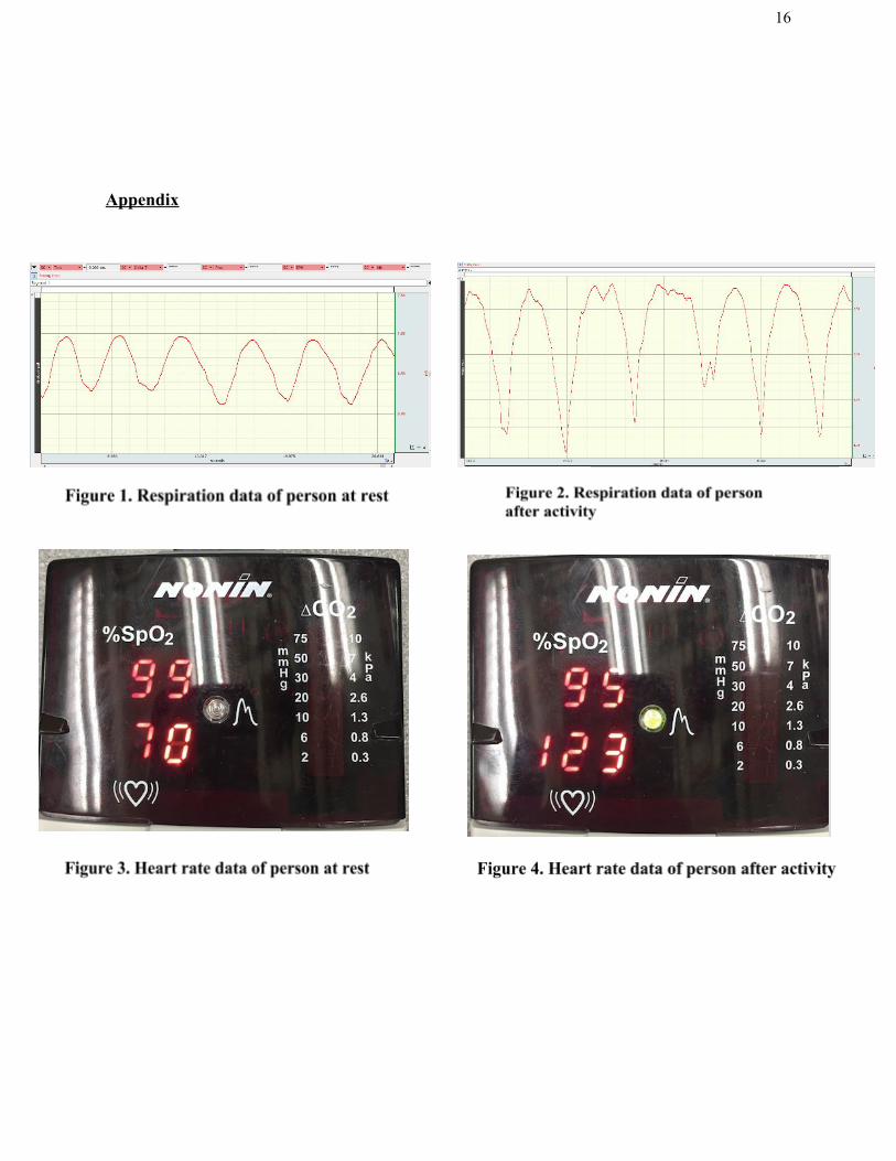
The change in respiration and heart rate can be measured using the pulse oximeter and
BIOPAC devices listed in the Materials section. Figures 1 and 2 show the respiration data of a
group member both before and after the following activity stimulus. To induce a physiological
response different from the resting state, the group member ran up and down five flights of stairs
before respiration and heart rate measurements were taken. Comparing figures 1 and 2, one can
see that the group member’s respiration increased both in frequency and amplitude after the
activity compared to the baseline measurements. Pulse Oximeter data was also affected by the
activity. Before the group member performed the activity, pulse was 70 bpm. After the

8
performed activity, pulse was 123 bpm. This positive control data is evidence that physiological
data such as heart rate and respiration could be measured both before and after a stimulus is
applied.
Negative Control
For each participant, the physiological baseline of respiration and heart rate was
measured by having a group member sit in a chair without armrests, with their eyes closed, and
relaxed breathing for one minute.
Results
Subject Characteristics
A total of 30 subjects comprised of 7 males and 23 females with ages ranging from 19 to
39 participated in the experiment. The majority of the participants (27 out of 30) were students
enrolled in Physiology 435 at the University of Wisconsin-Madison. Before the participants
underwent testing, they all were asked to sign a consent form detailing the purpose of the
experiment, physiological factors being measured, and potential triggers of the experiment.
Participants had an option to opt out of participating in the experiment after reading the consent
form.
Physiological Data Results
Subjects heart rate and respiration rate were- on average- shown to increase from the first
and second maze trials to the third “scream” condition (Fig. 5a and 5b). However, there was no
correlation with increased heart rate or respirations with faster response times in the reflex

9
response task (Fig. 5c.). For all figures we included the average values as well as the maximum
and minimum values to illustrate the range experienced by participants.
Heart rate measurements from trials 1 and 2 compared to trial 3 produced p-values of
.0268 and .0093 respectively using a paired t-test with an alpha=.05, and a 95% confidence
interval. This data shows that heart rate from trial 3 significantly increased from the two control
trials.
Respiration rate was also significantly increased in trial 3 compared to trials 1 and 2.
Using a paired t-test with a 95% confidence interval and alpha=.05, the p-values were .050 and
.046 respectively.
Two paired t-tests with 95% confidence intervals and alpha=.05 were used to compare
the magnitude of the difference in response times between trial 1 and trial 3, and trial 2 and trial
3. The t-test comparing the response times from trial 1 to trial 3 produced a p-value of 0.513. The
p-value produced from comparing trial 2 to trial 3 was 0.129. Based on these p-values there is no
significant difference in reflex response times from the control trials 1 and 2 to the “post-scream”
trial 3.
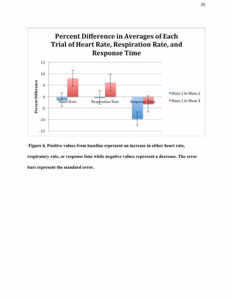
Figure 6 compares the percent difference in averages for each trial tested from maze 1 to
maze 2 and maze 1 to maze 3. The maze 1 trial is interpreted as the baseline measurement. The
figure represents a decrease in all three variables tested from maze 1 to maze 2 and an increase in
heart rate and respiration rate tested from maze 1 to maze 3. While reaction time decreased from
baseline for both maze 2 and 3, it decreased less after the auditory stimulus compared to just a
second “non-stimulus” test. Averages from Figures 5a-5c were taken and the percent differences
between the maximum and minimum were found for each variable tested. 30 participants results

10
were averaged for each trial. The maximum and minimum value for each trial is shown in the
graphs as well.
Discussion
Overall, it was determined that the data collected was not statistically significant. The
purpose of our study was to determine if skeletal muscle response times would decrease after
activation of the sympathetic nervous system by increasing heart rate and respiration rate. While
we were able to evoke a sympathetic response in most participants, skeletal muscle response
times did not decrease in response to the stimuli. There were no statistically significant decreases
in skeletal muscle reaction times found. These findings provide evidence that a sympathetic
nervous response does not reduce skeletal muscle reflex response times.
We believe there could have been a variety of factors interfering with the validity of the
results. The method by which the data was collected and the objective of the study should remain
the same, however the use of a more startling stimulus may have elicited a stronger response
throughout more of the participants. After the startling stimulus was heard, there was a
statistically significant increase in heart rate and respirations throughout the participants.
However, a more startling stimulus may have potentially caused heart rate and respirations to
further increase, leading to a stronger sympathetic nervous system response. This stronger
sympathetic nervous system response may lead to different results of skeletal muscle reaction
time.
Our results also may have been affected by participants having prior knowledge of our
experiment before partaking in it due to communication from other students in the course. If
11
subjects had knowledge that there was a startling stimulus during the experiment, they may have
been anticipating it in the process. Because of the anticipation, they may not have had quite as
large of a sympathetic reaction to the sound as intended, therefore their skeletal muscle reaction
times would not decrease as we had expected.
Looking at our experiment from a different perspective, our startling stimulus may have
led to null results due to distracting the participants from the reaction task. After our startling
stimulus played in the headphones, there were many different reactions from the participants.
While some participants showed no noticeable reaction, others jumped in their chairs, and some
even giggled and looked around the room confused. Immediately after the startling stimulus was
played the participants were instructed to begin their third reaction test. Due to the unexpected
stimulus, participants may have been distracted and therefore not as focused as previous trials as
they began their last reaction test.
The reaction task we chose was a computer task in which the participants had to click the
mousepad in response to a color change on the screen, and their reaction time in responding to
the color change was automatically measured and averaged. Like stated above, a possible
confound in our study may have been participant distractions due to the startling stimulus while
doing the third reaction test. Because of the activation of the sympathetic nervous system in the
participants, their focus on the computer task may have been hindered which could explain the
increased reaction times. For future studies, developing a method to test reaction time in a more
active manner may better encompass the reactions of the sympathetic nervous system.
It is important to note that before beginning the task, a brief explanation of how to do the
reaction test was given to the participants, but the participants weren’t able to practice the task

12
for themselves. Participant’s reaction times decreased from trial 1 to trial 2 of the task, and this
could be due to the possibility of a training effect. Completing this training and allowing the
participant’s to become more familiar with the task prior to starting may have led to clearer
results in the study.
Researchers may also consider widening their range of participants as the majority of our
contributors came from the Physiology 435 course in Spring 2018, which could have an effect on
the preparation or expectations of the study they will be participating in. Students who completed
this experiment as subjects may have conversed with Physiology 435 peers about the parameters
and tipped off future subjects to the surprise stimulus. Comfort is also an important aspect to this
study because we had two researchers in the room at a time with one participant. Acknowledging
the concept of social facilitation, the procedure of this study could be altered in future studies by
having researchers leave the room. The concept of social facilitation was studied by Dashiell
where he found that participants’ performance was either improved or impaired in the presence
of evaluators depending on the task given to them (Dashiell, 1935).
Additionally, future studies and research on the sympathetic nervous system and skeletal
response time could potentially benefit from testing response time using electromyogram
(EMG), which can measure elapsed time to either a relaxation or contraction (Yotani 2014). An
EMG may produce clearer results, as this measurement assesses the processing time of the
nervous system, which could be useful in analyzing smaller differences in response time. Skin
conductance and blood pressure are two other variables with potential to add useful data to the
results. Skin conductance measures autonomic nerve responses, which would be useful in

13
determining the impact of the auditory stimuli on the nervous system, while blood pressure
would also give researchers a similar conclusion.
Conclusion
After analyzing our results obtained from this experiment, we failed to reject the null
hypothesis. It was found that an auditory stimulation of the sympathetic nervous system did not
make a statistically significant difference in reaction times. Our results show no relationship
between increased sympathetic nervous response and faster skeletal muscle reaction time.

14
References
Akpan, N. (2015). Here's why human screams make your skin crawl. Retrieved March 06, 2018,
from https://www.pbs.org/newshour/science/brains-love-hate-screams
Dashiell, J. F. (1935). Experimental studies of the influence of social situations on the behavior
of individual human adults .
Fisher, L. D., & Tucker, D. C. (1991, March). Air jet noise exposure rapidly increases blood
pressure in young borderline hypertensive rats. Retrieved March 06, 2018, from
https://www.ncbi.nlm.nih.gov/pubmed/1851791
Mahmood, R., Parveen, N., Jillani, G., Safi, A., Din, S., Haq, I., Rehman, J., & Haq, A. (2011).
EFFECT OF NOISE ON HEART RATE. Journal Of Postgraduate Medical Institute
(Peshawar - Pakistan), 20 (1). Retrieved from
http://www.jpmi.org.pk/index.php/jpmi/article/view/129/41
Marshall, J.M., (1982). The influence of the sympathetic nervous system on individual vessels of
the microcirculation of skeletal muscle of the rat. Department of Physiology, University
of Birmingham. Retrieved from:
http://onlinelibrary.wiley.com/doi/10.1113/jphysiol.1982.sp014408/epdf
McCorry, L. K. (2007). Physiology of the Autonomic Nervous System. American Journal of
Pharmaceutical Education , 71(4), 78.

15
Schmidt, R. A., & Wrisberg, C. A. (2008). Motor Learning and Performance (4th ed., pp.
31-169).
Yotani, Kengo et al. “Muscle Contraction and Relaxation-Response Time in Response to on or
off Status of Visual Stimulus.” Journal of Physiological Anthropology 33.1 (2014): 23.
PMC . Web. 10 Apr. 2018.
Your lungs and exercise. (2016). Breathe, 12(1), 97–100.
http://doi.org/10.1183/20734735.ELF121
16
Appendix

17
Figure 5a. Average heart rate was shown to increase from trials 1 and 2 to trial 3,
providing evidence of a sympathetic response. Blue values indicate the averages
across all participants. Red values are maximum and orange values are minimum
values for each trial.

18
Figure 5b. Average respiration rate was shown to increase from trials 1 and 2 to
trial 3, providing evidence of a sympathetic response. Blue values indicate the
averages across all participants. Red values are maximum and orange values are
minimum values for each trial.

19
Figure 5c. Average reflex response time did not significantly change across 3
trials. The sympathetic response did not elicit a faster response time. Blue
values indicate the averages across all participants. Red values are maximum
and orange values are minimum values for each trial.
20
Figure 6. Positive values from baseline represent an increase in either heart rate,
respiratory rate, or response time while negative values represent a decrease. The error
bars represent the standard error.