Embed Size (px)
Citation preview
biochemical Pharmac&gy,Vol. 32,No. 11,~. 1789-1791, 1983. 00062952/83 $3.00 + 00 Printed in Great Britain. @ 1983 Pergamon Press Ltd.
THE EFFECTIVENESS OF DIFFERENT SULFATE PRECURSORS IN SUPPORTING EXTRAHEPATIC SULFATE
CONJUGATION
JANETR.DAWSON,* KAISANORBECK and PETERMOLD~US~
Department of Forensic Medicine, Karolinska Institutet, S-104 01 Stockholm, Sweden
(Received 21 October 1982; accepted 27 December 1982)
Abstract-The effectiveness of different sulfur-containing compounds in supplying inorganic sulfate for sulfate conjugation was studied in isolated ceils from rat small intestine, kidney and lung. With cells isolated from the small intestine and kidney, inorganic sulfate was by far the most effective source for intracellular active sulfate as judged by the ability to support sulfate conjugation of 7-hydroxycoumarin. Kidney cells could also use cysteine, N-acetylcysteine and glutathione as a sulfate source, whereas isolated small intestinal cells did not seem to break down and use these sulfur-containing compounds. With isolated lung cells cysteine was the most efficient sulfate precursor. Of the other precursors N- acetylcysteine and inorganic sulfate were used for sulfate conjugation to some extent.
Conjugation with inorganic sulfate is one of the most common of the so-called phase II reactions of drug metabolism [l]. Compounds which contain one or more hydroxyl group(s) are particularly good sub- strates for sulfate conjugation, especially those with a phenolic hydroxyl group [l, 21. The liver is the tissue which generally has the largest capacity for drug metabolic reactions, but extrahepatic tissues can have important qualitative roles to play. It has been shown that the small intestine has the ability to conjugate a number of xenobiotics [3,4] and could readily cope with the conjugation of ingested phe- nolic material [5]. Sulfate conjugation in the liver is readily saturated [2,6], and even though glucuron- idation in the liver generally takes over, at high substrate concentration it is possible that the kidney could play an important role in metabolizing circu- lating xenobiotics. Although, in general, the lung has a relatively small capacity for drug metabolism, small quantities of inhaled compbunds could be effectively metabolized at the site of absorption.
Sulfate conjugation is regulated by a number of factors including substrate concentration [2, 61, availability of inorganic sulfate [2,7,8] and the syn- thesis of adenosine 3’-phosphate 5’-sulfatophosphate (PAPS), which is the ‘active form’ in which sulfate is transferred to the substrate. The availability of inorganic sulfate has been studied almost exclusively in connection with hepatic availability [2,7,8]. Extrahepatic tissues, however, are also active in sulfate conjugation and could have important qual- itative differences in the various control processes. In the present investigation the effectiveness of dif- ferent sulfur-containing compounds as precursors of PAPS in extrahepatic tissues was studied. These studies were performed using isolated cells from the relevant tissues. Use of isolated cells facilitates changes in extracellular fluid composition, eliminates
* Present address: Department of Pharmacology, State University of Groningen, Groningen, The Netherlands.
t To whom correspondence should be addressed.
any hepatic influence and allows easy collection of metabolites.
MATERIALSANDMETHODS
Reduced glutathione, L-cysteine hydrochloride, N-acetylcysteine, protease type VII, sulfatase type H-l and Hepes (N-2-hydroxyethylpiperazine-N’-2- ethanesulfonic acid) were purchased from the Sigma Chemical Co. (St. Louis, MO). Collagenase was purchased from Boehringer Mannheim (F.R.G.). 7-Hydroxycoumarin (umbelliferone) was purchased from Fluka AG. (Switzerland). [U-14C]phenol (71.1 mCi/mmole) was purchased from Amsterdam, U.K. Lumagel, scintillation fluid and scintillation vials (6ml volume) were purchased from Lumac B.V. (Schaesberg, The Netherlands). All other chemicals were of analytical or reagent grade and purchased through local chemical suppliers.
Animals. Male Sprague-Dawley rats, 180-220 g, and allowed free access to food and water, were used throughout.
Cell preparations. Kidney cells were isolated by a collagenase perfusion method as described by Jones et al. [9], and were used throughout at a concentra- tion of l-2 X lo6 cells/ml.
Lung cells were isolated by a recently developed method involving perfusion of a protease solution via the trachea [lo]. Lung cells were used at a con- centration of 3 or 4 X lo6 cells/ml.
Cells were isolated from the small intestine by the method of Dawson and Bridges [ll], slightly modi- fied, as follows. The exerted pieces of intestine were incubated (20 min at 37”) in a Ca*+- and Mg*‘-free Hanks buffer (50 ml) containing protease type VII (1 mg/ml) and EDTA (1 mM). The portions of intes- tine were then transferred to (SOml) Krebs-Hepes buffer containing 1% bovine serum albumin (w/v) and glucose (5 mM), and incubated for a further 10 min at 37”. The cells from both flasks were pooled and gave a cell yield of 350-400 x lo6 cells from 60 cm of rat small intestine. The intestinal cells were used at a concentration of 2 x lo6 cells/ml buffer.
1789
1790 J. R. DAWSON, K. NORBECK and P. MOLD~US
Metabolism experiments. The buffer used through- out these experiments was a sulfate-free Krebs- Hepes buffer, pH 7.4, with the MgS04 in the buffer replaced by MgC12. Cell incubations were performed in rotating, round-bottomed flasks at 37”. Kidney and small intestinal cells were incubated under a carbogen (95% 02, 5% CO2) atmosphere and lung cells were incubated under air.
7-Hydroxycoumarin sulfation was determined as described by Dawson and Bridges [4]. In brief, samples (2 ml) were withdrawn from the incubation at the times required, and the free, unreacted sub- strate was removed by repeated extractions with ether containing 1.5% isoamyl alcohol (3 x 7ml). The aqueous layer was then acidified and deconju- gated overnight with sulfatase (1 mg/ml) in the pres- ence of saccarolactone (1 mM). The 7-hydroxycou- marin released was then determined fluorimetrically.
[?Z]Phenol sulfate was determined by the thin layer chromatographic method described by Shirkey et al. [3].
RESULTS
With cells isolated from rat small intestine, inor- ganic sulfate appeared to be the only effective source for intracellular active sulfate. The amino acids cys- teine and N-acetylcysteine, and the tripeptide glu- tathione did not appear to be broken down and used for sulfate conjugation of 7-hydroxycoumarin in the time studied (Fig. 1). A small amount of sulfate conjugates is produced, even in the absence of an extracellular sulfate source. This probably represents the intracellular sulfate store.
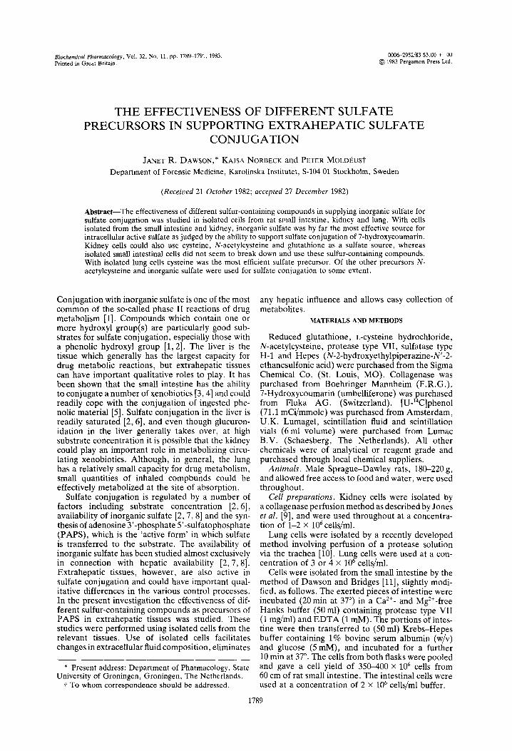
Although again inorganic sulfate was by far the most effective source for PAPS, kidney cells, how- ever, did effectively catabolize the amino acids and the glutathione to inorganic sulfate (Fig. 2). Cys- teine, as may be expected, was the most effective of the three, whilst N-acetylcysteine and glutathione
Fig. 1. Sulfate conjugation of 7-hydroxycoumarin by iso- lated small intestinal cells. Cells isolated from rat small intestine were incubated at a concentration of 2 X lo6 cells/ml in Krebs-Heues buffer with 100 uM 7-hvdroxv- , _ coumarin. Present in the incubation were: no sulfur source. 0; 1 mM NazSOI, 0; 1 mM cysteine, 0; 1 mM N-acetyl-
cysteine, n ; 1 mM reduced glutathione, A.
time I min)
Fig. 2. Sulfate conjugation of 7-hydroxycoumarin by iso- lated kidney cells. Cells isolated from rat kidney were incubated at a concentration of 1-2 x 10” cells/ml in Krebs-Hepes buffer with 10 uM 7-hydroxycoumarin. Pres- ent in the incubation were: no sulfur source, 0; 1 mM Na2S04, 0; 1 mM cysteine, 0; 1 mM N-acetylcysteine,
n ; 1 mM reduced glutathione, A.
were equally efficient precursors (Fig. 2). This reflects the need for one (N-acetylcysteine) or two (glutathione) additional steps in the catabolic route to sulfate.
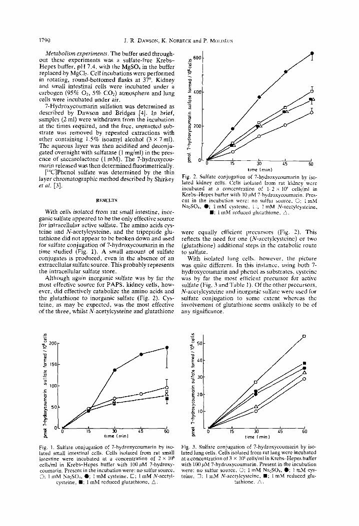
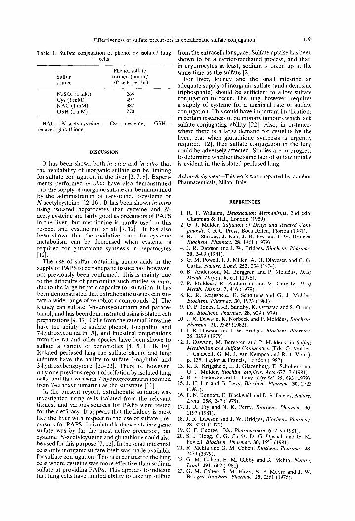
With isolated lung cells, however, the picture was quite different. In this instance, using both 7- hydroxycoumarin and phenol as substrates, cysteine was by far the most efficient precursor for active sulfate (Fig. 3 and Table 1). Of the other precursors, N-acetylcysteine and inorganic sulfate were used for sulfate conjugation to some extent whereas the involvement of glutathione seems unlikely to be of any significance.
Fig. 3. Sulfate conjugation of 7-hydroxycoumarin by iso- lated lung cells. Cells isolated from rat lung were incubated at a concentration of 3 x lo6 cells/ml in Krebs-Hepes buffer with 100 PM 7-hydroxycoumarin, Present in the incubation were: no sulfur source, 0; 1 mM Na2S04, 0; 1 mM cys- teine, 0; 1 mM N-acetylcysteine, n ; 1 mM reduced glu-
tathione, A.
Effectiveness of sulfate precursors in extrahepatic sulfate conjugation 1791
Table 1. Sulfate conjugation of phenol by isolated lung cells
Sulfur source
NaSOl (1 mM) Cys (1 mM) NAC (1 mM) GSH (1 mM)
NAC = N-acetylcysteine, reduced glutathione.
Phenol sulfate formed (pmole/ 106 cells per hr)
266 497 382 270
Cys = cysteine, GSH =
DISCUSSION
It has been shown both in vivo and in vitro that the availability of inorganic sulfate can be limiting for sulfate conjugation in the liver [2,7,8]. Experi- ments performed in vivo have also demonstrated that the supply of inorganic sulfate can be maintained by the administration of L-cysteine, D-cysteine or N-acetylcysteine [12-161. It has been shown in vitro using isolated hepatocytes that cysteine and N- acetylcysteine are fairly good as precursors of PAPS in the liver, but methionine is hardly used in this respect and cystine not at all [7, 121. It has also been shown that the oxidative route for cysteine metabolism can be decreased when cysteine is required for glutathione synthesis in hepatocytes ]121.
The use of sulfur-containing amino acids in the supply of PAPS to extrahepatic tissues has, however, not previously been confirmed. This is mainly due to the difficulty of performing such studies in vivo, due to the large hepatic capacity for sulfation. It has been demonstrated that extrahepatic tissues can sul- fate a wide range of xenobiotic compounds [2]. The kidney can sulfate 7-hydroxycoumarin and parace- tamol, and has been demonstrated using isolated cell preparations [9,17]. Cells from the rat small intestine have the ability to sulfate phenol, 1-naphthol and 7-hydroxycoumarin [3], and intestinal preparations from the rat and other species have been shown to sulfate a variety of xenobiotics [4, 5, 11,18,19]. Isolated perfused lung can sulfate phenol and lung cultures have the ability to sulfate l-naphthol and 3-hydroxybenzpyrene [20-231. There is, however, only one previous report of sulfation by isolated lung cells, and that was with 7-hydroxycoumarin (formed from 7-ethoxycoumarin) as the substrate [lo].
In the present report, extrahepatic sulfation was investigated using cells isolated from the relevant tissues, and various sources for PAPS were tested for their efficacy. It appears that the kidney is most like the liver with respect to the use of sulfate pre- cursors for PAPS. In isolated kidney cells inorganic sulfate was by far the most active precursor, but cysteine, N-acetylcysteine and glutathione could also be used for this purpose [7, 121. In the small intestinal cells only inorganic sulfate itself was made available for sulfate conjugation. This is in contrast to the lung cells where cysteine was more effective than sodium sulfate at providing PAPS. This appears to indicate that lung cells have limited ability to take up sulfate
from the extracellular space. Sulfate uptake has been shown to be a carrier-mediated process, and that, in erythrocytes at least, sodium is taken up at the same time as the sulfate [2].
For liver, kidney and the small intestine an adequate supply of inorganic sulfate (and adenosine triphosphate) should be sufficient to allow sulfate conjugation to occur. The lung, however, requires a supply of cysteine for a maximal rate of sulfate conjugation. This could have important implications in certain instances of pulmonary tumours which lack sulfate-conjugating ability [22]. Also, in instances where there is a large demand for cysteine by the liver, e.g. when glutathione synthesis is urgently required [12], then sulfate conjugation in the lung could be adversely affected. Studies are in progress to determine whether the same lack of sulfate uptake is evident in the isolated perfused lung.
Acknowledgement-This work was supported by Zambon Pharmaceuticals, Milan, Italy.
1.
2.
3.
4.
5.
6.
7.
8.
9.
10.
11.
12.
13.
14. 15.
16.
17.
18.
19. 20.
21.
REFERENCES
R. T. Williams, Detoxication Mechanisms, 2nd edn. Chapman & Hall, London (1959). G. J. Mulder, Sulfation of Drugs and Related Com- pounds. C.R.C. Press, Boca Raton, Florida (1981). R. J. Shirkey, J. Kao, J. R. Fry and J. W. Bridges, Biochem. Pharmac. 28, 1461 (1979). J. R. Dawson and J. W. Bridges, Biochem. Pharmac. 30, 2409 (1981). G. M. Powell, J. J. Miller, A. H. Olavesen and C. G. Curtis, Nature, Lond. 252, 234 (1974). B. Andersson, M. Berggren and P. Mold&s, Drug Metab. Dispos. 6, 611 (1978). P. Mold&, B. Andersson and V. Gergely, Drug Metab. Discos. 7. 416 (1979). K. R. Kriigsheld, E. ‘Schohens and G. J. Mulder. Biochem. Pharmac. 30, 1973 (1981). D. P. Jones, G.-B. Sundbv. K. Ormstad and S. Orren- ius, Biochem. Pharmac. ii, 929 (1979). J. R. Dawson, K. Norbeck and F: Mold&s, Biochem. Pharmac. 31. 3549 (1982). J. R. Dawson and J: W. ‘Bridges, Biochem. Pharmac. 28, 3299 (1979). J. Dawson, M. Berggren and P. Mold&s, in Sulfate Metabolism and Sulfate Conjugation (Eds. G. Mulder. J. Caldwell, G. M.-J. van Kempen and R. J. Vonk), P. 135. Tavlor & Francis. London (1982). K. R. Krijgsheld, E. J. Glazenburh, E. Scholtens and G. J. Mulder, Biochim. biophys. Acta 677, 7 (1981). R. E. Galinskv and G. Levv. Life Sci. 25. 693 (1979). J. H. Lin and’G. Levy, B&hem. Pharmac. 36, 27i3 (1981). F. N. Bennett, E. Blackwell and D. S. Davies. Nature, Lond. 258, 247 (1975). J. R. Fry and N. K. Perry, Biochem. Pharmac. 30, 1197 (1981). J. R. Dawson and J. W. Bridges, Biochem. Pharmac. 28, 3291 (1979). C. F. George, Chn. Pharmacokin. 6, 259 (1981). S. I. Hogg, C. G. Curtis, D. G. Upshall and G. M. Powell, Biochem. Pharmac. 30. 1551 (1981). R. Mehta and G. M. Cohen, L&ocher& Phhrmac. 28, 2479 (1979).
22. G. M. Cohen, E. M. Gibby and R. Mehta, Nature, Lond. 291, 662 (1981).
23. G. M. Cohen, S. M. Haws, B. F. Moore and J. W. Bridges, Biochem. Pharmac. 25, 2561 (1976).