Embed Size (px)
DESCRIPTION
The Effects of Captivity on the Morphology of Captive,
Citation preview

Mammal Rev
. 2005, Volume 35, No. 3&4, 215–230.
Printed in Singapore
.
© 2005 Mammal Society,
Mammal Review
,
35,
215–230
Blackwell Publishing LtdOxford, UKMAMMammal Review0305-1838Blackwell Publishing Ltd, 2005
? 2005
35
3&4215230
Review Article
Effects of captivity on morphologyH. J. O’Regan
and A. C. Kitchener
Correspondence: H. J. O’Regan. E-mail: h.j.o’[email protected]
The effects of captivity on the morphology of captive, domesticated and feral mammals
HANNAH J. O’REGAN*
and
ANDREW C. KITCHENER†*
Research Centre in Evolutionary Anthropology and Palaeoecology, School of Biological & Earth Sciences, Liverpool John Moores University, Liverpool L3 3AF, UK,
†
Department of Natural Sciences, National Museums of Scotland, Chambers Street, Edinburgh EH1 1JF, UK and Institute of Geography, School of Geosciences, University of Edinburgh, Drummond Street,
Edinburgh EH8 9XP, UK
ABSTRACT1.
The effects of captivity on the behaviour of wild and domestic animals have been relativelywell studied, but little has been published on morphological changes in wild animals incaptivity. We review the evidence for changes in a wide variety of mammalian taxa, with non-mammalian examples where relevant.
2.
We consider the morphological effects of the process of domestication, and comparechanges in both hard and soft tissues in captive and domestic animals with those in theirwild counterparts. These include skull shape differences, brain size reduction, postcranialadaptations and digestive tract changes.
3.
We also summarize studies that have looked at morphological change in feral animals incomparison with their wild and domestic ancestors, and consider their use as an analoguefor morphological change in captive-bred animals that have been released into the wild.
4.
We then discuss the importance of this work for the wider aims of conservation ofendangered species and captive breeding over many generations, and emphasize the impor-tance of studying these changes now, while for many species, the process is just beginningrather than many generations down the line, or immediately prior to release, where survivalof captive-bred animals may be severely compromised.
Keywords
: captive breeding, domestication, feral, morphological change, phenotypic plastic-ity, reintroduction
INTRODUCTION
There is increasing awareness of the potential for ‘domestication’ to occur as a by-productof wild animals being kept and bred in captivity. Much of this work has been centred onbehavioural changes, such as loss of response to predators and alteration of defensive andsexual behaviours (e.g. Price, 1999, 2002). However, it is also apparent that morphologicaland physiological changes can occur in captivity, which could have important implicationsfor reintroductions and the management of wild and captive populations as a whole, but asyet these have received little attention. This may in part be because there is an essentialdifference between the aims of taxonomy/functional morphology and conservation; conser-vationists aim to preserve and propagate live animals, whereas skeletal morphologists rely ondead ones for their raw materials. Skeletons are also often viewed as rigid, genetically defined

216
H. J. O’Regan and A. C. Kitchener
© 2005 Mammal Society,
Mammal Review
,
35,
215–230
entities; whereas in reality, they are entirely plastic, responding significantly to behaviouraland environmental stimuli. There is also a perception that the remains of captive animals are‘atypical’, and should therefore not be included in morphological or taxonomic studies(Hollister, 1917; Howell, 1925).
Studies of captive-bred animals provide a unique opportunity to assess the relative impor-tance of environmental and genetic influences on morphology, which may have importanttaxonomic and evolutionary implications, and may affect the conservation and managementof wild-living and captive populations. This latter point will become increasingly importantas wild and captive populations for each species are managed together. Given that the currentaim of zoos is to maintain self-sustaining, genetically diverse populations for up to 200 years(Soulé
et al
., 1986), it is time that the consequences of morphological and physiologicalchanges in captivity are quantified and addressed.
We review briefly the common changes that occur in the skeletal morphology of wildanimals when domesticated, as these may provide important indicators of how we may expectmorphological changes to occur in species removed from the wild for many generations andwhich have often survived severe population bottlenecks when brought into zoos. We thenreview the variety of morphological changes, from shape changes to decreasing brain size,which have been observed in captive wild animals. Studies of feral domestic animals may alsoprove of benefit, as they provide natural experiments to help us find out if the morphologicalchanges that occur in multigenerational captive animals persist in the wild, or if they aresimply individual responses to the captive environment. Suggestions are then given for areaswhere further investigation is necessary.
DOMESTICATION
A variety of physical changes are thought to have occurred to wild mammals during domes-tication. These were listed by Clutton-Brock (1999) as changes in body and brain size,alteration of external appearance, the gaining of a fat layer beneath the skin and a reductionof the facial region. The majority of these changes are thought to result from increasingneoteny in domestic mammals, which is a form of paedomorphosis. The animals that we seetoday are the ultimate product of some 5000–15 000
+
years of domestication, and the differ-ences between them and their wild ancestors are likely to be marked, given the power ofartificial selection to bring about rapid morphological changes. This subject has been a topicof interest since the mid-19th century (Darwin, 1868), and evidence is gathering to suggestthat characteristics that are considered to be markers of domestication, such as floppy earsand shortened tails, can arise relatively quickly by selecting for behavioural characteristicsconducive to captivity. In 1959, Dmitry Belyaev began a breeding programme for silver foxes
Vulpes vulpes
which had been bred in fur farms for over 50 years. In this experiment, thetamest animals from each generation were selected to be the founders of the next generation.After 30–35 generations, foxes were produced with floppy ears (0.23%), rolled (9.4%) orshortened tails (0.14%) and maloccluded teeth, although the latter were still at very low levelswithin the population (percentages based on occurrences per 100 000 foxes after 40 years)(Trut, 1999). Moreover, many coat colour mutations (e.g. non-agouti, white spotting), whichare frequently seen in domestic mammals, also became more common (12.4%) in thesedomesticated foxes. After 40 years, 70–80% of the population was tame; animals activelysolicited human contact by whining and licking the hands of their keepers (Trut, 1999). Thisexperiment has shown that within 10–15 generations, significant morphological changesbegan to occur as a by-product of the selection of animals for tameness. We believe thatdomestication may be occurring inadvertently in captive wild mammals (albeit at a slower

Effects of captivity on morphology
217
© 2005 Mammal Society,
Mammal Review
,
35,
215–230
rate than artificial selection) through passive selection for animals that are behaviourally moresuited to captivity, thereby resulting in concomitant morphological changes over severalgenerations, which may have severe consequences for the ability of the descendants of today’scaptive endangered species to be successfully reintroduced to the wild after 200 years.
CAPTIVE BREEDING FOR CONSERVATION
Early efforts to consider the effects of captive breeding on wild mammals have concentratedon preserving the genetic variability of the population (e.g. Frankham
et al
., 1986). Whilethis is obviously important, it appears that captivity can also erode an animal’s ability tofunction in the wild. There are numerous examples of captive-release programmes failingthrough poor foraging and lack of predator recognition or, in the case of the golden liontamarin
Leontopithecus rosalia
, the loss of the ability for effective locomotion and orientationin their natural environment (reviewed by Menzel & Beck, 2000; Wallace, 2000). Trut (1999)observed that tamed silver foxes that had escaped from the breeding programme invariablyreturned to the farm, and suggested that they would have been unable to survive in the wild;similar behaviour has also been observed in escapee or released American mink
Mustelavison
. As Frankham
et al
. (1986, p. 136) commented, ‘inadvertent selection for tameness andadaptation to the captive environment are inevitable. This reduces the chances of successfulreintroduction of the population into the wild’. Populations of endangered species withinzoos may have been subject to severe genetic bottlenecks, and are clearly self-selecting forthose animals that can tolerate captivity. Only those animals that are able to adapt to captivelife will survive, and a still smaller proportion of those will reproduce. For example, in anattempt to repeat the silver fox domestication programme with wild otters
Lutra lutra
, only8 of 50 wild-caught otters reproduced successfully, while only 14% of wild brown rats
Rattusnorvegicus
produced progeny that survived to adulthood (Trut, 1999). Therefore, it is notsurprising that captive animals exhibit marked differences from their wild conspecifics rela-tively quickly, as they are founded from small populations and the selection pressures oncaptive animals are substantially different from those in the wild. We will now summarizethe morphological and physiological changes, which have been observed in a wide variety ofcaptive mammals, with the inclusion of some non-mammalian examples where relevant.
CHANGES RESULTING FROM THE EFFECTS OF CAPTIVITYDiet
Many of the early problems in captive animal husbandry were related to poor diet, and deathrates were often high; Sutton (1884), for example, post-mortemed 130 carnivores from Lon-don Zoo between January 1882 and February 1884. Sutton’s paper is one of the first toidentify pathologies in captive wild mammals, including rickets and tuberculosis. He foundindividuals in which the ‘teeth are large, defective in number and late in appearance’ (Sutton,1884, pp. 181–182), and also that paralysis of the hindquarters resulted from enlarged verte-bral discs pressing on the spinal column (similar to that of spondyloarthrosis, see below). Hisoverall diagnosis was that these problems were all caused by the same factors: ‘Bad hygienicconditions incident to the life of a captive’ (Sutton, 1884, p. 181).
Diets lacking in necessary vitamins and minerals were a source of many pathologicalconditions, such as osteodystrophia fibrosa, which caused gross cranial thickening in threecaptive subadult baboons
Papio
sp. (Cordy, 1957). The long bones of these animals showedenlargements of the proximal and distal ends of the diaphyses, but the skull was most severelyaffected with only the basicranium remaining at normal size. It was suggested that anabnormal calcium : phosphorus ratio in the diet could be responsible (Cordy, 1957). du

218
H. J. O’Regan and A. C. Kitchener
© 2005 Mammal Society,
Mammal Review
,
35,
215–230
Boulay & Crawford (1968) also looked at nutritional bone disease in a sample of 31 SouthAmerican primates, of which 25 showed changes observable in radiographs, such as bowedlimb bones and thickening of the skull vault, which were concluded to be due to a dietdeficient in calcium and vitamin D
3
.However, following extensive work on dietary nutrition in the 1960–70s, diseases such as
these have largely been eradicated (see Kleiman
et al
., 1996; section 2), although some animalssuch as big cats and callitrichids continue to cause concern. Baker & Lyon (1977) observedchanges in eight lion cub
Panthera leo
skulls, including thickening of the cranial vault(especially the parietals), cranial asymmetry and herniation associated with increasing paral-ysis and eyesight loss. More recently, Chandra
et al
. (1999) provided more evidence for cranialthickening in lions, especially of the parietals and the tentorium cerebelli, and reviewed theevidence that vitamin A deficiency may cause this type of problem. Other possibilitiesincluded a genetic origin, but there is, as yet, no consensus on the cause of this malformation.Progressive limb paralysis and cerebellar degeneration has also been noted in captive-bredcheetahs
Acinonyx jubatus
, but it appears that no examination of the crania of these animalshas been carried out and the cause remains unknown (Palmer
et al
., 2001).In addition to pathological changes, there are substantial differences related to body size
and rate of maturity between wild and captive animals, and there is anecdotal evidence tosuggest that better nutrition in captivity could be the cause. For example, Smuts, Anderson& Austin (1978) found that the cranial dimensions of a small sample of captive lion cubswere greater than those of their wild counterparts from the same geographical area. However,it is possible that the observed size differences were the result of faster maturation in thecaptive animals, as they only looked at specimens up to 36 months of age. Schaller (1972)also noted that wild Serengeti lion cubs were smaller than captive cubs of the same age, andthought this might be due to periodic starvation in the wild animals. A wild-caught, butcaptive-raised, cub was reported as being nearly twice the size of wild cubs of similar age(Schaller, 1972). Captive-bred chinchillas
Chinchilla laniger
have significantly longer andwider skulls with a greater degree of variation in skull profile in comparison with those ofwild individuals (Crossley & del Mar Miguélez 2001). The authors suggested that the largersize of the captive specimens was probably due to an unrestricted diet with all nutrientsavailable. In addition, some animals in captivity do appear to mature faster. A study of growthin wild and laboratory chimpanzees
Pan troglodytes
revealed that captive chimps grew biggerand faster than those from the wild (Kimura & Hamada, 1996). This difference occurred inthe limb bones, which grew longer in laboratory chimpanzees, whereas skull measurementsseemed to be unaffected. Kimura & Hamada (1996) concluded that a better, constant diet,coupled with health care in captivity, resulted in the observed size differences. In a study ofdental eruption in yellow baboons
Papio cynocephalus
it was found that the eruption sched-ules, which had been based on laboratory animals, underestimated the age of wild baboonsin Tanzania by up to 1.5 years (Phillips-Conroy & Jolly, 1988). It was concluded that thelaboratory animals matured faster than those in the wild and, in particular, the captive males’canines and lower third premolars erupted much earlier than those of wild animals. In thesame captive baboon population, menarche was also reached some 1.5 years earlier than inthe wild animals (Altmann, Altmann & Hausfater, 1981), while in tamed silver foxes, repro-ductive age was reached 1 month earlier than in their wild conspecifics (Trut, 1999). There isanecdotal evidence to suggest that better nutrition in captivity has led to golden-headed liontamarins
Leontopithecus chrysomelas
growing much larger in captivity (K. Leus, personalcommunication) and many callitrichids in captivity are tending to have litters of three ratherthan the normal two infants.

Effects of captivity on morphology
219
© 2005 Mammal Society,
Mammal Review
,
35,
215–230
However, not all animals grow larger in captivity, and morphological changes need not beuniform. Groves (1982) described morphological differences between the skulls of captiveand wild rhinoceroses, and found that captive male Indian rhinoceroses
Rhinoceros unicornis
had smaller skulls (even if originally wild-caught and brought into captivity before maturity)than those of wild animals. In contrast, a variety of changes were seen in five captive femaleSumatran rhinos
Dicerorhinus sumatrensis
; two were very large, two were average-sized andone was a dwarf (Groves, 1982). A study of linear body and limb measurements of rhesusmacaques
Macaca mulatta
from a free-ranging, but provisioned, population from CayoSantiago, Puerto Rico, and a captive-bred group derived from the same locality, showed thatcaptive-bred animals did not grow so large, possibly owing to a more restricted captive diet,which included lower total protein, although there may have also been some genetic influence,owing to a limited founding group in captivity (Gore, 1993). Unusually for captive mammals,few of the captive-bred macaques were overweight. An analysis of a small sample of equids(13 wild and 12 captive
Equus
spp.) showed that overall, captive animals had smaller craniathan those of wild individuals, with the exception of palate breadth, which was of similar sizein both groups (Groves, 1966). Poole, Carpenter & Simms (1980) found that grey kangaroos,
Macropus giganteus
and
M. fuliginosus
, that had been hand-reared, or were from captivity(or very extreme environments on the edge of the species’ ranges), had different cranial shapesfrom those of their wild counterparts, and they suggested that skull size and shape weremodified by early nutritional changes. Guthrie (1984) provided extra food for captive-raisedDall’s sheep
Ovis dalli
and found that their horn and body size almost reached the largersizes of those of their Pleistocene ancestors. A male captive-bred alpine ibex
Capra ibex
atKolmården Zoo, Sweden, developed such large heavy horns owing to a plentiful and richcaptive diet, that their weight resulted in arthroses to the cervical vertebrae, leading toconstriction of the foramen magnum and spinal cord and the resultant loss of motor ability(B. Röken, personal communication). There has also been some experimental work onpheasant chicks
Phasianus colchicus
which confirms that bone morphology is permanentlyaltered by early nutritional levels. Ohlsson & Smith (2001) found that the levels of protein inthe diet in the first 3 weeks of life had a permanent effect on the length and asymmetry ofthe tarsometatarsus in captive birds, and that the effects on the skeleton were persistent,although the experimental animals caught up in terms of body weight with those chicksreared on a full protein diet.
Mechanical properties of food
The mechanical properties of an animal’s food may have a distinct effect on its cranialmorphology and dental health, and there have been many studies on this subject in animalsas diverse as caterpillars, squirrel monkeys and hyraxes (see Lieberman
et al
., 2004 for areview of this area). The majority of these studies have involved feeding laboratory animalsa diet that is either hard or soft to assess its impact on facial and cranial anatomy.
Corruccini & Beecher (1982) conducted experiments on squirrel monkeys
Saimiri sciureus
and found that animals fed on soft diets had crowded premolars, more displaced teeth andnarrower muzzles, while those on hard diets had significantly broader maxillae. More recently,it has been shown that significant differences in the cranial shape of hyraxes
Procavia capensis
have resulted from eating different diets (Lieberman
et al
., 2004). These studies indicate thatthere is a relationship between the mechanical properties of food and occlusal and morpho-logical variation. Cheetahs inadvertently provided evidence for this in a non-experimentalsetting. Cats in zoological collections in North America are often fed on a pre-processed diet,which requires little or no chewing. It was thought that as a result of this soft diet cheetahs

220
H. J. O’Regan and A. C. Kitchener
© 2005 Mammal Society,
Mammal Review
,
35,
215–230
developed ‘focal palatine erosion’ (FPE) (Fitch & Fagan, 1982), where the lower carnassialsfail to occlude with the upper carnassials and impact instead upon the palate. In severe cases,this can lead to sinuses developing between the oral and nasal cavities (Fitch & Fagan, 1982).In the captive population, 15 out of 59 live cheetahs exhibited FPE, with 15 out of 20 fedsoft diets being affected (Fitch & Fagan, 1982). Until 2004, this condition had only beennoted in captive felids fed on pre-processed diets, but recent work by Marker & Dickman(2004) found some degree of palatine erosion in 144 of a sample of 208 live wild cheetahs inNamibia, with 85 of 208 showing severe erosion. However, only 13 of these animals hadperforated palates (6.3% of the total sample) (Marker & Dickman, 2004), in comparison witheight (13.6%) of the North American captive cheetahs (Fitch & Fagan, 1982).
Many diets fed to captive animals have been formulated to provide the nutritional require-ments for growth, but they often lack texture and interest (Lindburg, 1998). Also, in the wildfood is often not as highly nutritious and contains abrasive particles such as grit or phytoliths.The lack of abrasion in captive diets, resulting in reduced or abnormal toothwear, has led toproblems of malocclusion in herbivorous animals, such as rodents, lagomorphs and elephants,which have teeth designed to be worn down by chewing throughout their life.
Studies of both equids (Groves, 1966) and chinchillas (Crossley & Miguélez, 2001) haveshown that an insufficiently abrasive diet can have serious consequences for an animal’s oralhealth. In the case of the equids, the increasing height of the maxillary and mandibular molarsled to the incisors being held apart, so that the animals were unable to graze effectively(Groves, 1966). Like the equids, the tooth crowns of captive chinchillas were found to besignificantly higher than those of wild chinchillas, which led to difficulties in chewing and, insome cases, the tooth roots protruded into the nasal cavity (Crossley & Miguélez, 2001). Aslightly different problem is seen in captive elephants (most commonly in the Asian elephant
Elephas maximus
) as they have a very specialized dentition. Each molar erupts in sequence,and there are usually only one or two molars present in any one quadrant of the mouth atany time. However, if the diet is not abrasive enough, the next molar may erupt before thepreceding tooth is worn away, leading to impaction and malocclusion in the jaws (Fagan,Oosterhuis & Roocroft, 2001; A.C.K., personal observation). The most famous example ofthis problem was seen in ‘Jumbo’ the African elephant
Loxodonta africana
at London Zoo,whose bad temper as a result of this condition led to him being sold to Barnum and Bailey’sCircus in 1882 (Jolly, 1976).
A comparative study of brown bear
Ursus arctos
skulls from the Berne bear pit and thewild found that the captive bears had increased levels of attrition to the canines, possibly asa result of chewing cage bars, and also had a high prevalence of dental caries and calculusformation, especially in bears over 10 years of age (Wenker
et al
., 1999). It is thought thatthe diet of the Berne bears has contributed to the problems with their teeth, as they were fedon ‘mostly low-fibre, paste-like food’ (Wenker
et al
., 1999, p. 217). However, some animalsin the wild bear sample also had a large number of caries (up to 64% in one animal). Dentalcalculus may also be a problem in big cats, but providing whole carcasses to feed on andbones for them to chew on does appear to reduce its occurrence (Haberstroh
et al
., 1984).However, in the feeding experiment on squirrel monkeys, it was found that the incidences ofperiodontal disease, dental caries and tooth loss were greater in the group fed on hard diets(Corruccini & Beecher, 1982), probably owing to the dentition being adapted for a softerfrugivorous/insectivorous diet.
Tooth pathologies have also been noted in captive animals. These range from those causedby trauma to developmental defects such as dental enamel hypoplasia. In a study of wild andcaptive primates, Molnar & Ward (1975) found that all animals had some level of microstruc-

Effects of captivity on morphology
221
© 2005 Mammal Society,
Mammal Review
,
35,
215–230
tural tooth defects, including hypoplasia, but their incidences were higher in captive individ-uals. Recently, Franz-Odendaal (2004) has studied the incidence of hypoplasia in the teeth ofwild and captive giraffes
Giraffa camelopardalis
and found that those of wild giraffes lackeddefects, while those of captive animals showed lines that corresponded with periods of stresssuch as weaning. Fagan
et al
. (2001) discussed the incidence of traumatic breakages ofelephant
Loxodonta africana
and
Elephas maximus
tusks caused by the captive environment,and noted similar occurrences in walruses
Odobenus rosmarus
and babirusas
Babyrousababyrussa
.
Biomechanics of the skull
It appears that not all areas of the skull react equally to the circumstances of captivity. Avariety of morphometric studies have found that the skulls of captive animals are significantlydifferent from those of their wild conspecifics in some areas and yet other dimensions remainunchanged. Many of the areas that do appear different in captive animals are related to thefeeding apparatus, and this is probably a result of the different mechanical properties ofcaptive diets (see above). For example, Groves (1982) reported that the skulls of captive Indianrhinoceroses were smaller, yet had increased mastoid and zygomatic breadths and higheroccipital crests than those of wild individuals. He suggested that it could have been due tobadly placed feeding trays or the feeding of inappropriate foodstuffs. Duckler (1998) alsofound a thickening and change in shape of the sagittal crest in a series of captive tiger
Pantheratigris
skulls, and postulated that this might be a result of stereotypical behaviour, such asovergrooming.
Hollister (1917, 1918) described differences between the skulls of captive lions, whichhad been caught in the wild in East Africa, and those of wild lions from the same geo-graphical area. These differences included increased breadth of the zygomatic arches,shortening of the skull, and reduced cranial volumes in the captive-reared animals. Hollis-ter considered these changes to be due to a lack of exercise of the cranial muscles incaptivity. Howell (1925) re-examined Hollister’s specimens and concluded that these ani-mals were pathological, and suggested a dietary deficiency as the cause of the observeddifferences that also included a general increase in the overall thickness of the skull and areduction in the height of the foramen magnum. Despite the pathological nature of Hollis-ter’s lions, his observation of an increase in zygomatic breadth has been supported byO’Regan (2001). Further work on a larger sample of skulls found that the breadth of themuzzle was also significantly larger in both male and female captive lions and leopards
Panthera pardus
than in wild individuals (O’Regan & Turner, 2004). Finding this differencein two species of big cat suggests that there may be a biomechanical, age-related or behav-ioural reason for these changes, but without knowledge of the life history of the animals, itis difficult to be certain.
Captive crocodilians also exhibit cranial changes. In a study of American alligators
Alli-gator mississippiensis
, Meers (1996) found that the skulls of captive animals had broadermaxillae and premaxillae, a flatter profile and less sculpting or rugosity on the cranial surfacethan those of wild individuals. These changes were all related to the biomechanics of thefeeding apparatus. Captive reptiles are fed to ensure the maximum growth in the minimumamount of time, resulting in animals that have smaller skulls than those of wild alligators ofsimilar body size, and suggesting that the cranium cannot match the growth rate of the body(Meers, 1996). There are concerns that animals with this morphology that have been bred forrelease into the wild could out-compete the wild animals, as their larger body size would givethem an advantage over wild individuals when competing at carcasses (Meers, 1996). Their

222
H. J. O’Regan and A. C. Kitchener
© 2005 Mammal Society,
Mammal Review
,
35,
215–230
smaller skulls may, however, place them at a disadvantage in holding onto carcasses andtwisting off mouthfuls to swallow, especially when competing with others.
A study of the skulls of two wild and three captive populations of oldfield mice
Peromyscuspolionotus subgriseus
found that there were significant differences between the wild andcaptive populations (McPhee, 2004). Each captive population had different founders,although they were all drawn from the same geographical area, and they had been bred incaptivity for different lengths of time (2, 14 and 35 generations). McPhee (2004) found thateach population had its own trajectory (i.e. that different populations had different morpho-logical changes), and that these changes were not cumulative over time (i.e. increased palatebreadth in one group did not lead to even wider palates in the next). This has importantimplications as it suggests that populations held in isolation will develop significantly differentmorphological traits, and so conversely, it is possible that captive populations managed aswhole may all develop the same features. Most of the above studies have used morphometricsto look for differences between wild and captive specimens. However, other techniques canalso be informative. Armitage (1983) used radiographs to determine whether or not anarchaeological specimen came from a captive individual. This study of two wild and twocaptive weeper capuchin
Cebus olivaceus
monkey mandibles found that the wild animals hadgood cortical development and the medullary cavity contained few widely spaced and thicktrabeculae, whereas the captive animals had a porous and ‘poorly developed’ cortical layerand the medullary cavity was completely infilled with a mass of cancellous bone, which wasconsistent with the archaeological specimen being from a captive individual (Armitage, 1983).
Changes to soft tissues and physiology
Rich, nutritious and easily digestible captive diets may have a significant effect on gutmorphology and physiology, which could compromise the survival of captive-bred animalsreleased into the wild, where diets will be poorer and less easy to digest. This phenomenonappears to have only been studied in birds, but we have no knowledge of the consequencesof captive diets for mammals. For example, the concentration of the required nutrients in ahighly nutritious artificial-formula diet means that captive capercaillies
Tetrao urogallus
develop shorter intestines and caecae, and lighter hearts, livers and gizzards than those ofwild birds (Liukkonen-Anttila, Saartoala & Hissa, 2000). As a result, captive-reared birdsare less able to digest their natural foodstuffs, such as pine needles, and deal with the toxinsthey contain (Liukkonen-Anttila et al., 2000). In this case, therefore, the overly nutritiouscaptive diet has actually caused physical changes to the birds’ digestive systems and itsphysiological capacity to deal with wild diets. Similar changes in gut morphology have beenrecorded in red grouse Lagopus lagopus scotica (Moss, 1972), barnacle geese Branta leucopsis(Owen, 1975) and houbara bustards Chlamydotis spp. (J. Samour, personal communication).Studies on captive-bred Japanese quail Coturnix japonica showed that gut length is pheno-typically labile. A laboratory group reared on a high-fibre diet had longer gut lengths thanthose on a low-fibre diet. However, when the diets were switched between groups, it only took3–4 weeks for the gut length to increase in the new high-fibre group and to decrease in thoseon the low-fibre diet (Savory & Gentle, 1976). Wild birds may also show seasonal variationin gut length, which correlates with changing seasonal diets (e.g. spruce grouse Canachitescanadensis, Pendergast & Boag, 1973). It is unclear whether there is similar phenotypicplasticity in mammals. Schauenberg (1977) distinguished between domestic cats Felis catus,and European wildcats F. silvestris, on the basis of relative and absolute gut length. It hasbeen suggested that domestic cats have a longer gut length, in order to increase digestiontimes for a less carnivorous diet (Kitchener, 1998). However, despite a similar diet, wildcats

Effects of captivity on morphology 223
© 2005 Mammal Society, Mammal Review, 35, 215–230
and feral domestic cats have significantly different gut lengths where sympatric in Scotland(Balharry & Daniels, 1998). There are insufficient gut length data from African and Asianwildcats to be certain whether the domestic cat’s gut length has been determined by its directancestors, the African and Asian wildcats F.s. lybica and F.s. ornata, or whether it is anadaptation to captivity that may have become fixed within the population.
A study of wild (n = 10) and captive-bred (n = 4) marsupial feathertail gliders Acrobatespygmaeus found that captive-bred individuals differed both morphologically and physiolog-ically from two wild populations (Geiser & Ferguson, 2001). The captive-bred individualswere from a lineage that had been in captivity for four generations. The captive animals hadlonger tails and snouts than those of the wild individuals, but most importantly, they differedin their ability to enter torpor. Feathertail gliders enter torpor for up to several days in anyseason during periods of low air temperature. The captive-bred animals were active for muchlonger periods, and entered torpor at lower temperatures (15 °C) than the wild animals(20 °C). In addition, captive-bred gliders became hypothermic and had to be warmed exter-nally at lower temperatures, whereas the wild-caught animals were never hypothermic (Geiser& Ferguson, 2001). The captive-bred gliders’ inability to keep body temperatures stable inambient temperatures that they would encounter in the wild could threaten their survival, ifthey are not fully acclimatized to the local environment (Geiser & Ferguson, 2001).
Kitchener, Merryweather & Allchurch (1999) carried out a study to look at the differencesbetween the flight musculo-skeletal systems of mainly Rodrigues fruit bats Pteropus rodricen-sis. They found that the wild-caught founders and captive-bred animals showed no differencesin the development of the musculature associated with flight, but no wild-living animals wereavailable for comparison to see if diminished flight levels in captivity have resulted in poorerdevelopment of flight musculature there. No significant difference was recorded between thebody weights of captive and wild Rodrigues fruit bats. However, the captive bats accumulatedsubcutaneous fat steadily throughout their lives, until it comprised 30+% of body weight bythe age of 20 years, which would probably have prevented or severely affected flight capability.This suggests that the proportion of body mass attributable to the flight musculature is fargreater in wild-living bats.
Age, activity levels and arthropathiesMany animals live longer in captivity than they do in the wild. For example, gorillas Gorillagorilla survive for more than 50 years in captivity compared with 35 years in the wild (Nowak,1999; Macdonald, 2001). What is not clear, however, is whether the changes such as arthro-pathies that have been observed in captive animals are normal age-related changes, the resultof lack of activity, different nutrition, chronic infection or other environmental factors.
Rothschild, Rothschild & Woods (1998) found spondyloarthopathy in both wild andcaptive big cats, but osteoarthritis and calcium pyrophosphate deposition disease (CPPD)were only found in captive animals. Spondyloarthropathy is likely to result in a decrease inactivity for the affected animals, and would eventually compromise survival in the wild.Kolmstetter, Munson & Ramsay (2000) found that degenerative spinal disease was presentin eight of 37 big cat skeletons from Knoxville Zoo. All affected animals were recorded ashaving progressively decreased activity levels and weight loss prior to death/euthanasia. Allvertebral columns showed narrowed or collapsed disc spaces or disc mineralization, and fivehad spondylosis. Age at onset of clinical signs varied from 10 to 19 years, with a median ageof 18 years.
In a study of a variety of captive wild mammals ranging in size from the plains pocketgopher Geomys bursanus to the brown bear, Greer, Greer & Gillingham (1977) found osteoar-
224 H. J. O’Regan and A. C. Kitchener
© 2005 Mammal Society, Mammal Review, 35, 215–230
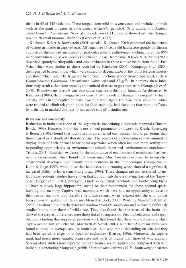
thritis in 45 of 145 skeletons. These ranged from mild to severe cases, and included animalssuch as the giant anteater Myrmecophaga tridactyla, gemsbok Oryx gazella and Arabiancamel Camelus dromedarius. None of the skeletons of 11 primates showed arthritic changes,nor the 28 small mammal skeletons (Greer et al., 1977).
Kitchener, Kolter & Brownstein (2001; see also Kitchener, 2004) examined the incidencesof various arthroses in captive bears. All bears over 15 years old had severe spondyloarthrosesand osteoarthroses with incidences of particular skeletal pathologies reaching more than 96%in 27 individuals of seven species (Kitchener, 2004). Kompanje, Klaver & de Vries (2000)described spondyloarthopathies and osteoarthroses in three captive bears from South-EastAsia, which were similar to those recorded by Kitchener (2004). Kompanje et al. (2000)distinguished between those which were caused by degeneration of the joints (osteoarthroses)and those which might be triggered by chronic infections (spondyloarthropathies), such asCampylobacter, Chlamydia, Mycoplasma, Salmonella and Shigella. In humans, these infec-tions may result either from sexually transmitted diseases or gastroenteritis (Kompanje et al.,2000). Roundworms Ascaris can also cause reactive arthritis in humans. As discussed byKitchener (2004), there is suggestive evidence that the skeletal changes may in part be due toactivity levels in the captive animals. Two Sumatran tigers Panthera tigris sumatrae, whichwere trained to climb telegraph poles for food each day, had skeletons that were unaffectedby arthritis, in marked contrast to less active cats of a similar age.
Brain size and complexityReduction in brain size is one of the key criteria for defining a domestic mammal (Clutton-Brock, 1999). However, brain size is not a fixed parameter, and work by Krech, Rosenweig& Bennett (1962) found that rats raised in an enriched environment had larger brains thanthose reared in a standard laboratory cage. The process of encouraging captive animals todisplay more of their normal behavioural repertoire, which often includes more activity andresponding appropriately to environmental stimuli, is termed ‘environmental enrichment’(Young, 2003). Empirical evidence for the importance of environmental enrichment has beenseen in experiments, which found that house mice Mus domesticus exposed to an enrichedenvironment developed significantly more neurones in the hippocampus (Kempermann,Kuhn & Gage, 1997), while those that had access to a running wheel showed a significantlyincreased ability to learn (van Praag et al., 1999). These changes are not restricted to justlaboratory rodents; studies have shown that London cab drivers (having learned the ‘knowl-edge’, Biegler et al., 2001), polygynous male voles, female cowbirds and food-storing birds,all have relatively large hippocampi owing to their requirement for above-normal spatiallearning and memory. Captive-bred mammals, which have had no opportunity to developtheir spatial memory, may therefore be disadvantaged when released into the wild, as hasbeen shown for golden lion tamarins (Menzel & Beck, 2000). Work by Marchetti & Nevitt(2003) has shown that hatchery-reared rainbow trout Oncorhynchus mykiss have significantlysmaller brains than those of wild trout. They also found that the areas of the brain thatshowed the greatest differences were those linked to aggression, feeding behaviour and repro-duction, a finding that supported previous work that found that these were the areas in whichcaptive-reared fish are deficient (Marchetti & Nevitt, 2003). Ranched American mink werefound to have, on average, smaller brain sizes than wild mink, depending on whether theyhad been reared in cages or in open-air enclosures (Kruska, 1996). Moreover, the captivemink had much more variable brain sizes and parts of brains than those of wild animals.Several other studies have reported reduced brain sizes in captive-bred compared with wildindividuals, including Mongolian gerbils Meriones unguiculatus (17.7% brain weight : carcass
Effects of captivity on morphology 225
© 2005 Mammal Society, Mammal Review, 35, 215–230
weight; Stuermer et al., 1997), pigs Sus scrofa (33% brain weight : carcass weight; Kruska &Röhrs, 1974), and llamas Lama glama (17% compared with guanacos, L. guanicoe of similarbody weight; Kruska, 1982). Przewalski’s wild horses Equus ferus przewalskii are now totallyderived from captive-bred individuals. Despite a captive breeding programme to maintaingenetic diversity and no attempt at deliberate domestication, these animals show a 16%decrease in brain weight or a 14% decrease in cranial volume, which is similar to that recordedfor domestic horses Equus caballus (Röhrs & Ebinger, 1998). Now that Przewalski’s horseshave been successfully reintroduced to Mongolia and China, it will be interesting to see iftheir cranial volumes revert to those of their wild ancestors. This has not been the case forferal ferrets Mustela furo in Britain and feral domestic cats in Australia, which still retainmuch smaller cranial volumes than their wild ancestors despite living for many generationsin the wild (Birks & Kitchener, 1999; Kitchener and Wagner, unpublished data; see below).Similar results have been recorded for feral dogs, goats and donkeys (Herre & Röhrs, 1990in Hemmer, 1990), and also for feral domestic pigs in the Galapagos Islands, which areotherwise said to be similar in external appearance to the European wild boar, but with thecolouration of the domestic pig (Kruska & Röhrs, 1974).
FERAL ANIMALS AS AN ANALOGUE FOR CAPTIVE RELEASEIt may be argued that many of the changes discussed above are simply the reaction of anindividual to captivity, and that such changes will not be passed onto their offspring. If thisis the case, then there should be no discernible difference between wild and feral individualsof the same species. With our growing knowledge of changes seen in captive animals, it isimportant to see if any of the observed changes will persist in the wild once animals havebeen released, as captive release programmes are expensive and time-consuming. Severalstudies have been undertaken to examine differences between wild, captive and feral animals,and these have produced some interesting results.
Pocock (1932) studied a sample of feral ferret skulls from the island of Mull, Scotland. Hewas unable to distinguish these from the skulls of wild polecats on the basis of their postor-bital breadths and cranial volumes. He suggested that the poor development of the jawmuscles in captive ferrets resulted in their narrower postorbital breadth and lower cranialvolume compared with wild polecats and feral ferrets. However, Birks & Kitchener (1999)found significant differences in cranial volume and postorbital breadths between polecats andferal ferrets, which suggested that these differences were not just the result of phenotypicplasticity. Lynch & Hayden (1995) reported differences between captive and feral Americanmink crania – captive mink have larger skulls, shorter palates and narrower postorbitalconstrictions. The skulls also displayed reduced sexual dimorphism, which suggested thatthere might be a relaxation of sexual selection, owing to selective breeding for larger animalsand lack of competition for resources in captivity. Feral mink can adapt to different environ-ments relatively quickly, as an examination of 15 populations of feral mink in Belarus foundthat there were substantial non-metric differences between the founder populations and thesubsidiary populations after only 30–40 years in the wild (Ulevièius, Sidorovich & Lauzhel,2001). Sexual dimorphism was also reduced in the domesticated foxes (Trut, 1999). In acomparison of wild and captive Coho salmon Oncorhynchus kisutch, Hard et al. (2000) foundthat captive fishes exhibited a reduction in sexual dimorphism and had smaller heads butlarger bodies than the wild salmon. Male salmon, which had been reared in captivity, werealso discriminated against by wild and captive female fishes, and were therefore less likely toreproduce than the wild males. These differences might be due to a lack of environmentalstimuli such as sea water, migration or increased density in the captive populations. However,

226 H. J. O’Regan and A. C. Kitchener
© 2005 Mammal Society, Mammal Review, 35, 215–230
although they are thought to be the result of environmental changes, a genetic differencebetween the populations could not be ruled out (Hard et al., 2000).
DISCUSSIONIn this review, we have covered a very wide range of morphological changes in a variety ofanimals. Some of the morphological differences between wild and captive animals can berectified with appropriate changes in husbandry (e.g. dietary nutrition and abrasion). How-ever, others imply a much greater modification in captive animals than may previously havebeen expected (e.g. brain size in feral mink). It may prove difficult to release animals whichhave adapted to captivity for many generations, if changes in brain size, speed of development,etc., have taken place. The changes in internal physiology such as that seen in the shorteningof bird’s intestines are obviously of great importance, and it will be worth looking to see ifsimilar modifications have occurred in captive mammals and whether they are reversible.Activity levels appear to have a great effect on captive animals, from pathology to increasingamounts of subcutaneous and deposited fat, and this is also something that needs to beaddressed. Environmental enrichment in the form of hiding food, scratching and climbingposts, etc., may well lead to changes in the prevalence of these problems, and the nextgeneration of captive animals will hopefully benefit from these changes. Kitchener (2004) hassuggested that environmental enrichment will be deemed successful when we can no longerdiscern any morphological differences between wild and captive-bred animals.
The lessening of sexual dimorphism in a variety of animals (silver foxes, mink and salmon)is also a cause for concern. Sexual dimorphism is central to many reproductive strategies,with the largest males having access to the most females. If this is disrupted, then animals ina population who would not normally be chosen to mate with will have as much opportunityto breed as those who display these traits. If the intention of a captive release programme isfor the wild animals to interbreed with the captive ones, then they may be at a disadvantagefrom the start. In addition, such breeding practices may result in captive populations diverg-ing from the wild ones at a much faster rate. Accelerated maturity may be of benefit if theobject is to breed as many animals as possible in a short time period, perhaps to restockhabitat from which they have been extirpated but where the niche is still present. If, however,they are to be held in captivity for a number of generations, the faster they breed, the morespace will be required to house them, and the process of morphological change from the wildto ‘domestic’ form may be speeded up. It is also possible that niche separation betweendifferent species, or between males and females of the same species, could be altered by achange in the size of captive animals, leading to potential competition between species orsexes owing to inevitable exploitation of similar-size prey.
It is time to think of these problems now, while most captive animals have only been inzoos for a few generations, rather than 200 years in the future. If behavioural abilities can beeroded in 20 years or so, for example, in golden lion tamarins, more effort needs to be putinto enriching the environments of all zoo animals, not just those destined for captive-release.The chances are that most species will become endangered at some point and that withoutfurther work, the stocks that we are perpetuating may not be able to be released in 200-yeartime.
CONCLUSIONA wide variety of animals show differences in captivity, but we are still a long way fromunderstanding what these changes are and their causes. They are important for a variety ofreasons, not just for the welfare of the animals, but also because they have the potential to
Effects of captivity on morphology 227
© 2005 Mammal Society, Mammal Review, 35, 215–230
provide information on the wider issues of mammalian evolution and morphology, particu-larly with regards to domestication. It is important to understand the changes that occur incaptive mammals, because for many species these are the only individuals available for study.The snow leopard Panthera uncia is a very rare cat, and there are few wild-caught individualsin museum collections. No one would suggest going out to collect more to feed the interestsof taxonomists, but then what is the alternative? Either to refuse to study that species becausethere is no ‘valid’ (i.e. wild-caught) material available, or attempt to understand whichstructures may have been altered by captivity and quantify that change? Unfortunately, theformer opinion often prevails.
There is also one very noticeable bias in these analyses – the majority have concentratedon crania, with very few studies looking at the rest of the skeleton. This is partly due tohistorical factors which have left museums filled with skulls, but very few postcranial skele-tons. However, in terms of locomotion and age-related pathologies, the appendicular skeletonis very important and this is an area that needs to be studied.
The majority of the case studies presented in this paper have been small scale, involve smallsample sizes or are mentioned in passing in papers written for other purposes. This is an areathat urgently needs addressing, and requires focused and carefully thought-out projects totry and unravel the complexities of relating morphological change to diet, environment,lifestyle, etc. in a large number of animals. The ideal pilot projects would be based on animalsthat have a discrete geographical range in the wild, are readily available in museum collections,have been kept as captive populations at a similar latitude, and breed relatively quickly sothat there is a good sample size (e.g. Lewis & Thomas, 2001; McPhee, 2004). Many rodentsand other small mammals could offer ideal models, whereby manipulation of the captiveenvironment would be possible over short time scales to see their effects on morphology andbehaviour. However, we also take the longer-term view and urge zoos, wildlife managers andmuseums to work together to take advantage of specimens that become available to studythe effects of captivity on larger and rarer species. The National Museums of Scotland hasbeen building such a collection of captive-bred, known-age specimens of a wide variety ofendangered species, in order to support long-term morphological studies as outlined above.
ACKNOWLEDGEMENTSWe would like to thank Filippo Aureli and David Wilkinson for reading and commenting ona draft of this paper, and two anonymous referees for making useful comments. A.C.K.would like to thank Graham Law for many constructive discussions about these topics, andis most grateful to the many zoos that have donated specimens to the National Museums ofScotland. He is also very grateful to Phil Howard, Alan Lothian, Ruth Pollitt and PeterSummers for preparing so many skeletons for the National Museums of Scotland’s researchcollections.
REFERENCESAltmann, J., Altmann, S. & Hausfater, G. (1981) Physical maturation and age estimates of yellow baboons,
Papio cynocephalus, in Amboseli National Park, Kenya. American Journal of Primatology, 1, 389–399.Armitage, P. (1983) Jawbone of a South America monkey from Brooks Wharf, City of London. London
Archaeologist, 4, 262–270.Baker, J.R. & Lyon, D.G. (1977) Skull malformation and cerebellar herniation in captive African lions.
Veterinary Record, 100, 154–156.Balharry, D. & Daniels, M. (1998) Wild living cats in Scotland. Scottish Natural Heritage Research, Survey
and Monitoring Report, No. 23.Biegler, R., McGregor, A., Krebs, J.R. & & Healy, S.D. (2001) A larger hippocampus is associated with longer-
lasting spatial memory. Proceedings of the National Academy of Sciences, USA, 98, 6941–6944.
228 H. J. O’Regan and A. C. Kitchener
© 2005 Mammal Society, Mammal Review, 35, 215–230
Birks, J.D.S. & Kitchener, A.C. (1999) The Distribution and Status of the Polecat Mustela putorius in Britainin the 1990s. The Vincent Wildlife Trust, London.
du Boulay, G.H. & Crawford, M.A. (1968) Nutritional bone disease in captive primates. Symposium of theZoological Society of London, 21, 223–236.
Chandra, A.M.S., Papendick, R.E., Schumacher, J., Homer, B.L. & Wollenman, P. (1999) Cerebellar herni-ation in captive lions (Panthera leo). Journal of Veterinary Diagnostic Investigation, 11, 465–468.
Clutton-Brock, J. (1999) A Natural History of Domesticated Mammals, 2nd edn. Cambridge University Press,Cambridge.
Cordy, D.R. (1957) Osteodystrophia fibrosa accompanied by visceral accumulation of lead. Cornell Veterinar-ian, 47, 480–490.
Corruccini, R.S. & Beecher, R.M. (1982) Occlusal variation related to soft diet in a nonhuman primate.Science, 218, 74–76.
Crossley, D.A. & del Mar Miguélez, M. (2001) Skull size and cheek–tooth length in wild-caught and captive-bred chinchillas. Archives of Oral Biology, 46, 919–928.
Darwin, C. (1868) The Variation of Animals and Plants under Domestication. John Murray, London.Duckler, G.L. (1998) An unusual osteological formation in the posterior skulls of captive tigers (Panthera
tigris). Zoo Biology, 17, 135–142.Fagan, D.A., Oosterhuis, J.E. & Roocroft, A. (2001) Captivity disorders in elephants impacted molars and
broken tusks. Der Zoologische Garten, 71, 218–303.Fitch, H.M. & Fagan, D.A. (1982) Focal palatine erosion associated with dental malocclusion in captive
cheetahs. Zoo Biology, 1, 295–310.Frankham, R., Hemmer, H., Ryder, O.A., Cothran, E.G., Soulé, M.E., Murray, N.D. & Snyder, M. (1986)
Selection in captive populations. Zoo Biology, 5, 127–138.Franz-Odendaal, T.A. (2004) Enamel hypoplasia provides insights into early systemic stress in wild and captive
giraffes (Giraffa camelopardalis). Journal of Zoology, London, 263, 197–206.Geiser, F. & Ferguson, C. (2001) Intraspecific differences in behaviour and physiology: effects of captive
breeding on patterns of torpor in feathertail gliders. Journal of Comparative Physiology B, 171, 569–576.
Gore, M.A. (1993) A comparison of morphometry from captive and free-ranging Macaca mulatta. Journal ofMedical Primatology, 22, 360–367.
Greer, M., Greer, J.K. & Gillingham, J. (1977) Osteoarthritis in selected wild mammals. Proceedings of theOklahoma Academy of Sciences, 57, 39–43.
Groves, C.P. (1966) Skull-changes due to captivity in certain Equidae. Zeitschrift für Säugetierkunde, 31, 44–46.
Groves, C.P. (1982) The skulls of Asian rhinoceroses: wild and captive. Zoo Biology, 1, 251–261.Guthrie, R.D. (1984) Alaskan megabucks, megabulls, and megarams: the issue of Pleistocene gigantism.
Carnegie Museum of Natural History Special Publication, 8, 482–510.Haberstroh, L.I., Ullrey, D.E., Sikarski, J.G., Richter, N.A., Colmery, B.H. & Myers, T.D. (1984) Diet and
oral health in captive Amur tigers (Panthera tigris altaica). Journal of Zoo Animal Medicine, 15, 142–146.Hard, J.J., Berejikian, B.A., Tezak, E.P., Schroder, S.L., Knudsen, C.M. & Parker, L.T. (2000) Evidence for
morphometric differentiation of wild and captively reared adult coho salmon: a geometric analysis. Envi-ronmental Biology of Fishes, 58, 61–73.
Hemmer, H. (1990) Domestication. Cambridge University Press, Cambridge.Herre, W. & Röhrs, M. (1990) Haustiere – Zoologisch Gesehen, 2nd edn. Gustav Fischer, Stuttgart.Hollister, N. (1917) Some effects of environment and habit on captive lions. Proceedings of the United States
National Museum, 53, 177–193.Hollister, N. (1918) East African mammals in the United States National Museum. Part I. Insectivora,
Chiroptera, and Carnivora. Bulletin of the United States National Museum, 99, 1–194.Howell, A.B. (1925) Pathologic skulls of captive lions. Journal of Mammology, 6, 163–168.Jolly, W.P. (1976) Jumbo. Constable, London.Kempermann, G., Kuhn, H.G. & Gage, F.H. (1997) More hippocampal neurons in adult mice living in an
enriched environment. Nature, 386, 493–495.Kimura, T. & Hamada, Y. (1996) Growth of wild and laboratory born chimpanzees. Primates, 37, 237–251.Kitchener, A.C. (1998) The Scottish wildcat – a cat with an identity crisis? British Wildlife, 9, 232–242.Kitchener, A.C. (2004) Problems with old bears in zoos. International Zoo News, 51, 282–293.Kitchener, A.C., Merryweather, J. & Allchurch, T. (1999) The effects of captivity on the flight musculo-skeletal
system of fruit bats, Pteropus spp. In: EEP Yearbook 1998/1999 Including the Proceedings of the 16th EAZAConference. Basel, 7–12 September 1999 (Ed. by F. Rietkerk, B. Hiddinga, K. Brouwer & S. Smits), pp.553–555. EAZA Executive Office, Amsterdam.

Effects of captivity on morphology 229
© 2005 Mammal Society, Mammal Review, 35, 215–230
Kitchener, A.C., Kolter, L. & Brownstein, D. (2001) Problems with old bears in zoos. In: EEP Yearbook 1999/2000 Including Proceedings of the 17th EAZA Conference, Aalborg 19–24 September 2000 (Ed. by B.Hiddinga & K. Brouwer), pp. 625–628. EAZA Executive Office, Amsterdam.
Kleiman, D.G., Allen, M.E., Thompson, K.V. & Lumpkin, S. (1996) Wild Mammals in Captivity – Principlesand Techniques. University of Chicago Press, Chicago.
Kolmstetter, C., Munson, L. & Ramsay, E.C. (2000) Degenerative spinal disease in large felids. Journal of Zooand Wildlife Medicine, 31, 15–19.
Kompanje, E.J.O., Klaver, P.S.J. & de Vries, G.Th. (2000) Spondyloarthropathy and osteoarthrosis in threeIndomalayan bears: Ursus ursinus Cuvier, 1823, Ursus thibetanus Raffles, 1821, and Ursus malayanus Shaw& Nodder, 1791 (Mammalia: Carnivora: Ursidae). Contributions to Zoology, 69, 259–269.
Krech, D., Rosenweig, M.R. & Bennett, E.L. (1962) Relations between brain chemistry and problem-solvingamong rats raised in enriched and impoverished environments. Journal of Comparative and PhysiologicalPsychology, 55, 801–807.
Kruska, D. (1982) Changes of brain size in Tylopoda during phylogeny and caused by domestication. Verhan-dlungen Deutschen Zoologischen Gesellschaft, 1982, 173–183.
Kruska, D. (1996) The effect of domestication on brain size and composition in the mink (Mustela vison).Journal of Zoology, London, 239, 645–661.
Kruska, D. & Röhrs, M. (1974) Comparative-quantitative investigations on brains of feral pigs from theGalapagos Islands and of European domestic pigs. Zeitschrift für Anatomie und Entwicklungsgeschichte,144, 61–73.
Lewis, O.T. & Thomas, C.D. (2001) Adaptations to captivity in the butterfly Pieris brassicae (L.) and theimplications for ex situ conservation. Journal of Insect Conservation, 5, 55–63.
Lieberman, D.E., Krovitz, G.E., Yates, F.W., Devlin, M. & St. Claire, M. (2004) Effects of food processingon masticatory strain and craniofacial growth in a retrognathic face. Journal of Human Evolution, 46, 655–677.
Lindburg, D.G. (1998) Enrichment of captive mammals through provisioning. In: Second Nature – Environ-mental Enrichment for Captive Animals (Ed. by D.J. Shepherdson, J.D. Mellen & M. Hutchins), pp. 262–276. Smithsonian Institution Press, Washington DC.
Liukkonen-Anttila, T., Saartoala, R. & Hissa, R. (2000) Impact of hand-rearing on morphology and physi-ology of the capercaillie (Tetrao urogallus). Comparative Biochemistry and Physiology Part A, 125, 211–221.
Lynch, J.M. & Hayden, T.J. (1995) Genetic influences on cranial form: variation among ranch and feralAmerican mink Mustela vison (Mammalia: Mustelidae). Biology Journal of the Linnean Society, 55, 293–307.
Macdonald, D. (2001) The New Encyclopedia of Mammals. Oxford University Press, Oxford.McPhee, M.E. (2004) Morphological change in wild and captive oldfield mice, Peromyscus polionotus subgri-
seus. Journal of Mammology, 85, 1130–1137.Marchetti, M.P. & Nevitt, G.A. (2003) Effects of hatchery rearing on the brain structures of rainbow trout,
Oncorhryncus mykiss. Environmental Biology of Fishes, 66, 9–14.Marker, L.L. & Dickman, A.J. (2004) Dental anomalies and incidence of palatal erosion in Namibian cheetahs
(Acinonyx jubatus jubatus). Journal of Mammology, 85, 19–24.Meers, M.B. (1996) Three Dimensional Analysis of Differences in Cranial Morphology between Captive and Wild
American Alligators. Available at: http://utweb.ut.edu/faculty/mmeers/res/gatorheads/index.html (accessedon 19 July 2004).
Menzel, C.R. & Beck, B.B. (2000) Homing and detour behavior in golden lion tamarin social groups. In: Onthe Move (Ed. by S. Boinski & P.A. Garber), pp. 299–326. Chicago University Press, Chicago.
Molnar, S. & Ward, S.C. (1975) Mineral metabolism and microstructural defects in primate teeth. AmericanJournal of Physical Anthropology, 43, 3–18.
Moss, R. (1972) Effects of captivity on gut lengths in red grouse, Lagopus lagopus. Journal of WildlifeManagement, 36, 99–104.
Nowak, R.M. (1999) Walker’s Mammals of the World, 6th edn. The Johns Hopkins University Press, Baltimoreand London.
O’Regan, H.J. (2001) Morphological effects of captivity in big cat skulls. In: Proceedings of the 3rd ZooResearch Symposium (Ed. by S. Wehnelt & C. Hudson), pp. 18–22. Chester Zoo, Chester.
O’Regan, H.J. & Turner, A. (2004) The interface between conservation biology, palaeontology and archaeo-zoology – morphometrics and population viability analysis. In: The Future from the Past: Archaeozoologyin Wildlife Conservation and Heritage Management (Ed. by R.C.G.M. Lauwerier & I. Plug), pp. 90–96.Oxbow Books, Oxford.
Ohlsson, T. & Smith, H.G. (2001) Early nutrition causes persistent effects on pheasant morphology. Physio-logical and Biochemical Zoology, 74, 212–218.
230 H. J. O’Regan and A. C. Kitchener
© 2005 Mammal Society, Mammal Review, 35, 215–230
Owen, M. (1975) Cutting and fertilizing grassland for winter goose management. Journal of Wildlife Manage-ment, 39, 163–167.
Palmer, A.C., Callanon, J.J., Gueri, L.A., Sheahan, B.J., Stonach, N. & Franklin, R.J.M. (2001) Progressiveencephalomyelopathy and cerebellar degeneration in 10 captive-bred cheetahs. Veterinary Record, 149, 49–54.
Pendergast, B.A. & Boag, D.A. (1973) Seasonal changes in the internal anatomy of Spruce Grouse in Alberta.The Auk, 90, 307–317.
Phillips-Conroy, J.E. & Jolly, C.J. (1988) Dental eruption schedules of wild and captive baboons. AmericanJournal of Primatology, 15, 17–29.
Pocock, R.I. (1932) Ferrets and polecats. Scottish Naturalist, 196, 97–108.Poole, W.E., Carpenter, S.M. & Simms, N.G. (1980) Multivariate analyses of skull morphometrics from the
two species of Grey Kangaroos, Macropus giganteus Shaw and M. fuliginosus (Desmarest). AustralianJournal of Zoology, 28, 591–605.
van Praag, H., Christie, B.R., Sejnowski, T.J. & Gage, F.H. (1999) Running enhances neurogenesis, learningand long-term potentiation in mice. Proceedings of the National Academy of Sciences, 96, 13427–13431.
Price, E.O. (1999) Behavioral development in animals undergoing domestication. Applied Animal BehaviourScience, 65, 245–271.
Price, E.O. (2002) Animal Domestication and Behavior. CABI Publishing, Wallingford.Röhrs, M. & Ebinger, P. (1998) Sind Zooprzewalskipferde Hauspferde? Berliner und Munchener Tierartliche
Wochenschrift, 111, 273–280.Rothschild, B.M., Rothschild, C. & Woods, R.J. (1998) Inflammatory arthritis in large cats: an expanded
spectrum of spondyloarthropathy. Journal of Zoo and Wildlife Medicine, 29, 279–284.Savory, C.J. & Gentle, M.J. (1976) Changes in food intake and gut size in Japanese quail in response to
manipulation of dietary fibre content. British Poultry Science, 17, 571–580.Schaller, G.B. (1972) The Serengeti Lion: A Study of Predator–Prey Relations. University of Chicago Press,
Chicago.Schauenberg, P. (1977) Longueur de l’intestin du chat forestier Felis silvestris Schreber. Mammalia, 41, 357–
360.Smuts, G.L., Anderson, J.L. & Austin, J.C. (1978) Age determination of the African lion (Panthera leo).
Journal of Zoology, London, 185, 365–373.Soulé, M., Gilpin, M., Conway, W. & Foose, T.J. (1986) The millennium ark: how long a voyage, how many
staterooms, how many passengers? Zoo Biology, 5, 101–113.Stuermer, I.W., Plotz, K., Wetzel, W., Wagner, T., Leybold, A. & Scheich, H. (1997) Reduced brainsize and
faster auditory discrimination learning in laboratory gerbils compared to wild Mongolian gerbils (Merionesunguiculatus). Society for Neuroscience (Abstracts), 23, 2067.
Sutton, J.B. (1884) On the diseases of the carnivorous mammals in the society’s gardens. Proceedings of theZoological Society of London, 177–187.
Trut, L.N. (1999) Early canid domestication: the farm-fox experiment. American Scientist, 87, 160–169.Ulevièius, A., Sidorovich, V. & Lauzhel, G. (2001) Specificity of non-metric parameters of American mink
(Mustela vison) populations in relation to habitat differences in Belarus. Mammalian Biology, 66, 35–47.Wallace, M.P. (2000) Retaining natural behaviour in captivity for re-introduction programmes. In: Behaviour
and Conservation (Ed. by L.M. Gosling & W.J. Sutherland), pp. 300–314. Cambridge University Press,Cambridge.
Wenker, C.J., Stich, H., Müller, M. & Lussi, A. (1999) A retrospective study of dental conditions of captivebrown bears (Ursus arctos spp.) compared with free-ranging Alaskan grizzlies (Ursus arctos horribilis).Journal of Zoo and Wildlife Medicine, 30, 208–221.
Young, R. (2003) Environmental Enrichment for Captive Animals. Blackwell Science, Oxford.
Submitted 5 October 2004; revision accepted 24 February 2005Editor: RM