Embed Size (px)
Citation preview
259
The effects of intensive, long-term
treadmill running on reproductivehormones, hypothalamus–pituitary–testis axis, and semen quality:a randomized controlled studyMohammad Reza Safarinejad, Kamran Azma1 and Ali Asgar Kolahi2
Urology and Nephrology Research Center, Shahid Beheshti University (MC), PO Box 19395-1849, Tehran, Iran1Department of Physical Medicine and Rehabilitation, Faculty of Medicine, Aja University of Medical Sciences, 195 698-9333 Tehran, Iran2Department of Health and Community Medicine, Faculty of Medicine, Shahid Beheshti University (MC), 158 684-4139 Tehran, Iran
(Correspondence should be addressed to M R Safarinejad; Email: [email protected])
Abstract
Effects of intensive exercise on hypothalamus–pituitary–testis
(HPT) axis remain controversial. Our aim was to determine
the effects of intensive, long-term treadmill running on
reproductive hormones, HPT axis, and semen quality. A total
of 286 subjects were randomly assigned to moderate-intensity
exercise (w60% maximal oxygen uptake (VO2max); group 1,
nZ143) and high-intensity exercise (w80% VO2max; group
2, nZ143) groups. The two groups exercised for 60 weeks in
five sessions per week, each session lasting 120 min. This was
followed by a 36-week low-intensity exercise recovery
period. All subjects underwent routine semen analysis.
Blood samples were drawn for the determination of the
levels of the following hormones: LH, FSH, prolactin,
testosterone (T), free testosterone (fT), inhibin B, and sex
hormone-binding globulin (SHBG). The HPT axis was
Journal of Endocrinology (2009) 200, 259–2710022–0795/09/0200–259 q 2009 Society for Endocrinology Printed in Great
assessed using GnRH and human chorionic gonadotropin
tests. After 24 weeks of exercise, the subjects exercising with
high intensity demonstrated significantly declined semen
parameters compared with those exercising with moderate
intensity (PZ0.03). Serum T and fT began to decrease, and
serum SHBG began to increase at the end of 12 weeks with
both moderate- and high-intensity exercises. The serum LH
and FSH concentrations decreased below the baseline level at
12 weeks in both groups (PZ0.07 in group 1 and 0.03 in
group 2). Both groups had blunted LH and FSH responses to
GnRH. These parameters improved to their pre-exercise
level during the recovery period. Long-term strenuous
treadmill exercises (overtraining syndrome) have a deleterious
effect on reproduction.
Journal of Endocrinology (2009) 200, 259–271
Introduction
There is little argument that physical activity is beneficial for
one’s health. Indeed, increasing the amount of physical
activity is associated with extensive health improvements.
However, strenuous exercise represents a physical stress that
challenges homeostasis. In response to this stressor, the
endocrine and nervous systems are known to react and
participate in the maintenance of homeostasis and the
development of physical fitness. Regardless of the many
known health benefits of exercise, there is a body of
evidence suggesting that endurance exercise is associated
with some health problems (Mastaloudis et al. 2004,
Scharhag et al. 2005). Despite the many advances in
‘andrology’ in the last decade, a considerable proportion of
‘male factor’ infertilities still remain unknown or idiopathic.
In recent years, prolonged strenuous exercise has been
proposed as a possible reason for male factor infertility (Arce
& De Souza 1993). A subset of endurance-trained men,
particularly runners, present with profound changes in their
GH (Kilic 2007), thyroid hormone and testosterone, LH,
dehydroepiandrosterone sulfate, cortisol (Tremblay et al.
2005), and prolactin (PRL; Bridge et al. 2003). It has been
reported that both hypothalamic and testicular endocrine
functions are suppressed during acute prolonged physical
exercise. The exercise-induced suppression of serum
testosterone is associated with suppressed endogenous
GnRH stimulation of gonadotropin release during exercise
(Kujala et al. 1990). Qualitatively and quantitatively normal
spermatogenesis is critically dependent on an intact hypo-
thalamus–pituitary–testis (HPT) axis. Androgens are essential
for the maintenance of normal spermatogenesis.
Reactive oxygen species (ROS) produced in metabolic and
physiological processes and harmful oxidative reactions may
lead to oxidation of biological molecules such as lipids,
proteins, and DNA (Halliwell & Gutteridge 1984). There is a
body of evidence suggesting that endurance exercise is
associated with oxidative stress. During the resting state, the
human body produces ROS continuously, but in healthy
individuals these ROS are produced at levels well within the
DOI: 10.1677/JOE-08-0477Britain Online version via http://www.endocrinology-journals.org
Downloaded from Bioscientifica.com at 03/14/2022 01:52:51AMvia free access
M R SAFARINEJAD and others . Treadmill running and male reproductive status260
capacity of the body’s antioxidant defense system. During
endurance exercise, there is a 10- to 20-fold increase in whole
bodyoxygen (O2) consumption (Astrand & Rodahl 1986), and
O2 uptake in the active skeletal muscle increases 100- to 200-
fold (Halliwell & Gutteridge 1999). This increase in O2
utilization may result in the production of ROS at the rates that
exceed the body’s capacity to detoxify them (Alessio 1993).
However, some studies have failed to observe exercise-induced
oxidative stress (Poulsen et al. 1996, Oztasan et al. 2004). An
increase in the formation of ROS decreases fertility, as the ROS
will attack the membranes of the spermatozoa, decreasing their
viability (Irvine 1996). Altogether these can impair sperma-
togenesis and fertility capacity in men. Less attention has been
directed toward identifying changes in spermatogenesis and
fertility capacity as a result of endurance training.
Treadmills offer many distinct advantages. Speed, slope,
and environmental factors can be easily controlled and data
from repeated running cycles can be collected. Treadmills
have therefore been used in many experimental studies. In this
study, we addressed the effects of prolonged high-intensity
treadmill running on semen quality, reproductive hormones,
and HPT axis.
Materials and Methods
Study participants
A total of 362 male volunteers between February 2002 and
July 2006 were recruited from sport facilities for this study.
Volunteers for the study were aged 20–40 years and recruited
through local advertisement. The trial requirements were
carefully explained to participants, who were given the
opportunity to withdraw at any time they desired. They
were habitual aerobic exercisers and reported training for
an average of 1.8 h per day, 5 days per week. Participation
was voluntary throughout the study period, and exercise
equipment and supplies were free of charge. Eligible
participants were invited to an orientation session. Individuals
who remained interested were assessed for eligibility and
signed a written informed consent form. The investigation
was approved by the local medical ethical committee, which
was conducted in accordance with the International
Conference on Harmonisation-Good Clinical Practice
(ICH-GCP) guidelines and the principles of the Declaration
of Helsinki.
Inclusion and exclusion criteria Subjects were included
in the study after fulfilling the following criteria: no known
medical condition that could interfere with their fertility
and total testicular volume (measured by ultrasound) R12 ml.
All participants were required to have ceased all medical
therapies at least 12 weeks before the start of the study.
Participants were excluded if they had abnormal semen
analysis according to the World Health Organization (WHO)
criteria (1992), abnormal reproductive endocrine profiles, or
Journal of Endocrinology (2009) 200, 259–271
positive antisperm antibody assay (ASA) test; present use of
ergogenic aids such as creatine monohydrate, herbal stimu-
lants, or any anabolic agents such as steroids; presence of genital
diseases (cryptorchidism, present genital inflammations, or
varicocele); a diagnosis of alcohol and/or drug dependence; Y
chromosome microdeletions or karyotype abnormalities; and
sexually transmitted disease or relevant genitourinary infec-
tion. Participants were also excluded if they had a body mass
index (BMI)R30; occupational and environmental exposures
to potential reproductive toxins; participation in reduced
caloric intake diets that may alter basal hormonal levels; and
were participating in another investigational study.
Evaluations All subjects were screened for nutritional habits
by a registered dietician. Subjects, who were unable to intake
adequate energy, including 30–40% energy from fat and 15–
20% energy from protein, were excluded from the study.
The participants underwent a thorough physical examination,
anthropometric measurements, urine analysis and serum
chemicals, hematological laboratory, and thyroid function
tests. Body weight was determined using a Detecto beam scale
(Brooklyn, NY, USA) accurate to 0.1 kg, and height was
determined using a Seca stadiometer accurate to 0.1 cm. Body
composition was estimated with Harpenden skinfold calipers
using the standard methods outlined in the Anthropometric
Standardization Reference Manual (Harrison et al. 1988).
Testicular volumes were measured with scrotal ultrasound
by a sonologist using electronic calipers. A volume of less than
12 ml was considered small. The presence of varicocele was
determined by Doppler ultrasonography of scrotum with the
Valsalva maneuver. Genetic testing included karyotype
analysis and Y chromosome microdeletion analysis.
All participants had at least two baseline semen analysis
following an abstinence period of 3–4 days. An average of two
for each subject and each parameter was taken for comparison
with the post-trial values. When the values differed by more
than 20%, a third test was done. All procedures and
interpretations used were in accordance with the established
WHO (1992) criteria, besides morphology, which was
established according to the Kruger parameters. The normal
WHO values included sperm concentration of 20!106
spermatozoa/ml or more and motility of 50% or more with
forward progression. Using the Kruger strict criteria, males
with greater than 14% normal forms were considered normal.
The total motile sperm count was determined by the formula:
(semen volume!sperm density!motility percentage)/100.
Total sperm per ejaculate was calculated as the product of
ejaculate volume and sperm concentration. Quality control
and proficiency criteria were set for !15% variation for
certifying the technicians trained to do semen analysis for the
study. In every subject, the immunobead test for ASA binding
was also performed.
Endocrinological studies Two early morning blood
samples were drawn from each subject after overnight fasting
at 20-min intervals for the determination of the resting levels
www.endocrinology-journals.org
Downloaded from Bioscientifica.com at 03/14/2022 01:52:51AMvia free access
Treadmill running and male reproductive status . M R SAFARINEJAD and others 261
of the following hormones: FSH, LH, inhibin B, PRL, total
testosterone (T), free testosterone (fT), and sex hormone-
binding globulin (SHBG).
The GnRH stimulation test was performed as follows:
100 mg GnRH analog (GnRHa; Decapeptyl, Ferring,
Germany) was administered by i.v. bolus. Blood was drawn
(for serum LH and FSH determination) at K15, 0, 20, 40, 60,
and 120 min after injection. We defined a more than 3-fold
increase in FSH and a more than 4-fold increase in LH in
response to Decapeptyl as GnRH positive, and increases of
less than 3-fold and 4-fold respectively as GnRH negative.
Three days after GnRH test, all participants received an
injection of human chorionic gonadotropin (hCG; Pregnyl,
50 IU/kg i.m., Organon, Oss, The Netherlands). Serum T
was determined before the hCG injection and on the third
day afterward. After the hCG injection, the serum T level
should rise in 3 days by a factor of 2–4 compared with the
basal value (positive test).
Laboratory methods Single-antibody, solid-phase RIA
was used to determine the immunoreactivity levels indicative
of blood concentrations of Tand fT (Diagnostic Products, Los
Angeles, CA, USA). Variances for the RIAs performed in this
investigation were as follows: total testosterone (intra-assay
%2.5%, interassayZ3.0%) and free testosterone (intra-assay
%2.5%, interassayZ9.6%). Serum LH and FSH were
measured using time-resolved immunofluorometric assay kits
(DELFIA hLH for LH and DELFIA hFSH for FSH; Wallac
Co., Turku, Finland). The intra- and interassay coefficients of
variation of the individual immunofluorometric assay method
was below 9% within the reference ranges. The reference
ranges were as follows: T, 9–38 nmol/l; fT, 90–310 pmol/l;
LH, 1.0–8.4 IU/l; and FSH, l.0–10.5 IU/l.
Serum SHBG was determined using 1235 AutoDELFIA
automatic system based on a time-resolved fluoroimmuno-
assay (AutoDELFIA SHBG, Wallac Co). The between-assay
coefficient of variation is 2.3–3.0%. Non-SHBG-bound fT
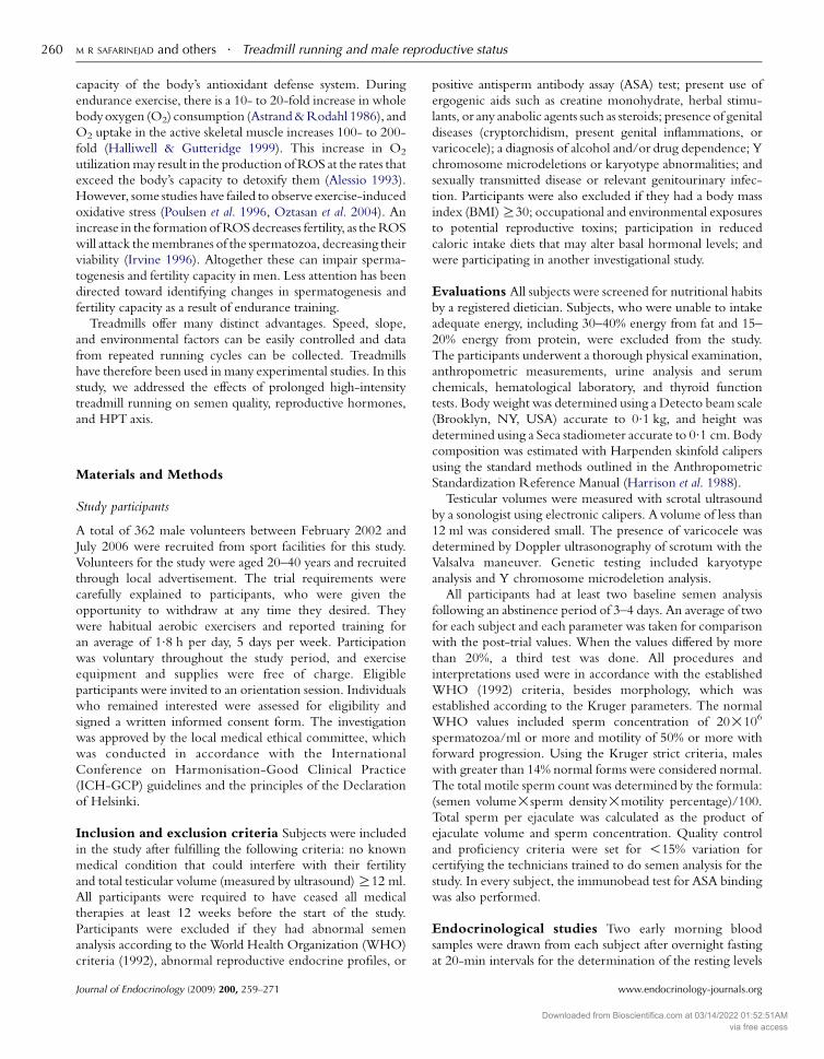
Figure 1 Study design.
www.endocrinology-journals.org
was obtained using the following formula: fT (pmol/l)ZT
(nmol/l)/(K!SHBG (nmol/l)C1)!1000, where K is the
equilibrium constant for T binding to SHBG (1.6!109
l/mol; Gluer et al. 1995). Serum levels of PRL were assayed by
commercial RIA kit. This commercial kit has been used
previously with inter- and intra-assay variations of less than
10%. The reference ranges for PRL was 92–697 pmol/l.
Serum Inhibin B was determined by ELISA method using kit
reagents and inhibin B standard (Oxford Bio-innovation Ltd,
Oxon, UK). The assay sensitivity was 4 pg/ml and the
between-assay variation was 15%. The antibodies bound to
the surface of the sperm were detected with the ‘immuno-
bead-binding test’. ASA was performed using specific beads
that bind separately to IgG and IgA. The ASA test was
considered positive when there was more than 20% sperm-
bound immunobeads as recommended by the WHO (1992).
Out of the 362 screened subjects, 304 met inclusion/
exclusion criteria and entered into the screening phase of
the study.
Study design
The study consisted of a 12-week screening and familia-
rization phase, a 60-week treadmill running phase (interven-
tion), and a 36-week recovery period (Fig. 1).
Screening phase All subjects were required to complete a
Physical Activities Readiness Questionnaire (Thomas et al.
1992) and the American College of Sports Medicine risk
stratification for coronary artery disease risk factors (Franklin
et al. 2000) to ensure that all were free from any known
cardiovascular, pulmonary, or musculoskeletal conditions.
All exercises were done on a motorized treadmill (HP
Cosmos Venus, Traunstein, Germany). The driving system
provided a range of constant speed from 1 to 22 km/h (by
minimum increments of 0.1 km/h). The maximal belt speed
error reached 7% with a subject of 100 kg walking at 6 km/h.
Journal of Endocrinology (2009) 200, 259–271
Downloaded from Bioscientifica.com at 03/14/2022 01:52:51AMvia free access
M R SAFARINEJAD and others . Treadmill running and male reproductive status262
The subjects visited the laboratory on two occasions and
completed a continuous, inclined treadmill test using the
modified Astrand protocol (Pollock et al. 1978) to volitional
exhaustion for the determination of maximum oxygen
consumption (VO2max). Oxygen consumption was measured
using indirect calorimetry. Metabolic measures were collected
using standard open-circuit spirometric techniques (meta-
bolic cart 2700Z; SensorMedics, Yorba Linda, CA, USA).
Maximum VO2 (VO2max) was considered to be attained when
the subjects met at least two of the following criteria: 1) a
plateau in VO2 with an increase in workload, 2) respiratory
exchange ratio greater than or equal to 1.15, and 3) maximum
age-predicted heart rate (220 – age) is attained. The highest
VO2 attained during the tests was registered as the VO2max
value.
For the purposes of this study, low-, moderate-, and high-
intensity exercises (HIE) were defined as 30, 60, and 80%
VO2max respectively. Exercise at 60% VO2max will be
sufficient to elicit clinically significant physiological changes
associated with exercise training; 80% VO2max will allow
those who wish to work at a higher intensity but not above
most individuals’ anaerobic threshold (NIH Consensus
Development Panel on Physical Activity and Cardiovascular
Health 1996, Snyder et al. 1997).
At the end of week 4, the subjects were recommended to
complete a ‘multi-stage’ incremental treadmill exercise in
which the running speed, incline, and duration were
increased gradually to the subject’s limit of tolerance. The
starting level and the rate of increments were voluntary.
However, the participants were required to increase the
exercise level to their pre-determined 80% VO2max for 2 h, 5
sessions per week, maximum within the 12-week screening
period. Out of the 304 participants who entered the screening
phase, 286 met the study protocol and continued partici-
pation with the intervention or running phase.
Running phase Participants were randomly assigned to
moderate-intensity exercise (MIE; w60% VO2max; group 1,
nZ143) and high-intensity exercise (HIE; w80% VO2max;
group 2, nZ143) groups. Assignment to exercise group was
performed using an interactive voice response system, which
followed a randomization table generated by the method of
random permuted blocks. Participant randomization numbers
were allocated to each site in an ascending sequence in blocks.
Randomization was not stratified by age. The two groups
exercised for 60 weeks in five sessions per week, each session
lasting 120 min. All subjects were asked to exercise to the
limit of the study. Other types of exercises such as swimming,
cycling, and sauna bathing were not allowed during the whole
study protocol. To avoid the confounding factors associated
with a training effect, the subjects agreed to maintain their
exercise regimen for the duration of the study period.
Throughout the running phase, the subjects were made aware
of the potential risks of exercises on reproductive capacity.
Out of the 286 subjects who participated in the running
phase, 264 (134 out of 143 in group 1, and 130 out of 143 in
Journal of Endocrinology (2009) 200, 259–271
group 2) were eligible and consented to continue with the
recovery phase of the study (Fig. 1). Subjects with any
violation of the study protocol, which could affect the study
results, were excluded according to the investigator’s
discretion.
Recovery phase The last 36 weeks consisted of the post-
intervention phase during which the long-term effects of the
intervention were assessed every 12 weeks. In this phase, all
subjects were asked to continue exercising only at low-
intensity exercise (LIE) level (w30% VO2max) if they so
desired. They were required to to exercise 120 min per day,
5 days per week, for 36 weeks. The subjects were free to
choose further participation with study protocol or dis-
continue any kind of participation.
Evaluations during the whole study protocol During
each exercise, surface skin thermistor probes were attached to
the skin surface at four locations (chest, triceps, thigh, and
calf) to determine the weighted mean skin temperature
(Ramanathan 1964). Excellent adherence to both interven-
tion and measurement is necessary. Each week, a tracking
report was generated to monitor adherence to exercise
prescription. In order to maintain high adherence rates,
flexibility is required. If a participant failed to meet the study
requirements for 1 week, the study requirement for the
following week (s) could be increased up to 1.5 kcal/kg per
week (KKW) to compensate. For the planned exercise
sessions, exercise intensity was quantified using the data from
the Polar XL HR monitor (Polar Heart Rate Monitor,
Kempele, Finland) worn by the participants. The appropriate
heart rate range for the prescribed intensity (30, 60, and 80%
VO2max) was calculated following completion of the exercise.
If a participant’s HR falls or rises out of range, the speed
and/or grade of the treadmill were being adjusted to maintain
the prescribed intensity. For monitoring daily activity outside
the assigned exercise session, each participant was given a step
counter (ACCUSPLIT Eagle, Japan) at baseline and
instructed to wear it at all times during waking hours. The
participants were asked to remove the step counter only
during planned exercise sessions, or for purposes of bathing,
sleeping, or dressing. At the end of each day, the participants
recorded the number of steps taken and reset the step counter
for the following day. The participants were also asked to
make note of any events resulting in significant changes in
activity, for example, illness or injury. In addition, physical
activity questionnaires were completed by the participants
during all three phases of the trial.
During the monthly visit, a medical history, a physical
examination, and a resting 12 lead electrocardiogram were
obtained, and a comprehensive health status questionnaire
was administered. Actual duration and frequency of exercises
was recorded in the diaries that were monitored for exercise
compliance during monthly visits. At follow-up visits (every
12 weeks, Fig. 2), two semen samples were collected within a
1- to 2-week period of each other and endocrinological
www.endocrinology-journals.org
Downloaded from Bioscientifica.com at 03/14/2022 01:52:51AMvia free access
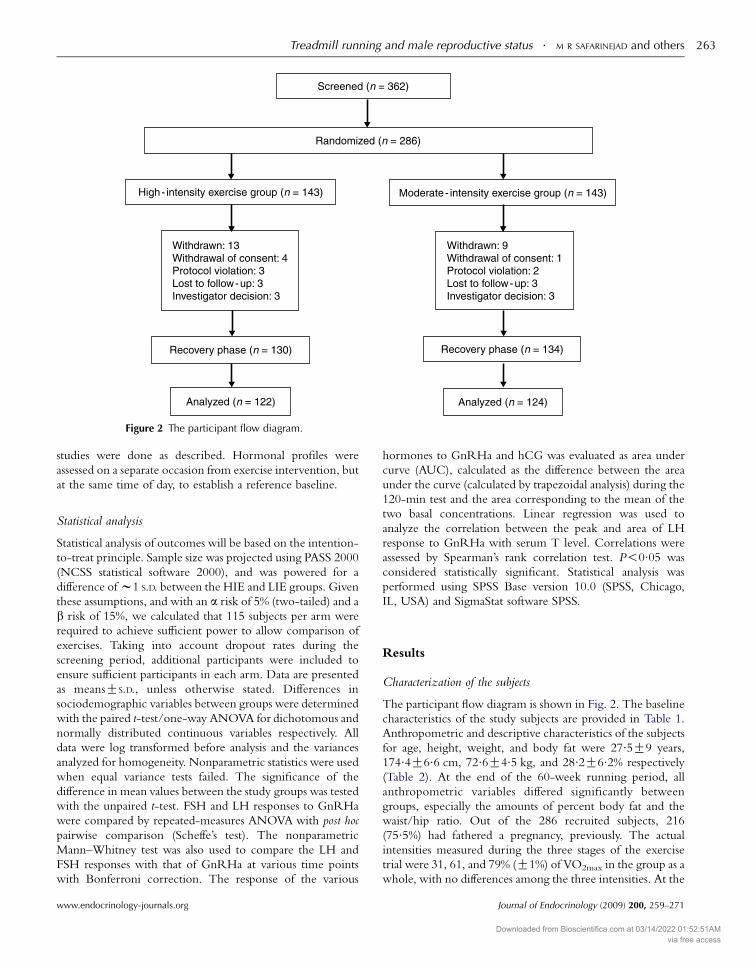
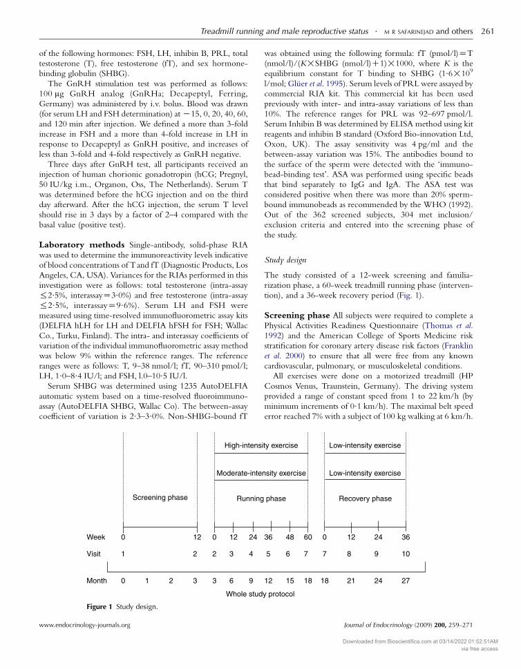
Figure 2 The participant flow diagram.
Treadmill running and male reproductive status . M R SAFARINEJAD and others 263
studies were done as described. Hormonal profiles were
assessed on a separate occasion from exercise intervention, but
at the same time of day, to establish a reference baseline.
Statistical analysis
Statistical analysis of outcomes will be based on the intention-
to-treat principle. Sample size was projected using PASS 2000
(NCSS statistical software 2000), and was powered for a
difference of w1 S.D. between the HIE and LIE groups. Given
these assumptions, and with an a risk of 5% (two-tailed) and a
b risk of 15%, we calculated that 115 subjects per arm were
required to achieve sufficient power to allow comparison of
exercises. Taking into account dropout rates during the
screening period, additional participants were included to
ensure sufficient participants in each arm. Data are presented
as meansGS.D., unless otherwise stated. Differences in
sociodemographic variables between groups were determined
with the paired t-test/one-way ANOVA for dichotomous and
normally distributed continuous variables respectively. All
data were log transformed before analysis and the variances
analyzed for homogeneity. Nonparametric statistics were used
when equal variance tests failed. The significance of the
difference in mean values between the study groups was tested
with the unpaired t-test. FSH and LH responses to GnRHa
were compared by repeated-measures ANOVA with post hoc
pairwise comparison (Scheffe’s test). The nonparametric
Mann–Whitney test was also used to compare the LH and
FSH responses with that of GnRHa at various time points
with Bonferroni correction. The response of the various
www.endocrinology-journals.org
hormones to GnRHa and hCG was evaluated as area under
curve (AUC), calculated as the difference between the area
under the curve (calculated by trapezoidal analysis) during the
120-min test and the area corresponding to the mean of the
two basal concentrations. Linear regression was used to
analyze the correlation between the peak and area of LH
response to GnRHa with serum T level. Correlations were
assessed by Spearman’s rank correlation test. P!0.05 was
considered statistically significant. Statistical analysis was
performed using SPSS Base version 10.0 (SPSS, Chicago,
IL, USA) and SigmaStat software SPSS.
Results
Characterization of the subjects
The participant flow diagram is shown in Fig. 2. The baseline
characteristics of the study subjects are provided in Table 1.
Anthropometric and descriptive characteristics of the subjects
for age, height, weight, and body fat were 27.5G9 years,
174.4G6.6 cm, 72.6G4.5 kg, and 28.2G6.2% respectively
(Table 2). At the end of the 60-week running period, all
anthropometric variables differed significantly between
groups, especially the amounts of percent body fat and the
waist/hip ratio. Out of the 286 recruited subjects, 216
(75.5%) had fathered a pregnancy, previously. The actual
intensities measured during the three stages of the exercise
trial were 31, 61, and 79% (G1%) of VO2max in the group as a
whole, with no differences among the three intensities. At the
Journal of Endocrinology (2009) 200, 259–271
Downloaded from Bioscientifica.com at 03/14/2022 01:52:51AMvia free access
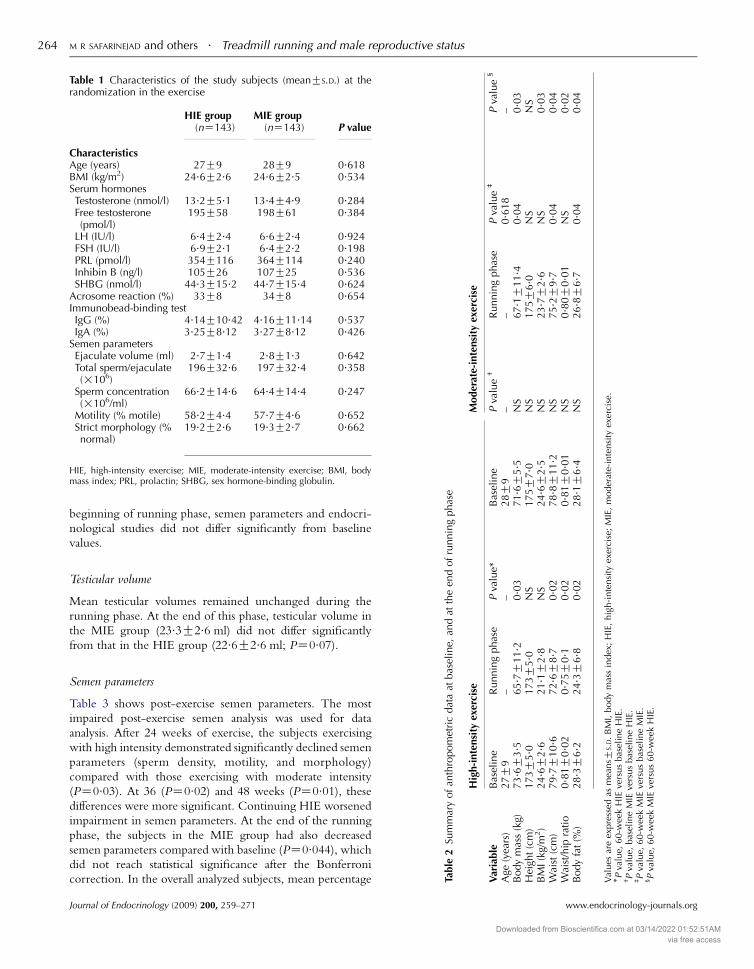
Table 1 Characteristics of the study subjects (meanGS.D.) at therandomization in the exercise
HIE group(nZ143)
MIE group(nZ143) P value
CharacteristicsAge (years) 27G9 28G9 0.618BMI (kg/m2) 24.6G2.6 24.6G2.5 0.534Serum hormonesTestosterone (nmol/l) 13.2G5.1 13.4G4.9 0.284Free testosterone(pmol/l)
195G58 198G61 0.384
LH (IU/l) 6.4G2.4 6.6G2.4 0.924FSH (IU/l) 6.9G2.1 6.4G2.2 0.198PRL (pmol/l) 354G116 364G114 0.240Inhibin B (ng/l) 105G26 107G25 0.536SHBG (nmol/l) 44.3G15.2 44.7G15.4 0.624
Acrosome reaction (%) 33G8 34G8 0.654Immunobead-binding testIgG (%) 4.14G10.42 4.16G11.14 0.537IgA (%) 3.25G8.12 3.27G8.12 0.426
Semen parametersEjaculate volume (ml) 2.7G1.4 2.8G1.3 0.642Total sperm/ejaculate(!106)
196G32.6 197G32.4 0.358
Sperm concentration(!106/ml)
66.2G14.6 64.4G14.4 0.247
Motility (% motile) 58.2G4.4 57.7G4.6 0.652Strict morphology (%normal)
19.2G2.6 19.3G2.7 0.662
HIE, high-intensity exercise; MIE, moderate-intensity exercise; BMI, bodymass index; PRL, prolactin; SHBG, sex hormone-binding globulin.
unnin
gphas
e
Moderate-intensity
exercise
Bas
elin
eP
valu
e†
Runnin
gphas
eP
valu
e‡
Pva
lue
§
28G
9–
–0. 6
18
–71. 6G
5. 5
NS
67. 1G
11. 4
0. 0
40. 0
3175G
7. 0
NS
175G
6. 0
NS
NS
24. 6G
2. 5
NS
23. 7G
2. 6
NS
0. 0
378. 8G
11. 2
NS
75. 2G
9. 7
0. 0
40. 0
40. 8
1G
0. 0
1N
S0. 8
0G
0. 0
1N
S0. 0
228. 1G
6. 4
NS
26. 8G
6. 7
0. 0
40. 0
4
erci
se;
MIE
,m
oder
ate-
inte
nsi
tyex
erci
se.
M R SAFARINEJAD and others . Treadmill running and male reproductive status264
beginning of running phase, semen parameters and endocri-
nological studies did not differ significantly from baseline
values.
ine,
and
atth
een
dof
r
phas
eP
valu
e*–
. 20. 0
3N
S8
NS
70. 0
21
0. 0
28
0. 0
2
ex;
HIE
,hig
h-i
nte
nsi
tyex
Testicular volume
Mean testicular volumes remained unchanged during the
running phase. At the end of this phase, testicular volume in
the MIE group (23.3G2.6 ml) did not differ significantly
from that in the HIE group (22.6G2.6 ml; PZ0.07).
Table
2Su
mm
ary
of
anth
ropom
etri
cdat
aat
bas
el
High-intensity
exercise
Variable
Bas
elin
eR
unnin
gA
ge(y
ears
)27G
9–
Body
mas
s(k
g)73. 6G
3. 5
65. 7G
11
Hei
ght
(cm
)173G
5. 0
173G
5. 0
BM
I(k
g/m
2)
24. 6G
2. 6
21. 1G
2.
Wai
st(c
m)
79. 7G
10. 6
72. 6G
8.
Wai
st/h
ipra
tio
0. 8
1G
0. 0
20. 7
5G
0.
Body
fat
(%)
28. 3G
6. 2
24. 3G
6.
Val
ues
are
expre
ssed
asm
eansG
S.D.
BM
I,body
mas
sin
d* P
valu
e,60-w
eek
HIE
vers
us
bas
elin
eH
IE.
†P
valu
e,bas
elin
eM
IEve
rsus
bas
elin
eH
IE.
‡P
valu
e,60-w
eek
MIE
vers
us
bas
elin
eM
IE.
§P
valu
e,60-w
eek
MIE
vers
us
60-w
eek
HIE
.
Semen parameters
Table 3 shows post-exercise semen parameters. The most
impaired post-exercise semen analysis was used for data
analysis. After 24 weeks of exercise, the subjects exercising
with high intensity demonstrated significantly declined semen
parameters (sperm density, motility, and morphology)
compared with those exercising with moderate intensity
(PZ0.03). At 36 (PZ0.02) and 48 weeks (PZ0.01), these
differences were more significant. Continuing HIE worsened
impairment in semen parameters. At the end of the running
phase, the subjects in the MIE group had also decreased
semen parameters compared with baseline (PZ0.044), which
did not reach statistical significance after the Bonferroni
correction. In the overall analyzed subjects, mean percentage
Journal of Endocrinology (2009) 200, 259–271 www.endocrinology-journals.org
Downloaded from Bioscientifica.com at 03/14/2022 01:52:51AMvia free access
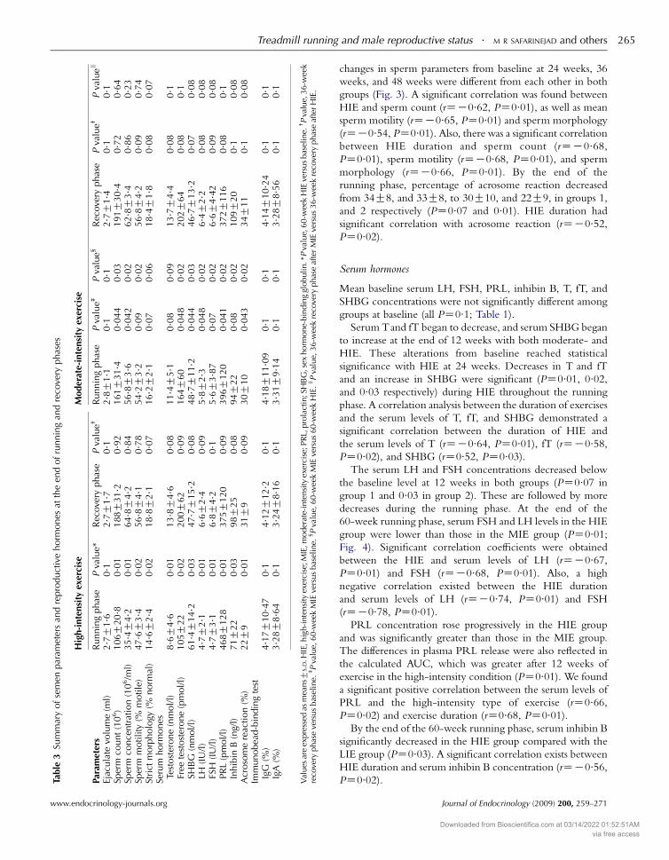
Table
3Su
mm
ary
of
sem
enpar
amet
ers
and
repro
duct
ive
horm
ones
atth
een
dof
runnin
gan
dre
cove
ryphas
es
High-intensity
exercise
Moderate-intensity
exercise
Parameters
Runnin
gphas
eP
valu
e*R
ecove
ryphas
eP
valu
e†R
unnin
gphas
eP
valu
e‡P
valu
e§
Rec
ove
ryphas
eP
valu
e†P
valu
es
Ejac
ula
tevo
lum
e(m
l)2. 7G
1. 6
0. 1
2. 7G
1. 7
0. 1
2. 8G
1. 1
0. 1
0. 1
2. 7G
1. 4
0. 1
0. 1
Sper
mco
unt
(10
6)
106G
20. 8
0. 0
1188G
31. 2
0. 9
2161G
31. 4
0. 0
44
0. 0
3191G
30. 4
0. 7
20. 6
4Sp
erm
conce
ntr
atio
n(1
06/m
l)35. 4G
4. 2
0. 0
164. 8G
4. 2
0. 8
456. 8G
3. 6
0. 0
42
0. 0
262. 8G
3. 4
0. 8
60. 2
3Sp
erm
moti
lity
(%m
oti
le)
47. 6G
3. 4
0. 0
256. 8G
4. 1
0. 7
854. 2G
3. 2
0. 0
90. 0
256. 8G
4. 2
0. 0
90. 7
4St
rict
morp
holo
gy(%
norm
al)
14. 6G
2. 4
0. 0
218. 8G
2. 1
0. 0
716. 2G
2. 1
0. 0
70. 0
618. 4G
1. 8
0. 0
80. 0
7Se
rum
horm
ones
Test
ost
erone
(nm
ol/
l)8. 6G
4. 6
0. 0
113. 8G
4. 6
0. 0
811. 4G
5. 1
0. 0
80. 0
913. 7G
4. 4
0. 0
80. 1
Free
test
ost
erone
(pm
ol/
l)105G
22
0. 0
2200G
62
0. 0
9164G
60
0. 0
48
0. 0
2202G
64
0. 0
80. 1
SHB
G(n
mol/
l)61. 4G
14. 2
0. 0
347. 7G
15. 2
0. 0
848. 7G
11. 2
0. 0
44
0. 0
346. 7G
13. 2
0. 0
70. 0
8LH
(IU
/l)
4. 7G
2. 1
0. 0
16. 6G
2. 4
0. 0
95. 8G
2. 3
0. 0
48
0. 0
26. 4G
2. 2
0. 0
80. 0
8FS
H(I
U/l
)4. 7G
3. 1
0. 0
16. 8G
4. 2
0. 1
5. 6G
3. 8
70. 0
70. 0
26. 6G
4. 4
20. 0
90. 0
8PR
L(p
mol/l)
468G
128
0. 0
1375G
120
0. 0
9396G
120
0. 0
41
0. 0
2372G
116
0. 0
80. 1
Inhib
inB
(ng/
l)71G
22
0. 0
398G
25
0. 0
894G
22
0. 0
80. 0
2109G
20
0. 1
0. 0
8A
croso
me
reac
tion
(%)
22G
90. 0
131G
90. 0
930G
10
0. 0
43
0. 0
234G
11
0. 1
0. 0
8Im
munobea
d-b
indin
gte
stIg
G(%
)4. 1
7G
10. 4
70. 1
4. 1
2G
12. 2
0. 1
4. 1
8G
11. 0
90. 1
0. 1
4. 1
4G
10. 2
40. 1
0. 1
IgA
(%)
3. 2
8G
8. 6
40. 1
3. 2
4G
8. 1
60. 1
3. 3
1G
9. 1
40. 1
0. 1
3. 2
8G
8. 5
60. 1
0. 1
Val
ues
are
expre
ssed
asm
eansG
S.D.H
IE,h
igh-i
nte
nsi
tyex
erci
se;M
IE,m
oder
ate-
inte
nsi
tyex
erci
se;P
RL,
pro
lact
in;S
HB
G,s
exhorm
one-
bin
din
ggl
obulin.*P
valu
e,60-w
eek
HIE
vers
us
bas
elin
e.†P
valu
e,36-w
eek
reco
very
phas
eve
rsus
bas
elin
e.‡P
valu
e,60-w
eek
MIE
vers
us
bas
elin
e.§P
valu
e,60-w
eek
MIE
vers
us
60-w
eek
HIE
.sP
valu
e,36-w
eek
reco
very
phas
eaf
ter
MIE
vers
us
36-w
eek
reco
very
phas
eaf
ter
HIE
.
Treadmill running and male reproductive status . M R SAFARINEJAD and others 265
www.endocrinology-journals.org
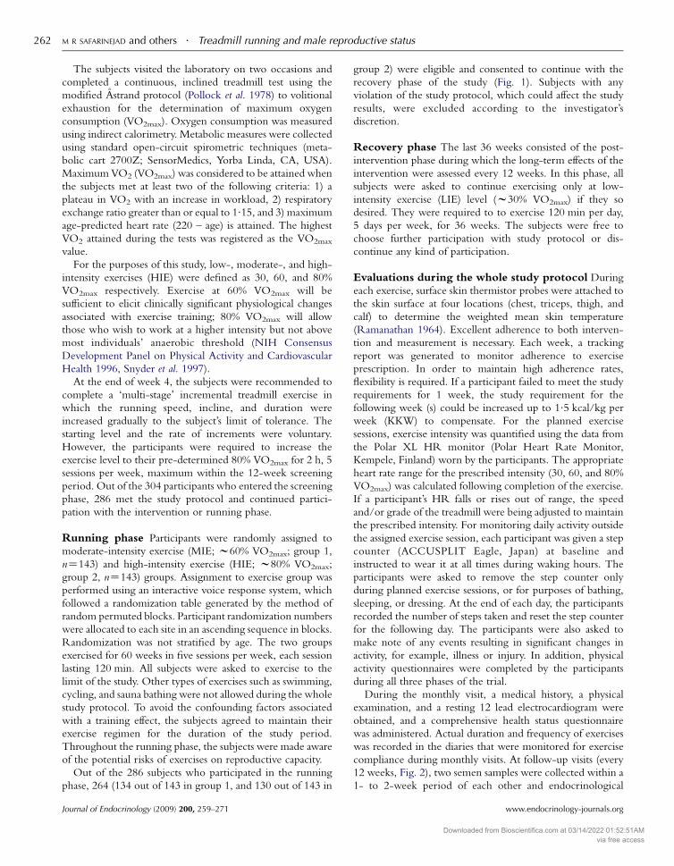
changes in sperm parameters from baseline at 24 weeks, 36
weeks, and 48 weeks were different from each other in both
groups (Fig. 3). A significant correlation was found between
HIE and sperm count (rZK0.62, PZ0.01), as well as mean
sperm motility (rZK0.65, PZ0.01) and sperm morphology
(rZK0.54, PZ0.01). Also, there was a significant correlation
between HIE duration and sperm count (rZK0.68,
PZ0.01), sperm motility (rZK0.68, PZ0.01), and sperm
morphology (rZK0.66, PZ0.01). By the end of the
running phase, percentage of acrosome reaction decreased
from 34G8, and 33G8, to 30G10, and 22G9, in groups 1,
and 2 respectively (PZ0.07 and 0.01). HIE duration had
significant correlation with acrosome reaction (rZK0.52,
PZ0.02).
Serum hormones
Mean baseline serum LH, FSH, PRL, inhibin B, T, fT, and
SHBG concentrations were not significantly different among
groups at baseline (all PZ0.1; Table 1).
Serum Tand fT began to decrease, and serum SHBG began
to increase at the end of 12 weeks with both moderate- and
HIE. These alterations from baseline reached statistical
significance with HIE at 24 weeks. Decreases in T and fT
and an increase in SHBG were significant (PZ0.01, 0.02,
and 0.03 respectively) during HIE throughout the running
phase. A correlation analysis between the duration of exercises
and the serum levels of T, fT, and SHBG demonstrated a
significant correlation between the duration of HIE and
the serum levels of T (rZK0.64, PZ0.01), fT (rZK0.58,
PZ0.02), and SHBG (rZ0.52, PZ0.03).
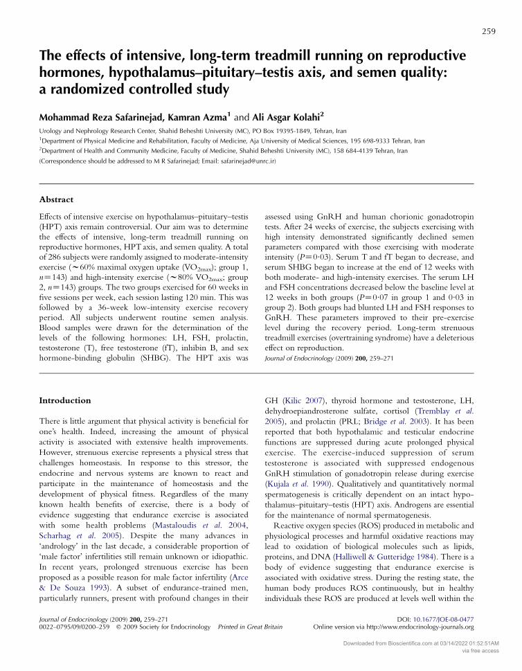
The serum LH and FSH concentrations decreased below
the baseline level at 12 weeks in both groups (PZ0.07 in
group 1 and 0.03 in group 2). These are followed by more
decreases during the running phase. At the end of the
60-week running phase, serum FSH and LH levels in the HIE
group were lower than those in the MIE group (PZ0.01;
Fig. 4). Significant correlation coefficients were obtained
between the HIE and serum levels of LH (rZK0.67,
PZ0.01) and FSH (rZK0.68, PZ0.01). Also, a high
negative correlation existed between the HIE duration
and serum levels of LH (rZK0.74, PZ0.01) and FSH
(rZK0.78, PZ0.01).
PRL concentration rose progressively in the HIE group
and was significantly greater than those in the MIE group.
The differences in plasma PRL release were also reflected in
the calculated AUC, which was greater after 12 weeks of
exercise in the high-intensity condition (PZ0.01). We found
a significant positive correlation between the serum levels of
PRL and the high-intensity type of exercise (rZ0.66,
PZ0.02) and exercise duration (rZ0.68, PZ0.01).
By the end of the 60-week running phase, serum inhibin B
significantly decreased in the HIE group compared with the
LIE group (PZ0.03). A significant correlation exists between
HIE duration and serum inhibin B concentration (rZK0.56,
PZ0.02).
Journal of Endocrinology (2009) 200, 259–271
Downloaded from Bioscientifica.com at 03/14/2022 01:52:51AMvia free access
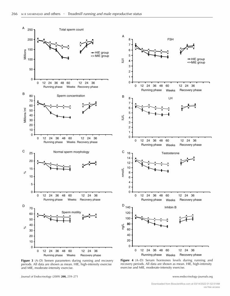
Figure 3 (A–D) Semen parameters during running and recoveryperiods. All data are shown as mean. HIE, high-intensity exerciseand MIE, moderate-intensity exercise.
Figure 4 (A–D) Serum hormones levels during running andrecovery periods. All data are shown as mean. HIE, high-intensityexercise and MIE, moderate-intensity exercise.
M R SAFARINEJAD and others . Treadmill running and male reproductive status266
Journal of Endocrinology (2009) 200, 259–271 www.endocrinology-journals.org
Downloaded from Bioscientifica.com at 03/14/2022 01:52:51AMvia free access
Treadmill running and male reproductive status . M R SAFARINEJAD and others 267
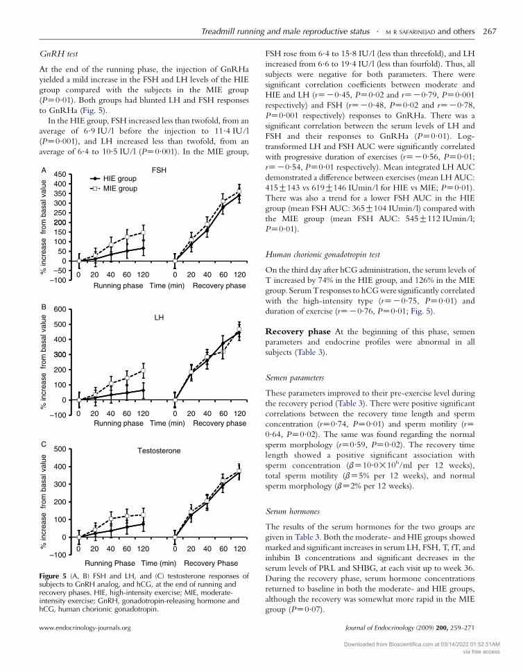
GnRH test
At the end of the running phase, the injection of GnRHa
yielded a mild increase in the FSH and LH levels of the HIE
group compared with the subjects in the MIE group
(PZ0.01). Both groups had blunted LH and FSH responses
to GnRHa (Fig. 5).
In the HIE group, FSH increased less than twofold, from an
average of 6.9 IU/l before the injection to 11.4 IU/l
(PZ0.001), and LH increased less than twofold, from an
average of 6.4 to 10.5 IU/l (PZ0.001). In the MIE group,
Figure 5 (A, B) FSH and LH, and (C) testosterone responses ofsubjects to GnRH analog, and hCG, at the end of running andrecovery phases. HIE, high-intensity exercise; MIE, moderate-intensity exercise; GnRH, gonadotropin-releasing hormone andhCG, human chorionic gonadotropin.
www.endocrinology-journals.org
FSH rose from 6.4 to 15.8 IU/l (less than threefold), and LH
increased from 6.6 to 19.4 IU/l (less than fourfold). Thus, all
subjects were negative for both parameters. There were
significant correlation coefficients between moderate and
HIE and LH (rZK0.45, PZ0.02 and rZK0.79, PZ0.001
respectively) and FSH (rZK0.48, PZ0.02 and rZK0.78,
PZ0.001 respectively) responses to GnRHa. There was a
significant correlation between the serum levels of LH and
FSH and their responses to GnRHa (PZ0.01). Log-
transformed LH and FSH AUC were significantly correlated
with progressive duration of exercises (rZK0.56, PZ0.01;
rZK0.54, PZ0.01 respectively). Mean integrated LH AUC
demonstrated a difference between exercises (mean LH AUC:
415G143 vs 619G146 IUmin/l for HIE vs MIE; PZ0.01).
There was also a trend for a lower FSH AUC in the HIE
group (mean FSH AUC: 365G104 IUmin/l) compared with
the MIE group (mean FSH AUC: 545G112 IUmin/l;
PZ0.01).
Human chorionic gonadotropin test
On the third day after hCG administration, the serum levels of
T increased by 74% in the HIE group, and 126% in the MIE
group. Serum Tresponses to hCG were significantly correlated
with the high-intensity type (rZK0.75, PZ0.01) and
duration of exercise (rZK0.76, PZ0.01; Fig. 5).
Recovery phase At the beginning of this phase, semen
parameters and endocrine profiles were abnormal in all
subjects (Table 3).
Semen parameters
These parameters improved to their pre-exercise level during
the recovery period (Table 3). There were positive significant
correlations between the recovery time length and sperm
concentration (rZ0.74, PZ0.01) and sperm motility (rZ0.64, PZ0.02). The same was found regarding the normal
sperm morphology (rZ0.59, PZ0.02). The recovery time
length showed a positive significant association with
sperm concentration (bZ10.0!106/ml per 12 weeks),
total sperm motility (bZ5% per 12 weeks), and normal
sperm morphology (bZ2% per 12 weeks).
Serum hormones
The results of the serum hormones for the two groups are
given in Table 3. Both the moderate- and HIE groups showed
marked and significant increases in serum LH, FSH, T, fT, and
inhibin B concentrations and significant decreases in the
serum levels of PRL and SHBG, at each visit up to week 36.
During the recovery phase, serum hormone concentrations
returned to baseline in both the moderate- and HIE groups,
although the recovery was somewhat more rapid in the MIE
group (PZ0.07).
Journal of Endocrinology (2009) 200, 259–271
Downloaded from Bioscientifica.com at 03/14/2022 01:52:51AMvia free access
M R SAFARINEJAD and others . Treadmill running and male reproductive status268
GnRH and hCG tests
The GnRH and hCG tests were repeated in all participants at
12, 24, and 36 weeks. GnRH test was positive in 54%, 87%,
and 94% of subjects in 12, 24, and 36 weeks of recovery phase
respectively. Recovery was faster in the MIE group
(PZ0.03). Of the participants in the recovery phase, 49%,
78%, and 92% had positive hCG tests, in 12, 24, and 36 weeks
of the recovery phase respectively. Recovery of GnRH and
hCG tests had significant correlation with recovery phase
length (rZ0.75, PZ0.01 and rZ0.78, PZ0.01 respectively).
Weighted mean skin temperature
Room temperature ranged between 22 and 25 8C during the
treadmill exercises. Mean skin temperature was increased
progressively during exercise in all three groups (P!0.001),
reaching 39.2G0.7 8C in the HIE group, 39.1G0.6 8C in
the MIE group, and 39.0G0.67 8C in the LIE group.
Discussion
The present investigation is a longitudinal study of
reproductive endocrine profile and HPT axis during long-
term HIE in men. The results of this study demonstrate that
strenuous long-term treadmill exercise caused a significant
decrease in plasma LH, FSH, and T concentrations. Our aim
was to include subjects with no known medical condition
(such as Y chromosome microdeletion) that could interfere
for their fertility. Therefore, Y chromosome microdeletion
analysis was carried out in all subjects. Yq microdeletions
have been previously reported in some studies of fertile males
(Kobayashi et al. 1994, Vogt et al. 1996). These microdele-
tions may be associated with suboptimal sperm count
(Andersen et al. 2000). Hence, deterioration of semen
parameters in these individuals cannot be attributed solely to
intensive treadmill running. There are some studies showing
data of the quality of semen in highly endurance-trained
groups.
Gebreegziabher et al. (2004) investigated the influence of
cycling on sperm characteristics. They concluded that
endurance cycling appears to be associated with a significant
alteration in sperm morphology. In another study, gonadal
hormones and semen quality were evaluated in male runners
(De Souza et al. 1994). In this study, the volume of training
was significantly correlated with sperm motility, density, and
number of round cells. Hormonal milieu was also assessed in
endurance-trained male athletes. Low serum FSH, LH, and T
values indicate hypothalamo-pituitary disease, i.e., hypogona-
dotropic hypogonadism. Lower basal levels of Tor fT have also
been reported in endurance-trained individuals in previous
studies (Cooper et al. 1998, Hackney et al. 1998). In another
study, 20 male marathon athletes were evaluated by hormonal
profiles, psychological testing, anthropomorphic indices, and
semen evaluations by Ayers et al. (1985). In this study, vigorous
Journal of Endocrinology (2009) 200, 259–271
endurance training was associated with significantly decreased
T values. The male reproductive system is regulated by the
HPT axis. The principal regulator of the HPT axis is GnRH.
GnRH stimulates pituitary FSH and LH secretion and,
subsequently induces spermatogenesis by seminiferous
tubules and testosterone secretion by the Leydig cells. Exercise
presents a serious challenge to the homeostasis of the body.
The pituitary hormone response to such exercise is often
described as a ‘stress response’. The stress system consists of
the hypothalamic–pituitary–adrenal (HPA) axis along with
the arousal and autonomic nervous systems. The stress response
in the neuroendocrine system includes release of cortico-
tropin-releasing hormone (CRH) from the hypothalamic
paraventricular nucleus and the secretion of pituitary ACTH,
leading to the secretion of glucocorticoid hormones by the
adrenal cortex (Carrasco & Van de Kar 2003). In response to
strenuous exercise, there is an enhanced secretion of cortisol,
stimulated by increased release of ACTH, which serves to
mobilize fuel stores (Deuster et al. 1989) and CRH-induced
proopiomelanocortin peptides, such as b-endorphin, and
inhibits hypothalamic GnRH secretion (Chen et al. 1992). In
addition, glucocorticoids suppress gonadal axis function at the
hypothalamic–pituitary level (Sakakura et al. 1975). CRH and
its receptors have also been identified in the Leydig cells of the
testis, where CRH exerts inhibitory actions on testosterone
biosynthesis (Fabri et al. 1990). In fact, CRH hyperactivity is
inferred to be associated with the overtraining syndrome
(Keizer 1998). An inhibitory effect of the HPA axis on the
female reproductive system has also been demonstrated
(Chrousos et al. 1998).
Despite the strong evidence for the health benefits of
exercise, serum markers do not necessarily support this
(Cooper et al. 1998). The exercise protocol used in this
study proved to be an effective means of examining the effects
of stress on reproductive function. Furthermore, the quality of
semen was negatively influenced by repeated HIE sessions over
a period of 60 weeks. The evaluation of fresh semen revealed
that the percentage of major defects, particularly abnormal
acrosome reaction started to rise 12 weeks after the beginning
of the intensive exercise. The most impaired post-exercise
semen analysis was used for data analysis. We also used the least
impaired semen analysis for statistical analysis. Findings were
similar between exercise groups. In addition, the changes in
semen parameters were all within the fertile range. In the
present study, we used a long bout of high-intensity treadmill
exercise to test the integrity of the HPT axis, and we tested
the hypothesis that long-duration HIE is associated with the
hyporesponsiveness of the HPTaxis as reflected by low serum
levels of LH, and FSH after the HPT axis activation. The
changes in gonadotropins and testosterone are clinically
relevant with hyporesponsive HPT axis. Kujala et al. (1990)
have reported that the exercise-induced suppression of serum
T is associated with two effects: suppressed endogenous
GnRH stimulation of gonadotropin release during exercise,
and decreased testicular capacity to secrete T during recovery
period. Assessment of sex hormone status in runners revealed
www.endocrinology-journals.org
Downloaded from Bioscientifica.com at 03/14/2022 01:52:51AMvia free access

Treadmill running and male reproductive status . M R SAFARINEJAD and others 269
diminished fT values attributable to strikingly increased
SHBG concentrations. In addition, direct RIA of fT
concentrations showed significant decreased values in the
HIE group, suggesting that the runners were, in fact,
hypogonadal. Our findings are consistent with the report of
Remes et al. (2004). They reported that long-term low- to
MIE had influence on serum estradiol, T, fT, or SHBG levels in
middle-aged men. SHBG concentrations have been observed
to increase acutely with aerobic physical activity exercises such
as cycle ergometry and longer term endurance training (Gray
et al. 1993). Several other hormones appear to be correlated
with SHBG levels. For instance, it has been reported that
estradiol significantly increases SHBG, and by contrast, insulin
and testosterone decrease SHBG (Haffner 1996). In the
present study, T levels (rZK0.37, PZ0.01) were negatively
associated with SHBG levels. In this study, baseline T and
fT concentrations, as expected, were not normally
distributed, and thus were log transformed prior to
regression and analysis. Obesity is associated with alterations
in the pituitary–adrenal and pituitary–gonadal functions.
(Glass, 1989) Visceral fat accumulation is associated with
decreases in testosterone concentrations in males and
reduces SHBG concentrations in both sexes (Lindstedt
et al. 1991, Haffner 2000). Significant negative Pearson
correlations between SHBG concentrations and fat body
phenotypes at baseline were found (rZK0.36, PZ0.01).
For the training response, significant negative correlations
were observed for SHBG with fat mass (rZK0.16,
PZ0.04) and BMI (rZK0.14, PZ0.03).
Elevated PRL levels were also noted by HIE. Since
multiple neural pathways that influence PRL secretion
converge on the hypothalamus from other parts of the
brain, the effect of exercise on the secretion of PRL may also
reflect the action of different neural inputs on the activity of
the hypothalamic–pituitary axis.
It has been reported that endurance exercise is associated with
oxidative stress (Mastaloudis et al. 2001). F2-isoprostanes is a
sensitive biomarker to assess lipid peroxidation in trained
subjects. Plasma F2-isoprostane levels increase significantly
during the 50 km ultramarathon. As we mentioned previously,
exercise may increase free radicals and ROS, which may interact
with lipids, DNA, and proteins (Mickelborough et al. 2000,
Hreljac & Stergiou 2002). Left unchecked, these ROS may
cause protein, lipid, and/or DNA damage. However, some
studies have suggested that exercise training enhances anti-
oxidant capacity (Child et al. 1998, Clarkson & Thompson
2000). Indeed, the machinery eliminating ROS adapts after
regular exercise and actually lowers the amount of ROS that is
produced, especially in the major organs (muscles) of oxygen
consumption and ROS production. Exercise training tends to
decrease ROS also in body fluids, although no data concerning
seminal fluid seem to be available. The possibility of ROS
affecting sperm quality remains, but participation of other,
maybe unknown factors seems more probable. Regardless of
the exercise protocol studied, increases in DNA damage in
peripheral human white cells have been reported, generating
www.endocrinology-journals.org
the consensus that exercise does indeed induce DNA damage
(Hartmann & Niess 2000).After an exercise bout,DNA damage
persists for up to 7 days (Tsai et al. 2001). The presence of high
ROS levels has been reported in the semen of between 25 and
40% of infertile men (Padron et al. 1997). This is because ROS,
at high levels, are potentially toxic to sperm quality and
function (Saleh & Agarwal 2002). Therefore, persistent ROS
formation during continuous strenuous exercise might be
harmful for normal spermatogenesis. In order to confirm this
link it is important to assess the oxidative stress and antioxidant
levels in the seminal plasma at baseline and after therapy. We did
not do this and this is one of the study limitations.
The intratesticular temperature should be lower than the
core temperature for normal sperm production (Hjollund
et al. 2002). External heating of the testes for short periods of
time results in a dramatic but temporary decrease in sperm
count after a delay of 6–8 weeks (Procope 1965). The adverse
effect of physical stress might be the result of increased body
temperature compromising testicular thermoregulation
during strenuous exercise.
After 1 year of exercise intervention, serum levels of T, fT,
LH, and FSH activity were significantly lower in the HIE
than MIE group. Testicular heating, ROS formation, and
gonadotropin suppression are all contributing factors to
semen changes. Another study limitation is that subfertile
men were not included and hence extrapolation of the falls in
the semen parameters to a subfertile population should be
made cautiously.
Because the changes in hormone levels returned to baseline
values in recovery phase, our intervention trial with LIE had no
influence on serum LH, FSH, T, fT, or SHBG levels in middle-
aged men. Our exercise program at low to high intensity
progressed following a 12-week retraining program and
began under close supervision. In addition, a strong point of
our study is the excellent adherence and compliance to the
intervention, as indicated by the low dropout rate.
However, we cannot generalize the present results to other
age groups and medically frail men. Furthermore, the influence
of the specific type of exercise training (e.g., bicycling versus
jogging) could not be evaluated.
Conclusion
Despite strong evidence for the health benefits of continued
aerobic exercise into later life, our results demonstrate that
long-term strenuous exercise (overtraining syndrome) impairs
reproductive capacity and the HPT axis in middle-aged
men. After cessation of HIE, impairment of the reproductive
system is fully recovered.
Declaration of interest
We declare that there is no conflict of interest that could be perceived as
prejudicing the impartiality of the research reported.
Journal of Endocrinology (2009) 200, 259–271
Downloaded from Bioscientifica.com at 03/14/2022 01:52:51AMvia free access
M R SAFARINEJAD and others . Treadmill running and male reproductive status270
Funding
This research did not receive any specific grant from any funding agency in the
public, commercial or not-for-profit sector.
Acknowledgements
We thank all of the subjects for participation in the study. We would like to
thank the two referees for their helpful comments on an earlier draft of
this article. We acknowledge the invaluable contributions of the following
individuals to the present project: Dr H Ziraksaz for his continuous support
and valuable contribution, Ms Shiva Safarinejad for critical reading of the
manuscript, Mrs Nayyer Shafiei for her laboratory assistance, Ms Saba
Safarinejad for her invaluable help during the data collection, Mr A R
Mohammadian for excellent technical assistance, and Dr S Y Hosseini for
body composition measurements in this study.
References
Alessio HM 1993 Exercise-induced oxidative stress. Medicine and Science in
Sports and Exercise 25 218–224.
Andersen AG, Jensen TK, Carlsen E, Jørgensen N, Andersson AM, Krarup T,
Keiding N & Skakkebaek NE 2000 High frequency of sub-optimal semen
quality in an unselected population of young men. Human Reproduction 15
366–372.
Arce JC & De Souza MJ 1993 Exercise and male factor infertility. Sports
Medicine 15 146–169.
Astrand PO & Rodahl K 1986 Circulation. In Textbook of Work Physiology:
Physiological Basis of Exercise, pp 170–175. Ed. DB van Dalen. New York:
McGraw Hill Book Company.
Ayers JW, Komesu Y, Romani T & Ansbacher R 1985 Anthropomorphic,
hormonal, and psychologic correlates of semen quality in endurance-
trained male athletes. Fertility and Sterility 43 917–921.
Bridge MW, Weller AS, Rayson M & Jones DA 2003 Ambient temperature
and the pituitary hormone responses to exercise in humans. Experimental
Physiology 88 627–635.
Carrasco GA & Van de Kar LD 2003 Neuroendocrine pharmacology of stress.
European Journal of Endocrinology 463 235–272.
Chen MD, O’Byrne KT, Chiappini SE, Hotchkiss J & Knobil E 1992
Hypoglycemic ‘stress’ and gonadotropin-releasing hormone pulse gen-
erator activity in the rhesus monkey: role of the ovary. Neuroendocrinology 56
666–673.
Child RB, Wilkinson DM, Fallowfield JL & Donnelly AE 1998 Elevated
serum antioxidant capacity and plasma malondialdehyde concentration in
response to a simulated half marathon run. Medicine and Science in Sports and
Exercise 30 1603–1607.
Chrousos GP, Torpy DJ & Gold PW 1998 Interactions between the
hypothalamic–pituitary–adrenal axis and the female reproductive system:
clinical implications. Annals of Internal Medicine 129 229–240.
Clarkson PM & Thompson HS 2000 Antioxidants: what role do they play in
physical activity and health? American Journal of Clinical Nutrition 72 637S–
646S.
Cooper C, Taaffe DR, Guido D, Parker E, Holloway L & Marcus R 1998
Relationship of chronic endurance exercise to the somatotropic and sex
hormone status of older men. European Journal of Endocrinology 138 517–
523.
Deuster PA, Chrousos GP, Luger A, DeBolt JE, Bernier LL, Trostmann UH,
Kyle SB, Montgomery LC & Loriaux DL 1989 Hormonal and metabolic
responses of untrained, moderately trained, and highly trained men to three
exercise intensities. Metabolism 38 141–148.
Fabri A, Tinajero JC & Dufau ML 1990 Corticotropin-releasing factor is
produced by rat Leydig cells and has a major local antireproductive role in
the testis. Endocrinology 127 1541–1543.
Journal of Endocrinology (2009) 200, 259–271
Franklin BA, Whaley MH & Howley ET 2000 ACSM’s Guidelines for Exercise
Testing and Prescription. edn 6, Philadelphia: Lippincott Williams & Wilkins.
Gebreegziabher Y, Marcos E, McKinon W & Rogers G 2004 Sperm
characteristics of endurance trained cyclists. International Journal of Sports
Medicine 25 247–251.
Glass AR 1989 Endocrine aspects of obesity. Medical Clinics of North America 73
139–160.
Gluer CC, Blak G, Lu Y, Blunt BA, Jergas M & Genant HK 1995 Accurate
assessment of precision errors: how to measure the reproducibility of bone
densitometry techniques. Osteoporosis International 5 262–270.
Gray AB, Telford RD & Weidemann MJ 1993 Endocrine response to intense
interval exercise. European Journal of Applied Physiology and Occupational
Physiology 66 366–371.
Hackney AC, Fahrner CL & Gulledge TP 1998 Basal reproductive hormonal
profiles are altered in endurance trained men. Journal of Sports Medicine and
Physical Fitness 38 138–141.
Haffner SM 1996 Sex hormone-binding protein, hyperinsulinemia, insulin
resistance and non-insulin-dependent diabetes. Hormone Research 45 233–
237.
Haffner SM 2000 Sex hormones, obesity, fat distribution, type 2 diabetes and
insulin resistance: epidemiological and clinical correlation. International
Journal of Obesity and Related Metabolic Disorders 24S 56–58.
Halliwell B & Gutteridge JM 1984 Lipid peroxidation, oxygen radicals, cell
damage, and antioxidant therapy. Lancet 1 1396–1397.
Halliwell B & Gutteridge JMC 1999 Free Radicals in Biology and Medicine.,
New York: Oxford University Press Inc..
Harrison GG, Buskirk ER, Lindsay Carter JE, Johnston FE, Lohman TG,
Pollock ML, Roche AF & Wilmore J 1988 Skinfold thicknesses and
measurement technique. In Anthropometric Standardization Reference Manual,
pp 55–70. Eds TG Lohman, AF Roche & R Martorell. Champaign, IL:
Human Kinetics.
Hartmann A & Niess A 2000 Oxidative DNA damage in exercise. In
Handbook of Oxidants and Antioxidants in Exercise, pp 195–217. Eds C Sen,
L Packer & O Hanninen. Amsterdam: Elsevier.
Hjollund NH, Storgaard L, Ernst E, Bonde JP & Olsen J 2002 The relation
between daily activities and scrotal temperature. Reproductive Toxicology 16
209–214.
Hreljac A & Stergiou N 2002 Phase determination during normal running
using kinematic data. Medical and Biological Engineering and Computing 38
503–506.
Irvine DS 1996 Glutathione as a treatment for male infertility. Reviews of
Reproduction 1 6–12.
Keizer HA 1998 Neurondocrine aspects of overtraining. In Overtraining in
Sport, pp 145–167. Eds RB Kreider, AC Fry & M O’Toole. Champaign,
IL: Human Kinetics Publishers.
Kilic M 2007 Effect of fatiguing bicycle exercise on thyroid hormone and
testosterone levels in sedentary males supplemented with oral zinc.
Neuroendocrinology Letters 28 681–685.
Kobayashi K, Mizuno K, Hida A, Komaki R, Tomita K, Matsushita I, Namiki
M, Iwamoto T, Tamura S & Minowada S 1994 PCR analysis of the Y
chromosome long arm in azoospermic patients: evidence for a second locus
required for spermatogenesis. Human Molecular Genetics 3 1965–1967.
Kujala UM, Alen M & Huhtaniemi IT 1990 Gonadotrophin-releasing
hormone and human chorionic gonadotrophin tests reveal that both
hypothalamic and testicular endocrine functions are suppressed during
acute prolonged physical exercise. Clinical Endocrinology 33 219–225.
Lindstedt G, Lundberg PA, Lapidus L, Lundgren H, Bengtsson C & Bjorntorp
P 1991 Low sex-hormonebinding globulin concentration as independent
risk factor for development of NIDDM. 12-year follow-up of population
study of women in Gothenburg, Sweden. Diabetes 40 123–128.
Mastaloudis A, Leonard SW & Traber MG 2001 Oxidative stress in athletes
during extreme endurance exercise. Free Radical Biology & Medicine 31
911–922.
Mastaloudis A, Yu TW, O’Donnell RP, Frei B, Dashwood RH & Traber MG
2004 Endurance exercise results in DNA damage as detected by the comet
assay. Free Radical Biology & Medicine 36 966–975.
www.endocrinology-journals.org
Downloaded from Bioscientifica.com at 03/14/2022 01:52:51AMvia free access
Treadmill running and male reproductive status . M R SAFARINEJAD and others 271
Mickelborough J, van der Linden ML, Richards J & Ennos AR 2000 Validity
and reliability of a kinematic protocol for determining foot contact events.
Gait & Posture 11 32–37.
NIH Consensus Development Panel on Physical Activity and Cardiovascular
Health 1996 Physical activity and cardiovascular health. Journal of the
American Medical Association 276 241–246.
Oztasan N, Taysi S, Gumustekin K, Altinkaynak K, Aktas O, Timur H, Siktar
E, Keles S, Akar S, Akcay F et al. 2004 Endurance training attenuates
exercise induced oxidative stress in erythrocytes in rat. European Journal of
Applied Physiology 91 622–627.
Padron OF, Brackett NL, Sharma RK, Lynne CM, Thomas AJ Jr & Agarwal A
1997 Seminal reactive oxygen species, sperm motility and morphology in
men with spinal cord injury. Fertility and Sterility 67 1115–1120.
Pollock ML, Wilmore JH & Fox SM III 1978 Health and Fitness Through
Physical Activity., New York: John Wiley & Sons, Inc.
Poulsen HE, Loft S & Vistisen K 1996 Extreme exercise and oxidative DNA
modification. Journal of Sports Sciences 14 343–346.
Procope BJ 1965 Effect of repeated increase of body temperature on human
sperm cells. International Journal of Andrology 10 333–339.
Ramanathan LM 1964 A new weighting system for mean surface temperature
of the human body. Journal of Applied Physiology 19 531–532.
Remes T, Vaisanen SB, Mahonen A, Huuskonen J, Kroger H, Jurvelin JS,
Penttila IM & Rauramaa R 2004 The association of bone metabolism with
bone mineral density, serum sex hormone concentrations, and regular
exercise in middle-aged men. Bone 35 439–447.
Sakakura N, Takebe K & Nakagawa S 1975 Inhibition of luteinizing hormone
secretion induced by synthetic LRH by long-term treatment with
glucocorticoids in human subjects. Journal of Clinical Endocrinology and
Metabolism 40 774–779.
Saleh R & Agarwal A 2002 Oxidative stress and male infertility: from research
bench to clinical practice. Journal of Andrology 23 737–752.
www.endocrinology-journals.org
Scharhag J, Herrmann M, Urhausen A, Haschke M, Herrmann W &
Kindermann W 2005 Independent elevations of N-terminal pro-brain
natriuretic peptide and cardiac troponins in endurance athletes after
prolonged strenuous exercise. American Heart Journal 150 1128–1134.
Snyder KA, Donnelly JE, Jacobsen DJ, Hertner G & Jakicic JM 1997 The effects
of long-term, moderate intensity, intermittent exercise on aerobic capacity,
body composition, blood lipids, insulin and glucose in overweight females.
International Journal of Obesity and Related Metabolic Disorders 21 1180–1189.
De Souza MJ, Arce JC, Pescatello LS, Scherzer HS & Luciano AA 1994 Gonadal
hormones and semen quality in male runners. A volume threshold effect of
endurance training. International Journal of Sports Medicine 15 383–391.
Thomas S, Reading J & Shephard R 1992 Revision of the physical activity
readiness questionnaire (PAR-Q). Canadian Journal of Sport Sciences 17
338–345.
Tremblay MS, Copeland JL & Van Helder W 2005 Influence of exercise
duration on post-exercise steroid hormone responses in trained males.
European Journal of Applied Physiology 94 505–513.
Tsai K, Hsu TG, Hsu KM, Cheng H, Liu TY, Hsu CF & Kong CW 2001
Oxidative DNA damage in human peripheral leukocytes induced by
massive aerobic exercise. Free Radical Biology & Medicine 31 1465–1472.
Vogt PH, Edelmann A, Kirsch S, Henegariu O, Hirschmann P, Kiesewetter F,
Kohn FM, Schill WB, Farah S, Ramos C et al. 1996 Human Y
chromosome azoospermia factors (AZF) mapped to different subregions in
Yq11. Human Molecular Genetics 7 933–943.
Received in final form 29 October 2008Accepted 27 November 2008Made available online as an Accepted Preprint3 December 2008
Journal of Endocrinology (2009) 200, 259–271
Downloaded from Bioscientifica.com at 03/14/2022 01:52:51AMvia free access