Embed Size (px)
Citation preview
The Effects of Neoadjuvant Chemoradiationon pTNM Staging and Its Prognostic Significancein Esophageal Cancer
Simon Law, M.S., M.A.(Cantab), M.B.B.Chir., F.R.C.S.(Edin), F.A.C.S.,Dora L.W. Kwong, M.B.B.S., F.R.C.R., Kam-Ho Wong, M.B.B.S., F.R.C.S.(Edin),Ka-Fai Kwok, M.B.B.S., F.R.C.S.(Edin), John Wong, M.D., Ph.D., F.R.A.C.S.
For esophageal cancer, it is not clear if pathologic TNM staging after chemoradiation and resection willhave the same prognostic significance compared with patients who undergo resection only. From 1995 to2004, prospectively collected data from 279 patients with intrathoracic squamous cell cancers were an-alyzed. Patients were given chemoradiation either as part of a randomized trial comparing neoadjuvantchemoradiation with surgical resection alone, or because of advanced disease at presentation. One hun-dred seventy patients had surgical resection only (surgery), and 109 had neoadjuvant chemoradiation(CRT plus surgery). In the surgery group, pT1, 2, 3, and 4 disease was found in 15, 17, 104, and 34 pa-tients, respectively; their respective pN1 rates were 13.3%, 29.4%, 57.7%, and 64.7%, P ! 0.01. In CRTplus surgery, pT0, T1, 2, 3, and 4 were found in 48, 12, 23, 21, and 5 patients, respectively; their respec-tive pN1 rates were 31.3%, 16.7%, 21.7%, 52.4%, and 20%, P 5 0.44. Logistic regression analysis offactors predictive of pN1 showed that pT stage correlated with pN1 status (P 5 0.005) in the surgerygroup, but not for the CRT plus surgery group. Cox regression analysis demonstrated that in the surgerygroup, pT, pN, and R category, and overall pTNM stage, were independent prognostic factors, whereaspN, R category, and gender were identified as relevant for CRT plus surgery. After chemoradiation, pTand overall pTNM stage groupings were not as clearly prognostic as in patients without prior therapy.Nodal status remains an important prognostic factor. ( J GASTROINTEST SURG 2006;10:1301–1311) � 2006 The Society for Surgery of the Alimentary Tract
KEY WORDS: Esophageal neoplasm, chemoradiation, multimodality treatment, staging, prognosis
Immediate surgical results of esophagectomy forcancer have improved. In dedicated centers, a mortal-ity rate of below 5% can be achieved.1–4 Prolonginglong-term survival is a goal more difficult to attain.Prognosis for esophageal cancer remains poorthroughout the world. In selected centers and in sub-groups of patients who undergo radical esophagec-tomy, 5-year survival rates of 40% or above couldbe achieved.5–7 Selection bias is difficult to disprove,and such encouraging results are infrequently seen.In most reports, a 20% 5-year survival rate isrecorded.8,9
In recent years, neoadjuvant therapy involvingchemotherapy and/or radiotherapy is commonly
used as an adjunct to surgical resection.10,11 Despitethe equivocal data from randomized controlled trialsthat these treatments can result in better prognosiscompared with surgery alone, they are frequentlyapplied with an aim to downstage tumordincreasingthe resection rate (especially R0 resection)dand toimprove survival.12–17 After neoadjuvant therapy,however, clinical restaging is difficult with conven-tional techniques such as CT scan or endoscopicultrasound. Positron emission tomography scanshows some promise, but how it should be integratedinto clinical practice, and whether it can be used topredict long-term survival or not requires furtherevaluation.18
Presented at the Forty-Seventh Annual Meeting of The Society for Surgery of the Alimentary Tract, Los Angeles, California, May 20–24, 2006(oral presentation).From the Division of Esophageal Surgery, Department of Surgery (S.L., K.-H.W., K.-F.K., J.W.) and Department of Clinical Oncology(D.L.W.K.), The University of Hong Kong, Queen Mary Hospital, Hong Kong.Reprint requests: Simon Law, M.S., M.A., M.B.B.Chir., F.R.C.S.Ed., F.A.C.S., Department of Surgery, The University of Hong Kong, QueenMary Hospital, Hong Kong. e-mail: [email protected]
� 2006 The Society for Surgery of the Alimentary Tract
Published by Elsevier Inc.
1091-255X/06/$dsee front matter
doi:10.1016/j.gassur.2006.06.009 1301
Among other factors investigated, pathologicalTNM stage after resection has been the gold stan-dard in prognosis stratification, and the relationshipbetween advancing pTNM stage and poor survival iswell established. It has been our observation how-ever, that after chemoradiation, the primary tumoris often sterilized, but persistence of nodal diseaseexists. It is therefore hypothesized that neoadjuvanttherapy may alter the relationship between thedifferent components of the pTNM system, suchas the intercorrelation of pT and pN status, andthat the postchemoradiation pTNM stages mayhave different prognostic implications comparedwith patients without prior therapy. These factorsare investigated in a large group of patients undergo-ing surgical resection, with or without neoadjuvantchemoradiation.
METHODS
From 1995–2004, 471 patients with intrathoracicsquamous cell carcinomas without prior treatmentswere managed at the Department of Surgery, TheUniversity of Hong Kong at Queen Mary Hospital.Patients who had cancers located in the cervicalesophagus, tumors that involved the gastroesopha-geal junction, and cancers of other cell types wereexcluded from this study. Patients with synchronousor history of nonesophageal malignancies were alsoexcluded, so that the influence of other unrelated tu-mors on survival was prevented. Surgical resectionwas carried out in 279 patients (59.2%), of whom170 had surgical resection only and 109 received pre-operative chemoradiation therapy. Data were cap-tured in a prospectively collected database. Thesepatients were the subjects of the present study.
The management rationale and protocols at theauthors’ institution have been described previ-ously.19 Patients were managed in an individualizedmanner determined by both patient (performancestatus, comorbidities) and tumor (stage, location)characteristics. Surgical treatment was the preferredtreatment option. Patients were selected for nonsur-gical treatment if they had locally advanced unresect-able disease, or nonlocal-regional metastases, whenmedical-surgical risks were prohibitive, or in thosewho declined surgery.
For tumor imaging and staging purposes, all pa-tients had a barium contrast study, an endoscopy,bronchoscopy, and since May 1996, endoscopicultrasound examination. An ultrasound of the neckand CT scan of the thorax and abdomen were carriedout. Positron emission tomography scans were avail-able for most patients since July 2002.
The surgical techniques are described in brief: formost tumors in the middle and lower third of theesophagus, a Lewis-Tanner esophagectomy via anabdominal-right thoracotomy approach was pre-ferred. For patients who had a tumor of the superiormediastinal segment, a three-phase esophagectomywas carried out. In this operation, usually a right-sided thoracotomy was performed first for esopha-geal mobilization; a synchronous laparotomy andleft cervical incision then provided access for gastricand cervical esophageal mobilization, followed bya gastric pull-up to the neck, either by the posteriormediastinal or by the retrosternal route for cervicalesophagogastrostomy. In patients who had limitedcardiopulmonary reserve for whom a thoracotomywas judged to be of high risk, a transhiatal esopha-gectomy was performed. This method was mainlyused for tumors of the lower esophagus. Thismethod was uncommonly performed in the study pe-riod because the preferred approach was transtho-racic and thoracoscopic esophagectomy has alsolargely replaced the need for transhiatal esophagec-tomy.20 Altogether, 16 patients underwent thoraco-scopic esophagectomy.
Lymphadenectomy usually involved a two-fieldlymphadenectomy with dissection of lymph nodesaround the celiac trifurcation, and also an infracari-nal mediastinal lymph node dissection. Lymph nodesof the superior mediastinum were sampled, but com-plete clearance of nodal tissues around the paratra-cheal area along the recurrent laryngeal nerves wasnot usually performed unless suspicious lymph nodeswere encountered. Similarly, cervical lymphadenec-tomy was not carried out routinely unless there wasevidence of disease because our study of recurrencepatterns suggested limited value of neck dissection,21
and that survival advantage of cervical lymphadenec-tomy was not proven.22,23 In patients with obviouslypalliative resection, a more limited lymphadenec-tomy was carried out.
Reconstruction of intestinal continuity was usu-ally restored with a gastric conduit placed in theright thoracic cavity (after Lewis-Tanner esophagec-tomy) or via the orthotopic route when the anasto-mosis was carried out in the neck. In the obviouslypalliative cases where residual mediastinal diseasewas evident, the retrosternal route was chosen. Thecolon was used in patients with a prior gastrectomy,the right ileocolon being the preferred conduit.24 Allthese surgical techniques have been described.25,26
Processing of the resected surgical specimensstarted in the operating room. The operatingsurgeons dissected the different nodal stations sepa-rately and labeled them for further histologicalexamination. Individual nodes were not dissected,
1302 Law et al.Journal of
Gastrointestinal Surgery
but only the fat and connective tissues containing thenodes at various anatomical locations were isolated.The tissue adjacent to the primary tumor was notdisturbed so that histological examination of the cir-cumferential margin would not be hampered. Theprimary tumor was serially sectioned for histologyby the pathologist.
During the study period, patients were given neo-adjuvant chemoradiation therapy as either part ofa randomized controlled trial comparing neoadju-vant chemoradiation and surgical resection alone,or when locally advanced tumor or nonregionalmetastastic spread, such as cervical lymphadenopa-thy, was encountered, whereby an R0 resection wasjudged improbable. The chemotherapy regimenconsisted of cisplatin at 100 mg/m2, by intravenousinfusion given on the first day and day 24, togetherwith 5-fluorouracil (5-FU), 500 mg/m2 per day bycontinuous infusion given from day 1–5, and day24–28. Radiotherapy was given as external beamirradiation at 40 Gy in 20 daily fractions of 2 Gyeach, delivered through anterior and posterioropposing fields to the primary tumor as defined byCT scan, endoscopy, and barium contrast study.Fields included the primary tumor with at least 1cm lateral margin on each side and proximal and dis-tal margins of at least 3 cm. Regional lymph nodeswere not prophylactically irradiated. Enlarged lymphnodes were irradiated in the fields for the primary ifthey were close to the primary, or separate radiationfields were used for palliation of symptoms. In thosewho demonstrated significant response, surgicalresection was carried out.
For the purpose of this study, 30-day mortalityrate was defined as any death after esophagectomywithin 30 days, and hospital mortality rate includedany deaths within the same hospital stay. Patientswere staged according to American Joint Committeeon Cancer (AJCC) classification,27 and the R cate-gory of resection was based on the InternationalUnion Against Cancer system.28
Statistical Analysis
Continuous variables are expressed as median(range). Chi-square and Fisher exact tests wereused to compare categorical data. Survival analyseswere performed using the Kaplan-Meier methodfrom the date of operation to the time of death ofany cause or to the time of last follow-up, at whichpoint the data were censored. Comparisons betweengroups were assessed by the log-rank test.
To evaluate the impact of various clinicopatho-logical parameters for long-term survival, potentialprognostic factors were analyzed with univariate
Cox regression analysis. The same factors were alsoused in Cox proportional hazard models fitted formultivariate analysis.
Statistical significance was accepted at the 5%level. All statistical analyses were performed withthe SPSS statistical package, version 11.5 (SPSSInc., Chicago, IL).
RESULTS
A total of 279 patients satisfied the inclusion crite-ria and underwent surgical resection. There were228 men and 51 women, the median age was 66 years(range, 38–86). Of these patients, 170 had surgicalresection only and 109 received neoadjuvant chemo-radiation therapy. Their demographics are shown inTable 1. The majority of patients underwent
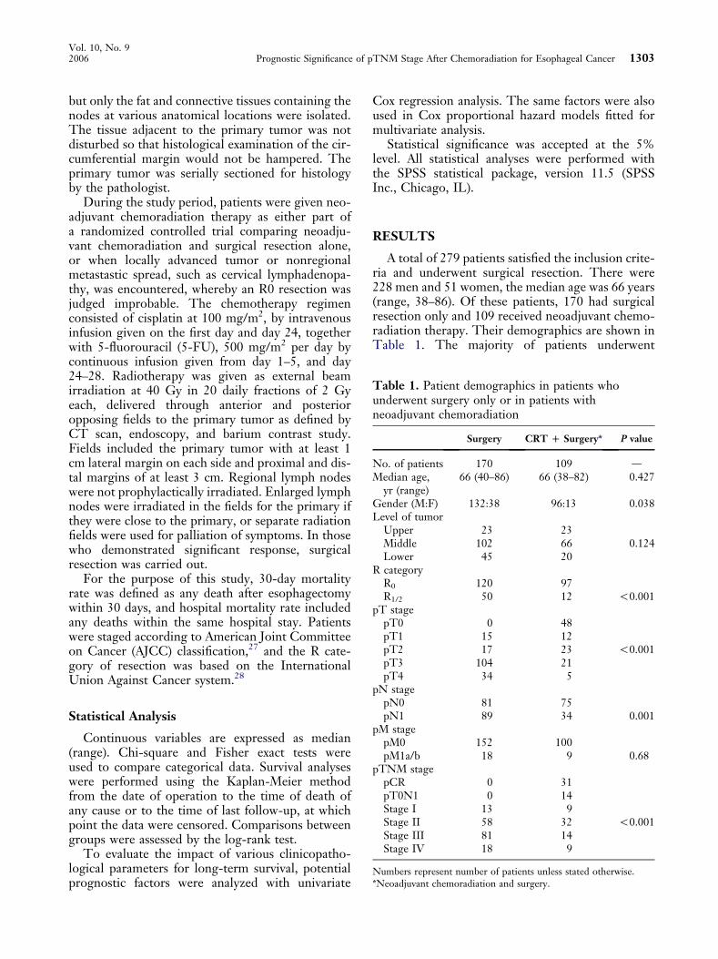
Table 1. Patient demographics in patients whounderwent surgery only or in patients withneoadjuvant chemoradiation
Surgery CRT D Surgery* P value
No. of patients 170 109 dMedian age,
yr (range)66 (40–86) 66 (38–82) 0.427
Gender (M:F) 132:38 96:13 0.038Level of tumor
Upper 23 23Middle 102 66 0.124Lower 45 20
R categoryR0 120 97R1/2 50 12 !0.001
pT stagepT0 0 48pT1 15 12pT2 17 23 !0.001pT3 104 21pT4 34 5
pN stagepN0 81 75pN1 89 34 0.001
pM stagepM0 152 100pM1a/b 18 9 0.68
pTNM stagepCR 0 31pT0N1 0 14Stage I 13 9Stage II 58 32 !0.001Stage III 81 14Stage IV 18 9
Numbers represent number of patients unless stated otherwise.*Neoadjuvant chemoradiation and surgery.
Vol. 10, No. 92006 Prognostic Significance of pTNM Stage After Chemoradiation for Esophageal Cancer 1303
a transthoracic esophagectomy (98.9%), with onlythree patients in the surgery group having a transhia-tal approach. Thirty-day mortality rate was 0.6%(one patient) in the surgery group, and 0% inpatients who received neoadjuvant therapy (P 5 1.0).In-hospital death rates were 2.4% (four patients)and 0%, P 5 0.16. R0 resections were possible in89% of the neoadjuvant therapy group and 70.6%of the surgery group, indicating significant tumordownstaging. This is also reflected in the pTNMstage groupings, with significantly lower diseasestage distributions in the neoadjuvant therapygroup.
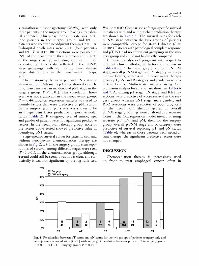
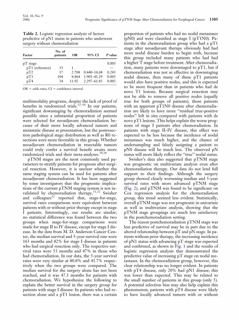
The relationship between pT and pN status isshown in Fig. 1. Advancing pT stage showed a clearlyprogressive increase in incidence of pN1 stage in thesurgery group (P ! 0.01). This correlation, how-ever, was not significant in the neoadjuvant group,P 5 0.44. Logistic regression analysis was used toidentify factors that were predictive of pN1 status.In the surgery group, pT status was shown to bean independent factor predictive of positive nodalstatus (Table 2). R category, level of tumor, age,and gender of patient were not significant predictivefactors. In the neoadjuvant therapy group, none ofthe factors above tested showed predictive value inidentifying pN1 status.
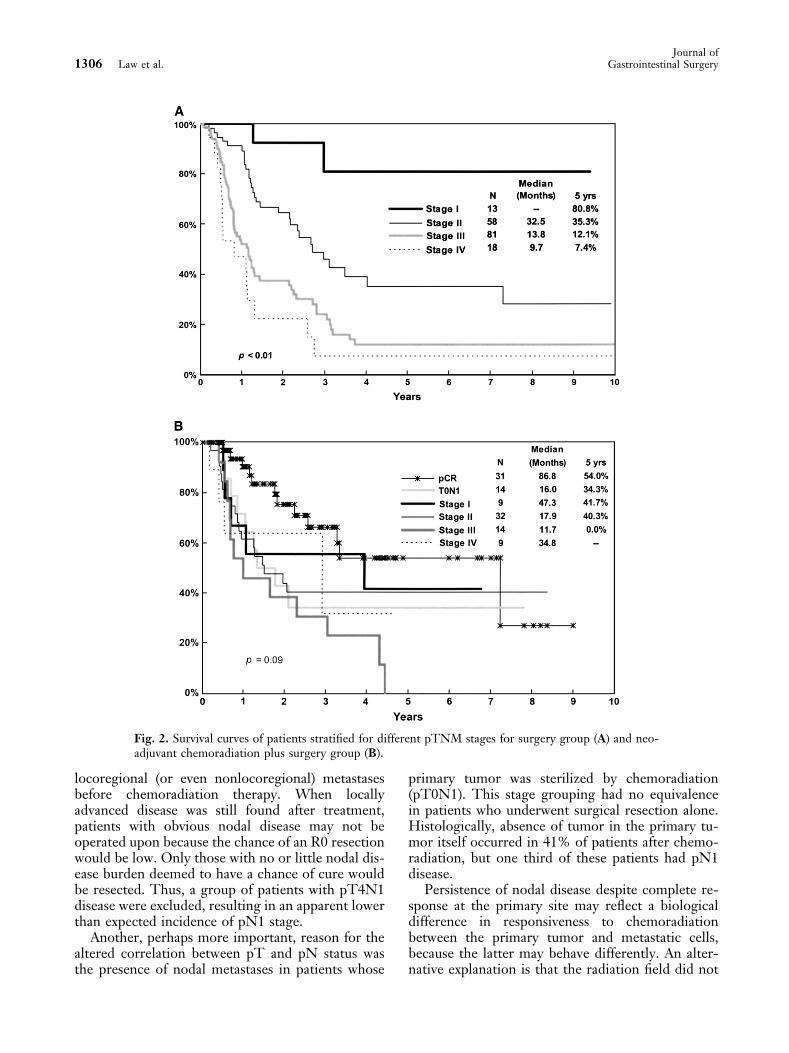
Stage-specific survival curves for patients with andwithout neoadjuvant chemoradiation therapy areshown in Fig. 2, a, b. In the surgery group, clear sepa-rations of survival among different stages were seen(P ! 0.01). In the chemoradiation group, althougha trend could still be seen, it was not as clear, and sta-tistically it was not significant by the log-rank test,
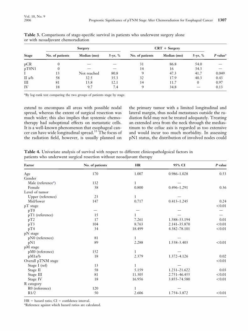
P value 5 0.09. Comparisons of stage-specific survivalin patients with and without chemoradiation therapyare shown in Table 3. The survival rates for eachpTNM stage between the two groups of patientswere comparable, except for stage I disease (P 5
0.0485). Patients with pathological complete responseand pT0N1 had no equivalent groupings in the sur-gery group and could not be directly compared.
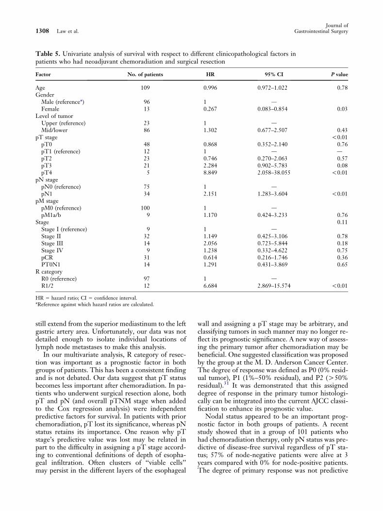
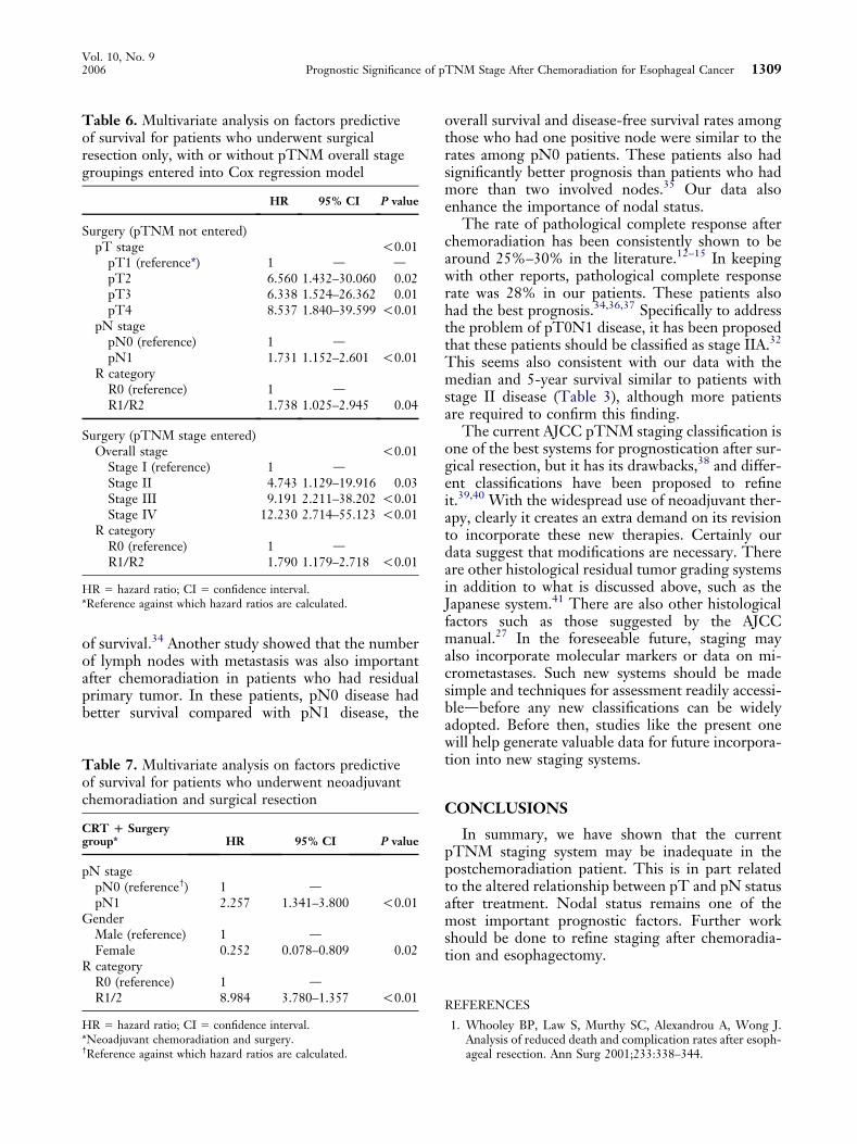
Univariate analyses of prognosis with respect todifferent clinicopathological factors are shown inTables 4 and 5. In the surgery group, pT, pN, pMstage, overall pTNM stage, and R category were sig-nificant factors, whereas in the neoadjuvant therapygroup, pT, pN, and R category and gender were pre-dictive factors. Multivariate analyses using Coxregression analysis for survival are shown in Tables 6and 7. Advancing pT stage, pN stage, and R1/2 re-sections were predictive of worse survival in the sur-gery group, whereas pN1 stage, male gender, andR1/2 resections were predictors of poor prognosisin the neoadjuvant therapy group. If overallpTNM stage groupings were analyzed as a separatefactor in the Cox regression model instead of usingseparate pT, pN, and pM, then for the surgerygroup, overall pTNM stage and R category werepredictive of survival replacing pT and pN status(Table 6), whereas in those patients with neoadju-vant therapy, the significant predictive factors werenot changed.
DISCUSSION
Chemoradiation therapy is increasingly usedup front to treat esophageal cancer, often in
Fig. 1. Relationship between pT status and pN status for the two groups of patients (surgery only andneoadjuvant chemoradiation [CRT] with surgery). Correlation between pT vs. pN in surgery group:P ! 0.01; in CRT þ surgery group: P 5 0.44.
1304 Law et al.Journal of
Gastrointestinal Surgery
multimodality programs, despite the lack of proof ofbenefits in randomized trials.12–16 In our patients,significant downstaging by chemoradiation seemedpossible since a substantial proportion of patientswere selected for neoadjuvant chemoradiation be-cause of their more locally advanced tumors andmetastatic disease at presentation, but the postresec-tion pathological stage distribution as well as R0 re-sections were more favorable in this group. Whetherneoadjuvant chemoradiation in resectable tumorscould truly confer a survival benefit awaits morerandomized trials and their meta-analyses.29,30
pTNM stages are the most commonly used pa-rameters to stratify patients for prognosis after surgi-cal resection. However, it is unclear whether thesame staging system can be used for patients afterneoadjuvant chemoradiation. It has been suggestedby some investigators that the prognostic implica-tions of the current pTNM staging system is not in-validated by chemoradiation therapy.31,32 Swisherand colleagues32 reported that, stage-for-stage,survival rates comparisons were equivalent betweenpatients with or without prior therapy except in stageI patients. Interestingly, our results are similar;no statistical difference was found between the twogroups when stage-for-stage comparisons weremade for stage II to IV disease, except for stage I dis-ease. In the data from M. D. Anderson Cancer Cen-ter, the median survival and 5-year survival rate were163 months and 82% for stage I disease in patientswho had surgical resection only. The respective sur-vival rates were 53 months and 47% in those whohad chemoradiation. In our data, the 5-year survivalrates were very similar at 80.8% and 41.7% respec-tively when the two groups were compared. Themedian survival for the surgery alone has not beenreached, and it was 47.3 months for patients withchemoradiation. We hypothesize the following toexplain the better survival in the surgery group forpatients with stage I disease: In patients who had re-section alone and a pT1 lesion, there was a certain
proportion of patients who had no nodal metastases(pN0) and were classified as stage I (pT1N0). Pa-tients in the chemoradiation group who had a pT1stage after neoadjuvant therapy obviously had hadmore nodal disease burden to begin with, becausethis group included many patients who had hada higher T stage before treatment. After chemoradia-tion, many patients were downstaged to pT1, but ifchemoradiation was not as effective in downstagingnodal disease, then many of these pT1 patientswould also have positive nodes, and this is expectedto be more frequent than in patients who had denovo T1 lesions. Because surgical resection maynot be able to remove all positive nodes (equallytrue for both groups of patients), those patientswith an apparent pT1N0 disease after chemoradia-tion are likely to have more ‘‘residual true-positivenodes’’ left in situ compared with patients with denovo pT1 lesions. This helps explain the worse prog-nosis of stage I patients after chemoradiation. Inpatients with stage II–IV disease, this effect wasexpected to be less because the incidence of nodalmetastases was much higher, and the chance ofundersampling and falsely assigning a patient topN0 disease will be much less. The observed pNstatus will more likely reflect the ‘‘true’’ nodal status.
Swisher’s data also suggested that pTNM stagewas prognostic on multivariate analysis even afterchemoradiation therapy. Our data do not lend fullsupport to their findings. Although the surgerygroup showed clearly worsening median and 5-yearsurvival rates with more advanced pTNM stage(Fig. 2), and pTNM was found to be significant onCox regression analysis for the chemoradiationgroup, this trend seemed less evident. Statistically,overall pTNM stage was not prognostic in univariateas well as multivariate analysis, showing that thepTNM stage groupings are much less satisfactoryin the postchemoradiation setting.
The reason postchemoradiation pTNM stage wasless predictive of survival may be in part due to thealtered relationship between pT and pN stage. In pa-tients without prior therapy, the increasing incidenceof pN1 status with advancing pT stage was expectedand confirmed, as shown in Fig. 1 and the results oflogistic regression analysis that demonstrated thepredictive value of increasing pT stage on nodal me-tastases. In the chemoradiation group, however, thisclear relationship was no longer evident. In patientswith pT4 disease, only 20% had pN1 disease; thiswas lower than expected. This may be related tothe small number of patients in this group (only 5).A potential selection bias may also help explain thisphenomenon; patients with pT4 disease were likelyto have locally advanced tumors with or without
Table 2. Logistic regression analysis of factorspredictive of pN1 status in patients who underwentsurgery without chemoradiation
FactorNo. of
patients OR 95% CI P value
pT stage 0.005pT1 (reference) 15 1 dpT2 17 2.708 0.440–16.68 0.283pT3 104 8.864 1.903–41.29 0.005pT4 34 11.92 2.297–61.83 0.003
OR 5 odds ratio; CI 5 confidence interval.
Vol. 10, No. 92006 Prognostic Significance of pTNM Stage After Chemoradiation for Esophageal Cancer 1305
locoregional (or even nonlocoregional) metastasesbefore chemoradiation therapy. When locallyadvanced disease was still found after treatment,patients with obvious nodal disease may not beoperated upon because the chance of an R0 resectionwould be low. Only those with no or little nodal dis-ease burden deemed to have a chance of cure wouldbe resected. Thus, a group of patients with pT4N1disease were excluded, resulting in an apparent lowerthan expected incidence of pN1 stage.
Another, perhaps more important, reason for thealtered correlation between pT and pN status wasthe presence of nodal metastases in patients whose
primary tumor was sterilized by chemoradiation(pT0N1). This stage grouping had no equivalencein patients who underwent surgical resection alone.Histologically, absence of tumor in the primary tu-mor itself occurred in 41% of patients after chemo-radiation, but one third of these patients had pN1disease.
Persistence of nodal disease despite complete re-sponse at the primary site may reflect a biologicaldifference in responsiveness to chemoradiationbetween the primary tumor and metastatic cells,because the latter may behave differently. An alter-native explanation is that the radiation field did not
Fig. 2. Survival curves of patients stratified for different pTNM stages for surgery group (A) and neo-adjuvant chemoradiation plus surgery group (B).
1306 Law et al.Journal of
Gastrointestinal Surgery
extend to encompass all areas with possible nodalspread, whereas the extent of surgical resection wasmuch wider; this also implies that systemic chemo-therapy had suboptimal effects on metastatic cells.It is a well-known phenomenon that esophageal can-cer can have wide longitudinal spread.33 The focus ofthe radiation field, however, is usually planned on
the primary tumor with a limited longitudinal andlateral margin, thus nodal metastases outside the ra-diation field may not be treated adequately. Treatingan extended area from the neck through the medias-tinum to the celiac axis is regarded as too extensiveand would incur too much morbidity. In assessingpN1 status, the distribution of involved nodes could
Table 3. Comparisons of stage-specific survival in patients who underwent surgery aloneor with neoadjuvant chemoradiation
Surgery CRT D Surgery
Stage No. of patients Median (mo) 5-yr, % No. of patients Median (mo) 5-yr, % P value*
pCR 0 d d 31 86.8 54.0 dpT0N1 0 d d 14 16 34.3 dI 13 Not reached 80.8 9 47.3 41.7 0.049II a/b 58 32.5 35.3 32 17.9 40.3 0.43III 81 13.8 12.1 14 11.7 0 0.97IV 18 9.7 7.4 9 34.8 d 0.13
*By log-rank test comparing the two groups of patients stage by stage.
Table 4. Univariate analysis of survival with respect to different clinicopatholgoical factors inpatients who underwent surgical resection without neoadjuvant therapy
Factor No. of patients HR 95% CI P value
Age 170 1.007 0.986–1.028 0.53Gender
Male (reference*) 132 1 dFemale 38 0.800 0.496–1.291 0.36
Level of tumorUpper (reference) 23 1 dMid/lower 147 0.717 0.413–1.245 0.24
pT stage !0.01pT0 d d d dpT1 (reference) 15 1 d dpT2 17 7.261 1.588–33.194 0.01pT3 104 8.763 2.141–35.870 !0.01pT4 34 18.499 4.382–78.101 !0.01
pN stagepN0 (reference) 81 1 dpN1 89 2.288 1.538–3.403 !0.01
pM stagepM0 (reference) 152 1 dpM1a/b 18 2.379 1.372–4.126 0.02
Overall pTNM stage !0.01Stage I (ref) 13 1 dStage II 58 5.159 1.231–21.622 0.03Stage III 81 11.305 2.751–46.455 !0.01Stage IV 18 16.956 3.855–74.580 !0.01
R categoryR0 (reference) 120 1 dR1/2 50 2.606 1.754–3.872 !0.01
HR 5 hazard ratio; CI 5 confidence interval.*Reference against which hazard ratios are calculated.
Vol. 10, No. 92006 Prognostic Significance of pTNM Stage After Chemoradiation for Esophageal Cancer 1307
still extend from the superior mediastinum to the leftgastric artery area. Unfortunately, our data was notdetailed enough to isolate individual locations oflymph node metastases to make this analysis.
In our multivariate analysis, R category of resec-tion was important as a prognostic factor in bothgroups of patients. This has been a consistent findingand is not debated. Our data suggest that pT statusbecomes less important after chemoradiation. In pa-tients who underwent surgical resection alone, bothpT and pN (and overall pTNM stage when addedto the Cox regression analysis) were independentpredictive factors for survival. In patients with priorchemoradiation, pT lost its significance, whereas pNstatus retains its importance. One reason why pTstage’s predictive value was lost may be related inpart to the difficulty in assigning a pT stage accord-ing to conventional definitions of depth of esopha-geal infiltration. Often clusters of ‘‘viable cells’’may persist in the different layers of the esophageal
wall and assigning a pT stage may be arbitrary, andclassifying tumors in such manner may no longer re-flect its prognostic significance. A new way of assess-ing the primary tumor after chemoradiation may bebeneficial. One suggested classification was proposedby the group at the M. D. Anderson Cancer Center.The degree of response was defined as P0 (0% resid-ual tumor), P1 (1%–50% residual), and P2 (O50%residual).31 It was demonstrated that this assigneddegree of response in the primary tumor histologi-cally can be integrated into the current AJCC classi-fication to enhance its prognostic value.
Nodal status appeared to be an important prog-nostic factor in both groups of patients. A recentstudy showed that in a group of 101 patients whohad chemoradiation therapy, only pN status was pre-dictive of disease-free survival regardless of pT sta-tus; 57% of node-negative patients were alive at 3years compared with 0% for node-positive patients.The degree of primary response was not predictive
Table 5. Univariate analysis of survival with respect to different clinicopathological factors inpatients who had neoadjuvant chemoradiation and surgical resection
Factor No. of patients HR 95% CI P value
Age 109 0.996 0.972–1.022 0.78Gender
Male (reference*) 96 1 dFemale 13 0.267 0.083–0.854 0.03
Level of tumorUpper (reference) 23 1 dMid/lower 86 1.302 0.677–2.507 0.43
pT stage !0.01pT0 48 0.868 0.352–2.140 0.76pT1 (reference) 12 1 d dpT2 23 0.746 0.270–2.063 0.57pT3 21 2.284 0.902–5.783 0.08pT4 5 8.849 2.058–38.055 !0.01
pN stagepN0 (reference) 75 1 dpN1 34 2.151 1.283–3.604 !0.01
pM stagepM0 (reference) 100 1 dpM1a/b 9 1.170 0.424–3.233 0.76
Stage 0.11Stage I (reference) 9 1 dStage II 32 1.149 0.425–3.106 0.78Stage III 14 2.056 0.723–5.844 0.18Stage IV 9 1.238 0.332–4.622 0.75pCR 31 0.614 0.216–1.746 0.36PT0N1 14 1.291 0.431–3.869 0.65
R categoryR0 (reference) 97 1 dR1/2 12 6.684 2.869–15.574 !0.01
HR 5 hazard ratio; CI 5 confidence interval.*Reference against which hazard ratios are calculated.
1308 Law et al.Journal of
Gastrointestinal Surgery
of survival.34 Another study showed that the numberof lymph nodes with metastasis was also importantafter chemoradiation in patients who had residualprimary tumor. In these patients, pN0 disease hadbetter survival compared with pN1 disease, the
overall survival and disease-free survival rates amongthose who had one positive node were similar to therates among pN0 patients. These patients also hadsignificantly better prognosis than patients who hadmore than two involved nodes.35 Our data alsoenhance the importance of nodal status.
The rate of pathological complete response afterchemoradiation has been consistently shown to bearound 25%–30% in the literature.12–15 In keepingwith other reports, pathological complete responserate was 28% in our patients. These patients alsohad the best prognosis.34,36,37 Specifically to addressthe problem of pT0N1 disease, it has been proposedthat these patients should be classified as stage IIA.32
This seems also consistent with our data with themedian and 5-year survival similar to patients withstage II disease (Table 3), although more patientsare required to confirm this finding.
The current AJCC pTNM staging classification isone of the best systems for prognostication after sur-gical resection, but it has its drawbacks,38 and differ-ent classifications have been proposed to refineit.39,40 With the widespread use of neoadjuvant ther-apy, clearly it creates an extra demand on its revisionto incorporate these new therapies. Certainly ourdata suggest that modifications are necessary. Thereare other histological residual tumor grading systemsin addition to what is discussed above, such as theJapanese system.41 There are also other histologicalfactors such as those suggested by the AJCCmanual.27 In the foreseeable future, staging mayalso incorporate molecular markers or data on mi-crometastases. Such new systems should be madesimple and techniques for assessment readily accessi-bledbefore any new classifications can be widelyadopted. Before then, studies like the present onewill help generate valuable data for future incorpora-tion into new staging systems.
CONCLUSIONS
In summary, we have shown that the currentpTNM staging system may be inadequate in thepostchemoradiation patient. This is in part relatedto the altered relationship between pT and pN statusafter treatment. Nodal status remains one of themost important prognostic factors. Further workshould be done to refine staging after chemoradia-tion and esophagectomy.
REFERENCES
1. Whooley BP, Law S, Murthy SC, Alexandrou A, Wong J.Analysis of reduced death and complication rates after esoph-ageal resection. Ann Surg 2001;233:338–344.
Table 6. Multivariate analysis on factors predictiveof survival for patients who underwent surgicalresection only, with or without pTNM overall stagegroupings entered into Cox regression model
HR 95% CI P value
Surgery (pTNM not entered)pT stage !0.01
pT1 (reference*) 1 d dpT2 6.560 1.432–30.060 0.02pT3 6.338 1.524–26.362 0.01pT4 8.537 1.840–39.599 !0.01
pN stagepN0 (reference) 1 dpN1 1.731 1.152–2.601 !0.01
R categoryR0 (reference) 1 dR1/R2 1.738 1.025–2.945 0.04
Surgery (pTNM stage entered)Overall stage !0.01
Stage I (reference) 1 dStage II 4.743 1.129–19.916 0.03Stage III 9.191 2.211–38.202 !0.01Stage IV 12.230 2.714–55.123 !0.01
R categoryR0 (reference) 1 dR1/R2 1.790 1.179–2.718 !0.01
HR 5 hazard ratio; CI 5 confidence interval.*Reference against which hazard ratios are calculated.
Table 7. Multivariate analysis on factors predictiveof survival for patients who underwent neoadjuvantchemoradiation and surgical resection
CRT D Surgerygroup* HR 95% CI P value
pN stagepN0 (reference†) 1 dpN1 2.257 1.341–3.800 !0.01
GenderMale (reference) 1 dFemale 0.252 0.078–0.809 0.02
R categoryR0 (reference) 1 dR1/2 8.984 3.780–1.357 !0.01
HR 5 hazard ratio; CI 5 confidence interval.*Neoadjuvant chemoradiation and surgery.†Reference against which hazard ratios are calculated.
Vol. 10, No. 92006 Prognostic Significance of pTNM Stage After Chemoradiation for Esophageal Cancer 1309
2. Siewert JR, Feith M, Werner M, Stein HJ. Adenocarcinomaof the esophagogastric junction: Results of surgical therapybased on anatomical/topographic classification in 1,002consecutive patients. Ann Surg 2000;232:353–361.
3. Swisher SG, Deford L, Merriman KW, et al. Effect of oper-ative volume on morbidity, mortality, and hospital use afteresophagectomy for cancer. J Thorac Cardiovasc Surg 2000;119:1126–1132.
4. Law S, Wong KH, Kwok KF, Chu KM, Wong J. Predictivefactors for postoperative pulmonary complications and mor-tality after esophagectomy for cancer. Ann Surg 2004;240:791–800.
5. Ando N, Ozawa S, Kitagawa Y, Shinozawa Y, Kitajima M.Improvement in the results of surgical treatment of advancedsquamous esophageal carcinoma during 15 consecutive years.Ann Surg 2000;232:225–232.
6. Watanabe H, Kato H, Tachimori Y. Significance of extendedsystemic lymph node dissection for thoracic esophageal carci-noma in Japan. Recent Results Cancer Res 2000;155:123–133.
7. Hagen JA, DeMeester SR, Peters JH, Chandrasoma P,DeMeester TR. Curative resection for esophageal adenocar-cinoma: Analysis of 100 en bloc esophagectomies. Ann Surg2001;234:520–530.
8. Ellis-FH J, Heatley GJ, Krasna MJ, Williamson WA,Balogh K. Esophagogastrectomy for carcinoma of the esoph-agus and cardia: A comparison of findings and results afterstandard resection in three consecutive eight-year intervalswith improved staging criteria. J Thorac Cardiovasc Surg1997;113:836–846.
9. Hulscher JB, Tijssen JG, Obertop H, van Lanschot JJ.Transthoracic versus transhiatal resection for carcinoma ofthe esophagus: A meta-analysis. Ann Thorac Surg 2001;72:306–313.
10. Daly JM, Fry WA, Little AG, et al. Esophageal cancer:Results of an American College of Surgeons Patient CareEvaluation Study. J Am Coll Surg 2000;190:562–572.
11. Suntharalingam M, Moughan J, Coia LR, et al. Outcome re-sults of the 1996-1999 patterns of care survey of the nationalpractice for patients receiving radiation therapy for carci-noma of the esophagus. J Clin Oncol 2005;23:2325–2331.
12. Bosset JF, Gignoux M, Triboulet JP, et al. Chemoradiother-apy followed by surgery compared with surgery alone insquamous-cell cancer of the esophagus. N Engl J Med1997;337:161–167.
13. Walsh TN, Noonan N, Hollywood D, Kelly A, Keeling N,Hennessy T. A comparison of multimodal therapy and sur-gery for esophageal adenocarcinoma. N Engl J Med 1996;335:462–467.
14. Urba SG, Orringer MB, Turrisi A, Iannettoni M,Forastiere A, Strawderman M. Randomized trial of preoper-ative chemoradiation versus surgery alone in patients withlocoregional esophageal carcinoma. J Clin Oncol 2001;19:305–313.
15. Le Prise E, Etienne PL, Meunier B, et al. A randomizedstudy of chemotherapy, radiation therapy, and surgery versussurgery for localized squamous cell carcinoma of the esoph-agus. Cancer 1994;73:1779–1784.
16. Burmeister BH, Smithers BM, Gebski V, et al. Surgery aloneversus chemoradiotherapy followed by surgery for resectablecancer of the oesophagus: A randomised controlled phase IIItrial. Lancet Oncol 2005;6:659–668.
17. Law S, Kwong DL, Kwok KF, et al. Improvement in treat-ment results and long-term survival of patients with esopha-geal cancer: Impact of chemoradiation and change intreatment strategy. Ann Surg 2003;238:339–348.
18. Swisher SG, Maish M, Erasmus JJ, et al. Utility of PET, CT,and EUS to identify pathologic responders in esophagealcancer. Ann Thorac Surg 2004;78:1152–1160.
19. Law S, Wong J. Current management of esophageal cancer.J GASTROINTEST SURG 2005;9:291–310.
20. Law S, Fok M, Chu KM, Wong J. Thoracoscopic esopha-gectomy for esophageal cancer. Surgery 1997;122:8–14.
21. Law SY, Fok M, Wong J. Pattern of recurrence after oeso-phageal resection for cancer: Clinical implications. Br JSurg 1996;83:107–111.
22. Law S, Wong J. Two-field dissection is enough for esopha-geal cancer. Dis Esophagus 2001;14:98–103.
23. Dresner SM, Wayman J, Shenfine J, Harris A, Hayes N,Griffin SM. Pattern of recurrence following subtotal oeso-phagectomy with two field lymphadenectomy. Br J Surg2000;87:362–373.
24. Davis PA, Law S, Wong J. Colonic interposition after esoph-agectomy for cancer. Arch Surg 2003;138:303–308.
25. Law S, Wong J. Esophagogastrectomy for carcinoma of theesophagus and cardia, and the esophageal anastomosis. InBaker RJ, Fischer JE, eds. Mastery of Surgery. Philadelphia,PA: Lippincott Williams & Wilkins, 2001, pp 813–827.
26. Law S, Wong J. Esophagectomy without thoracotomy. InBaker RJ, Fischer JE, eds. Mastery of Surgery. Philadelphia,PA: Lippincott Williams & Wilkins, 2001, pp 828–836.
27. American Joint Committee on Cancer. Esophagus. In:Greene FL, ed. AJCC Cancer Staging Manual. New York:Springer Verlag, 2002, pp 91–95.
28. Wittekind C, Compton CC, Greene FL, Sobin LH. TNMresidual tumor classification revisited. Cancer 2002;94:2511–2516.
29. Urschel JD, Vasan H. A meta-analysis of randomized con-trolled trials that compared neoadjuvant chemoradiationand surgery to surgery alone for resectable esophagealcancer. Am J Surg 2003;185:538–543.
30. Fiorica F, Di Bona D, Schepis F, et al. Preoperative chemo-radiotherapy for oesophageal cancer: A systematic reviewand meta-analysis. Gut 2004;53:925–930.
31. Chirieac LR, Swisher SG, Ajani JA, et al. Posttherapy path-ologic stage predicts survival in patients with esophageal car-cinoma receiving preoperative chemoradiation. Cancer 2005;103:1347–1355.
32. Swisher SG, Hofstetter W, Wu TT, et al. Proposed revisionof the esophageal cancer staging system to accommodatepathologic response (pP) following preoperative chemora-diation (CRT). Ann Surg 2005;241:810–817.
33. Akiyama H, Tsurumaru M, Udagawa H, Kajiyama Y. Radi-cal lymph node dissection for cancer of the thoracic esopha-gus. Ann Surg 1994;220:364–372.
34. Gaca JG, Petersen RP, Peterson BL, et al. Pathologic nodalstatus predicts disease-free survival after neoadjuvant chemo-radiation for gastroesophageal junction carcinoma. Ann SurgOncol 2006;13:340–346.
35. Gu Y, Swisher SG, Ajani JA, et al. The number of lymph nodeswith metastasis predicts survival in patients with esophageal oresophagogastric junction adenocarcinoma who receive preop-erative chemoradiation. Cancer 2006;106:1017–1025.
36. Rohatgi P, Swisher SG, Correa AM, et al. Characterization ofpathologic complete response after preoperative chemoradio-therapy in carcinoma of the esophagus and outcome afterpathologic complete response. Cancer 2005;104:2365–2372.
37. Donington JS, Miller DL, Allen MS, Deschamps C,Nichols FC III, Pairolero PC. Tumor response to inductionchemoradiation: Influence on survival after esophagectomy.Eur J Cardiothorac Surg 2003;24:631–636.
1310 Law et al.Journal of
Gastrointestinal Surgery

38. Rusch VW. Should the esophageal cancer staging systembe revised? J Thorac Cardiovasc Surg 2003;125:992–993.
39. Korst RJ, Rusch VW, Venkatraman E, et al. Proposed revi-sion of the staging classification for esophageal cancer.J Thorac Cardiovasc Surg 1998;115:660–669.
40. Rice TW, Blackstone EH, Rybicki LA, et al. Refining esoph-ageal cancer staging. J Thorac Cardiovasc Surg 2003;125:1103–1113.
41. Japanese Society for Esophageal Diseases. Guidelines forClinical and Pathologic Studies on Carcinoma of the Esoph-agus. Tokyo: Kanehara & Co Ltd, 2001.
Vol. 10, No. 92006 Prognostic Significance of pTNM Stage After Chemoradiation for Esophageal Cancer 1311


![Preoperative chemoradiation versus radiation alone for stage II … · 2017-12-19 · [Intervention Review] Preoperative chemoradiation versus radiation alone for stage II and III](https://img.pdfslide.net/doc/110x75/5f0251f77e708231d403afe0/preoperative-chemoradiation-versus-radiation-alone-for-stage-ii-2017-12-19-intervention.jpg)