Embed Size (px)
Citation preview

Advanced Review
The emerging role of triple helicesin RNA biologyNicholas K. Conrad∗
The ability of RNA to form sophisticated secondary and tertiary structures enablesit to perform a wide variety of cellular functions. One tertiary structure, the RNAtriple helix, was first observed in vitro over 50 years ago, but biological activitiesfor triple helices are only beginning to be appreciated. The recent determinationof several RNA structures has implicated triple helices in distinct biologicalfunctions. For example, the SAM-II riboswitch forms a triple helix that creates ahighly specific binding pocket for S-adenosylmethionine. In addition, a triple helixin the conserved pseudoknot domain of the telomerase-associated RNA TER isessential for telomerase activity. A viral RNA cis-acting RNA element called theENE contributes to the nuclear stability of a viral noncoding RNA by forminga triple helix with the poly(A) tail. Finally, a cellular noncoding RNA, MALAT1,includes a triple helix at its 3′-end that contributes to RNA stability, but surprisinglyalso supports translation. These examples highlight the diverse roles that RNAtriple helices play in biology. Moreover, the dissection of triple helix mechanismshas the potential to uncover fundamental pathways in cell biology. © 2013 John Wiley& Sons, Ltd.
How to cite this article:WIREs RNA 2014, 5:15–29. doi: 10.1002/wrna.1194
INTRODUCTION
RNA is most closely associated with its rolein the ‘central dogma’ of molecular biology,
DNA→RNA→protein. That is, it carries geneticinformation from the gene to the translationalmachinery where it is decoded into proteins. However,RNA does not serve solely as a molecular middle-manin protein synthesis. Transfer RNAs drive the decodingof the mRNA and, perhaps most impressively, thepeptidyl transferase reaction in protein synthesis isperformed using an RNA-based active site in theribosome.1 Spliceosomal snRNAs and snoRNAs arecentral players in pre-mRNA splicing and RNAmodification, respectively. Small and large noncodingRNAs (ncRNAs) serve as regulators of gene expressionby a variety of mechanisms. In addition to theseroles in protein production and gene regulation, many
∗Correspondence to: [email protected]
Department of Microbiology, UT Southwestern Medical Center,Dallas, TX, USA
Conflict of interest: The author has declared no conflicts of interestfor this article.
viruses use RNA as their genetic material. The uniquefunctional adaptability of RNA is derived in partfrom its capacity to form elaborate structures. Withits unique specificity and reversibility, RNA–RNAhybridization drives complex and dynamic tertiarystructure formation in RNA. This review focuses onthe discovery of novel functions for the RNA triplehelix, a specific RNA tertiary interaction in whichdouble-stranded RNA stems make hydrogen bondcontacts with a third strand of RNA.
The ability of RNA to adopt a triple-helical con-firmation was first reported in 1957 by AlexanderRich, Gary Felsenfeld, and David Davies.2,3 Whenmixing poly(A) and poly(U), the authors noted thattwo complexes were formed. The first complex, a typ-ical double helix, was the major form at an A:U ratioof approximately 1:1, while the formation of the triplehelix peaked when the ratio was 1:2. The reaction wasspecific for poly(U) as no other homopolymer tested(poly(A), poly(I), or poly(C)) formed a triple helix withA–U double strands. The authors speculated that simi-lar triple helices may form between DNA and a single-stranded RNA in cells and, indeed, such DNA–RNAtriple helices have been implicated in a variety of
Volume 5, January/February 2014 © 2013 John Wiley & Sons, Ltd. 15
Advanced Review wires.wiley.com/rna
cellular functions.4 For a more complete historical per-spective, readers are encouraged to see first-person ret-rospectives on the discovery of triple helices and otherearly investigations of RNA structural analysis.5,6
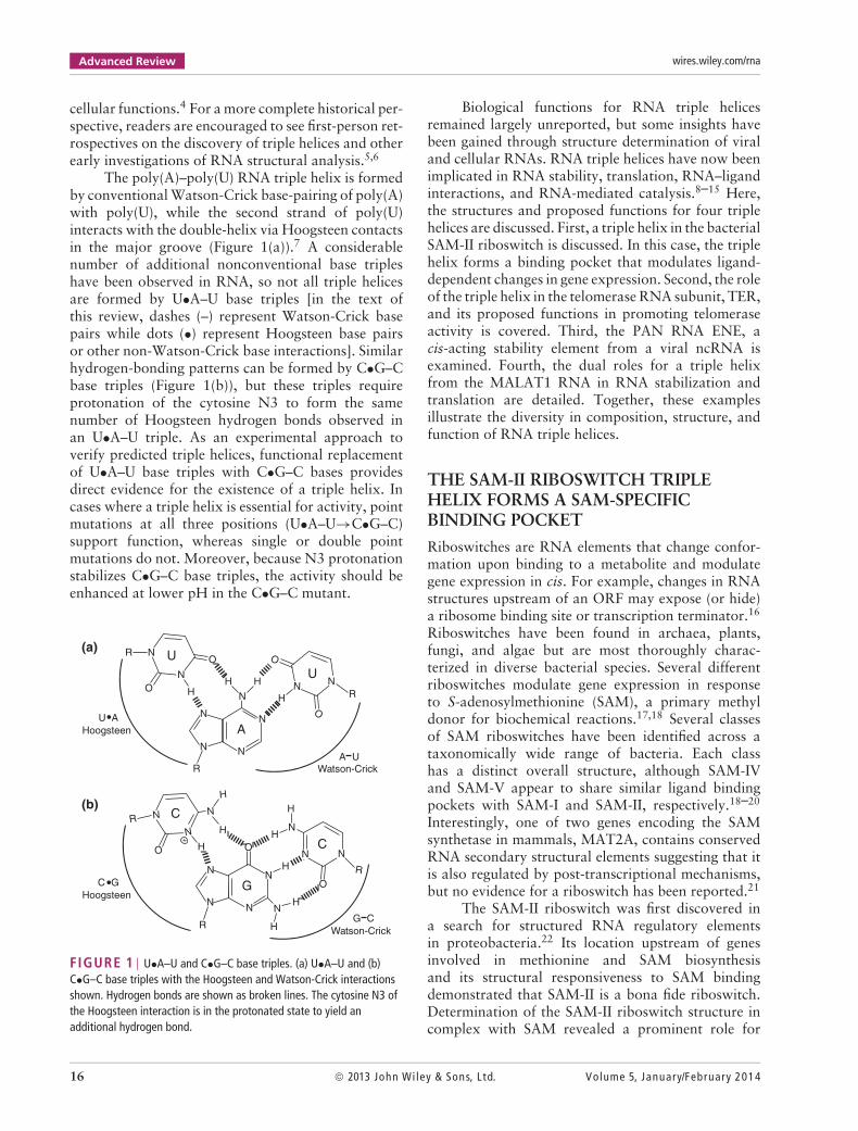
The poly(A)–poly(U) RNA triple helix is formedby conventional Watson-Crick base-pairing of poly(A)with poly(U), while the second strand of poly(U)interacts with the double-helix via Hoogsteen contactsin the major groove (Figure 1(a)).7 A considerablenumber of additional nonconventional base tripleshave been observed in RNA, so not all triple helicesare formed by U•A–U base triples [in the text ofthis review, dashes (–) represent Watson-Crick basepairs while dots (•) represent Hoogsteen base pairsor other non-Watson-Crick base interactions]. Similarhydrogen-bonding patterns can be formed by C•G–Cbase triples (Figure 1(b)), but these triples requireprotonation of the cytosine N3 to form the samenumber of Hoogsteen hydrogen bonds observed inan U•A–U triple. As an experimental approach toverify predicted triple helices, functional replacementof U•A–U base triples with C•G–C bases providesdirect evidence for the existence of a triple helix. Incases where a triple helix is essential for activity, pointmutations at all three positions (U•A–U→C•G–C)support function, whereas single or double pointmutations do not. Moreover, because N3 protonationstabilizes C•G–C base triples, the activity should beenhanced at lower pH in the C•G–C mutant.
N
N N
N
N
H H
R
N
N
R
O
O
H
O
O
HN N
R
U AHoogsteen
A UWatson-Crick
UU
A
(a)
(b)
N
N N
N
O
H
H
HN
R
R
NR
O
N
+
N
H
H H
H
NN
N
OGC GHoogsteen
C
C
G CWatson-Crick
H
FIGURE 1 | U•A–U and C•G–C base triples. (a) U•A–U and (b)C•G–C base triples with the Hoogsteen and Watson-Crick interactionsshown. Hydrogen bonds are shown as broken lines. The cytosine N3 ofthe Hoogsteen interaction is in the protonated state to yield anadditional hydrogen bond.
Biological functions for RNA triple helicesremained largely unreported, but some insights havebeen gained through structure determination of viraland cellular RNAs. RNA triple helices have now beenimplicated in RNA stability, translation, RNA–ligandinteractions, and RNA-mediated catalysis.8–15 Here,the structures and proposed functions for four triplehelices are discussed. First, a triple helix in the bacterialSAM-II riboswitch is discussed. In this case, the triplehelix forms a binding pocket that modulates ligand-dependent changes in gene expression. Second, the roleof the triple helix in the telomerase RNA subunit, TER,and its proposed functions in promoting telomeraseactivity is covered. Third, the PAN RNA ENE, acis-acting stability element from a viral ncRNA isexamined. Fourth, the dual roles for a triple helixfrom the MALAT1 RNA in RNA stabilization andtranslation are detailed. Together, these examplesillustrate the diversity in composition, structure, andfunction of RNA triple helices.
THE SAM-II RIBOSWITCH TRIPLEHELIX FORMS A SAM-SPECIFICBINDING POCKET
Riboswitches are RNA elements that change confor-mation upon binding to a metabolite and modulategene expression in cis. For example, changes in RNAstructures upstream of an ORF may expose (or hide)a ribosome binding site or transcription terminator.16
Riboswitches have been found in archaea, plants,fungi, and algae but are most thoroughly charac-terized in diverse bacterial species. Several differentriboswitches modulate gene expression in responseto S-adenosylmethionine (SAM), a primary methyldonor for biochemical reactions.17,18 Several classesof SAM riboswitches have been identified across ataxonomically wide range of bacteria. Each classhas a distinct overall structure, although SAM-IVand SAM-V appear to share similar ligand bindingpockets with SAM-I and SAM-II, respectively.18–20
Interestingly, one of two genes encoding the SAMsynthetase in mammals, MAT2A, contains conservedRNA secondary structural elements suggesting that itis also regulated by post-transcriptional mechanisms,but no evidence for a riboswitch has been reported.21
The SAM-II riboswitch was first discovered ina search for structured RNA regulatory elementsin proteobacteria.22 Its location upstream of genesinvolved in methionine and SAM biosynthesisand its structural responsiveness to SAM bindingdemonstrated that SAM-II is a bona fide riboswitch.Determination of the SAM-II riboswitch structure incomplex with SAM revealed a prominent role for
16 © 2013 John Wiley & Sons, Ltd. Volume 5, January/February 2014
WIREs RNA The emerging role of RNA triple helices
(b)
(a)
(c)
180°
C
U
U
U
UUU
U
U
U
G
G
G
G
G
G
GG
G
GC
A
A
A
AA
A
A
C
AA
C
A
C
A
C
A
GCA
G
G
C
GC
G
UUU
U
G
A
ASAM
A
5′
3′
10
20
4139
40
5
34
1514
31 A41
U40
A24
G8
C23
G42
A9 G22
C43
U44
U10
SAM
Met
A45
U11U21
U12U20
A46SAM
90°
U40 C23
U10
U10
G8
A41
SAM
(d)
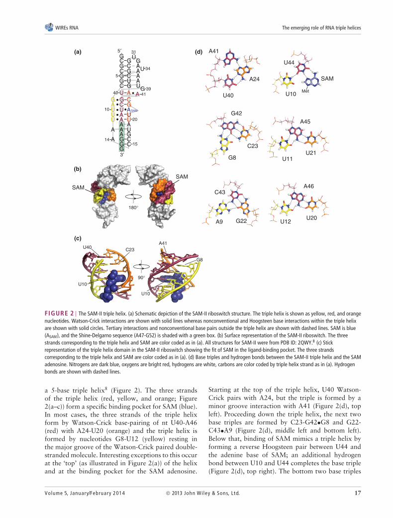
FIGURE 2 | The SAM-II triple helix. (a) Schematic depiction of the SAM-II riboswitch structure. The triple helix is shown as yellow, red, and orangenucleotides. Watson-Crick interactions are shown with solid lines whereas nonconventional and Hoogsteen base interactions within the triple helixare shown with solid circles. Tertiary interactions and nonconventional base pairs outside the triple helix are shown with dashed lines. SAM is blue(ASAM), and the Shine-Delgarno sequence (A47-G52) is shaded with a green box. (b) Surface representation of the SAM-II riboswitch. The threestrands corresponding to the triple helix and SAM are color coded as in (a). All structures for SAM-II were from PDB ID: 2QWY.8 (c) Stickrepresentation of the triple helix domain in the SAM-II riboswitch showing the fit of SAM in the ligand-binding pocket. The three strandscorresponding to the triple helix and SAM are color coded as in (a). (d) Base triples and hydrogen bonds between the SAM-II triple helix and the SAMadenosine. Nitrogens are dark blue, oxygens are bright red, hydrogens are white, carbons are color coded by triple helix strand as in (a). Hydrogenbonds are shown with dashed lines.
a 5-base triple helix8 (Figure 2). The three strandsof the triple helix (red, yellow, and orange; Figure2(a–c)) form a specific binding pocket for SAM (blue).In most cases, the three strands of the triple helixform by Watson-Crick base-pairing of nt U40-A46(red) with A24-U20 (orange) and the triple helix isformed by nucleotides G8-U12 (yellow) resting inthe major groove of the Watson-Crick paired double-stranded molecule. Interesting exceptions to this occurat the ‘top’ (as illustrated in Figure 2(a)) of the helixand at the binding pocket for the SAM adenosine.
Starting at the top of the triple helix, U40 Watson-Crick pairs with A24, but the triple is formed by aminor groove interaction with A41 (Figure 2(d), topleft). Proceeding down the triple helix, the next twobase triples are formed by C23-G42•G8 and G22-C43•A9 (Figure 2(d), middle left and bottom left).Below that, binding of SAM mimics a triple helix byforming a reverse Hoogsteen pair between U44 andthe adenine base of SAM; an additional hydrogenbond between U10 and U44 completes the base triple(Figure 2(d), top right). The bottom two base triples
Volume 5, January/February 2014 © 2013 John Wiley & Sons, Ltd. 17

Advanced Review wires.wiley.com/rna
are typical U-A•U triples (Figure 1(a)) formed by U21-A45•U11 and U20-A46•U12 (Figure 2(d), middleright and bottom right). Additional contacts betweenthe U21-A45•U11 and the positively charged sulfurgroup of SAM further support specific binding toSAM and aid in the discrimination between SAMand the related metabolite S-adenosylhomocysteine(SAH) (not shown).8,23 Thus, the SAM-II riboswitchtriple helix has evolved to form a highly specificbinding pocket for SAM, which then modulates geneexpression in response to metabolite concentrations.
The SAM-II triple helix is not simply a staticbinding pocket for SAM; formation and stabilizationof the triple helix is central to the responsiveness ofthe riboswitch. In the absence of SAM, the Shine-Dalgarno sequence (Figure 2(a), A47-G52, green box)is accessible for ribosome binding and translation.Chemical probing and structural data verify thatthe riboswitch adopts a more highly structuredconformation in the presence of SAM.8,22,24–26 TheShine-Dalgarno sequence becomes sequestered in thecompact structure resulting in the loss of translation(Figure 2(a)). Further biochemical and biophysicalassays led to the proposal that the SAM-II riboswitchemploys a conformational capture mechanism inresponse to SAM.24–26 The stable GC-rich doublehelix formed between the 5′-end of the RNA with ntG25-G30 forms in the absence of ligand. The presenceof magnesium promotes the formation of multipleconformers particularly with respect to the tertiaryinteractions surrounding that helix (Figure 2(a),dashed lines near the top of the molecule). BecauseSAM intercalates near the center of the triple helix,it is only in the presence of SAM that the hydrogenbonding networks are strong enough to capture astable triple helix structure. Formation of the stabletriple helix supports the interactions at the bottom ofthe molecule, resulting in sequestration of the Shine-Dalgarno sequence and repression of gene expression.The SAM-II riboswitch provides a striking examplein which an RNA triple helix forms the centralcomponent of a dynamic RNA regulatory element.
A TRIPLE HELIX IN TER IS ESSENTIALFOR TELOMERASE ACTIVITY
The primary mechanism for DNA replication isincapable of faithfully replicating chromosome ends,or telomeres. In the absence of alternate mechanismsfor telomere replication, the chromosomes shortenupon each round of cell division ultimately resultingin genome instability and loss of cell viability.In order to ensure proper telomere replication,eukaryotes encode an enzyme called telomerase that
extends the ends of DNA using a specialized reversetranscription mechanism.27 Telomerase activity isnecessary for continued cell proliferation and, assuch, it is strongly implicated in tumorigenesis.28,29 Infact, approximately 85–90% of tumor biopsies haveactivated telomerase. Therefore, understanding thedetailed mechanisms of telomerase activity may resultin discovery of novel therapeutic targets for cancer.
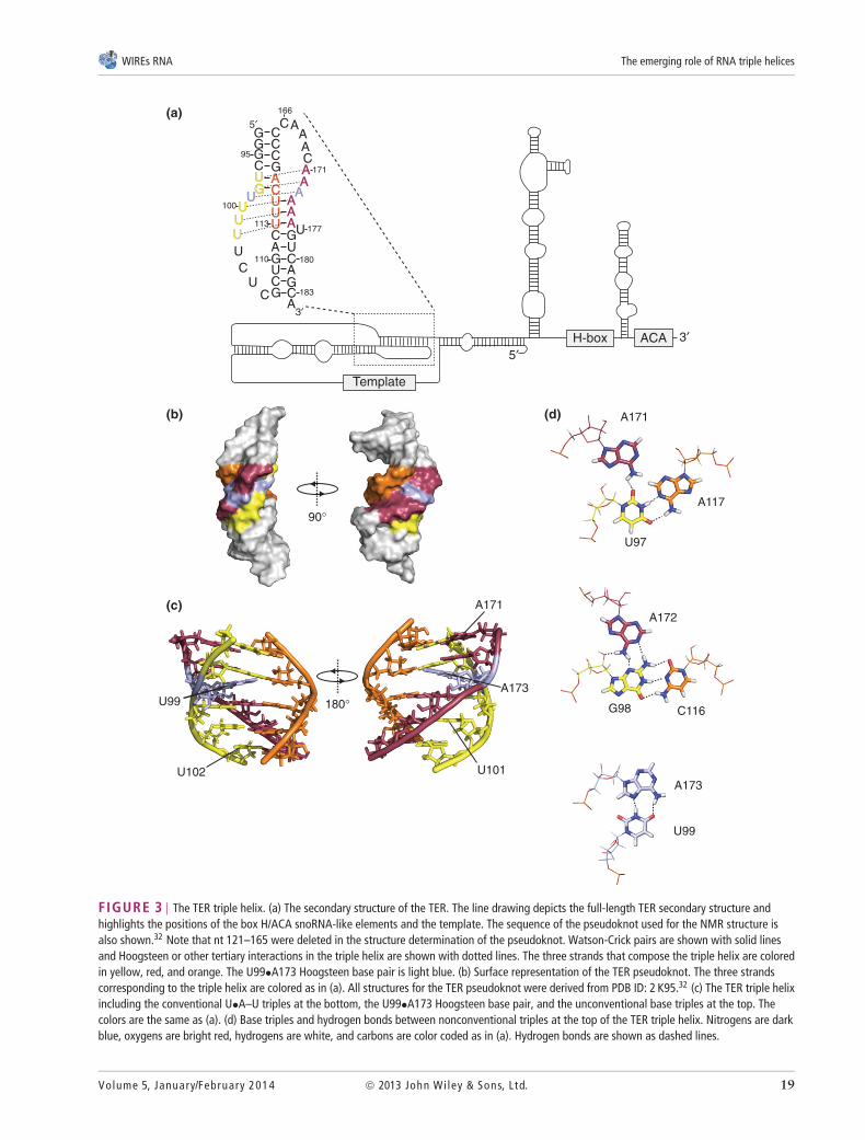
The human telomerase activity is provided bya ribonucleoprotein (RNP) complex minimally con-sisting of the protein TERT (Est2 in Saccharomycescerevisiae), and a ncRNA, TER (also known as hTR,TERC; Tlc1 in S. cerevisiae).27,30 The protein pro-vides the reverse transcriptase activity, and sharesstructural similarities with known retroviral reversetranscriptases.31 The RNA component has multiplefunctional domains including the template sequencethat is copied by TERT using the 3′-end of the chromo-some as a primer (Figure 3(a)). Iterative copying of thetemplate yields the short repeated sequence found ontelomeric DNA ([TTAGGG]n in mammals). TER fur-ther provides a boundary region essential for stoppingreverse transcription past the template. Telomerase-associated RNAs have cis-acting sequences importantfor their processing and stability, but the specific typesof sequences are not conserved. For example, humanTER assembles into a H/ACA snoRNA-like particlewhile yeast Tlc1 assembles into an Sm protein com-plex to promote RNA maturation and stability incells.33 Most importantly for this review, TER con-tains a highly conserved pseudoknot domain (Figure3(a)).34–36 While the precise molecular activity of thispseudoknot remains incompletely understood, muta-genesis studies and its conservation in ciliates, buddingyeast, and humans suggest an important function (seebelow).
Solution NMR revealed that an RNA triple helixis a central structural motif in the TER pseudoknot(Figure 3(b,c)).9,32 The triple helix is composed offive base triples (red, orange, and yellow) interruptedby a single A•U Hoogsteen base pair (light blue).Interestingly, the two triples at the top of the helixare formed by minor groove interactions. The firsttriple is formed from a single hydrogen bond betweenA171 and U97 while A117 contacts U97 in a Watson-Crick pair (Figure 3(d), top). Directly below that,A172 sits in the minor groove of the G98–C116Watson-Crick base pair (Figure 3(d), middle). Thetriple interactions are broken with a Hoogsteen basepair between A173 and U99 (Figure 3(d), bottom)which induces an approximately 30–38◦ bend (Figure3(c)). Immediately below the Hoogsteen pair are threecanonical U•A–U base triples (Figure 1(a)) in whichA174-A176 Watson-Crick pairs with U113–U115
18 © 2013 John Wiley & Sons, Ltd. Volume 5, January/February 2014
WIREs RNA The emerging role of RNA triple helices
(a)
(b)
(c)
(d)
5′3′
Template
H-box ACA
A171
A117
U97
A172
C116G98
A173
U99
90°
UU
G
GG
G
G
G
G
G
C A
C
A
C C
C
C
ACC
C
CA
C
CG
UU
U UU
UU
U
A
A
A
C
A
U
U
AAA
UAA
180
183
95
100
166
171
177
110
113
5′
3′
180°
U102
U99
U101
A173
A171
FIGURE 3 | The TER triple helix. (a) The secondary structure of the TER. The line drawing depicts the full-length TER secondary structure andhighlights the positions of the box H/ACA snoRNA-like elements and the template. The sequence of the pseudoknot used for the NMR structure isalso shown.32 Note that nt 121–165 were deleted in the structure determination of the pseudoknot. Watson-Crick pairs are shown with solid linesand Hoogsteen or other tertiary interactions in the triple helix are shown with dotted lines. The three strands that compose the triple helix are coloredin yellow, red, and orange. The U99•A173 Hoogsteen base pair is light blue. (b) Surface representation of the TER pseudoknot. The three strandscorresponding to the triple helix are colored as in (a). All structures for the TER pseudoknot were derived from PDB ID: 2 K95.32 (c) The TER triple helixincluding the conventional U•A–U triples at the bottom, the U99•A173 Hoogsteen base pair, and the unconventional base triples at the top. Thecolors are the same as (a). (d) Base triples and hydrogen bonds between nonconventional triples at the top of the TER triple helix. Nitrogens are darkblue, oxygens are bright red, hydrogens are white, and carbons are color coded as in (a). Hydrogen bonds are shown as dashed lines.
Volume 5, January/February 2014 © 2013 John Wiley & Sons, Ltd. 19

Advanced Review wires.wiley.com/rna
and Hoogsteen pairs with U100–U102 (yellow).Beyond these U•A–U triples, the Watson-Crick stemcontinues, but the triple helix is terminated.
The precise role of the triple helix in telomerasefunction remains unclear, but several lines ofevidence demonstrate that it is a critical componentof functional telomerase. The nucleotides in thetriple helix are conserved throughout vertebrates34
and triple helices are found in yeast as well.37,38
Importantly, loss of the triple helix leads to a less stableRNA structure and C•G–C substitutes for U•A–U tostabilize the pseudoknot structure in a pH-dependentfashion (Figure 1(b)).9,38 Similarly, C•G–C base triplescompensate for U•A–U triples in telomerase activityassays in vitro.9,37 While RNP formation is notaffected by mutations disrupting the triple helix, thetriple helix is located near the catalytic site.37 Theability of C•G–C to complement U•A–U in telomerasedemonstrates that the requirement for the triple helixis based on the triple helix structure, rather than thespecific sequence. In fact, Qiao and Cech37 showedthat the 2′-OH of A176 is necessary for catalysis invitro, suggesting the triple helix positions A176 2′-OHnear the catalytic site for activity. However, it shouldbe noted that methylation of the analogous hydroxylgroups in S. cerevisiae TLC1 had minimal or no effectstelomerase activity in vivo or on structural stability invitro.39,40 Rather, loss of a 2′-OH of a uridine nt fournt downstream of the A-rich strand of the buddingyeast triple helix had significant deleterious effects ontelomerase activity.39,40
Further studies of this domain are required toclarify the specific roles of the TER triple helix intelomerase function. For example, it is important tounderstand how 2′-OHs in or near the triple helixcontribute to telomerase catalysis. Furthermore, it isimportant to keep in mind that telomerase is a proces-sive enzyme. Thus, structural changes during binding,catalysis, and translocation of the RNA templaterequires sophisticated enzyme-substrate dynamics.Therefore, it will be interesting to determine whetherthe triple helix structure changes throughout the cat-alytic cycle of telomerase. Thus, exciting mechanisticinsights on telomerase may be revealed by continuedempirical examination of the telomerase triple helix.
THE PAN RNA ENE TRIPLE HELIXPROTECTS POLYADENYLATED RNAFROM NUCLEAR DECAY
The Kaposi’s sarcoma-associated herpesvirus (KSHV)encodes approximately 1.1 kb RNA called thepolyadenylated nuclear (PAN) RNA. As with otherherpesviruses, KSHV is long (approximately 140 kb)
nuclear dsDNA virus with a life cycle that includesboth latent and lytic phases.41–43 PAN RNA isexpressed during the lytic phase of infection to veryhigh levels. In fact, it is by far the most abundantRNA expressed in lytic phase, making up as muchas 70% of the polyadenylated RNA in lyticallyreactivated cell lines.44–46 As its name implies, PANRNA has a poly(A) tail and is primarily found in thenucleus. Consistent with an important role in viralreplication, knockdown of PAN RNA in infected celllines or deletion of PAN RNA from bacmid-basedinfectious clones severely decreases viral replicationand expression of viral lytic phase genes.47,48 Themechanisms and functions of PAN RNA are onlybeginning to be understood, but recent reports suggestthat it regulates transcription of viral and host genesto suppress immune responses and promote viral lyticgene expression.47–50 Because KSHV is an oncogenicvirus, understanding how viral factors modulate hostgene expression may provide clues to the pathogenesisof this clinically relevant virus.
PAN RNA is now classified as a viral long non-coding RNA, or lncRNA,51,52 but there were fewknown examples of nuclear polyadenylated ncRNAswhen PAN RNA was first described. PAN RNAincludes a 79-nt element near its 3′-end that isnecessary for PAN RNA accumulation and suffi-cient to increase the nuclear levels of an inefficientlyexported intronless β-globin reporter mRNA, but notits efficiently exported spliced counterpart.53 Becauseits activity depends on a canonical cleavage andpolyadenylation signal, it was initially proposed thatthe element enhanced RNA levels by increasing theefficiency of 3′-end formation. In addition, becausethe intronless β-globin mRNA accumulates in thenucleus, the element was dubbed the expression andnuclear retention element or ENE. However, laterwork showed that the primary function of the ENEis not to promote 3′-end formation or nuclear reten-tion directly. Instead, the ENE is a potent cis-actinginhibitor of nuclear RNA decay.54,55 Presumably, bothPAN RNA and intronless β-globin trigger nuclearRNA quality control pathways56–58 due to their inef-ficient export. Thus, the accumulation of intronless β-globin and PAN RNA in the nucleus is primarily due toprotection of nuclear-retained transcripts from nuclearRNA decay pathways. While these findings suggestthat ‘ENE’ is a misnomer, the element itself is a potentregulator of RNA metabolism and useful tool forstudying RNA decay pathways and nuclear ncRNAs.
The ENE is predicted to form a secondarystructure with two prominent stems (Figure 4(a), lightand dark blue) flanking a loop of nearly exclusivelyuridines (Figure 4(a), yellow and red). The presence
20 © 2013 John Wiley & Sons, Ltd. Volume 5, January/February 2014

WIREs RNA The emerging role of RNA triple helices
(a)
(c)
90°
180°
A
UG
C
A
A
A
A
A
A
AUUUU
U
UUUUU
G
G
GGG
GGG
CC
C
C
CC
C
C
C
C
UU
U
U
UUUU
U
G
G
G
G
GG
U
U
U
UU
U
CAC
A
C
CAC
G
UA
C
G
A
GCC
GU
888 966
888
903
908 944
949
953
916
927
AA
A
A
U
U
G
G
GGG
G
G
C
CC
C
C
C
U
U
U
UUU
UUU
G
CU
U
U
U
903
908 944
949
953
CG
G AAA
AAAAA
AAA
5′ 3′
5′A
UU A
G C
A
A-minor
(b)
(d)
A9
A9
FIGURE 4 | The PAN ENE triple helix. (a) Left Predicted secondary structure of the 79-nt PAN ENE. The numbers are relative to the PAN RNAtranscription start site; the polyadenylation site is at position 1077 (not shown). The upper and lower stems are colored in light blue and dark blue,respectively. The five-uridine stretches that participate in the triple helix are shown in yellow and red. Right : Schematic of the triple helix formed bythe core ENE in complex with oligoA9 (orange). The Hoogsteen interactions in the triple helix are shown with filled circles and Watson-Crick basepairs are depicted with solid lines. A-minor interactions are shown as dashed lines. (b) Surface representation of the PAN RNA ENE. Color-coding isthe same as in (a). All PAN RNA ENE structures were derived from PDB ID: 3P22.12 (c) The triple helix and lower stem of the PAN RNA ENE in complexwith A2–A9 of oligoA9. The U•A–U triple helix and the interface of the A-minor interactions between the minor groove of the lower G–C stem withA2-A4 are indicated. A952-U953 (gray) are ‘flipped out’ relative to the bases in the helix. (d) Close-up view of the 3′-most adenosine, A9 (orange,stick representation) as it fits in the pocket formed from the U-rich strands (yellow and red, surface representation) and the upper stem (light blue,surface representation).
of an unpaired U-rich loop and the ENE’s dependenceon a polyadenylation site led to the hypothesis that thepoly(A) tail hybridized with the U-rich portion of theENE to sequester it from exonucleases.55 In supportof this model, the poly(A) tail interacts with theENE in vitro and in RNPs purified from cells.12,54,55
In addition, the ENE protects transcripts fromexonucleases in nuclear extract, in recombinant invitro systems, and robustly stabilizes polyadenylatednuclear reporter transcripts in cells. Furthermore, theU-rich loops and stem structures are all essential forENE activity. These data provide a strong argument
that the ENE functions interact with the poly(A) tailin cis to protect transcripts from decay.
To better understand the structural basis forENE function, Mitton-Fry et al.12 determined thecrystal structure of the ‘core’ ENE in complex witha nine-adenosine oligonucleotide (oligoA9). The coreENE consists of the bottom stem, the U-rich loop,and four base pairs from the top stem (Figure 4(a),right). The top stem is extended by one C–G base pairand includes a GAAA tetraloop to stabilize the ENEstructure; the uppermost part of the ENE (nt 911–941)is not predicted to form a strong secondary structure
Volume 5, January/February 2014 © 2013 John Wiley & Sons, Ltd. 21
Advanced Review wires.wiley.com/rna
and was deleted for structural analysis. Importantly,the core ENE maintains the ability to interact withpoly(A) tails and inhibit decay in vitro, but the entireENE is necessary for full activity in cells.12,54
Determination of the structure of the ‘‘core’’ENE in complex with oligoA9 revealed that the U-rich sequences and the adenosines associate through afive-base triple helix (Figure 4(a–c)).12 The 3′-mostfive adenosines (oligoA9 is orange) are effectively‘‘clamped’’ between Watson-Crick base pairs withthe uridines on the right side of the U-rich loop(red) and Hoogsteen interactions with five uridines onthe left (yellow) forming typical U•A–U base triples(Figure 1(a)). Tertiary interactions between oligoA9and the ENE extend to the bottom stem with A2–A4interacting in the minor groove through A-minormotifs (Figure 4(c), orange and dark blue).12,59,60
The first adenosine of oligoA9 does not contact theENE. Based on its proposed function and mechanism,the ENE is predicted to restrict accessibility of the3′-end of the poly(A) tail. Indeed, it is difficult toimagine enzymatic access to this terminal nucleotide(A9) based on the ENE-oligoA9 structure (Figure4(d)). Thus, the ENE makes extensive tertiary contactswith oligoA9 to generate a stable structure and aninaccessible poly(A) tail 3′-end.
Determination of the ENE structure enablednovel bioinformatic strategies to identify ENE-likeelements in other viruses. Using a search querythat incorporated both structural and mutationalanalyses, Tycowski et al.61 identified six new viralENE-like elements. Two of these elements werefound in polydnaviruses, segmented dsDNA insectviruses. If one assumes that the ENE functionsto inhibit nuclear RNA decay in these cells, thisobservation strongly suggests conservation of nucleardecay mechanisms from insects to humans. Two moreelements were found in monkey (rhesus rhadinovirus,RRV) and horse (equine herpesvirus-2, EHV-2)gammaherpesviruses. Because of poor conservationat the sequence level, lncRNAs can be difficult toidentify based on sequence analysis. Unexpectedly,the identification of these new ENE-like elementsuncovered a lncRNA in RRV and supported theexistence of a lncRNA in EHV-2. The novel transcriptsand PAN RNA are encoded from syntenic positionson their respective viral genomes, strongly suggestingthey are viral lncRNA homologs with similar functionsin viral replication. The final two ENE-like elementswere found in mimivirus, a large dsDNA virus,62 andin picorna-like dicistrovirus (PSIV),63 a positive strandRNA virus with only a cytoplasmic phase. This latterdiscovery supports cytoplasmic functions for ENE-likestructures, a finding consistent with potential roles for
ENE-like structures in translation (see below). Thisstudy demonstrates that ENE-like triple helices maybe widespread in viruses and have both nuclear andcytoplasmic functions in viral RNA metabolism.
THE MALAT1 TRIPLE HELIXSTABILIZES A MAMMALIAN NUCLEARRNA
Viruses utilize host cell strategies to perform cellularfunctions, so Steitz and colleagues proposed thatcellular RNAs likely include ENE-like elements thatform triple helices.12 In fact, two reports recentlyshowed that the MALAT1 and MENβ nuclear RNAshave noncanonical 3′-ends that form triple helixstructures.14,64 The formation of triple helix structuresin both transcripts is well supported, but this reviewfocuses on the MALAT1 triple helix.
MALAT1, the metastasis-associated lungadenocarcinoma transcript 1 (also known as NEAT2),was originally identified due to its overexpression inmetastatic lung cancers65 and has since been associ-ated with several additional cancers.52,66 MALAT1is a conserved, abundant, nuclear ncRNA thatmodulates gene expression and cellular function atmultiple levels. Mechanistically, MALAT1 regulatesalternative splicing in cells by interacting with splicingregulatory proteins67 and this regulation can alter cellcycle progression.68 MALAT1 has additionally beenproposed to regulate cell cycle progression by modu-lating transcription.69 Surprisingly, mouse MALAT1knockout models have no strong phenotypes, so itsphysiological roles remain somewhat ambiguous.70–72
The 3′-ends of MALAT1 are generated using aunique endonucleolytic cleavage mechanism (Figure5(a)).73,74 A long (approximately 8.7 kb) polyadeny-lated MALAT1 transcript is generated using acanonical polyadenylation signal, but this transcriptrepresents only a small (<1%) fraction of theMALAT1 in the cell. Instead, the 3′-end of the abund-ant MALAT1 is generated by endonucleolytic cleavageof a 61-nt tRNA-like transcript called the MALAT1-associated small cytoplasmic RNA (mascRNA). ThemascRNA 5′ and 3′-ends are generated by the sameendonucleases involved in tRNA production, RNaseP, and RNase Z, respectively. MascRNA is exportedto the cytoplasm, but its function remains unknown.The RNase P cleavage site generates the mature 3′-endof the stable nuclear MALAT1 ncRNA. This regionincludes four domains conserved from Xenopus tohumans: two U-rich elements (Figure 5(a), yellowand red), a predicted stem (white), and a 3′-terminalA-rich motif (orange). The arrangement of U-richmotifs upstream of a terminal A-tract led to the
22 © 2013 John Wiley & Sons, Ltd. Volume 5, January/February 2014

WIREs RNA The emerging role of RNA triple helices
87078268
(a)
(b)
G
GG C
C
U
U
UU
UUU
U
U
UAAAAA
AA
A
GC CG
CA UC
G
A ACAAAA
CGA
CGAU
CC
UUUU
AAAA U
UUU
C
GG
AAG U5’
AA
AAAA
UUCGA UC
UU
3’G
GG
U
U
UU
UUU
U
U
UAAAAA
GC CG
AU
ACG
ACGAU
CC
UUUU
AAAA U
UUU
C
AA
A
A
3’
AU
A
AA
5’
MALAT1(human)
Comp14(mouse)
U-Rich U-Richstem A-RichMALAT1 aauaaa AAAAAAn
AAAAAAn
AAAAAAn
aauaaa
U-Rich U-Richstem A-RichMALAT1
RNase P
aauaaa
RNase Z
CCA
mascRNA(cytoplasmic)
stable MALAT1(nuclear)
CCA-addingEnzyme
83561
UUGUUUU
U
G
UU
U
U
G
FIGURE 5 | The MALAT1 ENE-like triple helix. (a) RNA processing pathway for MALAT1 and mascRNA. For simplicity, the polyadenylated speciesis shown as the primary transcript, but it is not clear whether the RNase P cleavage occurs on polyadenylated MALAT1 or co-transcriptionally.73 Thediagram is not to scale; the highlighted elements compose only a small fraction of the total MALAT1 length. The numbers refer to the humanMALAT1, but all the relevant domains are conserved. (b) Secondary structure and triple helix formation in the MALAT1 ENE-like element. Thestructure on the left corresponds to the full-length human MALAT1 ENE-like element, while the structure on the right is a minimal element derivedfrom murine MALAT1, Comp14.14 The U-rich sequence that forms Watson-Crick base pairs is shown in red, the A-rich stretch is orange, and theU-rich tract that forms Hoogsteen base triples is yellow. The Hoogsteen interactions in the triple helix are shown with filled circles and Watson-Crickbase pairs or triples are depicted with solid lines; predicted A-minor interactions are shown as dashed lines. The central C and G nucleotides areshown in light blue. The C•G–C is depicted as part of the helix based on pH profiles,64 but this triple was not observed in a structural model.14 Thelower G–C stem is shown in dark blue. The sequences important for translation are highlighted in the green box.
Volume 5, January/February 2014 © 2013 John Wiley & Sons, Ltd. 23

Advanced Review wires.wiley.com/rna
proposal that these elements may serve as an ENE-likeelement.73 This hypothesis has since been validatedby the demonstration that both structures form triplehelices and protect transcripts from decay.
Work from the Steitz and Sharp labsindependently uncovered the formation of a triplehelix at the 3′ terminus of MALAT1.14,64 Tofurther explore the processing of the MALAT1-mascRNA ncRNAs, Wilusz et al.14 inserted the 3′elements from mouse MALAT1 into a GFP-basedreporter system. They included both the U-richmotifs, the conserved stem, the A-rich tract, andthe mascRNA. Together, these elements recapitulatethe MALAT1 3′ processing pathway and producea stable transcript that accumulates primarily inthe cytoplasm, consistent with the idea that theseelements are not strong nuclear retention factors.Extensive mutational analysis further defined aminimal MALAT1 3′ stabilizing element (calledComp14) that included the U- and A-rich motifs with asmaller stem (Figure 5(b)). Brown and colleagues usedthe triple helix structure from the PAN RNA ENE asa guide to identification of similar elements in cellularRNAs.64 They focused on abundant nuclear ncRNAsthat have the same predicted secondary structuralfeatures as the ENE and identified the 3′ terminus ofMALAT1 as a candidate. Reporter assays confirmedthat the identified MALAT1 ENE-like element issufficient to stabilize RNA in cis. Interestingly, unlikePAN RNA, the mascRNA was also needed for fullactivity; that is, they did not stabilize a polyadenylatedtranscript produced using the canonical 3′-endformation machinery. Together, these studies definedpotential ENE-like triple helices in cellular RNAs.
BOX 1
HOW DO ENE-LIKE ELEMENTS PROTECTTRANSCRIPTS FROM DECAY?
The observation the MALAT1 ENE-like elementis significantly more active when the triplehelix has a blunt end strongly supports themodel that the triple helix functions bysequestration of the RNA 3′-end from the decaymachinery. This is in contrast to a flaviviruspseudoknot that stops the progression of theXRN1 5′-to-3′ exonuclease with a ‘roadblock’mechanism.75 If having the 3′-most adenosineblunt ended in the triple helix is essential toinhibit decay, then it is perplexing how thePAN RNA ENE, which contacts long poly(A)tails using exclusively U•A–U triples, inhibitsprocessive degradation. Perhaps, regions outside
of the core ENE,54 surrounding sequences inPAN RNA,53 or interplay with poly(A) bindingproteins48 contributes to the full activity ofPAN RNA ENE-mediated inhibition of RNAdecay. It is also not clear which enzyme(s) isinhibited by sequestration of the RNA 3′-ends.For example, GFP mRNAs with mutated MALAT1triple helices undergo uridylation-dependentdecay.14,76,77 Deletion of the ENE from PANRNA leads to poly(A) tail extension54 andunpublished data strongly support the idea thatthis hyperadenylation is coupled to transcriptdecay (Bresson and Conrad, PlosGenetics inpress). Taken together, these results raise thepossibility that the sequestration of the poly(A)tail is not protecting it from the exonucleasesdirectly, but may be keeping it from tailingactivities that are linked to exonucleolyticdecay.77,78 Further work with the ENE and ENE-like elements can therefore reveal importantmechanistic insights into cellular RNA decaypathways.
Even though a structure has not been solvedfor the MALAT1 ENE-like element, multiple lines ofevidence verify that it forms a triple helix betweenthe U- and A-rich conserved sequences. First, atertiary structural modeling approach predicts thatthis helix will form.14 Second, the melting profile ofthe MALAT1 ENE-like element is biphasic, consistentwith melting of the Hoogsteen triple followedby melting of the Watson-Crick strand.64 Third,U•A–U→C•G–C substitutions are functional in vivowhereas corresponding double mutations are not.14,64
Fourth, the co-conservation of U- and A-rich elementsand the similarity to the known structure of the PANRNA ENE support the formation of the triple helix.Taken together, these data demonstrate that MALAT1contains a conserved triple-helix structure at its 3′-end after processing and release of the mascRNA(Figure 5(a)).
The MALAT1 ENE-like element has ninepredicted U•A–U base triples, but these U•A–Utriples are interrupted by C–G and G–C base pairson the Watson-Crick strands and a single C on theHoogsteen strand (Figure 5(b), light blue). It is notimmediately obvious why the MALAT1 ENE-likeelement would include these GC nucleotides insteadof having an extended, and presumably more stable,U•A–U triple helical structure. While the structuralmodel does not predict the formation of a C•G–Ctriple helix,14 pH-dependent changes in biphasicmelting curves suggest the formation of a C•G–C
24 © 2013 John Wiley & Sons, Ltd. Volume 5, January/February 2014
WIREs RNA The emerging role of RNA triple helices
triple within the MALAT1structure.64 Mutationalanalyses suggest that U•A–U triples at the top andbottom are sufficient for function in the Comp14element, but triples in the central portion, includingthe putative C•G–C triple, are less critical.14 Evenso, the Watson-Crick pairing throughout the elevennucleotides is absolutely required for activity. Oneproposed function for the G–C base pairs is to ensurethat the 3′-most A forms a blunt end with the triplehelix. In fact, insertion of additional adenosines to the3′-end of the A-rich region to create a structure with asingle-stranded 3′ end extending from the triple helixdecreases RNA accumulation in vivo (see Box 1).64
THE MALAT1 TRIPLE HELIXPROMOTES TRANSLATION
Perhaps, the most surprising revelation regarding thefunction of the MALAT1 triple helix is that it sup-ports efficient translation.14 The GFP reporters usedto define the element produce ample levels of GFPprotein. In fact, protein production is comparable tocorresponding reporters with a conventional polyade-nylation signal in two different contexts. Moreover,the translational stimulation is not driven exclusivelyby increased RNA stability. In fact, mutations of thestems adjacent to the triple helix abrogate transla-tional stimulation but have no effect on cytoplasmicRNA accumulation (shaded green, Figure 5(b)).
The discovery that the MALAT1 triple helixsupports translation was unexpected for two primaryreasons. First, a poly(A) tail promotes efficienttranslation by stabilizing mRNAs in the cytoplasmand by recruiting the poly(A) binding protein, animportant translation factor.79 Second, the triple-helix containing MALAT1 transcript is primarilynuclear and presumed to be noncoding. However,sequences near the 5′-end of MALAT1 are protectedin ribosomal profiling assays.14 Therefore, a fractionof MALAT1 transcript may in fact be translated,even though no conserved ORFs have been identified.Other suspected ncRNAs have been shown to encodesmall polypeptides.80–82 Perhaps, these observationsregarding MALAT1 further question the assumptionthat all RNAs without an apparent coding region arebona fide ncRNAs.
RNA TRIPLE HELICES AS TOOLS FORDISCOVERY
Each of the four RNA triple helices described abovehas unique features that highlight the diversityof RNA structure and function. Moreover, each
example provides essential mechanistic insightsinto the particular biological process for whichit has evolved. However, triple helices may havesignificantly more widespread potential as tools tofurther our understanding of biological processes. Forexample, a triple helix structure has been identifiedin the catalytic domain of the Oceanobacillusiheyensis Group II self-splicing intron.13 In additionto elucidating the mechanism of the Group II intron,the structure yields important predictions regardingthe role of tertiary structural elements at the core ofthe human spliceosome.83 Thus, the structure of theO. iheyensis Group II self-splicing intron can serve asa framework for testing specific and detailed mech-anistic questions for human pre-mRNA splicing. Inaddition, examination of the PAN RNA ENE has ledto important insights into the nature of nuclear RNAdecay pathways, particularly with respect to the roleof the poly(A) tail.55 The MALAT1 ENE-like elementwas cleverly used to demonstrate that the presenceof a poly(A) tail was not an absolute requirement formiRNA-mediated translational silencing.14 Compari-son of the ends of GFP reporter mRNAs containing orlacking functional MALAT1 ENE-like element tran-scripts implicated uridylation as a factor in the decayof nonpolyadenylated transcripts.14 Given the rela-tively unexplored arena of RNA triple helix structures,exploration of the TER triple helix in atomic detailcould conceivably lead to novel drug developmentopportunities. Thus, mechanistic dissection of RNAtriple helices is important to understand the functionsfor each specific triple helix, but may additionallyuncover unexpected mechanisms in cell biology.
SUMMARY
The pioneering work of Felsenfeld, Davies, and Richrevealed that poly(A) and poly(U) could efficientlyform triple helix structures in vitro. More recently,RNA triple helices have been found in a numberof functional RNAs. The triple helix in the SAM-II riboswitch forms a highly specific ligand-bindingpocket for SAM and its formation is central to cis-acting gene regulation in the SAM-II riboswitch. Thefunction of the TER triple helix remains somewhatambiguous, but it is clear that this structure isessential for telomerase activity. Given the importanceof telomerase to cancer development, unraveling themechanistic details of the triple helix function of TERhas important implications for human health. ThePAN RNA ENE and the MALAT1 ENE-like triplehelices both contribute to the stability of long nuclearncRNAs by sequestration of the RNA 3′-ends. TheMALAT1 ENE-like element has an additional activity
Volume 5, January/February 2014 © 2013 John Wiley & Sons, Ltd. 25
Advanced Review wires.wiley.com/rna
that allows it to support translation. The examinationof these triple helix structures has contributed to ourknowledge of cell biology and has strong prospectsfor uncovering future discoveries. Over 50 years after
their initial description, it seems we are only beginningto understand the importance of RNA triple helices inbiology.
ACKNOWLEDGMENTS
Thanks to Drs. Neal Alto and Yunsun Nam for help with PyMOL. Dr. Conrad is funded by the NIH-NIAIDgrant AI081710, by the Cancer Prevention and Research Institute of Texas RP110132 and by Welch FoundationGrant I-1732. N.K.C. is a Southwestern Medical Foundation Scholar in Biomedical Research.
REFERENCES1. Cech TR. Structural biology. The ribosome is a
ribozyme. Science 2000, 289:878–879.
2. Rich A, Felsenfeld G. Formation of a threestrandedpolynucleotide molecule. J Am Chem Soc 1957,79:2023–2024.
3. Felsenfeld G, Rich A. Studies on the formation oftwo- and three-stranded polyribonucleotides. BiochimBiophys Acta 1957, 1000:87–98.
4. Buske FA, Mattick JS, Bailey TL. Potential in vivoroles of nucleic acid triple-helices. RNA Biol 2011,8:427–439. doi: 10.4161/rna.8.3.14999.
5. Rich A. Discovery of the hybrid helix and thefirst DNA-RNA hybridization. J Biol Chem 2006,281:7693–7696. doi: 10.1074/JBC.X600003200.
6. Rich A. The era of RNA awakening: structural biologyof RNA in the early years. Q Rev Biophys 2009,42:117–137. doi: 10.1017/S0033583509004776.
7. Hoogsteen K. The crystal and molecular structure ofa hydrogen-bonded complex between 1-methylthymineand 9-methyladenine. Acta Cryst 1963, 16:907–916.doi: 10.1107/S0365110X63002437.
8. Gilbert SD, Rambo RP, Van Tyne D, Batey RT.Structure of the SAM-II riboswitch bound to S-adenosylmethionine. Nat Struct Mol Biol 2008,15:177–182. doi: 10.1038/nsmb.1371.
9. Theimer CA, Blois CA, Feigon J. Structure of thehuman telomerase RNA pseudoknot reveals conservedtertiary interactions essential for function. Mol Cell2005, 17:671–682. doi: 10.1016/j.molcel.2005.01.017.
10. Chastain M, Tinoco I. Nucleoside triples from the groupI intron. Biochemistry 1993, 32:14220–14228.
11. Su L, Chen L, Egli M, Berger JM, Rich A. Minor grooveRNA triplex in the crystal structure of a ribosomalframeshifting viral pseudoknot. Nat Struct Biol. 1999,6:285–292. doi: 10.1038/6722.
12. Mitton-Fry RM, DeGregorio SJ, Wang J, Steitz TA,Steitz JA. Poly(A) tail recognition by a viral RNAelement through assembly of a triple helix. Science2010, 330:1244–1247. doi: 10.1126/science.1195858.
13. Toor N, Keating KS, Taylor SD, Pyle AM. Crystalstructure of a self-spliced group II intron. Science 2008,320:77–82. doi: 10.1126/science.1153803.
14. Wilusz JE, JnBaptiste CK, Lu LY, Kuhn CD, Joshua-TorL, Sharp PA. A triple helix stabilizes the 3’ ends of longnoncoding RNAs that lack poly(A) tails. Genes Dev2012, 26:2392–2407. doi: 10.1101/gad.204438.112.
15. Chastain M, Tinoco I. A base-triple structural domainin RNA. Biochemistry 1992, 31:12733–12741.
16. Serganov A, Nudler E. A decade ofriboswitches. Cell 2013, 152(1–2):17–24. doi:10.1016/j.cell.2012.12.024.
17. Roje S. S-Adenosyl-L-methionine: beyond the universalmethyl group donor. Phytochemistry 2006, 67:1686–1698. doi: 10.1016/j.phytochem.2006.04.019.
18. Wang JX, Breaker RR. Riboswitches that sense S-adenosylmethionine and S-adenosylhomocysteine.Biochem Cell Biol 2008, 86:157–168. doi:10.1139/O08-008.
19. Poiata E, Meyer MM, Ames TD, Breaker RR.A variant riboswitch aptamer class for S-adenosylmethionine common in marine bacteria.RNA 2009, 15:2046–2056. doi: 10.1261/rna.1824209.
20. Weinberg Z, Regulski EE, Hammond MC, Barrick JE,Yao Z, Ruzzo WL, Breaker RR. The aptamer core ofSAM-IV riboswitches mimics the ligand-binding siteof SAM-I riboswitches. RNA 2008, 14:822–828. doi:10.1261/rna.988608.
21. Parker BJ, Moltke I, Roth A, Washietl S, Wen J, KellisM, Breaker R, Pedersen JS. New families of humanregulatory RNA structures identified by comparativeanalysis of vertebrate genomes. Genome Res 2011,21:1929–1943. doi: 10.1101/gr.112516.110.
22. Corbino KA, Barrick JE, Lim J, Welz R, Tucker BJ,Puskarz I, Mandal M, Rudnick ND, Breaker RR.Evidence for a second class of S-adenosylmethionineriboswitches and other regulatory RNA motifs inalpha-proteobacteria. Genome Biol 2005, 6:R70. doi:10.1186/gb-2005-6-8-r70.
26 © 2013 John Wiley & Sons, Ltd. Volume 5, January/February 2014

WIREs RNA The emerging role of RNA triple helices
23. Doshi U, Kelley JM, Hamelberg D. Atomic-levelinsights into metabolite recognition and specificity ofthe SAM-II riboswitch. RNA 2012, 18:300–307. doi:10.1261/rna.028779.111.
24. Haller A, Rieder U, Aigner M, Blanchard SC,Micura R. Conformational capture of the SAM-IIriboswitch. Nat Chem Biol 2011, 7:393–400. doi:10.1038/nchembio.562.
25. Chen B, Zuo X, Wang Y-X, Dayie TK. Multipleconformations of SAM-II riboswitch detected withSAXS and NMR spectroscopy. Nucleic Acids Res 2012,40:3117–3130. doi: 10.1093/nar/gkr1154.
26. Kelley JM, Hamelberg D. Atomistic basis for theon-off signaling mechanism in SAM-II riboswitch.Nucleic Acids Res 2010, 38:1392–1400. doi:10.1093/nar/gkp1106.
27. Blackburn EH, Collins K. Telomerase: an RNPenzyme synthesizes DNA. Cold Spring Harb PerspectBiol 2011, 3:a003558–a003558. doi: 10.1101/cshper-spect.a003558.
28. Shay JW, Wright WE. Role of telomeres and telomerasein cancer. Semin Cancer Biol 2011, 21:349–353. doi:10.1016/j.semcancer.2011.10.001.
29. Gunes C, Rudolph KL. The role of telomeres instem cells and cancer. Cell 2013, 152:390–393. doi:10.1016/j.cell.2013.01.010.
30. Wyatt HDM, West SC, Beattie TL. InTERTpretingtelomerase structure and function. Nucleic Acids Res2010, 38:5609–5622. doi: 10.1093/nar/gkq370.
31. Gillis AJ, Schuller AP, Skordalakes E. Structureof the Tribolium castaneum telomerase catalyticsubunit TERT. Nature 2008, 455:633–637. doi:10.1038/nature07283.
32. Kim N-K, Zhang Q, Zhou J, Theimer CA, PetersonRD, Feigon J. Solution structure and dynamicsof the wild-type pseudoknot of human telomeraseRNA. J Mol Biol 2008, 384:1249–1261. doi:10.1016/j.jmb.2008.10.005.
33. Egan ED, Collins K. Biogenesis of telomeraseribonucleoproteins. RNA 2012, 18:1747–1759. doi:10.1261/rna.034629.112.
34. Chen JL, Blasco MA, Greider CW. Secondary structureof vertebrate telomerase RNA. Cell 2000, 100:503–514.
35. Chen J-L, Greider CW. An emerging consensus fortelomerase RNA structure. Proc Natl Acad Sci USA2004, 101:14683–14684. doi: 10.1073/pnas.0406204101.
36. Theimer CA, Feigon J. Structure and function oftelomerase RNA. Curr Opin Struct Biol 2006,16:307–318. doi: 10.1016/j.sbi.2006.05.005.
37. Qiao F, Cech TR. Triple-helix structure in telomeraseRNA contributes to catalysis. Nat Struct Mol Biol 2008,15:634–640. doi: 10.1038/nsmb.1420.
38. Shefer K, Brown Y, Gorkovoy V, Nussbaum T,Ulyanov NB, Tzfati Y. A triple helix within a
pseudoknot is a conserved and essential element oftelomerase RNA. Mol Cell Biol 2007, 27:2130–2143.doi: 10.1128/MCB.01826-06.
39. Huang C, Yu Y-T. Targeted 2’-O methylation at anucleotide within the pseudoknot of telomerase RNAreduces telomerase activity in vivo. Mol Cell Biol 2010,30:4368–4378. doi: 10.1128/MCB.00384-10.
40. Liu F, Theimer CA. Telomerase activity is sensitive tosubtle perturbations of the TLC1 pseudoknot 3′ stemand tertiary structure. J Mol Biol 2012, 423:719–735.doi: 10.1016/j.jmb.2012.08.025.
41. Mesri EA, Cesarman E, Boshoff C. Kaposi’s sarcomaand its associated herpesvirus. Nat Rev Cancer 2010,10:707–719. doi: 10.1038/nrc2888.
42. Cai Q, Verma SC, Lu J, Robertson ES. Molecularbiology of Kaposi’s sarcoma-associated herpesvirus andrelated oncogenesis. Adv Virus Res 2010, 78:87–142.doi: 10.1016/B978-0-12-385032-4.00003-3.
43. Wen KW, Damania B. Kaposi sarcoma-associatedherpesvirus (KSHV): molecular biology and onco-genesis. Cancer Lett 2010, 289:140–150. doi:10.1016/j.canlet.2009.07.004.
44. Song MJ, Brown HJ, Wu TT, Sun R. Transcriptionactivation of polyadenylated nuclear RNA by RTAin human herpesvirus 8/Kaposi’s sarcoma-associatedherpesvirus. J Virol 2001, 75:3129–3140. doi:10.1128/JVI.75.7.3129-3140.2001.
45. Sun R, Lin SF, Gradoville L, Miller G. Polyadeny-lylated nuclear RNA encoded by Kaposi sarcoma-associated herpesvirus. Proc Natl Acad Sci USA 1996,93:11883–11888.
46. Zhong W, Wang H, Herndier B, Ganem D. Restrictedexpression of Kaposi sarcoma-associated herpesvirus(human herpesvirus 8) genes in Kaposi sarcoma. ProcNatl Acad Sci USA 1996, 93:6641–6646.
47. Rossetto CC, Pari G. KSHV PAN RNA associateswith demethylases UTX and JMJD3 to activate lyticreplication through a physical interaction with thevirus genome. PLoS Pathog 2012, 8:e1002680. doi:10.1371/journal.ppat.1002680.t002.
48. Borah S, Darricarrere N, Darnell A, Myoung J, SteitzJA. A viral nuclear noncoding rna binds re-localizedpoly(A) binding protein and is required for late KSHVgene expression. PLoS Pathog 2011, 7:e1002300. doi:10.1371/journal.ppat.1002300.g009.
49. Rossetto CC, Pari GS. KSHV noncoding PAN RNAinteracts with virus and cellular-encoded proteinsand suppresses expression of genes involved inimmune modulation. J Virol 2011. doi: 10.1128/JVI.05886-11.
50. Rossetto CC, Tarrant-Elorza M, Verma S,Purushothaman P, Pari GS. Regulation of viraland cellular gene expression by Kaposi’s sarcomaassociated herpesvirus (KSHV) PAN RNA. J Virol2013. doi: 10.1128/JVI.03111-12.
Volume 5, January/February 2014 © 2013 John Wiley & Sons, Ltd. 27
Advanced Review wires.wiley.com/rna
51. Shamovsky I, Nudler E. Gene control by largenoncoding RNAs. Sci STKE 2006, 2006:pe40. doi:10.1126/stke.3552006pe40.
52. Batista PJ, Chang HY. Long noncoding RNAs: cellularaddress codes in development and disease. Cell 2013,152:1298–1307. doi: 10.1016/j.cell.2013.02.012.
53. Conrad NK, Steitz JA. A Kaposi’s sarcoma virusRNA element that increases the nuclear abundance ofintronless transcripts. EMBO J 2005, 24:1831–1841.doi: 10.1038/sj.emboj.7600662.
54. Conrad NK, Shu M-D, Uyhazi KE, Steitz JA. Mutationalanalysis of a viral RNA element that counteracts rapidRNA decay by interaction with the polyadenylate tail.Proc Natl Acad Sci USA 2007, 104:10412–10417. doi:10.1073/pnas.0704187104.
55. Conrad NK, Mili S, Marshall EL, Shu M-D, Steitz JA.Identification of a rapid mammalian deadenylation-dependent decay pathway and its inhibition by aviral RNA element. Mol Cell 2006, 24:943–953. doi:10.1016/j.molcel.2006.10.029.
56. Schmid M, Jensen TH. Quality control of mRNP inthe nucleus. Chromosoma 2008, 117:419–429. doi:10.1007/s00412-008-0166-4.
57. Fasken MB, Corbett AH. Mechanisms of nuclearmRNA quality control. RNA Biol 2009, 6:237–241.
58. Doma MK, Parker R. RNA quality controlin eukaryotes. Cell 2007, 131:660–668. doi:10.1016/j.cell.2007.10.041.
59. Doherty EA, Batey RT, Masquida B, Doudna JA. Auniversal mode of helix packing in RNA. Nat StructBiol 2001, 8:339–343. doi: 10.1038/86221.
60. Nissen P, Ippolito JA, Ban N, Moore PB, SteitzTA. RNA tertiary interactions in the large ribosomalsubunit: the A-minor motif. Proc Natl Acad Sci USA2001, 98:4899–4903. doi: 10.1073/pnas.081082398.
61. Tycowski KT, Shu M-D, Borah S, Shi M, SteitzJA. Conservation of a triple-helix-forming RNAstability element in noncoding and genomic RNAsof diverse viruses. CellReports 2012, 2:26–32. doi:10.1016/j.celrep.2012.05.020.
62. La Scola B, Audic S, Robert C, Jungang L, deLamballerie X, Drancourt M, Birtles R, Claverie J-M, Raoult D. A giant virus in amoebae. Science 2003,299:2033. doi: 10.1126/science.1081867.
63. Bonning BC, Miller WA. Dicistroviruses. Annu RevEntomol 2010, 55:129–150. doi: 10.1146/annurev-ento-112408-085457.
64. Brown JA, Valenstein ML, Yario TA, TycowskiKT, Steitz JA. Formation of triple-helical structuresby the 3’-end sequences of MALAT1 and MENβ
noncoding RNAs. Proc Natl Acad Sci 2012. doi:10.1073/pnas.1217338109.
65. Ji P, Diederichs S, Wang W, Boing S, Metzger R,Schneider PM, Tidow N, Brandt B, Buerger H, Bulk E,et al. MALAT-1, a novel noncoding RNA, and thymosin
beta4 predict metastasis and survival in early-stage non-small cell lung cancer. Oncogene 2003, 22:8031–8041.doi: 10.1038/sj.onc.1206928.
66. Gutschner T, Hammerle M, Diederichs S. MALAT1—aparadigm for long noncoding RNA function in cancer.J Mol Med 2013. doi: 10.1007/s00109-013-1028-y.
67. Tripathi V, Ellis JD, Shen Z, Song DY, Pan Q, WattAT, Freier SM, Bennett CF, Sharma A, Bubulya PA,et al. The nuclear-retained noncoding RNA MALAT1regulates alternative splicing by modulating SR splicingfactor phosphorylation. Mol Cell 2010, 39:1–14. doi:10.1016/j.molcel.2010.08.011.
68. Tripathi V, Shen Z, Chakraborty A, Giri S, Freier SM,Wu X, Zhang Y, Gorospe M, Prasanth SG, Lal A, et al.Long noncoding RNA MALAT1 controls cell cycleprogression by regulating the expression of oncogenictranscription factor B-MYB. PLoS Genet 2013,9:e1003368. doi: 10.1371/journal.pgen.1003368.
69. Yang L, Lin C, Liu W, Zhang J, Ohgi KA, GrinsteinJD, Dorrestein PC, Rosenfeld MG. ncRNA- and Pc2methylation-dependent gene relocation between nuclearstructures mediates gene activation programs. Cell2011, 147:773–788. doi: 10.1016/j.cell.2011.08.054.
70. Eißmann M, Gutschner T, Hammerle M, Gunther S,Caudron-Herger M, Groß M, Schirmacher P, RippeK, Braun T, Zornig M, et al. Loss of the abundantnuclear non-coding RNA MALAT1 is compatible withlife and development. RNA Biol 2012, 9:1076–1087.doi: 10.4161/rna.21089.
71. Zhang B, Arun G, Mao YS, Lazar Z, Hung G,Bhattacharjee G, Xiao X, Booth CJ, Wu J, ZhangC, et al. The lncRNA Malat1 is dispensable for mousedevelopment but its transcription plays a cis-regulatoryrole in the adult. CellReports 2012, 2:111–123. doi:10.1016/j.celrep.2012.06.003.
72. Nakagawa S, Ip JY, Shioi G, Tripathi V, Zong X, HiroseT, Prasanth KV. Malat1 is not an essential componentof nuclear speckles in mice. RNA 2012, 18:1487–1499.doi: 10.1261/rna.033217.112.
73. Wilusz JE, Freier SM, Spector DL. 3’ end processing ofa long nuclear-retained noncoding RNA yields a tRNA-like cytoplasmic RNA. Cell 2008, 135:919–932. doi:10.1016/j.cell.2008.10.012.
74. Wilusz JE, Spector DL. An unexpected ending:noncanonical 3’ end processing mechanisms. RNA2010, 16:259–266. doi: 10.1261/rna.1907510.
75. Pijlman GP, Funk A, Kondratieva N, Leung J, TorresS, van der Aa L, Liu WJ, Palmenberg AC, Shi P-Y,Hall RA, et al. A highly structured, nuclease-resistant,noncoding RNA produced by flaviviruses is requiredfor pathogenicity. Cell Host Microbe 2008, 4:579–591.doi: 10.1016/j.chom.2008.10.007.
76. Mullen TE, Marzluff WF. Degradation of histonemRNA requires oligouridylation followed by decappingand simultaneous degradation of the mRNA both 5’to 3’ and 3’ to 5’. Genes Dev 2008, 22:50–65. doi:10.1101/gad.1622708.
28 © 2013 John Wiley & Sons, Ltd. Volume 5, January/February 2014

WIREs RNA The emerging role of RNA triple helices
77. Scott DD, Norbury CJ. RNA decay via 3’ uridylation.Biochim Biophys Acta 2013. doi: 10.1016/j.bbagrm.2013.01.009.
78. Anderson JT. RNA turnover: unexpected consequencesof being tailed. Curr Biol 2005, 15:R635–8. doi:10.1016/j.cub.2005.08.002.
79. Kuhn U, Wahle E. Structure and function ofpoly(A) binding proteins. Biochim Biophys Acta2004, 1678(2–3):67–84. doi: 10.1016/j.bbaexp.2004.03.008.
80. Galindo MI, Pueyo JI, Fouix S, Bishop SA, Couso JP.Peptides encoded by short ORFs control developmentand define a new eukaryotic gene family. PLoS Biol2007, 5:e106. doi: 10.1371/journal.pbio.0050106.
81. Ingolia NT, Lareau LF, Weissman JS. Ribosome profil-ing of mouse embryonic stem cells reveals the complex-ity and dynamics of mammalian proteomes. Cell 2011,147:789–802. doi: 10.1016/j.cell.2011.10.002.
82. Xu Y, Ganem D. Making sense of antisense:seemingly noncoding RNAs antisense to the masterregulator of Kaposi’s sarcoma-associated herpesviruslytic replication do not regulate that transcript butserve as mRNAs encoding small peptides. J Virol 2010,84:5465–5475. doi: 10.1128/JVI.02705-09.
83. Keating KS, Toor N, Perlman PS, Pyle AM. Astructural analysis of the group II intron active site andimplications for the spliceosome. RNA 2009, 16:1–9.doi: 10.1261/rna.1791310.
Volume 5, January/February 2014 © 2013 John Wiley & Sons, Ltd. 29